IrAE – An asparaginyl endopeptidase (legumain) in the · PDF fileIrAE – An...
12
IrAE – An asparaginyl endopeptidase (legumain) in the gut of the hard tick Ixodes ricinus q Daniel Sojka a , Ondr ˇej Hajdus ˇek a , Jan Dvor ˇa ´k b , Mohammed Sajid b , Zdene ˇk Franta a , Eric L. Schneider c , Charles S. Craik c , Marie Vancova ´ a , Veronika Bures ˇova ´ a , Matthew Bogyo d , Kelly B. Sexton d , James H. McKerrow b , Conor R. Caffrey b , Petr Kopa ´c ˇek a, * a Institute of Parasitology, Biology Centre of the Academy of Sciences of the Czech Republic and Faculty of Biological Sciences, University of South Bohemia, C ˇ eske ´ Bude ˇjovice, CZ-370 05, Czech Republic b Sandler Center for Basic Research in Parasitic Diseases, University of California San Francisco, San Francisco, CA 94158, USA c Department of Pharmaceutical Chemistry, University of California San Francisco, San Francisco, CA 94720, USA d Department of Pathology, Stanford University School of Medicine, Stanford, CA 94305, USA Received 6 November 2006; received in revised form 11 December 2006; accepted 12 December 2006 Abstract Ticks are ectoparasitic blood-feeders and important vectors for pathogens including arboviruses, rickettsiae, spirochetes and protozoa. As obligate blood-feeders, one possible strategy to retard disease transmission is disruption of the parasite’s ability to digest host proteins. However, the constituent peptidases in the parasite gut and their potential interplay in the digestion of the blood meal are poorly under- stood. We have characterised a novel asparaginyl endopeptidase (legumain) from the hard tick Ixodes ricinus (termed IrAE), which we believe is the first such characterisation of a clan CD family C13 cysteine peptidase (protease) in arthropods. By RT-PCR of different tis- sues, IrAE mRNA was only expressed in the tick gut. Indirect immunofluorescence and EM localised IrAE in the digestive vesicles of gut cells and within the peritrophic matrix. IrAE was functionally expressed in Pichia pastoris and reacted with a specific peptidyl fluorogenic substrate, and acyloxymethyl ketone and aza-asparagine Michael acceptor inhibitors. IrAE activity was unstable at pH P 6.0 and was shown to have a strict specificity for asparagine at P1 using a positional scanning synthetic combinatorial library. The enzyme hydrolyzed protein substrates with a pH optimum of 4.5, consistent with the pH of gut cell digestive vesicles. Thus, IrAE cleaved the major protein of the blood meal, hemoglobin, to a predominant peptide of 4 kDa. Also, IrAE trans-processed and activated the zymogen form of Schis- tosoma mansoni cathepsin B1 – an enzyme contributing to hemoglobin digestion in the gut of that bloodfluke. The possible functions of IrAE in the gut digestive processes of I. ricinus are compared with those suggested for other hematophagous parasites. Ó 2007 Australian Society for Parasitology Inc. Published by Elsevier Ltd. All rights reserved. Keywords: Asparaginyl endopeptidase; Legumain; Protease; Tick; Midgut; Hemoglobin digestion; Ixodes ricinus 1. Introduction Ticks are blood-feeding ectoparasites that transmit a wide variety of pathogens including arboviruses, rickettsi- ae, spirochetes and protozoa to humans and domestic ani- mals (Kaufman, 1989). The hard tick Ixodes ricinus is a serious disease vector in Europe, especially for transmitting tick-borne encephalitis virus and the spirochete Borrelia burgdorferi, the causative agent of Lyme disease (Nutall, 1999). Digestion of host blood proteins, including hemo- globin, by gut-associated peptidases is an essential event for ticks that provides energy and nutrition for parasite molting and vitellogenesis (Grandjean, 1984). Also, 0020-7519/$30.00 Ó 2007 Australian Society for Parasitology Inc. Published by Elsevier Ltd. All rights reserved. doi:10.1016/j.ijpara.2006.12.020 q Note: The nucleotide sequence of the IrAE has been deposited in the GenBank database with Accession No. AY584752. * Corresponding author. Tel.: +420 38 777 2207; fax: +420 38 531 0388. E-mail address: [email protected] (P. Kopa ´c ˇek). www.elsevier.com/locate/ijpara International Journal for Parasitology 37 (2007) 713–724
Transcript of IrAE – An asparaginyl endopeptidase (legumain) in the · PDF fileIrAE – An...

www.elsevier.com/locate/ijpara
International Journal for Parasitology 37 (2007) 713–724
IrAE – An asparaginyl endopeptidase (legumain) in the gutof the hard tick Ixodes ricinus q
Daniel Sojka a, Ondrej Hajdusek a, Jan Dvorak b, Mohammed Sajid b, Zdenek Franta a,Eric L. Schneider c, Charles S. Craik c, Marie Vancova a, Veronika Buresova a,
Matthew Bogyo d, Kelly B. Sexton d, James H. McKerrow b,Conor R. Caffrey b, Petr Kopacek a,*
a Institute of Parasitology, Biology Centre of the Academy of Sciences of the Czech Republic and Faculty of Biological Sciences,
University of South Bohemia, Ceske Budejovice, CZ-370 05, Czech Republicb Sandler Center for Basic Research in Parasitic Diseases, University of California San Francisco, San Francisco, CA 94158, USA
c Department of Pharmaceutical Chemistry, University of California San Francisco, San Francisco, CA 94720, USAd Department of Pathology, Stanford University School of Medicine, Stanford, CA 94305, USA
Received 6 November 2006; received in revised form 11 December 2006; accepted 12 December 2006
Abstract
Ticks are ectoparasitic blood-feeders and important vectors for pathogens including arboviruses, rickettsiae, spirochetes and protozoa.As obligate blood-feeders, one possible strategy to retard disease transmission is disruption of the parasite’s ability to digest host proteins.However, the constituent peptidases in the parasite gut and their potential interplay in the digestion of the blood meal are poorly under-stood. We have characterised a novel asparaginyl endopeptidase (legumain) from the hard tick Ixodes ricinus (termed IrAE), which webelieve is the first such characterisation of a clan CD family C13 cysteine peptidase (protease) in arthropods. By RT-PCR of different tis-sues, IrAE mRNA was only expressed in the tick gut. Indirect immunofluorescence and EM localised IrAE in the digestive vesicles of gutcells and within the peritrophic matrix. IrAE was functionally expressed in Pichia pastoris and reacted with a specific peptidyl fluorogenicsubstrate, and acyloxymethyl ketone and aza-asparagine Michael acceptor inhibitors. IrAE activity was unstable at pH P 6.0 and wasshown to have a strict specificity for asparagine at P1 using a positional scanning synthetic combinatorial library. The enzyme hydrolyzedprotein substrates with a pH optimum of 4.5, consistent with the pH of gut cell digestive vesicles. Thus, IrAE cleaved the major protein ofthe blood meal, hemoglobin, to a predominant peptide of 4 kDa. Also, IrAE trans-processed and activated the zymogen form of Schis-
tosoma mansoni cathepsin B1 – an enzyme contributing to hemoglobin digestion in the gut of that bloodfluke. The possible functions ofIrAE in the gut digestive processes of I. ricinus are compared with those suggested for other hematophagous parasites.� 2007 Australian Society for Parasitology Inc. Published by Elsevier Ltd. All rights reserved.
Keywords: Asparaginyl endopeptidase; Legumain; Protease; Tick; Midgut; Hemoglobin digestion; Ixodes ricinus
1. Introduction
Ticks are blood-feeding ectoparasites that transmit awide variety of pathogens including arboviruses, rickettsi-
0020-7519/$30.00 � 2007 Australian Society for Parasitology Inc. Published b
doi:10.1016/j.ijpara.2006.12.020
q Note: The nucleotide sequence of the IrAE has been deposited in theGenBank database with Accession No. AY584752.
* Corresponding author. Tel.: +420 38 777 2207; fax: +420 38 531 0388.E-mail address: [email protected] (P. Kopacek).
ae, spirochetes and protozoa to humans and domestic ani-mals (Kaufman, 1989). The hard tick Ixodes ricinus is aserious disease vector in Europe, especially for transmittingtick-borne encephalitis virus and the spirochete Borrelia
burgdorferi, the causative agent of Lyme disease (Nutall,1999). Digestion of host blood proteins, including hemo-globin, by gut-associated peptidases is an essential eventfor ticks that provides energy and nutrition for parasitemolting and vitellogenesis (Grandjean, 1984). Also,
y Elsevier Ltd. All rights reserved.

714 D. Sojka et al. / International Journal for Parasitology 37 (2007) 713–724
hemoglobin hydrolysis derives antimicrobial peptides vitalfor the survival of both the hard and soft ticks Boophilus
microplus (Fogaca et al., 1999) and Ornithodoros moubata
(Nakajima et al., 2003), respectively. Therefore, preventingthe parasite’s ability to digest host proteins might provide auseful strategy to interfere with disease transmission bydecreasing the fecundity of ticks.
In contrast to insect blood feeders (Terra and Ferreira,1994), the contents of the tick gut are believed to be freeof extracellular digestive enzymes. Rather, host blood pro-teins are gradually endocytosed with digestion occurringwithin gut cells (Sonenshine, 1991). Thus far, both cysteine(Clan CA) and aspartic (Clan AA) peptidases are believedto contribute to hemoglobinolysis (Coons et al., 1986;Mendiola et al., 1996), however, few reports characterizingindividual peptidases exist (Renard et al., 2000, 2002;Boldbaatar et al., 2006).
Here, we identify and characterise an asparaginyl endo-peptidase (IrAE) from the hard tick I. ricinus. Theexpressed sequence tag (EST) encoding a C-terminal por-tion of this enzyme was first identified among those genesinduced by blood feeding, as determined by a subtractivecDNA hybridisation approach (Rudenko et al., 2005).
AEs were first described in leguminous plants (Ishiiet al., 1992) and are also known as legumains (clan CD,family C13; EC 3.4.22.34; Ishii, 1994). Since then, severalorthologues have been described in both protozoa and met-azoa (Muntz and Shutov, 2002; Leon-Felix et al., 2004;Caffrey et al., 2004; Watts et al., 2005) but to date, not inarthropods. AEs are generally lysosomal enzymes, activeat acid pH with a strict cleavage specificity for the carboxylside of an asparagine residue at the P1 position (Chenet al., 2000). Given this discrete specificity, therefore, func-tions demonstrated for or ascribed to AEs have focused ontheir ability to process other proteins (rather than indis-criminate hydrolysis), and include processing of peptideantigens prior to MHC II-loading (Manoury et al., 1998),processing of plant poly-proteins during seed germinationin legumes (the vacuolar processing enzyme – Hara-Nishimura et al., 1993) and activation of clan CA cathepsinzymogens in the gut of hematophagous helminths such asSchistosoma mansoni (Sajid et al., 2003; Caffrey et al.,2004) and Haemonchus contortus (Oliver et al., 2006). Pre-liminary data indicate that I. ricinus posseses orthologouspeptidases in the parasite gut. With this in mind, therefore,we have characterised the physico-chemical properties ofIrAE, localised its expression to the parasite gut and dem-onstrated its ability to process biologically relevant proteinsubstrates.
2. Materials and methods
2.1. Animals
Ixodes ricinus specimens were collected by flagging inwoodlands around Ceske Budejovice in the Czech Repub-lic. Adult females were fed on laboratory guinea pigs and
all animals were treated in accordance with the AnimalProtection Law of the Czech Republic No. 246/1992 Sb.
2.2. Preparation and screening of cDNA library
Ten adult tick females were allowed to feed naturally onguinea pigs for 5 days, then removed and dissected. TotalRNA was isolated from dissected guts using the TRIReagent� solution (Sigma) and its quality checked by 1·TBE agarose gel electrophoresis according to Kevil et al.(1997). The gut-specific cDNA libary was constructedusing the SMART� cDNA Library Construction Kit(Clontech, BD Biosciences) and Gigapack� III Gold Pack-aging Extract (Stratagene), following protocols providedby the manufacturers. Using gut-specific first strand cDNAas a template, gene-specific primers, forward 5 0-TCGGTGACGCTGAGAAGACTGAA-3 0 and reverse 5 0-TAGATTATGCCCGATGACTGTTGG-3 0, were designed toPCR amplify an EST of 239 bp encoding the C-terminalpart of IrAE (Rudenko et al., 2005; AY339983). The prod-uct was radio-labeled with [P32]dATP using the Deca LabelDNA Labeling System (MBI Fermentas) and used as aprobe in hybridisation screening of plaques in 2· sodiumchloride–sodium citrate (SSC), 0.5% SDS at 63 �C over-night. Positive clones were picked, PCR tested withIrAE-specific primers, and a secondary screening of thepositive plaques was performed. Single positive plaqueswere excised into Escherichia coli plasmids and sequencedusing an automated sequencer model CEQ 2000 and theCEQ Dye Terminator Cycle Sequencing kit (Beckman–Coulter) with appropriate sequencing primers.
2.3. Phylogenetic analysis
The primary sequences used for phylogenetic analysiscomprised the mature catalytic domain and C-terminalextension of IrAE (394 amino acids residues). Sequenceswere obtained from GenBank and aligned in the programClustalX 1.81 (Thompson et al., 1997). The alignmentwas manually checked using the BioEdit program (Hall,1999). Tree reconstruction employed the Neighbor Joining(NJ) method (Saitou and Nei, 1987) in the programMEGA 2.1 (Kumar et al., 2001). Nodal supports werecalculated with 500 replications.
2.4. Semi-quantitative RT-PCR
Total RNA was isolated from a variety of tissues dis-sected from adult female ticks fed for 5 days. First strandcDNA synthesis and the following PCRs were performedusing the Enhanced Avian HS RT-PCR Kit (Sigma) fol-lowing the instructions provided. Gene-specific primers,forward 5 0-GAGGCAGCGGGAAGGTAATC-3 0 andreverse 5 0-AGCGCCACAAACGACACG-3 0, were usedto amplify an IrAE product of 688 bp using an annealingtemperature of 56 �C. The identity of resulting PCR prod-ucts was confirmed by DNA sequencing. Amplification of

D. Sojka et al. / International Journal for Parasitology 37 (2007) 713–724 715
the ferritin mRNA was used as a loading control (Kopaceket al., 2003).
2.5. Expression and purification of recombinant IrAE
proenzyme and production of antibodies
To recombinantly express pro-IrAE, the E. coli bacterialexpression system, Champion� pET directional expressionkit (Invitrogen), was used. N-Terminal (6· His) taggedfusion IrAE was prepared using the pET100/D-TOPO�
expression vector and the primers: forward 5 0-CACCCTGGCGCTAGCTTCTCTTTTTc-3 0 and reverse 5 0-GTACAACACTAAACAATGCCG-3 0. The resultingexpression constructs were transformed into TOP10 cells(Invitrogen) and sequenced using the T7 forward and T7reverse sequencing primers. The correct constructs weretransformed into BL21 Star� (DE3) E. coli. Expression ofrecombinant proteins was undertaken in 100 ml bacterialculture by induction with 1 mM isopropyl-b-D-thiogalacto-pyranoside (IPTG) according to the Champion� pETExpression System protocol. The histidine-tagged fusionIrAE was purified from isolated inclusion bodies usingNi2+-chelating chromatography in the presence of 8 Murea. The purified protein was renatured by dialysis against0.4 M L-arginine, 0.15 M NaCl, 1 mM mercapthoethanol,pH 7.5 and gradually decreasing the concentration of urea(from 8 to 0 M) with a final dialysis against 25 mM Tris–HCl, pH 7.5. Rabbits were immunised with purified recom-binant IrAE using a standard immunisation protocol(Kopacek et al., 2003). The quality of immune sera, referredto as Ra · IrAE, was checked by dot blot and Western blotanalysis. Reducing SDS–PAGE and Western blotting wereperformed as already described (Kopacek et al., 1995).
2.6. Immunolocalisation of IrAE by indirect
immunofluorescent and EM
Semi-thin and ultra-thin sections of dissected tick gutswere prepared according to a standard protocol (Grunc-lova et al., 2006). The semi-thin sections (0.5 lm) wereblocked with 1% BSA and 5% non-fat dry milk solutionin PBS-Tween [0.3% (v/v) Tween 20] and incubated withRa · IrAE or pre-immune serum at a dilution of 1:25 inPBS-Tween in a wet chamber overnight at 4 �C. Goatanti-rabbit IgG conjugated with FITC (Sigma) diluted1:100 in PBS-Tween was used as a secondary antibody.Sections were than counterstained with Hochst-33 258(Molecular Probes), mounted in 2.5% DABCO (Sigma)and examined with an Olympus BX-51 fluorescence micro-scope equipped with the Olympus DP-70 CCD camera.
Ultra-thin sections of tick guts were blocked in 0.2% gly-cine, 0.1% NH4Cl, 0.05% Tween 20, 10% goat serum, 1%fish skin gelatin (Sigma) in PBS, incubated with Ra · IrAEor pre-immune serum diluted 1:10 in 1% fish skin gelatin/PBS for 2 h at room temperature and washed in PBS con-taining 0.05% Tween 20. Protein A conjugated with 10 nmgold particles (Aurion) diluted 1:30 in PBS was used to
detect the primary antibody. Finally, labeled and washedultra-thin sections were counterstained with uranyl acetateand lead citrate.
Specimens were examined in a TEM JEOL 1010 elec-tron microscope at accelerating voltage of 80 kV, equippedwith a MegaView III camera (SIS GMBh).
2.7. Expression of IrAE in Pichia pastoris
The starting position of the IrAE pro-region was predict-ed with the SignalP 3.0 server (http://www.cbs.dtu.dk/ser-vices/SignalP/) according to Bendtsen et al. (2004). IrAEzymogen was amplified using gut-specific first strandcDNA, Platinum� Pfx polymerase (Invitrogen) and the fol-lowing set of primers: forward 5 0-TCTCTCGAGAAAAGAACCTCCGTACCTACCTCC-3 0, containing anXhoI restriction site (underlined) and a Kex2 peptidasecleavage site, and reverse 5 0-ATTCTAGATCAATGATGA
TGATGATGATGAACAATGCCG-3 0, containing an XbaIrestriction site (underlined), termination codon (boldunderlined) and poly-histidine tag (in italics). PurifiedPCR products were ligated into the expression vector,pPICZaB (Invitrogen). The construct with correct sequencewas linearised with BstXI and the plasmid electroporatedinto the P. pastoris X33 strain. Expression was induced fol-lowing protocols provided by the manufacturer (Invitro-gen). Activity in medium was determined by fluorometricassay with the peptidyl substrate, benzyloxy carbonyl (Z)-Ala-Ala-Asn-7-amido-4-methyl-coumarin (Z-AAN-AMC)(see below). Expression medium was filtered (0.45 lm)and freeze-dried. One gram of the lyophilised medium wasdissolved in 7 ml of distilled water and concentrated to afinal volume of 500 ll in citrate–phosphate–salt (CPS) buff-er pH 4.5 (50 mM citric acid and 100 mM Na2HPO4 mixedin appropriate ratio, 100 mM NaCl) by repeated ultrafiltra-tion using a 15 ml Ultrafree� unit (30 kDa cut-off, Milli-pore). The IrAE was activated by addition of 4 mM DTTand incubation at room temperature (25 �C) overnight.This preparation was stored at �20 �C.
2.8. Protease activity assays
Protease activity was measured with Z-AAN-AMC andZ-FR-AMC (Bachem) for legumain (IrAE) and S. mansoni
cathepsin B (SmCB1), respectively. Assays were performedin black 96-well plates using an Infinite M200 fluorometer(TECAN) with excitation and emission wavelengths of 360and 465 nm, respectively. Typical assay conditions were asfollows: a 20 ll aliquot of adequately diluted, activatedIrAE was pre-incubated for 10 min at room temperaturein 80 ll of CPS buffer adjusted to pH 5.5, and containing4 mM DTT. The enzyme reaction was started by additionof 100 ll of 40 lM fluorescent substrate (diluted from10 mM stock solutions in dimethylsulfoxide (DMSO)) inthe CPS buffer of the same pH. For controls, the enzymewas replaced by the same volume of CPS buffer. Activitywas expressed relative to that of recombinant human
716 D. Sojka et al. / International Journal for Parasitology 37 (2007) 713–724
legumain (R&D Systems, Minneapolis, MN) where 20 llof activated IrAE at dilution 1:64, was equivalent to100 ng of human legumain, sufficient to yield 2000 relativefluorescent units (RFU)/min. Inhibition of IrAE was testedwith an aza-peptide Michael acceptor [CBz-Ala-Ala-(aza-Asn)ACH@CHACOOEt] previously described to inhibitmammalian AE (Ekici et al., 2004). This inhibitor, referredto as Aza-Asn-11a, was kindly provided by Drs. James C.Powers and Marion Gotz of the School of Chemistry andBiochemistry, Georgia Institute of Technology, Atlanta,Georgia.
2.9. IrAE specifity profiling using a positional scanning
synthetic combinatorial library
Synthesis of the positional scanning synthetic combina-torial library (PS-SCL) has been previously described(Choe et al., 2006). Randomised positions were incorporat-ed by addition of the isokinetic mixture of 20 amino acids(cysteine was omitted and norleucine included). Enzymeassays were carried out in black 96-well microtiter plates(Dynex Technologies) and initiated by addition of activat-ed recombinant IrAE (30 ll per well) in 70 ll CPS buffer,pH 6.0 or pH 4.0. Release of 7-amino-4-carbamoylmethyl-coumarin (ACC) was measured in a Perkin-Elmer LS50Bluminescence spectrometer with excitation and emissionwavelengths set to 380 and 460 nm, respectively.
2.10. Activity-based labeling of yeast-recombinant IrAE
The peptide portion of fluorescein-hexanoic acid-Pro-Asp-acyloxymethyl ketone (Fhx-PD-AOMK) wassynthesised as described (Kato et al., 2005). Followingdeprotection of the hexanoic acid, 4 equiv. FITC (Isomer1 from Invitrogen) and 7 equiv. diisopropylethylamine wereadded in dimethylformamide and shaken overnight. Resinwas then washed and the compound was cleaved from theresin by treatment with trifluoroacetic acid/triisopropylsi-lane/H2O (95/2.5/2.5) for 2 h. The cleavage solution wascollected and concentrated in vacuo. The compound wasthen purified by HPLC and verified by liquid chromatogra-phy-mass spectrometry with a final yield of 1.2%. Thisprobe was used to specifically label the IrAE active site.
Aliquots of 20 ll of activated IrAE were mixed with 20 llof CPS buffer pH 6.0 containing 4 mM DTT, and 10 ll of25 lM Fhx-PD-AOMK in distilled water. Prior inhibitionof IrAE was assessed with samples containing 50 lM Aza-Asn-11a and incubated for 20 min prior to labeling withFhx-PD-AOMK for 2 h at room temperature. Samples werethen heated to 70 �C in the reducing sample buffer (1% DTT)and separated by SDS–PAGE on 5–17.5% gradient gels.Gels were washed 3 · 15 min in water and then placed in asolution of 30% methanol, 10% acetic acid for 1 h. Afterwashing for 5 · 5 min in water, fluorescence was detectedin a Typhoon 8600 Variable Mode Imager (GE Amersham)using excitation at c = 532 nm (green laser) and a cut-offfilter set to c = 526 nm.
2.11. trans-Activation and labeling of S. mansoni
pro-cathepsin B1
The Clan CA peptidase active site-directed probe, DCG-04 (Greenbaum et al., 2000), was radio-iodinated as previ-ously described (Xing et al., 1998). Activated IrAE (5 ll)was mixed with 5 ll (1 lg) of yeast recombinant pro-SmCB1(Sajid et al., 2003) and 40 ll of CPS buffer of pH 4.5, 6.0 and7.5 containing 4 mM DTT. Samples were incubated at roomtemperature for 4 h in the presence or absence of Aza-Asn-11a inhibitor (50 lM, final concentration). In order to visu-alise SmCB1, samples were then incubated for 1 h withapproximately 100 nM radio-iodinaed DCG-04 probe inCPS buffer, 4 mM DTT, pH 5.5 and resolved by reducingSDS–PAGE on a 7% gel. Gels were then vacuum-driedand labeled proteins visualised in a Typhoon 8600 VariableMode Imager in phosphor imaging mode.
2.12. Hydrolysis of human hemoglobin by IrAE
Digestion of hemoglobin (Sigma) by IrAE was assayedovernight at room temperature (25 �C) by incubating 5 llof hemoglobin (20 mg/ml) and 5 ll of activated IrAE in50 lL CPS buffer, pH 4.5, 6.0 or 7.5, containing 4 mMDTT. Inhibition of IrAE was assessed with 50 lM ofAza-Asn-11a. The reaction mixture was resolved by reduc-ing SDS–PAGE and staining with Coomassie blue.
3. Results
3.1. Isolation and characterisation of full-length IrAE
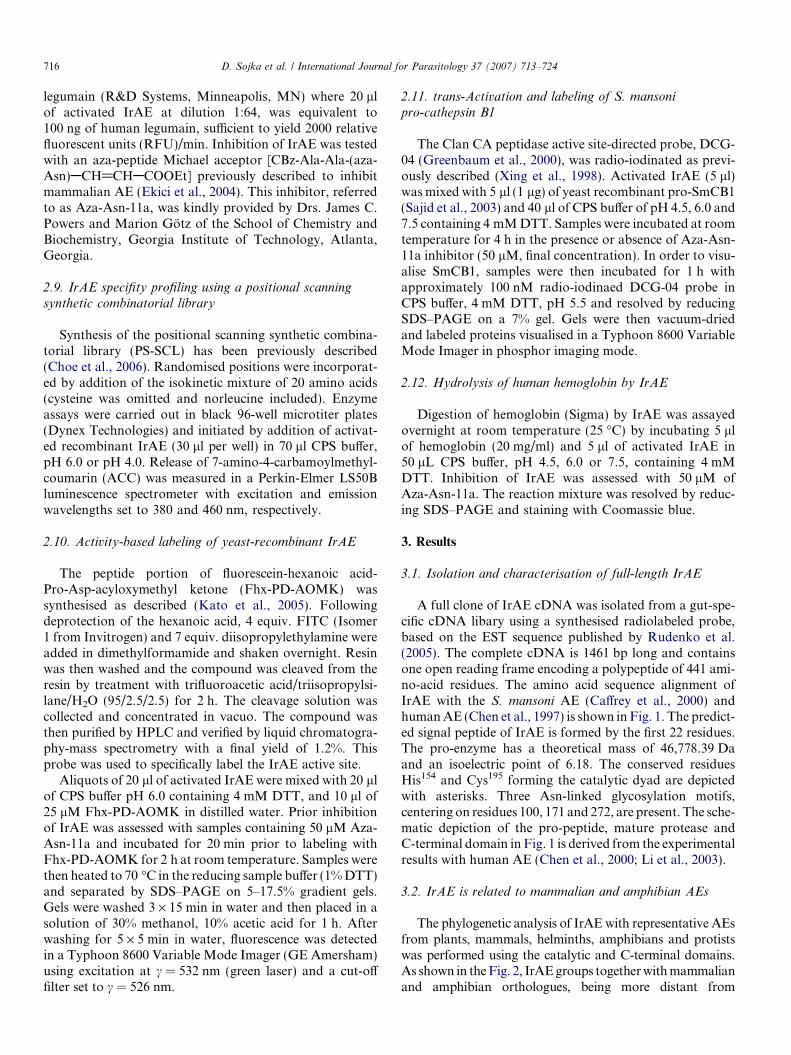
A full clone of IrAE cDNA was isolated from a gut-spe-cific cDNA libary using a synthesised radiolabeled probe,based on the EST sequence published by Rudenko et al.(2005). The complete cDNA is 1461 bp long and containsone open reading frame encoding a polypeptide of 441 ami-no-acid residues. The amino acid sequence alignment ofIrAE with the S. mansoni AE (Caffrey et al., 2000) andhuman AE (Chen et al., 1997) is shown in Fig. 1. The predict-ed signal peptide of IrAE is formed by the first 22 residues.The pro-enzyme has a theoretical mass of 46,778.39 Daand an isoelectric point of 6.18. The conserved residuesHis154 and Cys195 forming the catalytic dyad are depictedwith asterisks. Three Asn-linked glycosylation motifs,centering on residues 100, 171 and 272, are present. The sche-matic depiction of the pro-peptide, mature protease andC-terminal domain in Fig. 1 is derived from the experimentalresults with human AE (Chen et al., 2000; Li et al., 2003).
3.2. IrAE is related to mammalian and amphibian AEs
The phylogenetic analysis of IrAE with representative AEsfrom plants, mammals, helminths, amphibians and protistswas performed using the catalytic and C-terminal domains.As shown in the Fig. 2, IrAE groups together with mammalianand amphibian orthologues, being more distant from
Fig. 1. Multiple alignment of Ixodes ricinus asparaginyl endopeptidase (IrAE) with Schistosoma mansoni and human asparaginyl-endopeptidases. Thehard tick I. ricinus, this work (GenBank AY584752); the blood fluke S. mansoni (GenBank AJ250582); human (Homo sapiens; GenBank Y09862). Theconserved His and Cys residues forming the catalytic dyad of AE are marked with an asterisk. Space symbol indicates the possible N-glycosylation sites.Arrows depict the cleavage sites of the N- and C-terminal pro-domains experimentally determined for human AE. The bold dot marks the predicted Asnresidue cleavage of the IrAE C-terminal domain. The lower-case partial sequence shows the experimental N-terminal sequence of an enriched IrAEfragment.
D. Sojka et al. / International Journal for Parasitology 37 (2007) 713–724 717
helminth and plant AEs. An identical phylogeny was obtainedusing only the primary sequences of the mature domains (datanot shown). The multiple alignments are available uponrequest, or at http://www.paru.cas.cz/fig/sojka1.zip.
3.3. IrAE is expressed solely in the gut
Semi-quantitative RT-PCR profiling of IrAE mRNAlevels in different tissues of semi-engorged I. ricinus femalesrevealed that enzyme message was present specifically inthe gut (Fig. 3). Ferritin mRNA, levels of which are inde-pendent of blood-feeding (Kopacek et al., 2003), was usedas a loading control.
3.4. IrAE is localised to the digestive vesicles and peritrophic
matrix of the gut
N-Terminal histidine-tagged IrAE was expressed in E. co-
li, purified from inclusion bodies using Ni2+ chelatingsepharose in the presence of 8 M urea and refolded asdescribed in Section 2. The protein remained soluble andmigrated at 50 kDa by reducing SDS–PAGE (data notshown), thus agreeing with the predicted mass of
52,114 Da. By immunoblotting, the resulting rabbit anti-se-rum reacted with the corresponding antigen whereas nocross-reactivity was found with the pre-immune serum (datanot shown). This recombinant protein did not display anyhydrolytic activity of Z-AAN-AMC substrate even afterextensive pre-incubation at pH 4.5 in the presence of DTT.
The general structure of the sections from the semi-en-gorged female (5 days of feeding) is shown in Fig. 4A. Byimmuno-fluorescence microscopy with specific anti-serum,IrAE was localised in the digestive vesicles of the gut epi-thelium cells and within the peritrophic matrix (Fig. 4B,C). No staining was observed with pre-immune sera (datanot shown). The presence of IrAE within the peritrophicmatrix (boundary between the gut epithelium and lumen)was further confirmed by immuno-gold EM, where theIrAE-specific gold particles were found to be clearly associ-ated with the microvilli (Fig. 4D).
3.5. Functional expression and characterisation
of yeast-recombinant IrAE
IrAE, including a C-terminal histidine tag, wasexpressed in P. pastoris and the expression medium concen-

M. musculus
R. norvegicus
H. sapiens
X. laevis
I. ricinus
C. ensiformis
O. sativa
A. thaliana
V. radiata
H. contortus
S. mansoni
S. japonicum
F. hepatica
T. vaginalis 1
T. vaginalis 2
100
100
100
96
98
100
100
87
99
88
79
54
0.1Substitutions / site
Fig. 2. Phylogenetic comparison of Ixodes ricinus asparaginyl endopep-tidase (IrAE) with selected AEs. Trees were reconstructed using theNeighbor Joining method using amino acid sequences spanning across theputative mature enzymes and C-terminal extensions. Mammal – Mus
musculus (O89017), Rattus norvegicus (Q9R0J8), Homo sapiens (Q99538);amphibian – Xenopus laevis (AAH56842); tick – I. ricinus (AAS94231);plant – Canavalia ensiformis (P49046), Oryza sativa (Q8GS39), Arabidopsis
thaliana (P49047), Vigna radiata (Q9AUD9); helminth – Haemonchus
contortus (CAJ45481), Schistosoma mansoni (Q9NFY9), Schistosoma
japonicum (CAA50304), Fasciola hepatica (CAC85636); protist – Tricho-
monas vaginalis 1 and 2 (GenBank AAQ93039, AAQ93040, respectively).The horizontal bar represents a distance of 0.1 substitutions per site.Numbers at the branches represent bootstrap support.
Fig. 3. Tissue expression profile of Ixodes ricinus asparaginyl endopep-tidase (IrAE) in female I. ricinus. Messenger RNA levels were determinedby semi-quantitative two-step RT PCR. Tissues were dissected and pooledfrom 10 semi-engorged females fed for 5 days on guinea pigs. Ixodes
ricinus ferritin mRNA was used as a loading control. For details, seeSection 2.
718 D. Sojka et al. / International Journal for Parasitology 37 (2007) 713–724
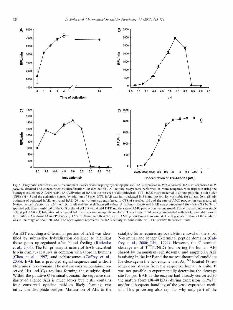
trated by ultrafiltration. Endopeptidase activity in this con-centrated medium was detectable using the peptidyl sub-strate Z-AAN-AMC prior to activation with 4 mM DTT.AE activity was never detected in non-transformed yeastclones processed similarly (data not shown). However,activity could be increased 3- to 4-fold in the presence ofDTT, reaching a maximum after 3–4 h and remaining sta-ble for at least 20 h at room temperature (Fig. 5A). The pHoptimum of the activated IrAE in the concentrated yeastexpression medium was approximately 5.5 and decreasedsharply above pH 6.0 (Fig. 5B). The steep decline in activ-ity was likely due to the irreversible inactivation of IrAE atpH P 6.0 as determined by pre-incubating activated IrAE
in CPS buffers over a pH range of 3.0–7.0 and subsequentlymeasuring residual activity at pH 5.5 (Fig. 5C). ActivatedIrAE was effectively inhibited by the specific azapeptidylinhibitor, Aza-Asn-11a, with an IC50 of approximately500 nM (Fig. 5D). Despite being able to measure its activ-ity, recombinant IrAE could not be detected as a distinctband by SDS–PAGE and silver staining on the backgroundof proteins from the concentrated yeast expression medium(Fig. 6, lane 1). However, specific rabbit antiserum inimmunoblots reacted with a protein band of approximately38–40 kDa consistent with the Mr of fully activated IrAE(Fig. 6, lane 2). Also, a protein band of about 40 kDawas also detected in the concentrated yeast medium bythe fluorescent activity-based probe, Fhx-PD-AOMK(Fig. 6, lane 3). Labeling of IrAE by Fhx-PD-AOMKwas completely inhibited by prior reaction with the Aza-Asn-11a inhibitor (Fig. 6, lane 4). Thus, we interpret thatthis band likely represents the glycosylated mature formof IrAE. No alteration in the migration of the 40 kDaFhx-PD-AOMK-labeled protein was observed after activa-tion in the presence of DTT (see above).
Attempts to purify intact recombinant IrAE zymogenfrom the concentrated yeast medium through use of aC-terminal polyhistidine tag and Ni2+ sepharose chroma-tography were unsuccessful. By SDS–PAGE and silverstaining, the only protein detected in the column eluatemigrated approximately as a 17 kDa band (data notshown). Edman degradation determined the N-terminusto be a mixture of two proteins; one unknown and theother IrAE, with the sequence VALGDAE(K)TXQ(Fig. 1). This sequence commences 15 residues downstreamfrom the theoretical cleavage site (Asn345) between the cat-alytic and C-terminal domains, as predicted using the serv-er ‘NetAEP: Predicting Asparaginyl Endopeptidasespecificity’ (http://theory.bio.uu.nl/kesmir/AEP/). Thus, itseems that the IrAE zymogen had already auto-activated,perhaps during expression in the yeast or subsequenthandling prior to Ni2+ sepharose chromatography.
3.6. IrAE has a strict specificity for Asn at P1
The mapping of IrAE specificity using the PS-SCL atpH 6.0 defined the specificity of IrAE for Asn at positionP1 (Fig. 7). At P2, partial preferences for Thr at P2 andAla at P3 were revealed and no preference for any residuewas noted at P4 (Fig. 7). Use of the PS-SCL at pH 4.0yielded similar results (not shown).
3.7. Processing of macromolecular substrates by the
activated IrAE
IrAE trans-processed both the full-length and intermedi-ate forms of recombinant SmCB1 zymogen (Sajid et al.,2003) to the active mature form as visualised by the ClanCA activity-based probe, DCG-04 (Fig. 8 and inset, rightpanel). Processing was maximal at pH 4.5, being progres-sively less efficient at pH 6.0 and 7.5. trans-Processing

Fig. 4. Localisation of Ixodes ricinus asparaginyl endopeptidase (IrAE) in the gut of female I. ricinus by indirect immunofluorescence microscopy andimmunogold EM. Sections were prepared from guts dissected from semi-engorged I. ricinus females (5 days of feeding). (A) Semi-thin sections stained withtoluidine blue – general structure of the tick gut showing the boundary between the gut epithelium (GE) digestive vesicles and the gut lumen (GL),containing hemoglobin crystals (Hb) and digestive gut cells (dGC); Nc – nuclei; Vs – digestive vesicles. (B) Semi-thin section labeled with Ra · IrAE serum(1:25) and fluorescein isothiocyanate-conjugated anti-rabbit antibody merged with Hochst 33–258 staining (blue). Note, the IrAE-specific signal wasmarkedly enriched within the peritrophic matrix (PM) and also present intracellularly in the digestive vesicles. (C) A detailed image of a digestive gut cell inthe phase of detachment from the gut epithelium, same staining as in (B). (D) EM of ultrathin sections labeled with Ra · IrAE serum (1:25) and protein Aconjugated with immunogold particles. IrAE-specific labeling within the peritrophic matrix showing the association of IrAE with microvilli (Mv).
D. Sojka et al. / International Journal for Parasitology 37 (2007) 713–724 719
was inhibited by pre-incubation of IrAE with the Aza-Asn-11a inhibitor (Fig. 8 inset, middle panel). IrAE digestedhuman hemoglobin to a major product of 4 kDa at pH4.5 (Fig. 9). Hemoglobinolysis was less efficient at pH6.0, yielding two products at 6 and 4 kDa, and did notoccur at pH 7.0. Edman N-terminal sequence analysis ofboth products resulted in a mixed signal due to the pres-ence of at least two peptides. Nevertheless, it was possibleto identify two fragments (in bold face) of the appropriatemolecular mass resulting from the cleavage of the a-hemo-globin by IrAE: DKTN10/VKAA. . .ALTN69 (6332.16 Da)and DPVN98/FKLL. . .SKYR142 (4797.7 Da) for the 6 and4 kDa bands, respectively (data not shown). No hemoglo-bin digestion occurred when IrAE was pre-incubated withthe Aza-Asn-11a inhibitor (Fig. 9).
4. Discussion
The hard tick I. ricinus is a major vector of tick-borneencephalitis virus and Lyme disease and, consequently, ofconcern to public health in Europe and northern Asia(Nutall, 1999). An improved understanding of the molecu-
lar physiology of I. ricinus should conceivably offer novelopportunities for interrupting the transmission of theseserious diseases. With this in mind and because hard ticksare obligate blood-feeders, we have focused on characteriz-ing digestive peptidases in the gut with which host proteins,particularly hemoglobin, are degraded to absorbable pep-tides and amino acids for parasite molting and egg produc-tion (Grandjean, 1984; Coons et al., 1986). Hematophagyin endo- and ecto-parasites, such as in the bloodflukeS. mansoni (Caffrey et al., 2004; Delcroix et al., 2006), theintestinal nematodes Ancylostoma caninum and H. contor-
tus, (Williamson et al., 2003) and other ticks (Renardet al., 2000, 2002; Boldbaatar et al., 2006), involves a num-ber of aspartic and cysteine peptidases to completelydegrade ingested proteins. Thus far for I. ricinus, however,nothing is known regarding component alimentary pepti-dases, their substrate specificities or potential redundancyof action.
Here, we report the full sequence, isolation and charac-terisation of an AE (a Clan CD, Family C13 cysteine pro-tease), found in the gut of I. ricinus. To our knowledge, thisis the first characterisation of an AE from an arthropod.
Time of activation
0 1 2 3 4 20
RF
U/m
in
1000
2000
3000
4000
5000
6000
7000
8000
pH
2.5 3.0 3.5 4.0 4.5 5.0 5.5 6.0 6.5 7.0 7.5
RF
U/m
in
0
500
1000
1500
2000
2500
3000
3500
Incubation pH
3.0 3.5 4.0 4.5 5.0 5.5 6.0 6.5 7.0
RF
U/m
in
0
200
400
600
800
1000
1200
1400
1600
1800
Concentration of Aza-Asn-11a [nM]
25000 5000 1000 500 100 20 4 0.8 0.16 0
RF
U/m
in
0
500
1000
1500
2000
2500
A B
DC
Fig. 5. Enzymatic characteristics of recombinant Ixodes ricinus asparaginyl endopeptidase (IrAE) expressed in Pichia pastoris. IrAE was expressed in P.
pastoris, desalted and concentrated by ultrafiltration (30 kDa cut-off). All activity assays were performed at room temperature in triplicate using thefluorogenic substrate Z-AAN-AMC. (A) Activation of IrAE in the presence of dithiothreitol (DTT). IrAE was transferred to citrate–phosphate–salt buffer(CPS) pH 4.5 and the activation started by addition of 4 mM DTT. IrAE was fully activated in 3 h and the activity was stable for at least 20 h. (B) pHoptimum of activated IrAE. Activated IrAE (20 h activation) was transferred to CPS of specified pH and the rate of AMC production was measured.Notice the loss of activity at pH > 6.0. (C) IrAE stability at different pH values. An aliquot of activated IrAE was pre-incubated for 4 h in CPS buffer ofspecified pH, then transferred to the CPS buffer of pH 5.5 with 4 mM DTT and the rate of AMC production was measured. The activated IrAE was stableonly at pH < 6.0. (D) Inhibition of activated IrAE with a legumain-specific inhibitor. The activated IrAE was pre-incubated with 2-fold serial dilutions ofthe inhibitor Aza-Asn-11A in CPS buffer, pH 5.5 for 30 min and then the rate of AMC production was measured. The IC50 concentration of the inhibitorwas in the range of about 500 nM. The open symbol represents the IrAE activity without inhibitor. RFU, relative fluorescent units.
720 D. Sojka et al. / International Journal for Parasitology 37 (2007) 713–724
An EST encoding a C-terminal portion of IrAE was iden-tified by subtractive hybridisation designed to highlightthose genes up-regulated after blood feeding (Rudenkoet al., 2005). The full primary structure of IrAE describedherein displays features in common with those in humans(Chen et al., 1997) and schistosomes (Caffrey et al.,2000). IrAE has a predicted signal sequence and a shortN-terminal pro-domain. The mature enzyme contains con-served His and Cys residues forming the catalytic dyad.Within the putative C-terminal domain, the sequence sim-ilarity of aligned AEs is much lower but it still containsfour conserved cysteine residues likely forming twointrachain disulphide bridges. Maturation of AEs to the
catalytic form requires autocatalytic removal of the shortN-terminal and longer C-terminal peptide domains (Caf-frey et al., 2000; Ishii, 1994). However, the C-terminalcleavage motif T322N/N(D) (numbering for human AE)shared by mammalian, schistosomal and amphibian AEsis missing in the IrAE and the nearest theoretical candidatefor cleavage in the tick enzyme is at Asn345 located 18 res-idues downstream from the respective human AE site. Itwas not possible to experimentally determine the cleavagesite for pro-IrAE as the enzyme had already converted tothe mature form (38–40 kDa) during expression in Pichia
and/or subsequent handling of the yeast expression medi-um. This processing also explains why only part of the

Fig. 6. Visualisation of active recombinant Ixodes ricinus asparaginylendopeptidase (IrAE) by specific antiserum and the activity-based probe,Fhx-PD-AOMK. Desalted and concentrated IrAE in Pichia pastoris
medium (see above) was resolved by reducing, gradient SDS–PAGE. Partsof the gel were silver stained or electroblotted onto polyvinylidene fluoridemembrane and visualised with either Ra · IrAE antibodies or the activity-based fluorescent probe, Fhx-PD-AOMK. Lane 1 – Silver stain of desaltedand concentrated IrAE in P. pastoris medium; lane 2 – Western blot usingRa · IrAE serum (1:100), swine · Ra-IgG – peroxidase conjugate anddiaminobenzidine as substrate. Lanes 3 and 4 – IrAE in P. pastoris
medium pre-incubated with Fhx-PD-AOMK in the absence and presenceof a legumain-specific inhibitor Aza-Asn-11a, respectively. For details, seeSection 2.
Fig. 8. trans-Processing of Schistosoma mansoni pro-cathepsin B1 byIxodes ricinus asparaginyl endopeptidase (IrAE). Graph – S. mansoni
cathepsin B1 zymogen (pro-SmCB1) (expressed in Pichia pastoris) waspre-incubated with or without activated IrAE at the specified pH and theSmCB1 activity assayed with the fluorogenic substrate Z-Phe-Arg-AMC.Inset – processing of pro-SmCB1 was visualised using a radio-iodinatedversion of the DCG-04 activity-based probe. Processing could be inhibitedby prior incubation with the legumain-specific inhibitor, Aza-Asn-11a.SmCB1pm, SmCB1pmi, SmCB1m refer to the zymogen, partiallyprocessed SmCB1 zymogen and the mature form of SmCB1, respectively.RFU, relative fluorescent units. For details, see Section 2.
D. Sojka et al. / International Journal for Parasitology 37 (2007) 713–724 721
C-terminal propeptide was captured on the Ni2+-chelatingcolumn via the engineered polyhistidine tag. For the same
Fig. 7. Specificity profile of Ixodes ricinus asparaginyl endopeptidase (IrAspecificities were determined with a synthetic peptidyl library in which randomiamino acids. RFU, relative fluorescent units.
reason, the precise processing site for cleavage of the N-ter-minal propeptide can only be hypothesised as beingbetween Asp28 and Ala29, based on the experimentallydemonstrated site at Asp25/Gly26 for human AE (Chenet al., 2000; Li et al., 2003).
E) using a positional scanning synthetic combinatorial library. P1–P4sed positions were incorporated by addition of the isokinetic mixture of 20

Fig. 9. Digestion of hemoglobin by activated Ixodes ricinus asparaginylendopeptidase (IrAE). Human hemoglobin (10 lg) was incubated withactivated IrAE in citrate–phosphate–salt buffers of specified pH values inthe presence (+) or absence (�) of the legumain-specific inhibitor,Aza-Asn-11a. The reaction mixture was resolved on reducing, gradientSDS–PAGE and stained with Coomassie brilliant blue. Digestion ofhemoglobin was most efficient at pH 4.5 and was inhibited by Aza-Asn-11a. The scale on the right of the panel indicates the positions ofpre-stained molecular weight standards.
722 D. Sojka et al. / International Journal for Parasitology 37 (2007) 713–724
The enzymatic properties of IrAE expressed in P. pasto-
ris accord with the data reported for purified or recombi-nant mammalian (Chen et al., 1997, 1998; Li et al., 2003)and S. mansoni AEs (Caffrey et al., 2000). IrAE has a pHoptimum for hydrolysis of Z-AAN-AMC at pH 5.5 andactivity declines sharply above pH 6.0. This phenomenonis most likely due to the instability of activated IrAE atneutral pH, which has also been reported for plant (Ishii,1994) and mammalian legumains (Chen et al., 1997). Theactivity of IrAE was effectively inhibited with a legumain-specific Aza-peptidyl Michael acceptor inhibitor (Ekiciet al., 2004) and active IrAE could be clearly visualisedusing the fluorescent activity-based probe, Fhx-PD-AOMK.
Mapping of the IrAE hydrolytic specificity by the PS-SCL revealed a strict specificity for N at P1, partial prefer-ences for T and A at P2 and P3, respectively, and a broadspecificity at P4. The preferred sequence at positions P4through P1 of X-A-T-N reveals a specificity similar tothose mapped for the human (P-T-N; P3 through P1)and S. mansoni AEs (A/T-A-N; P3 through P1) using sim-ilar libraries (Mathieu et al., 2002). The identified a-hemo-globin fragments revealed cleavage sites that were inreasonable agreement with the preferred sequence deter-mined by PS-SCL. Two of the three cleavage sites containT at P2 along with the required N at P1. One site with N atP1 (position 79) on the a-chain seemed not to be cleaved byIrAE, possibly due to the presence of an unfavorable P atP2, as judged by the PS-SCL. Although the b-chain con-tains six N residues, it is interesting to note that no frag-ments have thus far been identified. Many of thepotential cleavage sites contain unfavorable P2 residues,including G which appears twice. A favorable A at P2appears once near the C-terminus, but cleavage would
result in a fragment only seven residues in length, too shortto identify by SDS–PAGE.
In contrast to the optimal hydrolysis of the peptidylAMC substrate at pH 5.5, the cleavage of protein sub-strates including IrAE self-activation, the trans-processingof S. mansoni pro-cathepsin B1 and the hydrolysis ofhemoglobin was most efficient at pH 4.0–4.5, which is con-sistent with the acidic environment within the digestive ves-icles of tick gut cells (Coons et al., 1986; Lara et al., 2005).The trans-processing of pro-SmCB1 by IrAE is similar tothat demonstrated in vitro by endogenous S. mansoni AE(Sajid et al., 2003), whereby fully activated and matureSmCB1 only appeared in the presence of IrAE. As has beenhypothesised for Schistosoma (Sajid et al., 2003), it is pos-sible that IrAE may contribute to the activation of putativeClans CA (cysteine) and AA (aspartic) in the tick gut viatrans-processing. Preliminary evidence suggests that, likeparasitic helminths, I. ricinus expresses a number of gutClan CA and AA peptidases (Sojka, unpublished). Like-wise, the discrete hydrolysis of hemoglobin to a major frag-ment of 4 kDa by IrAE at pH 4.5 is remarkably similar tothat demonstrated for the S. mansoni orthologue in wormregurgitant (Delcroix et al., 2006). SmAE is postulated tosynergise with a network of Clan CA and AA proteasesto complete the breakdown of hemoglobin to absorbablepeptides and amino acids (Delcroix et al., 2006) and it ispossible that a similar system operates in the tick gut.The discrete processing activity of hemoglobin by IrAE isconsistent with its restricted P1-N specificity and contrastswith the complete hydrolysis of the same substrate by ClanCA peptidases from B. microplus (Mendiola et al., 1996) orwith the Clan AA peptidase, longepsin, from Haemaphysa-
lis longicornis (Boldbaatar et al., 2006). Importantly, thepresent data suggest that IrAE is not responsible for thegeneration of anti-bacterial peptides from hemoglobin(Fogaca et al., 1999; Nakajima et al., 2003) as none ofthe described peptides resulted from hydrolysis with N atP1.
Of interest was the finding that IrAE was extracellularlylocalised within the peritrophic matrix of partiallyengorged females (after the fifth day of feeding). Our datasupport previous results of morpho-functional (Coonset al., 1986; Grigor’eva, 2003, 2004) and histochemicalstudies (Agyei et al., 1992) showing that gut cells grow, dif-ferentiate and finally detach from the gut lumen during theslow feeding period (up to 6 days after tick attachment tothe host). Released contents of the detached cells apparent-ly become constituents of the peritrophic matrix on the gutepithelial surface. Entrapment and immobilisation ofsecreted enzymes at the surface of the peritrophic mem-brane has been demonstrated in insects (Ferreira et al.,1994). Thus, it is conceivable that IrAE is exported fromthe lysosomal vesicles onto the peritrophic matrix. WhetherIrAE retains catalytic activity on the peritrophic matrix isas yet unclear. With respect to the remarkable instabilityof IrAE above pH 6.0, we conclude that IrAE is unlikelyto be active in the gut lumen as the pH of gut contents
D. Sojka et al. / International Journal for Parasitology 37 (2007) 713–724 723
of several tick species has been reported to be at or above7.0 (Podboronov, 1991).
To conclude, the demonstration of an AE in the gut ofI. ricinus suggests a function for this enzyme associatedwith the degradation of host hemoglobin. That ortholo-gous AEs are also found in the gut of other hematopha-gous parasites such as S. mansoni (Caffrey et al., 2000)and H. contortus (Oliver et al., 2006) suggests a conservedfunction(s) for this discrete specificity of action, perhaps bycontributing directly to cleavage of hemoglobin and/or theprocessing of other gut peptidase zymogens (Clans CA andAA) that then act to complete the degradation of the sub-strate (Dalton and Brindley, 1996; Caffrey et al., 2004;Delcroix et al., 2006). Overall, the conservation of ClanCA, CD and AA gut peptidases across diverse hematopha-gic invertebrate parasites is remarkable and it is possiblethat one or more of these enzyme components representtargets for chemo- or immuno-therapeutic intervention(Sajid and McKerrow, 2002; Caffrey et al., 2004), therebydecreasing the incidence and prevalence of tick-borne dis-eases. Characterisation of other I. ricinus peptidases willbe the subject of future reports.
Acknowledgements
This work was supported by grants to P.K.: No.A6022307 from the Grant Agency of the Academy of Scienc-es of the Czech Republic; No. 206/06/0865 from the GrantAgency of the Czech Republic and Research Centre No.LC06009. We thank the Sandler Family Supporting Foun-dation for financial support of research visits by D.S. tothe San Francisco laboratory. J.H.M., C.S.C. and E.L.S.were supported by the NIH Grant AI35707. D.S. was sup-ported by project FRG3/3646/2005 and PhD program No.524/03/H133 from Ministry of Education, Youth and Sportsof the Czech Republic. Research at the Institute of Parasitol-ogy, BC ASCR and the Faculty of Biological Sciences, USBis covered by research plans Z60220518 and MSMT6007665801, respectively. We thank James C. Powers andMarion Gotz, School of Chemistry and Biochemistry, Geor-gia Institute of Technology, GA, for the gift of Aza-Asn-11a.
References
Agyei, A.D., Runham, N.W., Blackstock, N., 1992. Histochemicalchanges in the midgut of two ixodid tick species Boophilus microplus
and Rhipicephalus appendiculatus during digestion of the blood meal.Exp. Appl. Acarol. 13, 187–212.
Bendtsen, J.D., Nielsen, H., von Heijne, G., Brunak, S., 2004. Improvedprediction of signal peptides: SignalP 3.0. J. Mol. Biol. 340, 783–795.
Boldbaatar, D., Sikalizyo Sikasunge, C., Battsetseg, B., Xuan, X.,Fujisaki, K., 2006. Molecular cloning and functional characterizationof an aspartic protease from the hard tick Haemaphysalis longicornis.Insect Biochem. Mol. Biol. 36, 25–36.
Caffrey, C.R., Mathieu, M.A., Gaffney, A.M., Salter, J.P., Sajid, M.,Lucas, K.D., Franklin, C., Bogyo, M., McKerrow, J.H., 2000.Identification of a cDNA encoding an active asparaginyl endopepti-dase of Schistosoma mansoni and its expression in Pichia pastoris.FEBS Lett. 466, 244–248.
Caffrey, C.R., McKerrow, J.H., Salter, J.P., Sajid, M., 2004. Blood ‘n’guts: an update on schistosome digestive peptidases. Trends Parasitol.20, 241–248.
Chen, J.M., Dando, P.M., Rawlings, N.D., Brown, M.A., Young, N.E.,Stevens, R.A., Hewitt, E., Watts, C., Barrett, A.J., 1997. Cloning,isolation, and characterization of mammalian legumain, an asparag-inyl endopeptidase. J. Biol. Chem. 272, 8090–8098.
Chen, J.M., Dando, P.M., Stevens, R.A., Fortunato, M., Barrett, A.J.,1998. Cloning and expression of mouse legumain, a lysosomalendopeptidase. Biochem. J. 335, 111–117.
Chen, J.M., Fortunato, M., Barrett, A.J., 2000. Activation of humanprolegumain by cleavage at a C-terminal asparagine residue. BiochemJ. 352, 327–334.
Choe, Y., Leonetti, F., Greenbaum, D.C., Lecaille, F., Bogyo, M.,Bromme, D., Ellman, J.A., Craik, C.S., 2006. Substrate profiling ofcysteine proteases using a combinatorial peptide library identifiesfunctionally unique specificities. J. Biol. Chem. 281, 12824–12832.
Coons, L.B., Rosell-Davis, R., Tarnowski, B.I., 1986. Bloodmeal diges-tion in ticks. In: Sauer, J.R., Hair, J.A. (Eds.), Morphology,Physiology, and Behavioral Biology of Ticks. Ellis Horwood, Ltd.,Chichester, England, pp. 248–279.
Dalton, J.P., Brindley, P.J., 1996. Schistosome asparaginyl endopeptidaseSM32 in hemoglobin digestion. Parasitol. Today 12, 125.
Delcroix, M., Sajid, M., Caffrey, C.R., Lim, K.C., Dvorak, J., Hsieh, I.,Bahgat, M., Dissous, C., McKerrow, J.H., 2006. A multienzymenetwork functions in intestinal protein digestion by a platyhelminthparasite. J. Biol. Chem. 281, 39316–39329.
Ekici, O.D., Gotz, M.G., James, K.E., Li, Z.Z., Rukamp, B.J., Asgian,J.L., Caffrey, C.R., Hansell, E., Dvorak, J., McKerrow, J.H.,Potempa, J., Travis, J., Mikolajczyk, J., Salvesen, G.S., Powers, J.C.,2004. Aza-peptide Michael acceptors: a new class of inhibitors specificfor caspases and other clan CD cysteine proteases. J. Med. Chem. 47,1889–1892.
Ferreira, C., Capella, A.N., Sitnik, R., Terra, W.R., 1994. Digestiveenzymes in midgut cells, endo- and ectoperithrophic contents, andperitrophic membranes of Spodoptera frugiperda (Lepidoptera) larvae.Arch. Insect Biochem. Physiol. 26, 299–313.
Fogaca, A.C., da Silva, P.I. Jr., Miranda, M.T., Bianchi, A.G., Miranda,A., Ribolla, P.E., Daffre, S., 1999. Antimicrobial activity of a bovinehemoglobin fragment in the tick Boophilus microplus. J. Biol. Chem.274, 25330–25334.
Grandjean, O., 1984. Blood digestion in Ornithodoros moubata Murraysensu stricto Walton (Ixodoidea: Argasidae) females. I. Biochemicalchanges in the midgut lumen and ultrastructure of the midgut cells,related to intracellular digestion. Acarologia 25, 147–165.
Greenbaum, D., Medzihradszky, K.F., Burlingame, A., Bogyo, M., 2000.Epoxide electrophiles as activity-dependent cysteine protease profilingand discovery tools. Chem. Biol. 7, 569–581.
Grigor’eva, L.A., 2003. Morphofunctional changes in the midgut of tickfemales of the genus Ixodes (Acari: Ixodidae) during and after feeding.Parazitologiia 37, 169–176 (in Russian).
Grigor’eva, L.A., Amosova, L.I., 2004. Peritrophic matrix in the midgutof tick females of the genus Ixodes (Acari: Ixodidae). Parazitologiia 38,3–11 (in Russian).
Grunclova, L., Horn, M., Vancova, M., Sojka, D., Franta, Z., Mares, M.,Kopacek, P., 2006. Two secreted cystatins of the soft tick Ornithodoros
moubata: differential expression pattern and inhibitory specificity. Biol.Chem. 387, 1635–1644.
Hall, T.A., 1999. BioEdit: a user-friendly biological sequence alignmenteditor and analysis program for Windows 95/98/NT. Nucleic AcidsSymp. Ser. 41, 95–98.
Hara-Nishimura, I., Takeuchi, Y., Nishimura, M., 1993. Molecularcharacterization of a vacuolar processing enzyme related to aputative cysteine proteinase of Schistosoma mansoni. Plant Cell 5,1651–1659.
Ishii, S., Abe, Y., Mitta, M., Matsushita, H., Kato, I., 1992. A novelprotease from jack bean seeds: asparaginyl endopeptidase. J. ProteinChem. 11, 367–368.

724 D. Sojka et al. / International Journal for Parasitology 37 (2007) 713–724
Ishii, S-I., 1994. Legumain: asparaginyl endopeptidases. Methods Enzy-mol. 244, 604–615.
Kato, D., Boatright, K.M., Berger, A.B., Nazif, T., Blum, G., Ryan, C.,Chehade, K.A., Salvesen, G.S., Bogyo, M., 2005. Activity-basedprobes that target diverse cysteine protease families. Nat. Chem. Biol.1, 33–38.
Kaufman, W.R., 1989. Tick-host interaction: a synthesis of currentconcepts. Parasitol. Today 13, 165.
Kevil, C.G., Walsh, L., Laroux, F.S., Kalogeris, T., Grisham, M.B.,Alexander, J.S., 1997. An improved, rapid Northern protocol.Biochem. Biophys. Res. Commun. 238, 277–279.
Kopacek, P., Weise, C., Gotz, P., 1995. The prophenoloxidase from thewax moth Galleria mellonella: purification and characterization of theproenzyme. Insect Biochem. Mol. Biol. 25, 1081–1091.
Kopacek, P., Zdychova, J., Yoshiga, T., Weise, C., Rudenko, N., Law,J.H., 2003. Molecular cloning, expression and isolation of ferritinsfrom two tick species – Ornithodoros moubata and Ixodes ricinus.Insect Biochem. Mol. Biol. 233, 103–113.
Kumar, S., Tamura, K., Jakobsen, I.B., Nei, M., 2001. MEGA2:molecular evolutionary genetics analysis software. Bioinformatics 17,1244–1245.
Lara, F.A., Lins, U., Bechara, G.H., Oliveira, P.L., 2005. Tracing heme ina living cell: hemoglobin degradation and heme traffic in digest cells ofthe cattle tick Boophilus microplus. J. Exp. Biol. 208, 3093–3101.
Leon-Felix, J., Ortega-Lopez, J., Orozco-Solis, R., Arroyo, R., 2004. Twonovel asparaginyl endopeptidase-like cysteine proteinases from theprotist Trichomonas vaginalis: their evolutionary relationship withinthe clan CD cysteine proteinases. Gene 335, 25–35.
Li, D.N., Matthews, S.P., Antoniou, A.N., Mazzeo, D., Watts, C., 2003.Multistep autoactivation of asparaginyl endopeptidase in vitro andin vivo. J. Biol. Chem. 278, 38980–38990.
Manoury, B., Hewitt, E.W., Morrice, N., Dando, P.M., Barrett, A.J.,Watts, C., 1998. An asparaginyl endopeptidase processes a microbialantigen for class II MHC presentation. Nature 396, 695–699.
Mathieu, M.A., Bogyo, M., Caffrey, C.R., Choe, Y., Lee, J., Chapman,H., Sajid, M., Craik, C.S., McKerrow, J.H., 2002. Substrate specificityof schistosome versus human legumain determined by P1–P3 peptidelibraries. Mol. Biochem. Parasitol. 121, 99–105.
Mendiola, J., Alonso, M., Marquetti, M.C., Finlay, C., 1996. Boophilus
microplus: multiple proteolytic activities in the midgut. Exp. Parasitol.82, 27–33.
Muntz, K., Shutov, A.D., 2002. Legumains and their functions in plants.Trends Plant Sci. 7, 340–344.
Nakajima, Y., Ogihara, K., Taylor, D., Yamakawa, M., 2003. Antibac-terial hemoglobin fragments from the midgut of the soft tick,Ornithodoros moubata (Acari: Argasidae). J. Med. Entomol. 40, 78–81.
Nutall, P.A., 1999. Pathogen-tick-host interactions: Borrelia bugdorferi
and TBE virus. Zentralbl. Bakteriol. 289, 492–505.Oliver, E.M., Skuce, P.J., McNair, C.M., Knox, D.P., 2006. Identification
and characterization of an asparaginyl proteinase (legumain) from theparasitic nematode, Haemonchus contortus. Parasitology 133, 237–244.
Podboronov. V.M., Berdyev, A., 1991. Protective mechanisms of Ixodideatick and their vertebrate hosts (host-parasite interface). (Ashgabad,Ylym) (in Russian).
Renard, G., Garcia, J.F., Cardoso, F.C., Richter, M.F., Sakanari, J.A.,Ozaki, L.S., Termignoni, C., Masuda, A., 2000. Cloning andfunctional expression of a Boophilus microplus cathepsin L-likeenzyme. Insect Biochem. Mol. Biol. 30, 1017–1026.
Renard, G., Lara, F.A., de Cardoso, F.C., Miguens, F.C., Dansa-Petretski, M., Termignoni, C., Masuda, A., 2002. Expression andimmunolocalization of a Boophilus microplus cathepsin L-like enzyme.Insect Mol. Biol. 11, 325–328.
Rudenko, N., Golovchenko, M., Edwards, M.J., Grubhoffer, L., 2005.Differential expression of Ixodes ricinus tick genes induced by bloodfeeding or Borrelia burgdorferi infection. J. Med. Entomol. 42, 36–41.
Saitou, N., Nei, M., 1987. The neighbor-joining method: a new method forreconstructing phylogenetic trees. Mol. Biol. Evol. 4, 406–425.
Sajid, M., McKerrow, J.H., 2002. Cysteine proteases of parasiticorganisms. Mol. Biochem. Parasitol. 120, 1–21.
Sajid, M., McKerrow, J.H., Hansell, E., Mathieu, M.A., Lucas, K.D.,Hsieh, I., Greenbaum, D., Bogyo, M., Salter, J.P., Lim, K.C.,Franklin, C., Kim, J.H., Caffrey, C.R., 2003. Functional expressionand characterization of Schistosoma mansoni cathepsin B and itstrans-activation by an endogenous asparaginyl endopeptidase. Mol.Biochem. Parasitol. 131, 65–75.
Sonenshine, D.E., 1991. Biology of Ticks, vol. 1. Oxford university Press,New York.
Terra, W.R., Ferreira, C., 1994. Insect digestive enzymes: properties,compartmentalization and function. Comp. Biochem. Physiol. B 109,1–62.
Thompson, J.D., Gibson, T.J., Plewniak, F., Jeanmougin, F., Higgins,D.G., 1997. The CLUSTAL X windows interface: flexible strategiesfor multiple sequence alignment aided by quality analysis tools.Nucleic Acids Res. 25, 4876–4882.
Watts, C., Matthews, S.P., Mazzeo, D., Manoury, B., Moss, C.X., 2005.Asparaginyl endopeptidase: case history of a class II MHC compart-ment protease. Immunol. Rev. 207, 218–228.
Williamson, A.L., Brindley, P.J., Knox, D.P., Hotez, P.J., Loukas, A.,2003. Digestive proteases of blood-feeding nematodes. Trends Paras-itol. 9, 417–423.
Xing, R., Addington, A.K., Mason, R.W., 1998. Quantification ofcathepsins B and L in cells. Biochem. J. 332, 499–505.