IOS Press Residual vision activation and the brain-eye ... · 768 B.A. Sabel et al. / Residual...
25
Restorative Neurology and Neuroscience 36 (2018) 767–791 DOI 10.3233/RNN-180880 IOS Press 767 Residual vision activation and the brain-eye-vascular triad: Dysregulation, plasticity and restoration in low vision and blindness – a review Bernhard A. Sabel a,∗ , Josef Flammer b and Lotfi B. Merabet c a Institute of Medical Psychology, Medical Faculty, Otto-von-Guericke University of Magdeburg, Magdeburg, Germany b Department of Ophthalmology, University of Basel, Basel, Switzerland c Department of Ophthalmology, The Laboratory for Visual Neuroplasticity, Massachusetts Eye and Ear Infirmary, Harvard Medical School, Boston, USA Abstract. Vision loss due to ocular diseases such as glaucoma, optic neuropathy, macular degeneration, or diabetic retinopathy, are generally considered an exclusive affair of the retina and/or optic nerve. However, the brain, through multiple indirect influences, has also a major impact on functional visual impairment. Such indirect influences include intracerebral pressure, eye movements, top-down modulation (attention, cognition), and emotionally triggered stress hormone release affecting blood vessel dysregulation. Therefore, vision loss should be viewed as the result of multiple interactions within a “brain-eye-vascular triad”, and several eye diseases may also be considered as brain diseases in disguise. While the brain is part of the problem, it can also be part of the solution. Neuronal networks of the brain can “amplify” residual vision through neuroplasticity changes of local and global functional connectivity by activating, modulating and strengthening residual visual signals. The activation of residual vision can be achieved by different means such as vision restoration training, non-invasive brain stimulation, or blood flow enhancing medications. Modulating brain functional networks and improving vascular regulation may offer new opportunities to recover or restore low vision by increasing visual field size, visual acuity and overall functional vision. Hence, neuroscience offers new insights to better understand vision loss, and modulating brain and vascular function is a promising source for new opportunities to activate residual vision to achieve restoration and recovery to improve quality of live in patients suffering from low vision. Keywords: Glaucoma, optic neuropathy, recovery, plasticity, brain, vision restoration, vascular dysregulation 1. Scope of the problem for visual impairment Visual impairments (VI) and blindness are among the most feared medical conditions and a ∗ Corresponding author: Prof. Dr. Bernhard A. Sabel, Otto- von-Guericke University, Medical Faculty, Institute of Medical Psychology, Leipziger Str. 44, 39120 Magdeburg, Germany. Tel.: +49 391 67 21800; Fax: +49 391 67 21803; E-mail: bernhard.sabel @med.ovgu.de. growing problem in our aging societies (AFB web- page; Rosenberg & Sperazza, 2008). While visual impairment caused by diseases affecting the eye’s optics (e.g. cornea and lens) can be treated by cor- rective lenses or surgery, there are many other causes of VI which are not typically amenable to being improved or restored, namely those caused by dam- age to the neural visual pathway including the retina, optic nerve (ON), and different regions of the brain such as the thalamus, optic radiations, and visual 0922-6028/18/$35.00 © 2018 – IOS Press and the authors. All rights reserved This article is published online with Open Access and distributed under the terms of the Creative Commons Attribution Non-Commercial License (CC BY-NC 4.0).
Transcript of IOS Press Residual vision activation and the brain-eye ... · 768 B.A. Sabel et al. / Residual...

Restorative Neurology and Neuroscience 36 (2018) 767–791DOI 10.3233/RNN-180880IOS Press
767
Residual vision activation and thebrain-eye-vascular triad: Dysregulation,plasticity and restoration in low visionand blindness – a review
Bernhard A. Sabela,∗, Josef Flammerb and Lotfi B. MerabetcaInstitute of Medical Psychology, Medical Faculty, Otto-von-Guericke University of Magdeburg,Magdeburg, GermanybDepartment of Ophthalmology, University of Basel, Basel, SwitzerlandcDepartment of Ophthalmology, The Laboratory for Visual Neuroplasticity, Massachusetts Eyeand Ear Infirmary, Harvard Medical School, Boston, USA
Abstract. Vision loss due to ocular diseases such as glaucoma, optic neuropathy, macular degeneration, or diabetic retinopathy,are generally considered an exclusive affair of the retina and/or optic nerve. However, the brain, through multiple indirectinfluences, has also a major impact on functional visual impairment. Such indirect influences include intracerebral pressure,eye movements, top-down modulation (attention, cognition), and emotionally triggered stress hormone release affecting bloodvessel dysregulation. Therefore, vision loss should be viewed as the result of multiple interactions within a “brain-eye-vasculartriad”, and several eye diseases may also be considered as brain diseases in disguise. While the brain is part of the problem, itcan also be part of the solution. Neuronal networks of the brain can “amplify” residual vision through neuroplasticity changesof local and global functional connectivity by activating, modulating and strengthening residual visual signals. The activationof residual vision can be achieved by different means such as vision restoration training, non-invasive brain stimulation,or blood flow enhancing medications. Modulating brain functional networks and improving vascular regulation may offernew opportunities to recover or restore low vision by increasing visual field size, visual acuity and overall functional vision.Hence, neuroscience offers new insights to better understand vision loss, and modulating brain and vascular function isa promising source for new opportunities to activate residual vision to achieve restoration and recovery to improve qualityof live in patients suffering from low vision.
Keywords: Glaucoma, optic neuropathy, recovery, plasticity, brain, vision restoration, vascular dysregulation
1. Scope of the problem for visualimpairment
Visual impairments (VI) and blindness areamong the most feared medical conditions and a
∗Corresponding author: Prof. Dr. Bernhard A. Sabel, Otto-von-Guericke University, Medical Faculty, Institute of MedicalPsychology, Leipziger Str. 44, 39120 Magdeburg, Germany. Tel.:+49 391 67 21800; Fax: +49 391 67 21803; E-mail: [email protected].
growing problem in our aging societies (AFB web-page; Rosenberg & Sperazza, 2008). While visualimpairment caused by diseases affecting the eye’soptics (e.g. cornea and lens) can be treated by cor-rective lenses or surgery, there are many other causesof VI which are not typically amenable to beingimproved or restored, namely those caused by dam-age to the neural visual pathway including the retina,optic nerve (ON), and different regions of the brainsuch as the thalamus, optic radiations, and visual
0922-6028/18/$35.00 © 2018 – IOS Press and the authors. All rights reservedThis article is published online with Open Access and distributed under the terms of the Creative Commons Attribution Non-Commercial License (CC BY-NC 4.0).

768 B.A. Sabel et al. / Residual vision activation and the brain-eye-vascular triad
cortex. These VIs vary in severity from partial (inmost cases) to profound and complete blindness,impacting activities of daily living such as reading,identifying objects, orientation and mobility. Andthere are secondary, associated risks of psycholog-ical/psychiatric problems such as anxiety, depressionand social withdrawal (Rosenberg & Sperazza, 2008).
The burden of low vision and blindness is immenseas it impacts quality of life, cognitive function,well-being as well as society (Quigley, 2011; Chen,Bhattacharya & Pershing, 2017). This includesemployment, education opportunities, and healtheconomics (Stevens et al., 2013). According to WHOstatistics, 285 million people worldwide are visuallyimpaired (defined as a best corrected Snellen visualacuity of 20/60 or worse in the better seeing eye).Of these, 39 million are classified as blind, definedas visual acuity of 20/200 or worse in the better eyewith corrective lenses, or visual field restriction of20 degrees diameter or less in the better eye. Thelarge majority (82%) of individuals are aged 50 andolder (Pascolini & Mariotti, 2012; WHO Fact Sheet,2014; Boyers et al., 2015). In developed countries,the prominent causes of VI are age-related macu-lar degeneration (AMD; 1 to 4%), glaucoma (2%),diabetic retinopathy (1%); all of which are chronicconditions with no established cures (WHO FactSheet 2014; Bourne, Karimkhani, Hilton, Richheimer& Dellavalle, 2017). This is in stark contrast to thesituation in developing (i.e. middle to low income)countries where 80% of visual impairment is avoid-able or curable such as uncorrected refractive errorsand cataracts (WHO Fact Sheet, 2014; Bourne et al.,2017).
Though modern rehabilitation strategies may helppatients learn compensatory strategies to remainfunctionally independent (Dagnelie, 2013), little canbe done to improve the vision they have lost. There-fore, new concepts and efforts are urgently neededto develop better sensory-motor compensatory andvision restoration strategies by leveraging theinherent ability of the brain to adapt to injury.
VIs following diseases of the eye (e.g. glau-coma, optic neuropathy, macular degeneration,diabetic retinopathy) are typically managed byophthalmologists, optometrists, and other eye careprofessionals. In contrast, VIs associated with neu-rological problems are attended to by neurologistsand neurosurgeons. They include diffuse brain tis-sue damage after stroke or traumatic brain injury,neurodegenerative diseases such as Parkinson’s andother dementias, or multiple sclerosis. However, the
eye and brain should not be studied, nor treated,in isolation, and effective care necessitates a com-bined ophthalmological and neurological approach.Specifically, the three-way reciprocal interaction ofboth organs with the cardio-vascular system needto be better understood. Only when appreciating themany interactions within the brain-eye-vascular triadcan we fully understand and adequately modulateboth the physiological and psychological state ofour patients to achieve vision recovery and restora-tion. Though many direct causes of vision loss arewell established (like ocular disease, trauma, geneticpredispositions, etc.), this review paper discussesindirect factors related to the brain-eye-vascular triadand how they impact vision recovery: vascular sys-tem alterations and stress hormone influence thereon,brain mechanisms of neuroplasticity, cerebrospinalfluid (CSF) pressure, emotional stress etc. Thoughindirect, they are by no means less relevant for ourunderstanding of low vision and its treatment toachieve recovery and repair.
2. Neurological influences in “ocular”diseases
Partial vision loss, with sometimes considerable“residual vision” (Fig. 1) is by far more prevalent(>95%) than total vision loss. When vision loss devel-ops in ocular diseases affecting the retina and opticnerve, this has repercussions at the level of brain pro-cessing as well. Though it is textbook knowledgethat the retina is part of the brain, the complexityof the interaction between eye and brain is seldomconsidered and not such a simple matter. Here weneed to ask on the one hand (i) how much neu-ronal information reaches the brain, and on the otherhand we also should learn (ii) how the brain pro-cesses incoming (incomplete) visual information byignoring, enhancing (amplifying) or inhibiting (sup-pressing) it, and (iii) how visual cues are interpretedby emotions and cognition and how this impacts over-all vision. On a systems level, the brain, by releasingstress hormones influence vascular tone, particularlyin and around the optic nerve and thereby impairvascular autoregulation and perfusion of the wholebody, including itself and that of the eyes (Flammeret al., 2013a). Furthermore, the brain is involved inother ways in eye pathology a partner in the intraoc-ular (IOP)/ intracranial pressure (ICP) balance whichdirectly or indirectly impacts the consequences of eyepressure conditions (Hou et al., 2016). If any of these

B.A. Sabel et al. / Residual vision activation and the brain-eye-vascular triad 769
Fig. 1. Residual functions and subtle deficits in visual fields. Visual fields are not just black and white (blind and seeing fields) have “shadesof grey”: hidden potentials with residual vision but also hidden problems in the “intact” sector. Blindsight is the phenomenon that patientsare able to correctly guess that stimuli were presented without being aware of them. While correctly responding to vision signals, they reportseeing nothing; unconscious seeing without knowing. Areas of residual vision are those with uncertain responses where patients respondonly occasionally. These regions of the visual field are quite variable during repeated testing and are characterized by increased thresholdsand longer response time. The hidden deficits in the “seeing field” make patients “sightblind”, but this can only be measured by tests thatare sensitive to higher cognitive dysfunctions.
brain regulation mechanisms are altered, the brain-eye-vascular triad has a sensitive balancing act tomanage which determines if, how, and when a visionproblem is first diagnosed, whether it progresses,or whether it can be activated for recovery andrestoration.
2.1. Residual vision
A neurobiological substrate that supports visualfield recovery and restoration are regions of partialvisual functions, termed “areas of residual vision”(Sabel, Fedorov, Henrich-Noack & Gall, 2011a).These are regions in the visual field which are nei-ther blind nor seeing normally (graphically displayedin visual field charts in black or white color, respec-tively). Rather, visual fields typically have different“shades of grey” where function is neither completelylost nor normal (see Fig. 1). Most patients have someresidual structures and functions spared by the dam-age; complete vision loss in both eyes (“blackblind”)is extremely rare. Residual (partial) vision – wherevision is not lost but impaired – can easily be sub-jectively reported by patients and quantified withperimetry testing. They are measurable as a “relativescotoma” inside or at the border of the scotoma. Suchresidual areas are characterized by higher responsethresholds, lower contrast sensitivity, reduced acu-ity, foggy vision, incomplete perceptions or slowed
reaction times (Bola et al., 2013). It is these partiallydamaged regions of residual vision that have hiddenpotentials for activation and recovery.
The phenomenon of “blindsight” is an interest-ing and long-known example of residual vision:blindsight patients are able to correctly guess thatstimuli were presented without being aware of them.While they correctly respond to vision signals, thepatients report seeing nothing (“blind”); it is a case ofunconscious seeing without knowing (Poppel, Held& Frost, 1973; Sanders, Warrington, Marshall &Weiskrantz, 1974). Other areas of residual vision arethose with uncertain or less sensitive responses whichare shown in grey in visual field charts. Here patientsrespond only sometimes to stimuli. The responseis quite variable during repeated testing (Flammer,Drance & Fankhauser, 1984a; Flammer, Drance &Schulzer, 1984b) and characterized by increasedthresholds and longer response times. But visualfields may also have hidden deficits inside the pre-sumably “intact” areas which cannot be captured bystandard perimetric testing. Such patients are „sight-blind“ (Bola, Gall & Sabel, 2013). However, we donot propose a clear-cut border between “blindsight”and “residual vision”. Both may represent differentlevels of visual system activation supporting differentlevels of awareness to be analysed on a network level(Hadid & Lepore 2017; Mazzi, Savazzi & Silvanto,2018), an issue that needs to be further explored.
770 B.A. Sabel et al. / Residual vision activation and the brain-eye-vascular triad
2.2. Brain degeneration in ocular disease
Nervous system degeneration in (normal) agingcan affect the structure and function anywhere alongthe visual eye-to-brain axis (Haas, Flammer &Schneider, 1986). Obviously, what the brain per-ceives is a function of how much retinal input itreceives. But if the brain is also affected by any addi-tional loss of neurons, loss of functional connectionsin down-stream visual structures, or vascular autoreg-ulation problems, then the complexity of the “eyeproblem” is far greater (Faiq, Dada, Kumar, Saluja &Dada, 2016). This is the case when, for example, gen-eral cell loss occurs during normal aging with specificvulnerabilities of the brain’s visual system structures(Owsley, 2011). Even in traditionally ocular diseases(such as glaucoma), a selective degeneration of cellsin visual brain nuclei has been shown as indicated byatrophy of the LGN, optic radiation and visual cor-tex (Duncan, Sumple, Weinreb, Bowd & Zangwill,2007; Gupta, Ang, de Tilly, Bidaisee & Yucel, 2006;Gupta & Yucel, 2007; Engelhorn et al., 2011; Yucel,2013; Yu et al., 2013, Schoemann et al., 2014), and
even non-visual brain structures that control emotions(e.g. amygdala) (Wang et al., 2016).
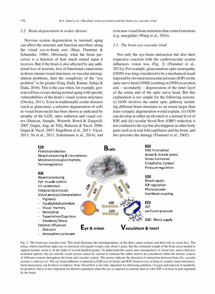
2.3. The brain-eye-vascular triad
Not only the eye-brain interaction but also theirrespective coaction with the cardiovascular systeminfluences vision loss (Fig. 2) (Flammer et al.,2013a). For example, glaucomatous optic neuropathy(GON) was long considered to be a mechanical insultimposed by elevated intraocular pressure (IOP) on theoptic nerve head (ONH) resulting in ONH excavationand – secondarily – degeneration of the inner layerof the retina and of the optic nerve head. But thisexplanation is too simple for the following reasons:(i) GON involves the entire optic pathway includ-ing different brain structures to an extent larger thantrans-synaptic degeneration would explain, (ii) GONcan develop at either an elevated or a normal level ofIOP, and (iii) occular blood flow (OBF) reduction isnot confined to the eye but also happens in other bodyparts such as in nail fold capillaries and the brain, andthis precedes the damage (Flammer et al., 2002).
Fig. 2. The brain-eye-vascular triad. This triad illustrates the interdependency of the three organ systems and their role in vision loss. Theretina, which transforms light rays to electrical cell signals weighs only about 1 gram. But the estimated weight of the brain areas needed tosupport normal vision is on the order of several hundred grams. To understand the causes and consequences of vision loss, and to find newtreatment options, the eye and the visual system cannot be viewed in isolation but rather need to be considered within the holistic contextof different systems throughout the brain and vascular system. The arrows indicate the direction of interaction between brain (b), vascularsystem (v) and eye (e). The eye-brain influence is denoted as E2B (eye-to-brain) and B2E (brain-to-eye or brain-to-central visual structures).Such interactions can be direct or indirect. Note: blood flow is not only important for delivering nutrition / oxygen and removal of metabolicby-products, but it is also important for thermo-regulation when the eye is exposed to extreme heat or cold. IOP is at least in part regulatedby the brain.

B.A. Sabel et al. / Residual vision activation and the brain-eye-vascular triad 771
Fig. 3. Emotional stress and the “Flammer Syndrome”. The term Flammer Syndrome (FS) describes a phenotype characterized by thepresence of primary vascular dysregulation with a cluster of additional symptoms and signs. Symptoms and signs include the following:prolonged sleep onset time, prolonged blood flow cessation in the finger capillaries after cooling, disturbed autoregulation of ocular bloodflow, increased prevalence of optic disc hemorrhages and activated retinal astrocytes, increased retinal venous pressure, increased stiffnessof retinal vessels, higher spatial irregularities in retinal vessels, increased resistance in retroocular vessels, increased oxidative stress, alteredgene expression as measured in lymphocytes, and altered activity of the autonomic nervous system (beat-to-beat variations of the heart).
The response to vascular challenges varies amongindividuals. It is influenced by environmental fac-tors and genetic predispositions as exemplified bythe Flammer Syndrome (FS). Such persons havea tendency to react differently (particularly with theirblood vessels) to a number of stimuli such as cold-ness or emotional stress (Konieczka et al., 2014).Hence, we need to appreciate the important role ofthe brain-vascular interaction in vision loss.
Embryologically, the eye develops from brain tis-sue, and both blood flow (blood brain barrier) andcell types (neurons) are similar. But retinal vesselslack innervation by the autonomic nervous system(ANS) and the blood brain barrier is incomplete in theoptic nerve head, rendering it more sensitive to cir-culating vasoactive molecules such as Endothelin-1.The term Flammer Syndrome describes a phenotypecharacterized by the presence of primary vasculardysregulation with a cluster of additional symptomsand signs. FS is protective against certain diseasessuch as arteriosclerosis but contributes to other dis-eases such as normal tension glaucoma (Konieczka& Erb, 2017).
FS occurs more often in females than in males, inslender people then in obese subjects, in people withindoor rather than outdoor jobs, and in academicsthan in labourers (Mozaffarieh et al., 2010). Individu-als with FS are characterized by additional signs andsymptoms which include the following: prolongedsleep onset time (Pache et al., 2001), prolonged bloodflow cessation in the finger capillaries after cooling(Mahler, Saner, Wurbel & Flammer, 1989), disturbedautoregulation of ocular blood flow (Gherghel et al.,1999), increased prevalence of optic disc haemor-rhage (Grieshaber, Terhorst & Flammer, 2006) andactivated retinal astrocytes, increased retinal venouspressure (Fang, Baertschi & Mozzaffarieh, 2014),increased stiffness of retinal vessels, higher spatialirregularities in retinal vessels (Kochkorov et al.,2006), increased resistance in retroocular vessels(Gherghel et al., 1999), increased oxidative stress(Mozaffarieh et al., 2008), altered gene expression asmeasured in the lymphocytes (Yeghiazaryan, Flam-mer, Orgul, Wunderlich & Golubnitschaja, 2009)and altered activity of the ANS (beat-to-beat vari-ation of the heart) (Flammer & Konieczka, 2017).

772 B.A. Sabel et al. / Residual vision activation and the brain-eye-vascular triad
Individuals with FS also have generally increasedsensitivities to high altitudes (Baertschi, Dayhaw-Barker & Flammer, 2016) and changes in weatherconditions (atmospheric pressure), vibration, as wellas pain sensation and muscle cramps. There are alsotypical psychological characteristics as individualstend to be worrisome, remarkably assiduous andperfectionists (Konieczka & Flammer, 2016).
Figure 3 illustrates the most characteristic symp-toms and signs. Its clinical relevance lies in itsassociation with eye diseases (Flammer, Pache &Resink, 2001) such as normal tension glaucoma,retinitis pigmentosa (Konieczka, Koch, Schoetzau &Todorova, 2016a), increased retinal venous pressure(Fang et al., 2014), retinal vein occlusion (Flammer& Konieczka, 2015), brain diseases such as multi-ple sclerosis (Konieczka, Koch, Binggeli, Schoetzau& Kesselring, 2016b), optic nerve compartment syn-drome (Flammer, Konieczka & Flammer, 2013b),preoperative ischemic optic neuropathy (Bojinova,Konieczka, Meyer & Todorova, 2016), and innerear diseases such as tinnitus or sudden hearing loss(Flammer et al., 2013b).
Of note, while the FS affects many patients thatsuffer vision loss, the relationship between FS andvision loss is one of association, not necessarily oneof causality. This requires further study, but currentevidence suggests that excessive mental stress maybe one of the main underlying cause of both (Sabel,Wang, Cardenas-Morales, Faiq & Heim, 2018).
2.4. Emotions, mental stress and visionimpairment
Many of the FS signs are quite typical also forthe response to emotional stress which suggests thatmental stress may not only be the consequence butalso the cause of VI (Sabel et al., 2018). In fact,in clinical practice many patients report that theirvision loss occurred at the time of heightened or pro-longed mental stress and anxiety. Hence, how stressaffects the eye, optic nerve, and brain is an issuethat should not to be ignored. On the one hand, thechanges of beat-to-beat variations of the heart indi-cate an involvement of the autonomic nervous system(Kurysheva, Ryabova & Shlapak, 2018) and explainthe response of the densely innervated retroocularvessels and the choroid to stress. On the other hand,vessels in and around the optic nerve become con-stricted by vasoconstrictors such as angiotensin II,endothelin, or adrenalin which reduce blood flow dur-ing emotional excitement (Sossi & Anderson, 1983).
Indeed, FS patients have higher levels of the stresshormone endothelin (Flammer & Konieczka, 2015)and may suffer from AION under stress (Flammeret al., 2013b). The following accounts are anecdotaland meant to illustrate how acute stress can influenceblood flow and vision loss. One of the authors (J.F.)saw three bankers who developed AION when theshare-values on the stock market suddenly droppedand observed a 12 year old girl with AION after majorschool related stress. Another case was an operasinger who was examined with capillary-microscopyafter exposing the latter patient to cold provocationwhich made the blood flow stop for 20 sec. Whenshe later revealed her marital problems, blood flowstopped again for three minutes. That glaucoma isassociated with psychosomatic components is sup-ported by the observation that women showing FSsigns (e.g. cold extremities, prolonged sleep onsetlatency) have a tendency to suppress anger becauseof stereotypic feminine gender socialization (van Arbet al., 2009), and normal-tension glaucoma patientsshow significantly more complaints and emotionalinstability (Erb et al., 1999).
Another frequent observation in the clinical con-text is that patients report their subjective impressionthat stress influences vision loss as they noticed thecoincidence of a history of excessive mental stressand the time of their vision loss and worsening of thevisual field during or after acute stress.
Thus, the striking resemblance of FS and psycho-logical adaptation problems in response to emotionalstressors suggests that VI may have an important,yet little appreciated, psychosomatic component.Stress is known to trigger autonomic respiratory andcardiovascular changes, and – from an evolution-ary perspective – stress is adaptive to prepare forthe “fight-or-flight” response. But long-lasting psy-chological stress unfavorably impacts the vascularsystem in the eye and brain as shown in both labanimals and humans. For example, acute or chronicstress can alter motor and sensory performance in lab-oratory rats (Metz, Schwab & Welzl, 2001), and coldhands in humans are one of the bodily reactions tomental stress (e.g. during job interviews, oral exams,or stage anxiety).
The reactivity to stressors is controlled by thebrain, where the dorsomedial hypothalamus (DMH)(Dampney, 2015) translates mental (psychological)states to a biological (medical) response. DMHreceives neuronal input from emotional circuits of thecortex, amygdala, and other forebrain structures andmediates the cognitive and emotional appraisal by the

B.A. Sabel et al. / Residual vision activation and the brain-eye-vascular triad 773
brain, triggering a cascade of autonomic, respiratory,vascular and neuroendocrine responses. And there arephylogenetically ancient midbrain structures control-ling the reflexive survival systems when exposed tosudden and threatening stimuli: the periaqueductalgray, the colliculi, and the basal ganglia that con-trol the orienting and the cardiorespiratory responses.Known bodily reactions to stress include vessel con-traction in the skin, skeletal muscles tension, andvarious visceral reactions. Blood vessel spasms cancause not only visual problems (Flammer et al., 2001)but also sudden hearing loss, vertigo or stroke.
Thus, emotional stress may trigger the manifesta-tion of vascular dysregulation in the eye and/or brainwith subsequent increase of oxidative stress or mayeven lead to cell death (Shily, 1987). Clearly, there isa psychosomatic component to VI because the stressresponse has a remarkable, though indirect, influenceon eye and brain vasculature. This, in turn, contributesto – or maybe even be the major causes of – vision loss(Sabel et al., 2018), especially in AION or glaucoma(Flammer et al., 2013b). The remedy is to managemental stress by relaxation exercises such as medi-tation and yoga which can reduce anxiety, normalizeIOP, and improve blood flow (Backon et al., 1990;Shemagonov & Sidorenko, 2000; Chen et al., 2012;Tang, Holzel & Posner, 2015; Dada et al., 2018).
Clearly, at this point the relationship betweenpsychological factors, vascular factors and ocularimpairments is still vague. Stress has been proposedto be both a consequence and major (though by nomeans unique) cause of vision loss (for further details,see Sabel et al., 2018). The role of stress in the eti-ology of various eye diseases requires further studywith the proof of causality still to be explored further(Dada et al., 2018).
2.5. Intracranial pressure in eye diseases
Another proposed brain contribution to retina andoptic nerve health is intracranial pressure (ICP),though this is still a matter of debate. Accordingto recent proposals, the cerebrospinal fluid (CSF)exchange in the optic nerve subarachnoid space (ON-SAS) is a kind of “communicating” channel betweenthe cerebral and the ocular pressure compartments.When ICP drops, so does the ON-SAS pressure.However, when lowering ICP by CSF shunting innormal dogs below a critical threshold, the pressuredecline in the optic nerve chamber stops, indicatingCSF flow arrest where CSF can no longer flow freelyfrom the brain cavity into the optic nerve chamber
(Hou et al., 2016). Nutrients are no longer delivered,and metabolic products excreted by the cells are nolonger flushed out, leading to an unhealthy biochemi-cal ecosystem much like a “dead pond” lacking freshwater inflow (Hou et al., 2016). Thus, the traditionalview that the IOP rise is the sole cause of damage tothe optic nerve head in glaucoma is too limited, unlessthe ICP and perfusion of the eye are also considered.This is especially true for normal tension glaucoma(Berdahl, Allingham & Johnson, 2008). Indeed, acuteICP reduction damages RGC axons (Zhang, Kedar,Lynn, Newman & Biousse, 2006) and when lower-ing ICP by CSF shunting in monkeys over the courseof one year the optic nerve is damaged (Yang et al.,2014). The IOP/ICP balance is therefore anotherexample how major “ocular diseases” may also be“brain diseases” in disguise. In patients with an opticnerve compartment syndrome (Killer et al., 2007),a particularly frequent condition in subjects with FS(Flammer et al., 2013b), there is a proven segregationof CSF between the ON-SAS and the intracranial sub-arachnoid space. This leads to measurable differencesin fluid composition and pressure and this is oftenreversible if FS is treated (Konieczka et al., 2016c).
But how is brain pressure regulated? Interestingly,neurons in the dorsomedial hypothalamus (DMH)(Samuels et al., 2012), the structure mediating thestress response and the surrounding perifornical area(mentioned above) influence both the ICP and IOP.Thus, it is the brain that regulates the balance betweenIOP and ICP, but when the neural activity in theDMH/PeF is impaired or stimulated, the ICP and IOPmay be uncoupled and regulated independently (Houet al., 2016).
2.6. Brain and eye movement control
Voluntary and involuntary eye and head move-ments mostly visit objects that are relevant to theaction and gaze control. This requires the precisecoordination of different muscles of the eyes and/orhead and trunk which are tightly tuned by midbrainand frontal (cortical) regions to ascertain both flexi-bility of movement and stability of gaze and assist theplanning, coordination and execution of behaviouralresponses (Land 2006; Proudlock & Gottlob, 2007).If eye movements or eye-head coordination areimpaired, this could be an important source of visionproblems in strabismus, double vision, fixation prob-lems, saccadic behavior, and nystagmus. Especiallymicrosaccades are altered in classic “eye” dis-eases such as glaucoma (Kanjee, Yucel, Steinbach,

774 B.A. Sabel et al. / Residual vision activation and the brain-eye-vascular triad
Gonzalez & Gupta, 2012; Faiq et al., 2016) andamblyopia (Shi et al., 2012). Also in differentdementias microsaccadic eye movements are ofinterest as they may have clinical utility in earlydetection (MacAskill & Anderson, 2016). For exam-ple, microsaccades (MS), small, fast, jerk-like eyemovements that happen once or twice per second(Martinez-Conde, Otero-Millan & Macknik, 2013)serve the function of counteracting foveal and periph-eral fading, which is critical for high-acuity vision(Winterson & Collewijn, 1976). In fact, one of theauthors (B.S.) recently uncovered that microsac-cades are critical for “cortical refreshment” of visualprocessing, a mechanism, if disturbed, is expectedto directly impact visual functions (Gao, Huber &Sabel, 2018). While the role of larger saccades ineye diseases has been studied extensively, the roleof microsaccades in ophthalmological and neuro-logical problems is only now being explored. Ina recent study by Gao and Sabel (2017) hemianopicstroke patients showed microsaccade enlargementand impaired binocular conjugacy. This was inter-preted to indicate that malfunctioning microsaccadiccontrol circuits worsen over time and an apparentmicrosaccade bias towards the seeing field was sug-gestive of greater allocation of attention to acceleratestimulus detection as the brain may try to com-pensate the vision impairment. Interestingly, besidescounteracting fading (i.e. refreshment of the retinalphotoreceptors firing) microsaccades may also playa role in controlling brain physiological activity byresetting oscillations in the alpha frequency band(Gao et al., 2018). Of note, microsaccades interactwith the vascular system because they are coupledto the heartbeat (Ohl, Wohltat, Kliegl, Pollatos &Engbert, 2016).
2.7. Top-down control of residual vision
Whatever the cause of vision loss may be, thekey question is how the brain can best handleresidual visual signals when the damage is alreadydone. According to the “residual vision activationtheory” (Sabel et al., 2011b) residual (partially dam-aged) structures, for example in optic neuropathy,are disadvantaged in several ways: (i) partially dam-aged regions have fewer neurons which reducesphysiological summation post-synaptically, (ii) theirdysfunctional state comprises a perceptual “dis-traction” so that the brain preferentially allocatesattentional resources to intact visual field sectors toreduce ambiguity, and (iii) their temporal processing
is impaired due to desynchronized brain networks. Inaddition, there is remote damage beyond the injuredregion, and this magnifies the problem: (i) evenpresumably “intact” visual field sector have subtleperceptual impairments (“sightblind”) (Bola et al.,2013) and (ii) long-range functional connectivitynetworks are disturbed as shown by EEG record-ings (Bola, Gall & Sabel, 2015). This is compatiblewith reports of distant activation changes after locallesions as shown with functional magnetic resonanceimaging (fMRI) (Marshall et al., 2008). Thus, notonly “bottom-up” (retinofugal) but also “top-down”(cognitive/attention) brain mechanisms are part of theproblem in vision loss of “eye” diseases.
One important “top-down” mechanism that mod-ulates visual perception is attention. The brain hasa natural tendency to direct its attention towardthe intact visual field sector, “ignoring” the dam-aged sectors of the visual field to reduce ambiguity.This leads to “non-use” of residual structures nearthe scotoma which reduces their neuronal activityand synaptic transmission. But that such “neglected”residual structures can – in principle – be reacti-vated is suggested by the following observations:(i) lifting pressure from the optic nerve in Graves’orbitopathy (Gasser & Flammer, 1986) or from thechiasm following removal of a pituitary adenomaleads to rapid recovery of vision (Gnanalingham,Bhattacharjee, Pennington, Ng, Mendoza, 2005),(ii) focusing attention onto areas of residual vision(relative scotomas) in hemianopia instantaneouslyimproves visual detection in the attended visual fieldsector (Poggel, Kasten, Muller-Oehring, Bunzen-thal & Sabel, 2006a), (iii) daily attention trainingimproves vision permanently (Poggel, Kasten &Sabel, 2004), and (iv) hemianopia patients with fastervisual processing (reduced reaction time) in areas ofresidual vision show a microsaccade direction biastowards the intact hemifield (Gao et al., 2018).
Besides attention, there are other “top-down” brainmodulating mechanisms of residual vision such asfatigue, acute anxiety, expectation, and cross modal(auditory) co-activation. Yet another sign of “top-down” influences are pseudo-hallucinations (suchas the Charles-Bonnet-Syndrome) in patients withvision loss (Kolmel, 1986; Schultz & Melzack, 1991),especially at the time when their vision recovers(Poggel et al., 2006b; Tan, Sabel & Goh, 2006).
Clearly, vision loss is not just a function of howmany cells survive (e.g. after optic nerve damage), butif and how well the top-down brain network processesany residual input surviving the damage. To use an

B.A. Sabel et al. / Residual vision activation and the brain-eye-vascular triad 775
analogy: whether we hear music from the stage is notjust a matter of the microphone but also one of theamplifier.
3. Visual problems in neurological diseases
Visual and eye movement dysfunctions are alsofrequent problems in different neurological diseasesbecause many brain regions are critical for visuallyguided performance. While a complete discussionof this field is beyond the scope of this review,some examples are mentioned here where non-visualdiseases have visual system involvement. In trau-matic brain injury and stroke, for example, a varietyof visual diseases result in diplopia (i.e. doublevision due to ocular misalignment), visual field sizereductions (i.e. hemianopia) or different perceptualand attention related disturbances. But also “non-visual” neurodegenerative diseases impact visualfunction. For example, the progressive CNS demyeli-nation in multiple sclerosis (MS) is often associatedwith vision (Meienberg, Flammer & Ludin, 1982;Costello, 2016), motor and cognitive impairments.Here, optic neuritis, an early MS symptom, leads toreduced visual acuity, sensitivity to contrast, impairedcolour discrimination, and central visual field losson the one hand and oculomotor deficits (e.g. inter-nuclear ophthalmoplegia) with double vision on theother hand. Another example is Parkinson’s disease(PD), a movement disorder caused by degenerationof the extrapyramidal motor system’s dopaminergicneurons, which also manifests deficits in visual acu-ity, contrast sensitivity, colour discrimination, eyemovement, visuospatial and motion perception andvisual processing speed (Bodis-Wollner, 1990; Arm-strong & Kergoat, 2015). Even dementias, such asAlzheimer’s disease (AD), can cause visual impair-ments, particularly in the elderly (Armstrong &Kergoat, 2015). Here, abnormal -amyloid proteindepositions in the brain leads not only to the well-known cognitive deficits but also to a variety ofvisual impairments including decreased visual acu-ity, colour vision and visual fields, or impairments ineye fixation and smooth and saccadic eye movements(Armstrong & Syed, 1996).
As with other degenerative conditions, patho-logical changes have been observed at the levelof the eye, subcortical visual pathways as wellas visual cortical processing areas. Interestingly,Wostyn, Audenaert & De Deyn (2010) argue thatAD also shares certain features with glaucoma in that
both conditions lead to RGC death. Other neuropsy-chiatric disorders with visual system disturbance arepsychosis/schizophrenia, autism, and dyslexia (Pam-mer, 2014; Stein, 2014; Williams, Fink, Zamora &Borchert, 2014; Silverstein & Rosen, 2015; Morriset al., 2015). Finally, neurodevelopmental conditionsuch as cerebral palsy and Down’s Syndrome alsomanifest characteristic visual disturbances. A studyof 120 children with cerebral palsy found that 50%had strabismus and/or significant refractive error and11% had visual field defects (Black, 1982). Crucially,children with cerebral palsy are often affected byunique visual/behavioural dysfunctions, e.g. visuo-spatial and motion processing, complexity/crowding,and attention deficits (Armstrong, 2011).
4. Brain reorganization, vision recoveryand restoration
The numerous cases of overlap between eye andbrain problems suggests that neuroscience may holdthe key for major progress the field of vision loss andhelp propel clinical care beyond eye drops or surgery.It is through the modulation of top-down influencesof higher-up brain regions that the brain can help toamplify residual vision by neuronal network plastic-ity and reorganization. The brain has a now widelyrecognized ability to modify its structure and func-tion, even in adulthood (Freund, Sabel & Witte, 1997;Pascual-Leone, Amedi, Fregni & Merabet, 2005).Yet, much effort is made to also improve vision lossby experimentally repairing the damaged structureitself by way of neuronal regeneration or stem cellimplantation.
4.1. Regeneration, stem cells, and retinalimplants
Protecting, replacing or regenerating cells torestore vision is currently a very active researchfield. However, adequately summarizing this basicresearch field is beyond the scope of this reviewand readers should refer to other sources (Fernandes,Diniz, Ribeiro & Humayun, 2012; Shepherd, Shiv-dasani, Nayagam, Williams & Blamey, 2013; Lewis& Rosenfeld, 2016; Rachitskaya & Yan, 2016; Bosk-ing, Beauchamp & Yoshor, 2017; Cheng, Greenberg& Borton, 2017; Chun & Cestari, 2017; Benowitz,He & Goldberg, 2017; Calkins, Pekny, Cooper &Benowitz, 2017; Pardue & Allen 2018; NajarpourForoushani, Pack & Sawan, 2018). Briefly, clinical

776 B.A. Sabel et al. / Residual vision activation and the brain-eye-vascular triad
trials on axonal regeneration, though promisingexperimentally, have not been carried out due to thestill too limited regeneration potential (de Lima, Hab-boub & Benowitz, 2012). Other approaches includethe cell replacement and visual restoration by retinaltissue sheets aimed at increasing visual responsive-ness to light by establishing synaptic connectionsbetween the transplant and the host (Seiler & Ara-mant, 2012). But stem cell transplantation was onlytried in few patients and is not ready for wide scaleclinical application (Whiting, Kerby, Coffey, da Cruz& McKernan, 2015).
Other attempts to improve or augment the dam-aged part of the visual system are prosthetic devicesusing either electrode arrays or electronic chips aimedat stimulating neuronal tissue. The first implantswere used to stimulate visual cortex directly (seeFernandes et al., 2012; Lewis & Rosenfeld, 2016for reviews). Here, focal electrical stimulation weredelivered to create sensations of discrete points oflight (called “phosphenes”) that are supposed tomimic pixels of vision. An image captured by cam-era or chip defined multi-site patterns of electricalstimulation with the aim to mirror the geometricalpattern of the visual world into neurophysiologicalimpulses to create the perception of shapes, images,or objects, such as letters. However, technical and sur-gical complications of brain electrode implants (risksof invasive surgery, neural coding problems, etc.;Merabet, 2011; Hadjinicolaou, Meffin, Maturana,Cloherty & Ibbotson, 2015) have hampered earlyefforts. But new technological advancements such aselectrode design and image processing have gener-ated renewed interest in this approach (Bosking et al.,2017; Najarpour Foroushani et al., 2018) and clin-ical trials are in progress (https://clinicaltrials.gov/ct2/show/NCT03344848).
Alternatively, miniature prosthetic devices havebeen implanted in the eye to stimulate the retinawith the aim to improve vision. This has beenstudied in retinal pathologies such as retinitis pig-mentosa (RP) and age-related macular degeneration(ARMD) (Merabet, 2011; Mills, Jalil & Stanga,2017). Briefly, the fundamental idea is to substi-tute damaged photoreceptor function by stimulatingretinal ganglion cells directly. Early clinical testsof the Argus II Retinal Prosthesis Study and othershave been encouraging and show, at least in princi-ple, that patterned electrical stimulation can evokepatterned light perceptions (Rachitskaya & Yuan,2016; Cheng et al., 2017). However, if and to whatextent the artificial visual impulses are meaningfully
processed by the brain to create useful functionalvision and improve quality of life needs further study(Merabet, Rizzo, Amedi, Somers & Pascual-Leone,2005, 2007; Shepherd et al., 2013). Other remainingissues include the risk of neurodegeneration due tolong surgical sessions, the effect of chronic electri-cal stimulation, and the concern if completely blindpatients really benefit from artificial vision given thatthey have learned to compensate by using their othersenses.
4.2. Cross-modal plasticity
Individuals living with blindness have to make dra-matic behavioral and compensatory adjustments inorder to remain functionally independent in a worldthat relies heavily on vision (Merabet & Pascual-Leone, 2010). In cases of congenital or early onsetblindness patients rely more on non-visual sensessuch as hearing and touch which is associated withdramatic neuroplastic changes in brain structure andfunction (Bauer et al., 2017). In particular, regionsof the brain normally ascribed to visual processingare co-opted to process tactile and auditory informa-tion. There is also evidence that blind individuals mayshow greater performance on higher order cognitivefunctions such as language and memory and, in somecases, outperform normally sighted peers (Merabet& Pascual-Leone, 2010). This functional recruit-ment of occipital cortex for non-visual processing isreferred to as “cross-modal plasticity”. For example,neuroimaging studies revealed that blind individu-als show robust activation in occipital cortical areaswhile performing a variety of nonvisual tasks such asBraille reading (Sadato et al., 1996), sound localiza-tion (Gougoux, Zatorre, Lassonde, Voss & Lepore,2005), odor perception (Kupers et al., 2011), orhigher order cognitive tasks such as language pro-cessing (Roder, Stock, Bien, Neville & Rosler, 2002;Bedny, Pascual-Leone, Dodell-Feder, Fedorenko &Saxe, 2011) and verbal memory recall (Amedi, Floel,Zohary & Cohen, 2003).
Evidence for the functional significance of occip-ital cortex recruitment arises from both clinicaland experimental studies. For example, a congen-itally blind, highly proficient Braille reader wasrendered alexic for Braille reading following bilat-eral occipital cortex stroke (Hamilton, Keenan, Catala& Pascual-Leone, 2000). Experimental studies oftranscranial magnetic stimulation (TMS) pulses cannon-invasively and reversibly disrupt localized corti-cal activity when delivered to occipital cortical areas

B.A. Sabel et al. / Residual vision activation and the brain-eye-vascular triad 777
of the blind that carry out Braille reading (Cohenet al., 1997), auditory localization (Collignon et al.,2011) or language processing (Amedi et al., 2004).But the importance of this cross modal recruitmentin terms of impacting the value of sight restorationefforts (e.g. retinal prostheses) or residual func-tion activation in the partially blind remains largelyunknown and deserves more careful consideration.Thus, the study of cross modal plasticity may provideinsight towards the development of post-implantationrehabilitative strategies and can be used to optimizebehavioral outcomes (Merabet et al., 2005; Merabet& Pascual-Leone, 2010). However, we need to beaware that some instances of plasticity may be mal-adaptive which is of concern for visual rehabilitation(Merabet & Pascual-Leone, 2010).
4.3. Reactivation of silenced cells
It seems a straight forward thought that after dam-age there are not only healthy and dying but also“silent” neurons. These silent neurons can be con-sidered too healthy to die, but too sick to functionnormally. Activating these “silent” neurons couldmaximize potential for recovery. How could this beexplained? When stimulation of neurons, i.e. by flick-ering light onto the retina, the neurons need to firevigorously which requires additional glucose andoxygen. If this stimulation is not accompanied byappropriate upstream blood vessel dilation (becauseneurovascular coupling is impaired by vascular dys-regulation (VD)), the neurons get “locked-in” a hypo-metabolic “resting” state and cannot rapidly fireaction potentials and remain “silent”. As discussedabove, VD is found in many diseases and the questionneeds to be studied if, or to what extent, VD is rate-limiting for neuronal activation of “silenced” neuronsand if this hypo-metabolic state can be reversed asa possible source of activating visual reserve.
The evidence for the existence of dormant (silent)neurons or neural networks is still rather indirect: (i)there is often a surprising degree of natural recoveryin the early phase after the lesion (Sabel, 1999; Zhanget al., 2006) which cannot be explained by edemaresolution or axon regeneration, (ii) residual visioncan be improved by vision training, drugs or non-invasive brain current stimulation (discussed below),and (iii) functional connectivity networks of the braincan reorganize after damage (Gilbert & Wiesel, 1992;Bola et al., 2014) (see Table 1). This proposal ofa hypometabolic state was already implied by experi-mental finding in glaucomatous monkeys that showed
that optic atrophy is associated with a non-selectivereduction of metabolism of neurons in afferent visualstructures (Harwerth et al, 2002). Clearly, the conceptof reactivating hypo-metabolic neurons as a basis forrecovery has a certain charm because reactivated neu-rons have been involved in visual processing beforeand thus are pretuned to respond to visual stimuli.
4.4. Perceptual learning and vision training
The neurobiological basis of normal learning andmemory formation is the strengthening of synapticconnections through repetitive activation of neuronalassemblies (Cotman & Berchtold, 2002). When neu-rons repeatedly fire action potentials, this will causethe release of neurotrophic factors from the post-synaptic membrane which stimulates pre-synapticsynapse formation and facilitates neurotransmission.But because learning and plasticity of vision is notrestricted to early childhood but extends into adult-hood and old age, perceptual learning – a changein performance following training – can improveperformance in detection threshold tasks (Marra &Flammer, 1991), gratings, hyperacuity, motion, ortexture to super-normal levels (Fahle, 2002). Forexample, Michelson asked normal volunteers to traintheir vision with ten 30 min sessions for visionacuity and contrast sensitivity and this increasedperformance by 32% and 47%, respectively (Otto& Michelson, 2014). Likewise, vision training canstrengthen residual vision in patients with visualsystem damage, no matter what caused the visionloss such as posterior stroke optic nerve damage(Kasten, Wust, Behrens-Baumann & Sabel, 1998),glaucoma (Sabel & Gudlin, 2014), amblyopia (Polat,Ma-Naim, Belkin & Sagi, 2004), and macular degen-eration (reviewed in Sabel et al., 2011a; Anastassiou,Schneegans, Selbach & Kremmer, 2013). In hemi-anopia, vision restoration training of the scotomaborder (Kasten et al., 1998; Romano, Schulz &Kenkel, 2008) or deep inside the blind field (Sahraieet al., 2006; Jobke, Kasten & Sabel, 2009) andcan significantly improve performance which isaccompanied by increased oxygenation (Marshallet al., 2008). Visual training stimuli typically includenear- or super-threshold detection tasks (perimetry),visual acuity, contrast sensitivity, movement detec-tion (Huxlin et al., 2009), or reading (Sabel et al.,2011a); in other paradigms multisensory (auditory)cues were used as well (Bolognini, Rasi, Coccia &Ladavas, 2005). However, some studies did not findevidence that vision training improves visual field

778 B.A. Sabel et al. / Residual vision activation and the brain-eye-vascular triad
Table 1
Selected publications on methods to improve visual fields in low vision following either vision training (behavioral exercises), non-invasivebrain current stimulation, or drug treatment (see reviews by Sabel et al. 2011b; Matteo et al. 2016; de Haan et al. 2014 and further studies
in the reference list)
Reference Indication Treatment / study design Improvements
Vision Training
Polat (2004) Amblyopia Training with Gabor patterns (n = 77)vs. control (n = 16), RCS
contrast sensitivity, acuity
Sabel (2014) Glaucoma VRT (n = 15) vs. visualdiscrimination training (n = 15) 6x/week, 2x daily for 30 min, RCS
Detection accuracy in perimetry,reaction time
Sahraie (2006) cortical blindness Visual detection training (n = 12)Daily for 3 months, RCS
Detection accuracy, contrastsensitivity, visual awareness,threshold perimetry
Kasten (1998) Post-chiasma & opticneuropathy
VRT (n = 19) vs. control group(n = 19) 1 hr./day for 6 months,RCS
Detection accuracy, visual fieldborder shift
Jobke (2009) Post-chiasmatic extra-striate VRT vs. standard VRT(n = 18) in a cross-over study(n = 8/10), RCS
Detection accuracy, letteridentification, threshold perimetry,quality of life measures
Poggel (2004) Post-chiasmatic Attention cue VRT (n = 9) vs.standard VRT (n = 10), RCS
Detection accuracy, thresholdperimetry, visual field size
Dundon (2015) Post-chiasmatic Visual stimulation combined withauditory cuing (n = 8, 4 hrs. dailyfor 2 weeks), RCS
visual detection and exploration
Brain current stimulation
Sabel (2011a) Optic Neuropathy ACS (n = 12) vs. control (n = 10); 10Tage for 20–40 min., RCS
Detection accuracy, reaction time,threshold perimetry, near- andfar-vision
Gall (2011) Opticus-Neuropathy ACS (n = 24) vs. control (n = 18), 10Tage a 20–40 min., RCS
Detection accuracy, visual field size,foveal threshold
Gall (2016) Opticus-Neuropathy/glaucoma
ACS (n = 49) vs. control (n = 37), 10Tage a 20–40 min., RCS
Detection accuracy, foveal(5
)threshold
Plow (2012) Opticus-Neuropathy/glaucoma
Direct current plus VRT vs. VRTalone (n = 8) 3 × 1 hr./week, for 3months), RCS
Visual field expansion, activities ofdaily living
Drugs
Flammer (1983a) Early glaucoma Acetazolamide 3 × 250 mg Tabletsover 12 hrs.; Single case, studiedtwice with 5 months interval
Perimetric threshold at 24 hrs. aftereach of the two test which were 5months apart
Konieczka (2016c) POAG & ON compartmentsyndrome
Case series (n = 7), low dose calciumchannel blocker (nifedipine,amlodipine) and magnesium
Optic nerve width, assessed by eyesocket echography; perimetry(only single case)
Abbreviations: PAOG (primary open angle glaucoma), ACS (alternating current stimulation), DCS (direct current stimulation), VRT (visionrestoration training), RCS (randomized, controlled study).
function, but these studies used training paradigmsthat were either too short (Balliett, Blood & Bach-y-Rita, 1985) or too difficult (Reinhard et al., 2005,Roth et al., 2009). Criticisms that eye movement arte-facts are responsible for apparent, but not real, visualfield improvements were raised, but they did not standthe test of experimental verification (Kasten, Bun-zenthal & Sabel, 2006; Kasten, Guenther & Sabel,2008). For a recent review on this topic, see Matteo,Vigano, Cerri & Perin (2016) and de Haan, Heutink,Melis-Dankers & Tucha (2014).
4.5. Brain networks, synchronization andamplification
Normal visual perception requires the interac-tion of many brain structures, not just the activityof a few local regions, such as the primary visualcortex. Vision is rather the product of both localand global mechanisms in the brain. Throughcomplex interactions among different visual and non-visual structures, conscious visual experience is theresult of synchronized mental activity among these

B.A. Sabel et al. / Residual vision activation and the brain-eye-vascular triad 779
different systems (Uhlhaas & Singer, 2006), and itsexpression is dependent on visually-elicited (motor)responses and cognitive processing. As Palva andPalva (2017) point out, it is through neuronal oscilla-tions of brain networks that synchronization betweendifferent brain areas takes place. This allows the coor-dination of neuronal communication in a distributedbrain network, and it is by way of cross-frequencyphase synchrony (coupling) that oscillatory activityof different phases and frequencies are coordinatedto permit the interactions of different functions andmodalities, linking, for example, sensory informationwith attentional and executive control.
What does that mean practically? Even a most sim-ple dot detection task, as used in perimetry, involvesthe interaction of a host of different brain structuresand functions: in the eye, on the one hand, pho-tons are translated into neurophysiological impulsesand pre-processed by retinal mechanisms, travellingthrough the optic nerve to the brain. On the brain-side,the situation is much more complex, because visualperception requires (i) sufficient vigilance (being“awake”), (ii) motivation (“I want to do the test”), (iii)attention to the task (“concentrate on detecting the tar-get dots”), (iv) fixating (“keep your eyes steady”), (v)Gestalt perception (“identifying the shape of a dot”),(vi) visual memory (“remember the dot”), (vii) cogni-tive decision making (“it was the target”) and, finally,(viii) a motor response (“move the finger to press thebutton”). Other modulating factors are (ix) anxiety,(x) responses biases, and (xi) response fluctuations(Flammer, Drance & Zulauf, 1984c) due to vascu-lar status, time of day or even weather conditions(atmospheric pressure).
Therefore, whatever number of residual visual sig-nals arrive in the brain, the question is: how can thebrain network amplify and interpret them in a holis-tic context with all its functions and sub-functions,to execute a (motor) response? Each of the manysensory and neuropsychological functional circuitsneeds to be properly synchronized in time and spaceto achieve a visual evoked network response withina short time of 200-500 msec (Bola & Sabel, 2015;Hadid & Lepore, 2017). This is achieved by the inter-action of many different brain structures. As Figure 4and Table 2 show, the execution of visually elicitedperception and behaviour is an affair of many dif-ferent structures within a complex vision networkcomprised of the retina in the eye, subcortical struc-tures, and cortical areas of the brain which interactwith other important brain regions, like the frontalcortex. The list in Table 2, which is not exhaustive,
serves to illustrate the important role of different brainregions in processing normal vision and Fig. 2 showsthe many different psychological modulators of thebrain affecting visual performance.
This complex interaction among different brainregions needs to be synchronized for vision to func-tion properly (Fig. 4). But when the brain network isde-synchronized (Fig. 5), for example by distractions,lack of attention, fatigue or because of morphologicaldamage (Bola et al., 2015), visually guided perfor-mance may be impaired (for example in glaucomaor optic neuropathy). Thus, vision loss is a com-bination of both, the tissue damage (“anatomical”loss or hypomethabolic state) plus any alterations inthe brain network organization and synchronization(“functional” loss). Even a perfectly normal retinalsignal may not be properly recognized by the brainif the brain network is desynchronized. Especiallyareas of residual vision, known as relative scotomas,are susceptible to many such influences which easilycause response fluctuations due to alterations in bloodflow regulation, disorganization of connectivity net-works and so on (Flammer et al., 1984a, b; Bola et al.,2015). Even if the damaged retinal tissue cannot befixed, the brain network synchronization “behind theeye” lesion can be modulated – an opportunity forrecovery of vision (Connell & Merabet, 2014; Bolaet al., 2014).
To understand mechanisms of vision recovery andrehabilitation, the brain’s network state needs to bebetter understood to be able to learn whether func-tional changes are due to the formation of newconnections, or due to functional recruitment of exist-ing pathways (or a combination of the two). Coweyand colleagues (Bridge, Thomas, Jbabdi & Cowey,2008) studied this topic in blindsight patients prob-ing connectivity after visual cortex lesions. Theyfound that alternative brain regions processing visualinformation were used following cortical damage(in this case, since childhood) as documented byMRI evidence of a contralateral LGN->V5 pathwayand bilateral cortico-cortical V5 connections, both ofwhich were not seen in normal controls.
4.6. Brain network reorganization after braincurrent stimulation
Brain functional reorganization is thus a poten-tial mechanism to amplify residual vision even inthe absence of regenerating cells or axons. Just asa microphone problem can be compensated for bymodulating the signal with an amplifier that filters

780 B.A. Sabel et al. / Residual vision activation and the brain-eye-vascular triad
Fig. 4. The brain’s network to control vision. Many structures of the brain need to interact synchronously to execute visually elicitedperformance. The vision network is comprised of the retina, subcortical structures, and cortical areas of the brain with multiple interactionswith each other. This graph depicts some of the most important brain regions and their presumed functions. The structures and some of theirmain functions are depicted in Table 2.
Table 2
Brain structures of vision. This Table illustrates how different visual areas are involvedin different functions and sub-function sub serving sensory, cognitive and executive functions
involved in visually elicited behaviour
Structure Abbr. Function
Retina Ret phototransduction, preprocessingOptic nerve ON signal transfer to brainLateral geniculate LGN bundles different functional channelsSuprachiasmatic nucleus SCN circadian rhythm controlPulvinar nucleus Pul orchestrates neural processing of cortexsuperior colliculus SC sensorimotor integration, orientationprimary visual cortex V1 feature analysis (pixels and edges)area V2 V2 binocularityarea V3/V3A V3/V3A global motionarea V4 V4 color processinginferior temporal cortex IT object identification (e.g. faces)middle temporal cortex MT (or V5) motion perception and integrationlateral intraparietal cortex LIP eye movements (saccades)frontal eye field FEF visual attention & voluntary eye movementsdorsal lateral prefrontal cortex DLPFC executive functions and planning
out noise or increases its volume, it is the deaffer-ented brain network ”behind” the zone of damagewhich has an untapped potential to recover vision bystrengthening and interpreting the visual signals bet-ter again. A de-synchronized brain network fails tosufficiently “amplify” residual visual signals whereasa synchronized network can elevate them above the
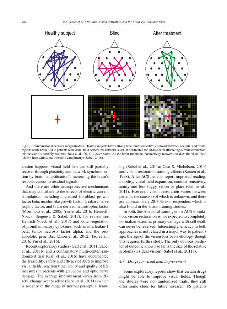
threshold of conscious perception (Fig. 5). If and howwell residual vision is consciously perceived dependson both local and global (downstream) brain networkmechanisms. The long-range alpha-synchronizationconnecting occipital and frontal cortical regionsseems to be a key in the vision recovery process(Fig. 6) (Sabel et al., 2011b; Bola et al., 2014).

B.A. Sabel et al. / Residual vision activation and the brain-eye-vascular triad 781
Fig. 5. Residual vision and brain network amplification. (A) This graph serves only as a conceptual guide to appreciate the nature of residualvision and the interactions of retina and brain by neuronal oscillatory activity. Accordingly, vision loss (e.g. measured by detection ability)depends on how many cells are lost: the greater the cell loss, the greater is the defect in different regions of the visual field. Areas of residualvision (ARVs; shown in grey) correspond to regions of partial damage with or without vascular dysregulation. They are found in all kinds ofvisual field defects such as after stroke (e.g. hemianopia) or retinal or optic nerve damage (e.g. glaucoma). Black areas represent completedamage. Note, however, that many black regions may, in fact, have some residual visual function as well. (B) Whether or not visual stimuliprocesses by the retina are consciously perceived by the brains is not only determined by the strength of the neuronal signals sent by the retinato the brain, but it also depends on how the brain processes this information through synchronization, amplification and interpretation. Neuralactivity of the retina is represented here by a simple sine wave. If the brain network is disorganized (illustrated here by non-synchronized,out-of-phase brain sine waves), the sum of retinal and brain signals is too low to surpass the perceptual threshold and the visual stimulus isnot perceived. When the brain is synchronized, this elevates (amplifies) the same residual visual signal to above-threshold perception, thusimproving or restoring conscious vision.
One opportunity to modify brain connectivity isby way of treating patients with alternating currentstimulation (ACS) (sometimes also referred to astransorbital, transcranial, or transpalpebral stimula-tion). ACS aims at activating residual vision andimprove visual fields (Sabel et al., 2011b; Bola et al.,2014; Gall et al., 2016) (Fig. 7). After optic nervedamage, for example ACS treatment for ten dayscan improve functional connectivity networks in thealpha-frequency range (Bola et al., 2014) and enlargevisual fields. This was shown with several studiesin Germany (Gall et al., 2011; Sabel et al., 2011b;Gall et al., 2016), Russia (Shandurina & Panin,1990) and Japan (Fujikado et al., 2006) includingsmall trails with nonarteritic ischemic neuropathy,primary open angle glaucoma, retinal artery occlu-sion and retinal dystrophies (Gall et al., 2011; Sabelet al., 2011b; Gall et al., 2016), macular degeneration(Anastassiou et al., 2013), and retinitis pigmentosa(Schatz et al., 2011, Bittner & Seger 2018). In hemi-anopia after stroke also direct current stimulation hasbeen used (Halko et al., 2011; Plow, Obretenova,Fregni, Pascual-Leone & Merabet, 2012). Indeed, the
approach to treat vision loss with electric current isnot new: almost 150 years ago vision recovery fol-lowing electric stimulation was already published inGermany (Erb, 1882; Mann, 1904).
The proposed mechanism of ACS action isa “learning-like” synaptic strengthening by repetitiveactivation of residual neurons in the brain’s network.ACS is applied near the eyes which forces RGCs tofire in the rhythm of the ACS frequency band (Her-rmann, Rach, Neuling & Struber, 2013; Foik et al.,2015). In this way brain oscillations may be mod-ulated and their repetitive use leads to long-lastingchanges (“long-term potentiation”) which outlaststhe time of stimulation (“after-effect”) (Sabel et al.,2011a; Herrmann et al., 2013). By way of thisbrain modulation (synchronization), residual vision isstrengthened through network reorganization whichcorrelates with the extent of visual field improve-ments (Sabel et al., 2011a; Bola et al., 2014). Thisis not proof of causality that network reorganizationis the only cause of recovery; both may have a com-mon (third) mechanism, for example, blood flowchanges. In any event, if no morphological regen-
782 B.A. Sabel et al. / Residual vision activation and the brain-eye-vascular triad
Fig. 6. Brain functional network reorganization. Healthy subjects have a strong functional connectivity network between occipital and frontalregions of the brain. But in patients with visual field defects this network is lost. When treated for 10 days with alternating current stimulation,this network is partially restored (Bola et al., 2014). Lower panel: As the brain functional connectivity recovers, so does the visual field(shown here with supra-threshold campimetry) (Sabel, 2016).
eration happens, visual field loss can still partiallyrecover through plasticity and network synchroniza-tion by brain “amplification”, increasing the brain’sresponsiveness to residual signals.
And there are other neuroprotective mechanismsthat may contribute to the effects of electric currentstimulation, including increased fibroblast growthfactor beta, insulin-like growth factor-1, ciliary nervetrophic factor, and brain-derived neurotrophic factor(Morimoto et al., 2005; Yin et al., 2016; Henrich-Noack, Sergeeva & Sabel, 2017), for review seeHenrich-Noack et al., 2017) and down-regulationof proinflammatory cytokines, such as interleukin-1beta, tumor necrosis factor alpha, and the pro-apoptotic gene Bax (Zhou et al., 2012; Tao et al.,2016; Yin et al., 2016).
Recent exploratory studies (Gall et al., 2011; Sabelet al., 2011b) and a confirmatory multi-center, ran-domized trial (Gall et al., 2016) have documentedthe feasibility, safety and efficacy of ACS to improvevisual fields, reaction time, acuity and quality of lifemeasures in patients with glaucoma and optic nervedamage. The average improvement varies from 20-40% change over baseline (Sabel et al., 2011a) whichis roughly in the range of normal perceptual learn-
ing (Sabel et al., 2011a; Otto & Michelson, 2014)and vision restoration training effects (Kasten et al.,1998). After ACS patients report improved reading,mobility, visual field expansion, contrast sensitivity,acuity and less foggy vision or glare (Gall et al.,2011). However, vision restoration varies betweenpatients, the cause(s) of which is unknown, and thereare approximately 20-30% non-responders which isalso found in the vision training studies.
In both, the behavioral training or the ACS stimula-tion, vision restoration is not expected to completelynormalize vision as primary damage with cell deathcan never be reversed. Interestingly, efficacy in bothapproaches is not related in a major way to patient’sage, the age of the vision loss or its etiology, thoughthis requires further study. The only obvious predic-tor of outcome known so far is the size of the relativescotoma (residual vision) (Sabel et al., 2011a).
4.7. Drugs for visual field improvement
Some exploratory reports show that certain drugsmight be able to improve visual fields. Thoughthe studies were not randomized trials, they stilloffer some clues for future research: FS patients

B.A. Sabel et al. / Residual vision activation and the brain-eye-vascular triad 783
Fig. 7. Activating residual vision. Examples of visual field recovery of three patients before and after treatment with alternating currentstimulation (ACS); Top and middle panel: visual fields of a case with diabetic retinopathy and open-angle glaucoma before and after 10days. The visual fields on the bottom is from a 27 year old male suffering from traumatic brain and optic nerve damage before and after 10days of ACS with an additional 3 months of relaxation and eye yoga exercises. Note that visual field recovery emerges mostly from the greyregions (relative scotomas or “areas of residual vision”). Red circles indicate regions of vision recovery.
treated with calcium channel blockers (nifedipine oramlodipine) (Gasser & Flammer, 1990; Konieczkaet al., 2016c) or acetazolamide (Flammer & Drance,1983a, b) to increase blood flow who rapidlyimproved visual field function. Drugs that improvedthe visual fields (VF) of glaucoma patients hadthe following characteristics and limitations: (i) the
short-term changes were not related to IOP changes;(ii) one could observe threshold shifts in relative sco-tomas (best seen with the help of the Bebie curve), butvery rarely – though occasionally – changes in the sizeof absolute scotomas, (iii) all the drugs that improvedVF had in common that they improved blood flowin the eye (and probably also in the brain), and (iv)

784 B.A. Sabel et al. / Residual vision activation and the brain-eye-vascular triad
improvements were more likely seen in FS+ but onlyinfrequently in FS- cases (Flammer & Konieczka,2017).
5. A neurovascular hypothesis of residualvision activation and restoration
In the present paper, we have discussed three fun-damental topics: the brain-eye-vascular triad, residualvision and its activation, and visual system plastic-ity (Fig. 1). These fields are interconnected as thefollowing hypothesis outlines.
To process visual information, neurons of thevisual pathway need to process and transmitelectrophysiological information from the eye to thebrain and within the brain by firing action potentials.Without visual stimulation, neurons are in a “restingstate”, firing action potentials only intermittently andat low rates. However, when visual stimuli need to beprocessed (for example when there is flickering light),neurons are activated and fire action potentials muchmore rapidly. This, however, requires a healthy cel-lular state and immediate upstream dilation of bloodvessels to increase oxygen and glucose supply. Neu-rovascular coupling (NC) is the mechanism wherebyneural activity and upstream vessel dilation are syn-chronized so that sufficient oxygen and glucose levelsreach the firing neurons. A key signal responsible forthis upstream vessel dilation is extracellular potas-sium which is elevated in the extracellular space whenneurons fire and which, through capillary potassium-sensing, initiates retrograde hyperpolarization. Sincepotassium ions are charged, it is an “electric” sig-nal that triggers upstream dilation (Longden et al.,2017).
But if vascular coupling is impaired, we proposethis can especially impact very small microvesselslike those found in the eye, inner ear, or brain.Vascular dysregulation (besides arteriosclerosis orvessel inflammation) then deprives neurons of oxy-gen and glucose and they get locked-in (“silenced”)in a hypo-metabolic state. Neurons are then unable tofire action potentials at sufficient rates and/or dura-tions to propagate a visual impulse. Though thesecells are too healthy to die (“silent survivors”), theysurvive but are unable to fire action potentials, leadingto a functional impairment.
We propose that areas of residual vision asoutlined above suffer not only from partial celldeath, but contain many cells which are silentsurvivors which – because of cellular or metabolic
stress and lack of energy (oxygen and glucose) –remain dormant for a long time (years). Visual fieldregions with such residual capacities appear to beblind perimetrically, but they provide an untappedpotential. Activating these silent survivors showsthat there is more vision potential behind the “blackcurtain” of blindness, but vision improvement ispossible using by visual training, medications, orelectrical stimulation. Interestingly, all these threetherapies have one thing in common: they improveblood flow. Visual training activates neurons to firevisual impulses and thus provokes blood flow locally,medications improve blood flow by pharmacologicalmeans, and electrical stimulation using alternatingcurrents stimulates neuronal firing and it mimics theendogenous electric mechanism of dilation directly.This proposal is compatible with the “residualvisual activation theory” (Sabel et al., 2011a) andexplains the following observations. (i) Behaviouraltraining takes a long time because each smalltraining stimulus (dot) excites only a small regionof the retina, (ii) drug treatment can instantaneouslyimprove visual fields, and (iii) alternating currentstimulation forces all surviving neurons to fire withrapid improvements in only a few days of treatment.
This cellular scenario is of relevance for regionsof the visual system directly damaged by the pathol-ogy, but also for down-stream neuronal networks ofthe brain. Healthy neurovascular coupling is thereforecritical for all visual functions and sub-functions, andwithout it there may be no plasticity.
To sum up, here we propose that vascular dys-regulation is both the problem and the solutionof vision loss. We believe that it is a fun-damental mechanism of restoration and neuralrecovery with a system-wide impact on neu-ronal reserve function. Further studies are nowneeded to substantiate this hypothesis to determine(i) if, or to what extent, vascular dysregulationis rate-limiting for neuronal activation because ofsilencing neurons, (ii) how vascular dysregulationaffects sensory-cognitive interactions, and (iii) howtreatment of it can reverse hypo-metabolic states,providing a possible source of sensory-cognitiverecovery and reserve.
6. Conclusions and perspectives
A better knowledge of the many interactionsbetween the eye, brain, and cardiovascular systemare key for a better understanding of how to treat

B.A. Sabel et al. / Residual vision activation and the brain-eye-vascular triad 785
vision loss. Exploring these interactions in the brain-eye-vascular triad (Fig. 1) will not only better explaindifferent pathological conditions, but it may also shedmore light on hitherto unexplained phenomena inclinical care (such as fluctuating and/or recoveryingvisual fields).
The brain has many direct and indirect influenceson eye diseases: direct influences are (i) the ampli-fication of (residual) retinofugal impulses, (ii) themodification and interpretation via top-down influ-ences (e.g. cognition, attention, emotion), and (iii)the control of eye movements. Indirect influences are(v) the modification of the vascular system throughstress hormones and their impact at different levelsof the vascular physiology, (vi) the nervous system,(vii) the CSF pressure conditions, and, finally, (viii)their relation to emotional states (stress, anxiety).
A more holistic medical and psychologicalapproach to VI is needed to address these differ-ent mechanisms of vision loss. This more holisticapproach might help pave the way for a fresh new per-spective of how to modulate multiple mechanisms, anuntapped potential for innovation in technology andpatient care. We need to grasp the meaning of visionimpairments in neurological diseases on the one handand appreciate the brain’s contribution in “visual”(eye) disorders on the other hand, particularly theirrole in activating residual vision to achieve recovery.
7. Future directions
This new understanding of a possible interaction ofbrain plasticity and vascular dysregulation in visionrecovery is a starting point to conceive of and testnovel therapeutic options to prevent disease pro-gression and/or restore visual functions. We are atthe beginning of a journey that includes the scienceof both brain physiology and vascular regulation inunderstanding and treating low vision.
Here are some suggestions for future directions:
– Further explore the role of transmodal brain plas-ticity to compensate for VI;
– Study hypo-metabolic mechanisms of cell inacti-vation (“silent” neurons);
– Search for both local (at or near the site of dam-age) and global mechanism of recovery of vision,e.g. brain functional networks and vascular sys-tem modulations.
– Study the mechanisms of vision training and braincurrent stimulation with regard to their impact on
residual vision activation and its relationship toregional and whole brain network synchroniza-tion and reorganization;
– Explore the potential to normalize vascular dys-regulation by way of drugs or other means;
– Evaluate psychological treatments for stress-reduction such as relaxation techniques likemeditation and yoga, physical exercise, and psy-chotherapeutic interventions and determine theirimpact on visual field fluctuations and visual fieldrecovery;
– Develop holistic concepts for the treatment of lowvision that are complementary to eye drops orsurgery and integrate the medical and psycho-logical state of the patient, including questionsrelated to emotions and lifestyles, issues whichare seldom addressed (Stew, 1997).
We should have an open mind that neurology,neuroscience and psychology could assist ophthal-mology to advance the field with a fresh newlook at the brain-eye-vascular triad. A more holis-tic approach to vision loss will unveil new treatmentoptions based on combined medical/psychologicalconcepts that follow a more global and not just aneye-centric perspective. Vision loss is a holistic mat-ter and the brain not just an “appendix” of the eye.There is clearly more wiggle room to improve visualfield defects and patient care and there is more lightat the end of the tunnel. The human suffering andthe burden to society inflicted by blindness justifiesthat fields outside of ophthalmology, such as neu-roscience/neurology and psychology, help push themission forward to restore vision and improve qual-ity of life. In this regard, the brain deserves a betterreputation for its role in ophthalmologic diseases ofthe “person behind the eye”.
References
AFB. Senior Site Press Release Archive. American Foundation forthe Blind Launches Web Site to Help People with Vision LossMaintain Independence. http://www.afb.org/info/programs-and-services/professional-development/experts-guide/press-release-archive-3641/1235 (accessed 03.04.2007).
Amedi, A., Raz, N., Pianka, P., Malach, R. & Zohary, E. (2003).Early ‘visual’ cortex activation correlates with superior verbalmemory performance in the blind. Nature Neuroscience, 6,758-766.
Amedi, A., Floel, A., Knecht, S., Zohary, E. & Cohen, L.G.(2004). Transcranial magnetic stimulation of the occipi-tal pole interferes with verbal processing in blind subjects.Nature Neuroscience, 7, 1266-1270.

786 B.A. Sabel et al. / Residual vision activation and the brain-eye-vascular triad
Anastassiou, G., Schneegans, A.L., Selbach, M. & Kremmer,S. (2013). Transpalpebral electrotherapy for dry age-relatedmacular degeneration (AMD): An exploratory trial. Restora-tive Neurology and Neuroscience, 31, 571-578.
Armstrong, R.A. (2011). Visual signs and symptoms of progressivesupranuclear palsy. Clinical & Experimental Optometry, 94,150-160.
Armstrong, R. & Kergoat, H. (2015). Oculo-visual changes andclinical considerations affecting older patients with dementia.Ophthalmic & Physiological Optics, 35, 352-376.
Armstrong, R.A. & Syed, A.B. (1996). Alzheimer’s disease andthe eye. Ophthalmic & Physiological Optics, 16, S2-8.
Backon, J., Matamoros, N., Ramirez, M., Sanchez, R.M., Ferrer,J., Brown, A. & Ticho, U. (1990). A functional vagotomyinduced by unilateral forced right nostril breathing decreasesintraocular pressure in open and closed angle glaucoma.British Journal of Ophthalmology, 74, 607-609.
Baertschi, M., Dayhaw-Barker, P. & Flammer, J. (2016). Theeffect of hypoxia on intra-ocular, mean arterial, retinal venousand ocular perfusion pressures. Clinical Hemorheology andMicrocirculation, 63, 293-303.
Balliett, R., Blood, K.M. & Bach-y-Rita, P., (1985). Visual fieldrehabilitation in the cortically blind? Journal of Neurology,Neurosurgery, and Psychiatry, 48, 1113-1124.
Bauer, C.M., Hirsch, G.V., Zajac, L., Koo, B.B., Collignon, O.& Merabet, L.B. (2017). Multimodal MR-imaging revealslarge-scale structural and functional connectivity changes inprofound early blindness. PLoS One, 12, e0173064.
Bedny, M., Pascual-Leone, A., Dodell-Feder, D., Fedorenko, E.& Saxe, R. (2011). Language processing in the occipital cor-tex of congenitally blind adults. Proceedings of the NationalAcademy of Sciences of the United States of America, 108,4429-4434.
Benowitz, L.I., He, Z. & Goldberg, J.L. (2017). Reaching thebrain: Advances in optic nerve regeneration. ExperimentalNeurology, 287, 365-373.
Berdahl, J.P., Allingham, R.R. & Johnson, D.H. (2008). Cere-brospinal fluid pressure is decreased in primary open-angleglaucoma. Ophthalmology, 115, 763-768.
Bittner, A.K. & Seger, K. (2018). Longevity of visual improve-ments following transcorneal electrical stimulation andefficacy of retreatment in three individuals with retinitis pig-mentosa. Graefe’s Archive for Clinical and ExperimentalOphthalmology, 256, 299-306.
Black, P. (1982). Visual disorders associated with cerebral palsy.British Journal of Ophthalmology, 66, 46-52.
Bodis-Wollner, I. (1990). The visual system in Parkinson’s disease.Research publications – Association for Research in Nervousand Mental Disease, 67, 297-316.
Bojinova, R.I., Konieczka, K., Meyer, P. & Todorova, M.G. (2016).The trilateral link between anaesthesia, perioperative visualloss and Flammer syndrome. BMC Anesthesiology, 16, 10.
Bola, M. & Sabel, B.A. (2015). Dynamic reorganization of brainfunctional networks during cognition. Neuroimage, 114, 398-413.
Bola, M., Gall, C. & Sabel, B.A. (2013). “Sightblind”: Perceptualdeficits in the “intact” visual field. Frontiers in Neurology, 4,80.
Bola, M., Gall, C. & Sabel, B.A. (2015). Disturbed temporaldynamics of brain synchronization in vision loss. Cortex, 67,134-146.
Bola, M., Gall, C., Moewes, C., Fedorov, A., Hinrichs, H. & Sabel,B.A. (2014). Brain functional connectivity network break-down and restoration in blindness. Neurology, 83, 542-551.
Bolognini, N., Rasi, F., Coccia, M. & Ladavas, E. (2005). Visualsearch improvement in hemianopic patients after audio-visualstimulation. Brain, 128, 2830-2842.
Bosking, W.H., Beauchamp, M.S. & Yoshor, D. (2017). ElectricalStimulation of Visual Cortex: Relevance for the Develop-ment of Visual Cortical Prosthetics. Annual Review of VisionScience, 3, 141-166.
Bourne, R.R.A., Flaxman, S.R., Braithwaite, T., Cicinelli, M.V.,Das, A., Jonas, J.B., Keeffe J., Kempen, J.H., Leasher, J.,Limburg, H., Naidoo, K., Pesudovs, K., Resnikoff, S., Sil-vester, A., Stevens, G.A., Tahhan, N., Wong, T.Y. & Taylor,H.R., Vision Loss Expert Group (2017). Magnitude, temporaltrends, and projections of the global prevalence of blind-ness and distance and near vision impairment: A systematicreview and meta-analysis. The Lancet Global Health, 5, e888–897.
Boyers, L.N., Karimkhani, C., Hilton, J., Richheimer, W. &Dellavalle, R.P. (2015). Global burden of eye and vision dis-ease as reflected in the Cochrane Database of SystematicReviews. JAMA Ophthalmology, 133, 25-31.
Bridge, H., Thomas, O., Jbabdi, S. & Cowey, A. (2008). Changesin connectivity after visual cortical brain damage underliealtered visual function. Brain, 131, 1433-1444.
Calkins, D.J., Pekny, M., Cooper, M.L. & Benowitz, L. (2017).The challenge of regenerative therapies for the optic nervein glaucoma. Lasker/IRRF Initiative on Astrocytes and Glau-comatous Neurodegeneration Participants. Experimental EyeResearch, 157, 28-33.
Chen, S.P., Bhattacharya, J. & Pershing, S. (2017). Association ofVision Loss With Cognition in Older Adults. JAMA Ophthal-mology, 135, 963-970.
Chen, K.W., Berger, C.C., Manheimer, E., Forde, D., Magidson, J.,Dachman, L. & Lejuez, J.W. (2012). Meditative Therapies forReducing Anxiety: A Systematic Review and Meta-analysisof Randomized Controlled Trials. Depression and Anxiety,29, 545-562.
Cheng, D.L., Greenberg, P.B. & Borton, D.A. (2017). Advances inRetinal Prosthetic Research: A Systematic Review of Engi-neering and Clinical Characteristics of Current ProstheticInitiatives. Current Eye Research, 42, 334-347.
Chun, B.Y. & Cestari, D.M. (2017). Advances in experimentaloptic nerve regeneration. Current Opinion in Ophthalmology,28, 558-563.
Cohen, L.G., Celnik, P., Pascual-Leone, A., Corwell, B., Falz, L.,Dambrosia, J., Honda, M., Sadato, N., Gerloff, C., Catala,M.D. & Hallett, M., (1997). Functional relevance of cross-modal plasticity in blind humans. Nature, 389, 180-183.
Collignon, O., Vandewalle, G., Voss, P., Albouy, G., Charbonneau,G., Lassonde, M. & Lepore, F. (2011). Functional special-ization for auditory-spatial processing in the occipital cortexof congenitally blind humans. Proceedings of the NationalAcademy of Sciences of the United States of America, 108,4435-4440.

B.A. Sabel et al. / Residual vision activation and the brain-eye-vascular triad 787
Connell, N. & Merabet, L.B., (2014). Uncovering the connectivityof the brain in relation to novel vision rehabilitation strategies.Neurology, 83, 484-485.
Costello, F., (2016). Vision Disturbances in Multiple Sclerosis.Semin Neurol, 36, 185-195.
Cotman, C.W. & Berchtold, N.C. (2002). Exercise: A behavioralintervention to enhance brain health and plasticity. Trends inNeuroscience, 25, 295-301.
Dada, T., Mittal, D., Mohanty, K., Faiq, M.A., Bhat, M.A., Yadav,R.K., Sihota, R., Sidhu, T., Velpandian, T., Kalaivani, M.,Pandey, R.M., Gao, Y., Sabel, B.A., Dada, R. (2018). Mind-fulness Meditation Reduces Intraocular Pressure, LowersStress Biomarkers and Modulates Gene Ex-pression in Glau-coma: a randomized controlled trial. Journal of Glaucoma,in press.
Dagnelie, G. (2013). Age-related psychophysical changes and lowvision. Investigative Ophthalmology and Visual Sciences, 54,ORSF88-93.
Dampney, R.A. (2015). Central mechanisms regulating coor-dinated cardiovascular and respiratory function duringstress and arousal. American Journal of Physiology. Reg-ulatory, Integrative and Comparative Physiology, 309,R429-443.
de Haan, G.A., Heutink, J., Melis-Dankers, B.J. & Tucha, O.(2014). Brouwer WH. Spontaneous recovery and treatmenteffects in patients with homonymous visual field defects: Ameta-analysis of existing literature in terms of the ICF frame-work. Survey of Ophthalmology, 59, 77-96.
de Lima, S., Habboub, G. & Benowitz, L.I. (2012). Combina-torial therapy stimulates long-distance regeneration, targetreinnervation, and partial recovery of vision after optic nerveinjury in mice. International Review of Neurobiology, 106,153-172.
Duncan, R.O., Sample, P.A., Weinreb, R.N., Bowd, C. & Zangwill,L.M. (2007). Retinotopic organization of primary visual cor-tex in glaucoma: Comparing fMRI measurements of corticalfunction with visual field loss. Progress in Retinal and EyeResearch, 26, 38-56.
Dundon, N.M., Ladavas, E., Maier, M.E. & Bertini, C. (2015). Mul-tisensory stimulation in hemianopic patients boosts orientingresponses to the hemianopic field and reduces attentionalresources to the intact field. Restorative Neurology and Neu-roscience, 33, 405-419.
Engelhorn, T., Michelson, G., Waerntges, S., Struffert, T., Haider,S. & Doerfler, A. (2011). Diffusion Tensor Imaging DetectsRarefaction of Optic Radiation in Glaucoma Patients. Acada-demic Radiology, 18, 764-769.
Erb, W. (1882). Handbuch der Elektrotherapie. Verlag V.C.W.Vogel; Leipzig.
Erb, C., Batra, A., Lietz, A., Bayer, A.U., Flammer, J. & Thiel,H.J. (1999). Psychological characteristics of patients withnormal-tension glaucoma. Graefe’s Archive for Clinical andExperimental Ophthalmology, 237, 753-757.
Fahle, M. (2002). Perceptual learning: Gain without pain? NatureNeuroscience, 5, 923-924.
Faiq, M.A., Dada, R., Kumar, A., Saluja, D. & Dada, T. (2016).Brain: The potential diagnostic and therapeutic target forGlaucoma. Current Drug Targets, CNS and Neurological Dis-orders, 15, 839-844.
Fang, L., Baertschi, M. & Mozaffarieh, M. (2014). The effect offlammer-syndrome on retinal venous pressure. BMC Ophthal-mology, 14, 121.
Fernandes, R.A.B., Diniz, B., Ribeiro, R. & Humayun, M. (2012).Artificial vision through neuronal stimulation. NeuroscienceLetters, 519, 122-128.
Flammer, J. & Drance. S.M. (1983a). Effect of acetazolamide onthe differential threshold. Archives of Ophthalmology, 101,1378-1380.
Flammer, J. & Drance, S.M. (1983b). Reversibility of a glaucoma-tous visual field defect after acetazolamide therapy. CanadianJournal of Ophthalmology, 18, 139-141.
Flammer, J. & Konieczka, K. (2015). Retinal venous pressure: Therole of endothelin. EPMA Journal, 6, 21.
Flammer, J. & Konieczka, K. (2017). The discovery of the Flam-mer syndrome: A historical and personal perspective. EPMAJournal, 8, 75-97.
Flammer, J., Drance, S.M. & Fankhauser, F. (1984a). Differentiallight threshold. Short- and long-term fluctuation in patientswith glaucoma, normal controls, and patients with suspectedglaucoma. Archives of Ophthalmology, 102, 876-879.
Flammer, J., Drance, S.M. & Schulzer, M. (1984b). Covariates ofthe Long-term Fluctuation of the Differential Light Thresh-old. Archives of Ophthalmology, 102, 880-882.
Flammer, J., Drance, S.M. & Zulauf, M. (1984c). Differentiallight threshold. Short- and long-term fluctuation in patientswith glaucoma, normal controls, and patients with suspectedglaucoma. Archives of Ophthalmology, 102, 704-706.
Flammer, J., Konieczka, K. & Flammer, A.J. (2013b). The pri-mary vascular dysregulation syndrome: Implications for eyediseases. EPMA Journal, 4, 14.
Flammer, J., Pache, M. & Resink, T. (2001). Vasospasm, its role inthe pathogenesis of diseases with particular reference to theeye. Progress in Retinal and Eye Research, 20, 319-349.
Flammer, J., Orgul, S., Costa, V.P., Orzalesi, N., Krieglstein, G.K.,Serra, L.M., Renard, J.P. & Stefansson, E. (2002). The impactof ocular blood flow in glaucoma. Progress in Retinal and EyeResearch, 21, 359-393.
Flammer, J., Konieczka, K., Bruno, R.M., Virdis, A., Flammer,A.J. & Taddei, S. (2013a). The eye and the heart. EuropeanHeart Journal, 34, 1270-1278.
Foik, A.T., Kublik, E., Sergeeva, E.G., Tatlisumak, T., Rossini,P.M., Sabel, B.A. & Waleszczyk, W.J. (2015). Retinal originof electrically evoked potentials in response to transcornealalternating current stimulation in the rat. Investigative Oph-thalmology and Visual Scinces, 56, 1711-1718.
Freund, H.-J., Sabel, B.A. & Witte, O. (Eds.) (1997). Brain Plas-ticity. Lippincott/Raven Press.
Fujikado, T., Morimoto, T., Matsushita, K., Shimojo, H., Okawa,Y. & Tano, Y. (2006). Effect of transcorneal electrical stimu-lation in patients with nonarteritic ischemic optic neuropathyor traumatic optic neuropathy. Japanese Journal of Ophthal-mology, 50, 266-273.
Gall, C., Sgorzaly, S., Schmidt, S., Brandt, S., Fedorov, A. &Sabel, B.A. (2011). Noninvasive transorbital alternating cur-rent stimulation improves subjective visual functioning andvision-related quality of life in optic neuropathy. Brain Stim-ulation, 4, 175-188.

788 B.A. Sabel et al. / Residual vision activation and the brain-eye-vascular triad
Gall, C., Schmidt, S., Schittkowski, M.P., Antal, A., Ambrus, G.G.,Paulus, W., Dannhauer, M., Michalik, R., Mante, A., Bola, M.,Lux, A., Kropf, S., Brandt, S.A. & Sabel, B.A. (2016). Alter-nating current stimulation for vision restoration after opticnerve damage: A randomized clinical trial. PLoS One, 11,e0156134.
Gao, Y. & Sabel, B.A. (2017). Microsaccade dysfunction and adap-tation in hemianopia after stroke. Restorative Neurology andNeuroscience, 35, 365-376.
Gao, Y., Huber, C. & Sabel, B.A. (2018). Stable microsaccadesand microsaccade-induced alpha band phase reset across thelife span. Investigative Ophthalmology and Visual Sciences,59, 2032-2041.
Gasser, P. & Flammer, J. (1986). Optic neuropathy of Graves’disease. A report of a perimetric follow-up. Ophthalmologica,192, 22-27.
Gasser, P. & Flammer, J. (1990). Short- and long-term effectof nifedipine on the visual field in patients with presumedvasospasm. The Journal of International Medical Research,18, 334-339.
Gherghel, D., Orgul, S., Dubler, B., Lubeck, P., Gugleta, K. &Flammer, J. (1999). Is vascular regulation in the central reti-nal artery altered in persons with vasospasm? Archives ofOphthalmology, 117, 1359-1362.
Gilbert, C.D. & Wiesel, T.N. (1992). Receptive field dynamics inadult primary visual cortex. Nature, 356, 150-152.
Gnanalingham, K.K., Bhattacharjee, S., Pennington, R., Ng, J. &Mendoza, N. (2005). The time course of visual field recov-ery following transphenoidal surgery for pituitary adenomas:Predictive factors for a good outcome. Journal of Neurology,Neurosurgery, and Psychiatry, 76, 415-419.
Gougoux, F., Zatorre, R.J., Lassonde, M., Voss, P. & Lep-ore, F. (2005). A functional neuroimaging study of soundlocalization: Visual cortex activity predicts performance inearly-blind individuals. PLoS Biology, 3, e27.
Grieshaber, M.C., Terhorst, T. & Flammer, J. (2006). Thepathogenesis of optic disc splinter haemorrhages: A newhypothesis. Acta Ophthalmologica Scandinavica, 84, 62-68.
Gupta, N., Ang, L.C., de Tilly, L.N., Bidaisee, L. & Yucel, Y.H.(2006). Human glaucoma and neural degeneration in intracra-nial optic nerve, lateral geniculate nucleus, and visual cortex.British Journal of Ophthalmology, 90, 674-678.
Gupta, N., Yucel & Y. H. (2007). Glaucoma as a neurode-generative disease. Current Opinion in Ophthalmology, 18,110-114.
Guthauser, U., Flammer, J. & Mahler, F. (1988). The relationshipbetween digital and ocular vasospasm. Graefe’s Archive forClinical and Experimental Ophthalmology, 226, 224-226.
Haas, A., Flammer, J. & Schneider, U. (1986). Influence of ageon the visual fields of normal subjects. American Journal ofOphthalmology, 101, 199-203.
Hadid, V. & Lepore, F. (2017). From Cortical Blindness to Con-scious Visual Perception: Theories on Neuronal Networksand Visual Training Strategies. Frontiers in Systems Neuro-science, 11, 64.
Hadjinicolaou, A.E., Meffin, H., Maturana, M.I., Cloherty, S.L.& Ibbotson, M.R. (2015). Prosthetic vision: Devices, patientoutcomes and retinal research. Clinical & ExperimentalOptometry, 98, 395-410.
Halko, M.A., Datta, A., Plow, E.B., Scaturro, J., Bikson, M.& Merabet, L.B. (2011). Neuroplastic changes followingrehabilitative training correlate with regional electrical fieldinduced with tDCS. Neuroimage, 57, 885-891.
Hamilton, R., Keenan, J.P., Catala, M. & Pascual-Leone, A. (2000).Alexia for Braille following bilateral occipital stroke in anearly blind woman. Neuroreport, 11, 237-240.
Harwerth, R.S., Crawford, M.L.J., Frishman, L.J., Viswanathan,S., Smith, Earl L. III & Carter-Dawson, L. (2002). Visualfield defects and neural losses from experimental glaucoma.Progress in Retinal and Eye Research, 21, 91-125.
Henrich-Noack, P., Sergeeva, E. & Sabel, B.A. (2017). Non-invasive electrical brain stimulation: From acute to late-stagetreatment of CNS damage. Neural Regeneration Research,12, 1590-1594.
Herrmann, C.S., Rach, S., Neuling, T. & Struber, D. (2013).Transcranial alternating current stimulation: A review of theunderlying mechanisms and modulation of cognitive pro-cesses. Frontiers in Human Neuroscience, 7, 279.
Hou, R., Zhang, Z., Yang, D., Wang, H., Chen, W., Li, Z., Sang, J.,Liu, S., Cao, Y., Xie, X., Ren, R., Zhang, Y., Sabel, B.A.& Wang, N. (2016). Intracranial pressure (ICP) and opticnerve subarachnoid space pressure (ONSP) correlation in theoptic nerve chamber: The Beijing Intracranial and IntraocularPressure (iCOP) study. Brain Research, 1635, 201-208.
Huxlin, K.R., Martin, T., Kelly, K., Riley, M., Friedman, D.I.,Burgin, W.S. & Hayhoe, M. (2009). Perceptual relearning ofcomplex visual motion after V1 damage in humans. Journalof Neuroscience, 29, 3981-3991.
Jobke, S., Kasten, E. & Sabel, B.A. (2009). Vision restorationthrough extrastriate stimulation in patients with visual fielddefects. Neurorehabilitation and Neural Repair, 23, 246-255.
Kanjee, R., Yucel, Y.H., Steinbach, M.J., Gonzalez, E.G. & Gupta,N. (2012). Delayed saccadic eye movements in glaucoma. Eyeand Brain, 4, 63-68.
Kasten, E., Bunzenthal, U. & Sabel, B.A. (2006). Visual fieldrecovery after vision restoration therapy (VRT) is indepen-dent of eye movements: An eye-tracker study. BehaviouralBrain Research, 175, 18-26.
Kasten, E., Guenther, T. & Sabel, B.A. (2008). Inverse stimuliin perimetric performance reveal larger visual field defects:Implications for vision restoration. Restorative Neurologyand Neuroscience, 26, 355-364.
Kasten, E., Wust, S., Behrens-Baumann, W. & Sabel, B.A. (1998).Computer-based training for the treatment of partial blind-ness. Nature Medicine, 4, 1083-1087.
Killer, H.E., Jaggi, G.P., Flammer, J., Miller, N.R., Huber, A.R. &Mironov, A. (2007). Cerebrospinal fluid dynamics betweenthe intracranial and the subarachnoid space of the optic nerve.Is it always bidirectional? Brain, 130, 514-520.
Kochkorov, A., Gugleta, K., Zawinka, C., Katamay, R., Flam-mer, J. & Orgul, S. (2006). Short-term retinal vessel diametervariability in relation to the history of cold extremities. Inves-tigative Ophthalmology and Visual Sciences, 47, 4026-4033.
Kolmel, H.W. (1985). Complex visual hallucinations in thehemianopic field. Journal of Neurology, Neurosurgery, andPsychiatry, 48, 29-38.
Konieczka, K. & Erb, C. (2017). Diseases potentially related toFlammer syndrome. EPMA Journal, 8, 327-332.

B.A. Sabel et al. / Residual vision activation and the brain-eye-vascular triad 789
Konieczka, K. & Flammer, J. (2016). Phenomenologyand Clinical Relevance of the Flammer Syndrome.Klinische Monatsblatter fur Augenheilkunde, 233,1331-1336.
Konieczka, K., Ritch, R., Traverso, C.E., Kim, D.M., Kook, M.S.,Gallino, A., Golubnitschaja, O., Erb, C., Reitsamer, H.A.,Kida, T., Kurysheva, N. & Yao, K. (2014). Flammer syn-drome. EPMA Journal, 5, eCollection.
Konieczka, K., Koch, S., Binggeli, T., Schoetzau, A. & Kesselring,J. (2016b). Multiple sclerosis and primary vascular dysregu-lation (Flammer syndrome). EPMA Journal, 7, 13.
Konieczka, K., Koch, S., Schoetzau, A. & Todorova, M.G. (2016a).Increased Prevalence of Flammer Syndrome in Patients withRetinitis Pigmentosa. Klinische Monatsblatter fur Augen-heilkunde, 233, 448-452.
Konieczka, K., Todorova, M.G., Bojinova, R.I., Binggeli, T.,Chackathayil, T.N. & Flammer, J. (2016c). UnexpectedEffect of Calcium Channel Blockers on the Optic NerveCompartment Syndrome. Klinische Monatsblatter fur Augen-heilkunde, 233, 387-390.
Kupers, R., Beaulieu-Lefebvre, M., Schneider, F.C., Kassuba, T.,Paulson, O.B., Siebner, H.R. & Ptito, M. (2011). Neuralcorrelates of olfactory processing in congenital blindness.Neuropsychologia, 49, 2037-2044.
Kurysheva, N.I., Ryabova, T.Y. & Shlapak, V.N. (2018). Heart ratevariability: The comparison between high tension and normaltension glaucoma. EPMA Journal, 9, 35-45.
Land, M.F. (2006). Eye movements and the control of actions ineveryday life. Progress in Retinal and Eye Research, 25, 296-324.
Lewis, P.M. & Rosenfeld, J.V. (2016). Electrical stimulationof the brain and the development of cortical visual pros-theses: An historical perspective. Brain Research, 1630,208-224.
Longden, T.A., Dabertrand, F., Koide, M., Gonzales, A.L.,Tykocki, N.R., Brayden, J.E., Hill-Eubanks, D. & Nelson,M.T. (2017). Capillary K+-sensing initiates retrograde hyper-polarization to increase local cerebral blood flow. NatureNeuroscience, 20, 717-726.
MacAskill, M.R. & Anderson, T.J. (2016). Eye movements in neu-rodegenerative diseases. Current Opinion in Neurology, 29,61-68.
Mahler, F., Saner, H., Wurbel, H. & Flammer, J. (1989). Local cool-ing test for clinical capillaroscopy in Raynaud’s phenomenon,unstable angina, and vasospastic visual disorders. Vasa, 18,201-204.
Mann, L. (1904). Uber elektrotherapeutische Versuche beiOptikuserkrankungen. Zeitschr fur diat phys Therapie, 8, 416-427.
Marra, G. & Flammer, J. (1991). The learning and fatigue effectin automated perimetry. Graefe’s Archive for Clinical andExperimental Ophthalmology, 229, 501-504.
Marshall, R.S., Ferrera, J.J., Barnes, A., Zhang, X., O’Brien, K.A.,Chmayssani, M., Hirsch, J. & Lazar, R.M. (2008). Brainactivity associated with stimulation therapy of the visual bor-derzone in hemianopic stroke patients. Neurorehabilitationand Neural Repair, 22, 136-144.
Martinez-Conde, S., Otero-Millan, J. & Macknik, S.L. (2013). Theimpact of microsaccades on vision: Towards a unified the-
ory of saccadic function. Nature reviews. Neuroscience, 14,83-96.
Matteo, B.M., Vigano, B., Cerri, C.G. & Perin, C. (2016). Visualfield restorative rehabilitation after brain injury. Journal ofVision, 16, 11.
Mazzi, C., Savazzi, S. & Silvanto, J. (2018). On the “blindness” ofblindsight: What is the evidence for phenomenal awareness inthe absence of primary visual cortex (V1)? Neuropsychologia,in press.
Meienberg, O., Flammer, J. & Ludin, H.P. (1982). Subclinicalvisual field defects in multiple sclerosis. Demonstration andquantification with automated perimetry, and comparisonwith visually evoked potentials. Journal of Neurology, 227,125-133.
Merabet, L.B. (2011). Building the bionic eye: An emerging realityand opportunity. Progress in Brain Research, 192, 3-15.
Merabet, L.B. & Pascual-Leone, A. (2010). Neural reorganizationfollowing sensory loss: The opportunity of change. NatureReviews. Neuroscience, 11, 44-52.
Merabet, L.B., Rizzo, J.F. 3rd, Pascual-Leone, A. & Fernandez,E. (2007). ‘Who is the ideal candidate?’ Decisions and issuesrelating to visual neuroprosthesis development, patient testingand neuroplasticity. Journal of Neural Engineering, 4, S130-135.
Merabet, L.B., Rizzo, J.F., Amedi, A., Somers, D.C. & Pascual-Leone, A. (2005). What blindness can tell us about seeingagain: Merging neuroplasticity and neuroprostheses. NatureReviews. Neuroscience, 6, 71-77.
Metz, G.A., Schwab, M.E. & Welzl, H. (2001). The effects of acuteand chronic stress on motor and sensory performance in maleLewis rats. Physiology & Behavior, 72, 29-35.
Mills, J.O., Jalil, A. & Stanga, P.E. (2017). Electronic retinalimplants and artificial vision: Journey and present. Eye (Lon-don), 31, 1383-1398.
Morimoto, T., Miyoshi, T., Matsuda, S., Tano, Y., Fujikado, T. &Fukuda, Y. (2005). Transcorneal electrical stimulation rescuesaxotomized retinal ganglion cells by activating endogenousretinal IGF-1 system. Investigative Ophthalmology and VisualSciences, 46, 2147-2155.
Morris, S.L., Foster, C.J., Parsons, R., Falkmer, M., Falkmer, T. &Rosalie, S.M. (2015). Differences in the use of vision and pro-prioception for postural control in autism spectrum disorder.Neuroscience, 307, 273-280.
Mozaffarieh, M., Schoetzau, A., Sauter, M., Grieshaber, M.,Orgul, S., Golubnitschaja, O. & Flammer, J. (2008). Cometassay analysis of single-stranded DNA breaks in circulat-ing leukocytes of glaucoma patients. Molecular Vision, 14,1584-1588.
Mozaffarieh, M., Fontana Gasio, P., Schotzau, A., Orgul, S., Flam-mer, J. & Krauchi, K. (2010). Thermal discomfort with coldextremities in relation to age, gender, and body mass indexin a random sample of a Swiss urban population. PopulationHealth Metrics, 8, 17.
Najarpour Foroushani, A., Pack, C.C. & Sawan, M. (2018). Cor-tical visual prostheses: From microstimulation to functionalpercept. Journal of Neural Engineering, 15, 021005.
Ohl, S., Wohltat, C., Kliegl, R., Pollatos, O. & Engbert, R. (2016).Microsaccades Are Coupled to Heartbeat. Journal of Neuro-science, 36, 1237-1241.

790 B.A. Sabel et al. / Residual vision activation and the brain-eye-vascular triad
Otto, J. & Michelson, G. (2014). Repetitive tests of visual functionimproved visual acuity in young subjects. British Journal ofOphthalmology, 98, 383-386.
Owsley, C. (2011). Aging and vision. Vision Research, 51, 1610-1622.
Pache, M., Krauchi, K., Cajochen, C., Wirz-Justice, A., Dubler,B., Flammer, J. & Kaiser, H.J. (2001). Cold feet and pro-longed sleep-onset latency in vasospastic syndrome. Lancet,358, 125-126.
Palva, J.M. & Palva, S. (2017). Functional integration across oscil-lation frequencies by cross-frequency phase synchronization.European Journal of Neuroscience, in press.
Pammer, K. (2014). Temporal sampling in vision and the impli-cations for dyslexia. Frontiers in Human Neuroscience, 7,933.
Pardue, M.T. & Allen, R.S. (2018). Neuroprotective strategies forretinal disease. Progress in Retinal and Eye Research, 65,50-76.
Pascual-Leone, A., Amedi, A., Fregni, F. & Merabet, L.B. (2005).The plastic human brain cortex. Annual Review of Neuro-science, 28, 377-401.
Pascolini, D. & Mariotti, S.P. (2012). Global estimates of visualimpairment 2010. British Journal of Ophthalmology, 96, 614-618.
Plow, E.B., Obretenova, S.N., Fregni, F., Pascual-Leone, A. &Merabet, L.B. (2012). Comparison of visual field training forhemianopia with active versus sham transcranial direct corti-cal stimulation. Neurorehabilitation and Neural Repair, 26,616-626.
Poggel, D.A., Kasten, E. & Sabel, B.A. (2004). Attentional cueingimproves vision restoration therapy in patients with visualfield loss. Neurology, 63, 2069-2076.
Poggel, D.A., Kasten, E., Muller-Oehring, E.M., Bunzenthal, U.& Sabel, B.A. (2006a). Improving residual vision by atten-tional cueing in patients with brain lesions. Brain ResearchCognitive Brain Research, 1097, 142-148.
Poggel, D.A., Muller-Oehring, E.M., Gothe, J., Kenkel, S., Kas-ten, E. & Sabel, B.A. (2006b). Visual hallucinations duringspontaneous and training-induced visual field recovery. Neu-ropsychologia, 45, 2598-2607.
Polat, U., Ma-Naim, T., Belkin, M., Sagi, D. (2004). Improvingvision in adult amblyopia by perceptual learning. Proc NatlAcad Sci USA, 101, 6692-6697.
Poppel, E., Held, R. & Frost, D. (1973). Residual visual functionafter brain wounds involving the central visual pathways inman. Nature, 243, 295-296.
Proudlock, F.A. & Gottlob, I. (2007). Physiology and pathology ofeye-head coordination. Progress in Retinal and Eye Research,26, 486-515.
Quigley, H.A. (2011). Glaucoma. Lancet, 377, 1367-1377.
Rachitskaya, A.V. & Yuan, A. (2016). Argus II retinal prosthesissystem: An update. Ophthalmic Genetics, 37, 260-266.
Reinhard, J., Schreiber, A., Schiefer, U., Kasten, E., Sabel, B.A.,Kenkel, S., Vonthein, R. & Trauzettel-Klosinski, S. (2005).Does visual restitution training change absolute homonymousvisual field defects? A fundus controlled study. British Jour-nal of Ophthalmology, 89, 30-35.
Roder, B., Stock, O., Bien, S., Neville, H. & Rosler, F. (2002).Speech processing activates visual cortex in congenitallyblind humans. European Journal of Neuroscience, 16, 930-936.
Romano, J.G., Schulz, P. & Kenkel, S. (2008). Visual field changesafter a rehabilitation intervention: Vision restoration therapy.Journal of the Neurological Sciences, 273, 70-74.
Rosenberg, E.A. & Sperazza, L.C. (2008). The visually impairedpatient. American Family Physician, 77, 1431-1436.
Roth, T., Sokolov, A.N., Messias, A., Roth, P., Weller, M. &Trauzettel-Klosinski, S. (2009). Comparing explorative sac-cade and flicker training in hemianopia: A randomizedcontrolled study. Neurology, 72, 324-331.
Sabel, B.A. (1999). Restoration of vision I: Neurobiological mech-anisms of restoration and plasticity after brain damage –a review. Restorative Neurology and Neuroscience, 15, 177-200.
Sabel, B. (2016). Restoring Low Vision, Amazon, Charleston.
Sabel, B.A. & Gudlin, J. (2014). Vision Restoration Training forGlaucoma. A randomized Clinical Trial. JAMA Ophthalmol-ogy, 132, 381-389.
Sabel, B.A., Fedorov, A., Henrich-Noack, P. & Gall, C. (2011a).Vision restoration after brain damage: The “Residual VisionActivation Theory”. Progress in Brain Research, 192, 199-262.
Sabel, B.A., Wang, J., Cardenas-Morales, L., Faiq, M. & Heim,C. (2018). Mental stress as consequence and cause of visionloss: The dawn of psychosomatic ophthalmology – a review.EPMA Journal, 9, 133-160.
Sabel, B.A., Fedorov, A.B., Naue, N., Borrmann, A., Herrmann,C. & Gall, C. (2011b). Non-invasive alternating currentstimulation improves vision in optic neuropathy. RestorativeNeurology and Neuroscience, 29, 493-505.
Sadato, N., Pascual-Leone, A., Grafman, J., Ibanez, V., Deiber,M.P., Dold, G. & Hallett, M. (1996). Activation of the primaryvisual cortex by Braille reading in blind subjects. Nature, 380,526-528.
Sahraie, A., Trevethan, C.T., MacLeod, M.J., Murray, A.D., Olson,J.A. & Weiskrantz, L. (2006). Increased sensitivity afterrepeated stimulation of residual spatial channels in blind-sight. Proceedings of the National Academy of Sciences ofthe United States of America, 103, 14971-14976.
Samuels, B.C., Hammes, N.M., Johnson, P.L., Shekhar, A.,McKinnon, S.J. & Allingham, R.R. (2012). Dorsome-dial/Perifornical hypothalamic stimulation increases intraoc-ular pressure, intracranial pressure, and the translaminarpressure gradient. Investigative Ophthalmology and VisualSciences, 53, 7328-7335.
Sanders, M.D., Warrington, E.K., Marshall, J. & Weiskrantz, L.(1974). “Blindsight”: Vision in a field defect. Lancet, 1, 707-708.
Schatz, A., Rock, T., Naycheva, L., Willmann, G., Wil-helm, B., Peters, T., Bartz-Schmidt, K.U., Zrenner, E.,Messias, A. & Gekeler, F. (2011). Transcorneal electri-cal stimulation for patients with retinitis pigmentosa: Aprospective, randomized, sham-controlled exploratory study.Investigative Ophthalmology and Visual Sciences, 52, 4485-4496.

B.A. Sabel et al. / Residual vision activation and the brain-eye-vascular triad 791
Schoemann, J., Engelhorn, T., Waerntges, S., Doerfler, A., El-Rafei, A. & Michelson, G. (2014). Cerebral microinfarcts inprimary open-angle glaucoma correlated with DTI-derivedintegrity of optic radiation. Investgative Ophthalmology andVisual Sciences, 55, 7241-7247.
Schultz, G. & Melzack, R. (1991). The Charles Bonnet syndrome:‘Phantom visual images.’ Perception, 20, 809-825.
Seiler, M.J. & Aramant, R.B. (2012). Cell replacement and visualrestoration by retinal sheet transplants. Progress in Retinaland Eye Research, 31, 661-687.
Shandurina, A.N. & Panin, A.V. (1990). Clinical and physiologicalanalysis of periorbital transcutaneous electrical stimulationmethod for damaged optic nerve and retina. Fiziologiia Che-loveka, 16, 53-59.
Shemagonov, A.V. & Sidorenko, V.N. (2000). Can the MedicalResonance Therapy Music affect autonomous innervation ofcerebral arteries? Integrative Physiological and BehavioralScience, 35, 218-223.
Shepherd, R.K., Shivdasani, M.N., Nayagam, D.A., Williams, C.E.& Blamey, P.J. (2013). Visual prostheses for the blind. Trendsin Biotechnology, 31, 562-571.
Shi, X.F., Xu, L.M., Li, Y., Wang, T., Zhao, K.X. & Sabel, B.A.(2012). Fixational saccadic eye movements are altered inanisometropic amblyopia. Restorative Neurology and Neu-roscience, 30, 445-462.
Shily, B.G. (1987). Psychophysiological stress, elevated intraoc-ular pressure, and acute closed-angle glaucoma. AmericanJournal of Optometry and Physiological Optics, 64, 866-870.
Silverstein, S.M. & Rosen, R. (2015). Schizophrenia and the eye.Schizophrenia Research. Cognition, 2, 46-55.
Sossi, N. & Anderson, D.R. (1983). Blockage of axonal transportin optic nerve induced by elevation of intraocular pressure.Effect of arterial hypertension induced by angiotensin I.Archives in Ophthalmology, 101, 94-97.
Stein, J. (2014). Dyslexia: The Role of Vision and Visual Attention.Current Developmental Disorders Reports, 1, 267-280.
Stevens, G.A., White, R.A., Flaxman, S.R., Price, H., Jonas, J.B.,Keeffe, J., Leasher, J., Naidoo, K., Pesudovs, K., Resnikoff,S., Taylor, H. & Bourne, R.R.A. (2013). Global prevalenceof vision impairment and blindness: Magnitude and temporaltrends, 1990-2010. Ophthalmology, 120, 2377-2384.
Stew, W.C. (1997). Chronic Open-angle Glaucoma and Lifestyle.Progress in Retinal and Eye Research, 16, 567-590.
Tan, C.S., Sabel, B.A., Goh, K.Y. (2006). Visual hallucinationsduring visual recovery after central retinal artery occlusion.Arch Neurol 63, 598-600.
Tang, Y.-Y., Holzel, B. & Posner, M. (2015). The neuroscienceof mindfulness meditation. Nature Review Neuroscience, 16,213-225.
Tao, Y., Chen, T., Liu, Z.Y., Wang, L.Q., Xu, W.W., Qin, L.M.,Peng, G.H. & Yi-Fei, H. (2016). Topographic Quantifica-tion of the Transcorneal Electrical Stimulation (TES)-InducedProtective Effects on N-Methyl-N-Nitrosourea-Treated Reti-nas. Investigative Ophthalmology and Visual Sciences, 57,4614-4624.
Uhlhaas, P.J. & Singer, W. (2006). Neural synchrony in braindisorders: Relevance for cognitive dysfunctions and patho-physiology. Neuron, 52, 155-168.
van Arb, M., Gompper, B., Meyer, A.H., Stutz, E.Z., Orgul, S.,Flammer, J. & Krauchi, K. (2009). Relationship betweengender role, anger expression, thermal discomfort andsleep onset latency in women. BioPsychoSocial Medicine,3, 11.
Wang, J., Li, T., Sabel, B.A., Chen, Z., Wen, H., Li, J., Xie, X.,Yang, D., Chen, W., Wang, N., Xian, J. & He, H. (2016).Structural brain alterations in primary open angle glaucoma:A 3T MRI study. Science Reports, 6, 18969.
Whiting, P., Kerby, J., Coffey, P., da Cruz, L. & McKernan,R. (2015). Progressing a human embryonic stem-cell-basedregenerative medicine therapy towards the clinic. Philosoph-ical transactions of the Royal Society of London. Series B,Biological Sciences, 370, 20140375.
WHO Fact Sheet. Visual impairment and blindness. FactSheet N 282, updated August 2014. http://www.who.int/mediacentre/factsheets/fs282/en/
Williams, M.E., Fink, C., Zamora, I. & Borchert, M. (2014).Autism assessment in children with optic nerve hypoplasiaand other vision impairments. Developmental Medicine andChild Neurology, 56, 66-72.
Winterson, B.J. & Collewijn, H. (1976). Microsaccades duringfinely guided visuomotor tasks. Vision Research, 16, 1387-1390.
Wostyn, P., Audenaert, K. & De Deyn, P.P. (2010). Alzheimer’sdisease: Cerebral glaucoma? Medical Hypotheses, 74, 973-977.
Yang, D., Fu, J., Hou, R., Liu, K., Jonas, J.B., Wang, H., Chen,W., Li, Z., Sang, J., Zhang, Z., Liu, S., Cao, Y., Xie, X.,Ren, R., Lu, Q., Weinreb, R.N. & Wang, N. (2014). Opticneuropathy induced by experimentally reduced cerebrospinalfluid pressure in monkeys. Investigative Ophthalmology andVisual Sciences, 55, 3067-3073.
Yeghiazaryan, K., Flammer, J., Orgul, S., Wunderlich, K. & Gol-ubnitschaja, O. (2009). Vasospastic individuals demonstratesignificant similarity to glaucoma patients as revealed bygene expression profiling in circulating leukocytes. MolecularVision, 15, 2339-2348.
Yin, H., Yin, H., Zhang, W., Miao, Q., Qin, Z., Guo, S., Fu, Q., Ma,J., Wu, F., Yin, J., Yang, Y. & Fang, X. (2016). Transcornealelectrical stimulation promotes survival of retinal ganglioncells after optic nerve transection in rats accompanied byreduced microglial activation and TNF-alpha expression.Brain Research, 1650, 10-20.
Yu, L., Xie, B., Yin, X., Liang, M., Evans, A.C., Wang, J. & Dai,C. (2013). Reduced cortical thickness in primary open-angleglaucoma and its relationship to the retinal nerve fiber layerthickness. PLoS One, 8, e73208.
Yucel, Y. (2013). Central nervous system changes in glaucoma.Journal of Glaucoma, 22, S24-25.
Zhang, X., Kedar, S., Lynn, M.J., Newman, N.J. & Biousse, V.(2006). Natural history of homonymous hemianopia. Neurol-ogy, 66, 901-905.
Zhou, W.T., Ni, Y.Q., Jin, Z.B., Zhang, M., Wu, J.H., Zhu, Y.,Xu, G.Z. & Gan, D.K. (2012). Electrical stimulation ame-liorates light-induced photoreceptor degeneration in vitrovia suppressing the proinflammatory effect of microglia andenhancing the neurotrophic potential of Muller cells. Exper-imental Neurology, 238, 192-208.