Involvement of the Clock Gene Rev-erb alphain the ...
15
Involvement of the Clock Gene Rev-erb alpha in the Regulation of Glucagon Secretion in Pancreatic Alpha- Cells Elaine Vieira 1,2 *, Laura Marroquı´ 1,2 , Ana Lucia C. Figueroa 3 , Beatriz Merino 1,2 , Rebeca Fernandez-Ruiz 2,3 , Angel Nadal 1,2 , Thomas P. Burris 5 , Ramon Gomis 2,3,4 , Ivan Quesada 1,2 * 1 Instituto de Bioingenierı ´a, Universidad Miguel Hernandez de Elche, Elche, Spain, 2 CIBER de Diabetes y Enfermedades Metabo ´ licas Asociadas, Barcelona, Spain, 3 Diabetes and Obesity Laboratory, Institut d’investigacions Biome `diques August Pi i Sunyer, Barcelona, Spain, 4 Endocrinology and Diabetes Unit, Hospital Clinic, Universitat de Barcelona, Barcelona, Spain, 5 Department of Molecular Therapeutics, The Scripps Research Institute, Jupiter, Florida, United States of America Abstract Disruption of pancreatic clock genes impairs pancreatic beta-cell function, leading to the onset of diabetes. Despite the importance of pancreatic alpha-cells in the regulation of glucose homeostasis and in diabetes pathophysiology, nothing is known about the role of clock genes in these cells. Here, we identify the clock gene Rev-erb alpha as a new intracellular regulator of glucagon secretion. Rev-erb alpha down-regulation by siRNA (60–70% inhibition) in alphaTC1-9 cells inhibited low-glucose induced glucagon secretion (p,0.05) and led to a decrease in key genes of the exocytotic machinery. The Rev- erb alpha agonist GSK4112 increased glucagon secretion (1.6 fold) and intracellular calcium signals in alphaTC1-9 cells and mouse primary alpha-cells, whereas the Rev-erb alpha antagonist SR8278 produced the opposite effect. At 0.5 mM glucose, alphaTC1-9 cells exhibited intrinsic circadian Rev-erb alpha expression oscillations that were inhibited by 11 mM glucose. In mouse primary alpha-cells, glucose induced similar effects (p,0.001). High glucose inhibited key genes controlled by AMPK such as Nampt, Sirt1 and PGC-1 alpha in alphaTC1-9 cells (p,0.05). AMPK activation by metformin completely reversed the inhibitory effect of glucose on Nampt-Sirt1-PGC-1 alpha and Rev-erb alpha. Nampt inhibition decreased Sirt1, PGC-1 alpha and Rev-erb alpha mRNA expression (p,0.01) and glucagon release (p,0.05). These findings identify Rev-erb alpha as a new intracellular regulator of glucagon secretion via AMPK/Nampt/Sirt1 pathway. Citation: Vieira E, Marroquı ´ L, Figueroa ALC, Merino B, Fernandez-Ruiz R, et al. (2013) Involvement of the Clock Gene Rev-erb alpha in the Regulation of Glucagon Secretion in Pancreatic Alpha-Cells. PLoS ONE 8(7): e69939. doi:10.1371/journal.pone.0069939 Editor: Miguel Lo ´ pez, University of Santiago de Compostela School of Medicine - CIMUS, Spain Received May 14, 2013; Accepted June 13, 2013; Published July 25, 2013 Copyright: ß 2013 Vieira et al. This is an open-access article distributed under the terms of the Creative Commons Attribution License, which permits unrestricted use, distribution, and reproduction in any medium, provided the original author and source are credited. Funding: FP7- Marie Curie program, Ministerio de Educacio ´ n y Ciencia (BFU2010-21773; BFU2011-28358), Generalitat Valenciana (PROMETEO/2011/080) and European Foundation for the Study of Diabetes (Ref.: 94553). CIBERDEM is an initiative of the Instituto de Salud Carlos III. Spanish Ministry of Science and Innovation grant agreement number SAF 2010-19527. Government of Catalonia, grant agreement number 2009 SGR 1426. The funders had no role in study design, data collection and analysis, decision to publish, or preparation of the manuscript. Competing Interests: Co-author Dr. Angel Nadal is a PLOS ONE Editorial Board member. This does not alter our adherence to all the PLOS ONE policies on sharing data and materials. * E-mail: [email protected] (EV); [email protected] (IQ) Introduction Numerous biological processes such as body temperature, sleep/wake cycle, feeding, metabolism and hormone release display 24 hours rhythms that are driven by cell circadian clocks [1,2]. In mammals, the central pacemaker of the clock machinery is located in the hypothalamus, more precisely in neurons of the suprachiasmatic nuclei. Besides the central location in the brain, peripheral molecular clocks exist in several organs, including liver, kidneys, muscle, adipose tissue and pancreas [3,4,5,6]. The central and peripheral oscillators share a common molecular circuitry, with a battery of transcriptional activators and repressors forming a self-sustained transcriptional feedback loop. The primary loop is composed by the transcription factors CLOCK (circadian locomotor output cycles kaput) and BMAL1 (brain and Muscle arnt-like 1) which drive the transcription of the Per1 (period homolog drosophila 1) and Per2 (period homolog drosophila 2) and Cry1 (cryptochrome 1) Cry2 (cryptochrome 2) genes [7]. PER and CRY inhibit their own CLOCK: BMAL1-induced transcrip- tion, and turnover of PER and CRY allows this cycle to continue. Important nuclear receptors such as Rev-erb alpha (reverse- eritroblastosis virus alpha, nuclear receptor encoded by NR1D1) can also regulate CLOCK and BMAL1 expression. Besides its role in the control of the molecular clock, Rev-erb alpha has also been shown to regulate lipid metabolism and bile acid homeostasis in the liver [8,9], adipogenesis [10] gluconeogenic genes [11,12], as well as insulin secretion [13]. Thus, Rev-erb alpha is considered a good candidate to link circadian rhythms and metabolism. Disturbances in the regulation of circadian rhythms have been implicated in the development of metabolic disorders such as obesity and type 2 diabetes. For instance, CLOCK and BMAL1 disruption leads to alterations in the expression of beta-cell genes involved in growth, survival and synaptic vesicle assembly, which can trigger the onset of diabetes [14]. The regulation of glucagon secretion in response to glucose plays an essential role in the control of glycaemic levels. Alteration of the alpha-cell normal function is part of the events that are present in the pathophys- iology of diabetes mellitus [15]. Actually, hyperglucagonemia is typically found in diabetic patients, favoring hepatic gluconeo- genesis and hyperglycemia. Despite its importance, little is known PLOS ONE | www.plosone.org 1 July 2013 | Volume 8 | Issue 7 | e69939
Transcript of Involvement of the Clock Gene Rev-erb alphain the ...
Involvement of the Clock Gene Rev-erb alpha in theRegulation of Glucagon Secretion in Pancreatic Alpha-CellsElaine Vieira1,2*, Laura Marroquı1,2, Ana Lucia C. Figueroa3, Beatriz Merino1,2, Rebeca Fernandez-Ruiz2,3,
Angel Nadal1,2, Thomas P. Burris5, Ramon Gomis2,3,4, Ivan Quesada1,2*
1 Instituto de Bioingenierıa, Universidad Miguel Hernandez de Elche, Elche, Spain, 2 CIBER de Diabetes y Enfermedades Metabolicas Asociadas, Barcelona, Spain,
3 Diabetes and Obesity Laboratory, Institut d’investigacions Biomediques August Pi i Sunyer, Barcelona, Spain, 4 Endocrinology and Diabetes Unit, Hospital Clinic,
Universitat de Barcelona, Barcelona, Spain, 5 Department of Molecular Therapeutics, The Scripps Research Institute, Jupiter, Florida, United States of America
Abstract
Disruption of pancreatic clock genes impairs pancreatic beta-cell function, leading to the onset of diabetes. Despite theimportance of pancreatic alpha-cells in the regulation of glucose homeostasis and in diabetes pathophysiology, nothing isknown about the role of clock genes in these cells. Here, we identify the clock gene Rev-erb alpha as a new intracellularregulator of glucagon secretion. Rev-erb alpha down-regulation by siRNA (60–70% inhibition) in alphaTC1-9 cells inhibitedlow-glucose induced glucagon secretion (p,0.05) and led to a decrease in key genes of the exocytotic machinery. The Rev-erb alpha agonist GSK4112 increased glucagon secretion (1.6 fold) and intracellular calcium signals in alphaTC1-9 cells andmouse primary alpha-cells, whereas the Rev-erb alpha antagonist SR8278 produced the opposite effect. At 0.5 mM glucose,alphaTC1-9 cells exhibited intrinsic circadian Rev-erb alpha expression oscillations that were inhibited by 11 mM glucose. Inmouse primary alpha-cells, glucose induced similar effects (p,0.001). High glucose inhibited key genes controlled by AMPKsuch as Nampt, Sirt1 and PGC-1 alpha in alphaTC1-9 cells (p,0.05). AMPK activation by metformin completely reversed theinhibitory effect of glucose on Nampt-Sirt1-PGC-1 alpha and Rev-erb alpha. Nampt inhibition decreased Sirt1, PGC-1 alphaand Rev-erb alpha mRNA expression (p,0.01) and glucagon release (p,0.05). These findings identify Rev-erb alpha as a newintracellular regulator of glucagon secretion via AMPK/Nampt/Sirt1 pathway.
Citation: Vieira E, Marroquı L, Figueroa ALC, Merino B, Fernandez-Ruiz R, et al. (2013) Involvement of the Clock Gene Rev-erb alpha in the Regulation of GlucagonSecretion in Pancreatic Alpha-Cells. PLoS ONE 8(7): e69939. doi:10.1371/journal.pone.0069939
Editor: Miguel Lopez, University of Santiago de Compostela School of Medicine - CIMUS, Spain
Received May 14, 2013; Accepted June 13, 2013; Published July 25, 2013
Copyright: � 2013 Vieira et al. This is an open-access article distributed under the terms of the Creative Commons Attribution License, which permitsunrestricted use, distribution, and reproduction in any medium, provided the original author and source are credited.
Funding: FP7- Marie Curie program, Ministerio de Educacion y Ciencia (BFU2010-21773; BFU2011-28358), Generalitat Valenciana (PROMETEO/2011/080) andEuropean Foundation for the Study of Diabetes (Ref.: 94553). CIBERDEM is an initiative of the Instituto de Salud Carlos III. Spanish Ministry of Science andInnovation grant agreement number SAF 2010-19527. Government of Catalonia, grant agreement number 2009 SGR 1426. The funders had no role in studydesign, data collection and analysis, decision to publish, or preparation of the manuscript.
Competing Interests: Co-author Dr. Angel Nadal is a PLOS ONE Editorial Board member. This does not alter our adherence to all the PLOS ONE policies onsharing data and materials.
* E-mail: [email protected] (EV); [email protected] (IQ)
Introduction
Numerous biological processes such as body temperature,
sleep/wake cycle, feeding, metabolism and hormone release
display 24 hours rhythms that are driven by cell circadian clocks
[1,2]. In mammals, the central pacemaker of the clock machinery
is located in the hypothalamus, more precisely in neurons of the
suprachiasmatic nuclei. Besides the central location in the brain,
peripheral molecular clocks exist in several organs, including liver,
kidneys, muscle, adipose tissue and pancreas [3,4,5,6]. The central
and peripheral oscillators share a common molecular circuitry,
with a battery of transcriptional activators and repressors forming
a self-sustained transcriptional feedback loop. The primary loop is
composed by the transcription factors CLOCK (circadian
locomotor output cycles kaput) and BMAL1 (brain and Muscle
arnt-like 1) which drive the transcription of the Per1 (period
homolog drosophila 1) and Per2 (period homolog drosophila 2)
and Cry1 (cryptochrome 1) Cry2 (cryptochrome 2) genes [7]. PER
and CRY inhibit their own CLOCK: BMAL1-induced transcrip-
tion, and turnover of PER and CRY allows this cycle to continue.
Important nuclear receptors such as Rev-erb alpha (reverse-
eritroblastosis virus alpha, nuclear receptor encoded by NR1D1)
can also regulate CLOCK and BMAL1 expression. Besides its role
in the control of the molecular clock, Rev-erb alpha has also been
shown to regulate lipid metabolism and bile acid homeostasis in
the liver [8,9], adipogenesis [10] gluconeogenic genes [11,12], as
well as insulin secretion [13]. Thus, Rev-erb alpha is considered a
good candidate to link circadian rhythms and metabolism.
Disturbances in the regulation of circadian rhythms have been
implicated in the development of metabolic disorders such as
obesity and type 2 diabetes. For instance, CLOCK and BMAL1
disruption leads to alterations in the expression of beta-cell genes
involved in growth, survival and synaptic vesicle assembly, which
can trigger the onset of diabetes [14]. The regulation of glucagon
secretion in response to glucose plays an essential role in the
control of glycaemic levels. Alteration of the alpha-cell normal
function is part of the events that are present in the pathophys-
iology of diabetes mellitus [15]. Actually, hyperglucagonemia is
typically found in diabetic patients, favoring hepatic gluconeo-
genesis and hyperglycemia. Despite its importance, little is known
PLOS ONE | www.plosone.org 1 July 2013 | Volume 8 | Issue 7 | e69939
about the mechanisms that control glucose-dependent alpha-cell
glucagon release, particularly those that are involved in the
coupling of plasma glucose levels with alpha-cell metabolism and
exocytosis.
One of the molecular pathways by which glucose regulates
glucagon secretion is through the AMP-activated protein kinase
(AMPK) [16]. Interestingly, AMPK has been shown to link
metabolism and the Clock machinery. For instance, the AMPK-
Nampt (nicotinamide phosphoribosyltransferase)-Sirt1 (silent mat-
ing type information regulation 1 homolog) pathway has been
shown to change the core clock proteins in white adipose tissue
[17]. In skeletal muscle, AMPK activation changes the expression
pattern of clock genes and metabolism via AMPKc3 [5]. Since
AMPK is an important mediator of glucagon secretion and can
also modulate several clock components, we decided to study the
role of Rev-erb alpha in pancreatic alpha-cell glucagon secretion and
the potential involvement of AMPK in this process. Here, we
showed that the clock gene Rev-erb alpha is present in the pancreatic
alpha-cell, is glucose-modulated and participates in the regulation
of glucagon release in response to extracellular glucose changes
through the AMPK-Nampt-Sirt1 pathway. Thus, the present work
identifies the clock gene Rev-erb alpha as an important intracellular
player in the control of pancreatic alpha-cell function.
Materials and Methods
Ethics StatementAll animal work has been conducted according to national and
international guidelines. All protocols were approved by the
ethical committee of Miguel Hernandez University ‘‘Comision de
Etica en la Investigacion Experimental’’ and specifically reviewed
and approved this study (approval ID: IB-IQM-001-10).
Cell CultureThe glucagon-releasing alphaTC1-9 cells were purchased from
American Type Cultures Collection (ATCC CRL-2350; Barce-
lona, Spain). They were grown in DMEM (Invitrogen, Barcelona,
Spain) supplemented with 4 mM L-glutamine, 16 mM glucose,
19 mM NaHCO3, 10% FBS, 15 mM HEPES and 0.1 mM non-
essential amino acids. To measure circadian Rev-erb alpha gene
expression in vitro, we performed a short treatment with 50%
serum (serum shock) to the confluent, serum-starved alphaTC1-9
cells, according to previous studies [18,19]. After 2 hours of serum
shock, the medium was changed to DMEM serum-free medium
and RNA was extracted every 6 hours during 48 hours for gene
expression measurements. The experiments were done in serum-
free medium to discriminate between oscillations in cell cycle and
intrinsic circadian oscillations [18,19]. Cell viability was performed
in alphaTC1-9 cells in presence of 0.5 mM glucose and 11 mM
glucose for 24 and 48 h using Trypan Blue stain 0.4% (Life
technologies Ltd, Paisley, UK) and measured with CountlessHautomated cell counter (Invitrogen, Life technologies Ltd, Paisley,
UK).
Islet Isolation and Ca2+ MeasurementsAll protocols were approved by our animal Care Committee
according to national regulations (ethical number: IB-IQM-001-
10). Adult C57BL/6J mice were sacrificed at 8 weeks old by
cervical dislocation and islets were then isolated by collagenase
digestion [20] and hypothalamus and liver were collected for gene
expression measurements. Mice were kept under 12 h:12 h light
dark cycle (lights on at 07:00 and lights off at 19:00) with food ad
libitum. For Ca2+ experiments, islets were dispersed into single cells
in a Ca2+-deficient medium containing 0.5 mM EDTA and 0.05%
trypsin followed by brief shaking. Then, cells were cultured
overnight at 37uC in RPMI 1640 (Sigma, Madrid, Spain)
supplemented with 10% fetal calf serum, 100 IU/ml penicillin,
0.1 mg/ml streptomycin and 11 mM D-glucose [21]. Either
alphaTC1-9 cells or isolated mouse islet cells were loaded with
Fura-2 (5 mM) for 1 hour at room temperature. Cells were placed
on a perfusion chamber mounted on the microscope stage and
perfused at a rate of 1.5 ml/min with a modified Ringer solution
containing (in mM): 120 NaCl, 5 KCl, 25 NaHCO3, 1.1 MgCl2
and 2.5 CaCl2; pH = 7.4, gassed with 95% O2 and 5% CO2.
Calcium signals were recorded using an inverted epifluorescence
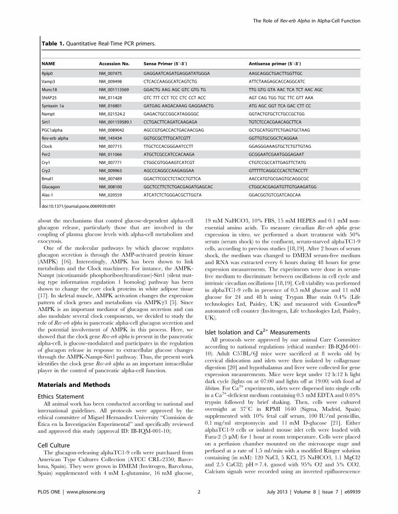
Table 1. Quantitative Real-Time PCR primers.
NAME Accession No. Sense Primer (59-39) Antisense primer (59-39)
Rplp0 NM_007475 GAGGAATCAGATGAGGATATGGGA AAGCAGGCTGACTTGGTTGC
Vamp3 NM_009498 CTCACCAAGGCATCAGTCTG ATTCTAAGAGCACCAGGCATC
Munc18 NM_001113569 GGACTG AAG AGC GTC GTG TG TTG GTG GTA AAC TCA TCT AAC AGC
SNAP25 NM_011428 GTC TTT CCT TCC CTC CCT ACC AGT CAG TGG TGC TTC GTT AAA
Syntaxin 1a NM_016801 GATGAG AAGACAAAG GAGGAACTG ATG AGC GGT TCA GAC CTT CC
Nampt NM_021524.2 GAGACTGCCGGCATAGGGGC GGTACTGTGCTCTGCCGCTGG
Sirt1 NM_001159589.1 CCTGACTTCAGATCAAGAGA TGTCTCCACGAACAGCTTCA
PGC1alpha NM_0089042 AGCCGTGACCACTGACAACGAG GCTGCATGGTTCTGAGTGCTAAG
Rev-erb alpha NM_145434 GGTGCGCTTTGCATCGTT GGTTGTGCGGCTCAGGAA
Clock NM_007715 TTGCTCCACGGGAATCCTT GGAGGGAAAGTGCTCTGTTGTAG
Per2 NM_011066 ATGCTCGCCATCCACAAGA GCGGAATCGAATGGGAGAAT
Cry1 NM_007771 CTGGCGTGGAAGTCATCGT CTGTCCGCCATTGAGTTCTATG
Cry2 NM_009963 AGCCCAGGCCAAGAGGAA GTTTTTCAGGCCCACTCTACCTT
Bmal1 NM_007489 GGACTTCGCCTCTACCTGTTCA AACCATGTGCGAGTGCAGGCGC
Glucagon NM_008100 GGCTCCTTCTCTGACGAGATGAGCAC CTGGCACGAGATGTTGTGAAGATGG
Alas-1 NM_020559 ATCATCTCTGGGACGCTTGGTA GGACGGTGTCGATCAGCAA
doi:10.1371/journal.pone.0069939.t001
The Role of Rev-erb Alpha in Alpha-Cell Function
PLOS ONE | www.plosone.org 2 July 2013 | Volume 8 | Issue 7 | e69939
The Role of Rev-erb Alpha in Alpha-Cell Function
PLOS ONE | www.plosone.org 3 July 2013 | Volume 8 | Issue 7 | e69939
microscope (Zeiss, Axiovert 200) and a Hamamatsu Digital
Camera C4742-95 (Hamamatsu Photonics, Barcelona, Spain).
Fluorescence records were expressed as the ratio of fluorescence
(R) at 340 nm and 380 nm (F340/F380). We analyzed the area-
under-the-curve (AUC) of the whole period of stimulation using
Origin software (Origin, Origin Lab Corporation, MA USA). This
parameter is an indicator of the global calcium increase in the cells
[22]. As previously reported, alpha-cells were identified by their
spontaneous calcium signalling activity at low glucose concentra-
tions and their characteristic response to epinephrine [13,23,24].
Glucagon Secretion and ContentCells were cultured for 2 days in DMEM prior to the
measurements of glucagon secretion. Then, cells were incubated
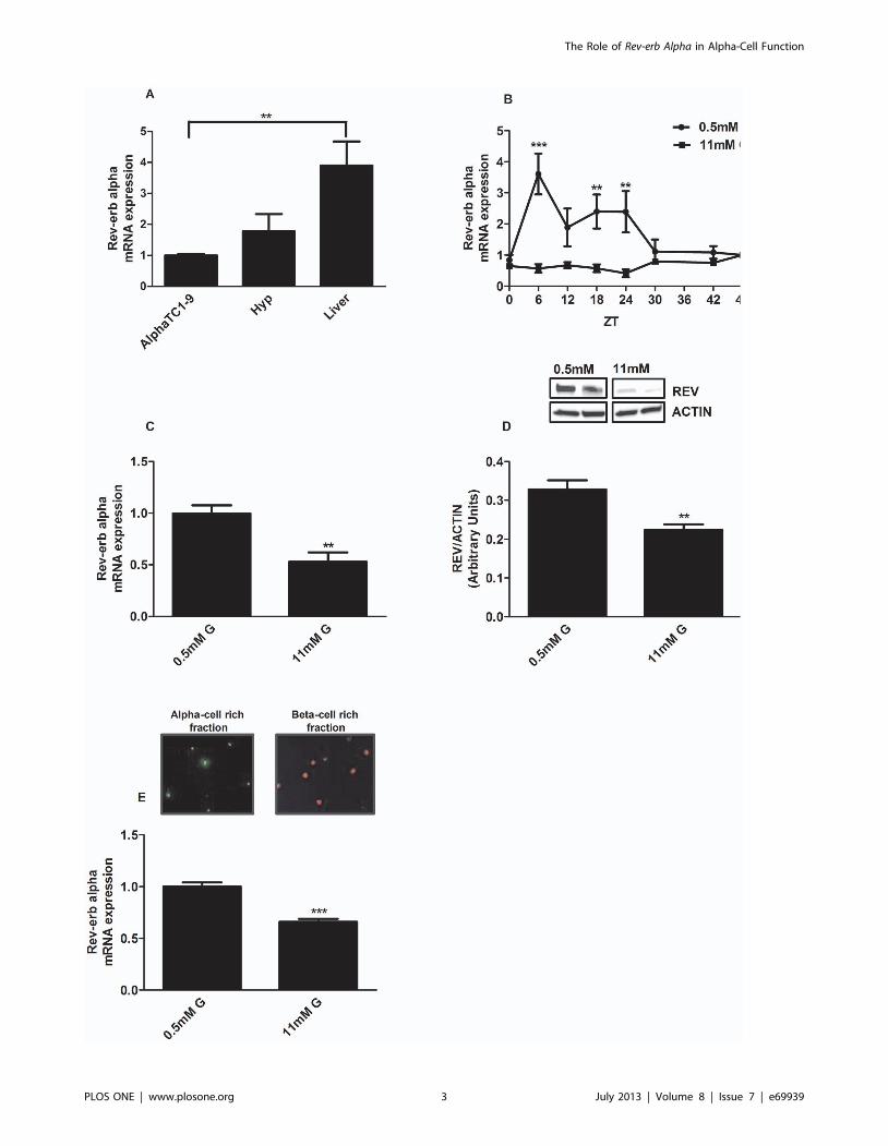
Figure 1. Glucose downregulates Rev-erb alpha gene expression in alphaTC1-9 cells and mouse pancreatic alpha cells. (A) Geneexpression of Rev-erb alpha in glucagon-secreting alphaTC1-9 cells, hypothalamus and liver (n = 5) The statistical significance was performedcomparing the expression levels in alphaTC1-9 cells with the other tissues. (B) Intrinsic oscillations of Rev-erb alpha gene expression during 48 hours.AlphaTC1-9 cells were treated with 0.5 mM glucose (circles) and 11 mM glucose (square) for 48 hours (n = 6). The statistical significance wasperformed between the ZT (Zeitgeber Time) times comparing 0.5 mM and 11 mM. (C) Rev-erb alpha gene expression in alphaTC1-9 cells treated with0.5 mM glucose and 11 mM glucose at ZT6 (n = 4–5). (D) Rev-erb alpha protein expression in alphaTC1-9 cells treated with 0.5 mM glucose and11 mM glucose at ZT6 (n = 4). (E) Rev-erb alpha gene expression in primary mouse alpha-cells separated by FACS sorting and treated with 0.5 mMglucose and 11 mM glucose at ZT6 (n = 4). Alpha-cell enriched fraction: 80% alpha cells and 4% beta cells. Beta-cells enriched fraction: 75% beta-cellsand no alpha-cells *p,0.05, **p,0.01, *** p,0.001. Data are expressed as mean 6S.E.M.doi:10.1371/journal.pone.0069939.g001
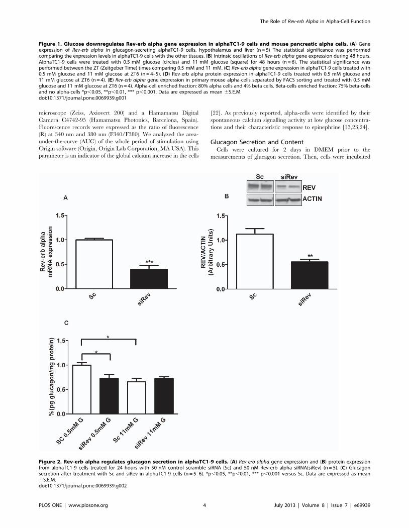
Figure 2. Rev-erb alpha regulates glucagon secretion in alphaTC1-9 cells. (A) Rev-erb alpha gene expression and (B) protein expressionfrom alphaTC1-9 cells treated for 24 hours with 50 nM control scramble siRNA (Sc) and 50 nM Rev-erb alpha siRNA(siRev) (n = 5). (C) Glucagonsecretion after treatment with Sc and siRev in alphaTC1-9 cells (n = 5–6). *p,0.05, **p,0.01, *** p,0.001 versus Sc. Data are expressed as mean6S.E.M.doi:10.1371/journal.pone.0069939.g002
The Role of Rev-erb Alpha in Alpha-Cell Function
PLOS ONE | www.plosone.org 4 July 2013 | Volume 8 | Issue 7 | e69939
at 37uC in 0.5 ml Krebs–Ringer bicarbonate buffer supplemented
with 15 mM HEPES, 0.5% BSA and 5.6 mM glucose, pH 7.4 for
30 min. Afterward, batches of 16106 alphaTC1-9 cells were
incubated for 1 h at 37uC in 0.5 ml of Krebs–Ringer bicarbonate
buffer supplemented with 0.5 mM, 11.2 mM glucose, the Rev-erb
alpha agonist GSK4112 (Sigma St Louis, USA), Hemin (Sigma St
Louis, USA), the Rev-erb alpha antagonist SR8278 (Sigma St Louis,
USA) or the Nampt inhibitor FK866 (Cayman Chemical, Ann
Arbor, USA). The supernatant was collected and glucagon
secretion was measured. Cells were then lysed with 50 ml of lysis
buffer (70% ethanol, 0.4% HCl at 30%, 24.6% distilled water) and
incubated overnight at 4uC. Samples were centrifuged at
2500 rpm for 5 min and the supernatant was collected for
glucagon content analysis. Glucagon was detected by ELISA
using a commercial kit (YK090; Gentaur, Brussels, Belgium).
Total protein was determined by the Bradford method.
Quantitative Real-time PCRQuantitative PCR assays were performed using CFX96 Real
Time System (Biorad, Hercules, USA). Reactions were carried out
in a final volume of 10 ml, containing 200 nM of each primer,
100 nM of endogenous control primer, 1 ml of cDNA and IQTM
SybrH Green Supermix (Biorad, Hercules, USA). Samples were
subjected to the following conditions: 10 min at 95uC, 40 cycles
(10 s 95uC/7 s 60uC/12 s 72uC), and a melting curve of 63 to
95uC with a slope of 0.1uC/s. The housekeeping gene rplp0
(ribosomal protein large P0, alias 36B4) was used as the
endogenous control for quantification [5]. The results were
analyzed with CFX Manager Version 1.6 (Biorad, Hercules,
USA) and values were expressed as the relative expression respect
to control levels (22DDct). Primers sequences are described in
Table 1.
Interference RNASiRNA treatment was performed in alphaTC1-9 cells as
previously described [25,26]. Cells were transfected overnight
with 50 nM of siRNAs SilencerH Pre-designed Rev-erb alpha
(Ambion, TX,USA) or 50 nM of SilencerH labelled negative
control #2 siRNA (Ambion, TX, USA) in optiMEMH I
(Invitrogen, Carlsbad, USA) culture medium without antibiotics
and 1% of Lipofectamine 2000 (Invitrogen, Carlsbad, USA). The
following Rev-erb alpha siRNA sequences were used (59-39):
GCAUCGUUGUUCAACGUGAtt, sense; UCACGUUGAA-
CAACGAUGCaa, antisense. After overnight incubation, the
transfection medium was replaced by DMEM culture medium
for 24 h before the start of the experiments.
Western-blot AnalysisCell pellets were obtained by centrifuging at 10006g for 10 min
and resuspended in 50 ml of lysis buffer (Cell Signaling Technol-
ogy, Danvers, MA). Cell extracts were subjected to sodium
dodecyl sulfate (SDS) polyacrylamide gel electrophoresis (Mini-
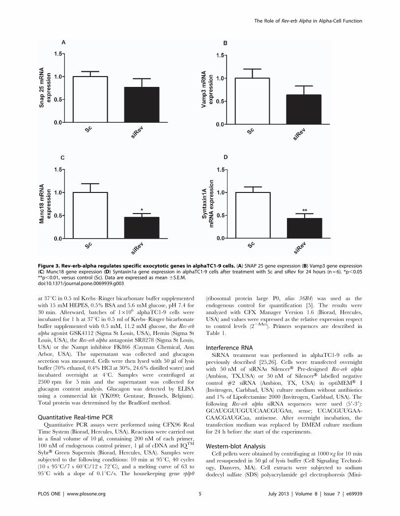
Figure 3. Rev-erb-alpha regulates specific exocytotic genes in alphaTC1-9 cells. (A) SNAP 25 gene expression (B) Vamp3 gene expression(C) Munc18 gene expression (D) Syntaxin1a gene expression in alphaTC1-9 cells after treatment with Sc and siRev for 24 hours (n = 6). *p,0.05**p,0.01, versus control (Sc). Data are expressed as mean 6S.E.M.doi:10.1371/journal.pone.0069939.g003
The Role of Rev-erb Alpha in Alpha-Cell Function
PLOS ONE | www.plosone.org 5 July 2013 | Volume 8 | Issue 7 | e69939

The Role of Rev-erb Alpha in Alpha-Cell Function
PLOS ONE | www.plosone.org 6 July 2013 | Volume 8 | Issue 7 | e69939

ProteanHTGXTM Precast Gel, 4–20% gels, Biorad). Pre-stained
SDS-PAGE standards were included for molecular mass estima-
tion. The transfer to PVDF membranes was performed at 125 mA
for 90 min in a buffer with 2.5 mM Tris base, 9 mM glycine, 20%
methanol. After membranes were blocked with 2% non-fat dry
milk, they were incubated with the following antibodies: rabbit
polyclonal anti-actin (1:1000; Sigma, Saint Louis, USA), anti-Rev-
erb alpha (1:500; Abcam, Cambridge, UK), pAMPK (Thr172)
(1:1000; Abcam, Cambridge, UK) and Nampt antibody (1:1000;
Abcam, Cambridge, UK). Membranes were incubated with
appropriate HRP-conjugated antibodies (Biorad, Hercules,
USA). Protein bands were revealed by using the ECL western
blot substrate (Thermo Fisher Scientific, Madrid, Spain). Intensity
of the bands was quantified using Scion image software (Frederick,
MD USA).
Cell SortingIsolated cells were placed in sorting buffer containing 1% BSA,
2.5 mM glucose, Ca2+, Mg2+ free PBS, 1 mM EDTA, 25 mM
Hepes and transferred to a fluorescence activated cell sorter
(FACS; BD FACSARIA SORP) equipped with 488 nm and
355 nm. The FACS was used to separate pancreatic beta-cells and
Figure 4. Activation of Rev-erb alpha stimulates glucagon secretion and calcium signals in alphaTC1-9 cells and pancreatic alpha-cells. (A) Glucagon secretion in alphaTC1-9 cells in the presence of 0.5 mM, 11 mM glucose and 10 mM of the Rev-erb alpha agonist GSK4112 (n = 7–8). (B) Intracellular calcium signals in alphaTC1-9 cells in the presence of 0.5 mM glucose and 10 mM of the Rev-erb alpha agonist GSK4112 (n = 42cells). (C) Area under the curve calculated from experiments illustrated in 4B with alphaTC1-9 cells. (D) Intracellular calcium signals in alphaTC1-9 cellsin the presence of 0.5 mM glucose and 10 mM of the Rev-erb alpha agonist GSK4112 (n = 24 cells). (E) Intracellular calcium measurements in primarymouse alpha cells in the presence of 0.5 mM glucose and 10 mM of Rev-erb alpha agonist GSK4112 (n = 14 cells). (F) Area under the curve calculatedfrom experiments illustrated in 4E with primary alpha-cells. *p,0.05, **p,0.01, *** p,0.001. Data are expressed as mean 6S.E.M.doi:10.1371/journal.pone.0069939.g004
Figure 5. Inhibition of Rev-erb alpha inhibits glucagon secretion and calcium signals in alphaTC1-9 cells and pancreatic alpha-cells.(A) Glucagon secretion from alphaTC1-9 cells in the presence with 0.5 mM, 11 mM glucose and 10 mM of the Rev-erb alpha antagonist SR8278 (n = 8).(B) Intracellular calcium signals in alphaTC1-9 cells in the presence of 0.5 mM glucose and 10 mM of the Rev-erb alpha antagonist SR8278 (n = 32cells). (C) Area under the curve calculated from experiment 5B. *p,0.05, **p,0.01. Data are expressed as mean 6S.E.M.doi:10.1371/journal.pone.0069939.g005
The Role of Rev-erb Alpha in Alpha-Cell Function
PLOS ONE | www.plosone.org 7 July 2013 | Volume 8 | Issue 7 | e69939
The Role of Rev-erb Alpha in Alpha-Cell Function
PLOS ONE | www.plosone.org 8 July 2013 | Volume 8 | Issue 7 | e69939

alpha cells by cellular autofluorescence as recently reported by
Kohler [27].
ImmunohistochemistryAfter sorting approximately 10.000 cells from each cell fraction
were subjected to cytospin in order to perform immunofluores-
cence analysis. Cells are fixed in slides with Paraformaldehyde 4%
and permeabilized with triton 0.2% for 45 minutes. Blocking was
then performed with PBS-BSA 0.1% for 30 minutes at room
temperature. Cells were incubated overnight at 4uC with insulin
and glucagon antibodies (DAKO), and subsequently incubated
with secondary antibodies Cy3 antiguineapig for insulin and Cy2
antirabbit for glucagon (Jackson Immunoresearch) and washed
with PBS. Analysis performed with Image J program. Alpha-cell
enriched fraction was composed of 80% alpha-cells and 4% beta-
cells while the beta-cell enriched fraction had 75% beta-cells and
no alpha-cells.
Statistical AnalysisData is shown as mean 6 SEM. Student’s t test, one-way
ANOVA or two-way ANOVA with Bonferroni correction were
performed as appropriate with a level of significance p,0.05.
Results
Glucose Regulates Rev-erb Alpha Gene and ProteinExpression in alphaTC1-9 Cells and in Mouse PancreaticAlpha Cells
To analyze the mRNA expression of several clock genes, we
used the mouse alpha-cell line alphaTC1-9 which has been
validated as a good model to study alpha cell function [16,25,28].
The mRNA levels in this cell line were compared with those of the
hypothalamus and liver. Rev-erb alpha expression in alphaTC1-9
cells was comparable with that in the hypothalamus whereas the
expression levels in the liver were much higher (Fig. 1A). The
expression of Clock, Bmal1 and Cry1 in alphaTC1-9 was similar
to that in the hypothalamus and liver (S 1). Per-1 expression was
lower in the liver compared with alphaTC1-9 cells whereas Per2
and Cry2 had lower expression levels in the hypothalamus (S 1).
To study whether alpha-cells exhibit an oscillatory pattern of Rev-
erb alpha expression along the day, we next performed mRNA
measurements every 6 hours during a 48 h, as previously
described [19]. To check that culture conditions were not affecting
cell viability, we measured this parameter at 0.5 and 11 mM
glucose after 24 and 48 hours of incubation. No differences were
found in cell viability between both glucose concentrations in any
of the time points measured (data not shown). Figure 1B shows
that Rev-erb alpha mRNA levels oscillated in vitro at low glucose
concentrations (0.5 mM) with an expression peak at ZT6
(Zeitgeber Time). Interestingly, in our experiments high glucose
concentrations (11 mM) inhibited Rev-erb alpha expression levels.
To further confirm the glucose effect on Rev-erb alpha at both the
mRNA and protein levels, we performed similar experiments at
ZT6. As expected, high glucose concentrations produced an
inhibitory action on both Rev-erb alpha gene (Fig. 1C) and protein
expression (Fig. 1D). We next studied the glucose effect at ZT6 in
sorted mouse primary alpha-cells. In agreement with the results in
alphaTC1-9 cells, high glucose concentrations down-regulated the
Rev-erb alpha mRNA levels compared to the effect of 0.5 mM
glucose in the alpha cell rich fraction of the primary isolated islet
cells (Fig. 1E). These findings indicate that both alphaTC1-9 cells
and mouse pancreatic alpha-cells express Rev-erb alpha and that
glucose regulates the expression of this clock gene. Additionally,
we show that alphaTC1-9 cells exhibit intrinsic circadian
oscillations of Rev-erb alpha, indicating that alpha-cells of the
endocrine pancreas have their own biological clock.
Rev-erb Alpha Regulates Glucagon Secretion inalphaTC1-9 Cells
To study the functional role of Rev-erb alpha in alpha-cells, we
used the siRNA technique to down-regulate this gene in mouse
alphaTC1-9 cells. Gene silencing efficiency was about 60–70%
compared with cells treated with a scramble siRNA (Sc; control
siRNA) (Fig. 2A). At these concentrations of siRNA (50 nM) we
previously observed no difference in cell viability between
scramble and siRNA treated cells [13]. The lower Rev-erb alpha
expression was associated with a decrease in protein level (Fig. 2B).
We next investigated whether Rev-erb alpha is involved in the
regulation of glucagon secretion. Figure 2C shows the glucose
effect on glucagon secretion from alphaTC1-9 cells after Rev-erb
alpha silencing. As previously shown in isolated mouse islets and
alphaTC1-9 cells [13,16], glucagon secretion was stimulated at
0.5 mM glucose whereas secretion was inhibited at 11 mM
glucose (Fig. 2C). After silencing the Rev-erb alpha gene in
alphaTC1-9 cells, glucagon secretion at low glucose concentra-
tions (0.5 mM) was decreased to the same extent as high glucose
concentrations (11 mM) (Fig. 2C). No significant differences were
observed either on glucagon content or glucagon gene expression
after Rev-erb alpha silencing (data not shown). Interestingly, when
we checked the mRNA expression of exocytotic genes such as
Vamp3, Munch18, SNAP25 and Syntaxin1a, there was a down
regulation of Munch18 and Syntaxin1a genes (and a tendency to
decrease in VAMP3) in alphaTC1-9 cells treated with siRev
compared with controls (Fig. 3A–D, respectively). Thus, the
decreased glucagon release in cells with down-regulated Rev-erb
alpha seems more related with changes in the mechanisms that
allow the release of glucagon rather than a consequence of an
altered glucagon synthesis.
The activity of Rev-erb alpha proteins can be modulated by its
natural ligand heme [12,29] or by the new synthetic Rev-erb alpha
agonist GSK4112 and the antagonist SR8278 [30,31]. To further
evaluate the regulatory role of Rev-erb alpha in alpha-cell function,
we checked the effect of GSK4112 and hemin on glucagon
secretion in alphaTC1-9 cells. At 0.5 mM glucose the agonist
GSK4112 had a stimulatory effect on glucagon secretion (Fig. 4A)
but GSK4112 was not able to revert the inhibitory effects of
11 mM glucose. (Fig. 4A). On the other hand, the natural Rev-
erbalpha agonist hemin stimulated glucagon secretion at both
0.5 mM and 11 mM glucose in isolated islets (S 2A). To test the
biological effect of hemin, we measured Alas-1 (d-aminolevulinate
synthase 1) a rate limiting enzyme in the heme biosynthetic
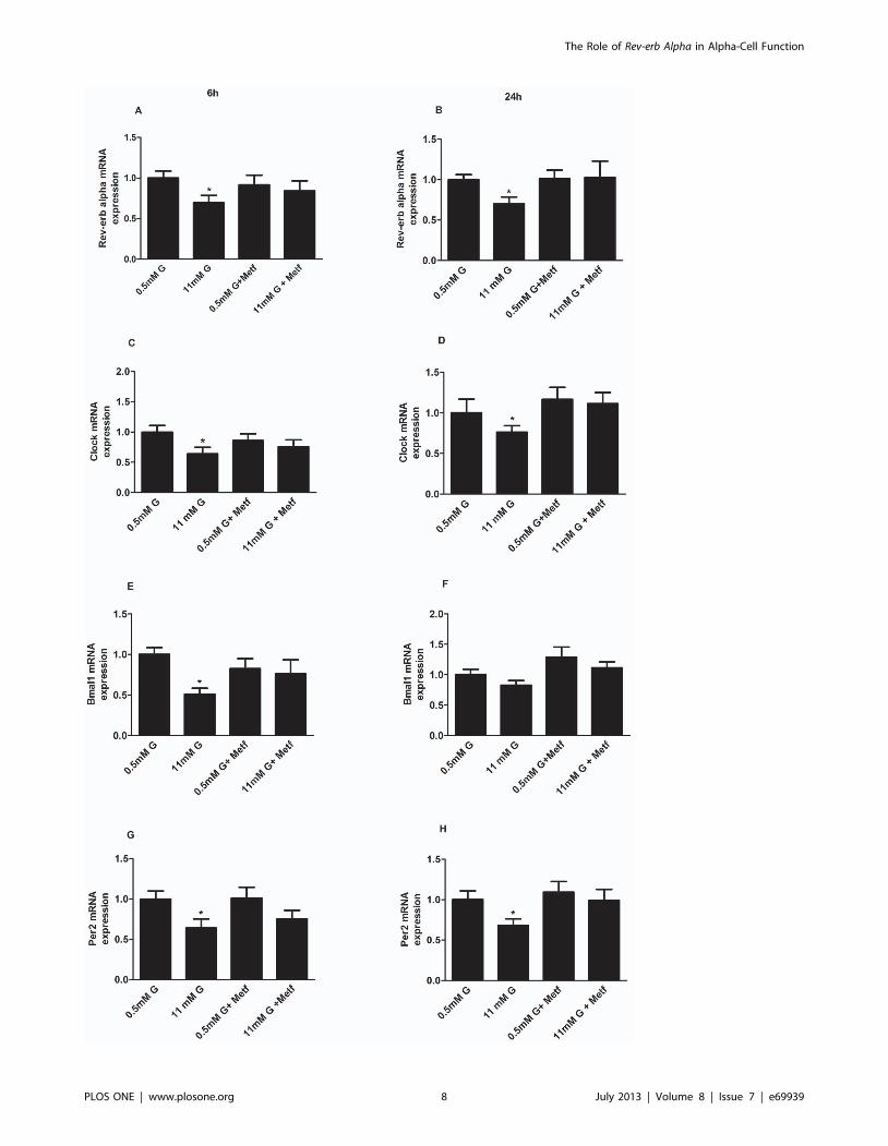
Figure 6. Activation of AMPK prevents glucose inhibition of clock genes in alphaTC1-9 cells. (A) Rev-erb alpha gene expression inalphaTC1-9 cells treated with 0.5 mM, 11 mM glucose or 500 mM Metformin for 6 hours (n = 5) and (B) 24 hours (n = 5). (C) Clock gene expression inalphaTC1-9 cells treated with 0.5 mM, 11 mM glucose or 500 mM Metformin for 6 hours (n = 5–6) and (D) 24 hours (n = 5–6). (E) Bmal1 geneexpression in alphaTC1-9 cells treated with 0.5 mM, 11 mM glucose or 500 mM Metformin for 6 hours (n = 5–6) and (F) 24 hours (n = 5–6). (G) Per2gene expression in alphaTC1-9 cells treated with 0.5 mM, 11 mM glucose or 500 mM Metformin for 6 hours (n = 5–6) and (H) 24 hours (n = 5–6).*p,0.05; **p,0.01. Data are expressed as mean 6S.E.M.doi:10.1371/journal.pone.0069939.g006
The Role of Rev-erb Alpha in Alpha-Cell Function
PLOS ONE | www.plosone.org 9 July 2013 | Volume 8 | Issue 7 | e69939

The Role of Rev-erb Alpha in Alpha-Cell Function
PLOS ONE | www.plosone.org 10 July 2013 | Volume 8 | Issue 7 | e69939
pathway. As expected the mRNA levels of Alas-1 was decreased in
presence of hemin in alphaTC1-9 cells (S 2B).
The stimulus-secretion coupling of pancreatic alpha-cells is still
controversial [20,23,32,33,34,35,36]. However, it is well accepted
that glucagon secretion is a calcium-dependent mechanism and
changes in calcium signaling regulate the exocytotic process in
alpha-cells [34,35,37,38]. Therefore, we checked whether the
stimulatory effect of the Rev-erb alpha agonist GSK4112 on
glucagon secretion could be via modulation of intracellular
calcium levels. At low glucose levels (0.5 mM) alphaTC1-9 cells
exhibited intracellular calcium oscillations (Fig. 4B). Addition of
GSK4112 increased these calcium signals (Fig. 4B) that were
statistically significant increased in the area under the curve
(Fig. 4C). In some alphaTC1-9 cells that were silent at low glucose
concentrations the addition of GSK4112 was able to trigger
intracellular calcium oscillations (Fig. 4D). Similar results were
achieved in mouse primary alpha-cells. Primary mouse alpha-cells
exhibited the characteristic oscillatory calcium pattern at 0.5 mM
glucose and addition of GSK4112 increased the signal frequency
or transformed calcium oscillations into a sustained pattern
(Fig. 4E–F). The synthetic Rev-erb alpha antagonist SR8278 had
the opposite effect on glucagon secretion and intracellular calcium
levels in alphaTC1-9 cells. At 0.5 mM glucose, SR8278 inhibited
glucagon secretion and had no additional effect at 11 mM glucose
(Fig. 5A). The effect of SR8278 on intracellular calcium was
evident by its inhibitory action at 0.5 mM glucose (Fig. 5B and
5C). These results demonstrated that modulation or inhibition of
Rev-erb alpha regulates glucagon secretion and alpha-cell calcium
signalling.
Glucose Inhibits Rev-erb Alpha Gene Expression viaAMPK-Nampt-Sirt1 Pathway
Since AMPK has been shown to be strongly regulated by
glucose in pancreatic alpha-cells, participates in glucagon release
[16], and modulates the expression of different clock genes in
skeletal muscle, we hypothesized that AMPK might be involved in
the mechanism by which glucose regulates Rev-erb alpha gene
expression. We treated alphaTC1-9 cells for 6 and 24 hours with
500 mM of the AMPK activator Metformin since the AMPK
activator AICAR had no effect on alphaTC1-9 cells, as previously
shown [16]. As expected, glucose down-regulated Rev-erb alpha
gene expression at both 6 and 24 hours of treatment (Fig. 6A and
B). Remarkably, while AMPK activation by metformin had no
effect at 0.5 mM glucose, it prevented the inhibitory effect of
11 mM glucose on Rev-erb alpha gene expression (Fig. 6A and B)
indicating that AMPK is involved in this process. Similar results
were obtained with others clock genes such as Clock (Fig. 6C and
D), Bmal1 (Fig. 6E and F) and Per2 (Fig. 6G and H).
It has been demonstrated in white adipose tissue that metformin
can control clock genes expression through AMPK-Nampt-Sirt1
pathway [17]. To investigate whether a similar mechanism takes
place in pancreatic alphaTC1-9 cells, we next studied this pathway
by treating alphaTC1-9 cells for 6 and 24 hours with 500 mM
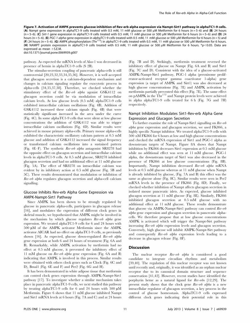
Metformin. Figure 6 shows that 11 mM glucose inhibited Nampt
and Sirt1 mRNA levels at 6 hours (Fig. 7A and C) and at 24 hours
(Fig. 7B and D). Strikingly, metformin treatment reversed the
inhibitory effect of glucose on Nampt (Fig. 6A and B) and Sirt1
(Fig. 7C and D). Consistent with the idea of a glucose-activated
AMPK-Nampt-Sirt1 pathway, PGC-1 alpha (peroxisome prolif-
erator-activated receptor gamma coactivator 1-alpha) gene
expression (a target of AMPK and Sirt1) was also decreased at
high glucose concentrations (Fig. 7E) and AMPK activation by
metformin partially prevented this effect (Fig. 7E). The same effect
on pAMPK in the Thr172 and Nampt protein levels were achieved
in alpha alphaTC1-9 cells treated for 6 h (Fig. 7G and 7H)
respectively.
Nampt Inhibition Modulates Sirt1-Rev-erb Alpha GeneExpression and Glucagon Secretion
To further examine the role of Nampt-Sirt1 signalling on Rev-erb
alpha gene expression and glucagon secretion, we used FK866, a
highly specific Nampt inhibitor. We treated alphaTC1-9 cells with
500 nM FK866 for 6 hours at low and high glucose concentrations
and checked the mRNA expression of Sirt1 and PGC-1 alpha as
downstream targets of Nampt. Figure 8A shows that Nampt
inhibition by FK866 decreases Sirt1 expression at 0.5 mM glucose
while no additional effect was seen at 11 mM glucose. PGC-1
alpha, the downstream target of Sirt1 was also decreased in the
presence of FK866 at low glucose concentrations (Fig. 8B).
Importantly, Nampt inhibition decreased Rev-erb alpha mRNA
levels at 0.5 mM glucose whereas at 11 mM glucose when Nampt
is already inhibited by glucose, (Fig. 7A and B) this effect was the
same as glucose alone (Fig. 8C). Similar results were seen in Per2
mRNA levels in the presence of FK866 (Fig. 8D). Finally, we
checked whether inhibition of Nampt affects glucagon secretion in
isolated mouse pancreatic islets. As expected, glucose inhibited
glucagon secretion at 11 mM glucose (Fig. 8E). Similarly, FK866
inhibitied glucagon secretion at 0.5 mM glucose with no
additional effect at 11 mM glucose. These results demonstrate
that glucose via AMPK-Nampt-Sirt1 pathway can control Rev-erb
alpha gene expression and glucagon secretion in pancreatic alpha-
cells. We therefore propose that at low glucose concentrations
AMPK is activated which will trigger the Nampt-Sirt pathway
increasing Rev-erb alpha expression levels and glucagon secretion.
Conversely, high glucose will inhibit AMPK-Nampt-Sirt pathway
and consequently Rev-erb alpha expression levels leading to a
decrease in glucagon release (Fig. 8F).
Discussion
The nuclear receptor Rev-erb alpha is considered a good
candidate to integrate circadian rhythms and metabolism
[39,40]. The regulation of this nuclear receptor was not known
until recently and, originally, it was identified as an orphan nuclear
receptor due to its canonical domain structure and sequence
conservation [41,42]. However, recent studies have identified the
porphyrin heme as a natural ligand for Rev-erbs [12,29]. The
present study shows that the clock gene Rev-erb alpha is a new
intracellular regulator of glucagon secretion, a key process in the
control of glucose homeostasis. AlphaTC1-9 cells expressed
different clock genes indicating their potential role in this
Figure 7. Activation of AMPK prevents glucose inhibition of Rev-erb alpha expression via Nampt-Sirt1 pathway in alphaTC1-9 cells.(A) Nampt gene expression in alphaTC1-9 cells treated with 0.5 mM, 11 mM glucose or 500 mM Metformin for 6 hours (n = 5–6) and (B) 24 hours(n = 5–6). (C) Sirt-1 gene expression in alphaTC1-9 cells treated with 0.5 mM, 11 mM glucose or 500 mM Metformin for 6 hours (n = 5–6) and (D) 24hours (n = 5–6). (E) PGC-1 alpha gene expression in alphaTC1-9 cells treated with 0.5 mM, 11 mM glucose or 500 mM Metformin for 6 hours (n = 5) and(F) 24 hours (n = 5–6). (G) AMPK phosphorylation (Thr172) in alphaTC1-9 cells treated with 0.5 mM, 11 mM glucose or 500 mM Metformin for 6 hours.(H) NAMPT protein expression in alphaTC1-9 cells treated with 0.5 mM, 11 mM glucose or 500 mM Metformin for 6 hours. *p,0.05. Data areexpressed as mean 6S.E.M.doi:10.1371/journal.pone.0069939.g007
The Role of Rev-erb Alpha in Alpha-Cell Function
PLOS ONE | www.plosone.org 11 July 2013 | Volume 8 | Issue 7 | e69939
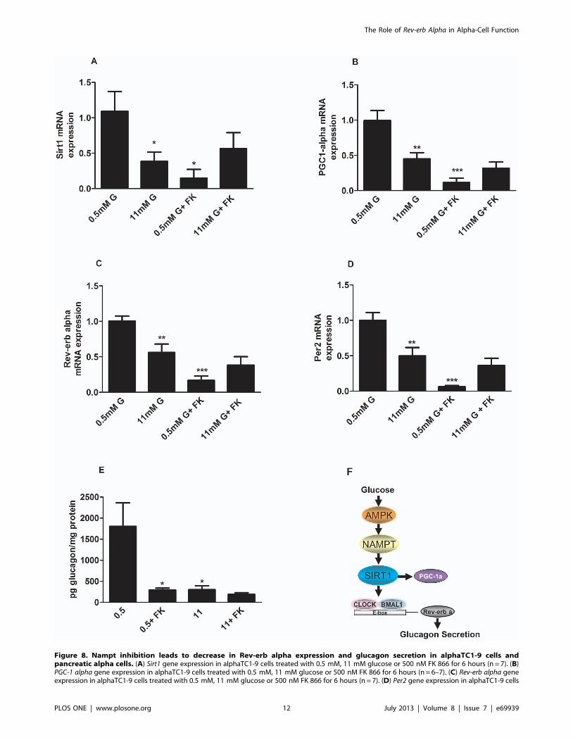
Figure 8. Nampt inhibition leads to decrease in Rev-erb alpha expression and glucagon secretion in alphaTC1-9 cells andpancreatic alpha cells. (A) Sirt1 gene expression in alphaTC1-9 cells treated with 0.5 mM, 11 mM glucose or 500 nM FK 866 for 6 hours (n = 7). (B)PGC-1 alpha gene expression in alphaTC1-9 cells treated with 0.5 mM, 11 mM glucose or 500 nM FK 866 for 6 hours (n = 6–7). (C) Rev-erb alpha geneexpression in alphaTC1-9 cells treated with 0.5 mM, 11 mM glucose or 500 nM FK 866 for 6 hours (n = 7). (D) Per2 gene expression in alphaTC1-9 cells
The Role of Rev-erb Alpha in Alpha-Cell Function
PLOS ONE | www.plosone.org 12 July 2013 | Volume 8 | Issue 7 | e69939

endocrine cell type. Particularly, the Rev-erb alpha mRNA levels
were found to have intrinsic 24 hours oscillations at low glucose
concentrations. The expression levels peaked at ZT6 and the
circadian pattern was completely inhibited by high glucose
concentrations. These results were confirmed at both the protein
level at ZT6 in alphaTC1-9 cells as well as at the mRNA level in
sorted mouse primary alpha-cells. In agreement with the present
findings, the inhibitory effect of glucose on the expression of Per1
and Per2 mRNA has been shown in rat1-fibroblasts [43]. On the
other hand, glucose has no effect on clock gene expression in rat
cardiomiocytes [44]. Taken together, our results demonstrate that
pancreatic alpha-cells present an intrinsic clock and that the clock
gene Rev-erb alpha can be induced or repressed according to the
glucose levels.
The involvement of Rev-erb alpha on glucagon secretion was
evident when this gene was down-regulated by siRNA. Reduction
of ,50% in protein and ,60% in mRNA levels were sufficient to
decrease glucagon release at low glucose concentrations. High
glucose levels were still capable of reducing glucagon secretion
after Rev-erb alpha silencing, probably because Rev-erb alpha levels
were already low at these glucose concentrations or due to other
signaling pathways different from Rev-erb alpha. Actually, several
mechanisms are involved in glucose modulation of glucagon
secretion as reported by us and others. [20,23,33,34,35,37,38]. In
any case, we identified Rev-erb alpha as a new intracellular mediator
of glucagon release, whose effects depend on extracellular glucose
levels, further supporting the hypothesis that glucose can directly
regulate alpha-cell secretion by several pathways [15]. The
involvement of Rev-erb alpha on glucagon secretion was further
confirmed by the stimulatory effect of the Rev-erb alpha agonist
GSK4112 and its natural ligant hemin and the opposite effect with
an antagonist. We have recently demonstrated that Rev-erb alpha
can regulate insulin secretion probably by downregulation of
exocytotic genes in the beta-cell [13], and similar findings have
been obtained with other clock genes [14]. In the case of
pancreatic alpha-cells, silencing of Rev-erb alpha led to a decrease of
specific exocytotic genes such as Munch18 and Syntaxin1a, genes
that are shown to be involved in the regulation of glucagon
exocytosis [45]. Additionally, our results also showed that
activation of Rev-erb alpha in alpha-cells led to an increase of
intracellular calcium concentrations whereas its inactivation
decreased calcium signaling. Given the importance of intracellular
calcium signals in the regulation of glucagon secretion
[20,24,33,35,36,38], our results indicate that Rev-erb alpha may
also regulate glucagon secretion by a calcium-dependent mecha-
nism. Thus, our findings give new insights regarding the regulation
of glucagon secretion showing that the clock gene Rev-erb alpha
modulates glucagon release in a glucose-modulated manner.
The stimulus secretion coupling of alpha-cells and the
mechanisms involved in glucagon release are still largely unknown.
Among the nutrients that regulate glucagon secretion, glucose is
considered one of the most important modulators [20,38].
However, the intracellular pathway by which glucose inhibits
glucagon release is still controversial. It has been proposed that
glucose can directly inhibit glucagon secretion independently of
paracrine signals from other cell types within the islet of
Langerhans [15,20,23,34,38]. On the other hand, several para-
crine mechanisms activated at high-glucose concentrations as a
result of beta and delta-cell stimulation have been shown to inhibit
glucagon release [15,33,35,36,46]. Here we show that modulation
of Rev-erb alpha both acutely (pharmacological modulation of Rev-
erb alpha) and chronically (inhibition of Rev-erb alpha by siRNA)
regulates glucagon secretion. The acute effects may involve
activation/inhibition of calcium channels by a still unknown
mechanism whereas the chronic effect may be due to the effects of
glucose on Rev-erb alpha gene expression through the AMPK-
Nampt-Sirt1 mechanism.
Another important signal that regulates glucagon secretion is
AMP-activated protein kinase [16]. It has been reported that
glucose inhibits AMPK activity by increasing the ATP/ADP ratio
in alphaTC1-9 cells. In addition, pharmacological activation of
AMPK with metformin stimulates glucagon secretion whereas
inhibition of the kinase leads to decreased glucagon release [16]. In
agreement with these previous reports, the use of metformin to
activate AMPK in alphaTC1-9 cells led to a decrease in several
metabolism and signalling genes. Recently, AMPK has been
implicated in the regulation of clock genes in other cell types
[5,17,47,48,49]. Consistent with these reports, we found that
AMPK activation by metformin in pancreatic alpha-cells could
prevent the inhibitory glucose effect on Rev-erb alpha gene
expression and other clock genes such as Clock, Bmal1 and Per2.
In white adipose tissue from obese mice, AMPK activation by
metformin rescued the defects on clock gene expression via an
AMPK-Nampt-Sirt1 mechanism [17]. Our results further support
this pathway in pancreatic alpha-cells. The mRNA levels of
Nampt, Sirt and PGC-1 alpha were all downregulated by high
glucose, a condition where the AMPK is inactivated in pancreatic
alpha-cells [16]. AMPK activation by metformin at low glucose
had no effect on Nampt, Sirt and PGC-1 alpha mRNA levels
because at these glucose levels the AMPK activity is already high
in these cells. However, when AMPK was activated by metformin
at high glucose, the mRNA levels of Nampt, Sirt and PGC-1 alpha
were increased indicating that the inhibitory effect of glucose on
these genes was very likely depending on AMPK. Recently, it was
demonstrated that AMPK activation increases Sirt1 activity by
increasing Nampt expression and NAD+ levels [17,50,51]. In
addition, the AMPK alpha1 and alpha 2 KO mice lack the cyclic
expression of Nampt and PGC-1 alpha [52]. Moreover, the NAD+
dependent deacetylase Sirt1 binds to the Clock/Bmal1 complex to
regulate the expression of clock genes [53,54]. Consistent with this
role of Nampt in the regulation of Sirt1 and clock genes, the highly
specific Nampt inhibitor FK866 inhibited Sirt1, PGC-1 alpha,
Per2 and Rev-erb alpha mRNA expression in alphaTC1-9 cells
and glucagon secretion in mouse pancreatic islets.
In conclusion, we demonstrated here that the clock gene Rev-
erb alpha is an intracellular regulator of glucagon secretion in
pancreatic mouse alpha-cells. Glucose, by regulating AMPK, may
modulate Nampt-Sirt1 signalling leading to the control of Rev-erb
alpha gene and glucagon secretion. Strategies to target the Nampt-
Sirt1-Rev-erb alpha in pancreatic alpha-cells can be useful for the
treatment of hyperglucagonemia present in diabetes.
treated with 0.5 mM, 11 mM glucose or 500 nM FK 866 for 6 hours (n = 7–8). (E) Glucagon secretion from mouse pancreatic islets stimulated for 1.5hour with 0.5 mM, 11 mM glucose or 500 nM FK 866 (n = 6). *p,0.05, **p,0.01, *** p,0.001. Data are expressed as mean 6S.E.M. (F) Proposedmodel for regulation of glucagon secretion via an AMPK-Nampt-Sirt1-Rev-erb alpha mechanism. At low glucose concentrations AMPK is activatedwhich will trigger the Nampt-Sirt pathway increasing Rev-erb alpha expression levels and glucagon secretion. Conversely, high glucose will inhibitAMPK-Nampt-Sirt pathway and consequently Rev-erb alpha expression levels leading to a decrease in glucagon release.doi:10.1371/journal.pone.0069939.g008
The Role of Rev-erb Alpha in Alpha-Cell Function
PLOS ONE | www.plosone.org 13 July 2013 | Volume 8 | Issue 7 | e69939

Supporting Information
Figure S1 Clock genes are expressed in AlphaTC1-9cells. Expression of (A) Clock (B) Bmal1 (C) Per1 (D) Per2 (E)
Cry1 mRNA in alphaTC1-9, hypothalamus and liver. **p,0.01,
*** p,0.001 compared to expression levels in alpha TC1-9. Data
are expressed as mean 6S.E.M.
(TIF)
Figure S2 The Rev-erbalpha agonist hemin stimulatesglucagon secretion. (A) Glucagon secretion from mouse
pancreatic islets stimulated for 1.5 hour with 0.5 mM, 11 mM
glucose or 30 mM Hemin (n = 6). (B) Alas-1 gene expression in
alphaTC1-9 cells treated with 0.5 mM glucose and 30 mM Hemin
for 6 hours (n = 4). *p,0.05, **p,0.01, *** p,0.001. Data are
expressed as mean 6S.E.M.
(TIF)
Acknowledgments
The authors thank, M.L. Navarro, Yaiza Esteban and Ainhoa Garcia
Alaman for their expert technical assistance.
Author Contributions
Conceived and designed the experiments: EV LM IQ. Performed the
experiments: EV LM ALCF BM RFR. Analyzed the data: EV LM ALCF
BM RFR IQ. Contributed reagents/materials/analysis tools: TPB AN RG
IQ. Wrote the paper: EV AN TPB RG IQ.
References
1. Green CB, Takahashi JS, Bass J (2008) The meter of metabolism. Cell 134: 728–
742.
2. Lowrey PL, Takahashi JS (2004) Mammalian circadian biology: elucidating
genome-wide levels of temporal organization. Annu Rev Genomics Hum Genet5: 407–441.
3. Muhlbauer E, Wolgast S, Finckh U, Peschke D, Peschke E (2004) Indication of
circadian oscillations in the rat pancreas. FEBS Lett 564: 91–96.
4. Panda S, Antoch MP, Miller BH, Su AI, Schook AB, et al. (2002) Coordinated
transcription of key pathways in the mouse by the circadian clock. Cell 109:307–320.
5. Vieira E, Nilsson EC, Nerstedt A, Ormestad M, Long YC, et al. (2008)Relationship between AMPK and the transcriptional balance of clock-related
genes in skeletal muscle. Am J Physiol Endocrinol Metab 295: E1032–1037.
6. Zylka MJ, Shearman LP, Weaver DR, Reppert SM (1998) Three period
homologs in mammals: differential light responses in the suprachiasmaticcircadian clock and oscillating transcripts outside of brain. Neuron 20: 1103–
1110.
7. Reppert SM, Weaver DR (2002) Coordination of circadian timing in mammals.
Nature 418: 935–941.
8. Le Martelot G, Claudel T, Gatfield D, Schaad O, Kornmann B, et al. (2009)
REV-ERBalpha participates in circadian SREBP signaling and bile acidhomeostasis. PLoS Biol 7: e1000181.
9. Raspe E, Duez H, Mansen A, Fontaine C, Fievet C, et al. (2002) Identification ofRev-erbalpha as a physiological repressor of apoC-III gene transcription. J Lipid
Res 43: 2172–2179.
10. Fontaine C, Dubois G, Duguay Y, Helledie T, Vu-Dac N, et al. (2003) The
orphan nuclear receptor Rev-Erbalpha is a peroxisome proliferator-activatedreceptor (PPAR) gamma target gene and promotes PPARgamma-induced
adipocyte differentiation. J Biol Chem 278: 37672–37680.
11. Estall JL, Ruas JL, Choi CS, Laznik D, Badman M, et al. (2009) PGC-1alpha
negatively regulates hepatic FGF21 expression by modulating the heme/Rev-Erb(alpha) axis. Proc Natl Acad Sci U S A 106: 22510–22515.
12. Yin L, Wu N, Curtin JC, Qatanani M, Szwergold NR, et al. (2007) Rev-erbalpha, a heme sensor that coordinates metabolic and circadian pathways.
Science 318: 1786–1789.
13. Vieira E, Marroqui L, Batista TM, Caballero-Garrido E, Carneiro EM, et al.
(2012) The clock gene Rev-erbalpha regulates pancreatic beta-cell function:modulation by leptin and high-fat diet. Endocrinology 153: 592–601.
14. Marcheva B, Ramsey KM, Buhr ED, Kobayashi Y, Su H, et al. (2010)Disruption of the clock components CLOCK and BMAL1 leads to
hypoinsulinaemia and diabetes. Nature 466: 627–631.
15. Quesada I, Tuduri E, Ripoll C, Nadal A (2008) Physiology of the pancreatic
alpha-cell and glucagon secretion: role in glucose homeostasis and diabetes.J Endocrinol 199: 5–19.
16. Leclerc I, Sun G, Morris C, Fernandez-Millan E, Nyirenda M, et al. (2011)
AMP-activated protein kinase regulates glucagon secretion from mouse
pancreatic alpha cells. Diabetologia 54: 125–134.
17. Caton PW, Kieswich J, Yaqoob MM, Holness MJ, Sugden MC (2011)
Metformin opposes impaired AMPK and SIRT1 function and deleteriouschanges in core clock protein expression in white adipose tissue of genetically-
obese db/db mice. Diabetes Obes Metab 13: 1097–1104.
18. Balsalobre A, Damiola F, Schibler U (1998) A serum shock induces circadian
gene expression in mammalian tissue culture cells. Cell 93: 929–937.
19. Nagoshi E, Brown SA, Dibner C, Kornmann B, Schibler U (2005) Circadiangene expression in cultured cells. Methods Enzymol 393: 543–557.
20. Quesada I, Todorova MG, Alonso-Magdalena P, Beltra M, Carneiro EM, et al.(2006) Glucose induces opposite intracellular Ca2+ concentration oscillatory
patterns in identified alpha- and beta-cells within intact human islets ofLangerhans. Diabetes 55: 2463–2469.
21. Soriano S, Gonzalez A, Marroqui L, Tuduri E, Vieira E, et al. (2010) Reducedinsulin secretion in protein malnourished mice is associated with multiple
changes in the beta-cell stimulus-secretion coupling. Endocrinology 151: 3543–
3554.
22. Soriano S, Gonzalez A, Marroqui L, Tuduri E, Vieira E, et al. (2011) Reduced
insulin secretion in protein malnourished mice is associated with multiplechanges in the beta-cell stimulus-secretion coupling. Endocrinology 151: 3543–
3554.
23. Liu YJ, Vieira E, Gylfe E (2004) A store-operated mechanism determines the
activity of the electrically excitable glucagon-secreting pancreatic alpha-cell. CellCalcium 35: 357–365.
24. Vieira E, Liu YJ, Gylfe E (2004) Involvement of alpha1 and beta-adrenoceptorsin adrenaline stimulation of the glucagon-secreting mouse alpha-cell. Naunyn
Schmiedebergs Arch Pharmacol 369: 179–183.
25. Marroqui L, Vieira E, Gonzalez A, Nadal A, Quesada I (2011) Leptin
downregulates expression of the gene encoding glucagon in alphaTC1–9 cellsand mouse islets. Diabetologia In press DOI 101007/s00125-010-2024-1.
26. Moore F, Colli ML, Cnop M, Esteve MI, Cardozo AK, et al. (2009) PTPN2, acandidate gene for type 1 diabetes, modulates interferon-gamma-induced
pancreatic beta-cell apoptosis. Diabetes 58: 1283–1291.
27. Kohler M, Dare E, Ali MY, Rajasekaran SS, Moede T, et al. (2012) One-step
purification of functional human and rat pancreatic alpha cells. Integr Biol(Camb) 4: 209–219.
28. Powers AC, Efrat S, Mojsov S, Spector D, Habener JF, et al. (1990) Proglucagon
processing similar to normal islets in pancreatic alpha-like cell line derived from
transgenic mouse tumor. Diabetes 39: 406–414.
29. Raghuram S, Stayrook KR, Huang P, Rogers PM, Nosie AK, et al. (2007)
Identification of heme as the ligand for the orphan nuclear receptors REV-ERBalpha and REV-ERBbeta. Nat Struct Mol Biol 14: 1207–1213.
30. Grant D, Yin L, Collins JL, Parks DJ, Orband-Miller LA, et al. (2010)
GSK4112, a small molecule chemical probe for the cell biology of the nuclear
heme receptor Rev-erbalpha. ACS Chem Biol 5: 925–932.
31. Kojetin D, Wang Y, Kamenecka TM, Burris TP (2010) Identification ofSR8278, a synthetic antagonist of the nuclear heme receptor REV-ERB. ACS
Chem Biol 6: 131–134.
32. Olsen HL, Theander S, Bokvist K, Buschard K, Wollheim CB, et al. (2005)
Glucose stimulates glucagon release in single rat alpha-cells by mechanisms thatmirror the stimulus-secretion coupling in beta-cells. Endocrinology 146: 4861–
4870.
33. Ravier MA, Rutter GA (2005) Glucose or insulin, but not zinc ions, inhibit
glucagon secretion from mouse pancreatic alpha-cells. Diabetes 54: 1789–1797.
34. Salehi A, Vieira E, Gylfe E (2006) Paradoxical stimulation of glucagon secretion
by high glucose concentrations. Diabetes 55: 2318–2323.
35. Tuduri E, Filiputti E, Carneiro EM, Quesada I (2008) Inhibition of Ca2+signaling and glucagon secretion in mouse pancreatic alpha-cells by extracellularATP and purinergic receptors. Am J Physiol Endocrinol Metab 294: E952–960.
36. Wendt A, Birnir B, Buschard K, Gromada J, Salehi A, et al. (2004) Glucoseinhibition of glucagon secretion from rat alpha-cells is mediated by GABA
released from neighboring beta-cells. Diabetes 53: 1038–1045.
37. Nadal A, Quesada I, Soria B (1999) Homologous and heterologous
asynchronicity between identified alpha-, beta- and delta-cells within intactislets of Langerhans in the mouse. J Physiol 517 (Pt 1): 85–93.
38. Vieira E, Salehi A, Gylfe E (2007) Glucose inhibits glucagon secretion by a directeffect on mouse pancreatic alpha cells. Diabetologia 50: 370–379.
39. Duez H, Staels B (2009) Rev-erb-alpha: an integrator of circadian rhythms and
metabolism. J Appl Physiol 107: 1972–1980.
40. Solt LA, Wang Y, Banerjee S, Hughes T, Kojetin DJ, et al. (2012) Regulation of
circadian behaviour and metabolism by synthetic REV-ERB agonists. Nature485: 62–68.
41. Miyajima N, Horiuchi R, Shibuya Y, Fukushige S, Matsubara K, et al. (1989)Two erbA homologs encoding proteins with different T3 binding capacities are
transcribed from opposite DNA strands of the same genetic locus. Cell 57: 31–39.
42. Miyajima N, Kadowaki Y, Fukushige S, Shimizu S, Semba K, et al. (1988)Identification of two novel members of erbA superfamily by molecular cloning:
the gene products of the two are highly related to each other. Nucleic Acids Res
16: 11057–11074.
The Role of Rev-erb Alpha in Alpha-Cell Function
PLOS ONE | www.plosone.org 14 July 2013 | Volume 8 | Issue 7 | e69939

43. Hirota T, Okano T, Kokame K, Shirotani-Ikejima H, Miyata T, et al. (2002)
Glucose down-regulates Per1 and Per2 mRNA levels and induces circadian geneexpression in cultured Rat-1 fibroblasts. J Biol Chem 277: 44244–44251.
44. Durgan DJ, Hotze MA, Tomlin TM, Egbejimi O, Graveleau C, et al. (2005)
The intrinsic circadian clock within the cardiomyocyte. Am J Physiol Heart CircPhysiol 289: H1530–1541.
45. Andersson SA, Pedersen MG, Vikman J, Eliasson L Glucose-dependent dockingand SNARE protein-mediated exocytosis in mouse pancreatic alpha-cell.
Pflugers Arch 462: 443–454.
46. Franklin I, Gromada J, Gjinovci A, Theander S, Wollheim CB (2005) Beta-cellsecretory products activate alpha-cell ATP-dependent potassium channels to
inhibit glucagon release. Diabetes 54: 1808–1815.47. Fulco M, Cen Y, Zhao P, Hoffman EP, McBurney MW, et al. (2008) Glucose
restriction inhibits skeletal myoblast differentiation by activating SIRT1 throughAMPK-mediated regulation of Nampt. Dev Cell 14: 661–673.
48. Lamia KA, Sachdeva UM, DiTacchio L, Williams EC, Alvarez JG, et al. (2009)
AMPK regulates the circadian clock by cryptochrome phosphorylation anddegradation. Science 326: 437–440.
49. Ando H, Kumazaki M, Motosugi Y, Ushijima K, Maekawa T, et al. (2011)
Impairment of Peripheral Circadian Clocks Precedes Metabolic Abnormalitiesin ob/ob Mice. Endocrinology.
50. Nakahata Y, Kaluzova M, Grimaldi B, Sahar S, Hirayama J, et al. (2008) The
NAD+-dependent deacetylase SIRT1 modulates CLOCK-mediated chromatinremodeling and circadian control. Cell 134: 329–340.
51. Nakahata Y, Sahar S, Astarita G, Kaluzova M, Sassone-Corsi P (2009)Circadian control of the NAD+ salvage pathway by CLOCK-SIRT1. Science
324: 654–657.
52. Um JH, Pendergast JS, Springer DA, Foretz M, Viollet B, et al. (2011) AMPKregulates circadian rhythms in a tissue- and isoform-specific manner. PLoS One
6: e18450.53. Asher G, Gatfield D, Stratmann M, Reinke H, Dibner C, et al. (2008) SIRT1
regulates circadian clock gene expression through PER2 deacetylation. Cell 134:317–328.
54. Ramsey KM, Yoshino J, Brace CS, Abrassart D, Kobayashi Y, et al. (2009)
Circadian clock feedback cycle through NAMPT-mediated NAD+ biosynthesis.Science 324: 651–654.
The Role of Rev-erb Alpha in Alpha-Cell Function
PLOS ONE | www.plosone.org 15 July 2013 | Volume 8 | Issue 7 | e69939