Lipid Peroxidation: Chemical Mechanism, Biological Implications
Upload
satya-prakash-guptaCategory
view
212download
0
ORIGINAL PAPER
Involvement of Nitric Oxide in Maneb- and Paraquat-InducedParkinson’s Disease Phenotype in Mouse: Is There Any Linkwith Lipid Peroxidation?
Satya Prakash Gupta • Suman Patel •
Sharawan Yadav • Anand Kumar Singh •
Seema Singh • Mahendra Pratap Singh
Accepted: 17 April 2010 / Published online: 9 May 2010
� Springer Science+Business Media, LLC 2010
Abstract The study aimed to investigate the involvement
of nitric oxide (NO) in maneb (MB)- and paraquat (PQ)-
induced Parkinson’s disease (PD) phenotype in mouse and
its subsequent contribution to lipid peroxidation. Animals
were treated intraperitoneally with or without MB and PQ,
twice a week for 3, 6 and 9 weeks. In some sets of
experiments (9 weeks treated groups), the animals were
treated intraperitoneally with or without inducible nitric
oxide synthase (iNOS) inhibitor-aminoguanidine, tyrosine
kinase inhibitor-genistein, nuclear factor-kappa B (NF-kB)
inhibitor-pyrrolidine dithiocarbamate (PDTC) or p38
mitogen activated protein kinase (MAPK) inhibitor-
SB202190. Nitrite content and lipid peroxidation were
measured in all treated groups along with respective con-
trols. RNA was isolated from the striatum of control and
treated mice and reverse transcribed into cDNA. RT-PCR
was performed to amplify iNOS mRNA and western blot
analysis was done to check its protein level. MB- and PQ-
treatment induced nitrite content, expressions of iNOS
mRNA and protein and lipid peroxidation as compared
with respective controls. Aminoguanidine resulted in a
significant attenuation of iNOS mRNA expression, nitrite
content and lipid peroxidation demonstrating the involve-
ment of nitric oxide in MB- and PQ-induced lipid peroxi-
dation. Genistein, SB202190 and PDTC reduced the
expression of iNOS mRNA, nitrite content and lipid per-
oxidation in MB- and PQ-treated mouse striatum. The
results obtained demonstrate that nitric oxide contributes to
an increase of MB- and PQ-induced lipid peroxidation in
mouse striatum and tyrosine kinase, p38 MAPK and NF-kB
regulate iNOS expression.
Keywords Parkinson’s disease phenotype �Maneb and paraquat � Nitric oxide � Lipid peroxidation
Introduction
Parkinson’s disease (PD) is characterized by an abnormal,
progressive and selective degeneration of dopaminergic
neurons of the nigrostriatal pathway [1–4]. Dopaminergic
neurons project into the striatum, therefore, the idiopathic
loss of dopaminergic neurons in the substantia nigra pars
compacta region of the brain reduces the level of dopamine
in the striatum [1–4]. The depletion of dopamine level in
the striatum leads to the loss of control over movement and
coordination [1–4]. PD is a multi-factorial disease, con-
tributed mainly by age, genetic and environmental factors
[3, 4]. Among environmental factors, herbicide and fun-
gicide exposures have been associated with PD phenotype
in animals [5–7]. Despite extensive research, the etiology
and the mechanism underlying selective neuronal loss have
not yet been clearly understood.
1,10-Dimethyl-4,40-bipyridinium (paraquat; PQ), an
herbicide, induces generation of free radicals and leads to
multi-organ toxicity, including neurotoxicity. PQ accepts
an electron and forms PQ radical that transfers its extra
electron to molecular oxygen and generates superoxide
anion [8, 9]. Superoxide anion gets converted to hydrogen
peroxide that in turn changes into hydroxyl radicals or is
Satya Prakash Gupta and Suman Patel have contributed equally to
this work.
S. P. Gupta � S. Patel � S. Yadav � A. K. Singh � S. Singh �M. P. Singh (&)
Indian Institute of Toxicology Research (Council of Scientific
and Industrial Research), Mahatma Gandhi Marg, Post Box-80,
Lucknow 226 001, UP, India
e-mail: [email protected]
123
Neurochem Res (2010) 35:1206–1213
DOI 10.1007/s11064-010-0176-5

directly detoxified by antioxidant enzymes [9, 10].
Hydroxyl radicals along with other free radicals readily
react with polyunsaturated fatty acids to yield lipid hydro-
peroxides that initiate the lipid radicals chain reaction,
which may cause oxidative damage to cells leading to
increased membrane fluidity, permeability and loss of
membrane integrity. Manganese ethylene-bis-dithiocarba-
mate (maneb, MB) is found to alter the function of mito-
chondria, therefore, expected to contribute to oxidative
stress and interfere with energy metabolism [3, 11]. Sys-
temic PQ exposure generates specific loss of dopaminergic
neurons and MB augments PQ-induced neuronal damage
[5, 12–14]. Co-treatment of MB and PQ selectively and
synergistically targets the nigrostriatal system leading to
significant reduction in motor activity, degeneration of
dopaminergic neurons, neuronal toxicity, increased oxida-
tive stress and lipid peroxidation and altered expression of
toxicant responsive genes [5, 7, 14–16].
Nitric oxide (NO), an important secondary mediator of
several biological functions, rapidly combines with
superoxide to form peroxynitrite, a potent and versatile
oxidant, which can attack a wide range of biological
targets [17, 18]. Although NO plays an important role in
neurotransmitter release, neurotransmitter re-uptake and
regulation of gene expression, its excessive production
leads to neurotoxicity [19]. NO mediates neurotoxicity by
excitotoxicity, DNA damage or post-translational modifi-
cation of proteins [20]. During microglial activation, an
increase in oxidative stress and nitric oxide synthase
(NOS)-mediated nitric oxide production are well known
[21]. The involvement of tyrosine kinase pathway in
iNOS-mediated nitrite oxide production is reported in a
study, which has shown reduction in lipopolysaccharide
and interferon-c induced iNOS activity in presence of
tyrosine kinase inhibitor [22]. The p38 mitogen activated
protein kinase (MAPK) inhibitor alters nitric oxide-med-
iated neurodegeneration confirming the role of p38 MAPK
in iNOS-mediated neuronal injury [23]. Similarly, nuclear
factor kappa B (NF-kB) is also found to regulate iNOS
transcriptional activity, as evidenced by the inhibition of
enhanced iNOS expression by a NF-kB inhibitor, pyrrol-
idine dithiocarbamate (PDTC), in several experimental
paradigms [24, 25].
Previously, we have reported an increased lipid perox-
idation activity in the mouse striatum co-treated with MB
and PQ as compared with saline treated controls [7]. The
present study aimed to investigate the contribution of nitric
oxide in MB- and PQ-induced lipid peroxidation and role
of secondary mediators regulating this phenomenon. The
study was performed with the striatum, the major site of
dopamine action and motor control, as the degeneration of
terminals of dopaminergic neurons initiates in the striatum
[16].
Experimental Procedures
Chemicals
Bovine serum albumin (BSA), Bradford’s reagent, sodium
dodecyl sulfate (SDS), ethylene-diamine-tetraacetic acid
(EDTA), ethylene glycol-bis (b-aminoethylether)-N, N, N0,N0-tetraacetic acid (EGTA), phenol, chloroform, MB, PQ,
ethidium bromide, agarose, ethanol, sodium nitrite, sodium
pyrophosphate, sodium fluoride, thiobarbituric acid (TBA),
Tris (hydroxymethyl) aminomethane, Triton X-100, ortho-
phosphoric acid (H3PO4), sulfanilamide, N-(1-naphthyl)
ethylene-diamine dihydrochloride, dimethyl sulfoxide
(DMSO), aminoguanidine, genistein, SB202190, PDTC,
Nonidet P-40 (NP-40), protease inhibitor cocktail, etc. were
procured from Sigma-Aldrich, St. Louis, USA. RT-PCR kits
were purchased from MBI Fermentas, Canada. Magnesium
chloride, dNTPs, Taq DNA polymerase, related buffers,
100 bp ladder, Western blot kits, 3,30,5,50-tetramethylbenz-
idine (TMB) and forward and reverse primers of iNOS and
glyceraldehyde-3-phosphate dehydrogenase (GAPDH)
were procured from Bangalore Genei Pvt. Ltd, India. Other
chemicals required for this study were purchased locally
from SISCO Research Laboratory (SRL), India.
Animal Treatment
Male Swiss albino mice (20–25 g) were obtained from the
animal colony of Indian Institute of Toxicology Research
(IITR), Lucknow. Animals were kept, maintained and
sacrificed in the animal house of IITR, Lucknow. The
institutional ethics committee for the use of laboratory
animals approved the study. Animals were fed commer-
cially available pellet diet along with water ad libitum.
Animals were treated intraperitoneally with PQ (10 mg/kg
body weight) and MB (30 mg/kg body weight) in combi-
nation for 3, 6 and 9 weeks along with saline treated
controls. The animals were treated with maneb ? paraquat
twice a week. In some sets of experiments, the animals
were treated intraperitoneally with aminoguanidine
(30 mg/kg body weight), genistein (10 mg/kg body
weight), SB202190 (5 mg/kg body weight) or PDTC
(50 mg/kg body weight) with respective controls in
9 weeks treated animals. Inhibitors were administered to
9 weeks maneb ? paraquat treated animals but not to 3
and 6 weeks treated animals, as the maximum effects on
nitrite and lipid peroxidation were observed in 9 weeks
treated animals. The animals were sacrificed via cervical
dislocation and the striatum from two animals were pooled
for biochemical analyses. TH-immunoreactivity was per-
formed with the brain dissected after cervical dislocation
and immediately kept at 4�C. A minimum of three sets of
experiments for each parameter was performed.
Neurochem Res (2010) 35:1206–1213 1207
123

Reverse Transcription Polymerase Chain Reaction
(RT-PCR)
Tri reagent was used for isolation of total RNA from mouse
striatum treated with and without MB and PQ. RNA was
reverse transcribed into cDNA using RT-PCR kit and oligo-
dT primers at 42�C for 1 h. GAPDH and iNOS were
amplified by PCR. The primers used for iNOS were: forward
primer ‘5TGTGTTCCACCAGGAGATGT3’ and reverse
primer ‘5AGGTGAGCTGAACGAGGAG3’ (Gene acces-
sion number: NM_010927), as described elsewhere [26].
The primers designed for GAPDH were: forward primer
‘5CTCATGACCACAGTCCATGC3’ and reverse primer
‘5CACATTGGGGGTAGGAACAC3’ (Gene accession
number: NM_ 0021284), as described elsewhere [27]. The
amplification was carried out as follows: 1 min at 95�C,
1 min at 94�C, 1 min at 60�C, 1 min at 72�C, repeated for 35
cycles and 1 min at 72�C for final extension. The PCR
products were visualized using ethidium bromide in agarose
gels. The images were captured and densitometry was per-
formed to quantify the band density. The values are
expressed as band density ratio of iNOS/GAPDH.
Western Blot Analysis
Western blot analysis was done as described elsewhere [28]
with some modifications. In brief, the 10% w/v tissue
homogenate was made in lysis buffer (20 mM Tris–HCl,
pH 7.4, 2 mM EDTA, 2 mM EGTA, 1 mM PMSF, 30 mM
NaF, 30 mM sodium pyrophosphate, 0.1 % SDS, 1 %
Triton X-100 and protease inhibitor cocktail) using poly-
tron homogenizer. Protein content was measured according
to Bradford’s procedure [29]. The proteins (200 lg) were
separated on 10% SDS–PAGE and proteins present in the
gel were electroblotted onto PVDF membrane. The mem-
brane was incubated with monoclonal antibodies for iNOS
or b-actin (Sigma Aldrich, USA; 1:1000) in Tris buffered
saline (TBS, pH 7.4) containing 5% non-fat dry milk,
overnight at 4�C. The blot was washed thrice with TBS
containing 0.2% Tween-20 to remove unbound antibodies.
The blot was further incubated with rabbit anti-mouse IgG
peroxidase conjugate (1:2000 dilution) for 2 h at room
temperature. The blot was washed thrice with TBS and
developed with TMB western blot kits (Bangalore Genei,
India).
Nitrite Estimation
Nitrite was estimated in the supernatant using standard
procedure [30]. In brief, supernatant of 10% w/v tissue
homogenate was incubated with ammonium chloride
(0.7 mM) and mixed with Griess reagent (0.1% N-naphthyl
ethylenediamine and 1% sulfanilamide in 2.5% phosphoric
acid). The tissue homogenate (10% w/v) was made in
0.7 M NH4Cl and centrifuged at 10,0009g at 4�C for
10 min. The reaction mixture was incubated at 37�C for
30 min, centrifuged, and the absorbance of the supernatant
was recorded at 550 nm. The nitrite content was calculated
using a standard curve for sodium nitrite (10–100 lM) in
terms of lmoles/ml.
Lipid Peroxidation
Lipid peroxidation was estimated using the method of
Ohkawa et al. [31] with slight modifications [7]. A 10%
microsomal fraction in SDS (10% w/v) was incubated for
5 min and glacial acetic acid (20% w/v) was added and
further incubated for 2 min. TBA (0.8% w/v) was added in
the reaction mixture and further incubated in a boiling
water bath for 1 h. Following centrifugation at 10,0009g
for 5 min at 4�C, supernatant was collected and the
absorbance was recorded at 532 nm against the control
blank. The values were calculated in terms of nmole
malonaldehyde/mg protein.
TH-Immunoreactivity
TH-immunoreactivity and counting of TH positive neurons
were performed as described previously [32] with slight
modifications. Unbiased cell counting was performed, as
the person who performed cell counting was not aware of
the treatment paradigms. Every third serial section of each
group was used for staining and counting [33]. Fixed
counting dimensions were used for each tracing to count
the cells bilaterally.
Statistical Analysis
Statistical analyses were performed with three or more
replicates of the pooled samples. Data are expressed in
means ± standard errors of means (SEM). Two-ways
analysis of variance (ANOVA) with Bonferroni post-test
was used for multiple comparisons (in Fig. 1). For single
variable comparisons, one-way ANOVA was used (Figs. 2,
3, 4). Differences were considered statistically significant,
when P values were \0.05.
Results
Expression of iNOS mRNA, Protein and Nitrite
Content
An increase in the expression of iNOS mRNA and protein
in mouse striatum after MB- and PQ-treatment was
observed as compared with respective controls (Fig. 1a–d).
1208 Neurochem Res (2010) 35:1206–1213
123
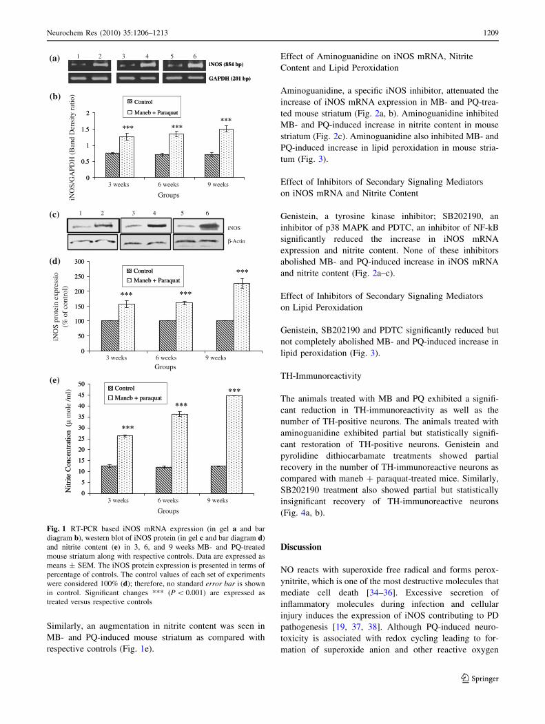
Similarly, an augmentation in nitrite content was seen in
MB- and PQ-induced mouse striatum as compared with
respective controls (Fig. 1e).
Effect of Aminoguanidine on iNOS mRNA, Nitrite
Content and Lipid Peroxidation
Aminoguanidine, a specific iNOS inhibitor, attenuated the
increase of iNOS mRNA expression in MB- and PQ-trea-
ted mouse striatum (Fig. 2a, b). Aminoguanidine inhibited
MB- and PQ-induced increase in nitrite content in mouse
striatum (Fig. 2c). Aminoguanidine also inhibited MB- and
PQ-induced increase in lipid peroxidation in mouse stria-
tum (Fig. 3).
Effect of Inhibitors of Secondary Signaling Mediators
on iNOS mRNA and Nitrite Content
Genistein, a tyrosine kinase inhibitor; SB202190, an
inhibitor of p38 MAPK and PDTC, an inhibitor of NF-kB
significantly reduced the increase in iNOS mRNA
expression and nitrite content. None of these inhibitors
abolished MB- and PQ-induced increase in iNOS mRNA
and nitrite content (Fig. 2a–c).
Effect of Inhibitors of Secondary Signaling Mediators
on Lipid Peroxidation
Genistein, SB202190 and PDTC significantly reduced but
not completely abolished MB- and PQ-induced increase in
lipid peroxidation (Fig. 3).
TH-Immunoreactivity
The animals treated with MB and PQ exhibited a signifi-
cant reduction in TH-immunoreactivity as well as the
number of TH-positive neurons. The animals treated with
aminoguanidine exhibited partial but statistically signifi-
cant restoration of TH-positive neurons. Genistein and
pyrolidine dithiocarbamate treatments showed partial
recovery in the number of TH-immunoreactive neurons as
compared with maneb ? paraquat-treated mice. Similarly,
SB202190 treatment also showed partial but statistically
insignificant recovery of TH-immunoreactive neurons
(Fig. 4a, b).
Discussion
NO reacts with superoxide free radical and forms perox-
ynitrite, which is one of the most destructive molecules that
mediate cell death [34–36]. Excessive secretion of
inflammatory molecules during infection and cellular
injury induces the expression of iNOS contributing to PD
pathogenesis [19, 37, 38]. Although PQ-induced neuro-
toxicity is associated with redox cycling leading to for-
mation of superoxide anion and other reactive oxygen
iNOS (854 bp)
GAPDH (201 bp)
iNOS (854 bp)
GAPDH (201 bp)
0
0.5
1
1.5
2
Control
Maneb + Paraquat
*** ******
0
0.5
1
1.5
2
GroupsiNO
S/G
APD
H (
Ban
d D
ensi
ty r
atio
)
Control
Maneb + Paraquat
*** ******
3 weeks 6 weeks 9 weeks
β-Actin
iNOS
0
50
100
150
200
250
300
Control
Maneb + Paraquat
******
***
0
50
100
150
200
250
300
iNO
S pr
otei
n ex
pres
sio
(% o
f co
ntro
l)
Control
Maneb + Paraquat
******
***
0
5
10
15
20
25
30
35
40
45
50Control
Maneb + paraquat***
***
***
0
5
10
15
20
25
30
35
40
45
50
Nitr
ite C
once
ntra
tion
(N
itrite
Con
cent
ratio
n (
μ m
ole
/ml)
Control
Maneb + paraquat***
***
***
2 1 3 4 5 6
2 1 3 4 5 6
Groups3 weeks 6 weeks 9 weeks
Groups
3 weeks 6 weeks 9 weeks
(a)
(b)
(c)
(d)
(e)
Fig. 1 RT-PCR based iNOS mRNA expression (in gel a and bar
diagram b), western blot of iNOS protein (in gel c and bar diagram d)
and nitrite content (e) in 3, 6, and 9 weeks MB- and PQ-treated
mouse striatum along with respective controls. Data are expressed as
means ± SEM. The iNOS protein expression is presented in terms of
percentage of controls. The control values of each set of experiments
were considered 100% (d); therefore, no standard error bar is shown
in control. Significant changes *** (P \ 0.001) are expressed as
treated versus respective controls
Neurochem Res (2010) 35:1206–1213 1209
123

species by one-electron reduction of PQ [39, 40], the role
of NO in MB- and PQ-mediated neurotoxicity in mouse
striatum remains to be explored.
MB- and PQ-induced expression of iNOS mRNA, its
protein and nitrite content in mouse striatum, suggests the
involvement of nitric oxide in MB- and PQ-induced
neurotoxicity. The elevation in iNOS mRNA expression
and nitrite content was attenuated by aminoguanidine
confirming the involvement of nitric oxide in MB- and
PQ-induced neurotoxicity in mouse striatum The results
obtained in this study are in accordance with the previous
investigations showing the role of nitric oxide in neuro-
degeneration and iNOS inhibitors in neuroprotection
[21, 41]. The involvement of iNOS in PD has also been
reported using iNOS-deficient mouse [42]. RT-PCR and
western blot analyses showed that the expression of iNOS
0
0.2
0.4
0.6
0.8
1
1.2
1.4
1.6
iNO
S/G
APD
H m
RN
A e
xpre
ssio
n (B
and
dens
ity R
atio
)
$$$
$$$ $$$ $$$
***
0
0.2
0.4
0.6
0.8
1
1.2
1.4
1.6
9 Weeks
iNO
S/G
APD
H m
RN
A e
xpre
ssio
n (B
and
dens
ity R
atio
)
$$$
$$$ $$$ $$$
***
0
10
20
30
40
50
60
*$$$
**$$
**$$
**$$$
***
*$$$
**$$
**$$
**$$$
***
- Inhibitor +Aminoguanidine + Genistein + SB202190 + PDTC N
itrite
Con
cent
ratio
n (
μ mol
e /m
l)
*$$$
**$$
**$$
**$$$
***
iNOS (854 bp)
GAPDH (201 bp)
1 2 3 4 5 6 7 8 9 10
9 Weeks
- Inhibitor +Aminoguanidine + Genistein + SB202190 + PDTC
Control
Maneb + Paraquat
Control
Maneb + Paraquat
(a)
(b)
(c)
Fig. 2 Effects of
aminoguanidine, genistein,
SB202190 and PDTC on iNOS
mRNA expression (in gel a and
bar diagram b) and nitrite
content (c) in 9 weeks MB- and
PQ-treated mouse striatum and
respective controls. Data are
expressed as means ± SEM.
Significant changes
* (P \ 0.05), ** (P \ 0.01) and
*** (P \ 0.001) are expressed
as treated versus respective
controls and $$ (P \ 0.01) and
$$$ (P \ 0.001) are expressed
as MB ? PQ without inhibitors
versus MB ? PQ in presence of
inhibitors
0
200
400
600
800
1000
1200
******$$
***$$ ***
$$$
*$$$
0
200
400
600
800
1000
1200
nmol
mal
onal
dehy
de/ m
g pr
otei
n
Control
Maneb + paraquat******$$
***$$ ***
$$$
*$$$
9 Weeks
- Inhibitor +Aminoguanidine + Genistein + SB202190 + PDTC
Fig. 3 Effects of
aminoguanidine, genistein,
SB202190 and PDTC on lipid
peroxidation in control and
9 weeks MB-and PQ-treated
mouse striatum. Data are
expressed as means ± SEM.
Significant changes
* (P \ 0.05), *** (P \ 0.001)
is expressed as treated versus
control and $$ (P \ 0.01) and
$$$ (P \ 0.001) are expressed
as MB ? PQ without inhibitors
versus MB ? PQ in presence of
inhibitors
1210 Neurochem Res (2010) 35:1206–1213
123

might be regulated both at the level of transcription as well
as translation, however, a direct correlation between NO
generation with iNOS mRNA indicated that mainly the
process is regulated at the level of transcription [43].
NO ultimately converts into peroxynitrite that increases
oxidative stress and neuronal cell death [44–46]. As per-
oxynitrite inhibits complexes I, III, IV and V of mito-
chondria; mitochondrial enzymes such as aconitase,
creatine kinase and superoxide dismutase and mitochon-
drial DNA synthesis [47], it could be possible that MB- and
PQ-mediated neurotoxicity occurs through induction of
iNOS and consequent NO generation. We have previously
demonstrated an induction in lipid peroxidation in the
striatum of MB- and PQ-treated mouse [7]. In this study,
we found that aminoguanidine, an inhibitor of iNOS, not
only reduced iNOS mRNA expression and nitrite but also
reduced MB- and PQ-induced lipid peroxidation. The
inhibition of lipid peroxidation by aminoguanidine indi-
cates that nitric oxide is involved in lipid peroxidation-
mediated injury of MB- and PQ-induced PD [48]. This is in
accordance with previous investigations that have shown
that aminoguanidine results in reduction of oxidative stress,
lipid peroxidation and possesses antioxidant property and
free radical scavenging activity [49–51]. Nitric oxide and
other reactive nitrogen species are known to contribute
significantly in the dopaminergic neurodegeneration. This
is evidenced by the abnormal nitrosylation of some key
proteins, such as Parkin, in PD [52]. Although the role of
nitric oxide is reported in 6-hydroxydopamine-induced PD,
as 7-nitroindazole and NG-nitro-L-arginine, NOS inhibi-
tors, improve the motor performance in treated animals, the
mechanism underlying the improvement in motor behavior
is not properly understood [53]. The significant restoration
of TH-immunoreactivity and number of TH-positive neu-
rons by aminoguanidine in MB- and PQ-induced PD phe-
notype suggests that nitric oxide plays a critical role in
dopaminergic neurodegeneration. Genistein and pyrolidine
dithiocarbamate, inhibitors of alternate pathways, showed
only partial recovery of the TH-immunoreactive neurons in
maneb ? paraquat-treated mice showing the involvement
of multiple molecular events including, tyrosine kinase and
NF-kB in maneb- and paraquat-induced neurotoxicity.
Blocking NO and iNOS is used to understand the
mechanism of iNOS-mediated biological events [54].
Genistein, a tyrosine kinase inhibitor, reduces nitric oxide-
mediated injury in neuronal cells [55]. Similarly, signifi-
cant reduction in iNOS expression in chemically induced
dopaminergic neuronal injury is achieved by selective
inhibition of p38 MAPK by SB202190 or NF-kB by PDTC
[25, 56]. In this study, genistein, SB202190 and PDTC
attenuated MB- and PQ-induced increase in nitrite content;
iNOS mRNA and protein level and lipid peroxidation,
ControlControl+Aminoguanidine Control+Genistein Control+SB202190 Control+PDTC
Maneb+ParaquatManeb+Paraquat+Aminoguanidine
Maneb+Paraquat+Genistein
Maneb+Paraquat+SB202190
Maneb+Paraquat+PDTC
0
20
40
60
80
100
120T
H p
ositi
ve n
euro
ns
(% o
f co
ntro
l)
ControlManeb + Paraquat
******
***** ***$$ $$
$$
(a)
(b)
Fig. 4 TH-immunoreactivity of
dopaminergic neurons in the
substantia nigra pars compacta
region of the brain in control
and MB- and PQ-treated mouse
with or without inhibitor (a) at
109 and number of TH positive
neuronal cells (b). Data are
expressed as means ± SEM.
The control values of each set of
experiments were considered
100%; therefore, no standard
error bar is shown in control.
Significant changes
** (P \ 0.01) and
*** (P \ 0.001) are expressed
as treated versus control and $$
(P \ 0.01) are expressed as
MB ? PQ without inhibitor vs.
MB ? PQ with inhibitor
Neurochem Res (2010) 35:1206–1213 1211
123

indicating that multiple pathways are responsible for the
regulation of iNOS expression. None of these inhibitors
reduced the increased lipid peroxidation completely, which
indicates that some other contributory factors might be
involved in the regulation of lipid peroxidation along with
these pathways. Although the inhibitors of the alternate
pathways reduced the nitrite content and lipid peroxidation,
they did not abolish the same completely suggesting that
there is a link between NO and lipid peroxidation,
however, NO alone does not regulate maneb ? paraquat-
induced increase in lipid peroxidation. This is in accor-
dance with previous observations that have shown the
contribution of toxicant responsive genes in the augmen-
tation of lipid peroxidation [7]. The study demonstrated
that MB- and PQ-induced neurotoxicity is also regulated
by NO dependent lipid peroxidation through multiple
pathways.
Acknowledgments The authors sincerely thank Department of
Biotechnology (DBT) for the financial support of the study. Authors
acknowledge Council of Scientific and Industrial Research (CSIR),
New Delhi, India for providing research fellowships to Suman Patel,
Sharawan Yadav and Anand Kumar Singh. Authors also thank Uni-
versity Grants Commission (UGC), New Delhi, India for providing
research fellowship to Seema Singh. The IITR communication
number of this article is 2780.
References
1. Marsden CD (1990) Parkinson’s disease. Lancet 335:948–952
2. Albin RL, Young AB, Penney JB (1989) The functional anatomy
of basal ganglia disorders. Trends Neurosci 12:366–375
3. Singh MP, Patel S, Dikshit M et al (2006) Contribution of
genomics and proteomics in understanding the role of modifying
factors in Parkinson’s disease. Indian J Biochem Biophys 43:69–
81
4. Singh C, Ahmad I, Kumar A (2007) Pesticides and metals
induced Parkinson’s disease: involvement of free radicals and
oxidative stress. Cell Mol Biol 53:19–28
5. Thiruchelvam M, Brockel BJ, Richfield EK et al (2000) Poten-
tiated and preferential effects of combined paraquat and maneb
on nigrostriatal dopamine systems: environmental risk factors for
Parkinson’s disease? Brain Res 873:225–234
6. Cory-Slechta DA, Thiruchelvam M, Barlow BK et al (2005)
Developmental pesticide models of the Parkinson disease phe-
notype. Environ Health Perspect 113:1263–1270
7. Patel S, Singh V, Kumar A et al (2006) Status of antioxidant
defense system and expression of toxicant responsive genes in
striatum of maneb-and paraquat-induced Parkinson’s disease
phenotype in mouse: mechanism of neurodegeneration. Brain Res
1081:9–18
8. Kappus H (1986) Overview of enzyme systems involved in bio-
reduction of drugs and in redox cycling. Biochem Pharmacol
35:1–6
9. Gragus Z, Klaassen CD (1996) Mechanisms of toxicity. In:
Klaassen CD, Andur MO, Doull J (eds) Casarett and Doulls
toxicology. The basic science of poisons. Macmillan, New York,
pp 41–48
10. Beckman JS, Beckman TW, Chen J et al (1990) Apparent
hydroxyl radical production by peroxynitrite: implications for
endothelial injury from nitric oxide and superoxide. Proc Natl
Acad Sci USA 87:1620–1624
11. Zhang J, Fitsanakis VA, Gu G et al (2003) Manganese ethylene-
bis-dithiocarbamate and selective dopaminergic neurodegenera-
tion in rat: a link through mitochondrial dysfunction. J Neuro-
chem 84:336–346
12. Brooks AI, Chadwick CA, Gelbard HA et al (1999) Paraquat
elicited neurobehavioral syndrome caused by dopaminergic
neuron loss. Brain Res 823:1–10
13. McCormack AL, Thiruchelvam M, Manning-Bog AB et al (2002)
Environmental risk factors and Parkinson’s disease: selective
degeneration of nigral dopaminergic neurons caused by the her-
bicide paraquat. Neurobiol Dis 10:119–127
14. Thiruchelvam M, Richfield EK, Baggs RB et al (2000) The
nigrostriatal dopaminergic system as a preferential target of
repeated exposures to combined paraquat and maneb: implica-
tions for Parkinson’s disease. J Neurosci 20:9207–9214
15. Patel S, Sinha A, Singh MP (2007) Identification of differentially
expressed proteins in striatum of maneb-and paraquat-induced
Parkinson’s disease phenotype in mouse. Neurotoxicol Teratol
29:578–585
16. Patel S, Singh K, Singh S et al (2008) Gene expression profiles of
mouse striatum in control and maneb ? paraquat-induced Par-
kinson’s disease phenotype: validation of differentially expressed
energy metabolizing transcripts. Mol Biotechnol 40:59–68
17. Beckman JS (1996) Oxidative damage and tyrosine nitration from
peroxynitrite. Chem Res Toxicol 9:836–844
18. Pryor WA, Squadrito GL (1995) The chemistry of peroxynitrite a
product from the reaction of nitric oxide with superoxide. Am J
Physiol 268:L699–L722
19. Dawson VL, Dawson TM (1998) Nitric oxide in neurodegener-
ation. Prog Brain Res 118:215–229
20. Zhang L, Dawson VL, Dawson TM (2006) Role of nitric oxide in
Parkinson’s disease. Pharmacol Ther 109:33–41
21. Liberatore GT, Jackson-Lewis V, Vukosavic S et al (1999) Inducible
nitric oxide synthase stimulates dopaminergic neurodegeneration in
the MPTP model of Parkinson disease. Nat Med 5:1403–1409
22. Paul A, Pendreigh RH, Plevin R (1995) Protein kinase C and
tyrosine kinase pathways regulate lipopolysaccharide-induced
nitric oxide synthase activity in RAW 264.7 murine macro-
phages. Br J Pharmacol 114:482–488
23. Xu Z, Wang B, Wang X et al (2006) ERK1/2 and p38 mitogen-
activated protein kinase mediate iNOS-induced spinal neuron
degeneration after acute traumatic spinal cord injury. Life Sci
79:1895–1905
24. Koppal T, Drake J, Yatin S et al (1999) Peroxynitrite-induced
alterations in synaptosomal membrane proteins: insights into
oxidative stress in Alzheimer’s disease. J Neurochem 72:310–317
25. Liu W, Kato M, Itoigawa M et al (2001) Distinct involvement of
NF-kB and p38 mitogen-activated protein kinase pathways in
serum deprivation-mediated stimulation of inducible nitric oxide
synthase and its inhibition by 4-hydroxynonenal. J Cell Biochem
83:271–280
26. Karpuzoglu E, Fenaux JB, Phillips RA et al (2006) Estrogen
up-regulates inducible nitric oxide synthase, nitric oxide, and
cyclooxygenase-2 in splenocytes activated with T cell stimulants:
role of interferon-gamma. Endocrinology 147:662–671
27. Fan Q, Ding J, Zhang J et al (2004) Effect of the knockdown of
podocin mRNA on nephrin and a-actinin in mouse podocyte. Exp
Biol Med 229:964–970
28. Martin PY, Bianchi M, Roger F et al (2002) Arginine vasopressin
modulates expression of neuronal NOS in rat renal medulla. Am J
Physiol Renal Physiol 283:F559–F568
29. Bradford MM (1976) A rapid and sensitive method for the
quantitation of microgram quantities of protein utilizing the
principle of protein-dye binding. Anal Biochem 72:248–254
1212 Neurochem Res (2010) 35:1206–1213
123

30. Granger DL, Taintor RR, Boockvar KS, Hibbs JB (1996) Mea-
surement of nitrate and nitrite in biological samples using nitrate
reductase and Greiss reaction. Methods Enzymol 268:142–151
31. Ohkawa H, Ohishi N, Yagi K (1979) Assay for lipid peroxides in
animal tissues by thiobarbituric acid reaction. Anal Biochem
95:351–358
32. Singh S, Singh K, Patel DK et al (2009) The expression of
CYP2D22, an ortholog of human CYP2D6, in mouse striatum
and its modulation in MPTP-induced Parkinson’s disease phe-
notype and nicotine-mediated neuroprotection. Rejuvenation Res
12:185–197
33. Singh AK, Tiwari MN, Upadhyay G et al (2010) Long term
exposure to cypermethrin induces the nigrostriatal dopaminergic
neurodegeneration in adult rats: postnatal exposure enhances the
susceptibility during adulthood. Neurobiol Aging. doi:10.1016/
j.neurobiolaging.2010.02.018
34. Ischiropoulos H, Al-Mehdi AB (1995) Peroxynitrite-mediated
oxidative protein modifications. FEBS Lett 364:279–282
35. Przedborski S, Jackson-Lewis V, Djaldetti R et al (2000) The
parkinsonian toxin MPTP: action and mechanism. Restor Neurol
Neurosci 16:135–142
36. Torreilles F, Salman-Tabcheh S, Guerin M, Torreilles J (1999)
Neurodegenerative disorders: the role of peroxynitrite. Brain Res
Rev 30:153–163
37. Okuno T, Nakatsuji Y, Kumanogoh A et al (2005) Loss of
dopaminergic neurons by the induction of inducible nitric oxide
synthase and cyclooxygenase-2 via CD40: relevance to Parkin-
son’s disease. J Neurosci Res 81:874–882
38. Knott C, Stern G, Wilkin GP (2000) Inflammatory regulators in
Parkinson’s disease: iNOS, lipocortin-I and cyclooxygenases-1
and -2. Mol Cell Neurosci 16:724–739
39. Bus JS, Gibson JE (1984) Paraquat: model for oxidant-initiated
toxicity. Environ Health Perspect 55:37–46
40. Cohen GM, d’Arcy Doherty M (1987) Free radical mediated cell
toxicity by redox cycling chemicals. Br J Cancer Suppl 8:46–52
41. Good PF, Hsu A, Werner P et al (1998) Protein nitration in
Parkinson’s disease. J Neurophathol Exp Neurol 57:338–342
42. Dehmer T, Lindenau J, Haid S et al (2000) Deficiency of
inducible nitric oxide synthase protects against MPTP toxicity in
vivo. J Neurochem 74:2213–2216
43. Chen YQ, Fisher JH, Wang MH (1998) Activation of the RON
receptor tyrosine Kinase inhibits inducible nitric oxide synthase
(iNOS) expression by murine peritoneal exudate macrophages:
phosphatidylinositol-3 kinase is required for RON-mediated
inhibition of iNOS expression. J Immun 161:4950–4959
44. Noack H, Possel H, Rethfeldt C et al (1999) Peroxynitrite med-
iated damage and lowered superoxide tolerance in primary cor-
tical glial cultures after induction of the inducible isoform of
NOS. Glia 28:13–24
45. Denicola A, Radi R (2005) Peroxynitrite and drug-dependent
toxicity. Toxicology 208:273–288
46. Antunes F, Nunes C, Laranjinha J, Cadenas E (2005) Redox
interactions of nitric oxide with dopamine and its derivatives.
Toxicology 208:207–212
47. Brown GC (1999) Nitric oxide and mitochondrial respiration.
Biochim Biophys Acta 1411:351–369
48. Violi F, Marino R, Milite MT, Loffredo L (1999) Nitric oxide and
its role in lipid peroxidation. Diabetes Metab Res Rev 15:283–288
49. Giardino I, Fard AK, Hatchell DL, Brownlee M (1998) Amino-
guanidine inhibits reactive oxygen species formation, lipid
peroxidation, and oxidant-induced apoptosis. Diabetes 47:1114–
1120
50. Kedziora-Kornatowska KZ, Luciak M, Blaszczyk J, Pawlak W
(1998) Effect of aminoguanidine on the generation of superoxide
anion and nitric oxide by peripheral blood granulocytes of rats
with streptozotocin-induced diabetes. Clin Chim Acta 278:45–53
51. Courderot-Masuyer C, Dalloz F, Maupoil V, Rochette L (1999)
Antioxidant properties of aminoguanidine. Fundan Clin Phar-
macol 13:535–540
52. Tsang AH, Lee YI, Ko HS et al (2009) S-nitrosylation of XIAP
compromises neuronal survival in Parkinson’s disease. ProcNatl
Acad Sci USA 106:4900–4905
53. Padovan-Neto FE, Echeverry MB, Tumas V et al (2009) Nitric
oxide synthase inhibition attenuates L-DOPA-induced dyskine-
sias in a rodent model of Parkinson’s disease. Neuroscience
159:927–935
54. Vallance P, Leiper J (2002) Blocking NO synthesis: how, where
and why? Nat Rev Drug Discov 1:939–950
55. Kong LY, McMillian MK, Maronpot R, Hong JS (1996) Protein
tyrosine kinase inhibitors suppress the production of nitric oxide
in mixed glia, microglia-enriched or astrocyte-enriched cultures.
Brain Res 729:102–109
56. Jeohn GH, Cooper CL, Wilson B et al (2002) p38 MAP kinase is
involved in lipopolysaccharide-induced dopaminergic neuronal
cell death in rat mesencephalic neuron-glia cultures. Ann NY
Acad Sci 962:332–346
Neurochem Res (2010) 35:1206–1213 1213
123