Involvement of DNA methylation in the control of the expression of an estrogen-induced...
12
Involvement of DNA Methylation in the Control of the Expression of an Estrogen-Induced Breast-Cancer-Associated Protein (pS2) in Human Breast Cancers Vale ´ rie Martin, 1 Ste ´ phane Ribieras, 1 Xiu-Gin Song-Wang, 1 Yves Lasne, 2 Lucien Frappart, 3 Marie-Christine Rio, 4 and Robert Dante 1 * 1 Laboratoire de Ge ´ne ´tique, UMR 5641 CNRS, UCLB Lyon 1, Lyon, France 2 Laboratoire des Radioisotopes, Ho ˆpital Edouard Herriot, Lyon, France 3 Laboratoire Anatomie Pathologique, Ho ˆpital Edouard Herriot, Lyon, France 4 Laboratoire de Ge ´ne ´ tique Mole ´culaire des Eucaryotes, CNRS, INSERM U 184, Illkirch France Abstract pS2 gene has been used to investigate the relationship between alterations of DNA methylation patterns in human tumors and gene expression. The expression of pS2, which is transcriptionally controlled by estrogens in breast cancer cell lines, is restricted to estrogen-receptor-rich human breast tumors. We found that the CCGG site within the promoter/enhancer sequence of pS2 was hypomethylated in estrogen-receptor-rich breast tumors expressing this gene. The amount of DNA molecules unmethylated at this site was related to the amount of pS2 mRNA detected in the samples. The demethylation of this region, which contains the estrogen responsive element, was confirmed by genomic sequencing. Transient expression of functional human estrogen receptors stimulated the expression of the endogenous pS2 in HeLa cells, but failed, in BT-20 cells, to stimulate expression of this gene. Since the promoter/enhancer region of pS2 is unmethylated in HeLa cells and methylated in BT-20 cells, these data also support the hypothesis that DNA methylation might be involved in the control of pS2 expression. J. Cell. Biochem. 65:95–106. r 1997 Wiley-Liss, Inc. Key words: breast cancers; genomic sequencing; 5-methyldeoxycytidine; multiple component analysis; pS2; RT-PCR In mammalians, the methylation of DNA is accomplished by enzymes which catalyze the transfer of methyl groups from S-adenosylme- thionine to the carbon-5 of deoxycytidine at CpG sites [Adams, 1990]. This epigenetic modi- fication seems to have an essential role in nor- mal mammalian development since it has been shown that targeted mutation of the DNA meth- yltransferase gene results in embryonic lethal- ity [Li et al., 1992]. DNA methylation has been implicated in several cellular processes, includ- ing chromosome instability, hot spots for so- matic mutations, and genomic imprinting, and in the last 15 years evidence of an involvement of DNA methylation in control of gene expres- sion has been accumulated [for review see Ad- ams, 1990; Razin and Cedar, 1991]. Numerous studies have correlated, in vitro and in vivo, the hypomethylation of the 58 region of viral or cellular genes [Doerfler, 1983] with their expres- sion, and several experiments have shown that methylation can repress gene transcription [Bird, 1992]. Although global genomic and gene- specific hypomethylation are generally observed in tumor tissues [Gama-Sosa et al., 1983; Fein- berg and Vogelstein, 1983; Ribieras et al., 1994], simultaneous occurrence of CpG islands hyper- methylation also has been reported in several cell lines and human tumors [for review see Laird and Jaenisch, 1994]. Taken together, these findings suggest that alterations of DNA meth- Abbreviations used: bp, base pair; CAT, chloramphenicol acetyl transferase; E 2 , 17b-estradiol; EGF, epidermal growth factor; ER, estrogen receptor; ERE, estrogen responsive element; GAPDH, glyceraldehyde-3-phosphate dehydroge- nase; kb, kilobase pair; RT-PCR, reverse transcription– polymerase chain reaction; SV 40, simian virus 40; WCE, whole cell extract. Contract grant sponsors: Ligue Nationale pour la Recher- che contre le Cancer, the Association pour la Recherche contre le Cancer, the Institut National de la Sante ´ et de la Recherche Me ´dicale. *Correspondence to: Robert Dante, Laboratoire de Ge ´ne ´ti- que, UMR 5641 CNRS, UCLB Lyon 1, 8 avenue Rockefeller, 69373 Lyon Cedex 08, France. Received 20 June 1996; accepted 26 November 1996 Journal of Cellular Biochemistry 65:95–106 (1997) r 1997 Wiley-Liss, Inc.
-
Upload
valerie-martin -
Category
Documents
-
view
213 -
download
0
Transcript of Involvement of DNA methylation in the control of the expression of an estrogen-induced...

Involvement of DNAMethylation in theControl of the Expression of an Estrogen-InducedBreast-Cancer-Associated Protein (pS2) inHuman Breast CancersValerie Martin,1 Stephane Ribieras,1 Xiu-Gin Song-Wang,1 Yves Lasne,2
Lucien Frappart,3Marie-Christine Rio,4 and Robert Dante1*1Laboratoire de Genetique, UMR 5641 CNRS, UCLB Lyon 1, Lyon, France2Laboratoire des Radioisotopes, Hopital Edouard Herriot, Lyon, France3Laboratoire Anatomie Pathologique, Hopital Edouard Herriot, Lyon, France4Laboratoire de Genetique Moleculaire des Eucaryotes, CNRS, INSERM U 184, Illkirch France
Abstract pS2 gene has been used to investigate the relationship between alterations of DNAmethylation patternsin human tumors and gene expression. The expression of pS2, which is transcriptionally controlled by estrogens inbreast cancer cell lines, is restricted to estrogen-receptor-rich human breast tumors. We found that the CCGG site withinthe promoter/enhancer sequence of pS2 was hypomethylated in estrogen-receptor-rich breast tumors expressing thisgene. The amount of DNAmolecules unmethylated at this site was related to the amount of pS2 mRNA detected in thesamples. The demethylation of this region, which contains the estrogen responsive element, was confirmed by genomicsequencing. Transient expression of functional human estrogen receptors stimulated the expression of the endogenouspS2 in HeLa cells, but failed, in BT-20 cells, to stimulate expression of this gene. Since the promoter/enhancer region ofpS2 is unmethylated in HeLa cells and methylated in BT-20 cells, these data also support the hypothesis that DNAmethylation might be involved in the control of pS2 expression. J. Cell. Biochem. 65:95–106. r 1997 Wiley-Liss, Inc.
Key words: breast cancers; genomic sequencing; 5-methyldeoxycytidine; multiple component analysis; pS2; RT-PCR
In mammalians, the methylation of DNA isaccomplished by enzymes which catalyze thetransfer of methyl groups from S-adenosylme-thionine to the carbon-5 of deoxycytidine atCpG sites [Adams, 1990]. This epigenetic modi-fication seems to have an essential role in nor-mal mammalian development since it has beenshown that targetedmutation of theDNAmeth-
yltransferase gene results in embryonic lethal-ity [Li et al., 1992]. DNAmethylation has beenimplicated in several cellular processes, includ-ing chromosome instability, hot spots for so-matic mutations, and genomic imprinting, andin the last 15 years evidence of an involvementof DNA methylation in control of gene expres-sion has been accumulated [for review see Ad-ams, 1990; Razin and Cedar, 1991]. Numerousstudies have correlated, in vitro and in vivo, thehypomethylation of the 58 region of viral orcellular genes [Doerfler, 1983] with their expres-sion, and several experiments have shown thatmethylation can repress gene transcription[Bird, 1992].Although global genomic and gene-specific hypomethylation are generally observedin tumor tissues [Gama-Sosa et al., 1983; Fein-berg andVogelstein, 1983; Ribieras et al., 1994],simultaneous occurrence of CpG islands hyper-methylation also has been reported in severalcell lines and human tumors [for review seeLaird and Jaenisch, 1994]. Taken together, thesefindings suggest that alterations of DNAmeth-
Abbreviations used: bp, base pair; CAT, chloramphenicolacetyl transferase; E2, 17b-estradiol; EGF, epidermal growthfactor; ER, estrogen receptor; ERE, estrogen responsiveelement; GAPDH, glyceraldehyde-3-phosphate dehydroge-nase; kb, kilobase pair; RT-PCR, reverse transcription–polymerase chain reaction; SV 40, simian virus 40; WCE,whole cell extract.Contract grant sponsors: Ligue Nationale pour la Recher-che contre le Cancer, the Association pour la Recherchecontre le Cancer, the Institut National de la Sante et de laRecherche Medicale.*Correspondence to: Robert Dante, Laboratoire de Geneti-que, UMR 5641 CNRS, UCLB Lyon 1, 8 avenue Rockefeller,69373 Lyon Cedex 08, France.Received 20 June 1996; accepted 26 November 1996
Journal of Cellular Biochemistry 65:95–106 (1997)
r 1997Wiley-Liss, Inc.
ylationmay play a variety of roles in carcinogen-esis [Counts andGoodman, 1995] and that somespecific alterations of DNA methylation pat-terns might be involved in the expression oftumor-specific genes.In order to gain further insights into this
question, we analyzed the DNA methylationpatterns of the estrogen-induced breast-cancer-associated protein (pS2) gene in normal andtumoral human breast tissues. This gene waschosen for two reasons: pS2 expression is re-stricted to subclasses of human breast cancers[Rio et al., 1987], and the promoter/enhancersequence is not associated with a CpG island.This model therefore offers the possibility ofinvestigating the relationship between gene ac-tivation and hypomethylation in human can-cers.pS2 is not transcribed in normal breast tis-
sue and is predominantly expressed in estrogen-receptor (ER)-positive breast carcinomas [Rioet al., 1987]. In the MCF-7 mammary cell line,pS2 expression is specifically controlled at thetranscriptional level by estradiol [Masiakowskiet al., 1982], and in human mammary tumorsits expression is indicative of a favorable re-sponse to hormonotherapy [Schwartz et al.,1991]. However, in some other carcinomas andbenign tumors the presence of pS2 transcriptsor proteins is not correlated with the presenceof estrogen receptors [Dante et al., 1994; Rio etal., 1988; Welter et al., 1992; Wysocki et al.,1990]. This gene is physiologically expressed instomach and in regenerative tissues surround-ing ulcerous lesions of the human gastrointesti-nal tract. It has been recently suggested thatpS2 protein might be important in stimulatinggastrointestinal repair [Playford et al., 1996],since transgenic mice overexpressing the hu-man pS2 protein have an increased resistanceto intestinal damage. In addition, disruption ofthe pS2 gene by homologous recombination in-dicates that its expression is essential for nor-mal differentiation of the antral and pyloricgastric mucosa and that it may function as agastric-specific tumor suppressor gene [Lefeb-vre et al., 1996].In fact, the 58 flanking region of pS2 contains
a complex promoter/enhancer region (from posi-tion2428 to position2332) responsive to estro-gens, EGF, the phorbol ester tumor promoter,c-Ha-ras oncoprotein, and c-jun protein [Nunezet al., 1989]. The estrogen responsive element(ERE) has been characterized and localized in
the 58 flanking sequences from position2405 toposition2393 [Berry et al., 1989]. In contrast tothe other known ERE, this ERE is an imperfectpalindromic sequence of 13 bp (GGTCACGGTG-GCC). Although several in vitro [Berry et al.,1989; Nunez et al., 1989] and in vivo [Rio et al.,1987] studies indicate that pS2 is, in breasttumoral tissues, controlled by estrogen recep-tors, additional factors seem to be involved inthe control of its expression [Dante et al., 1994;Rio et al., 1987; Zajchowski and Sager, 1991].DNA methylation patterns of pS2 were ana-
lyzed by Southern blot and genomic sequencingexperiments in normal and tumoral breast tis-sues, normal endometrium, and stomach. Theeffect of DNAmethylation on the inducibility ofpS2 expression was investigated, in cell lines,by transient expression of the human estrogenreceptors.
MATERIALS AND METHODSSpecimens and Cell Lines
A total number of two normal breast tissuesamples, 29 breast primary adenocarcinomas,two normal total stomach tissue samples, andfive normal endometrium tissue samples wereanalyzed. After excision, a portion of the tissuewas snap-frozen and subsequently stored inliquid nitrogen. For both normal and pathologi-cal samples, the remainder of the tissue wasexamined at the pathology laboratory. Speci-mens were fixed in formalin, paraffin-embed-ded, sectioned, and stainedwith hemalun-eosin-safran. HeLa, MCF-7, and BT-20 cell lines weregrown as described [Nunez et al., 1989]. For allbreast tumors, estrogen receptor status wasdetermined by ligand binding assays. Samplescontaining less than 10 fmol of ER per milli-gram of total protein were considered to beER-negative.
RT-PCR Assay
Total RNAs were extracted from cell linesand tissue samples and simultaneous amplifica-tion of GAPDH (glyceraldehyde-3-phosphate-dehydrogenase) and pS2 transcripts were per-formed as previously described [Dante et al.,1994]. The RT-PCR products were analysed on2% agarose gel, and the ratio between pS2 andGAPDH signals was determined. Detection ofestrogen receptor RNA by RT-PCR was per-formed as described elsewhere [Fuqua et al.,1990] but using GAPDH primers as internalcontrol instead of b-actin primers.
96 Martin et al.
Southern Blot Analysis
High molecular weight DNA was extractedfrom cell lines and frozen tissues by standardprocedures [Schwartz et al., 1991]. DNA werecleaved with TaqI, and methylation status atCCGG sites was determined by the isoschizo-meric restriction enzymes MspI and HpaII, aspreviously described [Ribieras et al., 1994]. Allfilters were hybridized with a probe which re-veals a TaqI fragment without CCGG sites ofthe 38 region of the human G-g globin gene[Ribieras et al., 1994], in order to control theloading on each lane, and with a probe whichreveals the unmethylated CCGG sites of theexon 1 and the 58 sequence of the human cata-lase gene [Ribieras et al., 1994], to ensure thatpartial cleavages observed for the pS2 are notdue to an artefactual inhibition of HpaII. Hy-bridization probes 1 and 2 were purified fromgenomic cloned DNA fragments pS2(429/86)pG1 [Berry et al., 1989] and pS2-pBR322[Jeltsch et al., 1987]. Probe 1 is from nucleo-tides 2428 to 297, and probe 2 is from nucleo-tides 287 to 1325, according to the publishedsequence [Jeltsch et al., 1987].
Genomic Sequencing Analysis of 5-Methyl dCyd
Determination of the methylation status ofdCyd was performed essentially as previouslydescribed [Martin et al., 1995]. Briefly, alkali-denatured DNAwas incubated in 3 M NaHSO3
and 5 mM hydroquinone for 16 h at 50°C. Thischemical treatment converts all cytosines touracil, while the methylated cytosines remainunmodified. Aliquots were used for the amplifi-cation of the region of interest using strandspecific primers and a two-step PCR method[Martin et al., 1995]. PCR products were thencloned in pT7blue-T Vector (Novagen, Madison,WI). After screening, plasmids were sequencedby the dideoxy method. The frequency of cyto-sine methylation in the samples analyzed wascalculated for each CpG by the number of clonesmethylated at this site divided by the numberof clones analyzed.
Computer Analysis
Multiple components analysis was performedusing the ADE program library [Thioulouse etal., 1994].
Transfections and CAT Assays
Expression vectors used in these experi-ments were pS2-CAT [Kumar et al., 1987], an
expression vector containing the 58 flankingsequences of pS2 (position ,21100 to 110),HEO, an expression vector encoding the humanestrogen receptor [Kumar and Chambon, 1988],HEGO, an expression vector encoding the wild-type human estrogen receptor [Tora et al., 1989],and pU-lacZ, an expression vector containingthe bacterial b-galactosidase gene driven bythe SV40 promoter.Plasmids were introduced into HeLa and
BT-20 cells by the calcium phosphate precipita-tion method [Sambrook et al., 1989]. Twenty-four hours before transfection, cells were seeded(2.5 3 105/6 cm diameter or 1.5 3 106/10 cmdiameter dishes) in a medium containing 10nM of 17b-estradiol (E2). Cell extracts wereprepared 48 h after transfection.For the determination of pS2-CAT activity,
cells grown in 6 cm diameter dishes were cotrans-fected with 5 µg of pS2-CAT plasmid, 2 µg ofvector encoding the estrogen receptor HEO,and 2 µg of control vector encoding the bacterialb-galactosidase gene (pU-LacZ) as internal stan-dard. The CAT enzyme activity was determinedfrom cellular extracts, and nonacetylated and ace-tylated [14C] chloramphenicol products were an-alyzed by thin layer chromatography followedby autoradiography [Sambrook et al., 1989].For gel retardation experiments, whole cell
extracts (WCE)were prepared from 10 cmdiam-eter HeLa cell dishes transfected with an ex-pression vector encoding the human estrogenreceptorHEGOandwith pU-LacZ plasmidDNAas an internal control.
Immunohistology
One day after transfection, HeLa cells wereseeded into eight chamber tissue culture slides(Lab-Tek; Nunc, Naperville, IL) at a density of2 3 104 cells/chamber and grown for 2 days infresh medium supplemented with E2 (10 nM).After washing, cells were fixed for 10 min withparaformaldehyde (4% in phosphate bufferedsaline [PBS]) and permeabilized for 5 min withTriton X-100 (0.02% in PBS).After an abundantrinsing with PBS, fixed cells were incubated for1 h with one 1/50 dilution of pS2 monoclonalantibody p2802 [Rio et al., 1988] in fetal calfserum 10% PBS. Then cells were rinsed withfetal calf serum 10% PBS and incubated for 30min in the dark with a 1/100 dilution of anti-mouse rhodamine–conjugated secondary anti-bodies (Immunotech, France).After a final rins-ing with PBS, slides were examined using an
pS2Methylation in Breast Cancers 97
epifluorescent microscope. Controls were run inparallel.
RESULTS
The methylation status of CCGG sites of the58 region of pS2 was investigated on Southernblots using the pair of the isoschizomeric restric-tion endonucleases, HpaII and MspI. MspIyielded fragments resulting from cleavage of allCCGG sites, whereasHpaII cleaved only CCGGsites with unmethylated internal cytosine. Inorder to precisely map these sites, we alsocleaved all DNAs with TaqI, and filters weresuccessively hybridized with several probes.Within the pS2 region analyzed, which in-cluded the 58 flanking sequences, exon 1, andpart of intron 1, TaqI and MspI digestion didnot show any polymorphism. In contrast,HpaIIpolymorphisms were observed in practically allsamples analyzed.
Correlation Between the Methylation Status ofCCGG Sites of the 58 Region of pS2 and ItsExpression in Human Breast Cell Lines
The methylation status of CCGG sites of pS2was first investigated in two human breastcancer cell lines: the MCF-7 cell line, whichexpresses pS2 and contains estrogen receptors,and the BT-20 cell line, which is an estrogen-receptor-negative and pS2-negative cell line [Rioet al., 1987].Hybridization with probes 1 and 2 (Fig. 1C),
which map the 3 CCGG sites at positions 2354(M1), 284 (M2), and 220 (M3), respectively,revealed that these three sites were unmethyl-ated in DNA extracted from MCF-7 cells, sinceMspI patterns were identical toHpaII patterns(Fig. 1A,B; MspI digest (lanes 2) compared toHpaII digest [lanes 3]). In DNAs extracted fromBT-20 cells, the Taq1-Taq2 1.06 kb (Fig. 1A,lane 4) band, which contains the M1, M2, andM3 sites, was only partially cleaved to shorterfragments by HpaII; a consistent part of theDNA molecules was unmethylated at M2 sites(visualized with probe 1 by the 0.65 kb band inFig. 1A, lane 4) and at M3 sites, as indicated bythe 0.71 kb band in Fig. 1A (lane 4) and the 0.34and 0.4 kb bands in Fig. 1B (lane 4). However,M1 sites were found to be heavily methylated inthese cells, since the 0.27 and 0.38 kb bandscorresponding to unmethylated M1 sites werenot detected (Fig. 1A, lane 4).In intron I, the CCGG sites were methylated
in BT-20 cell lines, and in MCF-7 the M5 site
was partially methylated (data not shown).Taken together, these results suggest that themethylated status of pS2 is associated with itsexpression. In addition, the analysis of the 3CCGG sites M1, M2, and M3 within the 58
regulatory sequences further suggests that themethylation status of M1 sites might be corre-lated with pS2 expression. This hypothesis wasfurther investigated in human breast tissues.
Correlation Between the Methylation Status ofCCGG Sites of the 58 Region of pS2 and a
Reduced Level of Expression inHuman Breast Cancers
The amount of pS2 mRNA was evaluated byRT-PCR assays as previously described [Danteet al., 1994]. Immunohistochemical studies haveshown that the percentage of pS2-positive tu-mor cells varied greatly between tumors [Henryet al., 1991; Rio et al., 1987], and the percentageof 3–5% of pS2-positive tumor cells has beencommonly adopted as the threshold for pS2-positivity [Henry et al., 1991]. In order to evalu-ate the sensitivity of the RT-PCR method used,pS2 and GAPDH transcripts were amplifiedfrom samples containing RNA extracted fromthe pS2-positive MCF-7 cells and from the pS2-negative BT-20 cells mixed in various propor-tions. In the conditions used in these experi-ments, pS2 transcripts were not detected insamples containing less than 5% ofMCF-7RNA,and the pS2/GAPDH ratios were dependent onthe amount of MCF-7 RNA. From these dataand the analysis of a few samples in which pS2mRNA were quantitated by Northern blot ex-periments [Dante et al., 1994], samples wereclassified on a three point scale: negative sample(0), pS2/GAPDH ratio lower than 0.2; low levelof expression (11), 0.2 # pS2/GAPDH ratio#0.3 ($5% to #25% of pS2-positive RNA); andstrong expressing samples (21), pS2/GAPDHratio .0.3.As expected from the data obtained by the
Southern blot analysis of cell lines, the CCGGsites (M1, M2, and M3) located at the 58 end ofpS2 were hypomethylated in DNAs extractedfrom breast tumors expressing this gene at ahigh level (7/7 cases of pS2 21 analyzed; threerepresentative samples are shown in Fig. 1A,B, lanes 7–9). In DNAs extracted from ER-richtumors (.10 fmol of ER per milligram of totalprotein) exhibiting a low level of pS2 expression(11), these sites were also hypomethylated butto a lesser extent (two representative samples
98 Martin et al.
out of the eight cases of pS2 11 analyzed areshown in Fig. 1A,B, lanes 10,11). In contrast, inpS2-negative samples, two different DNAmeth-ylation patterns were observed at the 58 end ofpS2.In DNAs extracted from the two normal
breast samples (pS2-negative) the M1, M2, andM3 sites in the promoter/enhancer region wereheavilymethylated (Fig. 1A,B, lanes 5,6). Thesesites were also methylated in six out of eightcases of ER-rich/pS2-negative breast tumor
samples analyzed; in two cases these CCGG siteswere slightly demethylated (four representativesamples are shown in Fig. 1A,B, lanes 12–15).In estrogen-receptor-negative samples (,10
fmol of ER per milligram of total protein) whichwere also pS2-negative adenocarcinomasamples (5/5 cases; three representativesamples are shown in Fig. 1A,B, lanes 16–18),HpaII patterns were similar to those obtainedwith DNA extracted from the BT-20 cell line.Hypomethylation of M2 sites (Fig. 1B, lanes
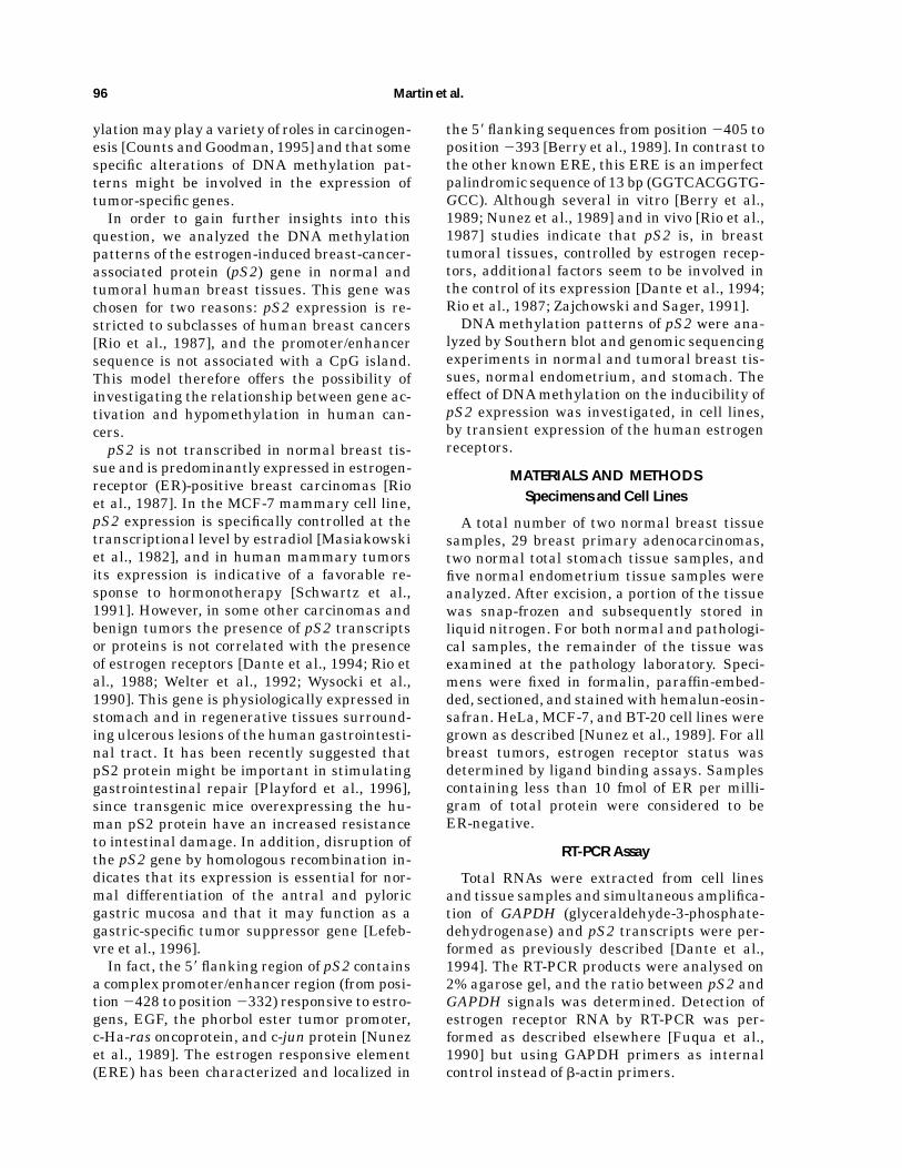
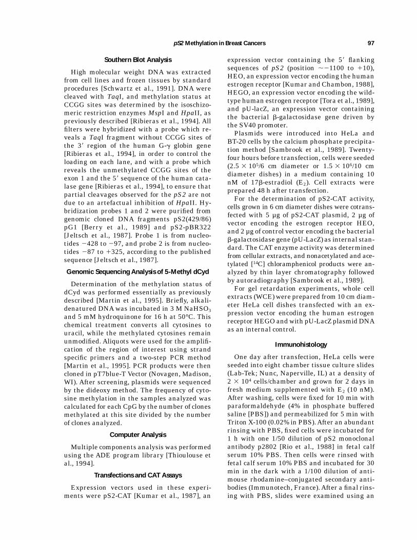
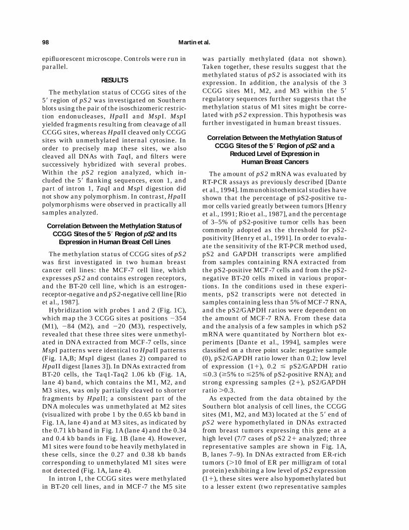
Fig. 1. DNAmethylation patterns of the 58 region of pS2 in celllines and human tissues. Southern blot analysis; DNA methyl-ation patterns shown are representative of those observed innormal and tumoral tissues studied. Detection of estrogen recep-tors (E. R.) in each sample is indicated by Y (yes), N (no), or ND(not determined), and the amount of pS2 mRNA by 0 (pS2-negative samples), 1 (low level of expression), and 11 (strongexpressing samples). DNA were cleaved with TaqI (lanes 1),TaqI-MspI (lanes 2), and TaqI-HpaII (lanes 3–22). A: Hybridiza-tion of DNA digests with probe 1 (see C). DNA were cleavedwith TaqI (lane 1), TaqI-MspI (lane 2), and TaqI-HpaII (lane
3–22). Human breast cancer cell lines (lanes 3,4, MCF-7 andBT-20, respectively), normal breast (lanes 5,6), breast adenocar-cinomas (lanes 7–18), normal endometrium (lanes 19–21), andnormal stomach (lane 22). B: Hybridization of the same filterswith probe 2 (C). C: Schematic representation of the pS2 gene,with positions of the three exons (black boxes), repeated se-quence elements (black arrows), and BamHI (B) sites. Thestudied region is presented on an expanded scale, with thepositions of TaqI (Tq) and MspI (M) sites and restriction mapanalysis. The sizes of TaqI-MspI and MspI-MspI fragments areindicated in base pair. The ERE is indicated by awhite circle.
pS2Methylation in Breast Cancers 99
16–18, 0.4 kb band) andM3 sites (Fig. 1B, lanes16–18, 0.34 kb band) were observed, while M1sites were heavilymethylated in these samples,since only very faint bands or none were detect-able at 0.27 kb and 0.38 kb with probe 1 (Fig.1A, lanes 16–18) and at 0.71 kb with probe 2(Fig. 1B, lanes 16–18).No correlation between the methylation sta-
tus of CCGG sites located in intron I and pS2expression was observed. In addition, the up-stream region of the promoter/enhancer wasmethylated in all samples analyzed, cell linesincluded (data not shown). Taken together, theseresults show that the methylation status of M1sites is correlated with the expression of pS2 inbreast tissues and that the amount of DNAmolecules possessing unmethylated M1 sites isrelated to the amount of pS2 mRNAdetected inthe samples by RT-PCR.The correlation between the hypomethyl-
ation of the 58 end of pS2 and its expression isalso observed in nonpathological samples. Innormal stomach tissue (2/2 cases analyzed),which physiologically express pS2 but do notpossess estrogen receptors, theM1,M2, andM3sites of the 58 region of pS2 were stronglycleaved by HpaII (Fig. 1A,B, lanes 22). In con-trast, in normal endometrium (5/5 cases), whichdo not express pS2 but possess estrogen recep-tors, the HpaII patterns (Fig. 1A,B, lanes 19–21), indicated that M1 sites were methylated inthese tissues. The hypomethylation of the pro-moter/enhancer sequences of pS2 seems, there-fore, associated with its expression rather thanthe presence of estrogen receptors.
Determination of the Methylation Pattern ofpS2 From Individual Chromosomes
In the region studied, only the hypomethyl-ation of the M1 site (located in the promoter/enhancer sequences) is specifically associatedwith pS2 expression, whereas M2 and M3 sitesare also hypomethylated in ER-negative andpS2-negative tumors. These data suggest thatthe methylation status of CpGs located down-stream of this sequence might be not associatedwith the methylation status of the M1 site orthat the demethylation of M2 and M3 sitesmight represent the first step in the demethyl-ation process leading to the demethylation ofthe 58 flanking sequences of pS2. To answer thisquestion, we analyzed DNA methylation pat-terns of individual chromosomes in a series ofbreast tumors.
The methylation status of the CpGs (from nt2427 to nt 146) of the upper strand of pS2 wasinvestigated using a ‘‘bisulphite genomic se-quencing method’’ [Frommer et al., 1992; Mar-tin et al., 1995]. This method was radically differ-ent from the other methods based on a randomand base-specific cleavage of DNAmolecules. Insingle-strand DNA, after bisulphite and alkalitreatment, the cytosines were deaminated touridin, while the methylated cytosines wereconverted to thymine at a very slow rate. DNAmethylation patterns were determined by se-quencing cloned DNAmolecules obtained afterPCR amplification of the region of interest. Thismethod provides, therefore, a positive identifi-cation of methylated cytosines of individualDNAmolecules. Data obtained from the analy-sis of 53 cloned DNAs obtained from two ER-rich tumors expressing pS2 at high level (P2and P3), two ER-negative/pS2-negative tumor(P4 and P5) molecules, and from a normal breasttissue sample (P1) are summarized in Figure 2A.CpGs sites very frequently contain cytosines
resistant to the deamination induced by bisul-phite (about 50%). Unmodified cytosines atother sites seem to be randomly distributed andare observed at a very low rate (less than 1%).In addition, no contamination by plasmid DNAwas observed, since the plasmid-specific meth-ylation at the EcoRII site [Martin et al., 1995]was not detected. Within the region analyzed,DNA methylation occurred, therefore, at CpGsites.Southern blot experiments usingHpaII/MspI
restriction enzymes have shown that in the
Fig. 2. Genomic sequencing andmethylation analysis of clonesderived from the pS2 promoter/enhancer sequence. A: Methyl-ation status of 12 CpGs at the 58 end of pS2. DNA moleculesobtained from individual chromosomes were analyzed using agenomic sequencing method (see Materials and Methods). CpGposition is as follows: a, 2400; b, 2388; c, 2370;M1, 2354; e,2301;M2, 284; g, 256;M3, 220; i, 213; j, 29; k, 110; and l,116. Black circles indicate unmethylated cytosines and opencircles methylated cytosines. P1, seven clones from a normalbreast tissue sample; P2 and P3, 9 and 14 clones from twoER-rich/pS2-positive (21) breast tumor samples; P4 and P5, 14and 9 clones from two ER-negative/pS2-negative breast tumorsamples. B: Representation of methylated and unmethylatedCpGs of the 58 end of pS2 arising from MCA (see Results). Sincein this analysis the methylation status is a qualitative datum,each site is represented, according to its methylation status, by1 (methylated) and 2 (unmethylated). To underline the clustersof points, circles are drawn around these clusters. F1 and F2axes represent the two main components of the variance vector(69.6% and 14.4%, respectively).
100 Martin et al.
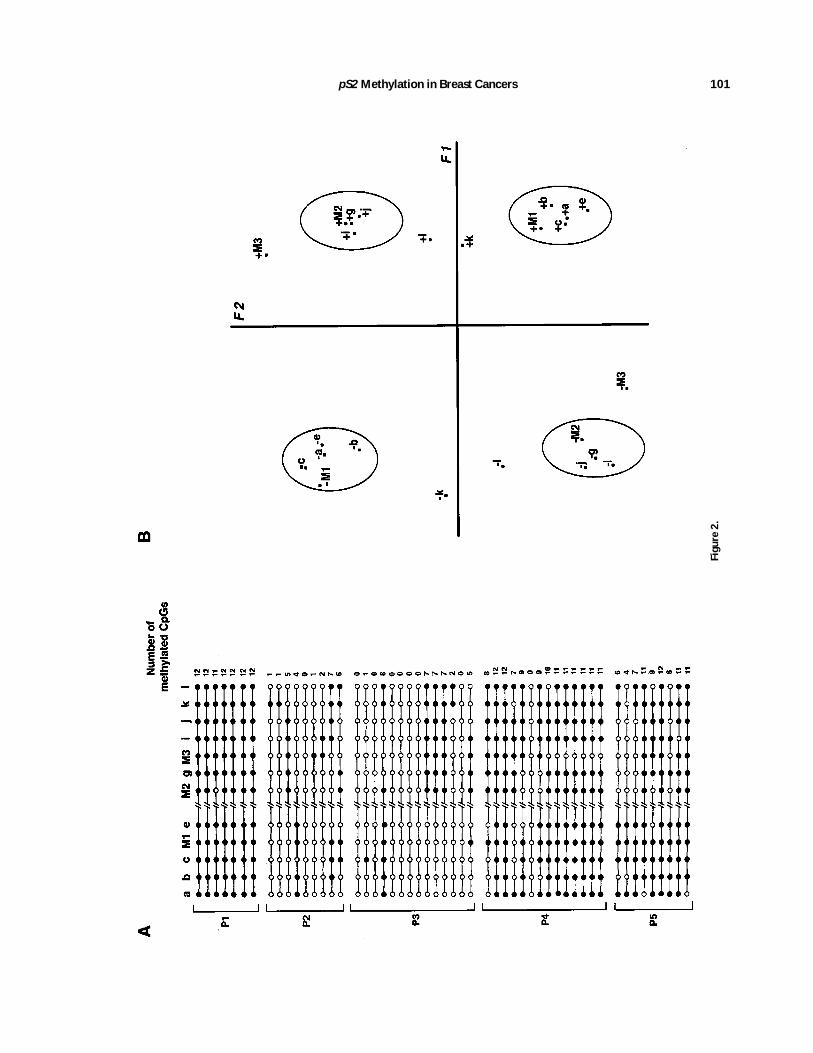
Figure2.
pS2Methylation in Breast Cancers 101
ER-negative/pS2-negative tumors the M1 site(position2354) was largely methylated and theM2 andM3 sites (position284 and220, respec-tively) were hypomethylated. In the great ma-jority of the clones (22/23) obtained from theseDNAsamples (P4 and P5), the cytosines (M1) atposition 2354 were found to be unchanged, andthe 284 (M2) and 220 (M3) sites were deami-nated in 8 out of 23 clones and 12 out of 23clones, respectively, indicating a good correla-tion between the data obtained from genomicsequencing and Southern blot experiments. Thiscorrelation was also observed for ER-rich/pS2-positive samples. The three CCGG sites (M1,M2, and M3) were hypomethylated in thesesamples, and the great majority of the DNAmolecules cloned from these samples did notexhibit methylated cytosines at these sites.However, in both subclasses of breast tumors,the demethylation of M1 sites is not alwaysassociated with the demethylation of M2 andM3 sites. These data, therefore, indicate thatthe demethylation of M2 andM3 sites is not thefirst step leading to the demethylation of the 58
end of pS2 gene.Although mosaic patterns of DNA methyl-
ation were observed in pathological samples,the subclasses of tumors can be distinguishedby their extent of global methylation levels.Clones obtained from ER-rich/pS2-positive tu-mors (P2 and P3) are significantly less methyl-ated than ER-negative/pS2-negative tumors(Student’s t-test, P , 0.001 for both samples),and ER-negative/pS2-negative tumors (P4 andP5 samples) are less methylated than clones(P1) obtained from the normal breast sample(Student’s t-test, P 5 0.058 and P 5 0.013, re-spectively).
Computerized Analysis of the MethylationPatterns of Individual ChromosomesIndicate Specific Associations Between
CpGs in the 58 Region of pS2
The patterns determined by genomic sequenc-ing of the 58 region of pS2 were analyzed withthe aid of the multiple component analysismethod (MCA). MCA is a computational tech-nique suitable for studying the pattern of distri-bution of objects characterized by more thantwo sets of categories [Lebart et al., 1984]. It istherefore a method of choice to process qualita-tive data and to show associations betweenresults. Starting from the data matrices con-
structed from cloned DNA molecules, the posi-tion of the sites, and the methylation status ofeach site, variables are defined by vectors. Thegraphical representation was obtained by theprojection in a plane of these vectors. The re-sults of MCA are presented in the graph (Fig.2B) that represents the positioning of the CpGs,in projection planes, according to the clone andits methylation status. These data have a sym-metrical part in the analysis. In this analysisthe variance vector is broken down into differ-ent components corresponding to fractions ofthe variance. These components are classifiedin decreasing order. In our case, the two axesF1and F2 drawn in this plane represent 69.6%and 14.4% of the variance explained by thesecomponents.Since in this analysis it is legitimate to inter-
pret the relative positions of the points ob-tained by the projection of these vectors, theassociation between the CpGs can be deduced.According to the first component symbolized bythe F1 axis (Fig. 2B), we observe an associationbetween, on the one hand, themethylated CpGsand, on the other hand, an association betweenthe unmethylated CpGs. This analysis indi-cates, as expected, that some clones possess amajority of unmethylated CpGs and that otherclones are essentially methylated.The second component (Fig. 2B, F2 axis) indi-
cates an association between unmethylatedCpGs (from a–e) at the promoter/enhancer se-quences and methylated CpGs (M2, g, i, and j)at the 38 region of this sequence. The reversesituation is also observed, since Figure 2B alsoindicates an association between methylateda–e CpGs and unmethylated M2, g, i, and jCpGs. From this representation it can be con-cluded that, in some cloned DNAmolecules, themethylation status of the CpGs (a–e) located atpromoter/enhancer sequences is not related tothe methylation status of the CpGs (M2, g, i,and j) located downstream of this sequence. Inaddition, some CpGs, M3, k and l, (Fig. 2B), donot seem to be associated with these two groupsof CpGs.These data show an association between, on
the one hand, the CpGs (a–e) located in thepromoter/enhancer region and, on the otherhand, between some CpGs (M2, g, i, and j)located in the 38 region, thereby suggestingthat the demethylation of the promoter/en-hancer of pS2 is not dependent on the surround-ing sequences.
102 Martin et al.
Transient Expression of Human ER Stimulates theExpression of Endogenous pS2 Gene inHeLa Cells But Not in BT-20 Cells
In the course of this study, we observed thatinHeLa cells the 58 region of pS2was hypometh-ylated at the M1, M2, and M3 sites and methyl-ated at the other CCGG sites (data not shown).This cell line was estrogen-receptor-negativeand pS2-negative (Fig. 3A, lane 1), and pS2proteins were undetectable by an immunohisto-chemical method (data not shown).We took thisopportunity to investigate the effect of the estro-gen receptors on the expression of the endog-enous pS2.Expression vector encoding the human estro-
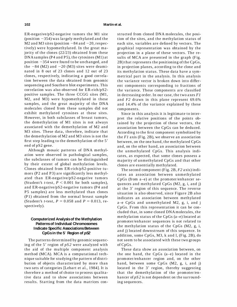
gen receptor (HE0) was transfected into HeLacells together with the pU-LacZ plasmid usedas an internal standard. RT-PCR analysis ofRNAextracted fromHeLa cells transfectedwiththe HE0 vector indicated that the transientexpression of this vector (Fig. 3A, lane 4) in-duced the expression of the endogenous pS2(Fig. 3A, lane 3). In addition, immunohistochemi-cal analysis (Fig. 3B) of the transfected HeLacells showed that pS2 mRNAs were efficientlytranslated, since the percentage (about 15%) ofcells expressingpS2wasequivalent to thepercent-age of cells expressing the b-galactosidase gene.In contrast, expression of the vector encoding
human estrogen receptor failed to induce the
endogenous pS2 in BT-20 cells. Control experi-ments showed that BT-20 cells were able toexpress the chimeric expression vector contain-ing the 58 flanking sequences of pS2 fused tothe CAT gene when cotransfected with the vec-tor encoding the human estrogen receptor (Fig.3C). The absence of expression of the endog-enous pS2 in transiently transfected BT-20 cellswas therefore due neither to the absence ofexpression of ER nor to a nonfunctional cellularmechanism. In addition, in HeLa and in BT-20cell lines, restriction maps of the pS2 gene,obtained from genomic DNAs or from PCR frag-ments, were identical to those obtained fromthe MCF-7 cell line or pS2-pBR322 plasmid,and no mutation was detected in the 58 region(nt2447 to nt166) of pS2 in the BT-20 cell line.Since in HeLa cells the promoter/enhancer
sequences were demethylated and the down-stream region was methylated, whereas inBT-20 cells the promoter/enhancer sequencesand the downstream region were methylated,these data are, therefore, strongly in favor of aninvolvement of the methylation of the 58 regionin the control of pS2 expression.
DISCUSSION
Numerous studies have suggested that alter-ations of DNA methylation may play a varietyof roles in carcinogenesis. However, the data
Fig. 3. Stimulation of the endogenous pS2 gene of HeLa cellsby transient expression of ER. HeLa and BT-20 cells weretransiently transfected with pU-LacZ and HE0 plasmids (HE01)or pS2-CAT plasmid (HE02). pS2 expression was studied byRT-PCR (A) and immunohistochemical methods (B) from thesame transfection experiment (see Materials and Methods). A:Total RNAwas extracted from HE02 (lanes 1,2) and from HE01
(lanes 3,4) HeLa cells. ER (lanes 2,4) and pS2 (lanes 1,3)transcripts were simultaneously amplified with GAPDH tran-
scripts as internal control. The RT-PCR specific products (pS2:208 pb; ER: 438 pb; GAPDH: 308 pb bands), are indicated byarrowheads. B: Identification of pS2-expressing HeLa cells byimmunohistochemical analysis. Shown here are a pS2-positiveHeLa cell, with strong cytoplasmic staining, and two pS2-negative HeLa cells. C: pS2-CAT activity was determined (asdescribed in Materials and Methods) in HeLa cells HE01 (lane1), HeLa cells HE02 (lane 2), BT-20 cells HEO1 (lane 3), andBT-20 cells HE02 (lane 4).
pS2Methylation in Breast Cancers 103
presented here are to our knowledge the first invivo demonstration of an involvement of DNAmethylation changes in the control of a hor-mono-regulated gene in human breast cancers.It had been shown that the gene studied
(pS2) is specifically expressed in subclasses ofbreast cancers (ER-rich tumors) and its detec-tion is a good indicator of the presence of func-tional estrogen receptors. In normal breast tis-sue pS2 expression is not detected [Rio et al.,1987; this report], and all the CCGG sites lo-cated at this locus are methylated.In contrast, analysis of the methylation pat-
terns at CCGG sites located at the 58 end of pS2show some variability between samples in hu-man breast adenocarcinomas. A short regioncontaining the promoter/enhancer sequences[Berry et al., 1989] is hypomethylated in breasttumors expressing pS2, and these sequencesretain a normal level of methylation in ER-richtumors which do not express pS2. In breasttumors the demethylation of the 58 end of pS2seems to be very specific. In ER-negative andpS2-negative tumors, M2 and M3 sites are alsodemethylated, while M1 (located in the pro-moter/enhancer sequences) remains methyl-ated. In addition, the upstream region of thepromoter/enhancer sequence was methylatedin all samples analyzed, cell lines included. Thehypomethylation of M1 sites is associated withpS2 expression; however, an involvement of M2and M3 sites cannot be ruled out. Analysis ofindividual chromosomes is also in favor of aninvolvement of the methylation in the control ofpS2 expression, since the CpGs located in the 58
region of this gene were essentially unmethyl-ated in the cloned DNA molecules obtainedfrom pS2-expressing tumor samples. Further,computer analysis of the data obtained by thegenomic sequencing method showed a correla-tion between themethylation status of the CpGswithin the promoter/enhancer region flankingthe M1 site and pS2 expression, suggestingthat the CpGs located outside theMspI site arealso involved in the control of pS2 expression.The amount of DNA molecules possessing
unmethylatedM1 sites is related to the amountof pS2 mRNA detected in these samples. How-ever, a complete cleavage by HpaII of the 1.06kb band containing the promoter/enhancer se-quences was never observed in the carcinomasamples analyzed. Since we have correlated thedemethylation of this region with pS2 expres-sion, these data suggest that the samples ana-
lyzed contained cells which do not express thisgene. In good agreement with this hypothesisare the observations that in pS2-positive breasttumors the percentage of tumors cells posi-tively stained by antibodies directed againstthis protein varied greatly [Henry et al., 1991;Rio et al., 1987]. Examination of primary breastcancers, using immunohistochemical methods,has shown that the percentage of tumors con-taining more than 50% of pS2-positive tumorcells was very low (4 out of 172 cases analyzed)and that the mean percentage for positivelystaining tumors was 14.9% [Henry et al., 1991].Among the 28 breast carcinoma samples ana-
lyzed, two ER-rich/pS2-negative samples exhib-ited hypomethylated M1 sites. The absence ofpS2 expression in these two samples might bedue to the absence of another factor requiredfor pS2 expression in these cells or to the pres-ence of nonfunctional ER. In this regard, thepresence of nonfunctional ER in human breastcancers has been observed by several authors[Fuqua et al., 1992; Scott et al., 1991]. More-over, it should be noted that transient expres-sion of the human estrogen receptor in HeLacells induces the expression of the unmethyl-ated endogenous pS2.The demethylation of the 58 end of pS2 does
not seem to be dependent on the presence of ERas has been described for the avian vitellogeninII gene. A single injection of estradiol to imma-ture chickens induces the demethylation of theestradiol/glucocorticoid-receptor binding sitesvia an active strand-specific mechanism [Saluzet al., 1986], whereas in human endometriumpS2 is methylated and not expressed despitethe presence of estrogen receptors. The analy-sis, by theMCAmethod, of the data obtained bygenomic sequencing indicated an associationbetween, on the one hand, the CpGs located inthe promoter/enhancer region and, on the otherhand, between some CpGs located downstreamthis region. These data therefore suggest thatthe methylation pattern of pS2 might be regu-lated by two different mechanisms. The mosaicpatterns of methylation does not seem to be atumor-specific phenomenon, since mosaic pat-terns were also observed during developmentin mouse for the imprinted Igf2 and H19 genes[Feil et al., 1994] and the hypoxanthine phos-phoribosyltransferase gene on the inactive Xchromosome [Park and Chapman, 1994]. Itwould be interesting therefore to determinewhether an active demethylation mechanism,
104 Martin et al.
as has been described during in vivo and invitro cellular differentiation [Jost, 1993; Kafriet al., 1993], is also induced in human breasttumors.Finally, all these data are in favor of an
involvement of DNAmethylation in the controlof pS2 expression. Since in these tumors theresponse to the estrogen defines subclasses oftumors, the data described in this report sug-gest that DNAmethylation changes are closelylinked to the neoplasic transformation of thesetissues.
ACKNOWLEDGMENTS
We thank P. Chambon, M. Berry, and E.Scheer, for pS2(429/86) pG1, pS2-pBR322, pS2-CAT, HEO, and HEGO plasmids and P. Cham-bon and D. Metzger for B10 antibodies againstER and advice for EMSA experiments. Thepresent work was supported by the Ligue Na-tional pour la Recherche contre le Cancer, theAssociation pour la Recherche contre le Cancer,and the Institut National de la Sante et de laRecherche Medicale. S. Ribieras and V. Martincontributed equally to this work.
REFERENCES
Adams RLP (1990): DNA methylation: The effect of minorbases on DNA-protein interactions. Biochem J 265:309–320.
Berry M, Nunez JM, Chambon P (1989): Estrogen-respon-sive element of the human pS2 gene is an imperfectlypalindromic sequence. Proc Natl Acad Sci U SA86:1218–1222.
Bird A (1992): The essentials of DNA methylation. Cell70:5–8.
Counts JL, Goodman JI (1995): Alteration in DNAmethyl-ation may play a variety of roles in carcinogenesis. Cell83:13–15.
Dante R, Ribieras S, Baldassini S, Martin V, Benzerara O,Bouteille C, Bremond A, Frappart L, Rio MC, Lasne Y(1994): Expression of an estrogen-induced breast cancerassociated protein (pS2) in benign and malignant humanovarian cysts. Lab Invest 71:188–192.
Doerfler W (1983): DNA methylation and gene activity.Annu Rev Biochem 52:93–124.
Feil R, Wakter J, Allen ND, Reik W (1994): Developmentalcontrol of allelic methylation in the imprintedmouse Igf2andH19 genes. Development 120:2933–2943.
Feinberg AP, Vogelstein B (1983): Hypomethylation distin-guishes genes of some human cancers from their normalcounterparts. Nature 301:89–92.
Frommer M, McDonald LE, Millar DS, Collis CM, Watt F,Grigg GW,Molloy PL, Paul CL (1992):Agenomic sequenc-ing protocol that yields a positive display of 5-methylcyto-sine residues in individual DNA strands. Proc Natl AcadSci U SA89:1827–1831.
Fuqua SAW, Falette NF, McGuire WL (1990): Sensitivedetection of estrogen receptor RNA by polymerase chainreaction assay. J Natl Cancer Inst 82:858–861.
Fuqua SAW, Fitzgerald SD, Allred DC, Elledge MR, NawazZ, McDonell DP, O’Malley BW, Greene GL, McGuire WL(1992): Inhibition of estrogen receptor action by a natu-rally occurring variant in human breast tumors. CancerRes 52:483–486.
Gama-Sosa M, Slagel VA, Trewyn RW, Oxenhandler R, KuoKC, Gehrke CW, Ehrlich M (1983): The 5-methylcytosinecontent of DNA from human tumors. Nucleic Acids Res11:6883–6894.
Henry JA, Piggott NH, Mallick UK, Nicholson S, FarndonJR,Westley BR,May FEB (1991): pNR-2/pS2 immunohis-tochemical staining in breast cancer: Correlation withprognostic factors and endocrine response. Br J Cancer63:615–622.
Jeltsch JM, Roberts M, Schatz C, Garnier JM, Brown A,Chambon P (1987): Structure of the human estrogen-responsive gene pS2. Nucleic Acids Res 15:1404–1414.
Jost JP (1993): Nuclear extracts of chicken embryos pro-mote an active demethylation of DNA by excision repairof 5-methyldeoxycytidine. Proc Natl Acad Sci U S A90:4684–4688.
Kafri T, Gao X, Razin A (1993): Mechanistic aspects ofgenome-wide demethylation in the preimplantationmouse embryo. Proc Natl Acad Sci U S A 90:10558–10562.
Kumar V, Chambon P (1988): The estrogen receptor bindstightly to its responsive element as a ligand-inducedhomodimer. Cell 55:145–156.
Kumar V, Green S, Stack G, Berry M, Jin JR, Chambon P(1987): Functional domains of the human estrogen recep-tor. Cell 51:941–951.
Laird PW, Jaenisch R (1994): DNAmethylation and cancer.HumMol Genet 3:1487–1495.
Lebart L, Morineau A, Warwick KM (1984): ‘‘MultivariateDescriptive Statistical Analysis. Correspondence Analy-sis and Related Techniques for Large Matrices.’’ NewYork: JohnWiley & Sons, Inc.
Lefebvre O, Chenard MP, Masson R, Linares J, Dierich A,LeMeur M, Wendling C, Tomasetto C, Chambon P, RioMC (1996): Gastricmucosa abnormalities and tumorigen-esis in mice lacking the pS2 trefoil protein. Science 274:259–262.
Li E, Bestor TH, Jaenisch R (1992): Targeted mutation ofDNAmethyltransferase gene results in embroyonic lethal-ity. Cell 6:915–926.
Martin V, Ribieras S, Song-Wang X, Rio MC, Dante R(1995): Genomic sequencing indicates a correlation be-tween DNA hypomethylation in the 58 region of the pS2and its expression in human breast cell lines. Gene157:261–264.
Masiakowski P, Breathnach R, Bloch J, Gannon K, KrustA,Chambon P (1982): Cloning of cDNA sequences of hor-mone-regulated genes from MCF-7 human breast cancercell lines. Nucleic Acids Res 19:7895–7903.
Nunez JM, Berry M, Imler JL, Chambon P (1989): The 58
flanking region of the pS2 contains a complex enhancerregion responsive to oestrogens, epidermal growth factor,a tumor promoter (TPA), the c-Ha-ras oncoprotein andthe c-jun protein. EMBO J 8:823–829.
pS2Methylation in Breast Cancers 105
Park JG, Chapman VM (1994): CpG island promoter regionmethylation patterns of the inactive-X-chromosome hypo-xanthine phosphoribosyltransferase (Hprt) gene.Mol CellBiol 14:7975–7983.
Playford RJ, Marchbank T, Goodlad RA, Chinery RA, Poul-som R, Hanby AM, Wright NA (1996): Transgenic micethat overexpress the human trefoil peptide pS2 have anincreased resistance to intestinal damage. Proc NatlAcadSci U SA93:2137–2142.
RazinA, CedarH (1991): DNAmethylation and gene expres-sion. Microbiol Rev 55:451–458.
Ribieras S, Song-Wang X-W, Martin V, Lointier P, FrappartL, Dante R (1994): Human breast and colon cancersexhibit alterations of DNA methylation patterns at sev-eral DNA segments on chromosomes 11p and 17p. J CellBiochem 56:86–96.
Rio MC, Bellocq JP, Gairard B, Rasmussen UB, Krust A,Koehl C, Calderoli H, Schiff V, Renaud R, Chambon P(1987): Specific expression of the pS2 in subclasses ofbreast cancers in comparisonwith expression of the estro-gen and progesterone receptors and the oncogene ERBB2.Proc Natl Acad Sci U SA84:9243–9247.
Rio MC, Bellocq JP, Daniel JY, Thomasetto C, Lathe R,Chenard MP, Batzenschlager A, Chambon P (1988):Breast cancer–associated pS2 protein: Synthesis and se-cretion by normal stomachmucosa. Science 241:705–707.
Saluz HP, Jiricny J, Jost JP (1986): Genomic sequencingreveals a positive correlation between the kinetics ofstrand-specific DNA demethylation of the overlappingestradiol/glucocorticoid-receptor binding site and the rateof avian vitellogeninmRNAsynthesis. Proc NatlAcad SciU SA83:7167–7171.
Sambrook J, Fritsch EF, Maniatis T (1989): ‘‘MolecularCloning: A Laboratory Manual,’’ 2nd ed. Cold SpringHarbor, NY: Cold Spring Harbor Laboratory.
Schwartz LH, Koerner FC, Edgerton SM, Sawicka JM, RioMC, Bellocq JP, Chambon P, ThorAD (1991): pS2 expres-sion and response to hormonal therapy in patients withadvanced breast cancer. Cancer Res 51:624–628.
Scott GK, Kushner P, Vigne JL, Benz CC (1991): Truncatedforms of DNA-binding estrogen receptors in human breastcancer. J Clin Invest 88:700–706.
Thioulouse J, Doledec S, Chessel D, Olivier JM (1994):ADEsoftware: Multivariate analysis and graphical display ofenvironmental data. In Guariso G, Rizzoli A (eds):‘‘Software per l’ambiente.’’ Bologna: Patron editore, pp57–62.
Tora L, Mullick A, Metzger D, Ponglitikmongkol M, Park I,Chambon P (1989): The cloned human oestrogen receptorcontains a mutation which alters its hormone bindingproperties. EMBO J 8:1981–1986.
Welter C, Theisinger B, Seitz G, Tomasetto C, Rio MC,Chambon P, Blin N (1992): Association of the humanspasmolytic polypeptide and an estrogen-induced breastcancer protein (pS2) with human pancreatic carcinoma.Lab Invest 66:187–192.
Wysoki SJ, Hahnel E, Masters A, Smith V, McCartney AJ,Hahnel R (1990): Detection of pS2 messenger RNA ingynecological cancers. Cancer Res 50:1800–1802.
Zajchowski DA, Sager R (1991): Induction of estrogen-regulated genes differs in immortal and tumorigenic hu-man mammary epithelial cells expressing a recombinantestrogen receptor. Mol Endocrinol 5:1613–1623.
106 Martin et al.