INVITED PAPER Catheter-BasedSystems ...rogersgroup.northwestern.edu/files/2015/Leeetal_2015.pdf ·...
8
INVITED PAPER Catheter-Based Systems With Integrated Stretchable Sensors and Conductors in Cardiac Electrophysiology We present novel mechanics, materials and integration strategies for this new class of bioelectronics onboard minimally invasive catheter based systems. Representative examples highlight the clinical significance soft biointegrated electronics. By Stephen P. Lee , Lauren E. Klinker , Leon Ptaszek , John Work , Cliff Liu , Fernando Quivara , Chad Webb , Canan Dagdeviren , John A. Wright , Jeremy N. Ruskin , Marvin Slepian , Yonggang Huang , Moussa Mansour , John A. Rogers, and Roozbeh Ghaffari ABSTRACT | Established classes of high-performance electro- nics have driven advances in interventional biomedicine. How- ever, the large size, planar geometry and stiff mechanical properties of standard conventional electronics employed in medical devices give rise to important integration challenges with soft biological tissue. Stretchable and flexible biointe- grated electronics could improve treatment procedures across a broad range of applications, including cardiac, neural and endovascular therapies. Here we present novel mechanics, materials and integration strategies for this new class of bio- electronics onboard minimally invasive catheter based systems. Co-located arrays of sensors and actuators affixed to cardiac and angioplasty balloon catheters capture new sensory infor- mation during ablation procedures, offering physicians the abi- lity to adjust placement and treatment intra-procedurally. New circuit topologies, enabled by stretchable electronics, also overcome long standing challenges associated with transmit- ting vast amounts of data through narrow catheter lumens, thus allowing for a large number of sensors to be multiplexed for mapping electrophysiological activity with high spatiotemporal resolution and with a minimal number of routing wires. We present representative examples that highlight the clinical sig- nificance of soft bio-integrated electronics, along with the mechanics and processes that enable this technology. KEYWORDS | Flexible electronics; biosensors; semiconductors; microfabrication; electrophysiology; vasculature; biomedical devices Bio-integrated electronics that interface with cellular and sub-cellular constituents of soft living tissues can fun- damentally change the way medical devices diagnose and deliver therapy [1]–[5]. Existing cardiac assist and deep- brain stimulation devices, which contain packaged electro- nic parts, have already improved the quality of life for patients with abnormal heart rhythm disorders, treatment- resistant depression, and Parkinson’s disease, but all of these existing solutions contain arrays of passive metal electrodes and wires that target large clusters of cells and nerve fibers indiscriminately with limited spatial pre- cision. These recording and stimulation electrodes are Manuscript received September 10, 2014; revised January 5, 2015; accepted January 26, 2015. Date of current version May 19, 2015. S. P. Lee, L. E. Klinker, J. Work, C. Liu, F. Quivara, J. A. Wright, and R. Ghaffari are with MC10 Inc., Cambridge, MA 02140 USA (e-mail: [email protected]). L. Ptaszek, J. N. Ruskin, and M. Mansour are with the Cardiac Arrhythmia Unit, Massachusetts General Hospital, Boston, MA 02140 USA. C. Webb, C. Dagdeviren, and J. A. Rogers are with the Department of Materials Science and Engineering, University of Illinois at Urbana-Champaign, Urbana, IL 61801 USA. M. Slepian is with the Departments of Medicine and Biomedical Engineering, Sarver Heart Center, University of Arizona, Tucson, AZ 85724 USA. Y. Huang is with the Department of Civil and Environmental Engineering, Northwestern University, Evanston, IL 60208 USA. Digital Object Identifier: 10.1109/JPROC.2015.2401596 0018-9219 Ó 2015 IEEE. Personal use is permitted, but republication/redistribution requires IEEE permission. See http://www.ieee.org/publications_standards/publications/rights/index.html for more information. 682 Proceedings of the IEEE | Vol. 103, No. 4, April 2015
Transcript of INVITED PAPER Catheter-BasedSystems ...rogersgroup.northwestern.edu/files/2015/Leeetal_2015.pdf ·...

INV ITEDP A P E R
Catheter-Based SystemsWith Integrated StretchableSensors and Conductors inCardiac ElectrophysiologyWe present novel mechanics, materials and integration strategies for this new class of
bioelectronics onboard minimally invasive catheter based systems. Representative
examples highlight the clinical significance soft biointegrated electronics.
By Stephen P. Lee, Lauren E. Klinker, Leon Ptaszek, John Work, Cliff Liu,
Fernando Quivara, Chad Webb, Canan Dagdeviren, John A. Wright,
Jeremy N. Ruskin, Marvin Slepian, Yonggang Huang, Moussa Mansour,
John A. Rogers, and Roozbeh Ghaffari
ABSTRACT | Established classes of high-performance electro-
nics have driven advances in interventional biomedicine. How-
ever, the large size, planar geometry and stiff mechanical
properties of standard conventional electronics employed in
medical devices give rise to important integration challenges
with soft biological tissue. Stretchable and flexible biointe-
grated electronics could improve treatment procedures across
a broad range of applications, including cardiac, neural and
endovascular therapies. Here we present novel mechanics,
materials and integration strategies for this new class of bio-
electronics onboardminimally invasive catheter based systems.
Co-located arrays of sensors and actuators affixed to cardiac
and angioplasty balloon catheters capture new sensory infor-
mation during ablation procedures, offering physicians the abi-
lity to adjust placement and treatment intra-procedurally. New
circuit topologies, enabled by stretchable electronics, also
overcome long standing challenges associated with transmit-
ting vast amounts of data through narrow catheter lumens, thus
allowing for a large number of sensors to be multiplexed for
mapping electrophysiological activity with high spatiotemporal
resolution and with a minimal number of routing wires. We
present representative examples that highlight the clinical sig-
nificance of soft bio-integrated electronics, along with the
mechanics and processes that enable this technology.
KEYWORDS | Flexible electronics; biosensors; semiconductors;
microfabrication; electrophysiology; vasculature; biomedical
devices
Bio-integrated electronics that interface with cellularand sub-cellular constituents of soft living tissues can fun-
damentally change the way medical devices diagnose and
deliver therapy [1]–[5]. Existing cardiac assist and deep-
brain stimulation devices, which contain packaged electro-
nic parts, have already improved the quality of life for
patients with abnormal heart rhythm disorders, treatment-
resistant depression, and Parkinson’s disease, but all of
these existing solutions contain arrays of passive metalelectrodes and wires that target large clusters of cells and
nerve fibers indiscriminately with limited spatial pre-
cision. These recording and stimulation electrodes are
Manuscript received September 10, 2014; revised January 5, 2015; accepted
January 26, 2015. Date of current version May 19, 2015.
S. P. Lee, L. E. Klinker, J. Work, C. Liu, F. Quivara, J. A. Wright, and R. Ghaffari arewith MC10 Inc., Cambridge, MA 02140 USA (e-mail: [email protected]).
L. Ptaszek, J. N. Ruskin, and M. Mansour are with the Cardiac Arrhythmia Unit,
Massachusetts General Hospital, Boston, MA 02140 USA.
C. Webb, C. Dagdeviren, and J. A. Rogers are with the Department of Materials
Science and Engineering, University of Illinois at Urbana-Champaign, Urbana,
IL 61801 USA.
M. Slepian is with the Departments of Medicine and Biomedical Engineering,
Sarver Heart Center, University of Arizona, Tucson, AZ 85724 USA.
Y. Huang is with the Department of Civil and Environmental Engineering,
Northwestern University, Evanston, IL 60208 USA.
Digital Object Identifier: 10.1109/JPROC.2015.2401596
0018-9219 � 2015 IEEE. Personal use is permitted, but republication/redistribution requires IEEE permission.See http://www.ieee.org/publications_standards/publications/rights/index.html for more information.
682 Proceedings of the IEEE | Vol. 103, No. 4, April 2015

prevalent in many existing catheter-based solutions andrepresent the gold standard of care in interventional medi-
cine [2]–[4]. However, these devices are largely restricted
to interactions with biological structures at scales that are
orders of magnitude larger and stiffer than individual
living cells. A powerful alternative would be to dramati-
cally reduce the size and mechanical hardness of devices
and sensors that directly interface with soft tissue and or-
gans [1], [3]. Soft, stretchable integrated circuits, sensorsand actuators that achieve intimate contact with target
cells at a high level of specificity constitute a viable solu-
tion for emerging medical systems.
This new class of electronics has significant implica-
tions for growing populations of patients stricken with
hypertension, heart disease [6], and neurological disorders.
Global forecasts predict a rapid rise in the number of pa-
tients with strokes, complex arrhythmias, as well as neuro-logical disorders, like epilepsy and Alzheimer’s [7], [8].
Highly localized methods of detection and therapy (e.g.,
electromodulation of neural signaling and ablation) are
becoming attractive treatment strategies in these patient
populations. The ability to instrument mechanical devices
(i.e., catheters) with arrayed forms of sensors and actuators
that exploit flexible and stretchable circuits offers oppor-
tunities to improve efficacy and to reduce procedure timeand cost.
In this report, we highlight recent developments in soft,
stretchable bio-integrated medical systems and demon-
strate catheter-based systems integrated with sensors that
exploit unusual mechanics and designs using existing man-
ufacturing processes. We provide recent examples of
catheter-based systems that have been applied across sev-
eral disciplines including cardiac, neural, and endovascularprocedures. By leveraging heterogeneous collections of
sensors and actuators, these catheter-based systems high-
light the opportunity to address highly localized target sites
to detect and/or modulate the body’s natural signaling
circuits.
I . MATERIALS, MECHANICS, ANDMICROFABRICATION PROCESSES
Development of ultrathin, stretchable arrays of sensors and
actuators for catheter-based systems begins with conven-
tional microfabrication and photolithography techniques,
as described previously [1], [3]. Ultrathin metal layers and
single-crystal inorganic nanomaterials released from donor
wafers are integrated with polymeric and elastomeric bal-
loon substrates using interfacial adhesives, plasma treat-ment, and transfer printing strategies [1]–[3]. Transfer
printing onto a three-dimensional balloon begins with a
soft, rubber-like stamp material called (poly)dimethylsilox-
ane (PDMS) that retrieves biosensor arrays and nanoma-
terials via van der Waals forces. This transfer process can be
performed over large areas of segmented arrays of sensors
using a flat stamp or over selected areas using a structured
stamp. In the next step, the picked biosensors and circuitry
are selectively placed on the complex three-dimensional
geometry of a balloon catheter while it is in its inflated
state. Thin glue layers, such as polyimide or epoxies, andplasma treatment promote high adhesion strength and
ruggedness, enabling multiple inflation and deflation cy-
cles during use. Once arrays of sensors are affixed to the
balloon surface, anisotropic conductive films are used to
connect sensor leads to copper wires, which electrically
route signals to an external data acquisition console.
These ultrathin designs achieve flexibility over very
small bending radii (G 1 mm). However, extreme bendingand stretching conditions could induce significantly greater
strains or fractures, particularly in instances where these
electronics must interface with soft tissues of the heart. For
example, electronics inside the chambers of the heart may
undergo large strains up to 20%–30%. Sensors and elec-
trodes on balloon catheters for minimally invasive proce-
dures thus require even higher stretchability [1]. To
alleviate the strains induced in these use-cases, serpentinelayouts of polymers, metals and nanomaterials are required
[1]–[3].
II . INSTRUMENTED CARDIACABLATION CATHETERS
Atrial fibrillation (AF) affects more than five million pa-
tients in the developed world and is the leading cause of
stroke in cardiac patients [10], [11]. With up to 400 000
Fig. 1. Multielectrode and balloon-based catheters used for ablating
and mapping electrical activity of cardiac tissue [3]. (a) Multielectrode
basket catheter (ConstellationTM, Boston Scientific; Boston, MA, USA)
provides high-density mapping of electrical activity within the atria.
(b) Multielectrode mapping and ablation catheter (MAACTM,
Medtronic; Minneapolis, MN, USA). (c) HeartLight laser-based
ablation balloon catheter (Cardiofocus Inc.; Marlborough, MA, USA).
(d) Cryoballoon ablation catheter (Medtronic; Minneapolis, MN, USA).
Lee et al. : Catheter-Based Systems With Integrated Stretchable Sensors and Conductors in Cardiac Electrophysiology
Vol. 103, No. 4, April 2015 | Proceedings of the IEEE 683

new cases diagnosed yearly [7], the adverse outcomes of AFrange from congestive heart failures to sudden death.
Pulmonary vein (PV) isolation with radio-frequency (RF)
energy ablation catheters has become a primary interven-
tion for complex arrhythmias [12], [13] and represents a
procedure that could benefit from localized sensing and
therapy. RF ablation involves delivery of energy in a point-
by-point manual fashion to specific locations within left
atrium, such as where the PVs intersect the atrial wall.Positioning mapping and/or ablation catheters containing
point electrodes [Fig. 1(a) and (b)] requires significant time
and dexterity, often leading to inconsistent outcomes
across multiple patients [13] and increasing the risk of
stroke and other clinical complications.
Emerging balloon catheter-based systems under-
development and in clinical trials rely [Fig. 1(c) and (d)]
solely on balloon substrates to conform to the anatomicalstructure of the heart [14]–[16]. These balloons inflate
within the left atrium and create continuous rings of con-
formal contact between the balloon and the tissue followed
by delivery of cryo- or laser-based ablation therapies. RF
and cryoablation energy destroy the targeted cells, thereby
correcting the aberrant electrical circuits that underliearrhythmias. Although conceptually straightforward, such
balloon ablation catheters fail to provide information on
the electrical state of intracardiac surfaces or sensory feed-
back about mechanical contact or temperature profile of
the target tissue [1]–[3]. The ‘‘instrumented’’ balloon
catheters with integrated conductors and active sensors
that allow multisensory feedback, pacing, and ablation
functionality could thus offer a significant advance overexisting balloon-based and multielectrode catheter devices.
Cardiac ablation serves as a compelling case study to
evaluate the benefits of soft biointegrated systems. In par-
ticular, the co-location of sensors with ablation electrodes
provides real-time intra-procedural feedback on the effi-
cacy of ablations without the use of additional catheters.
Flexible and stretchable sensor and actuator subcompo-
nents must be mechanically robust to match the dynamicalinflation and deflation cycles of balloon catheters. Ultra-
thin electronics, fabricated using flex processes and un-
packaged embedded die processes, can be made to stretch
as a network by using unique spring-like geometrical lay-
outs [Fig. 2(a)–(c)]. Thin film deposition, etching, and
Fig. 2. Multilayered stretchable interconnects. (a) Illustration of interconnect structures with meandering metal interconnects used to
accommodate strain in highly stretchable electronics (26). (b) Image strain relief structure encapsulated in elastomer. (c) Image of interconnect
structure experiencing �50% strain. Model simulation showing plastic strain distribution in response to 50% strain (26). (d) Representative
balloon catheter that employs a network of electrodes connected mechanically and electrically via spring-like meandering interconnects. The PI
and sensor layers in the stack interface with biological tissue, whereas the PU/PET layer constitutes the inner layer of balloon material.
Lee et al.: Catheter-Based Systems With Integrated Stretchable Sensors and Conductors in Cardiac Electrophysiology
684 Proceedings of the IEEE | Vol. 103, No. 4, April 2015
photolithographic patterning strategies are used to lay
stacks of polyimide, adhesives, silicon-based semiconduc-
tors, and metal layers, which allows for significant reduc-
tion in bending stiffness and enhanced flexibility due to
minimized stack thickness. The mechanical layouts of these
systems are driven by theoretical analysis with parameterstuned for reversible elastic response to large-strain
deformation [Fig. 2(c)] [17]. Strain concentration is limited
in the neutral mechanical plane, where sensors and actua-
tors are located, and constrained to the interconnected
regions alleviating stress from the sensitive elements and
providing further mechanical support. These mechanical
properties are well suited for three-dimensional balloon
structures, which undergo considerable stretching andbending during inflation/deflation cycles [Fig. 2(d)].
Kim et al. demonstrated stretchable impedance-based
contact sensors [Fig. 3(a)–(c)], temperature and pressure
sensors on balloon catheters to assess adequate contact
with the tissue [18]. The temperature sensors consist of
thin resistive films of titanium-platinum (5 nm/50 nm
thick). Here, the thin layers of Ti/Pt are deposited by elec-
tron beam evaporation and patterned by photolithographyand then lift-off creates intricate micro-patterns of sensors.
The Ti/Pt layer is strategically deposited on a flexible but
nonstretchable island of polyimide, which helps reduce the
effects of strain on the temperature measurement during
inflation and deflation [18]. Surface treatment of the PI
with UV/Ozone or deposition of a thin silicon dioxide
ðSiO2Þ layer (�50 nm) on top of PI improves the adhesion
of the Ti/Pt. These temperature sensors are located near the
ablation electrodes decorated on the balloon surface to
provide an accurate assessment of tissue temperatures dur-
ing ablations. Calibration measurements conducted under
extremely low (e.g., cryoablation) and elevated (e.g., RF
energy delivery) temperature conditions yielded a mono-
tonic relation between impedance and measured temper-ature, with a slope of �1:91 �=�C [18]. In the case of
irrigated RF ablation catheters and cryoballoons, this
sensitivity is sufficient to provide precise feedback on
thermal properties at the tissue-balloon interface.
Similarly, impedance-based contact sensors are fabricat-
ed on polyimide islands tied together by serpentine
interconnects. Lithographic processing and vertical trench
dry-etching techniques yielded isolated contact sensors(�1 mm2, and �12 �m total thickness of stack) that re-
main tethered to the underlying silicon or glass wafer
through ‘‘anchor’’ structures. This process can be used to
yield electrodes for mechanical contact sensing and electro-
gram measurements. Each contact sensor consists of a bipo-
lar pair of electrodes that measure a change in impedance
generated by an excitation current. For the experiment, the
excitation current was set to 10 uA and measurements weretaken at �1 kHz. An array of bipolar electrodes thus can be
used to evaluate contact according to changes in impedance,
which can increase by up to a factor of �1.5–2.
The individual sensors reside equatorially around the
balloon and are used to detect mechanical contact with
cardiac tissue to aid in catheter positioning and to confirm
contact during ablation events. These impedance-based
sensors can also be used to assess the contact pressure at
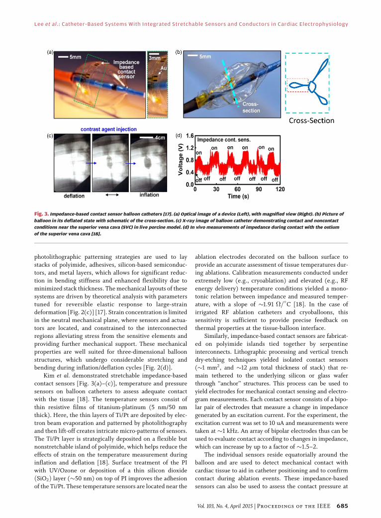
Fig. 3. Impedance-based contact sensor balloon catheters [17]. (a) Optical image of a device (Left), with magnified view (Right). (b) Picture of
balloon in its deflated state with schematic of the cross-section. (c) X-ray image of balloon catheter demonstrating contact and noncontact
conditions near the superior vena cava (SVC) in live porcine model. (d) In vivo measurements of impedance during contact with the ostium
of the superior vena cava [18].
Lee et al. : Catheter-Based Systems With Integrated Stretchable Sensors and Conductors in Cardiac Electrophysiology
Vol. 103, No. 4, April 2015 | Proceedings of the IEEE 685
the balloon-tissue interface. An array of contact sensors
provides feedback to physicians with significantly higher
accuracy than existing x-ray based imaging techniques[Fig. 3(d) and (e)].
III . HIGH SPATIAL RESOLUTIONMAPPING CATHETERS WITH FLEXIBLE/STRETCHABLE ACTIVE ELECTRODES
In more advanced arrhythmia cases, such as persistent AF,
the mechanisms underlying depolarization and hyperpola-
rization wave fronts remain poorly understood, and abla-tion targets are not well defined [19], [20]. As a result, this
form of AF is significantly more challenging to treat com-
pared to paroxysmal AF. Current ablation targets in per-
sistent AF include areas exhibiting complex fractionated
atrial electrograms (CFAEs). CFAEs are electrical record-
ings with a highly disorganized appearance, which may
represent rapid electrical activity from a nearby driving
force (rotor) [21], [22]. While these areas exhibit CFAEappearance, they do not play a major role in maintaining
AF. However, due to the lack of high density mapping
capabilities in the clinical setting, the distinction between
drivers of AF and passive CFAEs is not currently possible,
leading to erroneous ablation in a large number of patients.
Although basket catheters have a greater number of
electrodes compared to point ablation catheters, they
still lack the resolution necessary to map persistent
AF. The spatial density of electrodes is on the order of
0.1 electrode/cm2, whereas the action potential wave frontsdisperse at much finer resolution. By applying micro-
fabrication and photolithographic techniques, instru-
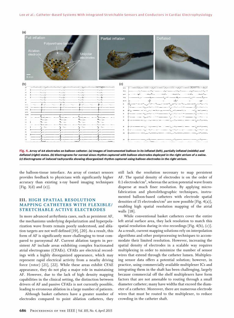
mented balloon-based catheters with electrode spatial
densities of 15 electrodes/cm2 are now possible [Fig. 4(a)],
enabling high spatial resolution mapping of the atrial
walls [18].
While conventional basket catheters cover the entire
left atrial surface area, they lack resolution to match thisspatial resolution during in vivo recordings [Fig. 4(b), (c)].
As a result, current mapping solutions rely on interpolation
algorithms and other postprocessing techniques to accom-
modate their limited resolution. However, increasing the
spatial density of electrodes in a scalable way requires
multiplexing in order to minimize the number of sensor
wires that extend through the catheter lumen. Multiplex-
ing sensor data offers a potential solution; however, inpractice, using commercially available multiplexer ICs and
integrating them in the shaft has been challenging, largely
because commercial off the shelf multiplexers have form
factors that are not amenable to routing through a small
diameter catheter; many have widths that exceed the diam-
eter of a catheter. Moreover, there are numerous electrode
wires that must be routed to the multiplexer, to reduce
crowding in the catheter shaft.
Fig. 4. Array of 64 electrodes on balloon catheter. (a) Images of instrumented balloon in its inflated (left), partially inflated (middle) and
deflated (right) states. (b) Electrograms for normal sinus rhythm captured with balloon electrodes deployed in the right atrium of a swine.
(c) Electrograms of induced tachycardia showing disorganized rhythm captured using balloon electrodes in the right atrium.
Lee et al.: Catheter-Based Systems With Integrated Stretchable Sensors and Conductors in Cardiac Electrophysiology
686 Proceedings of the IEEE | Vol. 103, No. 4, April 2015

The ability to integrate silicon-based electronics on ex-
tremely elastic substrates introduces ways to integrate
sensing, actuation, amplification, logic, and switching ca-
pabilities onboard cardiac balloon catheters. The underly-ing sensing technology has been previously developed
onboard catheters that employ these thin, conformal arrays
of sensory electronics embedded in deformable surfaces
(i.e., silicone or polyurethane balloon skins) [1], [5]. A
compelling solution is to develop distributed multiplexing
as shown in the circuit diagram in Fig. 5. Source followers,M1, buffers the signal, a CMOS switch, M2, opens or closes
each active electrode’s output to effectively multiplex its
Fig. 5. Distributed multiplexing. (a) An example of row/column selection using source-followers in a distributed multiplexing arrangement.
(b) Buffered, multiplexed electrodes on a polyimide sheet.
Fig. 6. Fabricated devices using a commercial SOI process. (a) The ‘‘donor’’ wafer undergoes a modified commercial fabrication process.
(b) and (c) After under-etch, devices are picked and transferred to an ‘‘acceptor’’ wafer. (d) Magnified image of transferred chiplets on
acceptor wafer. Interconnect traces are deposited following this CMOS processing stage.
Lee et al. : Catheter-Based Systems With Integrated Stretchable Sensors and Conductors in Cardiac Electrophysiology
Vol. 103, No. 4, April 2015 | Proceedings of the IEEE 687

respective amplifier/electrode creating a distributed multi-plexer on the surface of a sheet or balloon catheter
[Fig. 5(a)]. In this example, each electrode is addressed via
row/column select. Sixty-four electrodes thus contain 16
row column select lines yielding a savings in input/output
[Fig. 5(b)].
Fabrication of distributed multiplexers is an enhance-
ment of previous work by Kim et al. [23], achieved by the
following process steps: the amplifier multiplexer circuit isfabricated using a silicon-on-oxide foundry CMOS process
[Fig. 6(a)]. Each electrode amplifier/multiplexer pair is
fabricated within a trench and anchored to the trench walls
by tethers. The trench is vertically, wet etched away, leav-
ing the amplifier/multiplexer floating above the trench,
held in place by the anchors. An elastomeric stamp head
affixed to a modified pick and place machine [Fig. 6(b)]
performs the microtransfer printing by peeling theamplifier/multiplexer (5 �m thick) off the silicon ‘‘donor’’
wafer and transferring it to a polyimide substrate (1 �m
thick) on an ‘‘acceptor’’ wafer. Deposited metal traces con-
nect the array of amplifier/multiplier ‘‘chiplets’’ after
micro-transfer [Fig. 6(c) and (d)]. Serpentines and active
devices are then encapsulated with a polymer layer (e.g.,
polyurethane). The overall structure can accommodateradii of curvature of less than 1 mm based on analysis and
verified through experiment. The polyimide substrate con-
taining the array can be affixed to a balloon catheter or can
be used in sheet form [1], [5], [17], [18].
IV. CONCLUSION
Soft, stretchable biointegrated electronics have direct im-plications on the development of emerging balloon-based
catheter systems in cardiac electrophysiology. Integration
of sensors, actuators, amplification circuitry and multiplex-
ing could significantly reduce the size of catheter lumens to
reach additional biological locations while dramatically
enhancing sensing and signal to noise quality. In addition to
instrumented endovascular catheters, soft bioelectronics
can be applied to other substrates, including biofilms (e.g.,silk fibroin) that can conform to unique tissue geometries,
such as the sulci and gyri of the brain, and sutures in-
strumented with onboard flexible sensor electronics [24].
These examples highlight the vast set of applications of
stretchable, bio-integrated electronics to enhance cardiac,
neural, and endovascular procedures. h
REF ERENCE S
[1] D. H. Kim et al., ‘‘Multifunctional stretchabledevices on compliant balloon catheters forin-vivo electrophysiological mapping andablation therapy,’’ Nature Mater., vol. 10,no. 4, pp. 316–323, 2011, PMID: 21378969.
[2] M. Slepian, R. Ghaffari, and J. A. Rogers,‘‘Multifunctional balloon catheters of thefuture,’’ Intervent. Cardiol., vol. 3, no. 4,pp. 417–419, 2011.
[3] D. H. Kim, R. Ghaffari, N. Lu, andJ. A. Rogers, ‘‘Flexible and stretchableelectronics for biointegrated devices,’’ Annu.Rev. Biomed. Eng., vol. 14, pp. 113–1128, 2012.
[4] C. Famm, B. Litt, K. J. Tracey, E. Boyden, andM. Slaoui, ‘‘Drug discovery: A jumpstartfor electroceuticals,’’ Nature, vol. 496,pp. 159–161, 2013.
[5] J. Viventi et al., ‘‘A conformal, bio-interfacedclass of silicon electronics for mapping cardiacelectrophysiology,’’ Sci. Translat. Med., vol. 2,no. 24, 2010, doi:10.1126/scitranslmed.300073824ra22.
[6] World Health Organization Statistics, 2012.
[7] A. S. Go et al., (2001). Prevalence of diagnosedatrial fibrillation in adults: Nationalimplications for rhythm management andstroke prevention: The AnTicoagulation andRisk Factors in Atrial Fibrillation (ATRIA)Study. J. Amer. Med. Assoc. [Online]. 285(18),pp. 2370–2375. Available: http://www.ncbi.nlm.nih.gov/pubmed/11343485
[8] R. Brookmeyer, E. Johnson,K. Zeigler-Graham, and H. M. Arrighi,‘‘Forecasting the global burden ofAlzheimer’s disease,’’ Alzheimer’sDementia, vol. 3, pp. 186–191, 2007.
[9] H. M. De Boer, M. Mula, and J. W. Sander,‘‘The global burden and stigma of epilepsy,’’Epilepsy Behav., vol. 12, pp. 540–546,2008.
[10] M. D. Ezekowitz and J. A. Levine, ‘‘Preventingstroke in patients with atrial fibrillation,’’J. Amer. Med. Assoc., vol. 281, no. 19,pp. 1830–1835, 1999.
[11] V. Fuster et al., ‘‘ACC/AHA/ESC 2006Guidelines for the Management of Patientswith Atrial Fibrillation: A report of theAmerican College of Cardiology/AmericanHeart Association Task Force on PracticeGuidelines and the European Societyof Cardiology Committee for Practice,’’Circulation, vol. 114, no. 7, pp. e257–e354,2006, doi:10.1161/CIRCULATIONAHA.106.177292.
[12] M. HaBssaguerre et al., ‘‘Catheter ablationof long-lasting persistent atrial fibrillation:Clinical outcome and mechanisms ofsubsequent arrhythmias,’’ J. Cardiovasc.Electrophysiol., vol. 16, no. 11, pp. 1138–1147,2005, doi:10.1111/j.1540-8167.2005.00308.x.
[13] H. Calkins et al., ‘‘HRS/EHRA/ECASexpert consensus statement on catheterand surgical ablation of atrial fibrillation:Recommendations for personnel, policy,procedures and follow-up. A report of theHeart Rhythm Society (HRS) Task Forceon Catheter and Surgical Ablation of,’’Europace: Eur. Pacing, Arrhythmias, CardiacElectrophysiol.: J. Working Groups onCardiac Pacing, Arrhythmias, CardiacCellular Electrophysiol. Eur. Soc. Cardiol.,vol. 9, no. 6, pp. 335–379, 2007, doi:10.1093/europace/eum120.
[14] T. Neumann et al., ‘‘Circumferentialpulmonary vein isolation with the cryoballoontechnique results from a prospective 3-centerstudy,’’ J. Amer. College Cardiol., vol. 52, no. 4,pp. 273–278, 2008, doi:10.1016/j.jacc.2008.04.021.
[15] K. P. Phillips et al., ‘‘Anatomic location ofpulmonary vein electrical disconnectionwith balloon-based catheter ablation,’’ J.Cardiovasc. Electrophysiol., vol. 19, no. 1,pp. 14–18, 2008, doi:10.1111/j.1540-8167.2007.00964.x.
[16] W. Moreira et al., ‘‘Long-term follow-up aftercryothermic ostial pulmonary vein isolationin paroxysmal atrial fibrillation,’’ J. Amer.College Cardiol., vol. 51, no. 8, pp. 850–855,2008, doi:10.1016/j.jacc.2007.08.065.
[17] Y.-Y. Hsu et al., ‘‘A novel strain reliefdesign for multilayer thin film stretchableinterconnects,’’ IEEE Trans. Electron Devices,vol. 60, no. 7, pp. 2338–2345, Jul. 2013.
[18] D. H. Kim et al., ‘‘Electronic sensor andactuator webs for large-area complexgeometry cardiac mapping and therapy,’’Proc. Nat. Acad. Sci. USA, vol. 109,pp. 19 910–19 915, 2012.
[19] J. Jalife, ‘‘Mother rotors and fibrillatoryconduction: A mechanism of atrialfibrillation,’’ Cardiovasc. Res., vol. 54, no. 2,pp. 204–216, 2002, doi:10.1016/S0008-6363(02)00223-7.
[20] D. Katritsis, E. Giazitzoglou, D. Sougiannis,E. Voridis, and S. S. Po, ‘‘Complexfractionated atrial electrograms at anatomicsites of ganglionated plexi in atrialfibrillation,’’ Europace: Eur. Pacing,Arrhythmias, Cardiac Electrophysiol.: J.Working Groups Cardiac Pacing, Arrhythmias,Cardiac Cellular Electrophysiol. Eur. Soc.Cardiol., vol. 11, no. 3, pp. 308–315, 2009,doi:10.1093/europace/eup036.
[21] S. M. Narayan et al., ‘‘Treatment of atrialfibrillation by the ablation of localizedsources,’’ J. Amer. College Cardiol., vol. 60,no. 7, pp. 628–636, 2012, doi:10.1016/j.jacc.2012.05.022.
[22] S. M. Narayan, J. Patel, S. Mulpuru, andD. E. Krummen, ‘‘Focal impulse and rotormodulation ablation of sustaining rotorsabruptly terminates persistent atrialfibrillation to sinus rhythm with eliminationon follow-up: A video case study,’’ HeartRhythm: Official J. Heart Rhythm Soc.,vol. 9, no. 9, pp. 1436–1439, 2012,doi:10.1016/j.hrthm.2012.03.055.
[23] D. H. Kim et al., ‘‘Optimized structuraldesigns for stretchable silicon integratedcircuits,’’ Small, vol. 5, no. 24, pp. 2841–2847,2009.
[24] D. H. Kim et al., ‘‘Thin, flexible sensors andactuators integrated in surgical sutures fortargeted wound monitoring and therapy,’’Small, 2012, doi:10.1002/smll.201200933.
Lee et al.: Catheter-Based Systems With Integrated Stretchable Sensors and Conductors in Cardiac Electrophysiology
688 Proceedings of the IEEE | Vol. 103, No. 4, April 2015

ABOUT T HE AUTHO RS
Stephen P. Lee, photograph and biography not available at the time of
publication.
Lauren Klinker, photograph and biography not available at the time of
publication.
Leon Ptaszek, photograph and biography not available at the time of
publication.
John Work, photograph and biography not available at the time of
publication.
Cliff Liu, photograph and biography not available at the time of
publication.
Fernando Quivara, photograph and biography not available at the time
of publication.
Chad Webb, photograph and biography not available at the time of
publication.
Canan Dagdeviren, photograph and biography not available at the time
of publication.
John A. Wright, photograph and biography not available at the time of
publication.
Jeremy N. Ruskin, photograph and biography not available at the time
of publication.
Marvin Slepian, photograph and biography not available at the time of
publication.
Yonggang Huang, photograph and biography not available at the time of
publication.
Moussa Mansour, photograph and biography not available at the time
of publication.
John A. Rogers, photograph and biography not available at the time of
publication.
Roozbeh Ghaffari, photograph and biography not available at the time
of publication.
Lee et al. : Catheter-Based Systems With Integrated Stretchable Sensors and Conductors in Cardiac Electrophysiology
Vol. 103, No. 4, April 2015 | Proceedings of the IEEE 689