
Invisible treatment of a severe Class II deep over bite with narrow ... · deep bite case that...
11
Case Report Invisible treatment of a severe Class II deep over bite with narrow mandibular dental arch with multilingual bracket appliances Tadayoshi Fukui a, * , Kazumi Fukui a , Kana Yoshida a , Masahiko Tsuruta b , Maria Therese S. Galang-Boquiren c a Private Practice, Yokohama, Japan b Fujita Method Society, Yokohama, Japan c Department of Orthodontics, University of Illinois at Chicago College of Dentistry, Chicago, Illinois article info Article history: Received 16 January 2017 Accepted 15 April 2017 Available online 21 April 2017 Keywords: Adult orthodontic patient Deep over bite arch with multilingual bracket appliances Lingual orthodontic treatment abstract Background: This case report presents an invisible orthodontic treatment of a 26-year- old adult female patient with Class II deep bite with narrow mandibular arch. The patient did not wish to wear any visible appliances, so we selected the multilingual bracket appliance. Because she had a deep bite, it was impossible to place the multilingual bracket from the beginning of treatment. Methods: We had to correct molar occlusion, expand the mandibular arch, upright the premolars and molars distally, and intrude mandibular incisors to correct the deep bite. After this process, we were able to place the lingual brackets in the maxillary arch. We extracted both maxillary first premolars to reduce crowding and to correct the overjet. The total active treatment period was 31 months. Results: The amount of over bite and overjet was 1.5 and 1.5 mm, respectively. Overcorrection was achieved. Fixed retainer in the mandibular arch was used. Four years after the active treatment, the patient’s occlusion is stable. Conclusion: This is the first case report of an adult patient with Class II division 2 malocclusion using the multilingual bracket appliances. Ó 2017 World Federation of Orthodontists. 1. Introduction K. Fujita [1,2] introduced the multilingual bracket system in the orthodontic field for the first time via a publication in 1979. Recently, the multilingual bracket appliances have become pop- ular especially in Asian countries, including Japan and Korea [3e8]. Although most adult patients hesitate to wear the tradi- tional labial orthodontic fixed appliance, they accept the invisible treatment using the multilingual bracket system. Because their parents’ generation had not had the opportunity to receive or- thodontic treatment in Japan, they had not recognized the ne- cessity of orthodontic treatment until they themselves noticed their malocclusion. The demand for invisible orthodontic treat- ment has increased [6e9]. The Fujita lingual bracket appliance has been improved five times within 40 years. Now we use the fifth- generation multilingual bracket, which provides two main slots for the archwire and one vertical slot for the auxiliary appliances and ligature wire (Fig. 1). Each uniquely shaped Fujita bracket provides three slots: an occlusal slot (0.019 0.019 inch), a hori- zontal slot (0.018 0.025 inch), and a vertical slot (0.016 0.016 inch) [8]. The causes of deep over bite have been classified as skeletal and/or dental in nature. The skeletal factors are flat mandibular plane and short lower facial height. The dental cause is typically a deep curve of Spee [10e13], which is caused by the retro- clination and extrusion of incisors, as well as the mesial tipping and infra-eruption of the mandibular buccal segments [10]. Ideal intercuspation of the teeth cannot be achieved due to the dis- harmonic dental arch width at the buccal segments. Currently, there are no published case reports with deep bite orthodontic treatment using the multilingual bracket appliances. If the lingual bracket was placed on the lingual surface at the maxillary anterior teeth, the patient would not be able to occlude and would remain open. In this case report, first we placed a bi-helix appliance, and then a lingual arch to upright and distally move the second molars to correct the deep over bite in the first treatment phase. Consequently, lingual brackets could then be bonded on the maxillary and mandibular teeth to solve the crowding and the large overjet. * Corresponding author: 8-6 Kitadai, Higashiterao, Tsurumi-Ku, Yokohama 230e0016, Japan. E-mail address: [email protected] (T. Fukui). Contents lists available at ScienceDirect Journal of the World Federation of Orthodontists journal homepage: www.jwfo.org 2212-4438/$ e see front matter Ó 2017 World Federation of Orthodontists. http://dx.doi.org/10.1016/j.ejwf.2017.04.002 Journal of the World Federation of Orthodontists 6 (2017) 69e79
Transcript of Invisible treatment of a severe Class II deep over bite with narrow ... · deep bite case that...

Contents lists available at ScienceDirect
Journal of the World Federation of Orthodontists
journal homepage: www.jwfo.org
Journal of the World Federation of Orthodontists 6 (2017) 69e79
Case Report
Invisible treatment of a severe Class II deep over bite with narrowmandibular dental arch with multilingual bracket appliances
Tadayoshi Fukui a,*, Kazumi Fukui a, Kana Yoshida a, Masahiko Tsuruta b,Maria Therese S. Galang-Boquiren c
a Private Practice, Yokohama, Japanb Fujita Method Society, Yokohama, JapancDepartment of Orthodontics, University of Illinois at Chicago College of Dentistry, Chicago, Illinois
a r t i c l e i n f o
Article history:Received 16 January 2017Accepted 15 April 2017Available online 21 April 2017
Keywords:Adult orthodontic patientDeep over bite arch with multilingualbracket appliancesLingual orthodontic treatment
* Corresponding author: 8-6 Kitadai, Higashiter230e0016, Japan.
E-mail address: [email protected] (T. Fukui).
2212-4438/$ e see front matter � 2017 World Federahttp://dx.doi.org/10.1016/j.ejwf.2017.04.002
a b s t r a c t
Background: This case report presents an invisible orthodontic treatment of a 26-year- old adult femalepatient with Class II deep bite with narrow mandibular arch. The patient did not wish to wear any visibleappliances, so we selected the multilingual bracket appliance. Because she had a deep bite, it wasimpossible to place the multilingual bracket from the beginning of treatment.Methods: We had to correct molar occlusion, expand the mandibular arch, upright the premolars andmolars distally, and intrude mandibular incisors to correct the deep bite. After this process, we were ableto place the lingual brackets in the maxillary arch. We extracted both maxillary first premolars to reducecrowding and to correct the overjet. The total active treatment period was 31 months.Results: The amount of over bite and overjet was 1.5 and 1.5 mm, respectively. Overcorrection wasachieved. Fixed retainer in the mandibular arch was used. Four years after the active treatment, thepatient’s occlusion is stable.Conclusion: This is the first case report of an adult patient with Class II division 2 malocclusion using themultilingual bracket appliances.
� 2017 World Federation of Orthodontists.
1. Introduction
K. Fujita [1,2] introduced the multilingual bracket system in theorthodontic field for the first time via a publication in 1979.Recently, the multilingual bracket appliances have become pop-ular especially in Asian countries, including Japan and Korea[3e8]. Although most adult patients hesitate to wear the tradi-tional labial orthodontic fixed appliance, they accept the invisibletreatment using the multilingual bracket system. Because theirparents’ generation had not had the opportunity to receive or-thodontic treatment in Japan, they had not recognized the ne-cessity of orthodontic treatment until they themselves noticedtheir malocclusion. The demand for invisible orthodontic treat-ment has increased [6e9]. The Fujita lingual bracket appliance hasbeen improved five times within 40 years. Now we use the fifth-generation multilingual bracket, which provides two main slotsfor the archwire and one vertical slot for the auxiliary appliances
ao, Tsurumi-Ku, Yokohama
tion of Orthodontists.
and ligature wire (Fig. 1). Each uniquely shaped Fujita bracketprovides three slots: an occlusal slot (0.019 � 0.019 inch), a hori-zontal slot (0.018 � 0.025 inch), and a vertical slot (0.016 � 0.016inch) [8].
The causes of deep over bite have been classified as skeletaland/or dental in nature. The skeletal factors are flat mandibularplane and short lower facial height. The dental cause is typicallya deep curve of Spee [10e13], which is caused by the retro-clination and extrusion of incisors, as well as the mesial tippingand infra-eruption of the mandibular buccal segments [10]. Idealintercuspation of the teeth cannot be achieved due to the dis-harmonic dental arch width at the buccal segments. Currently,there are no published case reports with deep bite orthodontictreatment using the multilingual bracket appliances. If thelingual bracket was placed on the lingual surface at the maxillaryanterior teeth, the patient would not be able to occlude andwould remain open. In this case report, first we placed a bi-helixappliance, and then a lingual arch to upright and distally movethe second molars to correct the deep over bite in the firsttreatment phase. Consequently, lingual brackets could then bebonded on the maxillary and mandibular teeth to solve thecrowding and the large overjet.

Fig. 1. Form of fifth-generation Fujita lingual bracket.
T. Fukui et al. / Journal of the World Federation of Orthodontists 6 (2017) 69e7970
Because of the nature of this patient’s malocclusion, we placed alabial appliance on the terminal molars for a short period. None-theless, her appliances remained invisible during her orthodontictreatment period. We were able to provide the best treatment tomeet her requirement. This article describes the successful treat-ment of an adult patient with Class II division 2 malocclusion usingthe multilingual bracket appliances.
Fig. 2. Pretreatment facial an
2. Case report
2.1. Diagnosis and etiology
The patient was a 26-year-old womanwith good general health.Her chief complaints were maxillary and mandibular dental archcrowding and upper lip protrusion. The patient refused to receiveorthodontic treatment using conventional labial bracketappliances.
Facial photographs before treatment showed a convex-typeprofile. Her upper lip was slightly prominent and her mandiblewas retrognathic. Her smile indicated normal gingival display. Herocclusion presented overlapped upper central incisors and herbilateral premolars exhibited a scissors or Brodie bite. Bothmandibular second premolars were submerged (Fig. 2). Herexcessive over bite was 10.1 mm and the overjet was 2.2 mm at thecentral incisors. The overjet at the lateral incisor was 8.8 mm. Themandibular dental arch showed an excessive curve of Spee, 6.0 mm,with a constricted narrow dental arch, which showed a saddle-shaped arch. Mandibular dental arch width was 27.4 mm (Japa-nese mean ¼ 35.01 mm) [14]. The molar relationship was AngleClass II on both sides. The arch length discrepancies of the maxillaryand mandibular arches were �6.7 and �18.1 mm, respectively(Fig. 3).
The cephalometric analysis showed a sella nasion point A (SNA)angle of 84.0�, a sella nasion point B (SNB) angle of 75.5�, and anANB (between SNA and SNB) angle of 8.5�, which indicatedmandibular retrognathia. The Frankfort mandibular angle (FMA)was 28.0� and Gonial angle was 117.0�. Both angles were within thenormal range for Japanese. The inclination of maxillary incisors of
d intraoral photographs.

Fig. 3. Pretreatment dental casts.
T. Fukui et al. / Journal of the World Federation of Orthodontists 6 (2017) 69e79 71
the SN plane angle was 77.1� and the inclination of mandibularincisors was 81.0�. Both maxillary and mandibular central incisorswere over-retroclined (Table 1, Fig. 4) [14]. These results indicatedthat the patient was diagnosed with a severe Class II division 2 withnarrow mandibular dental arch.
The panoramic radiograph revealed that all teeth were presentexcept two maxillary third molars. Both right and left mandibularthird molars were impacted. Mandibular premolars and molarswere inclined mesially (Table 1, Fig. 5).
There were no signs or symptoms of temporomandibular jointdisorders during mandibular movement.
2.2. Treatment objectives
The treatment objectives were to correct (1) the narrowmandibular arch, (2) the mesial inclinations of the mandibularpremolars and molars, (3) the excessive curve of Spee, (4) theanterior crowding, (5) the large over bitewith over-retroclination ofmaxillary central incisors.
Table 1Cephalometric analysis
Measurements (degree) Norm SD Pretreatment Posttreatment
Skeletal patternSNA 81.35 2.95 84.0 83.0SNB 79.24 2.98 75.5 74.8ANB 2.11 2.06 8.5 8.2FMA 27.08 5.19 28.0 29.5Gonial angle 121.62 5.96 117.0 116.5OP/FH 10.75 4.04 14.0 16.5
Denture patternInterincisal angle 127.92 8.63 168.5 134.3U1-SN 104.34 5.75 77.1 87.5L1-MP 93.02 6.17 81.0 104.5
Japanese standards are from Nagaoka and Kuwahara [14].ANB, A point, nasion, B point; FMA, Frankfort mandibular plane angle; L1-MP, lowercentral incisor to mandibular plane; OP/FH, occlusal plane/Frankfort horizontalplane; SD, standard deviation; SNA, sella nasion point A; SNB, sella nasion point B;U1-SN, upper central incisor to sella nasion line.
2.3. Treatment alternatives
The patient strongly requested an invisible appliance; however,it was difficult to place the multilingual bracket on the lingualsurface of the maxillary anterior teeth due to her deep over bite.We
Fig. 4. Pretreatment panoramic radiograph.

Fig. 5. Pretreatment lateral cephalometric radiograph.
T. Fukui et al. / Journal of the World Federation of Orthodontists 6 (2017) 69e7972
encouraged the patient to accept ceramic conventional bracketsand tooth colorecoated archwire, but she refused our treatmentplan. She clearly stated she would forgo any orthodontic treatmentif invisible appliances could not be used. We therefore selected thelingual archetype appliances at the beginning of treatment, fol-lowed by the multilingual bracket appliances.
The treatment plan and approach of invisible orthodontictreatment is quite different from conventional labial brackettreatment. If she permitted placement of the conventional labial
Fig. 6. (A) Bi-helix appliance in the mandibular arch. (B) Lingu
bracket, we would correct the over-retroclination of maxillaryanterior teeth, and subsequently protrude them. After increasingthe overjet, it would be possible to bond the brackets in themandibular teeth to eliminate the deep curve of Spee. In this case,because the patient selected invisible treatment with lingual ap-pliances, we could directly resolve the deep curve of Spee in themandible without needing to procline the maxillary incisors first.Intrusion of mandibular incisor is the most suitable treatment for adeep bite case that shows adults with normal gingival display and
al arches and small sectional arches with open coil spring.
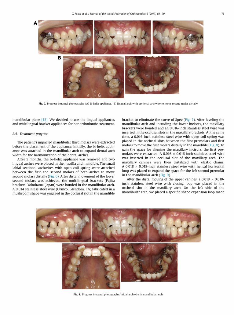
Fig. 7. Progress intraoral photographs. (A) Bi-helix appliance. (B) Lingual arch with sectional archwire to move second molar distally.
T. Fukui et al. / Journal of the World Federation of Orthodontists 6 (2017) 69e79 73
mandibular plane [15]. We decided to use the lingual appliancesand multilingual bracket appliances for her orthodontic treatment.
2.4. Treatment progress
The patient’s impacted mandibular third molars were extractedbefore the placement of the appliance. Initially, the bi-helix appli-ance was attached in the mandibular arch to expand dental archwidth for the harmonization of the dental arches.
After 5 months, the bi-helix appliance was removed and twolingual arches were placed in the maxilla and mandible. The smalllabial sectional archwires with open coil spring were attachedbetween the first and second molars of both arches to movesecond molars distally (Fig. 6). After distal movement of the lowersecond molars was achieved, the multilingual brackets (Fujitabrackets, Yokohama, Japan) were bonded in the mandibular arch.A 0.014 stainless steel wire (Ormco, Glendora, CA) fabricated in amushroom shape was engaged in the occlusal slot in the mandible
Fig. 8. Progress intraoral photographs:
bracket to eliminate the curve of Spee (Fig. 7). After leveling themandibular arch and intruding the lower incisors, the maxillarybrackets were bonded and an 0.016-inch stainless steel wire wasinserted in the occlusal slots in the maxillary brackets. At the sametime, a 0.016-inch stainless steel wire with open coil spring wasplaced in the occlusal slots between the first premolars and firstmolars to move the first molars distally in the mandible (Fig. 8). Togain the space for aligning the maxillary incisors, the first pre-molars were extracted. A 0.016 � 0.016-inch stainless steel wirewas inserted in the occlusal slot of the maxillary arch. Themaxillary canines were then distalized with elastic chains.A 0.018 � 0.018-inch stainless steel wire with helical horizontalloop was placed to expand the space for the left second premolarin the mandibular arch (Fig. 9).
After the distal moving of the upper canines, a 0.018 � 0.018-inch stainless steel wire with closing loop was placed in theocclusal slot in the maxillary arch. On the left side of themandibular arch, we placed a specific shape expansion loop made
initial archwire in mandibular arch.

Fig. 9. Progress intraoral photographs: maxillary canine retraction, expand with helical horizontal loop for the left mandibular second premolar.
T. Fukui et al. / Journal of the World Federation of Orthodontists 6 (2017) 69e7974
with a 0.018 � 0.018-inch stainless steel wire to avoid occlusalinterference with the maxillary premolar and molar. A smalllingual button was bonded on the lingual surface of the left secondpremolar to pull buccally using elastic thread and also to reinforcethe expansion effect (Fig. 10). Finally, 0.016 � 0.016-inch idealmushroom archwires were placed in the occlusal slot of bracketsof both arches. We were then able to achieve an overcorrection ofthe incisors (Fig. 11).
The total active treatment period was 31 months (Fig. 12).Following bracket removal, a fixed retainer of 0.016 � 0.022-inchrectangular wire was bonded on the lingual surface of teeth fromright premolars to the left premolars to keep the expandedmandible arch width. In addition, a removal thermoformingretainer (Tru-tain, Rochester, MN) was made for the patient toretain the mandibular and maxillary arches (Fig. 13). The patientwas instructed to wear the removable retainers 24 hours a day forthe first year and subsequently only at night.
Fig. 10. Progress intraoral photographs: maxillary canine retraction, expand
2.5. Treatment results
Facial photographs after active treatment showed goodimprovement in the profile and frontal view of the patient’s facedue to the results of the treatment. The upper lip was slightly ret-ruded (Fig. 12). The left side occlusion showed Class II molar andClass I canine relationships, and the right-side occlusion presentedClass II relationships for both canine and first molar. The amount ofover bite and overjet was 1.5 and 1.5 mm, respectively. Over-correction was achieved (Fig. 14).
Cephalometric analysis showed the skeletal measurement valuesslightlychanged. FMAangle increasedby1.5� withclockwise rotation.U1 to SN and L1 to MP angles showed proclined incisors, particularlythe mandibular incisor increased by 23.5�. Those changes wereconfirmed in the superimposed pretreatment and post treatmentlateral cephalometric tracings. Intrusionandflaringof themandibularincisors contributed to correcting the deep bite. Extraction of the
with helical horizontal loop for the left mandibular second premolar.

Fig. 11. Progress intraoral photographs: ideal treatment phase.
T. Fukui et al. / Journal of the World Federation of Orthodontists 6 (2017) 69e79 75
maxillary first premolars contributed to correction of the crowding.The maxillary incisors proclined and intruded as well (Fig. 15).
The extremely narrow mandibular arch width expanded from27.0 mm to 37.2 mm. The change of curve of Spee was from 6.0 mmto 0.5 mm. The second molars were uprighted. However, the right-
Fig. 12. Posttreatment facial a
side intercuspation was still cusp-to-cusp occlusion in the canineand premolar (Fig. 14).
Panoramic radiograph revealed that an acceptable root paral-lelism was achieved while slight root resorption of the maxillaryand mandibular incisors occurred (Fig. 16).
nd intraoral photographs.

Fig. 13. A removal thermoforming retainer with mandibular fixed retainer.
T. Fukui et al. / Journal of the World Federation of Orthodontists 6 (2017) 69e7976
After 4 years of retention, the occlusionwas deemed to be stable.Fixed type wire retainer is intact and must remain bonded for aslong as possible (Fig. 17).
3. Discussion
Treatment with multilingual bracket appliance has been con-traindicated for patients with deep bite occlusion mainly becauselingual brackets could not be placed on the lingual surface in themaxillary incisors. No case report has been found on a patient withClass II division 2 deep bite occlusion with multilingual brackettreatment.
The etiology of deep over bite usually comes from complex anddiverse causes. This patient did not have any deep bite facial
Fig. 14. Posttreatme
patterns and gingival display. Mandibular plane angle, Gonialangle, and lower facial height were within the Japanese normalrange. However, there were many dental problems. The maxillarycentral incisors were over-retroclined and mandibular incisorswere also retroclined. Mandibular dental arch width wasextremely narrow and showed excessive curve of Spee. Deep bitecan be treated orthodontically by intrusion or flaring of incisors,extrusion or passive eruption of the buccal segments, or a com-bination of these in general [11e13,15,16]. Mostafa et al. [11] sug-gested that a deep curve of Spee was the highest contribution forthe dental factor, confirming the importance of intruding themandibular incisors in deep bite mechanotherapy. Mandibularincisor intrusion is the most suitable deep bite treatment foradults with normal mandibular plane [15]. An intrusive force that
nt dental casts.

Fig. 15. Posttreatment lateral cephalometric radiograph and superimposed tracing.
T. Fukui et al. / Journal of the World Federation of Orthodontists 6 (2017) 69e79 77
is closer to the center of resistance of the mandibular incisorswould affect to intrude them with lingual bracket appliance incomparison with the conventional labial bracket appliance (Fig. 18)[8,17]. The intrusive force applied on the occlusal slot of mandib-ular incisors works effectively to prevent the overproclination ofthe teeth [9]. In this case, the proclination of the mandibular in-cisors contributed to resolve the large arch length discrepancy.However, incisor flaring has been thought to increase the inci-dence of relapse [18]. Berg [19] suggested that the interincisalangle should be less than 140� at the end of treatment for stabilityof deep bite correction [20]. Interincisal angle was decreased from168.5� to 134.3� in this case. Although this treatment result isreasonable with respect to the correction of the deep bite maloc-clusion, superimposition of the pretreatment and posttreatment
Fig. 16. Posttreatment pa
images demonstrated slightly unfavorable clockwise rotation ofthe mandible. The cause of this phenomenon was possibly theexpansion and uprighting of the mandibular buccal segments. Itcould have been prevented by placing temporary skeletalanchorage devices for tooth intrusion [21,22]. In summary, themalocclusion was corrected by intrusion, flaring of maxillary andmandibular incisors, and extrusion and uprighting of the buccalsegments. These corrections were effective to maintain theocclusal stability in this nongrowing patient. The fixed retainerplayed a very important role in keeping the mandibular dentalarch width.
The mushroom-shape archwire makes it difficult to adjust thecircumference of the lingual side of the dental arch. It is necessaryto bend a large offset between canines and premolars in
noramic radiograph.

Fig. 17. Photographs 4 years after the end of active treatment.
Fig. 18. Applied intrusive force. (A) Lingual bracket. (B) Conventional labial bracket.C.R., center of resistance.
T. Fukui et al. / Journal of the World Federation of Orthodontists 6 (2017) 69e7978
comparisonwith conventional labial bracket treatment. In addition,the length of interbracket span of archwire is shorter than theconventional labial edgewise archwire. These factors failed toaccurately control the mandibular premolar angulation on the rightside and this resulted in a less than ideal occlusal relationship onthat side.
4. Conclusions
The invisible multilingual bracket treatment achieved accept-able results to correct a severe Class II division 2 deep bite case.Although the right side has less than ideal intercuspation, theaccomplished occlusion has been stable for many years.
References
[1] Fujita K. New orthodontic treatment with lingual bracket mushroom archwireappliance. Am J Orthod 1979;76:657e75.
[2] Fujita K. Multilingual-bracket and mushroom archwire technique. Am JOrthod 1982;82:120e40.
[3] Fukui T, Choi YB, Yamaguchi H, Tsuruta M. Treatment of a horizontal open bitewith an invisible multiloop appliance in a girl with tooth trauma. Am J OrthodDentofacial Orthop 2009;136:596e606.
[4] Hong RK, Hong HP, Koh HS. Effect of reverse curvemushroom archwire on lowerincisors in adult patients: a prospective study. Angle Orthod 2001;71:425e32.
[5] Hong RK, Heo JM, Ha YK. Lever-arm and mini-implant system for anteriortorque control during retraction in lingual orthodontic treatment. AngleOrthod 2005;75:129e41.

T. Fukui et al. / Journal of the World Federation of Orthodontists 6 (2017) 69e79 79
[6] Fukui T, Tsuruta M, Choi Y, Kuwahara Y. Multilingual bracket treatmentcombined with orthognathic surgery in a skeletal class III patient with facialasymmetry. Am J Orthod Dentofacial Orthop 1999;115:654e9.
[7] Fukui T, Tsuruta M. Invisible treatment of a class III female adult patient withsevere crowding and cross-bite. J Orthod 2002;29:267e75.
[8] Fukui T, Suga K, Fukui K, Tsuruta M, Galang MT. Invisible treatment of a severeClass I crowding with multilingual bracket system using new double mush-room archwire technique. J World Fed Orthod 2015;4:151e61.
[9] Deguchi T, Terao F, Aonuma T, et al. Outcome assessment of lingual and labialappliances compared with cephalometric analysis, peer assessment rating,and objective grading system in Angle Class II extraction cases. Angle Orthod2015;85:400e7.
[10] Sangchaream Y, Chrsitopher HO. Effect of incisors angulation on overjet andoverbite in Class II camouflage treatment. Angle Orthod 2007;77:1011e8.
[11] El-Dawiatly MM, Fayed MMS, Mostafa YA. Deep overbite malocclusion:analysis of the underlying components. Am J Orthod Dentofacial Orthop2012;142:473e80.
[12] Al-Buraiki H, Sadowsky C, Schneider B. The effectiveness and long-term sta-bility of overbite correction with incisor intrusion mechanics. Am J OrthodDentofacial Orthop 2005;127:47e55.
[13] Baydas B, Yavuz L, Atasaral N, Ceylan I, Dagsuyu L. Investigation of the changes inthe positions of upper and lower incisors, overjet, overbite, and irregularity indexin subjects with different depths of curve of Spee. Angle Orthod 2004;74:349e55.
[14] Nagaoka K, Kuwahara Y. Normal standards for various roentgen cephalo-metric and cast model analyses in present day Japanese adults: part 1. NihonKyosei Shika Gakkai Zasshi 1993;52:467e80.
[15] Varlik SK, Alpakan OO, Turkoz C. Deepbite correction with incisor intrusion inadults: a long-term cephalometric study. Am J Orthod Dentofacial Orthop2013;144:414e9.
[16] Brian BE, Kuftinec MM, Baker IM. The relationship between bite depth andincisor angular change. Angle Orthod 1990;60:55e8.
[17] Burstone CJ. Application of Bioengineering to Clinical Orthodontics. In:Graber TM, Vanarsdall RL, editors. Orthodontics Current Principles andTechniques. Trd Ed. St.Louis: Mosby; 2000. p. 259e92.
[18] Dake ML, Sinclair PM. A comparison of the Rickets and Tweed-type archleveling techniques. Am J Orthod Dentofacial Orthop 1989;95:72e8.
[19] Berg R. Stability of deep overbite correction. Eur J Orthod 1983;5:75e83.[20] Houston WJB. Incisor edge-centroid relationships and overbite depth. Eur J
Orthod 1989;11:139e43.[21] Ishihara Y, Kuroda S, Sugawara Y, Balam TA, Takano-Yamamoto T,
Yamashiro T. Indirect usage of miniscrew anchorage to intrude overeruptedmandibular incisors in a Class II patient with a deep overbite. Am J OrthodDentofacial Orthop 2013;143:S113e24.
[22] Kim TW, Kim H, Lee SJ. Correction of deep overbite and gummy smile by usinga mini-implant with a segmented wire in a growing Class II Division 2 patient.Am J Orthod Dentofacial Orthop 2006;130:676e85.