Investigations of kanuka and manuka essential oils for in vitro treatment of disease and cellular...
8
ORIGINAL ARTICLE Investigations of kanuka and manuka essential oils for in vitro treatment of disease and cellular inflammation caused by infectious microorganisms Chien-Chia Chen a , Sui-Hing Yan a , Muh-Yong Yen a , Pei-Fang Wu b , Wei-Ting Liao c , Tsi-Shu Huang d,e,f , Zhi-Hong Wen g, *, Hui-Min David Wang b,h, ** a Department of Internal Medicine, Renai Branch, Taipei City Hospital, Taiwan, ROC b Department of Fragrance and Cosmetic Science, Kaohsiung Medical University, Kaohsiung, Taiwan, ROC c Department of Biotechnology, Kaohsiung Medical University, Kaohsiung, Taiwan, ROC d Section of Microbiology, Department of Pathology and Laboratory Medicine, Kaohsiung Veterans General Hospital, Kaohsiung, Taiwan, ROC e Department of Medical Laboratory Sciences and Biotechnology, Fooyin University, Taiwan, ROC f Institute of Clinical Medicine, National Yang-Ming University, Taipei, Taiwan, ROC g Department of Marine Biotechnology and Resources, National Sun Yat-sen University, Kaohsiung, Taiwan, ROC h Graduate Institute of Natural Products, Kaohsiung Medical University, Kaohsiung, Taiwan, ROC Received 30 April 2013; received in revised form 3 December 2013; accepted 18 December 2013 KEYWORDS Bacteria; Fungi; Inflammation; Kanuka essential oils; Manuka essential oils Background: Diseases caused by infectious and inflammatory microorganisms are among the most common and most severe nosocomial diseases worldwide. Therefore, developing effec- tive agents for treating these illnesses is critical. In this study, essential oils from two tea tree species, kanuka (Kunzea ericoides) and manuka (Leptospermum scoparium), were evaluated for use in treating diseases and inflammation caused by microorganism infection. Methods: Isolates of clinically common bacteria and fungi were obtained from American Type Culture Collection and from Kaohsiung Veterans General Hospital. Minimum inhibitory concen- trations for Trichosporon mucoides, Malassezia furfur , Candida albicans, and Candida tropica- lis were determined by the broth microdilution method with Sabouraud dextrose broth. The * Corresponding author. Department of Marine Biotechnology and Resources, National Sun Yat-sen University, 70 Lien-Hai Road, Kaohsiung 80424, Taiwan, ROC. ** Corresponding author. Department of Fragrance and Cosmetic Science, Kaohsiung Medical University, 100 Shiquan First Road, Kaohsiung 80708, Taiwan, ROC. E-mail addresses: [email protected] (Z.-H. Wen), [email protected] (H.-M. David Wang). + MODEL Please cite this article in press as: Chen C-C, et al., Investigations of kanuka and manuka essential oils for in vitro treatment of disease and cellular inflammation caused by infectious microorganisms, Journal of Microbiology, Immunology and Infection (2014), http:// dx.doi.org/10.1016/j.jmii.2013.12.009 1684-1182/$36 Copyright ª 2014, Taiwan Society of Microbiology. Published by Elsevier Taiwan LLC. All rights reserved. http://dx.doi.org/10.1016/j.jmii.2013.12.009 Available online at www.sciencedirect.com ScienceDirect journal homepage: www.e-jmii.com Journal of Microbiology, Immunology and Infection (2014) xx,1e8
Transcript of Investigations of kanuka and manuka essential oils for in vitro treatment of disease and cellular...

+ MODEL
Journal of Microbiology, Immunology and Infection (2014) xx, 1e8
Available online at www.sciencedirect.com
ScienceDirect
journal homepage: www.e- jmii .com
ORIGINAL ARTICLE
Investigations of kanuka and manukaessential oils for in vitro treatment ofdisease and cellular inflammation caused byinfectious microorganisms
Chien-Chia Chen a, Sui-Hing Yan a, Muh-Yong Yen a,Pei-Fang Wu b, Wei-Ting Liao c, Tsi-Shu Huang d,e,f,Zhi-Hong Wen g,*, Hui-Min David Wang b,h,**
a Department of Internal Medicine, Renai Branch, Taipei City Hospital, Taiwan, ROCb Department of Fragrance and Cosmetic Science, Kaohsiung Medical University,Kaohsiung, Taiwan, ROCc Department of Biotechnology, Kaohsiung Medical University, Kaohsiung, Taiwan, ROCd Section of Microbiology, Department of Pathology and Laboratory Medicine,Kaohsiung Veterans General Hospital, Kaohsiung, Taiwan, ROCe Department of Medical Laboratory Sciences and Biotechnology, Fooyin University, Taiwan, ROCf Institute of Clinical Medicine, National Yang-Ming University, Taipei, Taiwan, ROCg Department of Marine Biotechnology and Resources, National Sun Yat-sen University,Kaohsiung, Taiwan, ROCh Graduate Institute of Natural Products, Kaohsiung Medical University, Kaohsiung, Taiwan, ROC
Received 30 April 2013; received in revised form 3 December 2013; accepted 18 December 2013
KEYWORDSBacteria;Fungi;Inflammation;Kanuka essential oils;Manuka essential oils
* Corresponding author. DepartmenKaohsiung 80424, Taiwan, ROC.** Corresponding author. DepartmenKaohsiung 80708, Taiwan, ROC.
E-mail addresses: [email protected]
Please cite this article in press as: Chand cellular inflammation caused bdx.doi.org/10.1016/j.jmii.2013.12.00
1684-1182/$36 Copyright ª 2014, Taiwhttp://dx.doi.org/10.1016/j.jmii.2013
Background: Diseases caused by infectious and inflammatory microorganisms are among themost common and most severe nosocomial diseases worldwide. Therefore, developing effec-tive agents for treating these illnesses is critical. In this study, essential oils from two tea treespecies, kanuka (Kunzea ericoides) and manuka (Leptospermum scoparium), were evaluatedfor use in treating diseases and inflammation caused by microorganism infection.Methods: Isolates of clinically common bacteria and fungi were obtained from American TypeCulture Collection and from Kaohsiung Veterans General Hospital. Minimum inhibitory concen-trations for Trichosporon mucoides, Malassezia furfur, Candida albicans, and Candida tropica-lis were determined by the broth microdilution method with Sabouraud dextrose broth. The
t of Marine Biotechnology and Resources, National Sun Yat-sen University, 70 Lien-Hai Road,
t of Fragrance and Cosmetic Science, Kaohsiung Medical University, 100 Shiquan First Road,
.edu.tw (Z.-H. Wen), [email protected] (H.-M. David Wang).
en C-C, et al., Investigations of kanuka and manuka essential oils for in vitro treatment of diseasey infectious microorganisms, Journal of Microbiology, Immunology and Infection (2014), http://9
an Society of Microbiology. Published by Elsevier Taiwan LLC. All rights reserved..12.009

2 C.-C. Chen et al.
+ MODEL
Please cite this article in press as: Chand cellular inflammation caused bdx.doi.org/10.1016/j.jmii.2013.12.00
antibacterial susceptibility of Staphylococcus aureus, Streptococcus sobrinus, Streptococcusmutans, and Escherichia coli were determined by the broth microdilution method. A humanacute monocytic leukemia cell line (THP-1) was cultured to test the effects of the essentialoils on the release of the two inflammatory cytokines, tumor necrosis factor-a and inter-leukin-4.Results: Multiple analyses of microorganism growth confirmed that both essential oils signifi-cantly inhibited four fungi and the four bacteria. The potent fungicidal properties of the oilswere confirmed by minimum inhibitory concentrations ranging from 0.78% to 3.13%. The oilsalso showed excellent bactericidal qualities with 100% inhibition of the examined bacteria.In THP-1 cells, both oils lowered tumor necrosis factor-a released after lipopolysaccharidestimulation. Finally, the antimicrobial and anti-inflammatory effects of the oils were obtainedwithout adversely affecting the immune system.Conclusion: These results indicate that the potent antimicroorganism and anti-inflammationproperties of kanuka and manuka essential oils make them strong candidates for use in treatinginfections and immune-related disease. The data confirm the potential use of kanuka and man-uka extracts as pharmaceutical antibiotics, medical cosmetology agents, and food supple-ments.Copyright ª 2014, Taiwan Society of Microbiology. Published by Elsevier Taiwan LLC. All rightsreserved.
Introduction
Kanuka (Kunzea ericoides) and manuka (Leptospermumscoparium) tea trees are small trees or shrubs distributedthroughout New Zealand in widely varying climates, alti-tudes, and population densities. Early New Zealand recordsreport the use of the bark, leaves, sap, and seed capsules ofthese plants in beverage supplements and in pharmaceuticaland medicinal preparations.1 Kanuka and manuka arecommonly known as “tea trees” because many early NewZealand settlers made tea from the leaves.2 Accumulatingevidenceof theuniquebiofunctional activities andmedicinalproperties of these oils has recently generated renewed in-terest in their potential commercial applications.3 Tradi-tional medical applications of the white leaf gums of kanukaand manuka include internal or external sedatives, feverreducers, and cough suppressants. A decoction of the seedcapsules canalso beused to treat inflammation. A liquid formof the decoction alleviates dysentery, diarrhea, and colicpain. The capsules or leaves could also be chewed to relievedysentery.4 A decoction formed by boiling the leaves andbark has proven effective for treating breast inflammation,back stiffness, and eye problems. A poultice formed frompounded capsules of the plants was used to dry open woundsor running sores and to treat scald and burn injuries. Finally,the liquid could be used as a mouthwash or gargle to treatmouth and throat sores.1
A troubling global public health issue that has emergedin recent decades is hospital-acquired infections or non-socomial illnesses caused by microorganisms.5 Pathogenicfungi and bacteria are able to survive for extended periodson human superficial skin, mucosa, or environmental sur-faces, and have been implicated in infectious outbreaks inhospitals, medical facilities, and institutions in manycountries.6,7 As the use of broad-spectrum antibiotics in-creases, the most alarming characteristic of these micro-organisms is their apparent resistance to almost allcommercially available antimicrobial drugs. As a result,many of these microorganisms are now classified as highly
en C-C, et al., Investigations of ky infectious microorganisms, Jou9
antibiotic-resistant.3,8 Therefore, the search for newtherapeutic modalities has increased requirements in nat-ural medicinal therapy. Malassezia furfur is a lipophilicyeast that lives on the normal skin flora of many animals,including humans. Because the growth of this fungus re-quires a fat source, it is most common in areas with manysebaceous glands, including the scalp, face, and upperbody. Opportunistic infections involving some species of M.furfur may cause hypopigmentation in the trunk and otherlocations in humans.9 Trichosporon mucoides is usuallyfound in soil and water. Although it can contaminate humanskin, its effects in otherwise healthy individuals are usuallyeither harmless or are limited to superficial skin and nailinfections. In immunocompromised patients or in patientscurrently undergoing immunosuppressive therapy, howev-er, an opportunistic infection with Trichosporon can belethal.10 Another genus of yeast is Candida, in which theCandida albicans and Candida tropicalis species are com-mon and easily recognized medical yeast pathogensbecause they are normal constituents of the humanflora.11,12 The most common species of Staphylococcus isStaphylococcus aureus, which causes staphylococcal in-fections and is frequently found in the human respiratorytract and on skin surfaces. The emergence of antibiotic-resistant forms of pathogenic S. aureus (e.g., methicillin-resistant S. aureus) is a worldwide problem in clinicalmedicine.13 Another species in the Streptococcus genus isStreptococcus sobrinus, which is a spherically-shapedanaerobic and Gram-positive bacterium. They grow inpairs or chains, and they are not motile and do not formspores. Streptococcus mutans is a facultatively anaerobic,Gram-positive, coccus-shaped bacterium commonly foundin the human oral cavity and is a significant contributor totooth decay. The most intensively studied prokaryoticmodel organism is Escherichia coli, a Gram-negative, rod-shaped bacterium commonly found in the lower intestine ofwarm-blooded organisms. This bacterium is easily andinexpensively grown in a laboratory setting and has beenstudied intensively in the past half century.14
anuka and manuka essential oils for in vitro treatment of diseasernal of Microbiology, Immunology and Infection (2014), http://

Anti-infection properties of tea tree oils 3
+ MODEL
The human immune system can be defined as a mecha-nism for protecting the biochemistry of an organism againstsickness, including viruses, microorganisms, worms, al-lergies, inflammation, and cancer. An important componentof the immune system is the T-helper (Th) cell, which ini-tiates and regulates immune responses.15,16 The four majorsubtypes of Th cells in humans are: Th1, which regulatesinflammatory responses to infections; Th2, which modu-lates allergic responses; T regulatory cells, which have apivotal role in immune suppression; and Th17, which isassociated with autoimmunity. The inflammatory responsesof Th1 are mainly triggered by cytokines from monocytes/macrophages. When monocytes polarize to macrophages,cells recognize parasitical antigens and secrete proin-flammatory cytokines such as tumor necrosis factor-a (TNF-a) and interleukins (ILs).17 TNF-a and IL-6 released frommonocytes/macrophages then encounter and activateantigen-specific natural killer T cells in a process known asthe Th1 response. By contrast, in response to allergenexposure, macrophages alternatively secrete Th2 cyto-kines, such as IL-4, and activate Th2-mediated allergicresponses.18e20 Given the negative feedback between Th1and Th2 (i.e., activation of one inactivates the other), ananti-inflammatory agent might also exhibit an increased butparallel allergic effect.21 Lipopolysaccharide (LPS) frombacteria is a model compound with a well-known role ininflammatory effectiveness, whereas TNF-a initiates Th1-mediated inflammatory responses, and IL-4 inhibits in-flammatory responses and initiates allergic responses. Thisstudy detected both of these major cytokines, TNF-a andIL-4, which were generated by a macrophage cell line,human acute monocytic leukemia cell line (THP-1).
The antimicrobial and anti-inflammatory characteristicsof plant extracts have been studied and applied for thou-sands of years.19,22 The use of plant-derived constitutes aspreservatives, cosmetics, and pharmaceuticals haverecently attracted increased interest. The present workanalyzed two essential oil extracts to identify their anti-microbial activities in four fungi and four bacteria. Thereleases of inflammatory factors TNF-a and IL-4 in THP-1cells were also examined. The potent anti-microbial andanti-inflammatory properties observed in these oils confirmtheir strong potential use as medicinal pharmaceuticals.
Materials and methods
Reagents and oil samples
Dimethyl sulfoxide (DMSO) and LuriaeBertani broth werepurchased from Sigma-Aldrich Chemical Inc. (St Louis, MO,USA). Sabouraud dextrose (SD) agar and SD broth werepurchased from Creative Media Products, Ltd. (WuguShiang, Taiwan, R.O.C.). Dulbecco modified Eagle’s me-dium, fetal bovine serum, penicillin, and streptomycinwere purchased from Life Technologies Co., Ltd. (Gibco,Grand Island, NY, USA). XTT [2,3-bis-(2-methoxy-4-nitro-5-sulfophenyl)-2H-tetrazolium-5-carboxanilide] assay kit waspurchased from Roche Diagnostics (Mannheim, Germany).Kanuka and manuka essential oils (100%) were provided by alocal company (YO-HOON Trading Co, Ltd, Kaohsiung,Taiwan). Both essential oils were extracted from the leaves
Please cite this article in press as: Chen C-C, et al., Investigations of kand cellular inflammation caused by infectious microorganisms, Joudx.doi.org/10.1016/j.jmii.2013.12.009
of plants by steam distillation. All buffers and other re-agents were of the highest purity commercially available.
Bacterial and fungal species
Six microorganism strains used in this study were purchasedfrom American Type Culture Collection (ATCC), included T.mucoides (ATCC 204094), C. tropicalis (ATCC 9968), S.aureus (ATCC 29213), S. mutans (ATCC 25175), S. sobrinus(ATCC 33478), and E. coli (ATCC 35218). Clinical isolates ofother microorganisms, M. furfur and C. albicans, wereprovided by Kaohsiung Veterans General Hospital, a 1400-bed tertiary referral medical center in Taiwan. The THP-1cell line used in this study, which was purchased fromATCC (TIB-202), was derived from the peripheral blood of a1-year-old human male with acute monocytic leukemia.
Miniaturized broth dilution susceptibility test ofanti-fungal activity
The SD broth was used to examine the susceptibilities ofthe minimum inhibitory concentration (MIC) of kanuka andmanuka essential oils by the microdilution method in sterile96-well microtiter plates covered with lids. For analyzingoils, this method provides better reproducibility comparedto the agar plate diffusion and dilution techniques used tostudy fungi.5,20,22,23 The MIC values of each strain weredetermined by dissolving the testing samples in DMSO atvarious concentrations with two-fold serial dilutions inbroth. DMSO was used as a blank vehicle control solvent,and untreated microorganisms were used as negative con-trols. To ensure that DMSO did not affect the assays, DMSOconcentrations used in the experiment were 5% lower thanthose in controls. Comparisons showed no significant dif-ference between samples with and without DMSO.
Briefly, after incubation at 35�C for 2e7 days, M. furfur,T. mucoides, C. tropicalis, and C. albicans colonies from theSD agar plates were mixed with sterile water and then uni-formly rotated. The fungal suspension was adjusted to anapproximate turbidity of 1 � 106e5 � 106 CFU/mL(0.5 McFarland suspension, VITEK Special DR100 Colorimeter52-1210; Hach Company, Loveland, CO, USA). Varying folddilutions of the fungal suspension in sterile water(1 � 104e5 � 104 CFU/mL) were then added to 0.05 ml SDbroth. After adding 0.01 mL of fungal solution to eachwell ofa 96-well microtiter plate, the final fungi concentrationswere 1 � 102e5 � 102 CFU/well. Addition of 0.05 mL kanukaormanuka essential oil then obtained concentrations rangingfrom 0.01% to 12.5%. After incubation in darkness for 2e7days at 35�C, the MIC values of the two oils were establishedfor each of the four fungi.24 All experiments were performedin triplicate with the blank vehicle control, negative control,and experimental control groups.
Determination of antibacterial properties
The antimicrobial properties of the kanuka and manukaessential oils were investigated using previously describedmethods.5,13 Briefly, 105 CFU/mL microbial suspensions offour bacteria, S. aureus, S. mutans, S. sobrinus, and E. coli,were incubated in each well at 37�C for 24 hours. The
anuka and manuka essential oils for in vitro treatment of diseasernal of Microbiology, Immunology and Infection (2014), http://
4 C.-C. Chen et al.
+ MODEL
microbial bacteria were then harvested in normal salineand adjusted to McFarland 0.5 (1.5 � 108 CFU/mL). Onemilliliter of each bacterial sample suspension was centri-fuged at 470g for 5 minutes and then treated with 0.3 mL ofan essential oil to obtain a final concentration of 10% v/v.Reactions were then compared at 25�C at intervals of 5seconds, 30 seconds, 60 seconds, 180 seconds, 300 seconds,and 900 seconds. A well containing DMSO was used as thegrowth blank vehicle control; a well containing mediumonly was used as the negative control; and all the otherwells contained the experimental groups treated withessential oils. A test was considered valid if the well for thegrowth control group was positive and those for othergroups were negative. After the specified reaction times,the bacteria were centrifuged for 2 minutes, washed once,and then suspended in sterile saline water. A 104-folddilution of the bacterial suspension (100 mL), was thenplated on blood agar plates. After a 24-hour incubationperiod, the bactericidal effects of the essential oils weredetermined in each sample by measuring bacterial growthin culture.21 The inhibition of bacterial growth was thenmeasured by comparison with normal growth observed inmicrobes not treated with the samples.
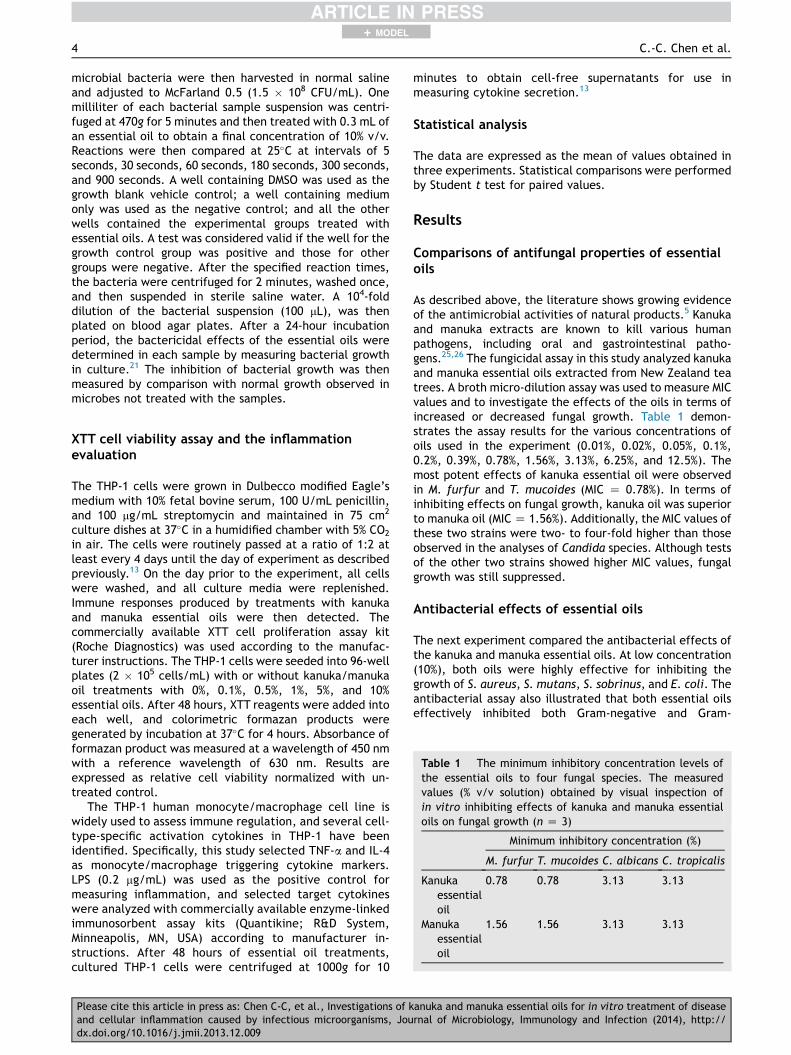
Table 1 The minimum inhibitory concentration levels ofthe essential oils to four fungal species. The measuredvalues (% v/v solution) obtained by visual inspection ofin vitro inhibiting effects of kanuka and manuka essentialoils on fungal growth (n Z 3)
Minimum inhibitory concentration (%)
M. furfur T. mucoides C. albicans C. tropicalis
Kanukaessentialoil
0.78 0.78 3.13 3.13
Manukaessentialoil
1.56 1.56 3.13 3.13
XTT cell viability assay and the inflammationevaluation
The THP-1 cells were grown in Dulbecco modified Eagle’smedium with 10% fetal bovine serum, 100 U/mL penicillin,and 100 mg/mL streptomycin and maintained in 75 cm2
culture dishes at 37�C in a humidified chamber with 5% CO2
in air. The cells were routinely passed at a ratio of 1:2 atleast every 4 days until the day of experiment as describedpreviously.13 On the day prior to the experiment, all cellswere washed, and all culture media were replenished.Immune responses produced by treatments with kanukaand manuka essential oils were then detected. Thecommercially available XTT cell proliferation assay kit(Roche Diagnostics) was used according to the manufac-turer instructions. The THP-1 cells were seeded into 96-wellplates (2 � 105 cells/mL) with or without kanuka/manukaoil treatments with 0%, 0.1%, 0.5%, 1%, 5%, and 10%essential oils. After 48 hours, XTT reagents were added intoeach well, and colorimetric formazan products weregenerated by incubation at 37�C for 4 hours. Absorbance offormazan product was measured at a wavelength of 450 nmwith a reference wavelength of 630 nm. Results areexpressed as relative cell viability normalized with un-treated control.
The THP-1 human monocyte/macrophage cell line iswidely used to assess immune regulation, and several cell-type-specific activation cytokines in THP-1 have beenidentified. Specifically, this study selected TNF-a and IL-4as monocyte/macrophage triggering cytokine markers.LPS (0.2 mg/mL) was used as the positive control formeasuring inflammation, and selected target cytokineswere analyzed with commercially available enzyme-linkedimmunosorbent assay kits (Quantikine; R&D System,Minneapolis, MN, USA) according to manufacturer in-structions. After 48 hours of essential oil treatments,cultured THP-1 cells were centrifuged at 1000g for 10
Please cite this article in press as: Chen C-C, et al., Investigations of kand cellular inflammation caused by infectious microorganisms, Joudx.doi.org/10.1016/j.jmii.2013.12.009
minutes to obtain cell-free supernatants for use inmeasuring cytokine secretion.13
Statistical analysis
The data are expressed as the mean of values obtained inthree experiments. Statistical comparisons were performedby Student t test for paired values.
Results
Comparisons of antifungal properties of essentialoils
As described above, the literature shows growing evidenceof the antimicrobial activities of natural products.5 Kanukaand manuka extracts are known to kill various humanpathogens, including oral and gastrointestinal patho-gens.25,26 The fungicidal assay in this study analyzed kanukaand manuka essential oils extracted from New Zealand teatrees. A broth micro-dilution assay was used to measure MICvalues and to investigate the effects of the oils in terms ofincreased or decreased fungal growth. Table 1 demon-strates the assay results for the various concentrations ofoils used in the experiment (0.01%, 0.02%, 0.05%, 0.1%,0.2%, 0.39%, 0.78%, 1.56%, 3.13%, 6.25%, and 12.5%). Themost potent effects of kanuka essential oil were observedin M. furfur and T. mucoides (MIC Z 0.78%). In terms ofinhibiting effects on fungal growth, kanuka oil was superiorto manuka oil (MICZ 1.56%). Additionally, the MIC values ofthese two strains were two- to four-fold higher than thoseobserved in the analyses of Candida species. Although testsof the other two strains showed higher MIC values, fungalgrowth was still suppressed.
Antibacterial effects of essential oils
The next experiment compared the antibacterial effects ofthe kanuka and manuka essential oils. At low concentration(10%), both oils were highly effective for inhibiting thegrowth of S. aureus, S. mutans, S. sobrinus, and E. coli. Theantibacterial assay also illustrated that both essential oilseffectively inhibited both Gram-negative and Gram-
anuka and manuka essential oils for in vitro treatment of diseasernal of Microbiology, Immunology and Infection (2014), http://

Anti-infection properties of tea tree oils 5
+ MODEL
positive bacteria. Specifically, for each microbe, whetherGram-negative or Gram-positive, growth was 100% inhibitedby exposures of 5 seconds, 30 seconds, 60 seconds, 180seconds, 300 seconds, and 900 seconds. The antibacterialeffects of the oils still remained potent after long incuba-tion times (data not shown). The data confirm that, inaddition to their suppressive effects on bacterial growth,both oils have strong inhibiting effects. Additionally, theremarkable biofunctions of the kanuka and manuka tea treeoil samples analyzed within this study included both anti-fungal and antibacterial activities.
Anti-inflammatory properties of kanuka andmanuka essential oils
After confirming the antimicrobial effects of kanuka andmanuka essential oils, this study further investigated othereffects such as suppression or inhibition of inflammatoryreactions. Fig. 1 demonstrates that, after 48-hour exposureto test concentrations of kanuka and manuka essential oilsranging from 0.1% to 10%, the viabilities of THP-1 cellsexceeded 100%. This reveals that the oils have no majortoxic side effects on THP-1 cells. In the absence of LPSstimulation, 48-hour treatments with kanuka essential oil
(A)
(B)
Figure 1. XTT assay results for THP-1 cells after 48 hourstreatment with 0%, 0.1%, 0.5%, 1%, 5%, and 10% (A) kanuka and(B) manuka essential oils with dark green color, respectively;LPS (0.2 mg/mL) was used as the positive control for measuringinflammation reactions. *p < 0.05 versus untreated vehiclecontrol group; mean � standard deviation.LPS Z lipopolysaccharide; XTT Z 2,3-bis-(2-methoxy-4-nitro-5-sulfophenyl)-2H-tetrazolium-5-carboxanilide; THP-1 Z human acute monocytic leukemia cell line.
Please cite this article in press as: Chen C-C, et al., Investigations of kand cellular inflammation caused by infectious microorganisms, Joudx.doi.org/10.1016/j.jmii.2013.12.009
concentrations ranging from 0.1% to 10% did not signifi-cantly change the release of cytokines, TNF-a, or IL-4 fromcultured THP-1 cells (Fig. 2). The bacterial endotoxin LPS(0.2 mg/mL) elicits a strong inflammatory response in THP-1cells. Cotreatment with LPS and 0.1e10% kanuka oil duringcell culture was performed to display the effects on THP-1cytokine secretion. Kanuka essential oil treatment signifi-cantly reduced the LPS-induced TNF-a release from THP-1cells, but did not affect IL-4. Similarly, manuka essentialoil treatment had an anti-inflammatory effect on LPS-induced release of TNF-a, but with no influence on IL-4(Fig. 3).
Discussion
Although some essential oils such as tea tree oils are knownto have antimicrobial effects, clinical evidence of theirefficacy for treating bacterial, fungal, or viral infections islimited. Kanuka is a large tree or shrub abundantthroughout New Zealand and manuka is distinguishable byits relatively larger leaves and flowers, but smaller overall
Figure 2. Enzyme-linked immunosorbent assay results fortumor necrosis factor (TNF-a) cytokine released from THP-1cells after stimulation with LPS (20 mg/mL) and 48 hourstreatment with 0%, 0.1%, 0.5%, 1%, 5%, and 10% (A) kanuka and(B) manuka essential oils; **p < 0.05 versus untreated vehiclecontrol group; *p < 0.05 versus group treated with 0.1e10% ofthe two oils after LPS incubation; mean � standard deviation.LPS Z lipopolysaccharide; THP-1 Z human acute monocyticleukemia cell line.
anuka and manuka essential oils for in vitro treatment of diseasernal of Microbiology, Immunology and Infection (2014), http://

Figure 3. Enzyme-linked immunosorbent assay results forIL-4 cytokines released from THP-1 cells after stimulation withLPS (20 mg/mL) and 48 hours treatment with 0%, 0.1%, 0.5%,1%, 5%, and 10% of (A) kanuka and (B) manuka essential oilsversus untreated control samples. IL Z interleukin; THP-1 Z human acute monocytic leukemia cell line.
6 C.-C. Chen et al.
+ MODEL
size. The Maori people and early New Zealand settlers usedboth plants in traditional medicinal preparations.1,27
Maddocks-Jennings et al27 reported verification suggestingthat a gargle or mouthwash containing the essential oils ofkanuka and manuka extracts could delay the developmentof mucositis and reduce associated health problems. Ac-cording to these lecture reports, both essential oils werewith low irritations to human skin, functions on improveoral infection, benefits in ease-breath, and help in coolingdown user’s body.1,28 Commercial applications of tea treeessential oils for their antimicrobial and antifungal prop-erties are in soaps, creams, mouthwashes, toothpastes, andother preparations. Internal and external use of these oilsby herbalists and aroma therapists has as well increased inthe past twenty years.4,11
The recent emergence of new strains of bacteria andfungi warrants intensive study of therapeutic agents fortheir control and eradication.29,30 Due to the widespreaduse of tea tree oils for body massage and aromatherapy,in vivo studies have been performed to investigate theirpharmacological and antimicrobial activities in guinea pigileum, rat uterus, rat skeletal muscle, chick biventermuscle, and rat phrenic nerve diaphragm.4 Studies ofmanuka honey produced by New Zealand tea trees
Please cite this article in press as: Chen C-C, et al., Investigations of kand cellular inflammation caused by infectious microorganisms, Joudx.doi.org/10.1016/j.jmii.2013.12.009
suggested that, whereas the antibacterial properties ofhoney products are derived from hydrogen peroxide, thoseof manuka oil are derived through other mechanisms.31,32
Our data indicate that both oils might possess variousproperties to inhibit microorganism growth. Unique manukafactor is currently the global standard for identifying andmeasuring the antibacterial strength of manuka in specificstrains of bacteria.11 The antibacterial activity of manukahoney is now standardized in terms of phenol concentrationequivalent, which is expressed as a unique manuka factorvalue.30,33 However, further studies are needed to identifythe antimicroorganism mechanisms of these two targetessential oils.
Our data specify that neither oil treatment had stimu-latory effects on cytokine release in untreated THP-1macrophages. Interestingly, both oils showed strong inhib-iting effects on inflammation induced by LPS treatment.Because TNF-a release from monocytes/macrophages reg-ulates Th1-mediated inflammatory responses, short-termnontoxic dosages of the two essential oils may be effectivefor treating inflammation. For example, the oils may beeffective for treating lesions caused by insect bites or forrepairing infectious wounds. Both essential oils reduced theTNF-a production (Fig. 2) and did not cause cytotoxicity toThP-1. It is possible that essential oil may directly interactwith the toll-like receptor 4, which is an LPS receptor.However, we did not have enough evidence for this inter-action within the present study. We cannot rule out thepossibility for the involvement of the toll-like receptor 4competition with essential oils, and this needs to be furtherinvestigated in the future. Therefore, we focused on thechanges of inflammatory outcome, the TNF-a marker. Theoil treatments did not significantly affect IL-4 release,which suggests that, in addition to the anti-inflammatoryproperties, the oils have potential applications as anti-allergic agents. Because of their anti-inflammatory prop-erties and their absence of adverse allergic reactionsresulting from cytokine release, kanuka and manuka oilsmay also be effective in human epidermal-relatedproducts.
Aromatherapy is an alternative medicine practice inwhich volatile plant materials, known as essential oils, andother aromatic compounds are used to improve theemotional state, cognitive function, or physical health.4
However, the literature on the efficacy of aromatherapiesfor treating medical conditions, especially studies thathave applied a rigorous methodology, is very limited.Nevertheless, some data indicate that essential oils mighthave therapeutic applications.15 Essential oils are occa-sionally used to describe fragrant oils extracted from plantmaterials by any solvent-based extraction methods. Aro-matherapists and other practitioners need to understandthat the clinical use of kanuka or manuka oils for thevarying compositions of the extracts.2 Another issue thatarises when new pharmaceutical applications of plantcomponents are introduced is their modulating effectswhen used in combined therapy with commercial antibi-otics or other anti-inflammatory medicines. Although somestudies have reported synergistic effects of natural plantextracts and antibiotics when used in combined therapy,5
further study is needed to identify the efficacy of essen-tial oils for inhibiting microbial growth and inflammation
anuka and manuka essential oils for in vitro treatment of diseasernal of Microbiology, Immunology and Infection (2014), http://

Anti-infection properties of tea tree oils 7
+ MODEL
and particularly the chemical and taxonomic properties ofkanuka and manuka. Given the enormous potential of theclinical applications of essential oils and the growing evi-dence of their antimicrobial and anti-inflammatory effects,continuing study of potential medical applications of theseand other locally produced extracts is needed. The prom-ising findings of this study warrant further evaluation of thecurrent model for use in objectively measuring a contami-nated wound environment and for assessing the mecha-nisms of medicinal applications of kanuka and manuka oils.However, long-term tests of the toxicity and ion-specificeffects of these oils are needed to verify their safety andeffectiveness for clinical use as wound healing agents andfor aromatherapy.
Conflicts of interest
No contributing author has a conflict of interest in thepublication of this study.
Acknowledgments
This work was financially supported by a grant from theNational Science Council, Republic of China (NSC-99-2221-E-037-006-MY3); and by two joint grants from National SunYat-sen University and Kaohsiung Medical University (NSY-SUKMU 101-007). The authors would like to thank Paul SteveLugue for English editing.
References
1. Porter NG, Wilkins AL. Chemical, physical and antimicrobialproperties of essential oils of Leptospermum scoparium andKunzea ericoides. Phytochemistry 1998;50:407e15.
2. Maddocks-Jennings W, Wilkinson JM, Shillington D,Cavanagh H. A fresh look at manuka and kanuka essential oilsfrom New Zealand. Int J Aromather 2005;15:141e6.
3. Lee H, Churey JJ, Worobo RW. Antimicrobial activity of bac-terial isolates from different floral sources of honey. Int J FoodMicrobiol 2008;126:240e4.
4. Lis-Balchin M, Hart SL, Deans SG. Pharmacological and anti-microbial studies on different tea-tree oils (Melaleuca alter-nifolia, Leptospermum scoparium or Manuka and Kunzeaericoides or Kanuka), originating in Australia and New Zealand.Phytother Res 2000;14:623e9.
5. Wang HM, Chen CY, Chen HA, Huang WC, Lin WR, Chen TC,et al. Zingiber officinale (ginger) compounds with tetracyclinehave synergistic effects against clinical extensively-drugresistant Acinetobacter baumannii. Phytotherapy Research2010;24:1825e30.
6. Peleg AY, Seifert H, Paterson DL. Acinetobacter baumannii:emergence of a successful pathogen. Clin Microbiol Rev 2008;21:538e82.
7. Munoz-Price LS, Weinstein RA. Acinetobacter infection. NewEngl J Med 2008;358:1271e81.
8. Doi Y, Husain S, Potoski BA, McCurry KR, Paterson DL. Exten-sively drug-resistant Acinetobacter baumannii. Emerg InfectDis 2009;15:980e2.
9. Wi HS, Na EY, Yun SJ, Lee JB. The antifungal effect of lightemitting diode on Malassezia yeasts. J Dermatol Sci 2012;67:3e8.
Please cite this article in press as: Chen C-C, et al., Investigations of kand cellular inflammation caused by infectious microorganisms, Joudx.doi.org/10.1016/j.jmii.2013.12.009
10. Lacasse A, Cleveland KO. Trichosporon mucoides fungemia in aliver transplant recipient: case report and review. TransplInfect Dis 2009;11:155e9.
11. Agarwal V, Lal P, Pruthi V. Effect of plant oils on Candidaalbicans. J Microbiol Immunol Infect 2010;43:447e51.
12. Basson NJ, Grobler SR. Antimicrobial activity of two SouthAfrican honeys produced from indigenous Leucospermum cor-difolium and Erica species on selected micro-organisms. BMCComplement Altern Med 2008;8:41.
13. Jenkins R, Burton N, Cooper R. Manuka honey inhibits cell di-vision in methicillin-resistant Staphylococcus aureus. J Anti-microb Chemother 2011;66:2536e42.
14. Tan HT, Rahman RA, Gan SH, Halim AS, Hassan SA,Sulaiman SA, et al. The antibacterial properties of Malaysiantualang honey against wound and enteric microorganisms incomparison to manuka honey. BMC Complement Altern Med2009;9:34.
15. Harrington LE, Hatton RD, Mangan PR, Turner H, Murphy TL,Murphy KM, et al. Interleukin 17-producing CD4þ effector Tcells develop via a lineage distinct from the T helper type 1 and2 lineages. Nat Immunol 2005;6:1123e32.
16. Chen BH, Wu PY, Chen KM, Fu TF, Wang HM, Chen CY. Anti-allergic potential on RBL-2H3 cells of some phenolic con-stituents of Zingiber officinale (Ginger). J Nat Prod 2009;72:950e3.
17. Bischofberger AS, Dart CM, Perkins NR, Dart AJ. A preliminarystudy on the effect of manuka honey on second-intentionhealing of contaminated wounds on the distal aspect of theforelimbs of horses. Vet Surg 2011;40:898e902.
18. Mantovani A, Sica A, Sozzani S, Allavena P, Vecchi A, Locati M.The chemokine system in diverse forms of macrophage acti-vation and polarization. Trends Immunol 2004;25:677e86.
19. Liao WT, Huang TS, Chiu CC, Pan JL, Liang SS, Chen BH, et al.Biological properties of acidic cosmetic water from seawater.Int J Mol Sci 2012;13:5952e71.
20. Yu TH, Tsai CN, Lai MW, Chen CC, Chao HC, Lin CW, et al.Antigenemia and cytokine expression in rotavirus gastroen-teritis in children. J Microbiol Immunol Infect 2012;45:265e70.
21. Edris AE. Pharmaceutical and therapeutic potentials of essen-tial oils and their individual volatile constituents: A review.Phytother Res 2007;21:308e23.
22. Policegoudra RS, Divakar S, Aradhya SM. Identification ofdifurocumenonol, a new antimicrobial compound from mangoginger (Curcuma amada Roxb.) rhizome. J Appl Microbiol 2007;102:1594e602.
23. Christoph F, Kaulfers PM, Stahl-Biskup E. A comparative studyof the in vitro antimicrobial activity of tea tree oils s.1. withspecial reference to the activity of b-triketones. Planta Med2000;66:556e60.
24. Li CF, Liu MF, Shi ZY, Hsueh PR, Liao CH, Jang TN, et al.Changing trends in antimicrobial susceptibility of Strepto-coccus pneumoniae isolates in Taiwan, 2006e2007. J MicrobiolImmunol Infect 2012;45:305e10.
25. Takarada K, Kimizuka R, Takahashi N, Honma K, Okuda K,Kato T. A comparison of the antibacterial efficacies of essentialoils against oral pathogens. Oral Microbiol Immunol 2004;19:61e4.
26. Lin SM, Molan PC, Cursons RT. The controlled in vitro suscep-tibility of gastrointestinal pathogens to the antibacterial effectof manuka honey. Eur J Clin Microbiol Infect Dis 2011;30:569e74.
27. Maddocks-Jennings W, Wilkinson JM, Cavanagh HM,Shillington D. Evaluating the effects of the essential oils Lep-tospermum scoparium (manuka) and Kunzea ericoides(kanuka) on radiotherapy induced mucositis: A randomized,placebo controlled feasibility study. Eur J Oncol Nurs 2009;13:87e93.
anuka and manuka essential oils for in vitro treatment of diseasernal of Microbiology, Immunology and Infection (2014), http://

8 C.-C. Chen et al.
+ MODEL
28. Bardy J, Molassiotis A, Ryder WD, Mais K, Sykes A, Yap B, et al.A double-blind, placebo-controlled, randomised trial of activemanuka honey and standard oral care for radiation-inducedoral mucositis. Br J Oral Maxillofac Surg 2012;50:221e6.
29. Warnke PH, Becker ST, Podschun R, Sivananthan S, Springer IN,Russo PA, et al. The battle against multi-resistant strains: Re-naissance of antimicrobial essential oils as a promising force tofight hospital-acquired infections. J Craniomaxillofac Surg2009;37:392e7.
30. Sherlock O, Dolan A, Athman R, Power A, Gethin G, Cowman S,et al. Comparison of the antimicrobial activity of Ulmo honeyfrom Chile and Manuka honey against methicillin-resistant
Please cite this article in press as: Chen C-C, et al., Investigations of kand cellular inflammation caused by infectious microorganisms, Joudx.doi.org/10.1016/j.jmii.2013.12.009
Staphylococcus aureus, Escherichia coli and Pseudomonasaeruginosa. BMC Complement Altern Med 2010;10:47.
31. Lusby PE, Coombes AL, Wilkinson JM. Bactericidal activity ofdifferent honeys against pathogenic bacteria. Arch Med Res2005;36:464e7.
32. Badet C, Quero F. The in vitro effect of manuka honeys ongrowth and adherence of oral bacteria. Anaerobe 2011;17:19e22.
33. Kato Y, Umeda N, Maeda A, Matsumoto D, Kitamoto N,Kikuzaki H. Identification of a novel glycoside, leptosin, as achemical marker of manuka honey. J Agric Food Chem 2012;60:3418e23.
anuka and manuka essential oils for in vitro treatment of diseasernal of Microbiology, Immunology and Infection (2014), http://