Investigation of the endodontic space in maxillary first ...
8
Vol. 17, Br. 4 UDC 616.31:572.772 CODEN: ASCRBK 1983. YU ISSN: 0001— 7019 Original paper Investigationof theendodonticspace inmaxillary first permanent molars Jozo Šuta!© Department of Dental pathology Faculty of Stomatology University of Zagreb Summary The purpose of the investigation was to establish the exact number of root canals in the maxillary first permanent molar, their direction, type and relation, based upon a large number of random samples. Randomly chosen 443 maxillary first permanent molars (221 left and 222 right) were investigated with the aim to get more knowledge into the morpho logy of the endodontic space. One canal in the mesiobuccal root appeared in 366 teeth or 82.62% and three canals in mesiobuccal root were found in 25 teeth or 5.64%. In the distobuccal root two canals were found in 8 teeth or 1.81%. In palatal root two canals were found in 4 teeth or 0.9%. Analizing the obtained data it has been established that there was not a significant correlation between the appearence of two canals in the mesiobuccal root and the side of the jaw. There '\s also no relation between the type of the canals and splitting of the mesiobuccal root at the apex. Key words: pulp, root canals, morphology INTRODUCTION A thorough knowledge of the morphology of the endodontic space is a supposi tion for successful treatment of pulpal affections. Owing to the complexity and va riability of the pulpal chamber and root canals endodontia of molars is in general dental practice often avoided. Maxillary first permanent molars being the first teeth to erupt in the childhood are therefore more exposed to caries and dental pathosis more often develops. Unfortunately many molars are extracted, because of various reasons. There is no doubt that to perform an exact endodontic treatment requires knowledge and practical skill of the dental surgeon combined with patience and apprecia tion of the person undergoing the complicated endodontic treatment. It is my opinion that every dental student should become aware of the impor tance during his special education of endodontia, and to get the best knowledge of the endodontic morphology and become qualified to perform even the compli cated endodontic treatment of molar teeth. aucixx stomjcctoLo gixxL ccoaiiocc, Acta stom. croat., VoJ. 17, Br. 4, 1983. 271
Transcript of Investigation of the endodontic space in maxillary first ...
Vol. 17, Br. 4 UDC 616.31:572.772
CODEN: ASCRBK
1983. YU ISSN: 0001—7019
Original paper
Investigation of the endodontic space in maxillary
first permanent molars
Jozo Šuta!©
Department of Dental pathology Faculty of Stomatology University of Zagreb
Summary
The purpose of the investigation was to establish the exact number of root canals in the maxillary first permanent molar, their direction, type and relation, based upon a large number of random samples.
Randomly chosen 443 maxillary first permanent molars (221 left and 222 right) were investigated with the aim to get more knowledge into the morphology of the endodontic space. One canal in the mesiobuccal root appeared in 366 teeth or 82.62% and three canals in mesiobuccal root were found in 25 teeth or 5.64%. In the distobuccal root two canals were found in 8 teeth or 1.81%. In palatal root two canals were found in 4 teeth or 0.9%. Analizing the obtained data it has been established that there was not a significant correlation between the appearence of two canals in the mesiobuccal root and the side of the jaw. There '\s also no relation between the type of the canals and splitting of the mesiobuccal root at the apex.Key words: pulp, root canals, morphology
INTRODUCTION
A thorough knowledge of the m orphology of the endodontic space is a supposition fo r successful treatm ent of pu lpal affections. O w ing to the complexity and variab ility of the pu lpal cham ber and root canals endodontia of molars is in general denta l practice often avoided. M axilla ry firs t perm anent molars being the firs t teeth to erupt in the childhood are therefore more exposed to caries and denta l pathosis more often develops.
U nfortunate ly many molars are extracted, because of various reasons. There is no doubt tha t to perform an exact endodontic treatm ent requires knowledge and practica l skill of the denta l surgeon combined w ith patience and app rec ia tion of the person undergoing the com plicated endodontic treatm ent.
It is my op in ion tha t every denta l student should become aware of the im portance during his special education of endodontia , and to get the best knowledge of the endodontic m orphology and become qua lified to perform even the com plicated endodontic treatm ent of molar teeth.
aucixxstomjcctoLo gixxL ccoaiiocc,
Acta stom. croat., VoJ. 17, Br. 4, 1983. 271
J. Šutalo Endodontic space in m axillary firs t .molars
For some time the endodontic treatm ent was not regarded a d ifficu lt task, because the denta l profession presumed each tooth has one canal only, based on the investigation of C a rabe lli1 in 1844..
Numerous investigations have been performed w ith the aim to throw more ligh t in to this problem (Lenhossek2, Barrett3, O kum ura4, Hess5, Meyer6, Skilling7, W eine8, W heeler9). But in spite of all the efforts of various experts in dental morphology many fa ilures are encountered after endodontic treatm ent of the molar teeth.
Having in mind the fact tha t for a successful endodontic treatm ent it is o b lig a tory to find and negotia te all root canals in the tooth to be treated and a fte rwards fill herm etica lly a ll the empty endodontic space, the task of the present in vestigation was a d irect observation of the root canal in extracted m axillary first perm anent molars p lacing endodontic files in mesiobuccal canals and d rilling theroot long itud ina ly until the contact w ith the file. It was possible in this mannerto expose a ll the existing root canals.
The purpose of this investigations was to established:
1. The number of canals in the mesiobuccal root
2. The number of canals and the ir types in the distobuccal root
3. The number of canals in the pa la ta l root
4. The relation between root canals and side of jaw
MATERIAL AND METHODS
A random sample of 443 m axillary firs t perm anent molars (221 left and 222 right) was collected fo llow ing the extraction and stored in glass dishes filled w ith 5% form alin solution. A fterwards a ll samples were rinsed w ith running w ater and c leaned to remove rem aining parodonta l tissue and kept in a dish of 3% hydrogen peroxid fo r 24 hours, and then rinsed w ith 70% alcohol and dried w ith warm air.
A ll teeth were opened on the occlusal surface using high speed a ir tu rb ine w ith a cylindrica l d iam ond bur w ith w ater spray. The roof of the pulp chamber was removed w ith a low rota ting fissure bu r No. 2. A fter the removal of pulp tissue the entrance of the mesiobuccal, d istobuccal and pa la ta l canals were made visible. The entrance to the add itiona l secondary mesiobuccal canal often can not be seen due to the inc lina tion of the mesioaproximal surface of the pu lpal wall, which covers the entrance to mesiobuccal add itiona l-secondary canal. In order to expose this canal a part of the mesiobuccal surface of the pu lpal wall was removed starting from the primary mesiobuccal canal towards the pa la ta l wall approxim ately 1 mm wide and 2 -4 mm long.
Kerr reamers No 0 8 and 0 10 were placed in the exposed canals. This was followed by long itud ina l d rillin g of the mesiobuccal root until the firs t contact w ith reamers along the entire length of the root. Root canals were then dyed to make a better observation of re lation and directions of root canals possible.
272 Acta stom. croat., Vol. 17, Br. 4, 1983.

Tables No 1, 2 and 3 show the occurence of a ll canals in the mesiobuccal, d is to buccal and pa la ta l roots, and the percentage in re lation to the ir to ta l appearance.
Table 1
J. Šutalo Endodontic space in m axillary firs t molars
Mesiobuccal root Occurrence Percentage
1 canal 52 11.742 canals 366 82.623 canals 25 5.64T o t a l 433 100.00
Photographs 1, 2, 3 show the mesiobuccal root w ith various numbers of canals.
Fig. 1 One canal in the Fig. 2 Two canals in the Fig. 3 Three canals in themesiobuccal root mesiobuccal root mesiobuccal root
Table 2
Distobuccal root Occurrence Percentage
1 car I 435 98.192 car ; 8 1.81T o t a l 443 100.00
Table 3
Palatal root Occurrence Percentage
1 canal 439 99.12 canals 4 0.9T o t a l 443 100.00
Acta stom. croat., Vo'l. 17, Br. 4, 1983. 273
J. Šutalo Endodontic space in m axillary firs t ,molars
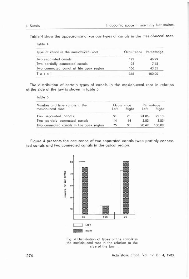
Table 4 show the appearance of various types of canals in the mesiobuccal root.
Table 4
Type of canal in the mesio'buccal root Occurrence Percentage
Two separated canals 172 46.99Two partially connected canals 28 7.65Two connected canal at hte apex region 166 43 35
T o t a l 366 100.00
s d is tribu tion of certa in types of canals i side of the jaw is shown in tab le 5.
in the mesiobuccal root in rel
Table 5
Number and type canals in the Occu rrence Percentagemesiobuccal root Left Right Left Right
Two separated canals 91 81 24.86 22.13Two partialy connected canals 14 14 3.83 3.83Two connected canals in the apex region 75 91 20.49 100.00
Figure 4 presents the occurence of two separated canals twoo partia ly connected canals and two connected canals in the ap ica l region.
Fig. 4 Distribution of types of the canals in the mesiobuccal root in the relation to the
side of the jaw
274 Acta stom. croat., Vol. 17, Br. 4, 1983.

J. Šutalo Endodontic space in m axillary firs t molars
Figure 5, 6, 7 presents the section of the mesiobuccal root w ith d iffe ren t types of the ir canals.
Fig. 7 Two connected canals in the mesiobuccal
root at the apex region
Fig. 5 Two separate canals in the mesiobuccal
root
Fig. 6 Two partially connected canals in the me
siobuccal root
It was established tha t the m axillary firs t perm anent molars can have in their mesiobuccal root two separate canals, two pa rtia lly connected canals and two canals which jo in in the apex region. This phenomenon does not depend upon the side of the jaw. In a ll types of canals in the mesiobuccal root a marked sp litting on the apex of the root up to 1 mm was observed (Fig. 8).
Fig. 8 Distribution of the three types of canals in the mesiobuccal root in relation to the marked splitting of the apex of the root
Acta stom. croat., Vol. 17, Br. 4, 1983. 275

J. Šutalo Endodontic space in m axillary firs t molars
The obta ined and analysed data show tha t the type of the canals does not; depend on sp litting of the root apex. This substantiates the hypothesis tha t in separated add itiona l secondary canals three is the same percentage of teeth w ith sp litting of the apex of the root and teeth w ithou t sp litting (p robab ility pi = 0.81, p2 — 0.86).
Three canals were found in the mesiobuccal root in 5.6% They can be d ifferently o rie n ta te d :a. Three canals connected at th ird of the apexb. The th ird canal connected w ith the primary canalc. Secondary canal connected w ith the primary canal, andd. Three separated canals.
My observation regarding the distobuccal root of the m axillary first perm anent molars. Two canals have found in 1.81%. In the pa la ta l root two canals have found in 0,9% of the cases. These canals are also d iffe rn tly orientated.
DISCUSSION
Numerous investigations performed during the past years have revealed tha t the m axillary firs t perm anent molar has a very complex endodontic morphology. This refers especially to the mesiobuccal root.
Various data on the presence secondary canal in the mesiobuccal root of the m axillary perm anent molar have been published. This variety is due to d iffe rent methods app lied in the investigations and to the fact th a t in the invests gations the firs t and second molars were considered together. According to my c lin ica l experience m axillary first and second perm anent molars show a series of m orphological d ifferences which lead to d iffe ren t relations w ith in the pulp space. Lenhossek7 reports tha t the secondary canal was found in mesiobuccal root of the m axillary firs t molars in 620/o. Hesss investigated m axillary first and second molars together, and concluded th a t the secondary canal was present in 53% o f the cases only.
G reen10 reports double canals in the mesiobuccal root in 50%, O kum ura4 in 52%, W eine8 in 51.5% of the cases. Nosonowitz and Brenner11 in the ir in vivo reported 69%, and Pineda and Kuttle r13 in 74.70/o of the cases. Acosta and Tru- geda14 reported on the presence of the secondary canal in 68.40%.
The comparison of the above data on occurrence of the secondary canals in the m esiobuccal root w ith my results shows somewhat a h igher appearance of the add itiona l secondary canals, which is due to sim plic ity of my method used and the ob jectiv ity of evaluation based upon d irect observation using the d iscribed method.
The occurrence of the two separated canals varies from 8.2% (Bence15) to 47% (P ineda16). In th is study two separated canals in mesiobuccal root have been found in 46.99% of the cases. In comparison the shape of the mesiobuccal root w ith the type of the canal, a regularity in the appearance of a marked saplit- ting on the apex of the root has been observed. However, by analyzing the data it was established tha t the type of the canal does not depend on the presence of
276 Acta stom. croat., Vol. 17, Br. 4, 1983.

J. Šutalo Endodontic space in m axilla ry firs t molars
a sp litting of the root. Kerekes and Tronstad17, on the basis of the investigation carried out on 40 m axillary firs t perm anent molars, have reported about in 10% two canals in the pa la ta l root. Com paring this result w ith my own I believe tha t this percentage is too high, because I could not verify it on 443 m axillary first perm anent molars (Šutalo18). My results are identica l w ith results C ede19 and al. being 0.9%.
Further thoroughly investigations are necessary to c la rify the problem of endodontic space in m axillary firs t perm anent molars.
CONCLUSION
Based on results in the investigation of 443 random ly chosen m axillary first perm anent molars, the fo llow ing may be d raw n:1. One canal in the mesiobuccal root was found in 52 samples or 11.74%.2. The occurence two canals in the mesiobuccal root was found in 366 samples
or 82.62%.3. Three canals in the mesiobuccal root of the m axillary firs t molars were found
in 25 or 5.6% of the cases.4. Two canals in the distobuccal root were found in 8 samples or 1.81%.5. Two canals in the pa la ta l root were found in 4 specimen or 0.9%.
Analyzing the obta ined data, it has been established tha t there is no s ign ifican t d ifference between the occurrence of two canals and in the respect of the left and the righ t side of the jaw.. There has not been a re lation between the type of the canal and the sp litting at apex of the mesiobuccal root.
References
1. CARABELLI, G.: Systematisches Handbuch der Zahnheikunde Braumüller und Seidl, Wien, 1844.
2. LENHOSSEK, M.: Makroskopische Anatomie und Handb. der Zahnheilkunde, Bd. I. J. Scheff, Urban-Schwarzenberg, Wien—Leipzig, 1922.
3. BARRETT, M. T.: The internal anatomy of the teeth with special reference to the pulp with branches Dent. Cosmos, 67 : 581—582, 1925.
4. OKUMURA, T.: Anatomy of the root canals J. Am. Dent. Assoc. 6:632, 1927.
5. HESS, W.: The Anatomy of the root canals J. Am. Dent. Assoc., 23:1968, 1936.
6. MEYER, W. H.: Die Anatomie der Wurzelkanäle Dtsch. zahnartzl Z., 14:1239, 1959.
7. SKILLING, W., G.: Morphology of the root canals J. Am. Dent. Assoc., 19:719, 1932.
8. WEINE, F.: Endodontic therapy St. Louis, The C. V. Mosby Co. 1976.
9. WHEELER, R. C.: Pulp cavities of the permanent teeth W. B. Saunders Co Philadelphia, 1976.
10. GREEN, W.: Morphology of the root canals of the permanent teeth. Oral. Surg. 8:743, 1965.
11. NOSONOWITZ, D. M., BRENNER, M. R.: The mayor canals of the mesiobuccal root of the maxillary first and second molars. Ny. J. Dent. 43:12, 1973.
12. POIME'RANZ, H. H., FISHELBERG, G.: The secondary mesiobuccal canal of maxillary molars, J. Am. Dent. Assoc., 88:119, 1974.
13. PINEDA, F., KUTTLER, Y.: Roentgeno- graphic investigation on the mesiobuccal root of the maxillary first molar, Oral. Surg. 36:235, 1973.
Acta stom. croat., Vol. 17, Br. 4, 1983. 277
J. Šutalo Endodontic space in m axillary firs t mo'lars
14. ACOSTA, V. S., TRUGEDA, B. S.: Anatomy of the pulp chamber floor of the permanent maxillary first molars, J. Endodon., 4:214, 1978.
15. BENCE, R.: Clinical Endodontics, The C. V. Mosiby Co. St. Louis, 1976.
16. PINEDA, F.: Roentgenographic investigation of the mesiobuccal root of the maxillary first molar, Oral. Surg., 36:253, 1973.
17. KEREKES, K., TRONSTAD, L.: Morpho
metrie observation of the root canals of the human molars, J. Endodon., 3:114, 1977.
18. ŠUTALO, J.: Sekundarni kanal u mesio- bukalnom korijenu prvog gornjeg kutnjaka s posebnim osvrtom na endo- dontski tretman, Thesis, Zagreb 1979.
19. CECIC, P., HARTWELL, G., BELLIZI, R.: The multiple root canal system in the maxillary first molar: a case report, J. Endodon., 8:113, 1982.
Sažetak
U ovom je radu ispitivana morfologija endodontskih prostora prvih gornjih kutnjaka. Poznata je činjenica da se neuspjesi u endodontskom tretmanu prvog gornjeg kutnjaka osim neadekvatnog tehničkog pristupa mogu pripisati i nedovoljnom poznavanju morfologije endodontskog prostora.
Činjenica da se u stomatološkoj literaturi dugi niz godina raspravlja o en- dodonskoj morfologiji prvog gornjeg kutnjaka nije bila zapreka da se pristupi ovom istraživanju iz razumljivog razloga što su podaci iz literature vrlo različiti, a kadikad i oprečni. U nakani da se ustanovi stvaran broj kanala u korje- novima prvog gornjeg kutnjaka, njihov međusobni odnos kao i moguće morfološke varijacije, nasumce je uzeto 443 ekstrahiranih prvih gornjih kutnjaka (221 lijevih i 223 desnih). Nakon čišćenja i sušenja svi su uzorci otvarani na okluzijskoj plohi turbo-bušilicom i dijamantnim brusnim tijelom do krova pul- pne komorice, koji je zatim odstranjen čeličnim svrdlom br. 2 do potpunog nestanka rogova pulpe. Ulazi u primarni mezio — bukalni, disto-bukalni i palatinalni kanal su uvijek bili vidljivi, dok se ulaz u sekundarni mezio — bukalni kanal nije mogao vidjeti bez dodatnog uklanjanja dijela mezioaproksi- malne stijenke pulpne komorice.
U ovako prikazane kanale postavljeni su Kerr proširivači No 008-0010, a zatim je izvršeno uzdužno brušenje mezio-bukalnog i disto-bukalnoq kori'ena Ho p n/on kontakta s površinom p^oši riva ča. Nakon uklanjanja proširivača izvršeno ie bojenie kanala polikolor bojama.
U raspravi o rezultatima se aovori o broiu kanala u poiedinim korienovima prvog gornjeg kutnjaka. Najveću pažnju je privukao meziobukalni korijen zbog složenosti svoje morfologije. Od sveukupno ispitanih 443 prvih gornjih kutnjaka u meziobukalnom korijenu utvrđeno je 52 zuba s jednim kanalom ili11.74%, 366 zubi ili 82. 62% s dva kanala i 25 zubi ili 5.64% s tn kanala. U distobukalnom korijenu su dva kanala utvrđena u 8 zubi ili 1.81%, dok su u palatinalnom korijenu dva kanala nađena samo u 4 zuba ili 0.9%. Koriien- ^ki kanali moau imati različite međusobne odnose. U meziobukalnom kori- ienu s lijeću se potpuno separirana dva kanala u 172 uzorka ili 46.99%, dva dielomično spojena kanala u 28 uzoraka ili 7.65% u dva kanala koji se međusobno spajaiu u predielu aoeksne trećine koriiena u 166 uzoraka ili 43 35%.
U daljnjem tijeku rada se raspravljalo o povezanosti između pojave separi- arnih kanala u meziobukalnom korijenu sa znakovima kalanja na apeksu korijena, kao i korelaciji između pojave dvostrukih kanala i strane čeljusti. Utvrđeno je da ne postoji signifikantna razlika između ove dvije pojave.
Ključne riječi: pulpa, korjenski kanali, morfologija
278 Acta stom. croat., Vol. 17, Br. 4, 1983.