Investigation of in vivo effects by E. coli virulence factors using wild
25
Investigation of in vivo effects by E. coli virulence factors using wild type and mutant Caenorhabditis elegans as an invertebrate animal infection model system Malin Jonsson Degree project in biology, Master of science (2 years), 2009 Examensarbete i biologi 30 hp till masterexamen, 2009 Biology Education Centre, Uppsala University, and Umeå Centre of Microbial Research, Umeå University Supervisors: Bernt-Eric Uhlin and Sun Nyunt Wai
Transcript of Investigation of in vivo effects by E. coli virulence factors using wild

Investigation of in vivo effects by E. colivirulence factors using wild type and mutantCaenorhabditis elegans as an invertebrateanimal infection model system
Malin Jonsson
Degree project in biology, Master of science (2 years), 2009Examensarbete i biologi 30 hp till masterexamen, 2009Biology Education Centre, Uppsala University, and Umeå Centre of Microbial Research, UmeåUniversitySupervisors: Bernt-Eric Uhlin and Sun Nyunt Wai

Table of contents
1. Summary 2 2. Abbreviations 4 3. Introduction 5
3.1 Caenorhabditis elegans 5 3.2 Pore forming toxins 8 3.3 Lipopolysaccharides 11 3.4 Aims 13
4. Results
4.1 Survival assays 14 4.2 Effects on nematode physiology and behaviour 19 4.3 Analysis of bacterial cell proteins 21
5. Discussion 22
6. Materials and Methods 6.1 Bacterial strains, culture conditions and plasmids 28 6.2 Caenorhabditis elegans maintenance 30 6.3 Caenorhabditis elegans killing assay 31 6.4 Gel electrophoresis and Western blot analysis 32
7. Acknowledgements 33 8. References 35
1

1. Summary In this degree project an invertebrate animal in vivo model system was established to investigate virulence factors that cause damage to a host. This model system was the nematode Caenorhabditis elegans. Survival assays of the nematode combining the model organism and bacteria were carried out, measuring survival of nematodes daily, statistically managed by a Kaplan-Meyer statistical method. In eukaryotic cells the unfolded protein response (UPR) is triggered by unfolded or misfolded proteins in the endoplasmic reticulum (ER), sensed by three different transmembrane proteins, one of which is the endoribonuclease Inositol requiring enzyme (Ire-1a). The x-box binding protein 1 (Xbp-1) is a transcription factor regulated by Ire-1a, in turn regulating chaperone-encoding genes. Three different strains of nematodes were used; a wild type strain (N2) and two mutant strains with mutations inactivating the ire-1 or xbp-1 genes, thereby downregulating the UPR system. When these genes are downregulated the nematodes may become sensitive to virulence factors described below which can be observed in vivo. The virulence factors investigated were two pore forming toxins (PFT’s) that form pores in membranes: α-hemolysin and Cytolysin A. Pore-forming toxins are examples of virulence factors produced by bacteria causing urinary tract infections and septicemia in humans. Another major virulence factor is the lipopolysaccharide, LPS, in the outer membrane of Gram negative bacteria. Compositions of LPS differ between bacterial species and strains, affecting the properties of bacteria and their effects on a host. LPS is a major cause of bacterial pathogenicity and is usually referred to as endotoxin. Results from nematode survival assays showed that nematodes lacking the ire-1 and xbp-1 genes were more susceptible than wild type nematodes to bacteria expressing pore forming toxins HlyA and ClyA and with altered LPS. The result from these studies also showed that the nematodes had a shorter life span when the bacteria were grown on high nutrient media such as brain heart infusion agar in comparison with the ordinary growth media used for maintenance of the nematodes. Furthermore, the ire-1 and xbp-1 mutant nematodes were more sensitive to the stress caused by transfer of nematodes between plates. The expression of potential virulence factors by bacteria grown under different conditions was analysed by sodium dodecyl sulfate polyacrylamide gel electrophoresis (SDS-PAGE) and Western blot analysis of protein extract from the bacteria used in the survival assays showed no significant differences.
2

2. Abbreviations Abs – absorbance
DTT – dithiothreitol
ER – endoplasmic reticulum
LB – Luria Bertani medium
LPS – lipopolysaccharides
MDT – membrane damaging toxin
NGM – nematode growth medium
OD – optical density
PBS – phosphate buffered saline
PFT – pore-forming toxins
SDS-PAGE – sodium dodecyl sulfate polyacrylamide gel electrophoresis
UPR – unfolded protein response
3
3. Introduction Bacteria may express different virulence factors that contribute to the colonization and infection of a host. Examples of virulence factors are the fimbrial adhesion factors expressed by many bacteria and the different secreted protein toxins. The effect of bacterial ability to cause infection also includes the interplay with the host's innate immune response. Different bacteria have different kinds of virulence factors and one bacterium can have several different virulence factors (Madigan & Martinko 2005). 3.1 Caenorhabditis elegans During recent years the nematode Caenorhabditis elegans has become an established research organism for studies of bacterial pathogenesis (Kurz & Ewbank 2003). Mammalian model organisms such as rats, pigs and mice require special housing and complex experimental approaches, and are accompanied by large costs and necessitate ethical considerations.(Steinert et al. 2003) Therefore the usage of invertebrate organisms such as nematodes as models for in vivo testing of bacterial virulence has increased. In the 1970’s, the Nobel laureate Sidney Brenner chose C. elegans as a laboratory model organism and ever since then researchers in genetics and microbiology have been using it as a model using his methods (Steinert et al. 2003), (Hill et al. 2000). Some reasons why C. elegans is an attractive model organism are that it has a rapid generation time (ca. 3 days), produces a large number of offsprings from only a few original nematodes, its adult size of maximum 1.5 mm as an adult nematode, its low number of cells (less than 1000 cells) and the ease of growing and maintaining the nematode in the laboratory (Barr 2003). Caenorhabditis elegans is a nematode found in nutrient soil all over the world feeding mostly on bacteria (Altun & Hall 2005). There are several different stages during its lifecycle, including four larval stages (L1-L4) before it develops into an adult nematode (Fig 1) (Altun et al. 2005). There is also a fifth stage called Dauer larva (see figure 4). The nematode becomes a Dauer larva after the L2 stage when environmental conditions are not favourable for further growth (Altun et al. 2005). Environmental conditions such as lack of food and too high or low temperatures (over 27 °C and below 15 °C) act as signals that can trigger formation of a Dauer larva, but also the presence of a certain pheromone that indicates a highpopulation density triggers this formation (Altun et al. 2005). The Dauer larva stage is a reversible, non-aging state, meaning that the larva will resume its development when entering a more favourable environment again. Within 1 hour of accessing food the nematode exits the Dauer stage, starts to feed after 2-3 hours and after about 10 hours it enters the L4 stage (Altun et al. 2005). C elegans is a hermaphrodite, meaning it has the ability to reproduce it self. There are also just male nematodes that together with a hermaphrodite increase the number of offsprings. A hermaphrodite that reproduces on its own can generate about 300 offsprings due to the limited amount of sperm. But if a male nematode contributes to fertilizing, the number of offsprings can increase to 1200-1400. The adult hermaphrodite produces oocytes for ~4 days
4

and after this fertile period the adult nematode lives for about 10-15 days (Altun, Z. F. & Hall, D. H. 2005).
Figure 1. The life cycle of C. elegans at 22°C. After confinement of fertilized oocytes the embryo develops for 14 hrs to a stage 1 larva (L1). 12 hrs later it becomes a stage 2 larva (L2) if in preferable environment conditions, other vice it will enter a resting, reversible Dauer stage. As Dauer larva the nematode can be resting for several months until more favorable conditions occur. Stage 3 and 4 larvae occurs after this with 8 hrs apart and about 10 hrs after that it develops into a grown nematode. It is considered a full grown nematode after about 8 more hours (adapted and with permission from WormAtlas).
The sequence analysis of the entire genome of C. elegans was reported during 1998 (http://www.wormbase.org/db/gb2/gbrowse/c_elegans, 2011). It is easy to modify this genetic system, and make mutants (Hill et al., 2000). 3.2 The unfolded protein response The unfolded protein response (UPR) can be defined as a transcriptional up-regulation of genes that supplement the processing and protein folding capacity of the endoplasmic reticulum, ER (Todd et al. 2008). UPR occurs when proteins due to stress become unfolded or wrongly folded in the ER. This is sensed by three different transmembrane proteins in the ER: i) the protein-kinase and site-specific endoribonuclease inositol-requiring enzyme 1a (IRE-1a) ii) the eukaryotic translation initiation factor 2 kinase, PERK/PEK and iii) the transcriptional activator 6 (ATF6) (Rutkowski et al. 2007; Shen et al. 2001). The IRE-1 transmembrane protein is a direct activator of UPR. In cells with high levels of ER stress there might be a connection between UPR and apoptosis pathways. (Todd et al. 2008). The product of the ire-1 gene is a sensor for misfolded proteins in ER. When it is activated by the presence of such misfolded proteins, it causes alternative splicing of the pre-mRNA encoding the X-box binding protein 1 (Xbp-1). This results in a shift in the reading frame (Todd et al. 2008) and translation of active Xbp-1, which is a transcription factor that activates genes coding for ER-
5

chaperones. IRE1 and XBP1 together with some adaptor proteins are considered to be a signalling platform that can respond to ER stress rapidly (Todd et al. 2008). 3.3 Pore forming toxins Pore forming toxins (PFT’s) can be found in many different pathogenic bacteria and are common virulence factors. They are found in several human pathogens such as Streptococcus pyogenes, Clostridium perfringens and Staphylococcus aureus (Bischof et al. 2008) These toxins belong to a family of toxins called membrane damaging toxins (MDT’s) and are also known as cytolysins or cytolytic toxins. α-hemolysin is a multi-subunit PFT expressed by the Gram negative bacterium Escherichia coli that is associated with urinary tract infections and septicemia in humans. This 110 kDa cytolysin (Kerenyi et al. 2005) is exported through the membranes via the type I secretion system (Alouf 2003, Balsalobre et al. 2006). Cytolysin A, another PFT, is a 34 kDa hemolytic/cytolytic protein (Kerenyi et al. 2005) also expressed by E. coli. It is encoded by the gene clyA (also named hlyE or sheA). E. coli cells expressing ClyA have a cytotoxic effect upon direct contact of mammalian cells. ClyA together with lipid membranes forms an oligomeric pore-like assembly (Oscarsson et al. 1999). Wai et al (2003) have shown that outer membrane vesicles (OMV’s) released from E. coli contained Cytolysin A and define a vesicle mediated transport mechanism in the bacteria that is responsible for the activation and delivery of pathogenic effector proteins. Exactly how eukaryotic cells respond to these toxins is not really known but it was found that the unfolded protein response (UPR) in the endoplasmic reticulum (ER) is activated (Fig 2) during exposure of PFT’s such as cytolysin and α-hemolysin, in both mammalian cells and the nematode Caenorhabditis elegans (Bischof et al. 2008). 3.4 Lipopolysaccharides. Gram negative bacteria, such as E. coli, have two membranes, an inner and an outer membrane enclosing the cytoplasm (van der Ley et al. 1991). The outer membrane is a bilayer with an outer leaflet mainly composed of lipopolysacharide (LPS), with some phospholipids and proteins. LPS contains a polysaccharide chain composed of an inner core polysaccharide and an outer O-specific polysaccharide. The core polysaccharide consists of N-acetylglucoseamine, galactose, gluctose, heptoses and ketodeoxyoctonulate (KDO) and connected to this is the O-specific polysaccharide containing repeats of hexoses such as galactose, mannose and glucose, and dideoxy sugars such as colitose, paratose and abequose. The inner part of the core polysaccharides is Lipid A, a disaccharide composed of N-acetylglucosamine to which fatty acids are linked forming the outer leaflet of the membrane (Madigan & Martinko, 2005). Compositions of LPS, especially the core and O-specific polysaccharide, differ between species and strains and these differences affect the properties of bacteria and their effect on a host, giving a wide variety and strong immunogenicity (Nagy & Pál, 2008). LPS is a major cause of bacterial pathogenicity and is usually referred to as endotoxin. LPS is considered toxic to most animals with involvement causing septic shock in patients with Gram-negative septicaemia (Kim et al. 2006).
6

Figure 2. Schema representation (courtesy of Dr. R. Nakao) of the LPS of lipopolysacharide part (outer leaflet) of the outer membrane in four mutant derivatives (Nakao, R. , Ramstedt, M. , Wai, S.N. , Uhlin, B.E. 2011. Enhanced biofilm formation by Escherichia coli with mutations altering the core lipopolysaccharide composition. Manuscript; Ramstedt M., Nakao R., Wai S.N., Uhlin B.E., Boily J.-F. 2011. Monitoring surface chemical changes in the bacterial cell wall multivaraite analysis of cryo-X-ray photoelectron spectroscopy data. Manuscript submitted for publication) of E. coli BW25113 used in this thesis. Lipid A, core polysaccharide (inner + outer core) and the O specific polysaccharide chain are shown in the mutants. A) E. coli BW25113 wt with intact inner and outer membrane and core oligo saccharides. B) E. coli BW25113 ΔwaaE, a deep rough strain with no outer core and deleted PEtN and Hep from inner core. C) E. coli BW25113 ΔwaaL, a rough strain with intact core oligo saccharides but no long saccharide chain at the end. D) E. coli BW25113 ΔgalE, a smooth strain with Gal deletion in outer core.
The differences between deep rough strains and smooth strains are not quite so easy to determine. Smooth mutants that differ only in the O-specific polysaccharide chains are easier to construct in the lab and they occur in nature giving a high variety of serotypes. Mutants lacking this variable O-specific polysaccharide repeat and expressing only the deeper components of LPS (‘rough’ mutants) are expected to induce an immune response with broader specificity. (Nagy & Pál, 2008) I compared two nematode mutants with the wildtype C. elegans N2 strain, one with down regulation in the ire-1 gene called C. elegans ire-1 and another with down regulated xbp-1 gene called C. elegans xbp-1. I wanted to investigate when these genes are down regulated if the nematodes may become sensitive to pore forming toxins such as α-hemolysin and Cytolysin A. Since unfolded protein response occurs during stress we also considered the possibility that the mutant worms would be sensitive to expression of some modified LPS in outer membranes of the bacteria as well. 3.4 Aims By using an infection model based on the nematode C. elegans my aim was to to examine the effect of bacterial virulence factors, in particular PFTs and LPS, on survival of C. elegans. This was done by assays of nematode survival and documentation of development and effects during infection, to get an idea of the activity, stability and entry of these kinds of toxins in an infected host. I also aimed to optimize the experimental approach by monitoring if there were any changes in the bacteria being used over time with respect to expression of virulence factors after growth on different growth substrates.
7

4. Results 4.1 Survival assays By using Kaplan-Maier data analysis of survival assays of Caenorhabditis elegans the susceptibility of the nematodes to the bacterial pore forming toxins and LPS was measured. The toxins were expressed from plasmids carrying the genes for the α-hemolysin (HlyA) or the Cytolysin A (ClyA) (Fig 3). Initial experiments gave reasons to believe that a downregulation of C. elegans ire-1 and xbp-1 genes affected the susceptibility of the mutant nematodes to pore forming toxins such as α-hemolysin and Cytolysin A might increase the susceptibility to other bacterial virulence factors. The survival of mutant nematodes was clearly reduced in comparison with wildtype nematodes when they were feeding on E. coli MC1061 and E. coli W3110 and there was not much difference seen between the plasmid vector control strain and the PFT-expressing bacterial derivates (Fig. 3). The outer membrane components of the bacteria, in particular the lipopolysaccharides layer (LPS) was considered as a factor that might cause an effect on the mutant nematodes. Therefore I tested four different LPS mutant E. coli strains (Fig. 4). The strains used were E. coli BW25113 wild type with intact inner and outer membrane, E. coli BW25113 ΔwaaE, a deep rough strain with no outer core or oligosaccharides and phosphoethanolamine (PEtN) and Hepatose (Hep) deleted from inner core, E. coli BW25113 ΔwaaL, a rough strain with intact core oligosaccharides but no long saccharide chain in the end, and E. coli BW25113 ΔgalE, a smooth strain with Gal deletion in the outer core. I also tested the wild type E. coli strains MC1061, W3110, and OP50 without any plasmids expressing PFT’s. These bacterial strains also gave a reduced viability of the mutant nematodes (Fig.5).
8

.
Figure 3. Survival of nematodes (n=15) feeding on bacteria with plasmids expressing pore forming toxin ClyA or HlyA or the empty vector pUC18. A, D: wild type C. elegans; B,E C elegans xbp-1; C,F: C. elegans ire-1 . A-C: Nematodes were seeded on bacteria grown on NGM plates lacking cholesterol (NMG –Ch); D-F: Nematodes were seeded on bacteria grown on plates containing half strength LB (LB low nut.). Nematodes were fed with E. coli MC1061 pYMZ80 (ClyA) (green), E .coli MC1061 pANN202-812 (HlyA) (blue) and E .coli MC1061 pUC18 (vector control) (pink, empty vector). Transfer of nematodes between plates was done by using a small platinum wire picking up one or several nematodes at the time and moving them to a new plate. Data for the empty vector in panel D were incomplete and should be disregarded since an experimental error occurred.
9

Figure 4. Survival of nematodes (n=15) feeding on bacteria with varying LPS. A, D: wild type C. elegans; B,E C. elegans xbp-1; C,F: C. elegans ire-1 . A-C: Nematodes were seeded on bacteria grown on NGM plates lacking cholesterol (NMG –Ch); D-F: Nematodes were seeded on bacteria grown on plates containing half strength LB (LB low nut.). Nematodes were fed with E. coli LPS mutant strains BW25113 wt (grey), E. coli BW25113 ΔwaaE (green), E. coli BW25113 ΔwaaL (purple) and E. coli BW25113 ΔgalE (blue). Transfer of nematodes between plates was done by using a small platinum wire picking up one or several nematodes at the time and by moving them to a new plate. Data for graph A were not obtained after day 10.
10

Figure 5. Survival of nematodes (n=15) feeding on different wt strains of E. coli. A, D:
wild type C. elegans; B,E C elegans xbp-1; C,F: C. elegans ire-1 . A-C: Nematodes were seeded on bacteria grown on NGM plates lacking cholesterol (NMG –Ch); D-F: Nematodes were seeded on bacteria grown on plates containing half strength LB (LB low nut.). Nematodes were fed with E. coli MC1061 wt (pink), E. coli W3110 wt (mustard) and E. coli OP50 (blue). Transfer of nematodes between plates was done by using a small platinum wire picking up one or several nematodes at the time and by moving them to a new plate
These results suggested that some E. coli strains themselves could be harmful to the ire-1 and xbp-1 mutant nematodes regardless PFTs were overexpressed or not by the bacteria. Results from a series of tests showed that nematodes lacking ire-1 and xbp-1 were more susceptible to bacteria expressing PFT’s (HlyA and ClyA) and to bacteria with altered LPS than were the wild type nematodes. These mutant nematodes were also susceptible to wild type strains of E. coli MC1061 and E. coli W3110 that did not overexpress the PFTs. Even the non-pathogenic strain OP50 normally used as food for maintenance of the nematodes reduced the viability of the mutant nematodes (Fig 5). Mutant nematodes, as well as wild type nematodes, showed a reduction in survival when feeding on bacteria grown on Luria Bertani (LB) low nutrient plates in comparison with bacteria grown on nematode growth media plates (NGM), especially in the experiments with different LPS-mutants (Fig. 4). LB-medium is more nutrient-rich to E. coli than NGM; since it contains yeast extract. Usually, cholesterol is added to NGM medium but I made NGM without cholesterol (NGM –Ch) since cholesterol binds to Cytolysin A and therefore possibly could interfere with how well the toxin would be taken up by the nematodes. Wild type nematodes grew the fastest, with L4 larvae seen after two to three days; xbp-1 mutant nematodes grew more slowly with L4 larvae seen after three to four
11

days, and ire-1 mutants grew most slowly with L4 larvae seen after seven days. The ire-1 L4 larvae were much smaller than L4 larvae of the other two nematode strains and were overall more sensitive when transferred between plates. Transfer of the nematodes between plates was done by using a small platinum wire picking up one or several nematodes at the time and moving them to a new plate. I also performed a killing assay comparing the survival of nematodes feeding on E. coli strain OP50 growing on NGM plates without cholesterol (NGM -Ch) with that of nematodes feeding on OP50 growing on original NGM plates. Just as predicted there was a difference but only a small one, except for xbp-1 mutant nematodes which had exceptionally low survival when feeding on OP50 in both cases, Fig 6.
0 1 2 3 4 5 6 7 8 9 10 11 12 13 140
102030405060708090
100
N2 + OP50 on NGM
N2 + OP50 on NGM-Ch
xbp-1 + OP50 on NGM
xbp-1 + OP50 on NGM-Ch
ire-1 + OP50 on NGM
ire-1 + OP50 on NGM-Ch
Days
Per
cent
sur
viva
l
Figure 6. Survival of nematodes (n=20) feeding on E. coli OP50 grown on NGM vs. NGM -Ch plates. N2 are the wild type nematodes.
The ire-1- and xbp-1 mutants of C. elegans were killed within six to eight days, i.e. significantly earlier in comparison to wild type nematodes when feeding on the different bacterial strains. When nematodes were moved to new plates, after two to three days and after five to seven days, the death rate increased somewhat, indicating that other stress, such as being transferred from one plate to another also was a factor. Presumably the nematodes were more sensitive to mechanical stress when lacking ire-1 and xbp-1 genes. 5.2 Effects on nematodes physiology and behaviour In most cases the bacteria not just killed the mutant nematode strains more rapidly but also affected the appearance and development of the nematodes. Wild type nematodes always showed good growth, movements and activity regardless on what bacterial strain it grew. They also produced new oocytes and offsprings within a few days and handled a move to new plates without any problem (Fig 7 A). xbp-1- and ire-1 mutant nematodes showed low activity after just a few days of growth on HlyA-, ClyA- or altered LPS strains, just as on OP50. Low activity means they did not move around the plates as much as wild type nematodes even though they were still alive. A nematode was considered dead when it did not react to a gentle touch. When a nematode dies it lyses and disappears from the plate within a day or two. The mutant nematodes produced fewer oocytes and offsprings compared to wild type nematodes. They seemed less able to resist the mechanical stress during a transfer to new plates and
12
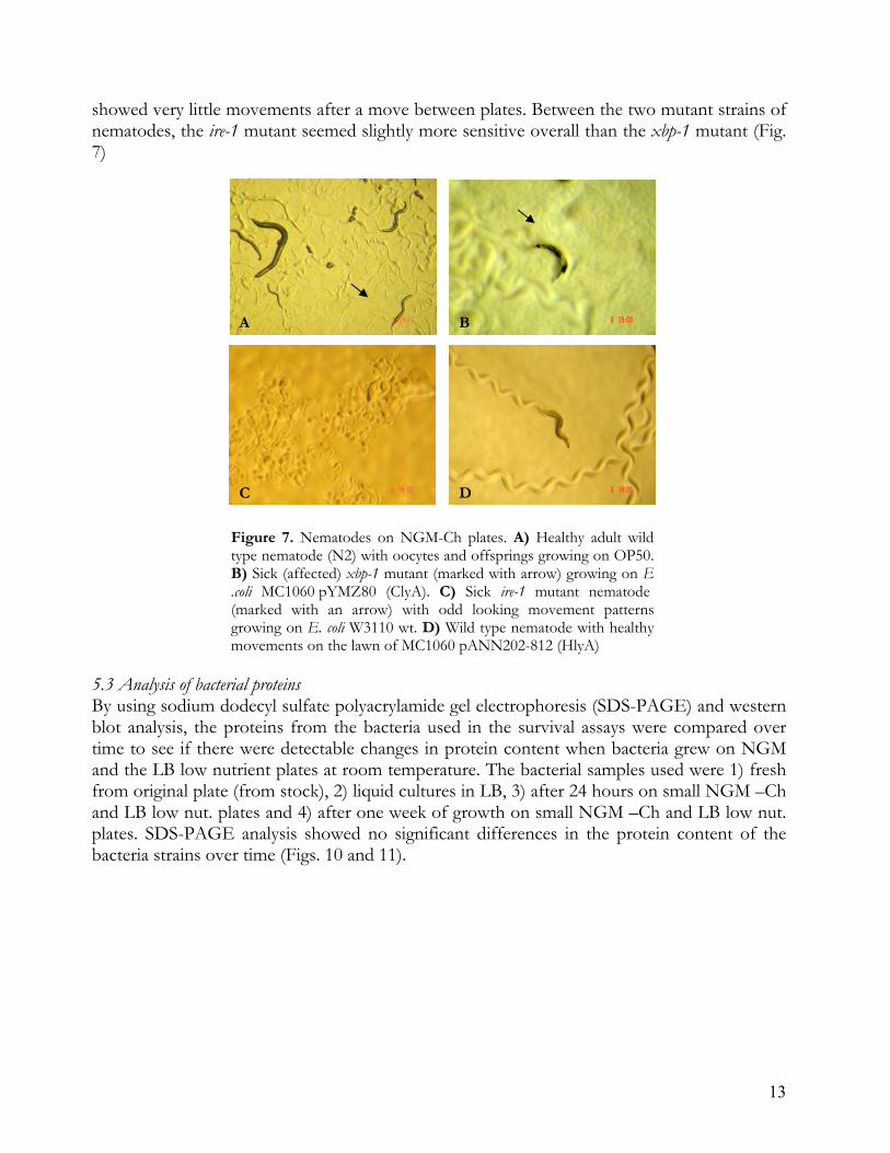
showed very little movements after a move between plates. Between the two mutant strains of nematodes, the ire-1 mutant seemed slightly more sensitive overall than the xbp-1 mutant (Fig. 7) A B C D Figure 7. Nematodes on NGM-Ch plates. A) Healthy adult wild
type nematode (N2) with oocytes and offsprings growing on OP50. B) Sick (affected) xbp-1 mutant (marked with arrow) growing on E .coli MC1060 pYMZ80 (ClyA). C) Sick ire-1 mutant nematode (marked with an arrow) with odd looking movement patterns growing on E. coli W3110 wt. D) Wild type nematode with healthy movements on the lawn of MC1060 pANN202-812 (HlyA)
5.3 Analysis of bacterial proteins By using sodium dodecyl sulfate polyacrylamide gel electrophoresis (SDS-PAGE) and western blot analysis, the proteins from the bacteria used in the survival assays were compared over time to see if there were detectable changes in protein content when bacteria grew on NGM and the LB low nutrient plates at room temperature. The bacterial samples used were 1) fresh from original plate (from stock), 2) liquid cultures in LB, 3) after 24 hours on small NGM –Ch and LB low nut. plates and 4) after one week of growth on small NGM –Ch and LB low nut. plates. SDS-PAGE analysis showed no significant differences in the protein content of the bacteria strains over time (Figs. 10 and 11).
13

Figure 10. SDS-PAGE analysis of proteins in E. coli MC1061 wild types, MC1061 expressing HlyA and
ClyA and W3110 wild types. Letters A, B, C and D represents the different times samples were taken A) fresh from original LB plate, 1: E. coli MC1061 wt, 2: E. coli W3110 wt, 3: E. coli MC1061 pANN202-812 (HlyA), 4: MC1061 pYMZ80 (ClyA) B) from liquid culture in LB (OD600 ~0.5); 1: E. coli MC1061 wt, 2: E. coli W3110 wt, 3: E. coli MC1061 pANN202-812 (HlyA), 4: MC1061 pYMZ80 (ClyA), 5: E. coli OP50. C) after 24 hrs on plates at room temperature; 1: E. coli MC1061 wt on LB low nut., 2: E. coli MC1061 wt on NGM –Ch, 3: E. coli W3110 wt on LB low nut., 4: E. coli W3110 wt on NGM –Ch, 5: E. coli MC1061 pANN202-812 (HlyA) on LB low nut. 6: E. coli MC1061 pANN202-812 (HlyA) on NGM -Ch, 7: MC1061 pYMZ80 (ClyA) on LB low nut, 8: MC1061 pYMZ80 (ClyA) on NGM -Ch, 9: E. coli OP50 on LB low nut., 10: E. coli OP50 on NGM -Ch D) after 1 week at room temperature; 1: E. coli MC1061 wt on LB low nut., 2: E. coli MC1061 wt on NGM –Ch, 3: E. coli W3110 wt on LB low nut., 4: E. coli W3110 wt on NGM –Ch, 5: E. coli MC1061 pann202-812 (HlyA) on LB low nut. 6: E. coli MC1061 pANN202-812 (HlyA) on NGM –Ch, 7: MC1061 pYMZ80 (ClyA) on LB low nut, 8: MC1061 pYMZ80 (ClyA) on NGM -Ch, 9: E. coli OP50 on LB low nut., 10: E. coli OP50 on NGM –Ch. A line in the first gel separates A and B.
14
Figure 11. SDS-PAGE analysis in proteins in E. coli BW25113 LPS mutant strains. Letters A, B, C and D represents the different times samples were taken A) fresh from original LB plate, 1: E. coli BW25113 wt, 2: E. coli BW25113 ΔwaaE, 3): E. coli BW25113 ΔwaaL, 4: E. coli BW25113 ΔgalE) B) from liquid culture in LB (OD600 ~0.5); 1 E. coli BW25113 wt, 2: E. coli BW25113 ΔwaaE, 3): E. coli BW25113 ΔwaaL, 4: E. coli BW25113 ΔgalE). C) after 24 hrs on plates at room temperature; 1: E. coli BW25113 wt on LB low nut., 2: E. coli BW25113 wt on NGM –Ch, 3: E. coli BW25113 ΔwaaE on LB low nut., 4: E. coli BW25113 ΔwaaE on NGM –Ch, 5: E. coli BW25113 ΔwaaL on LB low nut. 6: E. coli BW25113 ΔwaaL on NGM –Ch, 7: BW25113 ΔgalE on LB low nut, 8: BW25113 ΔgalE on NGM -Ch D) after 1 week at room temperature 1: E. coli BW25113 wt on LB low nut., 2: E. coli BW25113 wt on NGM –Ch, 3: E. coli BW25113 ΔwaaE on LB low nut., 4: E. coli BW25113 ΔwaaE on NGM –Ch, 5: E. coli BW25113 ΔwaaL on LB low nut. 6: E. coli BW25113 ΔwaaL on NGM –Ch, 7: BW25113 ΔgalE on LB low nut, 8: BW25113 ΔgalE on NGM –Ch. A line in the first gel separates A and B.

Western blot analyses were done to see whether any changes in protein expression levels occurred during the killing assays, using anti-ClyA (Fig 12) and anti-H-NS (Figs 13-14) antibodies. The results showed that there were no major changes in protein expression over time in the bacterial strains used throughout the experiments. A Western blot for HlyA was also performed but no specific bands were seen after exposure.
Figure 12. Western blot analysis of E. coli MC1061 pYMZ80 (ClyA). The ClyA protein is 34 kDa. Negative control was MC1061 wt - lacking ClyA. The different times when samples were taken were; 1) fresh from original stock on LB plate, 2) from liquid culture in LB (OD600 ~0.5), 3) after 24 hours on NGM-Ch plates and LB low nut. plates and 4) after 1 week at room temperature NGM-Ch plates and LB low nut. plates.
Figure 13. Western blot analysis of H-NS in E. coli BW25113 LPS mutant strains. A) E. coli BW25113 wt, B): E. coli BW25113 ΔwaaE, C): E. coli BW25113 ΔwaaL, D): E. coli BW25113 ΔgalE). The H-NS protein is a 14 kD. Negative control lacking H-NS was E. coli BSN29. The different times when samples were taken was; 1) fresh from original stock on LB plate, 2) from liquid culture in LB (OD600 ~0.5), 3) after 24 hours on NGM-Ch plates and LB low nut. plates and 4) after 1 week at room temperature NGM-Ch plates and LB low nut. plates.
15

Figure 14. Western blot analysis of H-NS in A) wt E. coli W3110 wt strains and B) E. coli MC1061. The H-NS protein is a 14 kD. Negative control lacking H-NS was E. coli BSN29. The different times when samples were taken was; 1) fresh from original stock on LB plate, 2) from liquid culture in LB (OD600 ~0.5), 3) after 24 hours on NGM-Ch plates and LB low nut. plates and 4) after 1 week at room temperature NGM-Ch plates and LB low nut. plates.
16

6. Discussion These studies with different nematode strains, different bacterial derivatives, and different growth media show that the C. elegans model system can be used for monitoring changes caused by bacteria in nematode survival due to altered conditions. However, the results also suggest that one need to be careful in choosing bacteria and growth media for such studies. Different media in the plates resulted in pronounced differences in survival of the mutant nematodes but there were also effects in the case of the wild type nematode. Nematodes on low nutrient LB media survived less well than on NGM plates, even the OP50 E. coli strain that was used as standard nematode food source. When bacteria grow to late exponential phase too quickly (due to very high nutrient uptake) they seem to become harmful to nematodes. Also when the layer of bacteria on the plate was too thick the nematodes disappeared into it and they were very hard to screen (count). This must be considered when choosing bacteria for in vivo experiments using C. elegans. Different bacteria grow different on different media. To test whether bacterial protein expression changed significantly in protein expression over time sodium dodecyl sulfate polyacrylamide gel electrophoresis (SDS-PAGE) and western blot were made. The results indicated that bacterial protein expression remained the same during the duration of the experiments. The results were inconclusive in the case of HlyA where I only got a very weak signal despite repeated experiments.This probably was due to non functional and too unspecific HlyA antibodies. New HlyA antibodies will probably be needed for further analyses in this case. Additional methods using C. elegans as experimental systems could include using fluorescence tagged gene and protein markers (e.g. GFP) introduced into the bacteria before feeding to the nematodes. This would enable tracking of the bacteria inside the nematodes. C. elegans are relatively transparent and it would be very interesting to locate where bacteria end up within the nematode. The results from killing assays ought to be considered somewhat preliminary and should merely serve as guidelines for further experiments since each experiment should be done at least three to four times to ensure reproducibility of the data. I tried out several different strains instead of focusing on the same strain several times throughout this degree project. Nematodes or bacteria may also be affected by conditions not according to the original scheme, like contamination and illness of the experiment holder. A median of several results of the same experiment would be preferred. After I did these experiments there hasn’t been anyone else following these up at this department. But as previously stated C. elegans is becoming a well liked research mechanism much because C. elegans low cost and logistically advantages but also through an ethical view with less laboratory animals. In today’s society science needs to adapt to more rules and using C. elegans in experiments is a way of doing this and results seems to follow (O'Callaghan & Vergunst, 2010). One major research field nowadays is antibiotic resistance in bacteria. We are facing resistance bacteria world wide and C. elegans have now been used in research to
17

understand these bacteria and to be able to fight them. One is using Staphylococcus aureus virulence and pathogenesis (Wu et al, 2010) where researchers are looking into toxicity and virulence of representative clinical isolates of the more wide spread methicillin-resistant S. aureus (MRSA) with no clear results yet, but one step closer to defeat the antibiotic resistance with the aid of the earth worm C. elegans.
18

7. Materials and Methods 7.1 Bacterial strains, culture conditions and plasmids Caenorhabditis elegans strains used throughout the experiments are shown in Table 1. The bacterial strains used throughout the experiments are shown in Table 2. The plasmids introduced by transformation into bacteria are shown in Table 3. Table 1. Caenorhabditis elegans strains
Strains Genotype Source or reference C. elegans N2
Wild type (Brenner 1974)
C. elegans xbp-1
xbp-1 (Bischof et al. 2008)
C. elegans ire-1 ire-1 (Bischof et al. 2008) Bacteria were spread on NGM-Ch plates (3 g NaCl, 2.5 g peptone and 30 g agar mixed in 1l MQ H2O; after autoclaving 1 ml 1M CaCl2, 1 ml 1M MgSO4 and 25 ml KH2PO4 was added) or LB low nut. plates (1.25 g NaCl, 0.625 g yeast extract, 1.25 g tryptone and 15 g agar, mixed in 500 ml MQ H2O). Bacteria were inoculated from regular LB plates (2.5 g NaCl, 1.5 g yeast extract, 2.5 g tryptone and 15 g agar, mixed in 500 ml MQ H2O) and incubated in ~10 ml liquid LB (2.5 g NaCl, 1.5 g yeast extract, 2.5 g tryptone, mixed in 500 ml MQ H2O) at 37 °C til OD600 of ~0.5 was reached. 50 µl was spread out on LB low nut. plates and 75 µl was spread on NGM -Ch plates. The bacteria were not spread all over the plate but as a lawn in the centre of the plate, whereafter the plates were stored at room temperature throughout the experiment. For bacteria containing plasmids, antibiotics were added to the agar media before it was poured into plates and grown to OD600 of ~0.5. Antibiotics used were carbenicillin for E. coli MC1061 pYMZ80 and MC1061 pUC18 and chloramphenicol for MC1061 pANN202 -812.
19

Table 2. Bacteria
Strains Genotype or Phenoype Source or reference E. coli MC1061 hsdR2 hsdM+ hsdS+ araD139
Δ(ara-leu)7697 Δ(lac)X74 galE15 galK16 rpsL (StrR) mcrA mcrB1
(Casadaban & Cohen, 1980)
E. coli W3110 F- λ- rph-1 INV(rrnD, rrnE) (Hill & Harnish, 1981) E. coli BW25113 lacIq rrnBT14 ΔlacZWJ16 hsdR514
ΔaraBADAH33 ΔrhaBADLD78
(Baba et al, 2006)
E. coli BW25113 ΔwaaE lacIq rrnBT14 ΔlacZWJ16 hsdR514 ΔaraBADAH33 ΔrhaBADLD78
ΔwaaE
(Baba et al, 2006)
E. coli BW25113 ΔwaaL lacIq rrnBT14 ΔlacZWJ16 hsdR514 ΔaraBADAH33 ΔrhaBADLD78
ΔwaaL
(Baba et al, 2006)
E. coli BW25113 ΔgalE lacIq rrnBT14 ΔlacZWJ16 hsdR514 ΔaraBADAH33 ΔrhaBADLD78
ΔgalE
(Baba et al, 2006)
E. coli BSN29 MC4100trp::Tn10Δhns, stpA::Kmr
(Johansson et al., 1998)
E. coli OP50 Uracil auxotrof (Brenner 1974)
Table 3. Plasmids
Plasmid Genotype Source and references pANN2020-812 hly gene cluster cloned in
pBR322, Cbr (Ludwig et al. 1987)
pYMZ80 clyA gene cloned in pUC18, Cbr
(Oscarsson et al. 1999)
pUC18 lacZ' bla (bla = the beta-lactamase gene mediating Cb resistance)
(Yanisch-Perron) et al 1985
7.2 Caenorhabditis elegans maintenance For growth of C. elegans I made small agar plates, 5.3 cm in diameter (Fig. 15) containing nematode growth medium (NGM) lacking cholesterol (NGM -Ch) or LB low nutrient plates Nematodes used in experiments were maintained at room temperature on NGM -Ch plates seeded with 100µg E. coli OP50. The bacteria were incubated on the plates for 24 hours where after nematodes were transferred to them and experiments started. At the start of a new experiment 20 (or 15) L4 larvae
20
Figure 15. The small agar plates used for C. elegans experiments. Plates were 5.3 cm in diameter, slightly smaller than a Swedish match.

were put on each plate. Further on during the experiment all surviving nematodes were transferred to new plates to separate the original nematodes from the offsprings. When moving nematodes to new plates, every other or third day, a small platinum wire was used (Fig. 16) carefully picking up the nematodes from underneath from one plate to another. 7.3 Caenorhabditis elegans survival assays For each experiment 20 L4 nematodes of each nematode strain were picked, when a large number of L4 nematodes were present, and seeded on plates (both LB low nut. and NGM –Ch) containing bacteria. In some experiments only 15 nematodes, instead of 20, were used in each assay because to too few L4 nematodes were available to start with. Plates were incubated at room temperature (22 °C) and scored for live nematodes every day for 14 days (a normal length of life cycle for C. elegans). A nematode was considered dead when it no longer responded to touch. Data were subjected to statistical analyses and plotted according to a Kaplan–Meier survival graph by using the program PRISM, version 4.0 (GraphPad, San Diego, USA)
Fig 16. Platinum wire used when picking up and tranferring C. elegance between plates.
7.4 Gel electrophoresis and western blot analysis Sodium dodecyl sulfate polyacrylamide gel electrophoresis: An SDS-polyacrylamide gel consists of a resolving and a stacking gel. A 12.5 % resolving gel was prepared by mixing 4.2 ml acrylamide:bisacrylamide 37.5:1 (Bio-Rad, Stockholm Sweden), 3.77 ml Tris-HCl pH 8.8, 1.89 ml MQ H2O, 100 µl 10% sodium dodecyl sulfate (SDS), 33.5 µl 10% ammonium persulfate (APS) and 6.7 µl TEMED (Sigma-Aldrish, Steinheim Germany). A stacking gel was prepared by mixing 1.25 ml acrylamide:bisacrlyamide 37.5:1, 0.94 ml Tris-HCl pH 6.8, 5.73 ml MQ H2O, 75 µl 10% SDS 10%, 18 µl 10% APS and 15 µl TEMED. Protein samples were mixed with 4SDS loading buffer containing 0.8 g SDS, 20 mg bromphenol blue, 2.6 ml MQ H2O, 2.5 ml Tris-HCl pH 6.8, 4 ml glycerol 50% and 100 µl β mercaptoethanol. Protein samples were boiled for 5 minutes and loaded on 12.5 % one-dimensional SDS for analysis. PageRuler Plus Prestained protein ladder (Fermentas, Vilnius Lithuania) was used as a molecular marker in gels to be stained. Gels were run at 12 mV through the stacking gel and 15 mV through the separation gel. After electrophoresis the gel was washed 3x10 minutes in MQ H2O and soaked in Coomassie Brilliant Blue, Page Blue Protein staining Solution (Fermentas, Vilnius Lithuania) for approx. 2 hours. After staining the gel was put in MQ H2O to remove the excess stain. The gel was saved digitally using a scanner and later dried on a vacuum-drier for 2 hours. Western blot: Proteins were transferred from an SDS-polyacrylamide gel to an polyscreen polyvinylidene fluoride (PVDF) transfer membrane, pore size 0.45 µm (PerkinElmer, Zaventem Belgium) in 1xTransfer Buffer (10x transfer buffer is 250 mM Tris, 100 mM glycine, 2.5 mM DTT). The transfer was carried out at 15 V, 150 mA for 1 hour, after which the membrane (now containing the proteins) was blocked with milk solution (5 g milk powder in 100 ml
21
phosphate-buffered saline (PBS)-Tween (10x transferbuffer is 10.9 g Na2HPO4, 3.2 g NaH2PO4, 90 g NaCl and 1000 ml destilled water and mixed to adjust to pH 7.4 (0.1% Tween) at 4°C over night. To detect the proteins, the membrane was washed in PBS-Tween and then first incubated in 10 ml PBS-Tween (1%) with 100 µl of the primary antibodies (see table 3) for 1 h at room temperature, and washed 3x15 minutes with 1 x PBS-Tween. Table 4. Antibodies
Antibody Dilution Source or Reference Rabbit anti-ClyA- antibodies 1:6000 (Oscarsson et al, 1999) Rabbit anti-H-NS- antibodies 1:5000 (Johansson et al. 1998) Donkey anti-rabbit IgG conjugated to horseradish peroxidase
1:20 000 GE-Healthcare, Buckinghamshire UK.
The membrane was then incubated another hour at room temperature with the secondary anti-rabbit IgG antibody (Table 4). For this incubation, 20 ml PBS-Tween + 1 µl antibody was used. The membrane was washed 3x15 minutes with 1x PBS-Tween (0.1% Tween). Bands were detectied using the ECL-chemiluminescence system (GE-Healthcare, Amersham™, Buckinghamshire, UK) for 5 minutes in dark, putting the membrane filter in a cassette. Photo film (Agfa HealthCare, Mortel Belgium) was processed and pictures made digitally by using ChemiDoc XRS system with Quantity One analysis software (BioRad, Stockholm Sweden). 8. Acknowledgements I would primary like to thank Prof. Bernt Eric Uhlin, my supervisor, for giving me the opportunity to do my degree project, involving my new favourite organism Caenorhabditis elegans, at the department of Molecular Biology at Umeå University. I had a whim, got the chance and took it. It has been great to move “back home” up north for a semester to this fantastic laboratory! I also want to thank Prof. Sun Nyunt Wai for always approaching me with new ideas and telling me what to do. Of course a BIG Thank you to Annika, Monica, Stina and Connie without whom I would not have made it in the lab. Thank you for always having the time for my never ending questions of how everything is supposed to be done in the lab. Thanks to all the rest in the cooperating lab groups; BEU, SNW, JJ and BS for the friendly and educational environment you make in the lab, weekly seminars and in the lunch room. And at last I would like to thank my family for always believing in me, whatever I am up to.
22

9. References C. elegans (current release): 11.6 kbp from III:9,060,076..9,071,672. Available at
http://www.wormbase.org/db/gb2/gbrowse/c_elegans/ [Accessed March 16, 2011].
Alouf, J., 2003. Molecular features of the cytolytic pore-forming bacterial protein toxins. Folia Microbiologica, 48: 5-16.
Altun, Z. F. & Hall, D. H., 2005. WormAtlas. Handbook of C. elegans Anatomy. Available at:
http://www.wormatlas.org/handbook/contents.htm [Accessed March 16, 2011]. Baba, T. Ara, T. Hasegawa, M. Takai, Y. Okumura, Y. Baba, M. Datsenko, K.A. Tomita, M. Wanner, B.L. Mori, H. 2006. Construction of Escherichia coli K-12 in-frame, single-gene knockout mutants: the Keio collection.Mol. Systems Biol. 10.1038/msb4100050 Balsalobre, C. Silvan J M., Berglund S., Mizunoe Y., Uhlin B-E., Wai S. N., 2006. Release of
the type I secreted α-hemolysin via outer membrane vesicles from Escherichia coli. Molecular Microbiology, 59: 99-112.
Barr, M.M., 2003. Super models. Physiol. Genomics, 13: 15-24. Bischof, L.J., KaoC-y., Los F C O., Gonzales M R., Shen Z., Briggs S P., van der Goot G.,
Aroian R V., 2008. Activation of the Unfolded Protein Response Is Required for Defenses against Bacterial Pore-Forming Toxin In Vivo. PLoS Pathogens, 4: e1000176.
Brenner, S., 1974. The genetics of Caenorhabditis elegans, Genetics, 77: 71-94. Casadaban, M.J. & Cohen, S.N., 1980. Analysis of gene control signals by DNA fusion and
cloning in Escherichia coli. Journal of Molecular Biology, 138: 179-207. Ludwig, A., Vogel, M., and Goebel, W.,1987, Mutations affecting activity and transport of
haemolysin in Escherichia coli. Mol Gen Genet 206: 238–245. Gábor Nagy & Tibor Pál, 2008. Lipopolysaccharide: a tool and target in enterobacterial
vaccine development. Biol Chem, 389: 513–520.
Hill, A.A., Hunter C P., Tsung B T., Tucker-Kellogg G., Brown E L., 2000. Genomic Analysis of Gene Expression in C. elegans. Science, 290: 809-812.
Hill, C.W. & Harnish, B.W., 1981. Inversions between ribosomal RNA genes of Escherichia
coli. Proceedings of the National Academy of Sciences of the United States of America, 78: 7069-7072.
23

24
Johansson, J., Dagberg B., Richet E., Uhlin B-E., 1998. H-NS and StpA Proteins Stimulate Expression of the Maltose Regulon in Escherichia coli. J. Bacteriol., 180: 6117-6125.
Kerenyi, M., Allison H E., Batai i., Sonnevend A.,Emödy L., Plaveczky N., Pál T., 2005.
Occurrence of hlyA and sheA Genes in Extraintestinal Escherichia coli Strains. J. Clin. Microbiol., 43: 2965-2968.
Kim, S.Jia W., PArreira V R., Bishop R E., Gyles C L., 2006. Phosphoethanolamine
substitution in the lipid A of Escherichia coli O157 : H7 and its association with PmrC. Microbiology, 152: 657-666.
Kurz, C.L. & Ewbank, J.J., 2003. Caenorhabditis elegans: an emerging genetic model for the
study of innate immunity. Nat Rev Genet, 4: 380-390. van der Ley P, H.J. & van der Ley P, Heckels JE, Virji M, Hoogerhout P, Poolman JT, 1991.
Topology of outer membrane porins in pathogenic Neisseria spp. Infection and immunity, 59: 2963–71.
Madigan, M. & Martinko, J., 2005. Brock Biology of Microorganisms 11th ed., Prentice Hall. O'Callaghan, D. & Vergunst, A., 2010. Non-mammalian animal models to study infectious
disease: worms or fly fishing? Current Opinion in Microbiology, 13: 79-85. Oscarsson, J. Mizunoe Y., Li L., Lai X-H., Wieslander Å., Uhlin B-E., 1999. Molecular analysis
of the cytolytic protein ClyA (SheA) from Escherichia coli. Molecular Microbiology, 32: 1226-1238.
Pimkin, M. & Markham, G.D., 2008. The CBS subdomain of inosine 5’-monophosphate
dehydrogenase regulates purine nucleotide turnover. Molecular microbiology, 68: 342–359. Steinert, M., Leippe, M. & Roeder, T., 2003. Surrogate hosts: protozoa and invertebrates as
models for studying pathogen-host interactions. International Journal of Medical Microbiology, 293: 321-332.
Yanisch-Perron C., Vieira J., Messing J. 1985. Improved M13 phage cloning vectors and host
strains: nucleotide sequences of the M13mp18 and pUC19 vectors. Gene 33:103-119. Wu. K., Conly J., McClure J.-A., Elsayed S., Louie T., K. Zhang, 2010. Caenorhabditis elegans as a
host model for community-associated ethicillin-resistant Staphylococcus aureus, Clinical Microbiological Infection, 16: 245–254


![[15] Recombineering: In Vivo Genetic Engineering in …biology.hunter.cuny.edu/molecularbio/Class Materials Fall 2010 710...[15] Recombineering: In Vivo Genetic Engineering in E. coli,](https://static.fdocuments.us/doc/165x107/5b0a5e027f8b9adc138bfadc/15-recombineering-in-vivo-genetic-engineering-in-materials-fall-2010-71015.jpg)


![[15] Recombineering: In Vivo Genetic Engineering in E. coli, S](https://static.fdocuments.us/doc/165x107/62064bdc8c2f7b1730065d41/15-recombineering-in-vivo-genetic-engineering-in-e-coli-s.jpg)