
Investigation of Immune Biomarkers Using Subcutaneous ... · Colorado State University, USA under...
8
IMMUNE NETWORK Vol. 15, No. 2: 83-90, April, 2015 http://dx.doi.org/10.4110/in.2015.15.2.83 pISSN 1598-2629 eISSN 2092-6685 ORIGINAL ARTICLE 83 Received on December 18, 2014. Revised on January 30, 2015. Accepted on February 6, 2015. CC This is an open access article distributed under the terms of the Creative Commons Attribution Non-Commercial License (http://creativecommons.org/licenses/by-nc/3.0) which permits unrestricted non-commercial use, distribu- tion, and reproduction in any medium, provided the original work is properly cited. *Corresponding Author. Rajpal S. Kashyap, Biochemistry Research Laboratory, Central India Institute of Medical Sciences, 88/2 Bajaj Nagar, Nagpur 440 010, India. Tel: +91-712-2233381/2236441; Fax: 0712-2236416; E-mail: raj_ciims@ rediffmail.com Abbreviations: MTB, M.tuberculosis; TB, Tuberculosis; BCG, Bacillus Calmette Guerin; Hsp, Heath Shock protein; Ag85B, Antigen 85B; ESAT-6, Early secretory antigenic target-6; CFP-10, Culture filtrate protein-10; PPD, Purified protein de- rivative Investigation of Immune Biomarkers Using Subcutaneous Model of M. tuberculosis Infection in BALB/c Mice: A Preliminary Report Aliabbas A. Husain 1 , Hatim F. Daginawala 1 , Shubangi R Warke 2 , Devanand R Kalorey 2 , Nitin V. Kurkure 2 , Hemant J Purohit 3 , Girdhar M Taori 1 and Rajpal S Kashyap 1 * 1 Biochemistry Research Laboratory, Central India Institute of Medical Sciences, 88/2 Bajaj Nagar, Nagpur 440 010, 2 Department of Veterinary Microbiology and Animal Biotechnology, Nagpur Veterinary College, Nagpur 440 006, 3 Environmental Genomic Unit, National Environmental Engineering Research Institute (NEERI), CSIR, Nehru Marg, Nagpur-440 020, India Evaluation and screening of vaccines against tuberculosis depends on development of proper cost effective disease models along with identification of different immune markers that can be used as surrogate endpoints of protection in pre- clinical and clinical studies. The objective of the present study was therefore evaluation of subcutaneous model of M.tuberculosis infection along with investigation of different immune biomarkers of tuberculosis infection in BALB/c mice. Groups of mice were infected subcutaneously with two different doses : high (2×10 6 CFU) and low doses (2×10 2 CFU) of M.tuberculosis and immune markers including hu- moral and cellular markers were evaluated 30 days post M.tuberculosis infections. Based on results, we found that high dose of subcutaneous infection produced chronic dis- ease with significant (p<0.001) production of immune mark- ers of infection like IFNγ, heat shock antigens (65, 71) and antibody titres against panel of M.tuberculosis antigens (ESAT-6, CFP-10, Ag85B, 45kDa, GroES, Hsp-16) all of which correlated with high bacterial burden in lungs and spleen. To conclude high dose of subcutaneous infection pro- duces chronic TB infection in mice and can be used as con- venient alternative to aerosol models in resource limited settings. Moreover assessment of immune markers namely mycobacterial antigens and antibodies can provide us val- uable insights on modulation of immune response post infection. However further investigations along with opti- mization of study protocols are needed to justify the outcome of present study and establish such markers as surrogate endpoints of vaccine protection in preclinical and clinical studies in future. [Immune Network 2015;15(2):83-90] Keywords: Mice, Tuberculosis, Subcutaneous route, Immune markers INTRODUCTION Bacillus Calmette Guerin (BCG), only licensed Tuberculosis (TB) vaccine, has afforded limited protection against TB de- spite of its widespread use in developing countries like India (1,2). To overcome this limitation, a number of different vac- cination strategies have been investigated using different ani- mal models of TB infection (3,4). However, animal models that strictly reproduce the human host-M.tuberculosis (MTB) relationship are required to evaluate new vaccines and thera- pies (5). Aerosol model by far is most widely used route to establish TB infection in mouse since it replicates immuno- logical events in human leading to slow progressive disease
Transcript of Investigation of Immune Biomarkers Using Subcutaneous ... · Colorado State University, USA under...

IMMUNE NETWORK Vol. 15, No. 2: 83-90, April, 2015
http://dx.doi.org/10.4110/in.2015.15.2.83
pISSN 1598-2629 eISSN 2092-6685ORIGINAL ARTICLE
83
Received on December 18, 2014. Revised on January 30, 2015. Accepted on February 6, 2015.CC This is an open access article distributed under the terms of the Creative Commons Attribution Non-Commercial
License (http://creativecommons.org/licenses/by-nc/3.0) which permits unrestricted non-commercial use, distribu-tion, and reproduction in any medium, provided the original work is properly cited.
*Corresponding Author. Rajpal S. Kashyap, Biochemistry Research Laboratory, Central India Institute of Medical Sciences, 88/2 Bajaj Nagar, Nagpur 440 010, India. Tel: +91-712-2233381/2236441; Fax: 0712-2236416; E-mail: raj_ciims@ rediffmail.com
Abbreviations: MTB, M.tuberculosis; TB, Tuberculosis; BCG, Bacillus Calmette Guerin; Hsp, Heath Shock protein; Ag85B, Antigen 85B; ESAT-6, Early secretory antigenic target-6; CFP-10, Culture filtrate protein-10; PPD, Purified protein de-rivative
Investigation of Immune Biomarkers Using Subcutaneous Model of M. tuberculosis Infection in BALB/c Mice: A Preliminary ReportAliabbas A. Husain1, Hatim F. Daginawala1, Shubangi R Warke2, Devanand R Kalorey2, Nitin V. Kurkure2, Hemant J Purohit3, Girdhar M Taori1 and Rajpal S Kashyap1*1Biochemistry Research Laboratory, Central India Institute of Medical Sciences, 88/2 Bajaj Nagar, Nagpur 440 010, 2Department of Veterinary Microbiology and Animal Biotechnology, Nagpur Veterinary College, Nagpur 440 006, 3Environmental Genomic Unit, National Environmental Engineering Research Institute (NEERI), CSIR, Nehru Marg, Nagpur-440 020, India
Evaluation and screening of vaccines against tuberculosis depends on development of proper cost effective disease models along with identification of different immune markers that can be used as surrogate endpoints of protection in pre-clinical and clinical studies. The objective of the present study was therefore evaluation of subcutaneous model of M.tuberculosis infection along with investigation of different immune biomarkers of tuberculosis infection in BALB/c mice. Groups of mice were infected subcutaneously with two different doses : high (2×10⁶ CFU) and low doses (2×10² CFU) of M.tuberculosis and immune markers including hu-moral and cellular markers were evaluated 30 days post M.tuberculosis infections. Based on results, we found that high dose of subcutaneous infection produced chronic dis-ease with significant (p<0.001) production of immune mark-ers of infection like IFNγ, heat shock antigens (65, 71) and antibody titres against panel of M.tuberculosis antigens (ESAT-6, CFP-10, Ag85B, 45kDa, GroES, Hsp-16) all of which correlated with high bacterial burden in lungs and spleen. To conclude high dose of subcutaneous infection pro-duces chronic TB infection in mice and can be used as con-venient alternative to aerosol models in resource limited settings. Moreover assessment of immune markers namely mycobacterial antigens and antibodies can provide us val-uable insights on modulation of immune response post
infection. However further investigations along with opti-mization of study protocols are needed to justify the outcome of present study and establish such markers as surrogate endpoints of vaccine protection in preclinical and clinical studies in future.[Immune Network 2015;15(2):83-90]
Keywords: Mice, Tuberculosis, Subcutaneous route, Immune markers
INTRODUCTION
Bacillus Calmette Guerin (BCG), only licensed Tuberculosis
(TB) vaccine, has afforded limited protection against TB de-
spite of its widespread use in developing countries like India
(1,2). To overcome this limitation, a number of different vac-
cination strategies have been investigated using different ani-
mal models of TB infection (3,4). However, animal models
that strictly reproduce the human host-M.tuberculosis (MTB)
relationship are required to evaluate new vaccines and thera-
pies (5). Aerosol model by far is most widely used route to
establish TB infection in mouse since it replicates immuno-
logical events in human leading to slow progressive disease

Investigation of Immune Markers in MiceAliabbas A. Husain, et al.
IMMUNE NETWORK Vol. 15, No. 2: 83-90, April, 201584
development. However, requirement of Biosafety level-3
(BSL-3) facilities and high maintenance cost limits their usage
in many resource limited settings of developing world. Other
routes of infection such as subcutaneous, intravenous and in-
tranasal have been investigated in other studies for develop-
ment of TB model in animals (6-8). Although subcutaneous
route may not mimic actual development of infection in ani-
mals, however, it can be used as convenient alternative to
aerosol route (9,10) in resource limited settings. Dose of MTB
infection used is another contributing factor, since outcome
of successful infection generally depends on amount of bac-
teria colonizing in lungs and other organs of host animal used
(11). Thus along with route of infection, standardization of
optimal dose required for infection is mandatory in develop-
ment of proper disease model of TB infection.
Another major aspect of vaccine evaluation is identifcation
of appropriate markers that can be used as surrogate end-
points of protection. Infection with MTB leads to diverse im-
mune response, thereby producing wide range of biomarkers
(11). Whether or not infection will lead to development of
disease depends on the outcome of a complex interaction be-
tween the pathogen and the host's immune response (12).
Therefore, based on our understanding of the pathogen-host
interactions, the design of superior vaccines or drugs against
mycobacterial infections can be facilitated. Assesment of bio-
markers, especially MTB antigens and antibodies produced
during infection provide us useful insight with respect to de-
velopment of disease and may also increase our under-
standing about their production during infection. Moreover,
with increase in ethical concerns regarding number of ex-
perimental animals used and sacrificed in vaccination studies,
identification and evaluation of such biomarkers as surrogate end-
points are need in preclinical evaluation of such studies in future.
Keeping the existing questions in mind, the objective of the
study was to evaluate subcutaneous model of TB using two
different doses of MTB infection in BALB/c mice. Apart from
evaluation of subcutaneous model, the study also focused on
evaluation of different immune markers post MTB infection
which can be used as surrogate endpoints for evaluation of
different vaccine candidates under preclinical and clinical
stage of development.
MATERIALS AND METHODS
Mice Female BALB/c mice, 6∼8 weeks old, were obtained from
National Institute of Nutrition (NIN, India), Hyderabad. Mice
were housed under aseptic conditions and provided with
food and sterile water. Prior to experiments, all mice were
acclimatized for 15∼20 days.
Antigens and antibodiesMTB H37Rv antigens Ag85B, ESAT-6, CFP-10, Gro-ES, and
Hsp16 along with Monoclonal antibodies against Hsp16
(alpha-crystalline like-Rv2031c, hspX), Hsp65 (Rv0440, cpn60.2,
GroEL) and Hsp71 (Rv0350, DnaK), were obtained from
Colorado State University, USA under the TB research materi-
als and vaccine testing contract (NO1-AI-75320). The secon-
dary antibody rabbit anti-mouse IgG-HRP was obtained from
Genei, Banglore, India. MTB purified protein derivative
(PPD) was obtained from Span Diagnostics, Banglore, India.
MTB infection of miceThe MTB H37Rv was grown in 7H9 Middlebrook Broth
(Himedia laboratories, India) to mid log phase. The bacterial
suspension was diluted in phosphate buffered saline (PBS)
and adjusted according to the number 1 McFarland scale.
Depending upon the load of mycobacteria to be infected, the
cultures were serially diluted in sterile saline. Mice were div-
ided into two different experimental groups (n=10, each
group), and infected subcutaneously with 2×10⁶ CFU (for
high dose), and 2×10² (low dose). All procedures of cultur-
ing and infection were carried out in BSL facilities. A control
group of mice (n=10) were separately maintained and sham
immunized with sterile saline. 30 days after MTB infection,
blood was collected to obtain serum and used for immuno-
logical marker analysis.
Analysis of Antigen and Antibody response MTB heat shock proteins (Hsps) 16, 65, 71 were detected us-
ing in house developed ELISA method as described elsewhere
(13). Total IgG was estimated using ELISA protocol as de-
scribed earlier by Husain et al. (2). The antibody response
against MTB antigens (Ag85, ESAT-6, CFP-10, Gro-ES, and
Hsp16) were evaluated using in house ELISA method devel-
oped by Kashyap et al. Briefly, the 96-well microtiter plates
(MaxisorpImmunoplate, Nalge Nunc International, Naperville,
IL, USA) were coated with MTB antigens. After 3 hours of
incubation at 37oC, the plates were washed and blocked with
0.25% bovine serum albumin (BSA) in PBS pH 7.4. After 2
hours of incubation plates were washed twice and kept over-
night at 4oC. Next day plates were incubated with serum sam-
Investigation of Immune Markers in MiceAliabbas A. Husain, et al.
IMMUNE NETWORK Vol. 15, No. 2: 83-90, April, 2015 85
ples (1:400 diluted) in PBS. After 30 minutes of incubation
plates were washed and incubated with rabbit anti-mouse
IgG, HRP conjugate (1:10,000) for 30 min. For colour devel-
opment tetramethyl benzidine in hydrogen peroxide
(TMB/H2O2) substrate was added and incubated for 10 min.
The reaction was terminated by adding 2.5 N sulphuric acid
and the absorbance of colour in each well was read at 450
nm. Anti-PPD titre was estimated by protocol for antibody de-
tection described earlier.
Enumeration of bacterial load in lungs and spleen Thirty days after MTB infection, mice from respective groups
(n=7) were sacrificed. Lungs and spleen were isolated, homo-
genized and serially diluted. These serially diluted homoge-
nates were innoculated in Middle brook 7H9 liquid medium
along with oleic acid, albumin, dextrose and catalase (OADC)
enrichment and antibiotic supplements in BacT culture bottles
(Biomeriux, France) and were incubated at 37oC in BacT/Alert
system (Biomerieux) for 28∼35 days. Mycobacterial load in
organs was determined in terms of mean time taken by organ
cultures from respective groups to become positive in BacT/Alert
system as described elsewhere by Kolibab et al. (14).
Cytokine estimation Cytokine Interferon gamma (IFN-γ) was assessed in spleeno-
cytes as per manufacturer's instruction (Bender Med System,
Austria). In brief, anti-IFN-γ-monoclonal coating antibody
was adsorbed onto micro wells. After two hours of incubation
at room temperature, the wells were washed and blocked
with 0.5% BSA in PBS. After one hour, spleenocytes (1:400
diluted) followed by biotin-conjugated anti-cytokine antibody
was added to the coated wells. After another two hours of
incubation, streptavidin-HRP (horseradish peroxidase) was
added to the wells. After one hour of incubation, substrate
solution reactive with HRP was added to the wells. The re-
action was terminated by the addition of 2.5 N sulphuric acid
and the absorbance of colour was read at 450 nm.
Observation of granulomas and necrosis in lungA section was prepared from the base of the apical lobe and
from the diaphragmatic lobe of the left lung, representing two
distinct regions of the organ. Duplicate sections were stained
with haematoxylin and eosin and Van Gieson in order to aid
visualization of fibrous tissue. The two sections were scored
in a blinded fashion for the following features: percentage of
the section occupied by granulomatous inflammation, healthy
lung, interstitial pneumonitis, and necrosis. The presence of
epithelioid cells and the extent of fibrosis and lymphocytic
infiltration (and whether around vessels or in granulomas in
both cases) were also assessed. Amount of granulomatous le-
sions were scored in terms of percentage (%) occupied by
them in lungs.
Ethical Committee ApprovalAll protocols for animal experiments were approved by
Institutional Animal Ethics Committee of Central India Institute
of Medical Sciences, Nagpur.
Statistical analysisData are expressed as mean±standard deviation (SD). Graphs
were plotted using Graph Pad Prism 6 software. For multiple
comparisons Paired t-test was used for obtaining statistical sig-
nificance in different dose groups. p<0.05 was considered
statistically significant (**) and p<0.001 for highly significant
(***) values.
RESULTS
High dose of MTB infection induced significant antibody response compared to low dose and control mice groupsAntibodies were estimated 30 days post MTB infection. Blood
was harvested from different groups of mice to obtain serum.
Fig. 1A and B indicates total IgG and anti-PPD titres in differ-
ent dose and control groups. Mice infected with high dose
showed significantly (p<0.001) high total IgG and anti-PPD
response comapred to low dose and control group. Antibody
titres against panel of MTB H37Rv antigens (ESAT-6, CFP-10,
GroES, Hsp-16, 45kD, Ag85B) showed similar results with sig-
nificantly higher titres against Ag85B, ESAT-6, and CFP-10 in
high dose group (Fig. 2). On the contrary, levels of anti-
bodies against GroES, 45kDa and Hsp16 were found to be
similar in both dose groups.
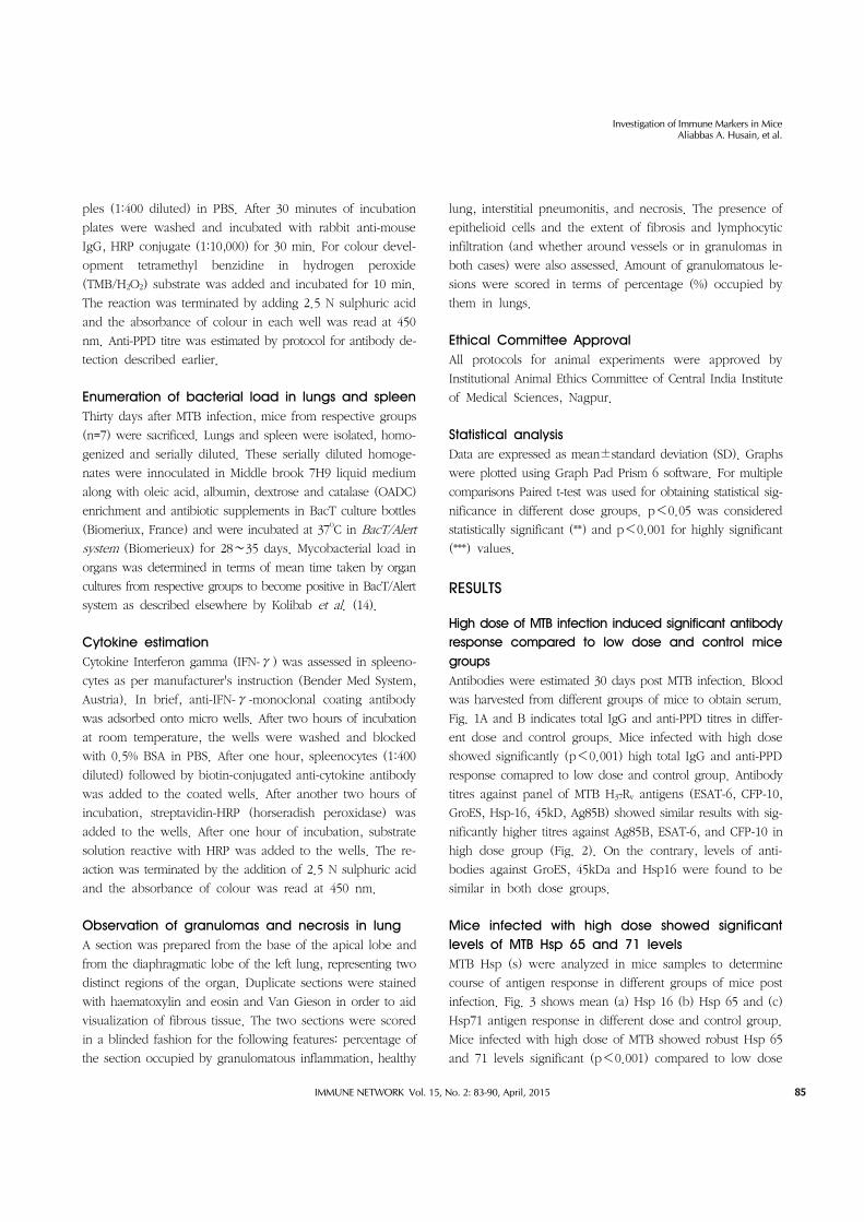
Mice infected with high dose showed significant levels of MTB Hsp 65 and 71 levelsMTB Hsp (s) were analyzed in mice samples to determine
course of antigen response in different groups of mice post
infection. Fig. 3 shows mean (a) Hsp 16 (b) Hsp 65 and (c)
Hsp71 antigen response in different dose and control group.
Mice infected with high dose of MTB showed robust Hsp 65
and 71 levels significant (p<0.001) compared to low dose

Investigation of Immune Markers in MiceAliabbas A. Husain, et al.
IMMUNE NETWORK Vol. 15, No. 2: 83-90, April, 201586
Figure 1. (A) Mean Total IgG and (B) anti-PPD levels in serum of mice (n=7, each group) in high dose, low dose and control groups. Anti-body levels were estimated 30 days after development of TB infection. Paired t-test was used to compare and obtain statistical significance. Error bars indicate standard error of mean. **Represents significant (p<0.05) and ***represents highly significant (p<0.001) values.
Figure 2. Scatter plot of antibody levels against panel of MTB H37Rv antigens (a) Ag85B (b) 45kDa (c) Hsp 16 (d) GroEs (e) ESAT-6 and (f) CFP-10 in serum of mice (n=7, each group) after TB infection. Paired t-test was used to compare and obtain statistical significance. Error bars indicate standard error of mean. **Represent significant (p<0.05) and ***represents highly significant (p<0.001) values.
groups, while both dose groups showed similar levels of Hsp
16 post TB infection.
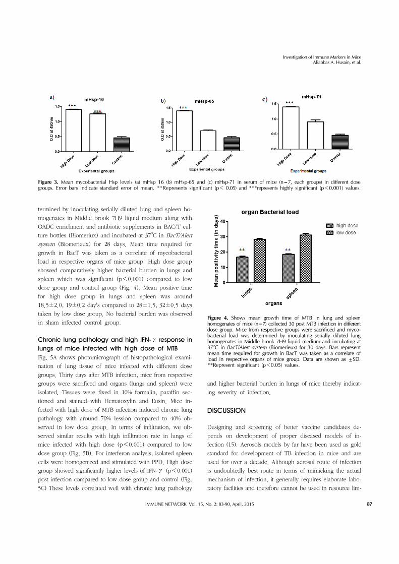
High dose of MTB infection produced higher bacterial burden in lungs and spleens of mice compared to low dose groupMice from respective groups (n=7) were sacrificed after 30
days of MTB infection and mycobacterial load was de-
Investigation of Immune Markers in MiceAliabbas A. Husain, et al.
IMMUNE NETWORK Vol. 15, No. 2: 83-90, April, 2015 87
Figure 3. Mean mycobacterial Hsp levels (a) mHsp 16 (b) mHsp-65 and (c) mHsp-71 in serum of mice (n=7, each groups) in different dose groups. Error bars indicate standard error of mean. **Represents significant (p< 0.05) and ***represents highly significant (p<0.001) values.
Figure 4. Shows mean growth time of MTB in lung and spleen homogenates of mice (n=7) collected 30 post MTB infection in different dose group. Mice from respective groups were sacrificed and myco-bacterial load was determined by inoculating serially diluted lung homogenates in Middle brook 7H9 liquid medium and incubating at 37oC in BacT/Alert system (Biomerieux) for 30 days. Bars represent mean time required for growth in BacT was taken as a correlate of load in respective organs of mice group. Data are shown as ±SD.**Represent significant (p<0.05) values.
termined by inoculating serially diluted lung and spleen ho-
mogenates in Middle brook 7H9 liquid medium along with
OADC enrichment and antibiotic supplements in BAC/T cul-
ture bottles (Biomeriux) and incubated at 37oC in BacT/Alert
system (Biomerieux) for 28 days. Mean time required for
growth in BacT was taken as a correlate of mycobacterial
load in respective organs of mice group. High dose group
showed comparatively higher bacterial burden in lungs and
spleen which was significant (p<0.001) compared to low
dose group and control group (Fig. 4). Mean positive time
for high dose group in lungs and spleen was around
18.5±2.0, 19±0.2 day's compared to 28±1.5, 32±0.5 days
taken by low dose group. No bacterial burden was observed
in sham infected control group.
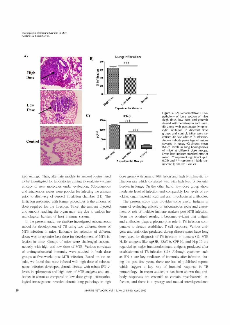
Chronic lung pathology and high IFN-γ response in lungs of mice infected with high dose of MTBFig. 5A shows photomicrograph of histopathological exami-
nation of lung tissue of mice infected with different dose
groups. Thirty days after MTB infection, mice from respective
groups were sacrificed and organs (lungs and spleen) were
isolated. Tissues were fixed in 10% formalin, paraffin sec-
tioned and stained with Hematoxylin and Eosin. Mice in-
fected with high dose of MTB infection induced chronic lung
pathology with around 70% lession compared to 40% ob-
served in low dose group. In terms of infiltration, we ob-
served similar results with high infiltration rate in lungs of
mice infected with high dose (p<0.001) compared to low
dose group (Fig. 5B). For interferon analysis, isolated spleen
cells were homogenized and stimulated with PPD. High dose
group showed significantly higher levels of IFN-γ (p<0.001)
post infection compared to low dose group and control (Fig.
5C) These levels correlated well with chronic lung pathology
and higher bacterial burden in lungs of mice thereby indicat-
ing severity of infection.
DISCUSSION
Designing and screening of better vaccine candidates de-
pends on development of proper diseased models of in-
fection (15). Aerosols models by far have been used as gold
standard for development of TB infection in mice and are
used for over a decade. Although aerosol route of infection
is undoubtedly best route in terms of mimicking the actual
mechanism of infection, it generally requires elaborate labo-
ratory facilities and therefore cannot be used in resource lim-
Investigation of Immune Markers in MiceAliabbas A. Husain, et al.
IMMUNE NETWORK Vol. 15, No. 2: 83-90, April, 201588
Figure 5. (A) Representative Histo-pathology of lungs section of mice (high dose, low dose and control) stained with hematoxylin and Eosin. (B) along with percentage lympho-cytic infiltarion in different dose groups and control. Mice were sa-crificed 30 days after MTB infection. Arrows indicate percentage of lesions covered in lungs. (C) Shows mean INF-γ levels in lung homogenates of mice at different dose groups. Errors bars indicate standard error of mean. **Represent significant (p<0.05) and ***represents highly sig-nificant (p<0.001) values.
ited settings. Thus, alternate models to aerosol routes need
to be investigated for laboratories aiming to evaluate vaccine
efficacy of new molecules under evaluation. Subcutaneous
and intravenous routes were popular for infecting the animals
prior to discovery of aerosol inhalation chamber (11). The
limitation associated with former procedures is the amount of
dose required for the infection. Since, the amount injected
and amount reaching the organ may vary due to various im-
munological barriers of host immune system.
In the present study, we therfore investigated subcutaneous
model for development of TB using two different doses of
MTB infection in mice. Rationale for selection of different
doses was to optimize best dose for development of MTB in-
fection in mice. Groups of mice were challenged subcuta-
neously with high and low dose of MTB. Various correlates
of antimycobacterial immunity were studied in both dose
groups at five weeks post MTB infection. Based on the re-
sults, we found that mice infected with high dose of subcuta-
neous infection developed chronic disease with robust IFN-γ
levels in splenocytes and high titers of MTB antigens and anti-
bodies in serum as compared to low dose group. Histopatho-
logical investigations revealed chronic lung pathology in high
dose group with around 70% lesion and high lymphocytic in-
filtration rate which correlated well with high load of bacterial
burden in lungs. On the other hand, low dose group show
moderate level of infection and comparably low levels of cy-
tokine, organ bacterial load and anti mycobacterial antibodies.
The present study thus provides some useful insights in
terms of evaluating efficacy of subcutaneous route and assess-
ment of role of multiple immune markers post MTB infection.
From the obtained results, it becomes evident that antigen
and antibodies plays a pleomorphic role in TB infection com-
parable to already established T cell response. Various anti-
gens and antibodies produced during disease states have long
been used for diagnosis of TB infection in humans (1). MTB
H37Rv antigens like Ag85B, ESAT-6, CFP-10, and Hsp-16 are
regarded as major immunodominant antigens produced after
establishment of TB infection (16). Although cytokines such
as IFN-γ are key mediators of immunity after infection, dur-
ing the past few years, there are lots of published reports
which suggest a key role of humoral response in TB
immunology. In recent studies, it has been shown that anti-
body responses are essential to contain mycobacterial in-
fection, and there is a synergy and mutual interdependence

Investigation of Immune Markers in MiceAliabbas A. Husain, et al.
IMMUNE NETWORK Vol. 15, No. 2: 83-90, April, 2015 89
between cell mediated and humoral immunity (17). Zuniga
et al., in their review article have discussed briefly about dif-
ferent cellular and humoral mechanisms evolved in control
of TB infection and also suggested a possible role of B cells
in modulating immune response in TB (18). Report by
Kozakiewicz et al. suggests B cell plays a vital role in shaping
immune response against MTB infection (19). Our results thus
are in agreement with reported evidences supporting the role
of antibodies and antigens in immune response after TB
infection. Assessment of multiple biomarkers including mark-
ers of humoral immunity may thus provide valuable evidence
regarding development of TB infection and can be used as
surrogate end points for screening of vaccine efficacy in vari-
ous candidate molecules.
Another major observation of the study is the correlation
of all the TB markers with lung pathology and MTB load in
organs of the mice. Each year, large number of animals are
utilized for studying and evaluating vaccines for TB and other
infectious diseases. Although there is no exact figure, it is es-
timated that vaccine research, development, production, and
quality control constitutes around 15% of the total number of
animals used in biomedical research (20). These animals are
used at various stages including screening to adjuvant se-
lection, immunogenicity & safety studies, tests for route &
dose of administration along with formulation etc. Also large
number of animals are sacrifice in order to study bacterial
burden in organ and for histopathological procedures. Thus
evaluation of markers that can be used as an alternatives to
such procedures are needed to minimize the number of ani-
mal sacrifice thereby reducing ethical constrains on use of an-
imal for vaccination studies. In the present study, we esti-
mated biomarkers of humoral and cellular immunity and
found that high titres of such biomarkers significantly corre-
lated with bacterial load and lung pathology. Our results
thereby support our mentioned hypothesis suggesting that
evaluated markers can be used as alternative to CFU studies
and histopathological procedures) especially in screening and
standardization assays in mice model. However these are just
preliminary observations, and require extensive further inves-
tigation in different animal models using same route of infection.
There are few limitations associated with study. Major limi-
tation was our inability to use other routes of infection such
as Intravenous for comparison of disease development and
biomarker outcome with subcutaneous route. Another limi-
tation was lack of evaluation of markers at different time
points after MTB infection.
To conclude, high dose of subcutaneous infection produces
chronic TB infection in mice and can be use as convenient
alternative to aerosol models in resource limited settings.
Moreover, assessment of immune markers such as MTB anti-
gens and antibodies post MTB infection provides valuable in-
sights on modulation of immune response post infection.
However further investigations along with optimization of
study protocols are needed to justify the present study out-
come and establish such markers as surrogate endpoints of
vaccine protection in preclinical and clinical studies in future.
ACKNOWLEDGEMENTS
AAH would like to thanks Indian Council of Medical Research
(ICMR), Govt. of India, for Award of senior research Fellowship.
All Authors would like to thank Central India Institute of
Medical Sciences for funding this study. Authors also ac-
knowledge Colorado State University, USA, for providing
MTB antigens and antibodies under the TB Research Materials
and Vaccine Testing Contract (NO1-AI-75320). Authors also
thank Ms Seema Shekhawat for her assistance in manuscript
corrections.
CONFLICT OF INTEREST
The authors have no financial conflict of interest.
REFERENCES
1. Kashyap, R. S., A. R. Nayak, A. A. Husain, H. M. Gaherwar, H. J. Purohit, G. M. Taori and H. F. Daginawala. 2013. Tuberculosis in India: the continuing challenge. Curr. Sci. 105: 597-606.
2. Husain, A. A., R. S. Kashyap, D. R. Kalorey, S. R. Warke, H. J. Purohit, G. M. Taori, and H. F. Daginawala. 2011. Effect of repeat dose of BCG vaccination on humoral re-sponse in mice model. Indian J. Exp. Biol. 49: 7-10.
3. Parida, S. K., and S. H. Kaufmann. 2010. Novel tuberculosis vaccines on the horizon. Curr. Opin. Immunol. 22: 374-384.
4. Hoft, D. F. 2008. Tuberculosis vaccine development: goals, immunological design, and evaluation. Lancet 372: 164-175.
5. Morais, F. D., R. S. Rosada, M. O. Paula, P. F. Wowk, L. H. Franco, E. G. Soares, C. L. Silva, and V. L. peron Bonato. 2010. Experimental tuberculosis: designing a better model to test vaccines against tuberculosis. Tuberculosis (Edinb.) 90: 135-142.
6. Sugawara, I., T. Udagawa, T. Aoki, and S. Mizuno. 2009. Establishment of a guinea pig model of latent tuberculosis with GFP-introduced Mycobacterium tuberculosis. Tohoku J. Exp. Med. 219: 257-262.
7. Srivastava, S., A. Ayyagari, T. N. Dhole, N. Krishnani, K. K.
Investigation of Immune Markers in MiceAliabbas A. Husain, et al.
IMMUNE NETWORK Vol. 15, No. 2: 83-90, April, 201590
Nyati, and S. K. Dwivedi. 2008. Progression of chronic pul-monary tuberculosis in mice intravenously infected with ethambutol resistant Mycobacterium tuberculosis. Indian J. Med. Microbiol. 26: 342-348.
8. Fremond, C. M., V. Yeremeev, D. M. Nicolle, M. Jacobs, V. F. Quesniaux, and B. Ryffel. 2004. Fatal Mycobacterium tu-berculosis infection despite adaptive immune response in the absence of MyD88. J. Clin. Invest. 114: 1790-1799.
9. Bonanni, D., L. Rindi, N. Lari, and C. Garzelli. 2005. Immunogenicity of mycobacterial PPE44 (Rv2770c) in Mycobacterium bovis BCG-infected mice. J. Med. Microbiol. 54: 443-448.
10. Sharpe, S. A., H. McShane, M. J. Dennis, R. J. Basaraba, F. Gleeson, G. Hall, A. McIntyre, K. Gooch, S. Clark, N. E. Beveridge, E. Nuth, A. White, A. Marriott, S. Dowall, A. V. Hill, A. Williams, and P. D. Marsh. 2010. Establishment of an aerosol challenge model of tuberculosis in rhesus mac-aques and an evaluation of endpoints for vaccine testing. Clin. Vaccine Immunol. 17: 1170-1182.
11. Gupta, U. D. and V. M. Katoch. 2009. Animal models of tu-berculosis for vaccine development. Indian J. Med. Res. 129: 11-18.
12. Flynn, J. L. 2004. Immunology of tuberculosis and im-plications in vaccine development. Tuberculosis (Edinb.) 84: 93-101.
13. Shekhawat, S. D., R. K Jain, H. M. Gaherwar, H.J. Purohit, G. M. Taori, H. F. Daginawala, and R. S Kashyap. 2014. Heat shock proteins: possible biomarkers in pulmonary and ex-trapulmonary tuberculosis. Hum. Immunol. 75: 151-158.
14. Kolibab, K., A. Yang, M. Parra, S. C. Derrick, and S. L.
Morris. 2014. Time to detection of Mycobacterium tuber-culosis using the MGIT 320 system correlates with colony counting in preclinical testing of new vaccines. Clin. Vaccine Immunol. 21: 453-455.
15. Ottenhoff, T. H., and S. H. Kaufmann. 2012. Vaccines against tuberculosis: where are we and where do we need to go? PLoS Pathog. 8: e1002607.
16. Kashyap, R. S., A. R. Nayak, S. Gaikwad, R. K. Jain, H. F. Daginawala, and G. M. Taori. 2013. Biomarkers in pulmo-nary and extra pulmonary tuberculosis. Proceedings of the International conference on Advances in developing Afforda-ble Invitro molecular Diagnostics. p. 73-86.
17. Abebe, F., and G. Bjune. 2009. The protective role of anti-body responses during Mycobacterium tuberculosis infection. Clin. Exp. Immunol. 157: 235-243.
18. Zuniga, J., D. Torres-Garcia, T. Santos-Mendoza, T. S. Rodriguez-Reyna, J. Granados, and E. J. Yunis. 2012. Cellular and humoral mechanisms involved in the control of tuber-culosis. Clin. Dev. Immunol. 2012: 193923.
19. Kozakiewicz, L., Y. Chen, J. Xu, Y. Wang, K. Dunussi- Joannopoulos, Q. Ou, J. L. Flynn, S. A. Porcelli, W. R. Jacobs, Jr., and J. Chan. 2013. B cells regulate neutrophilia during Mycobacterium tuberculosis infection and BCG vacci-nation by modulating the interleukin-17 response. PLoS Pathog. 9: e1003472.
20. Acosta, A., M. N. Norazmi, R. Hernandez-Pando, N. Alvarez, R. Borrero, J. F. Infante, and M. E. Sarmiento. 2011. The im-portance of animal models in tuberculosis vaccine develop-ment. Malays. J. Med. Sci. 18: 5-12.


