
Inverted photoelectron diffraction: a new technique for surface structure determination
7
ELSEVIER Journal of Electron Spectroscopy and Related Phenomena 70 (1995) 217-223 JOURNAL OF ELECTRONSPECTROSCOPY and Related Phenomena Inverted photoelectron diffraction: a new technique for surface structure determination Stephen Evans Electron Spectroscopy Laboratory, Institute of Earth Studies, Llandinam Building, University of Wales, Penglais, Aberystwyth, Dyfed SY23 3DB, UK First received28 February 1994;in final form 14 June 1994 Abstract A reversal of the conventional X-ray photoelectron diffraction (XPD) experiment is described in which an incident electron beam is diffracted to reach different sites within a crystal with differing relative probabilities, which are expected to mirror those for the escape of photoelectrons from the same sites in the XPD experiment. The diffracted-electron flux arriving at each site is monitored by measuring the intensity of the characteristic X-ray emission following core ionisation by the incident electrons. This new approach potentially offers advantages over XPD in respect of minimum sample size and angular resolution in relation to equipment costs. Preliminary results from a GaAs crystal confirm that a clear correlation exists between results from the new technique and from XPD. Keywords: IXPD; Novel; XPD 1. Introduction In recent years there has been increasing interest in the application of X-ray photoelectron diffraction (XPD) for the examination of surface and near-surface structures [1,2]. Nevertheless, the technique has two significant practical limitations: a requirement for relatively large crystals, because the area illuminated by a laboratory X-ray beam is necessarily fairly large, and (frequently) a restriction to moderate angular resolution, which is limited by the size and cost of the electron energy analyser. This paper describes a possible way to avoid both these difficulties, and demonstrates that the idea is experimentally viable. The new proposal consists essentially of reversing the direction of the diffracted electron beam. Incident electrons at any specified angle will be diffracted by the crystal, leading to differing probabilities of reaching crystallo- graphically differentiated sites in just the same way that photoelectrons ejected from these sites have differing probabilities of reaching the surface following emission in different directions. For any specified element (and electron energy), a fixed fraction of the electrons arriving at the site will induce core ionisation; and a fixed fraction of the resultant holes will decay to yield an X-ray photon, which may escape from the solid and be detected by an Si(Li) X-ray detector of the type routinely used in scanning electron microscopes. The incident beam should have an energy which is not greatly in excess of that required to ionise the relevant core shell of the target element(s) because as the beam 0368-2048/95/$09.50 © 1995 Elsevier Science B.V. All rights reserved SSD1 0368-2048(94)02234-8
-
Upload
stephen-evans -
Category
Documents
-
view
212 -
download
0
Transcript of Inverted photoelectron diffraction: a new technique for surface structure determination
E L S E V I E R Journal of Electron Spectroscopy and Related Phenomena 70 (1995) 217-223
JOURNAL OF ELECTRON SPECTROSCOPY
and Related Phenomena
Inverted photoelectron diffraction: a new technique for surface structure determination
S t e p h e n E v a n s
Electron Spectroscopy Laboratory, Institute of Earth Studies, Llandinam Building, University of Wales, Penglais, Aberystwyth, Dyfed SY23 3DB, UK
First received 28 February 1994; in final form 14 June 1994
Abstract
A reversal of the conventional X-ray photoelectron diffraction (XPD) experiment is described in which an incident electron beam is diffracted to reach different sites within a crystal with differing relative probabilities, which are expected to mirror those for the escape of photoelectrons from the same sites in the XPD experiment. The diffracted-electron flux arriving at each site is monitored by measuring the intensity of the characteristic X-ray emission following core ionisation by the incident electrons. This new approach potentially offers advantages over XPD in respect of minimum sample size and angular resolution in relation to equipment costs. Preliminary results from a GaAs crystal confirm that a clear correlation exists between results from the new technique and from XPD.
Keywords: IXPD; Novel; XPD
1. Introduction
In recent years there has been increasing interest in the application of X-ray photoelectron diffraction (XPD) for the examination of surface and near-surface structures [1,2]. Nevertheless, the technique has two significant practical limitations: a requirement for relatively large crystals, because the area illuminated by a laboratory X-ray beam is necessarily fairly large, and (frequently) a restriction to moderate angular resolution, which is limited by the size and cost of the electron energy analyser. This paper describes a possible way to avoid both these difficulties, and demonstrates that the idea is experimentally viable.
The new proposal consists essentially of reversing the direction of the diffracted electron
beam. Incident electrons at any specified angle will be diffracted by the crystal, leading to differing probabilities of reaching crystallo- graphically differentiated sites in just the same way that photoelectrons ejected from these sites have differing probabilities of reaching the surface following emission in different directions. For any specified element (and electron energy), a fixed fraction of the electrons arriving at the site will induce core ionisation; and a fixed fraction of the resultant holes will decay to yield an X-ray photon, which may escape from the solid and be detected by an Si(Li) X-ray detector of the type routinely used in scanning electron microscopes. The incident beam should have an energy which is not greatly in excess of that required to ionise the relevant core shell of the target element(s) because as the beam
0368-2048/95/$09.50 © 1995 Elsevier Science B.V. All rights reserved SSD1 0368-2048(94)02234-8
218 s. Evans/Journal of Electron Spectroscopy and Related Phenomena 70 (1995) 217-223
scatters, with energy loss, its directional integrity will become progressively degraded.
2. Experimental
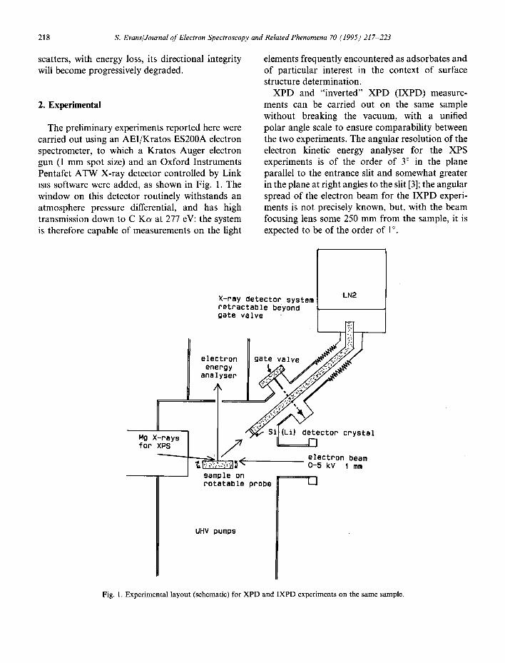
The preliminary experiments reported here were carried out using an AEI/Kratos ES200A electron spectrometer, to which a Kratos Auger electron gun (1 mm spot size) and an Oxford Instruments Pentafet ATW X-ray detector controlled by Link isis software were added, as shown in Fig. 1. The window on this detector routinely withstands an atmosphere pressure differential, and has high transmission down to C K s at 277 eV: the system is therefore capable of measurements on the light
elements frequently encountered as adsorbates and of particular interest in the context of surface structure determination.
XPD and "inverted" XPD (IXPD) measure- ments can be carried out on the same sample without breaking the vacuum, with a unified polar angle scale to ensure comparability between the two experiments. The angular resolution of the electron kinetic energy analyser for the XPS experiments is of the order of 3 ° in the plane parallel to the entrance slit and somewhat greater in the plane at right angles to the slit [3]; the angular spread of the electron beam for the IXPD experi- ments is not precisely known, but, with the beam focusing lens some 250 mm from the sample, it is expected to be of the order of 1°.
Mg X-rays for XPS
LN2 X-ray detector system retractable beyond gate valve
electron gate valve ~~'/~..,'7.~/~ energy y" analyser
"" Si](Lil detector crystal
S / ~ electron beam
l ~ ~ , ~" 0-5 kV I mm sample on rotatable probe
UHV pumps
Fig. 1. Experimental layout (schematic) for XPD and IXPD experiments on the same sample.
S. Evans/Journal of Electron Spectroscopy and Related Phenomena 70 (1995) 217-223 219
A wafer of (100)-oriented GaAs was used as sample for these initial experiments, with the axis of rotation parallel to (110) as in previous XPD experiments [4,5]: native oxide was removed using nitric acid, hydrochloric acid and water before mounting the sample in aluminium foil. The X- ray detector was racked down to within ~30 mm of the sample for IXPD data collection, which was carried out using an incident beam energy (E0) of 1400 eV. At this energy, a beam current of 2-3 #A was found to give the fastest data collection (dead time ~50%, data collection in "optimum resolution mode"). Two sets of X-ray spectra were collected at angular increments of 3.75 ° using 100 and 200 s live time (effective data acquisition time) respectively. The raw spectra were smoothed by convolution with a Gaussian of 50 eV full-width-at-half-maximum [6], and a continuum (bremsstrahlung) background esti- mated [7] as
f (E) = [k 1 ( E 0 - E ) / E n t- k 2 ( E 0 - E ) 2 / E 2]
X exp(-k3/E n)
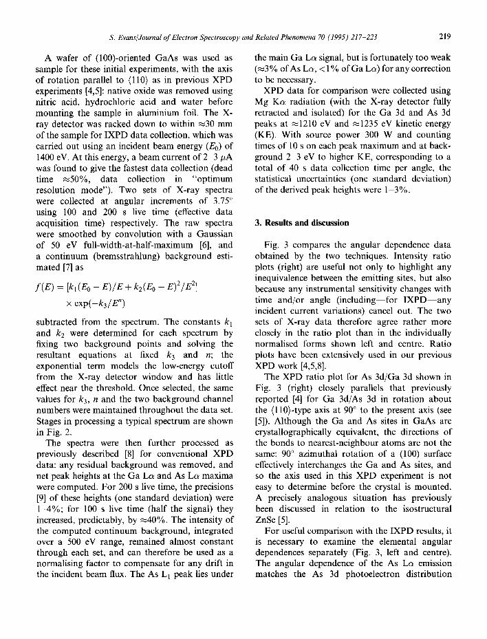
subtracted from the spectrum. The constants k 1 and k2 were determined for each spectrum by fixing two background points and solving the resultant equations at fixed k 3 and n; the exponential term models the low-energy cutoff from the X-ray detector window and has little effect near the threshold. Once selected, the same values for k3, n and the two background channel numbers were maintained throughout the data set. Stages in processing a typical spectrum are shown in Fig. 2.
The spectra were then further processed as previously described [8] for conventional XPD data: any residual background was removed, and net peak heights at the Ga L~ and As Lc~ maxima were computed. For 200 s live time, the precisions [9] of these heights (one standard deviation) were 1-4%; for 100 s live time (half the signal) they increased, predictably, by ~40%. The intensity of the computed continuum background, integrated over a 500 eV range, remained almost constant through each set, and can therefore be used as a normalising factor to compensate for any drift in the incident beam flux. The As L1 peak lies under
the main Ga Lc~ signal, but is fortunately too weak (~3 % of As Lc~, < 1% of Ga La) for any correction to be necessary.
XPD data for comparison were collected using Mg K s radiation (with the X-ray detector fully retracted and isolated) for the Ga 3d and As 3d peaks at ~1210 eV and ~1235 eV kinetic energy (KE). With source power 300 W and counting times of 10 s on each peak maximum and at back- ground 2-3 eV to higher KE, corresponding to a total of 40 s data collection time per angle, the statistical uncertainties (one standard deviation) of the derived peak heights were 1-3%.
3. Results and discussion
Fig. 3 compares the angular dependence data obtained by the two techniques. Intensity ratio plots (right) are useful not only to highlight any inequivalence between the emitting sites, but also because any instrumental sensitivity changes with time and/or angle (including--for IXPD--any incident current variations) cancel out. The two sets of X-ray data therefore agree rather more closely in the ratio plot than in the individually normalised forms shown left and centre. Ratio plots have been extensively used in our previous XPD work [4,5,8].
The XPD ratio plot for As 3d/Ga 3d shown in Fig. 3 (right) closely parallels that previously reported [4] for Ga 3d/As 3d in rotation about the (110)-type axis at 90 ° to the present axis (see [5]). Although the Ga and As sites in GaAs are crystallographically equivalent, the directions of the bonds to nearest-neighbour atoms are not the same: 90 ° azimuthal rotation of a (100) surface effectively interchanges the Ga and As sites, and so the axis used in this XPD experiment is not easy to determine before the crystal is mounted. A precisely analogous situation has previously been discussed in relation to the isostructural ZnSe [5].
For useful comparison with the IXPD results, it is necessary to examine the elemental angular dependences separately (Fig. 3, left and centre). The angular dependence of the As La emission matches the As 3d photoelectron distribution

220 S. Evans/Journal o f Electron Spectroscopy and Related Phenomena 70 (1995) 217-223
C.9
C.)l
(_
Q ul
I
r
0
~ ° ~
L 1
o o o o
o ° o o o
• o o
4~ ° ° ~
0 U
~ l @'~ °
s ~ u n
I
~JeJ~que / AIISNHINI
tgl
8
0
0
E . r '
e,0
e x "~
0
H
t" e-i
t L

S. Evans~Journal of Electron Spectroscopy and Related Phenomena 70 (1995) 217-223 221
~1.4
~1.0
50.e Q
~ 1.2
~ 1 . 0
z°O.8
(i) (ii)
i • . . , , , , . 0
0 20 40 80 80 POLAR ANGLE /DEGREES
1.4
1.2
1.0
0 .8
1.2
1.0
0 .8
0 .6
(~) GaL : :
, , , , , , , , , O
0 20 40 60 80 POLAR ANGLE /DEGREES
01 .4 ~_1.2 ~1 .o z o . 8 ~ I .4 81 .2
5 1 .0
(iii)
~) ;A~J./_.C~.. ', 1' : t
0 20 40 60 80 POLAR ANGLE /DEGREES
Fig. 3. Angular dependences of (i) the As 3d XPS peak height compared with that for As Lc~ (IXPD), (ii) the Ga 3d XPS peak height compared with that for Ga La and (iii) the As 3d/Ga 3d peak height ratio (XPD) compared with that for As La/Ga Lc~ (IXPD). For the X-ray data, open points were obtained using 200 s live time and crossed points with 100 s. Curves are drawn as an aid to the eye to pass through the (mean) data points in each plot.
quite closely, and with a similar extent of anisotropy, although the sharp feature at ~35 ° appears somewhat broadened (Fig. 3, left). In contrast, the Ga La distribution, unlike that for Ga 3d, is almost featureless (Fig. 3, centre); however, faint but distinct echoes of all the main Ga 3d features can just be discerned. As one might then expect, the IXPD ratio plot shows some, but by no means all, of the features of the XPD plot (Fig. 3, right); we need not consider this plot further.
The differences between the two sets of X-ray data in each elemental plot are not entirely the result of random count-rate fluctuations, but arise in part as a result of the normalisation process (division by the mean peak heights). A small beam alignment difference reduced the absolute count-rates at high angle for the 200 s set, so that the open points occur systematically above the crossed points at lower angles. All the trends noted above are visible in either data set.
The difference between the extent of angular modulation of the As La and Ga La signals is not entirely unexpected: the As 2p3/2 (L3) threshold occurs at ~1327 eV, only 73 eV below the energy of the incident beam, whereas the Ga 2p3/2 threshold, at ~ 1119 eV, is as much as 281 eV below the beam energy. The direction of the incident electron beam should be largely preserved when energy losses are relatively small (the most probable discrete energy loss in GaAs is ~20 eV),
but repeated inelastic collisions apparently randomise the incident electron direction so effectively that by 280 eV above threshold the IXPD effect is barely recognisable.
It is interesting to contrast this situation with that arising in electron-impact Auger electron diffraction (AED). There has been controversy over the importance of incident beam diffraction in this context [10], but it is now clear that strong incident beam angular effects do occur in GaAs [11] (as in other solids [10]), even when the incident electron energy is as high as 3 keV, nearly 2 keV above the ionisation threshold. The key difference between that and the situation described above for Ga IXPD is that the sampling depth in the AED case is limited by the escape depth of the Auger electrons. Auger electrons generated at greater depths by the (degraded) primary beam are inelastically scattered and do not contribute to the signal, whereas photons generated below the surface have a high escape probability.
Comparable surface sensitivity can, however, in principle be obtained for IXPD experiments by ensuring that the primary beam energy is only marginally above the ionisation threshold; the primary electrons then have a short inelastic mean free path, comparable with that for the Auger electrons, and if after only one scattering event their energy becomes too low to ionise the relevant core electron, surface sensitivity will be ensured. As the primary energy is increased,
222 s. Evans/Journal of Electron Spectroscopy and Related Phenomena 70 (1995) 217-223
however, inelastic collisions become possible without reducing the electron energy below the threshold, and the effective depth sampled will increase progressively as the angular resolution degrades.
For angular resolution and surface sensitivity to match those of conventional XPD, it will therefore be necessary to work with an incident beam of rather lower energy than that used in these preliminary experiments. This would appear to be possible; calculations using Gryzinski's formula [12] suggest that, at 10 eV above threshold, the X-ray yield would be ~6% of that at 73 eV, and the incident electron current can readily be increased (for many samples) above the present 2-3 #A. The signal-to-background ratio does, however, deteriorate as the energy is decreased, and this may cause difficulties. The present experiments were severely constrained in counting rate by the use of the "optimum resolution" mode of operation of the discriminators in the X-ray analysis system to ensure the best possible resolution of the Ga and As L~ peaks, which are separated by 184 eV. In this mode, the maximum data acquisition rate is ~3 x 10³ cps, whereas in the "optimum acquisition rate" mode up to 3 x 10⁴ cps can be processed, although with greatly reduced energy resolution. The Ga and As Lc~ peaks are then inadequately resolved. The limit- ing factor will, in fact, usually be not the maximum permissible incident current, but the maximum permissible X-ray count-rate. When peak overlap is not a problem, however, it should be possible to collect data much closer to threshold than 73 eV within an acceptable time frame. Moving the detector even closer to the sample would also be worthwhile; if it were possible to place the detector 10 mm from the sample, the sensitivity would increase approximately tenfold, permitting a much lower incident current for any desired count-rate.
The markedly greater intensity of the Ga La peak relative to As Lc~ (Fig. 2) must in part be a reflection of the increase in ionisation cross-section with excess energy above threshold, and in part a result of the X-ray generation by inelastically scattered electrons within the sample, as discussed above. It is, in fact, surprising that the Ga La peak
does not have an even greater relative intensity, since the cross-section effect alone would be expected [12] to be greater than that observed, and the loss of angular modulation discussed above indicates that the second process is also highly significant. Even for Ga, however, the cross-section is only ~20% of the maximum, reached when the incident electron energy is 3-4 times the binding energy [12]. For oxygen, in contrast, the cross-section in the present experi- ments is close to maximum; thus the relatively large O Ks peak in Fig. 2 represents only a very thin oxide overlayer, regenerated (after cleaning) during sample mounting and insertion. A corre- sponding O ls peak was detected in the X-ray photoelectron spectroscopic data. As in previous experiments [4,5], elastic scattering in this very thin oxide overlayer is negligible. A small amount of carbonaceous contamination was also detected by both techniques.
It may be helpful to clarify the relationship between the present experiments and the inverse photoemission spectroscopies. In the latter, the energy of an incident electron beam is scanned, and the intensity variation of the emitted photon flux is measured, to provide information on the structures of the unoccupied bands of the solid. When relatively high incident electron energies (~1-5 keV) are employed, and photons are detected at one fixed energy, the resulting tech- nique is usually known as bremsstrahlung iso- chromat spectroscopy (BIS) and essentially provides only information on the density-of-states rather than on individual bands [13]. A mechanism for a weak dependence of photon flux on the angle between the incident electron and the detected photon has, however, been described [14]. For experiments using much lower incident energies, the term "inverse photoemission spectroscopy" (IPES) is used; here k-conservation allows detailed maps of the unoccupied bands to be elucidated [13,15] and a strong angular dependence may be observed. In both experiments, however, the initial electronic excitation and the emission of the photon are linked: the core electrons are not excited. The present IXPD experiments are not inverse photoemission, but relate instead to a two-stage process: an ionisation event induced by

s. Evans/Journal of Electron Spectroscopy and Related Phenomena 70 (1995) 217-223 223
a relatively high-energy electron in which k- conservation is not important, so that the only angular dependence should be that resulting from diffraction of the incident beam, followed by a subsequent (incoherent) event as the core hole decays with emission of a characteristic X-ray photon. The photons which form the signal in the BIS experiment merely comprise a small part of the background in IXPD. This distinction is important and is the reason for terming the new technique "inverted" rather than "inverse" XPD; although it is desirable to emphasize the link between a reversed XPD experiment and IXPD, true inverse photoemission is a quite distinct phenomenon offering different information.
angular dependence of core-hole generation by angle-integrated Auger spectroscopy [16], a procedure which introduces an additional surface weighting in comparison with the present experiment. Much work thus remains to be done to evaluate fully the possible future role of IXPD in surface-structure determination, and further investigations are already in progress.
Acknowledgement
The SERC is thanked for Standard Grant G R / H 26864.
support under
4. Conclusions
The monitoring of electron diffraction in the keV range by the detection of X-rays released on decay of the core holes resulting from diffracted-electron impact is clearly feasible. To the author 's know- ledge, this has not previously been either suggested or demonstrated. The phenomenon closely parallels conventional XPD, thus facilitating the interpretation of future IXPD data. Potential advantages over conventional XPD include the use of smaller samples and the relatively inexpensive achievement of high angular resolution. It should be mentioned, however, that an alternative way of gaining an equivalent advantage in minimising sample size is to separate incident-beam and AED effects in electron-excited AED by the use of rotatable sample stages and electron analysers to maintain a constant electron exit direction while varying the incident-beam angle. This approach offers much greater freedom to choose the diffracted-electron wavelength, and it has in fact already been applied to GaAs [11], but it requires much more expensive equipment than IXPD. Yet another possibility is to monitor the
References
[1] C.S. Fadley, in R.Z. Bachrach (Ed.), Synchrotron Radia- tion Research: Advances in Surface Science, Plenum Press, New York, 1990.
[2] G. Grenet, Y. Jugne, H.C. Poon and Duc Tran Minh, Surf. Interface Anal., 14 (1989) 367.
[3] S. Evans and C.E. Riley, J. Chem. Soc. Faraday Trans. 2, 82 (1986) 541.
[4] S. Evans and M.D. Scott, Surf. Interface Anal., 3 (1981) 269.
[5] E.S. Crawford, S. Evans, E. Raftery and M.D. Scott, Surf. Interface Anal., 5 (1983) 28.
[6] S. Evans and A.G. Hiorns, Surf. Interface Anal., 8 (1986) 71.
[7] J.l. Goldstein, D.E. Newbury, P. Echlin, D.C. Joy, C. Fiori and E. Lifshin, Scanning Electron Microscopy and X-ray Microanalysis, Plenum Press, New York, 1981, pp. 396 401.
[8] L.A. Ash, S.L. Clark, S. Evans and A.G. Hiorns, J. Chem. Soc. Dalton Trans., (1988) 859 and references therein.
[9] S. Evans, Surf. Interface Anal., 18 (1992) 323. [10] S.A. Chambers, Surf. Sci. Rep., 16 (1992) 261. [11] Y. Gao and J. Cao, Phys. Rev. B, 43 (1991) 9692. [12] M. Gryzinski, Phys. Rev., 138 (1965) A336. [13] J.B. Pendry, J. Phys. C: Solid State Phys., 14 (1981) 1381. [14] O. Sipr, J. Phys.: Condensed Matter, 3 (1991) 8503. [15] See, for example, A.K. See, M. Thayer and R.A. Bartynski,
Phys. Rev. B, 47 (1993) 13722. [16] A. Stuck, M. Novicki, S. Mroz, D. Naumovic and J. Oster-
walder, Surf. Sci., 306 (1994) 21.