Introduction to ultrasound in emergency department A&E medical meeting 28/07/2011 Dr. David Tran (...
24
Introduction to Introduction to ultrasound ultrasound in emergency in emergency department department A&E medical meeting A&E medical meeting 28/07/2011 28/07/2011 Dr. David Tran Dr. David Tran ( ( Source: Ultrasound guide for emergency physician, Beatrice Source: Ultrasound guide for emergency physician, Beatrice Hoffmann Hoffmann ) www.sonoguide.com ) www.sonoguide.com
-
Upload
asher-wilkins -
Category
Documents
-
view
222 -
download
1
Transcript of Introduction to ultrasound in emergency department A&E medical meeting 28/07/2011 Dr. David Tran (...

Introduction to ultrasound Introduction to ultrasound in emergency departmentin emergency department
A&E medical meetingA&E medical meeting28/07/201128/07/2011Dr. David TranDr. David Tran
((Source: Ultrasound guide for emergency physician, Beatrice HoffmannSource: Ultrasound guide for emergency physician, Beatrice Hoffmann) ) www.sonoguide.comwww.sonoguide.com

History of ultrasound in medicineHistory of ultrasound in medicine
The first application of ultrasound as a medical The first application of ultrasound as a medical
diagnostic tool was published in 1942 by Karl and diagnostic tool was published in 1942 by Karl and
Friederich Dussik in ViennaFriederich Dussik in Vienna
The Austrian brothers attempted to locate brain tumors The Austrian brothers attempted to locate brain tumors
and the cerebral ventricles by measuring ultrasound and the cerebral ventricles by measuring ultrasound
transmission through the skull. They concluded that if transmission through the skull. They concluded that if
imaging of the ventricles was possible, the interior of the imaging of the ventricles was possible, the interior of the
human body could also be visualized using ultrasound. human body could also be visualized using ultrasound.
This marked the beginning of diagnostic ultrasonography This marked the beginning of diagnostic ultrasonography
in the medical fieldin the medical field

Evolution of Evolution of ultrasound in ultrasound in emergency emergency medicinemedicine
The portability of real-time bedside diagnosis has made The portability of real-time bedside diagnosis has made ultrasound an attractive tool for emergency medicine. ultrasound an attractive tool for emergency medicine. More and more emergency physicians have made More and more emergency physicians have made bedside sonography part of their clinical practice and bedside sonography part of their clinical practice and research activitiesresearch activities
Implementing this diagnostic test into our daily practice Implementing this diagnostic test into our daily practice can reduce morbidity and mortality for many medical and can reduce morbidity and mortality for many medical and surgical emergencies. In addition, emergency ultrasound surgical emergencies. In addition, emergency ultrasound education has become part of our specialty training education has become part of our specialty training

Efficiency and cost-effectivenessEfficiency and cost-effectiveness
Increased efficiency and cost-effectiveness in the evaluation of the blunt abdominal trauma patient with the use of ultrasound.Arrillaga A, Graham R, York JW, Miller RS. (Am. Surg. 1999, Jan)
Over a 9-month period, 331 patients suspected of sustaining blunt abdominal trauma were evaluated at a Level I trauma center by US, CT, and/or DPL. Cost data and time to disposition were determined for analysis. The sensitivity, specificity, and accuracy of US were similar to those reported in previous studies. There was a significant difference in time to disposition with the US group being significantly lower (P = 0.001). The total procedural cost was 2.8 times greater for the CT/DPL group than for the US group. US is not only effective in diagnosing blunt abdominal trauma, but it is also more efficient and cost-effective than is CT/DPL

Basic ultrasound physicsBasic ultrasound physics
Sound is a mechanical wave, Sound is a mechanical wave, which requires a medium in which requires a medium in which to travel. It is a series of which to travel. It is a series of pressure waves propagating pressure waves propagating through a medium.through a medium.
One cycle of the acoustic wave One cycle of the acoustic wave is composed of a complete is composed of a complete positive and negative pressure positive and negative pressure change. change.
The wavelength is the distance The wavelength is the distance traveled during one cycle, the traveled during one cycle, the frequency of the wave is frequency of the wave is measured in cycles per second measured in cycles per second or Hertz (Cycles/s, Hz).or Hertz (Cycles/s, Hz).

Speed propagation in differents Speed propagation in differents medium medium ((tissuestissues))
The speed with which an acoustic wave travels The speed with which an acoustic wave travels through a medium is determined by the density through a medium is determined by the density and stiffness of the medium. The greater the and stiffness of the medium. The greater the stiffness, the faster the wave will travel. This stiffness, the faster the wave will travel. This means that sound waves travel faster in solids means that sound waves travel faster in solids than liquids or gases. Acoustic waves are than liquids or gases. Acoustic waves are calculated to travel through human tissue at calculated to travel through human tissue at body temperature at approximately 1540 m/s body temperature at approximately 1540 m/s (about one mile per second).(about one mile per second).

Effects between the unhomogeneos Effects between the unhomogeneos border of two different mediumsborder of two different mediums
When traveling through a When traveling through a medium the sound waves' medium the sound waves' intensity and amplitude intensity and amplitude decreases. This is called decreases. This is called 'attenuation' and is the reason 'attenuation' and is the reason why echoes from deeper why echoes from deeper structures are weaker than structures are weaker than echoes from superficial areas. echoes from superficial areas.
The major cause of attenuation The major cause of attenuation in soft tissue is absorption, in soft tissue is absorption,
Other mechanisms are Other mechanisms are reflection, refraction and scatter.reflection, refraction and scatter.

Artifacts in ultrasoundArtifacts in ultrasound: Shadowing: Shadowing
Shadowing:Shadowing:This artifact is caused This artifact is caused by partial or total by partial or total reflection or absorption reflection or absorption of the sound energy. A of the sound energy. A much weaker signal much weaker signal returns from behind a returns from behind a strong reflector (air) or strong reflector (air) or sound-absorbing sound-absorbing structure (gallstone, structure (gallstone, kidney stone, bone).kidney stone, bone).

Posterior enhancementPosterior enhancement
Posterior Enhancement:Posterior Enhancement:In posterior enhancement, In posterior enhancement, the area behind an echo-the area behind an echo-weak or echo-free structure weak or echo-free structure appears brighter (more appears brighter (more echogenic) than its echogenic) than its surrounding structures. surrounding structures.
This occurs because This occurs because neighboring signals had to neighboring signals had to pass through more pass through more attenuating structures and attenuating structures and return with weaker echoes return with weaker echoes

Edge ShadowingEdge Shadowing
Edge Shadowing:Edge Shadowing:The lateral edge shadow is The lateral edge shadow is a thin acoustic shadow that a thin acoustic shadow that appears behind edges of appears behind edges of cystic structures. cystic structures.
Sound waves encountering Sound waves encountering a cystic wall or a curved a cystic wall or a curved surface at a tangential surface at a tangential angle are scattered and angle are scattered and refracted, leading to energy refracted, leading to energy loss and the formation of a loss and the formation of a shadow shadow

ReverberationReverberation
Reverberation:Reverberation:Reverberation occurs when Reverberation occurs when sound encounters two highly sound encounters two highly reflective layers. The sound reflective layers. The sound is bounced back and forth is bounced back and forth between the two layers between the two layers before traveling back. The before traveling back. The probe will detect a prolonged probe will detect a prolonged traveling time and assume a traveling time and assume a longer traveling distance and longer traveling distance and display additional display additional ‘reverberated’ images in a ‘reverberated’ images in a deeper tissue layer deeper tissue layer

Different probes Different probes ((transducertransducers)s)
Large Convex ProbeLarge Convex Probe:: Main ED utilization is transabdominal sonography. Main ED utilization is transabdominal sonography.
Sector ProbeSector Probe:: Sector probes are utilized especially for transthoracic sonography. Sector probes are utilized especially for transthoracic sonography.
Linear ProbeLinear Probe:: Main utilization is vascular sonography or evaluation of superficial Main utilization is vascular sonography or evaluation of superficial
soft tissue structures.soft tissue structures.

Ultrasound vocabularyUltrasound vocabulary
Anechoic Anechoic -- Complete absence of returning sound Complete absence of returning sound
waves, area is black.waves, area is black.
HypoechoicHypoechoic - - Structure has very few echoes and Structure has very few echoes and
appears darker than surrounding tissue.appears darker than surrounding tissue.
Hyperechoic / EchogenicHyperechoic / Echogenic - - Opposite of hypoechoic, Opposite of hypoechoic,
structure appears brighter than surrounding tissue.structure appears brighter than surrounding tissue.

Position of the probePosition of the probe Axial PlaneAxial Plane - - Separates the superior from Separates the superior from
the inferior, or, the head from the feet.the inferior, or, the head from the feet. Sagittal PlaneSagittal Plane - - Oriented perpendicular to Oriented perpendicular to
the ground, separating left from right. The the ground, separating left from right. The "midsagittal plane" is a sagittal plane that is "midsagittal plane" is a sagittal plane that is exactly in the middle of the body.exactly in the middle of the body.
Coronal PlaneCoronal Plane - - Also known as the frontal Also known as the frontal
plane, separates the anterior from the plane, separates the anterior from the posterior or the front from the back.posterior or the front from the back.
Oblique PlaneOblique Plane - - The probe is oriented The probe is oriented neither parallel to, nor at right angles from, neither parallel to, nor at right angles from, coronal, sagittal or transverse planes.coronal, sagittal or transverse planes.
Longitudinal PlaneLongitudinal Plane - - The longitudinal The longitudinal plane is perpendicular to the transverse plane is perpendicular to the transverse plane an can be either the coronal plane or plane an can be either the coronal plane or sagittal plane. sagittal plane.

FASTFAST: : FFocused ocused AAssessment with ssessment with SSonographic in onographic in TTrauma patientsrauma patients
Many trauma patients have injuries that are not apparent on the initial physical exam. Patients can present with distracting injuries or altered mental status. Significant bleeding into the peritoneal, pleural, or pericardial spaces may occur without obvious warning signs. The purpose of bedside ultrasound in trauma is to rapidly identify free fluid (usually blood) in the peritoneal, pericardial, or pleural spaces.
Recently, research studies have shown that bedside ultrasound is equivalent to, or better than, chest radiography for identifying a hemothorax or pneumothorax in trauma patients. For this reason some trauma centers have begun performing an extended FAST exam (EFAST), evaluating for pneumo- and hemothorax in addition to intraperitoneal injuries

Concept of FASTConcept of FAST
four transducer positionsfour transducer positions (1) pericardial area(1) pericardial area (2) right upper quadrant(2) right upper quadrant (3) left upper quadrant (3) left upper quadrant (4) pelvis.(4) pelvis.
The concept behind the FAST exam is that many life-threatening injuries cause bleeding. Although ultrasound is not 100% sensitive for identifying all bleeding, it is nearly perfect for recognizing intraperitoneal bleeding in hypotensive patients who need an emergent laparotomy and for diagnosing cardiac injuries from penetrating trauma
It is important to recognize the imperfect nature of such exams, but sonographers who master this challenge, will find it an invaluable tool in the care of trauma patients.

Right coronal viewRight coronal view
Right Coronal and Right Coronal and Intercostal Oblique Views: Intercostal Oblique Views: The easiest abdominal view The easiest abdominal view to obtain is the view of to obtain is the view of Morison’s pouch. To obtain Morison’s pouch. To obtain this view place the probe in this view place the probe in the mid-axillary line at the mid-axillary line at about the 8th to 11th about the 8th to 11th intercostal space with the intercostal space with the marker-dot pointed marker-dot pointed cephalad (Figure 4). This cephalad (Figure 4). This gives a gives a coronalcoronal view of the view of the interface between the liver interface between the liver and kidney and kidney
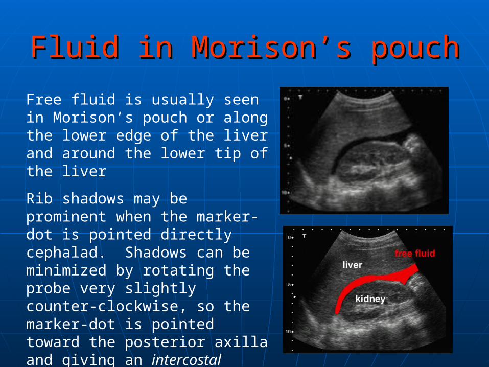
Fluid in Morison’s pouchFluid in Morison’s pouch
Free fluid is usually seen in Morison’s pouch or along the lower edge of the liver and around the lower tip of the liver
Rib shadows may be prominent when the marker-dot is pointed directly cephalad. Shadows can be minimized by rotating the probe very slightly counter-clockwise, so the marker-dot is pointed toward the posterior axilla and giving an intercostal oblique view.
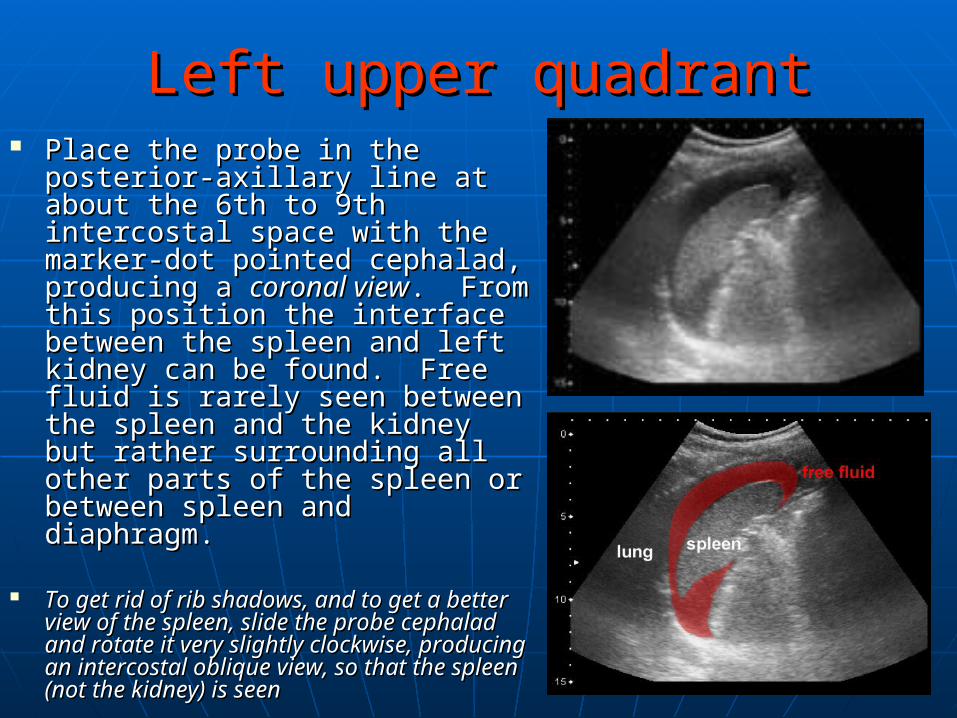
Left upper quadrantLeft upper quadrant Place the probe in the posterior-Place the probe in the posterior-
axillary line at about the 6th to axillary line at about the 6th to 9th intercostal space with the 9th intercostal space with the marker-dot pointed cephalad, marker-dot pointed cephalad, producing a producing a coronal viewcoronal view. From . From this position the interface this position the interface between the spleen and left between the spleen and left kidney can be found. Free fluid kidney can be found. Free fluid is rarely seen between the is rarely seen between the spleen and the kidney but rather spleen and the kidney but rather surrounding all other parts of surrounding all other parts of the spleen or between spleen the spleen or between spleen and diaphragm.and diaphragm.
To get rid of rib shadows, and to get a To get rid of rib shadows, and to get a
better view of the spleen, slide the better view of the spleen, slide the probe cephalad and rotate it very probe cephalad and rotate it very slightly clockwise, producing an slightly clockwise, producing an intercostal oblique view, so that the intercostal oblique view, so that the spleen (not the kidney) is seen spleen (not the kidney) is seen

Pelvis viewPelvis view Since the pelvis is the most Since the pelvis is the most
dependent part of the dependent part of the peritoneal space, it is the peritoneal space, it is the most likely place to see most likely place to see abdominal free fluid. It is abdominal free fluid. It is easy to obtain both easy to obtain both longitudinal and transverse longitudinal and transverse views of the pelvis. If the views of the pelvis. If the longitudinal view is longitudinal view is performed first, it is often performed first, it is often easier to understand the easier to understand the anatomy and obtain good anatomy and obtain good images. Place the probe in images. Place the probe in the midline just cephalad to the midline just cephalad to the pubic bone with the the pubic bone with the marker-dot pointed marker-dot pointed cephalad.cephalad.
Normal longitudinal view of bladder and uterus

Fluid in the Douglas Pouch Fluid in the Douglas Pouch ((malemale))
In a male, the Douglas pouch In a male, the Douglas pouch
is just behind the bladder. is just behind the bladder.
Blood can be seen just Blood can be seen just
posterior to the bladder.posterior to the bladder.
If the bladder is empty, it is If the bladder is empty, it is
very difficult to recognize very difficult to recognize
pelvic free fluid in a male. In a pelvic free fluid in a male. In a
female, the pouch of Douglas female, the pouch of Douglas
may still be identifiable, even may still be identifiable, even
when the bladder is empty.when the bladder is empty.
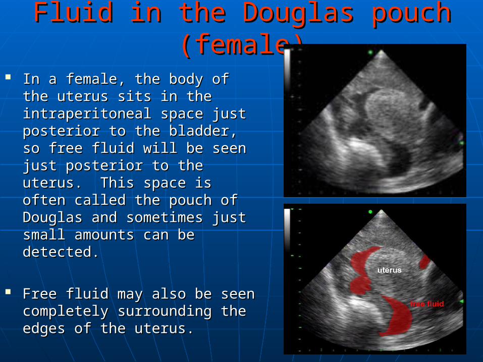
Fluid in the Douglas pouch (female)Fluid in the Douglas pouch (female)
In a female, the body of the In a female, the body of the uterus sits in the uterus sits in the intraperitoneal space just intraperitoneal space just posterior to the bladder, so free posterior to the bladder, so free fluid will be seen just posterior fluid will be seen just posterior to the uterus. This space is to the uterus. This space is often called the pouch of often called the pouch of Douglas and sometimes just Douglas and sometimes just small amounts can be small amounts can be detected. detected.
Free fluid may also be seen Free fluid may also be seen completely surrounding the completely surrounding the edges of the uterus.edges of the uterus.

Litterature review about FASTLitterature review about FAST
Arrillaga A, Graham R, York JW, Miller RS. Increased efficiency and cost-effectiveness in the evaluation of the blunt abdominal trauma patient with the use of ultrasound. The American Surgeon 1999; 65:31-5.
Boulanger BR, McLellan BA, Brenneman FD, Ochoa J, Kirkpatrick AW. Prospective evidence of the superiority of a sonography-based algorithm in the assessment of blunt abdominal injury. Journal of Trauma 1999; 47:632-7.
Melniker LA, Leibner E, McKenney MG, Lopez P, Briggs WM, Mancuso CA. al. Randomized controlled clinical trial of point-of-care, limited ultrasonography in the emergency department: the First Sonography Outcomes Assessment Program Trial. Academic Emergency Medicine 2006; 48:227-35.
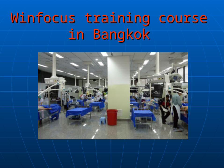
Winfocus training course Winfocus training course in Bangkokin Bangkok