
Introduction to Neuroscience: Introduction to Neuroscience ...
Upload
herlinanababanCategory
view
75download
13description

INTRODUCTION TO MYCOLOGY
The Study of Fungal Microorganisms

Only 300 of 70,000 fungal species cause human infections
Primary growth requirements – warm and moist environment
Classification of fungal infections Superficial mycoses – s. corneum or hair cuticle Cutaneous mycoses – also known as dermatophyte
infections; skin, hair, nails Subcutaneous mycoses – SQ tissue penetration Systemic mycoses – primarily on the respiratory tract
Overview

Involve only the outermost layers of the stratum corneum of the skin or the cuticle of the hair shaft.
Usually constitute cosmetic problems and rarely elicit an immune response from the host.
Patients are accidentally infected with these common organisms via indwelling catheters or intravenous lines.
Virtually nothing is known concerning the pathogenic mechanisms of these fungi.
Superficial Mycoses
Hortae werneckii
Tinea nigra – superficial fungal infection of the skin characterized by brown to black macules which usually occur on the palmar aspects of hands and occasionally the plantar and other surfaces of the skinFound in soil, compost, humus and on wood in humid tropical and sub-tropical regionsTreatment – topical Whitfield's ointment (benzoic acid compound) or an imidazole agent twice a day for 3-4 weeks
Superficial Mycoses

Malassezia furfur
Infections – pityriasis versicolor, pityriasis folliculitis, seborrheic dermatitis, dandruff, fungaemiaPart of the normal flora of the skinFungal colonization begins after birth peaking in late adolescence and early adulthood because of increased sebaceous gland activityTreatment – topical imidazole, ketoconazole, zinc pyrithione, propylene glycol
Superficial Mycoses
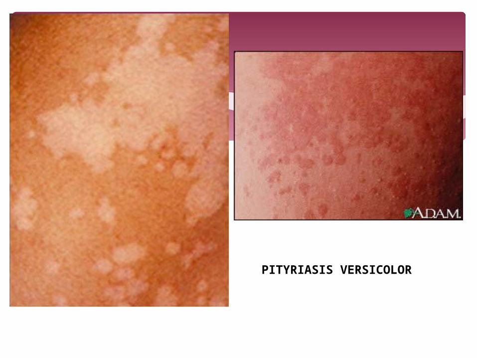
PITYRIASIS VERSICOLOR

SEBORRHEIC DERMATITIS
Piedra hortae
Black piedra – hard black nodules on the hair shafts (scalp, moustache, beard, pubic hair)Treatment – shave/cut hair + terbenafine 250 mg OD x 6 weeks
Trichosporon beigelli
White piedra – like black piedra but shows soft-grayish white nodules along the hair shaft (axilla, scalp, face, pubic hair)
Superficial Mycoses

DermatophytesDermatophytosis – tinea, ringworm of scalp, glabrous skin, nailsAffects the s. corneum with induction of allergic and inflammatory responseHuman agents
Epidermophyton floccosum T violaceum T soudanense Trichophyton rubrum T concentricum T
tonsurans T interdigitale T schoenleinii Microsporum audouinii M ferrugineum
Zoophilic agents – T mentagrophytes, T equinum, T erinacei, T verrucosum, M canis, M nanumSoil dermatophytes – M gypseum, M nanum, M cookie
Cutaneous Mycoses
Dermatophyte InfectionsTinea pedis – T rubrum, T interdigitale, E floccosumTinea cruris – same as aboveTinea unguium (dermatophyte onchomycosis) – T rubrum and T interdigitaleTinea corporis – T rubrum, M gypseumTinea capitis – M canis, T equinum, T schoenleinii, T verrucosum
TreatmentTerbinafine, itraconazole, fluconazole, griseofulvin
Cutaneous Mycoses

TINEA PEDIS
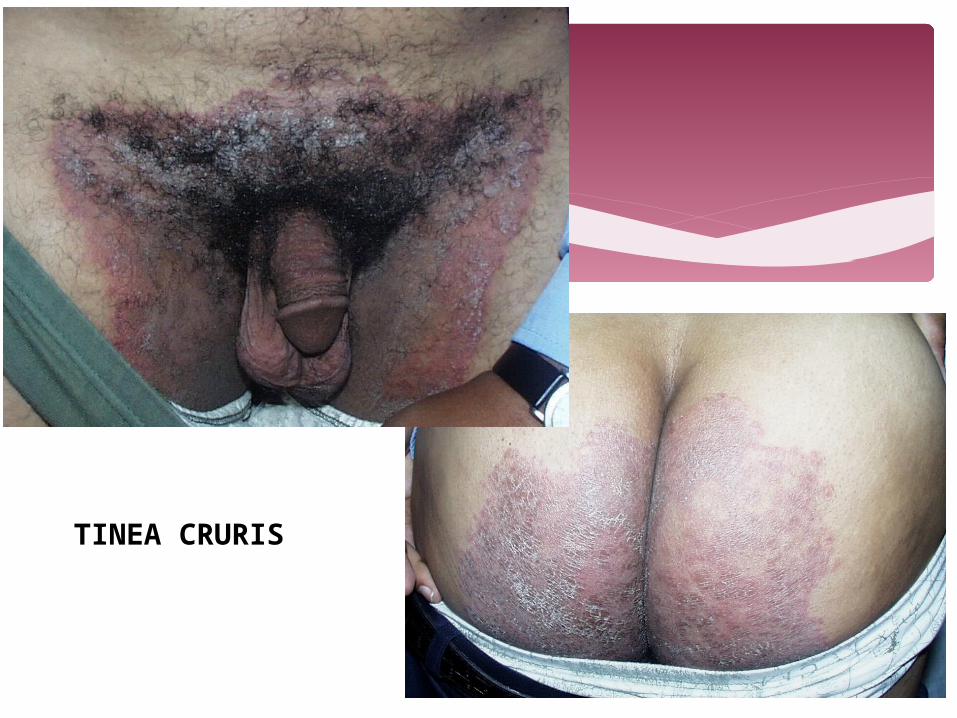
TINEA CRURIS
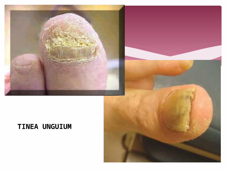
TINEA UNGUIUM

TINEA CORPORIS
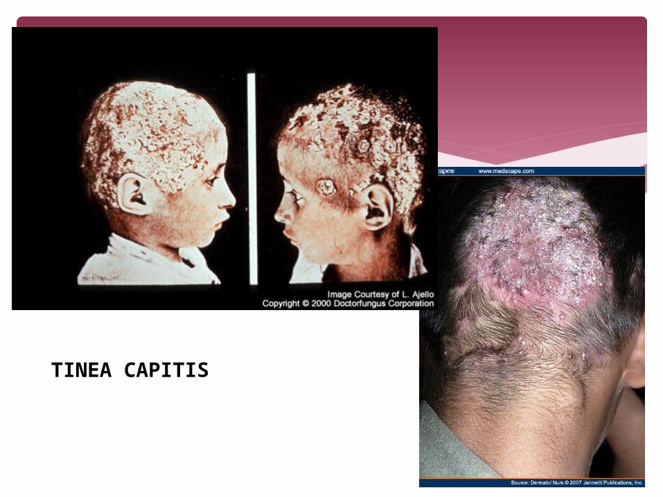
TINEA CAPITIS
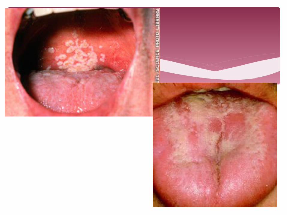
CANDIDIASIS – Candida albicans infections Oropharyngeal candidiasis – oral thrush, glossitis, stomatitis
and angular cheilitis (perleche) Cutaneous candidiasis - intertrigo, diaper candidiasis,
paronychia and onychomycosis. Intertriginous candidiasis - axillae, groin, inter- and sub-
mammary folds, intergluteal folds, interdigital spaces, and umbilicus
Diaper candidiasis – common in infants under unhygienic conditions of chronic moisture and local skin maceration
Paronychia of the finger nails - persons whose hands are subject to continuous wetting, especially with sugar solutions or contact with flour, that macerates the nail folds and cuticle.
Cutaneous Mycoses

INTERTRIGO

DIAPER CANDIDIASIS
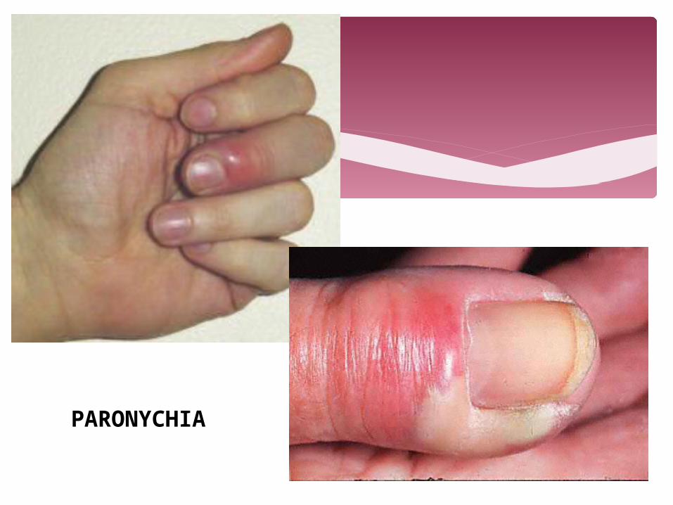
PARONYCHIA
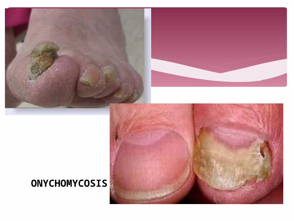
ONYCHOMYCOSIS
Vulvovaginal candidiasis – Pruritus, burning, erythema and dyspareunia with a creamy white, curd-like discharge.
Balanitis – erythema, pruritus and vesiculopustules on the glans penis or prepuce
Chronic mucocutaneous candidiasis - persistent candidiasis, usually caused by C. albicans, of the skin, nails and mucous membranes that occurs in patients with various metabolic disturbances to cell-mediated immunity (hypoparathyroidism, Addison's disease, hypothyroidism, diabetes, dysfunction of the thyroid and polyglandular autoimmune disease)
Cutaneous Mycoses

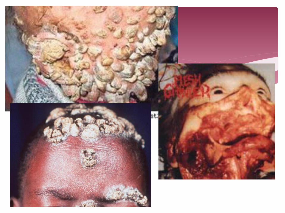
Candida granuloma - severe localized form which may occur with or without endocinopathy characterized by marked hyperkeratic granulomatous lesions
Neonatal and congenital candidiasis Acquired in utero and is confined to the skin in the
form of a generalized erythematous vesicular rash. Esophageal candidiasis - burning pain in the
substernal area, dysphagia, nausea and vomiting; lesions are white mucosal plaques with erythema resembling those seen in oral candidiasis.
Cutaneous Mycoses

Gastrointestinal candidiasis - ulcerations of the stomach and less commonly the duodenum and intestine which can lead to perforation, peritonitis and hematogenous spread to the liver, spleen and other organs.
Peritonitis - fever, abdominal pain, tenderness and a cloudy peritoneal dialysate containing greater than 100 leukocytes/mm3
Candida cystitis – caused by prolonged catheterization with concomitant antibiotic treatment, underlying diseases
Renal candidiasis (pyelonephritis) – due to an ascending infection or hematogenous dissemination from another organ focus
Cutaneous Mycoses
Pulmonary candidiasis - hematogenous dissemination causing a diffuse pneumonia or by bronchial extension in patients with oropharyngeal candidiasis; aspiration of yeasts
Meningitis – rare, predominantly seen in low birthweight neonates with septicemia and in patients with hematological malignancies, complicated neurosurgery or intracerebral prosthetic devices such as ventriculoperitoneal shunts - feverish meningeal irritation
Hepatic and hepatosplenic candidiasis – fever, hepato-splenomegaly and increased blood concentrations of alkaline phosphatases.
Cutaneous Mycoses
Endocarditis, myocarditis and pericarditis Fever, murmur, congestive heart failure, anemia
and splenomegaly Candidemia and disseminated candidiasis -
presence of yeasts in the blood with or without visceral involvement
Ocular candidiasis - localized lesions near the macula and patients complain of cloudy vision
Osteoarticular candidiasis - late sequel of candidemia in neonates or neutropenic patients esp. the knees
Cutaneous Mycoses


Other forms of candidiasis: Cutaneous, ocular and arthritic manifestations in
heroin addicts Fever, rash and myalgia associated with leukemia
patients Candida cholecystitis Candida prostatitis Pancreatic abscesses Epiglottitis and osteomyelitis
Cutaneous Mycoses
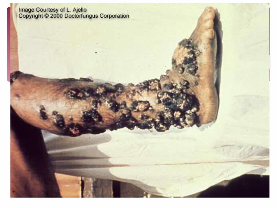
Chromoblastomycetes
Agents - Phialophora verrucosa, Fonsecaea pedrosoi, F. compacta and Cladophialophora carrioniiChromoblastomycosis - infections of the cutaneous and subcutaneous tissues characterized by the development in tissue of dermatiaceous (brown-pigmented), planate-dividing, rounded sclerotic bodiesInfections are caused by the traumatic implantation of fungal elements into the skin and are chronic, slowly progressive and localized. Tissue proliferation usually occurs around the area of inoculation producing crusted, verrucose, wart-like lesions.
Subcutaneous Mycoses
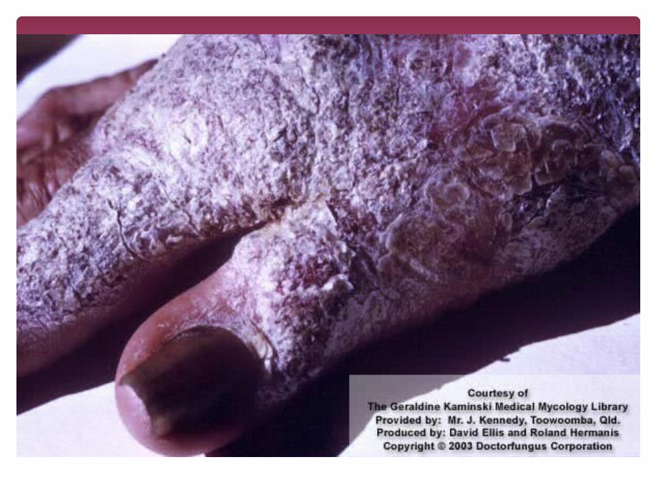

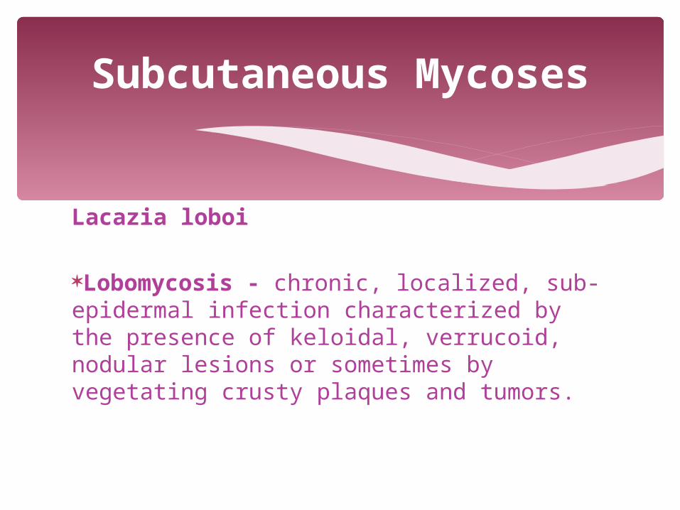
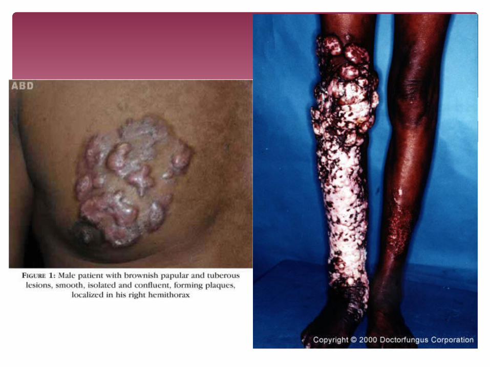
Lacazia loboi
Lobomycosis - chronic, localized, sub-epidermal infection characterized by the presence of keloidal, verrucoid, nodular lesions or sometimes by vegetating crusty plaques and tumors.
Subcutaneous Mycoses

Mycetomycetes
Agents - Madurella, Acremonium, Pseudallescheria, Exophiala, Leptosphaeria, Curvularia, Fusarium, and AspergillusMycetoma - infection of humans characterized by draining sinuses, granules and tumefaction. The disease results from the traumatic implantation of the etiologic agent and usually involves the cutaneous and subcutaneous tissue, fascia and bone of the foot or hand.
Subcutaneous Mycoses


Phaeohyphomycetes
Agents - Exophiala, Phialophora, Wangiella, Bipolaris, Exserohilum, Cladophialophora, Phaeoannellomyces, Aureobasidium, Cladosporium, Curvularia and Alternaria Phaeohyphomycosis - range from localized superficial infections of the stratum corneum (tinea nigra) to subcutaneous cysts (phaeomycotic cyst) to invasion of the brain
Subcutaneous phaeohyphomycosis Paranasal sinus phaeohyphomycosis Cerebral phaeohyphomycosis
Subcutaneous Mycoses


Sporothrix schenkii
Sporotrichosis - characterized by nodular lesions which may suppurate and ulcerate Present in soil, and in vital and decomposing plant material such as peat moss.Infections are caused by the traumatic implantation of the fungus into the skin, or very rarely, by inhalation into the lungs. Secondary spread to articular surfaces, bone and muscle is not infrequent, and the infection may also occasionally involve the central nervous system, lungs or genitourinary tract.
Subcutaneous Mycoses


Zygomycetes
Zygomycosis - any infection due to a member of the Zygomycetes
Mucorales - cause subcutaneous and systemic zygomycosis (Mucormycosis) and includes the species Rhizopus, Mycocladus (Absidia), Rhizomucor, Mucor, Cunninghamella, Saksenaea, Apophysomyces, Cokeromyces and Mortierella.
Entomophthorales – cause subcutaneous zygomycosis (Entomophthoromycosis) and includes the species Conidiobolus and Basidiobolus.
Subcutaneous Mycoses


Aspergillus
Highly aerobic fungi and are found in almost all oxygen-rich environments where they grow as molds on the surface of substratesAspergillosis - caused by Aspergillus fumigatus, A. flavus, A. niger, A. nidulans and A. terreus
Mycotoxicosis due to ingestion of contaminated foods Allergy and sequelae to the presence of conidia or
transient growth of the organism in body orifices Colonization without extension in preformed cavities
and debilitated tissues Invasive, inflammatory, granulomatous, narcotizing
disease of lungs, and other organs Systemic and fatal disseminated disease (rare)
Systemic Mycoses


Some Aspergillus species cause serious disease in humans and animals. A flavus - produces aflatoxin which is both a toxin
and a carcinogen and which can potentially contaminate foods such as nuts.
A fumigatus - most common causing allergic disease.
A clavatus – can also cause allergic diseases. Prevention and control include the following:
Aggressive medical or surgical management depending on the type of infection.
Concomittant treatment with systemic antifungals.
Systemic Mycoses
Blastomyces
Blastomycosis – chronic granulomatous and suppurative disease with a primary pulmonary stage with dissemination to skin and bonesContains receptors that bind estrogen which prevents the transformation of the mycelium into yeast phase
Systemic Mycoses
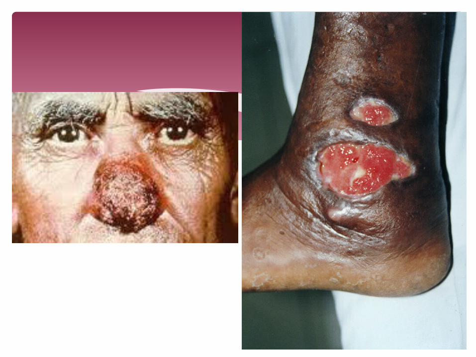
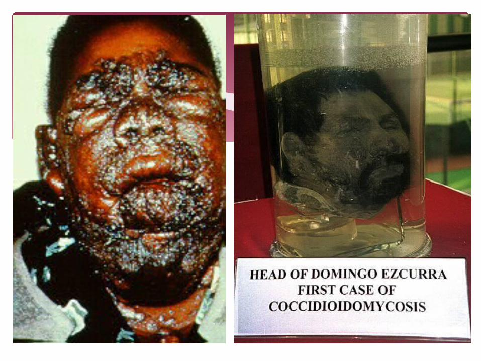
Coccidioides
Coccidioidomycosis – respiratory infection d/t inhalation of conidiaAgents – C immitis, C posadasiiComplications – meningitis, osteomyelitis, arthritis, infections of the cutaneous and SQ tissuesFilipinos have the highest risk of dissemination of progressive pneumonitis because of travel to endemic areas
Systemic Mycoses
Cryptococcus
Agents – C neoformans, C albidus, C gatii, C laurentiiCauses infections of the respiratory and nervous systems
Fungal meningitis Cryptococcosis of the respiratory tree, CNS, skin,
bones, eyes Pyelonephritis, prostatitis, adrenocortical lesions,
endocarditis, hepatitis, sinusitis, localized esophageal lesions
Systemic Mycoses

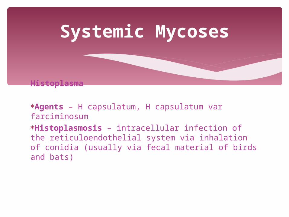
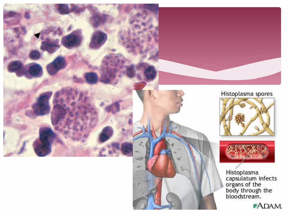
Histoplasma
Agents – H capsulatum, H capsulatum var farciminosumHistoplasmosis – intracellular infection of the reticuloendothelial system via inhalation of conidia (usually via fecal material of birds and bats)
Systemic Mycoses

Hyalohyphomyces
Agents – Penicillium, Phaecilomyces, Acremonium, Beauveria, Fusarium, ScopulariopsisThe clinical manifestations of hyalohyphomycosis are many ranging from harmless saprophytic colonization to acute invasive disease.Predisposing factors include prolonged neutropenia, especially in leukemia patients or in bone marrow transplant recipients, corticosteroid therapy, cytotoxic chemotherapy and to a lesser extent patients with AIDS.The typical patient is granulocytopenic and receiving broad-spectrum antibiotics for unexplained fever
Systemic Mycoses

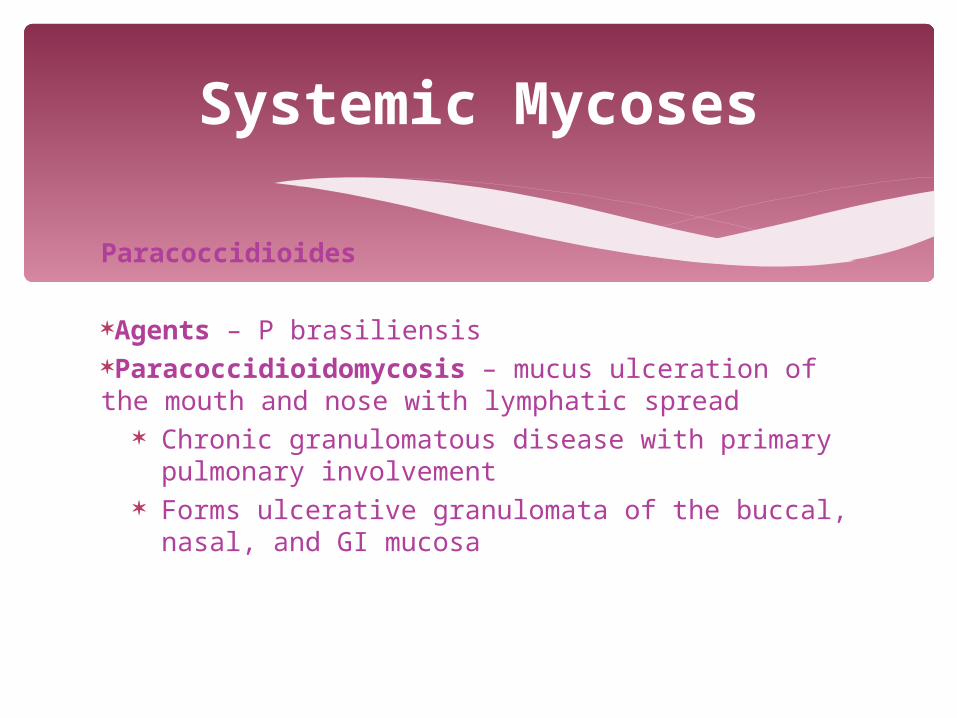
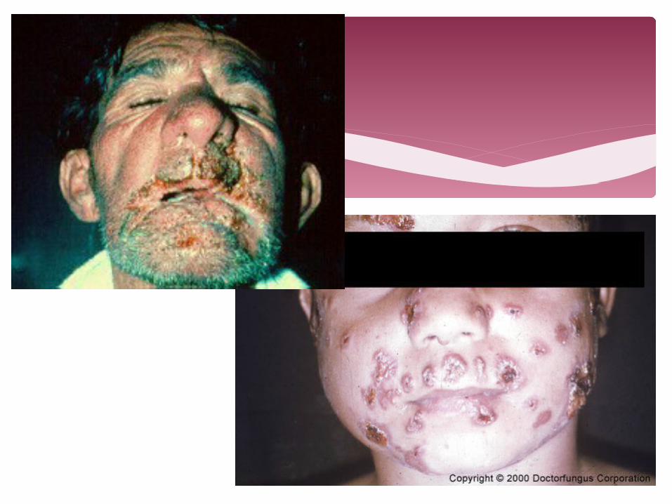
Paracoccidioides
Agents – P brasiliensisParacoccidioidomycosis – mucus ulceration of the mouth and nose with lymphatic spread
Chronic granulomatous disease with primary pulmonary involvement
Forms ulcerative granulomata of the buccal, nasal, and GI mucosa
Systemic Mycoses
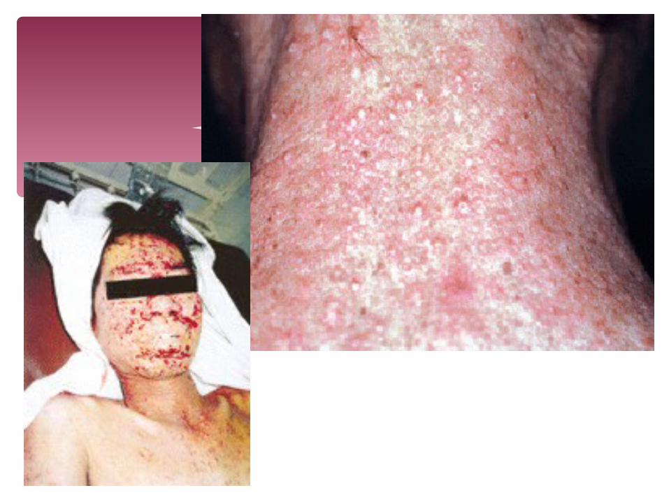
Penicilium merneffei
Grow in living tissues; a major opportunistic pathogen in HIV patientsOther predisposing factors - lymphoproliferative disorders, bronchiectasis and tuberculosis, autoimmune diseases and corticosteroid therapy
Systemic Mycoses

Zygomycetes: Pseudallescheria boydii and Scedosporium aurantiacum
Pseudallescheriasis - non-invasive colonization of the external ear and pulmonary colonization in patients with poorly draining bronchi or paranasal sinuses and "fungus ball" formation in pre-formed cavities similar to those seen in AspergillusInfections include invasive sinusitis, pneumonia, arthritis with osteomyelitis, cutaneous and subcutaneous granulomata, meningitis, brain abscesses, endophthalmitis, and disseminated systemic disease.
Systemic Mycoses

Zygomycetes: Scedosporium prolificans
Spectrum of clinical manifestations are similar to that of P. boydii.
Disseminated disease in immunosuppressed patients especially those with prolonged neutropenia and post-transplantation therapy.
Colonization of the external ear, paranasal sinuses and lung, including "fungus ball"
Systemic Mycoses