Image artifacts in Single Molecule Localization Microscopy ...
Upload
shavonne-baldwinCategory
view
218download
0
Introduction to Image Analysis
Presented to Microscopy and Microscopy Education
11 March 2000New Orleans, LA
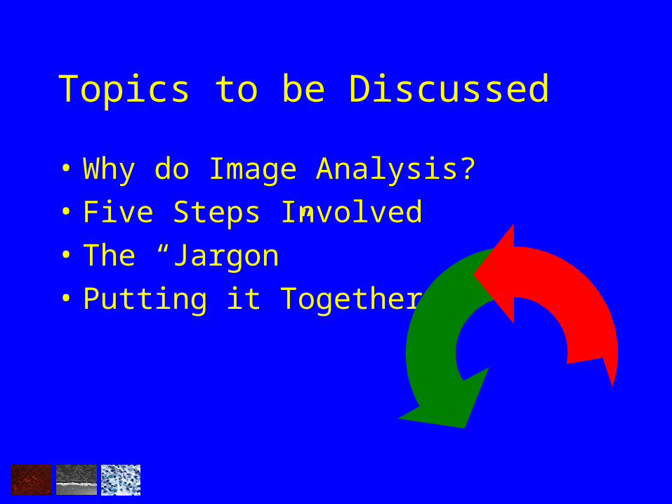
Topics to be Discussed
• Why do Image Analysis?
• Five Steps Involved
• The “Jargon”
• Putting it Together
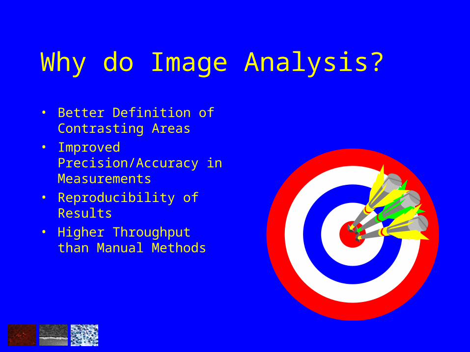
Why do Image Analysis?
• Better Definition of Contrasting Areas
• Improved Precision/Accuracy in Measurements
• Reproducibility of Results
• Higher Throughput than Manual Methods
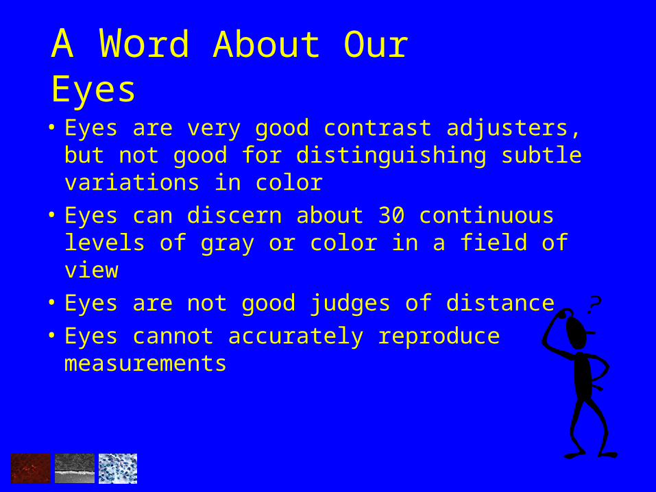
A Word About Our Eyes
• Eyes are very good contrast adjusters, but not good for distinguishing subtle variations in color
• Eyes can discern about 30 continuous levels of gray or color in a field of view
• Eyes are not good judges of distance
• Eyes cannot accurately reproduce measurements


The Steps Involved
• Acquiring Images
• Processing Images
• Measuring Regions of Interest
• Analysing Data
• Reporting Results

Acquiring the Image
Arguably, the most important aspect of all
• Proper setup of imaging apparatus is vital (microscope, camera, etc.)
• Obtain maximum contrast and dynamic range• Reduce “noise” and other unwanted artifacts

•Nice Dynamic Range•Excellent Contrast•No Apparent Noise•Uniform Lighting
A Good Image

Example of Image Artifacts
• Image ‘Vignetting’• Dirty Optics

Processing the Image
• Maximize Brightness/Contrast
• Reduce Artifacts
• Separate Objects from Background
• Enhance Non-Visual Data (data visible to camera but not eye)

Raw Image Contrast Adjusted
Adjusting Contrast

What is ‘Contrast Adjustment’
• Modifying Contrast, Brightness and Gamma Levels to Obtain Best Possible Image
Contrast- The degree of difference between the darkest
and lightest parts of the image Brightness- The overall amount of light in an image
Gamma- Improves contrast in very light or very dark areas of the image

Reducing Artifacts
Raw Image Flattening Filter Applied

What Filters Do
• Improve quality of image
• Remove artifacts introduced through instrumentation (noise, etc.)
• Enhance features within an image
• Remove background variations
• Enhance edges
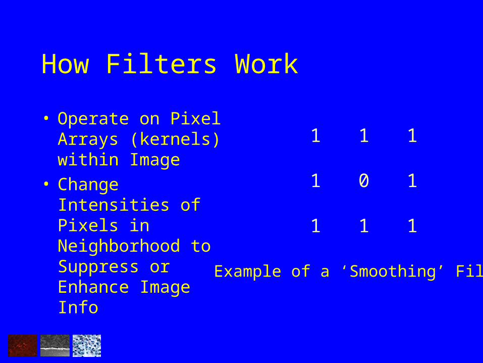
How Filters Work
• Operate on Pixel Arrays (kernels) within Image
• Change Intensities of Pixels in Neighborhood to Suppress or Enhance Image Info
1 1 1
1 0 1
1 1 1
Example of a ‘Smoothing’ Filter

Examples of Filtering
Before

Identifying Regions of Interest
• Determine whether looking for morphometric data or measuring lengths
• Select appropriate measurements• ‘Threshold’ image to select objects, if necessary• Apply measurement parameters to image

Thresholding an Image
• Select appropriate morphometric characteristics
• Identify regions of interest to analyse
• Mask regions

Reporting Data
Area
O
b
j
e
c
t
s
172 751.60 1331.20
0
20

Editing Data
“Autosplitting”

Determining Lengths
• Automatic or manual length determination
• Place markers• Report data
L1
L2
L3

Determining Thickness
• Automated or Manual Methods• Report Values inCalibrated Units

Summary
• Image acquisition is Critical!• Enhance brightness/contrast to reveal faint regions of interest• Apply image filters to improve image quality• Apply measurement factors and edit resulting data• Analyse results

References
Castleman, K. R. 1998. Concepts in Imaging and Microscopy: Color ImageProcessing for Microscopy. The Biological Bulletin. 194 (2): 100-107.
Russ, J.C. 1995. The Image Processing Handbook. 2nd ed. CRC Press. BocaRaton, FL.

Media Cybernetics8484 Georgia Ave. Suite 200
Silver Spring, MD 20910301.495 3305
www.mediacy.com