Introduction to Clinical Dentistry & Oral Diagnosis (Theory) · factors, such as alcohol and ......
31
Screening, patients’ files and assigning patients to students Preparing, receiving, treating, and dismissing the patient Dispensary Instruments handling, transport, packaging, sterilization Lab work prescription, disinfection Clinical safety protocols and potential hazards Introduction to Clinical Dentistry & Oral Diagnosis (Theory) Year 3 – summer semester
Transcript of Introduction to Clinical Dentistry & Oral Diagnosis (Theory) · factors, such as alcohol and ......
Screening, patients’ files and assigning patients to students
Preparing, receiving, treating, and dismissing the patient
Dispensary
Instruments handling, transport, packaging, sterilization
Lab work prescription, disinfection
Clinical safety protocols and potential hazards
Introduction to Clinical Dentistry & Oral Diagnosis (Theory)
Year 3 – summer semester

Caries Risk Assessment
The determination of the likelihood of the incidence of caries (ie the number
of new cavitated or incipient lesions) during a certain time period or the
likelihood that there will be a change in the size or activity of lesions already
present.
Caries-risk assessment models currently involve a combination of factors
including: diet, fluoride exposure, a susceptible host and microflora that
interplay with a variety of social, cultural and behavioural factors.
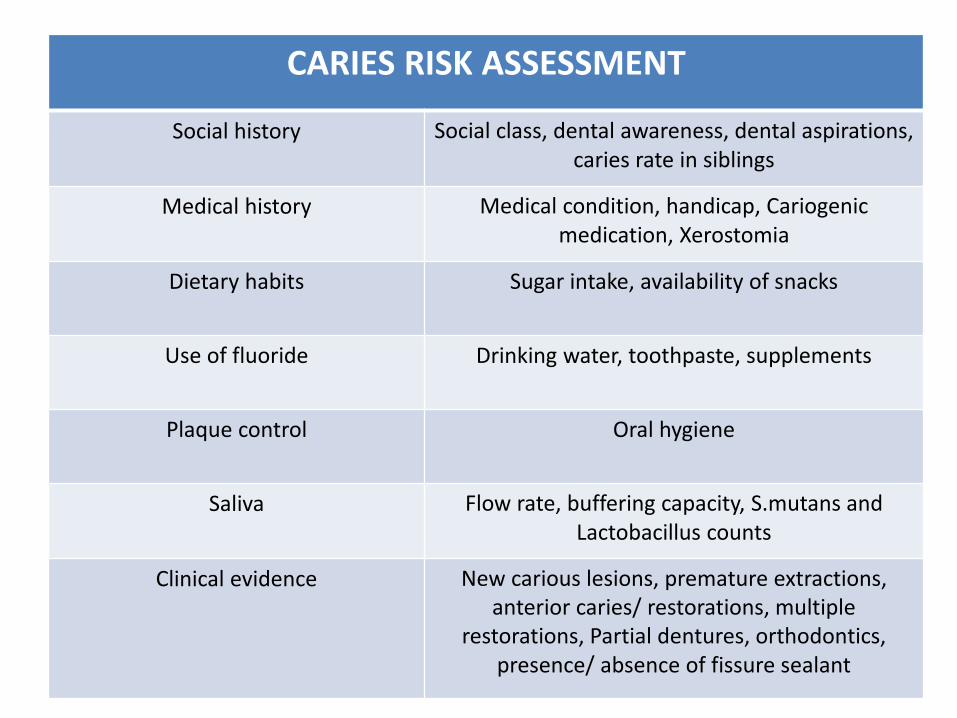
CARIES RISK ASSESSMENT
Social class, dental awareness, dental aspirations, caries rate in siblings
Social history
Medical condition, handicap, Cariogenic medication, Xerostomia
Medical history
Sugar intake, availability of snacks Dietary habits
Drinking water, toothpaste, supplements Use of fluoride
Oral hygiene Plaque control
Flow rate, buffering capacity, S.mutans and Lactobacillus counts
Saliva
New carious lesions, premature extractions, anterior caries/ restorations, multiple
restorations, Partial dentures, orthodontics, presence/ absence of fissure sealant
Clinical evidence

Caries Risk Assessment
It is now known that surgical intervention of dental caries alone does not
stop the disease process. Additionally, many lesions do not progress, and
tooth restorations have a finite longevity.
Modern management of dental caries should be more conservative and
includes:
early detection of noncavitated lesions
identification of an individual’s risk for caries progression
understanding of the disease process for that individual
application of preventive measures

Caries Risk Assessment
NICE recall intervals and oral health – 2004
Caries risk assessment form – American Dental Association 2009
Guideline on Caries-risk Assessment and Management for Infants,
Children, and Adolescents - AMERICAN ACADEMY OF PEDIATRIC
DENTISTRY 2013


The NICE guidelines 2004:
• For adult patients, NICE recommends a recall between three months and two
years, based on a risk assessment, taking into account a checklist of risk
factors, such as alcohol and tobacco use.
• The recommended interval for children is between three and 12 months.
• The actual interval should be a clinical decision by the dentist based on the
patient’s needs

Treatment planning – clinical cases:

PERSONAL DETAIILS Initials: G.S. Sex: Male Date of birth: 10/09/1949 Age at presentation: 62
PATIENT’S COMPLAINTS 1)“I lost my front bridge and I’m not
happy with how my teeth look” 2)“I sometimes struggle to chew
steaks”
RELEVANT MEDICAL HISTORY Fit and well. No allergies. No medication.
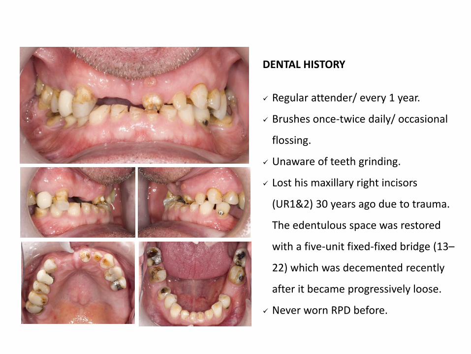
DENTAL HISTORY
Regular attender/ every 1 year.
Brushes once-twice daily/ occasional
flossing.
Unaware of teeth grinding.
Lost his maxillary right incisors
(UR1&2) 30 years ago due to trauma.
The edentulous space was restored
with a five-unit fixed-fixed bridge (13–
22) which was decemented recently
after it became progressively loose.
Never worn RPD before.
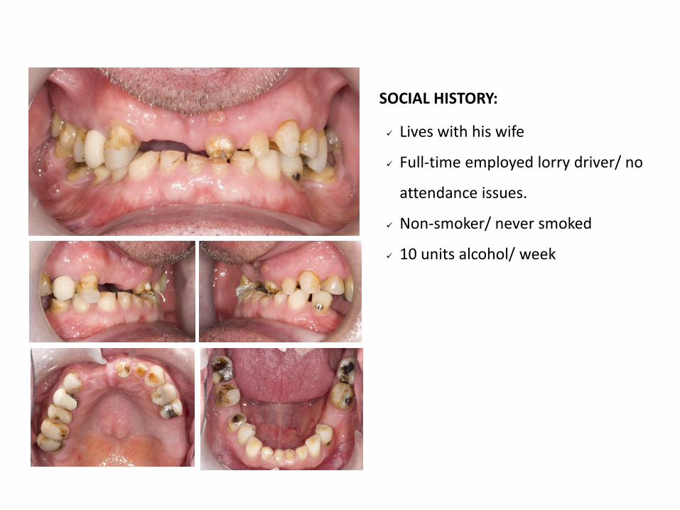
SOCIAL HISTORY:
Lives with his wife
Full-time employed lorry driver/ no
attendance issues.
Non-smoker/ never smoked
10 units alcohol/ week

Extra-oral examination:
TMJ: NAD
Muscles of Mastication: NAD
Facial symmetry: NAD
Lips: competent and low maxillary lip line.
Intra-oral exammination:
Soft tissues: Labial discharging sinus tract (UL1).
Hard tissues: 6-7mm Torus palatinus
BPE:
Bleeding index: 30%
Plaque index: 30%
Oral hygiene: Fair with some visible plaque and
calculus deposits.

Charted teeth present:
Comp: composite filling
Amg: amalgam filling
MC-Cr: metal-ceramic crown
#: fractured crown
TSL: tooth surface loss
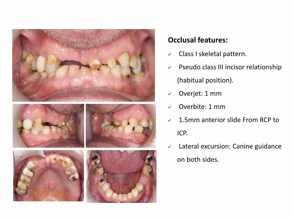
Occlusal features:
Class I skeletal pattern.
Pseudo class III incisor relationship
(habitual position).
Overjet: 1 mm
Overbite: 1 mm
1.5mm anterior slide From RCP to
ICP.
Lateral excursion: Canine guidance
on both sides.

SPECIAL INVESTIGATIONS:
Sensibility testing: (Endo frost and electric pulp tests):
Negative response:
Baseline records: maxillary and mandibular impressions, jaw
registration in CR and face-bow record.
1 1
1 2
8

RADIOGRAPHIC EXAMINATION

DIAGNOSES AND CLINICAL FINDINGS SUMMARY:
1. Generalized tooth surface loss (TSL).
2. Failing direct and indirect restorations:
3. Caries:
4. Necrotic pulp:
5. Chronic apical periodontitis:
6. Missing teeth:
7 6 5 4 3 4
8 7
7 6 5 3 1 2 3
8 7
1 2
8 1 1 1 2
1
1 2 6 7 8
6 5 6
8 2 1

TREATMENT OPTIONS:
1- Extraction of existing teeth and provision of complete dentures.
2- Restore the existing teeth with direct and indirect restorations and:
a) accept edentulous spaces .
b) restore edentulous spaces with a removable partial denture(s).
c) Restore the edentulous spaces with fixed partial bridges.
d) Restore the edentulous spaces with implant supported prostheses.

TREATMENT PLAN
1. Stabillization and prevention:
a- Improve oral hygiene and dietary habits.
b- Stabilize active caries and periodontal disease.
c- Extraction of teeth with hopeless prognosis.
2. Transitional:
a- Increase the OVD.
b- Improve function and aesthetics using direct restorations.
3. Definitive:
Restore the existing teeth with direct and indirect restorations at the new OVD.
4. Maintenance:
Maintain a healthy dentition and oral tissues.
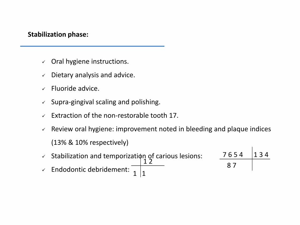
Stabilization phase:
Oral hygiene instructions.
Dietary analysis and advice.
Fluoride advice.
Supra-gingival scaling and polishing.
Extraction of the non-restorable tooth 17.
Review oral hygiene: improvement noted in bleeding and plaque indices
(13% & 10% respectively)
Stabilization and temporization of carious lesions:
Endodontic debridement:
1 3 4
8 7
7 6 5 4 1 2
1 1
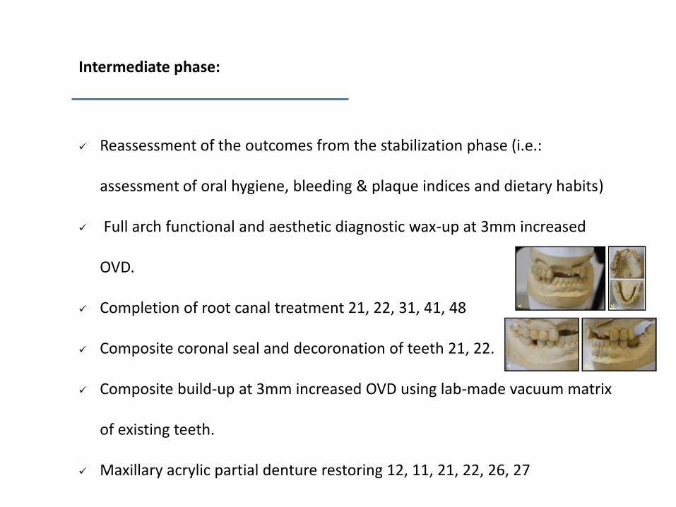
Intermediate phase:
Reassessment of the outcomes from the stabilization phase (i.e.:
assessment of oral hygiene, bleeding & plaque indices and dietary habits)
Full arch functional and aesthetic diagnostic wax-up at 3mm increased
OVD.
Completion of root canal treatment 21, 22, 31, 41, 48
Composite coronal seal and decoronation of teeth 21, 22.
Composite build-up at 3mm increased OVD using lab-made vacuum matrix
of existing teeth.
Maxillary acrylic partial denture restoring 12, 11, 21, 22, 26, 27


Definitive phase:
Maxillary definitive restorations:
1- Co-Cr RPD design .
2- Milled metal-ceramic crowns: teeth 13 and 23.
3- Milled gold crown: tooth 16.
4- Maxillary Cr-Co RPD insertion.
Definitive phase:
Mandibular definitive restorations:
1- Full gold crown: LR7.
2- Fixed-fixed metal-ceramic bridge from LL4 – LL7
(Edentulous 46 space was accepted and no restoration was planned).
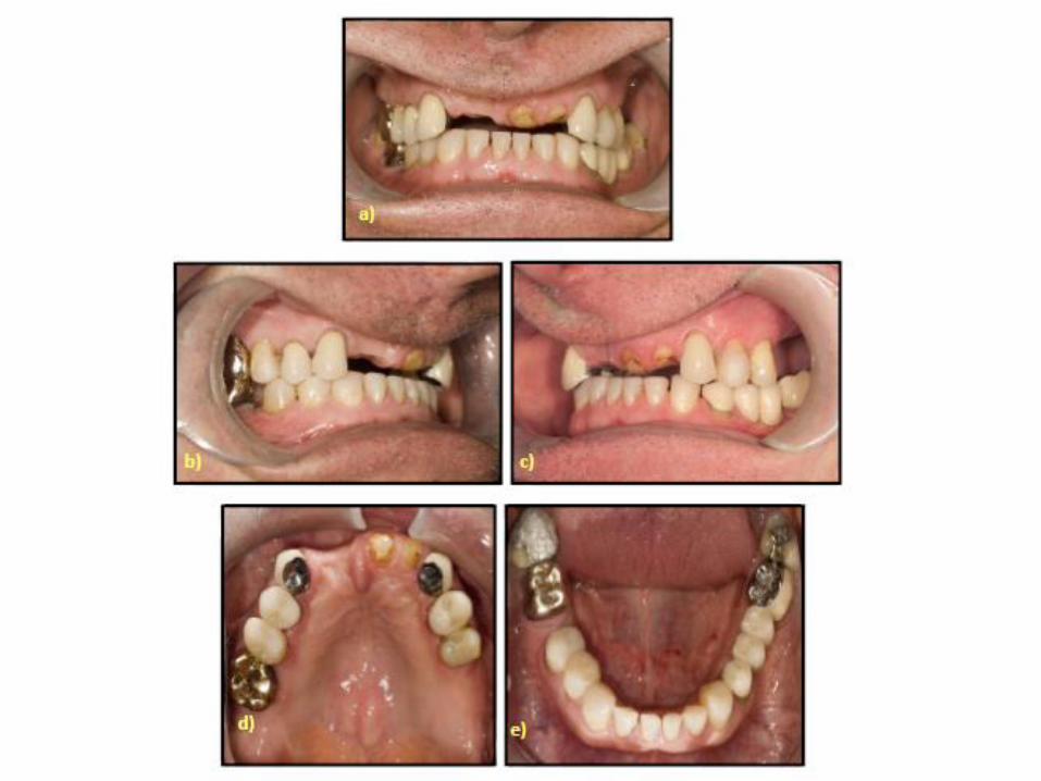


Maintenance phase:
At the 6-month review appointment:
Patient presented with good oral hygiene and healthy gingivae
(bleeding and plaque indices 11% and 10% respectively).
His occlusion was stable.
Periapical radiographs taken (after 1 year of completion of root canal
treatments) showed satisfactory periradicular healing