Introduction to Anatomy & Physiology Headings Vocabulary Important Info.
46
Introduction to Anatomy & Physiology Headings Vocabulary Important Info
-
Upload
jayson-parsons -
Category
Documents
-
view
221 -
download
2
Transcript of Introduction to Anatomy & Physiology Headings Vocabulary Important Info.

Introduction to
Anatomy & PhysiologyHeadings
Vocabulary
Important Info

•Anatomyoscience of
structure
orelationships revealed by dissection imaging techniques
•Physiology
oscience of body functions

Clinical Observational Techniques•Palpation
ofeel body surface with
hands
pulses and breathing
rates
•Auscultation
olisten to body sounds
with stethoscope
abnormal fluid in lungs
•Percussion
otap on body surface and
listen to echo
air in intestines

Levels of Organization
•Chemical
•Cellular
•Tissue
•Organs
•System Level
•Organismic Level

Levels of Structural Organization•Chemical Level - atomic and molecular level
•Cellular Level - smallest living unit of the body
•Tissue Level - group of cells and the materials
surrounding them that work together on one task
•4 basic tissue
oepithelium
omuscle
oconnective
tissue
onerve

•Organ Levelogrouping of 2 or more tissue types into a recognizable structure
with a specific function.
•Organ Systemocollection of related organs with a common function
osometimes an organ is part of more than one system
•Organismic Level - one living individual.
Interactions of Body Systems
•Example: Integumentary System & Skeletal System
oSkin produces vitamin D needed for CA absorption and bone
growth
oBone marrow produces cells which help the skin resist
infection.

Life Processes•Metabolism = sum of all
chemical processesobreakdown of large
molecules into small
obuilding new structural components (proteins)
oproviding chemical energy for cells
•Responsivenessodetect & respond to changes
in internal or external environment
osome typical responses muscle contraction, electrical
signals, hormone or glandular secretion

•Movement oany structural level
obody, organ, cell or cell component
•Growthoincrease in number or size
of cells or the material found between cells
•Differentiationospecialization of cells
for a specific function
ostem cells give rise to cells that specialize
•Reproductionoformation of new cells or
new individuals

Autopsy•Postmortem examination of body by dissection
•Purposeoconfirm or determine
cause of death
osupport findings of other tests
oprovide info on effects of drug usage
oeducate healthcare students
oreveal congenital defects

Homeostatis•Maintaining the internal
environment within physiological limits (internal balance)
•First described by French physiologist, 1813-1878
•Process named by Walter Cannon, 1871-1945
•Exampleoblood glucose level is kept
within narrow range 70-110/100ml

Homeostasis of Body Fluids
•Delineation of fluid compartments
•Intracellular Fluid (ICF) = w/i cells
•Extracellular Fluid (ECF) = o/s cells
Intercellular Fluid = tissue fluid =
interstitial fluid
Plasma = fluid portion of blood
•Composition of fluids change as
substances move between compartments
onutrients, oxygen, ions and wastes
move in both directions across
capillary walls

Control of Homeostasis•Homeostasis is continually
being disrupted by:oExternal Stimuli
intense heat, cold , and lack of oxygen
oInternal Stimuli psychological stresses
exercise
•Disruptions are usually mild & temporary
•If homeostasis is not maintained, death may result

Neural and Endocrine Controls•Maintaining a controlled
conditionosensory receptors detect change
in a monitored variable
onervous system and/or endocrine system responds
•Ex: Control of blood gas leveloexercise increases blood CO2
levels
osensory receptors detect change
onervous system increases heart and breathing rates to remove excess CO2
oadrenal gland releases epinephrine to increase heart and breathing rates

Components of Feedback Loop
•Receptor
omonitors a controlled condition
•Control Center
odetermines next action
•Effector
oreceives directions from the
control center
oproduces a response that
changes controlled condition

Negative & Positive Feedback Loops•Negative Feedback Loopooriginal stimulus reversed
omost feedback systems in the body are negative
oused for conditions that need frequent adjustment
obody temperature, blood sugar levels, blood pressure
•Positive Feedback Loopooriginal
stimulus intensified
onormal childbirth

Homeostasis of Blood Pressure
•Pressure receptors in walls of
certain arteries detect an increase in
BP
oBlood Pressure = force of blood on
walls of vessels
•Brain receives input and signals
heart and blood vessels
•Heart rate slows and arterioles
dilate (increase in diameter)
•BP returns to normal
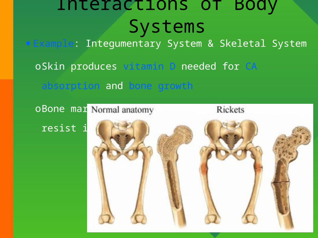
Positive Feedback during Childbirth
•Stretch receptors in walls of
uterus send signals to brain
•Brain releases hormone
(oxytocin) into bloodstream
•Uterine smooth muscle contracts
more forcefully
•More stretch, more hormone,
more contraction etc.
•Cycle ends with birth of the baby
& decrease in stretch

Homeostatic Imbalances•Disorder = abnormality of function
•Disease = homeostatic imbalance with distinct…oSymptoms: changes in body function felt by patient such as nausea
oSigns: changes in body function that can be observed by doctor such as rash or fever
•Diagnosis: skill of distinguishing one disease from another
•Epidemiology: how disease is transmitted
•Pharmacology: how drugs used to treat disease

Basic Anatomical Terminology
•Regions of the body
•Anatomical position
•Anatomical planes, sections and directional terms

Anatomical Position
•Standardized position
describing directional
terms
ostanding upright
ofacing the observer,
head level
oeyes facing forward
ofeet flat on the floor
oarms at the sides
opalms turned forward
•Prone Position = lying face
down
•Supine Position = lying face up

Common Regional Names
•Clinical terminology based on a Greek or Latin root word.
•Fill in worksheet to help remember the terms
She is standing in the Anatomical Position

Planes & Sections
•Plane: imaginary flat
surface that passes
through the body.
•Section: one of the 2
surfaces (pieces) that
results when the body is
cut by a plane passing
through it.

Sagittal Plane•Sagittal Plane
odivides the body or an organ into left and right sides
•Midsagittal Plane
oproduces equal halves
•Parasagittal Plane
oproduces unequal halves

Other Planes and Sections
•Frontal or Coronal Plane
odivides the body or an organ into front
(anterior) and back (posterior) portions
•Transverse or Horizontal Plane
ocross-sectional
odivides the body or an organ into upper
(superior) or lower (inferior) portions
•Oblique Plane
osome combination of 2 other planes

Planes and Sections of the Brain(3-D anatomical relationships revealed)
•Horizontal Plane
•Frontal Plane
•Midsagittal Plane

Major Directional Terms

Superior & Inferior
•Dorsal or Posterior
oBack of the body
oBrain is posterior to the forehead.
•Ventral or Anterior
oFront of the body
oSternum is anterior to the heart.
•Superior
oTowards the head
oEyes are superior to mouth.
•Inferior
oAway from head
oStomach is inferior to the
heart.
•Dorsal & Ventral
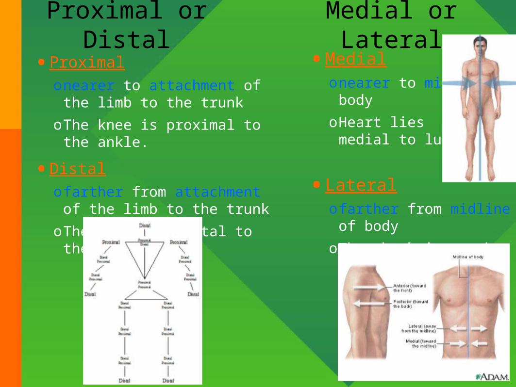
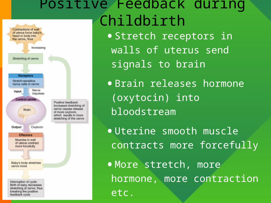
Medial or Lateral
•Medialonearer to midline of body
oHeart lies medial to lungs
•Lateralofarther from midline of body
oThe thumb is on the lateral side of the hand.
•Proximalonearer to attachment of the limb
to the trunk
oThe knee is proximal to the ankle.
•Distalofarther from attachment of the
limb to the trunk
oThe wrist is distal to the elbow.
Proximal or Distal

•Brain is posterior to the
forehead.•Eyes are superior to
mouth.•Stomach is inferior to the
heart.•Sternum is anterior to the heart.•The knee is proximal to the ankle.
•Heart lies medial to lungs
•The wrist is distal to the
elbow.•The thumb is on the lateral side of the hand.

Dorsal Body Cavity•Near dorsal surface of body
•2 subdivisions
oCranial Cavity holds the brain
formed by skull
oVertebral or Spinal Canal contains the spinal cord
formed by vertebral column
•Meninges (system of membranes) line dorsal body cavity

Ventral Body Cavity
•Near ventral surface of body
•Visceral Organs (viscera): A group of internal organs housed in the
ventral cavity
•2 subdivisions
oThoracic Cavity: above
diaphragm
oAbdominopelvic Cavity:
below diaphragm
•Diaphragm = large,
dome-shaped muscle
•Organs called viscera
•Organs covered with
serous membrane

Abdominopelvic Cavity
•Inferior portion of ventral body cavity below diaphragm
•Encircled by abdominal wall, bones & muscles of pelvis

Thoracic Cavity
•Encircled by ribs, sternum, vertebral column and muscle
•Divided into 2 pleural cavities by mediastinum
•Mediastinum contains all thoracic organs except lungs
o"middle" section of the chest cavity
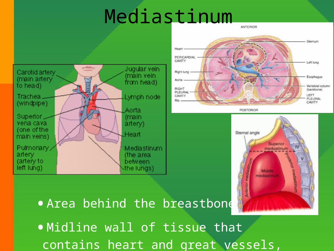
Mediastinum
•Area behind the breastbone
•Midline wall of tissue that contains heart and
great vessels, esophagus, trachea and thymus.
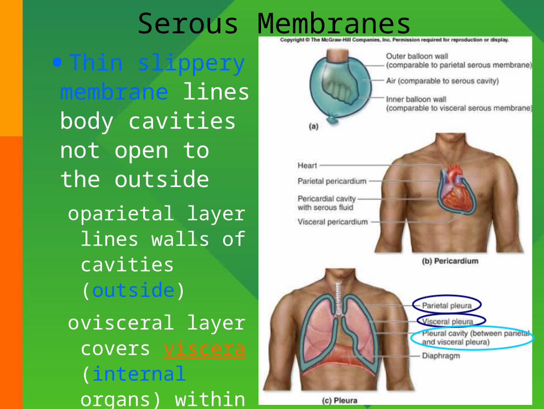
Serous Membranes
•Thin slippery membrane lines body cavities not open to the outsideoparietal layer lines
walls of cavities (outside)
ovisceral layer covers viscera (internal organs) within the cavities
•Serous fluid reduces friction

Pleural & Pericardial Cavities
•Visceral Pleura: clings to surface of lungs
•Parietal Pleura: lines chest wall
•Visceral Pericardium: covers heart
•Parietal Pericardium: lines pericardial sac

Peritoneum
•Visceral Peritoneum --- serous membrane that covers the abdominal viscera (organs)
•Parietal Peritoneum --- serous membrane that lines the abdominal wall
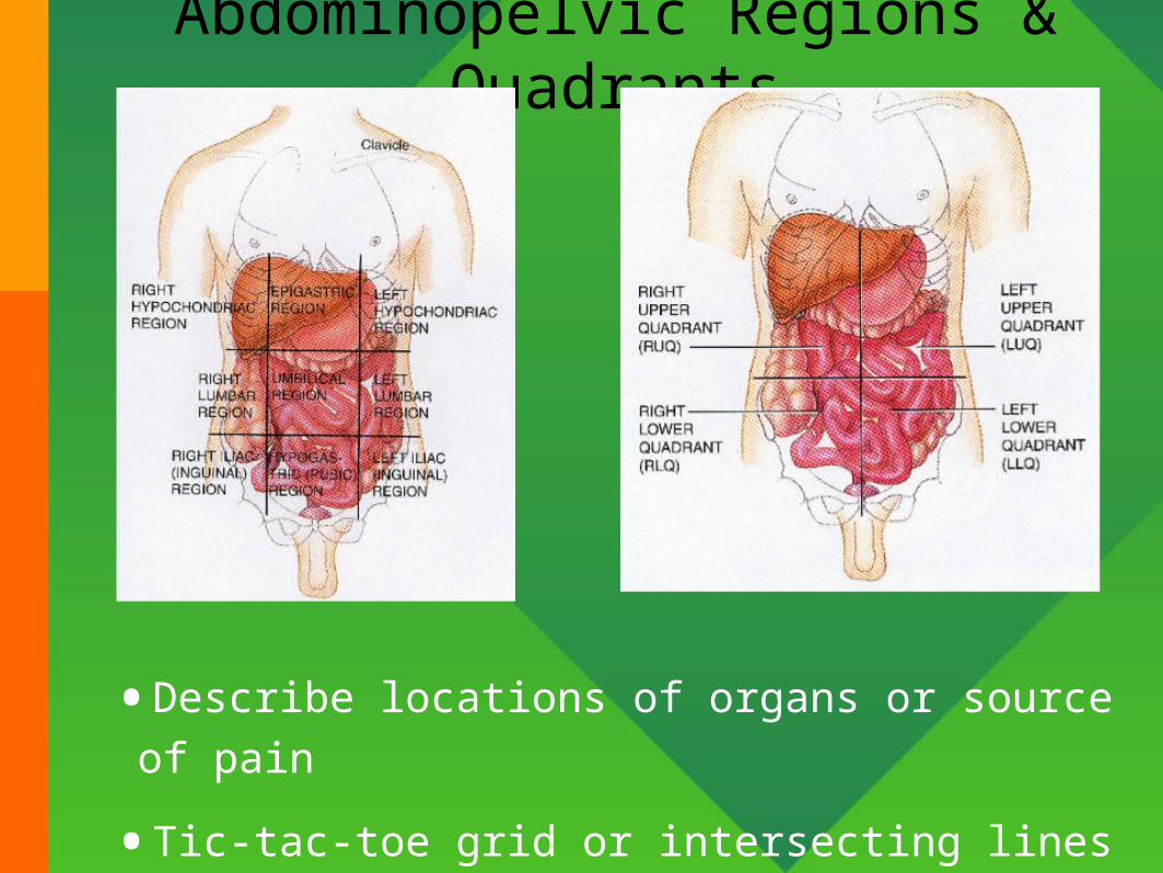
Abdominopelvic Regions & Quadrants
•Describe locations of organs or source of pain
•Tic-tac-toe grid or intersecting lines through navel

Medical Imaging
•Allows visualization of structures without
surgery
•Useful for
confirmation
of diagnosis
•Examples of
imaging
techniques

Conventional Radiography•A single burst of xrays
•Produces 2-D image on
film
•Known as radiography or
xray
•Poor resolution of soft
tissues
•Major use is Osteology:
study of bones

Computed Tomography (CT Scan)
•Moving x-ray beam
•Image produced on a video monitor of a cross-section through body
•Computer generated image reveals more soft tissue detailokidney & gallstones
•Multiple scans used to build 3D views

Digital Subtraction Angiography(DSA)
•Radiopaque material
injected into blood
vessels
•Before and after images
compared with a
computer program
•Image of blood vessel is
shown on a monitor

Ultrasound (US)•High-frequency sound waves
emitted by hand-held device
•Safe, noninvasive & painless
•Image or sonogram is displayed on video monitor
•Used for fetal ultrasound and examination of pelvic & abdominal organs, heart and blood flow through blood vessels

Magnetic Resonance Imaging (MRI)
•Body exposed to high-energy magnetic field
•Protons align themselves relative to magnetic field
•Pulse of radiowaves used to generate an image on video monitor
•Can not use on patient with metal in their body
•Reveals fine detail within soft tissues

Positron Emission Tomography(PET)
•Substance that emits
positively charged
particles is injected into
body
•Collision with negatively
charged electrons in
tissues releases gamma
rays
•Camera detects gamma
rays & computer
generates image
displayed on monitor