
Best Children Specialist In Virginia | Online Pediatricians Centre
Upload
hoanghuongCategory
view
214download
0
Introduction of the Image Gently Introduction of the Image Gently Campaign to Pediatricians using a Campaign to Pediatricians using a
series of Patient Presentation Modules series of Patient Presentation Modules Entitled Entitled ““Commonly Requested but Commonly Requested but
Misunderstood Imaging StudiesMisunderstood Imaging Studies”” and and self assessment examself assessment exam
Tiffany Lewis BATiffany Lewis BA1, 1, Julianne Dean BAJulianne Dean BA22,Lisa Lowe MD, FAAP,Lisa Lowe MD, FAAP3,43,4, , Trent Phan DOTrent Phan DO44, Cynthia Taylor MD, Cynthia Taylor MD3,43,4
1Kansas City University of Medicine and Biosciences, 2Midwestern University Arizona College of Osteopathic Medicine Glendale, AZ, 3Children’s Mercy
Hospitals and Clinics,4University of Missouri Kansas City School of Medicine, Kansas City, MO

Learning Objectives
After viewing this exhibit the learner should:• Become more familiar with the Image
Gently Campaign• Have increased knowledge on the
indications for commonly requested but misunderstood imaging studies
• Be aware of a series of newsletters free to the public online

Outline
• Educational Campaign information• Optional links to mini-discussions for
commonly ordered but misunderstood studies• Self-assessment exam• Conclusion • References

Introduction
• Recent studies suggests there has been a significant increase in the use of medical radiation, especially CT, from 2001 to 2006
• 7 million CT studies are done per year in the pediatric population and it increases 10% per year
• In 2004, a survey indicated only 9% of emergency physicians and 47% radiologists were aware of radiation risks from CT
• Multidetector CT and 3D imaging increase radiation dose by up to 30% or 3 to 10 times
Arch, Michael and Donald P. Frush. “Pediatric Body MDCT: A 5-year follow up survey of scanning parameters used by Pediatric Radiologists.” AJR 2008; 191: 611-617.http://www.pedrad.org/associations/5364/ig/index.cfm?page=393

Introduction
• In children, radiation induced cancer risk is increased due to younger age, lifespan, and increased sensitivity to radiation
• Our job as radiologists begins with helping clinicians determine study indications, and follows with ensuring appropriate imaging protocols
• Many practitioners are unaware of the Image Gently campaign
• A local education campaign can help get the word out about the Image Gently campaign

Purpose of Poster Presentation
• Given the increased use of medical imaging in children, especially CT, and the lack of knowledge regarding the risks to children in the general medical community, we set out to:– Educate practitioners on the image gently
campaign– Educate practitioners on commonly requested
but misunderstood studies• Hopefully our educational campaign will help
protect children from risk related to unnecessary radiation and sedation

Image Gently Campaign
• The Society for Pediatrics Radiology (SPR) together with the American Academy of Pediatrics (AAP) along with 33 other medical organizations have formed a multidisciplinary group, the Alliance for Radiation Safety in Pediatric Imaging
• This organization represents over 400,000 healthcare professionals promoting appropriate and high quality CT for children

Image Gently Campaign
• 4 simple guidelines:– Reduce or “child-size”– Scan only when necessary– Scan only the indicated region– Scan once; multiphase scanning is usually not necessary in
children• You can take the pledge online at:
– http://www.pedrad.org/associations/5364/ig
the amount of radiation utilized
Let’s increase the number pledged today!

Image Gently Pledge

Imaging Guidelines
• Imaging guidelines and appropriateness criteria can be found on the ACR website:– http://www.acr.org/s_acr/bin.asp?TrackID=&S
ID=1&DID=14800&CID=1848&VID=2&DOC=File.PDF

Imaging Guidelines--AAP
• Imaging guidelines can also be found on the American Academy of Pediatrics (AAP) website at http://www.aap.org/sections/radiology/default.cfm
• This also includes radiation safety information for parents
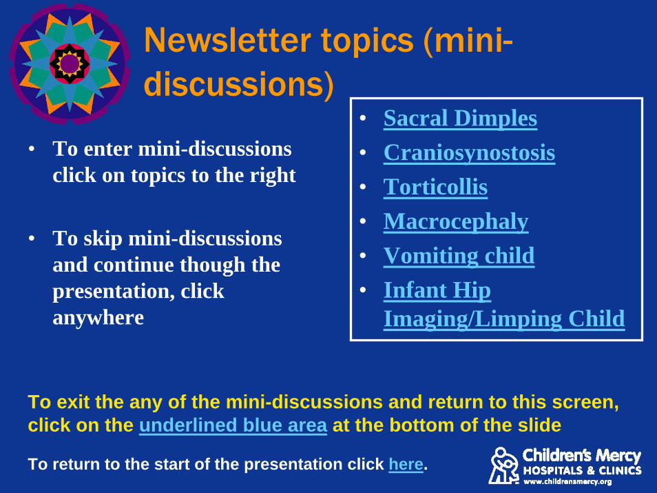
Newsletter topics (mini-discussions)
• To enter mini-discussions click on topics to the right
• To skip mini-discussions and continue though the presentation, click anywhere
• Sacral Dimples• Craniosynostosis• Torticollis• Macrocephaly• Vomiting child• Infant Hip
Imaging/Limping Child
To exit the any of the mini-discussions and return to this screen, click on the underlined blue area at the bottom of the slide
To return to the start of the presentation click here.

Sample newsletters:
• Image gently campaign

Sample newsletters:
• Imaging sacral dimples

Sample newsletters:
• Cranio-synostosis

Sample newsletters:
• Imaging torticollis

Sample newsletters:
• Imaging macrocephaly

Sample newsletters:
• Imaging the vomiting infant

Sample newsletters:
• Imaging the hip

CMH website with downloadable newsletters
• These newsletters are being distributed to community practitioners
• They are available on Children’s Mercy Hospital and Clinics Radiology website at:– http://www.childrensmercy.org/Content/view.aspx?id=183
• Use of these newsletters is open to the public
Return to newsletter page or click anywhere to continue with next topic

Self-Assessment Exam
• Welcome to the self assessment exam where you can sees what you have learned
• Please answer the next 5 questions by clicking True or False
• Follow prompts on screen to obtain the correct answer and move on to the next question

Self-assessment Exam: Question #1
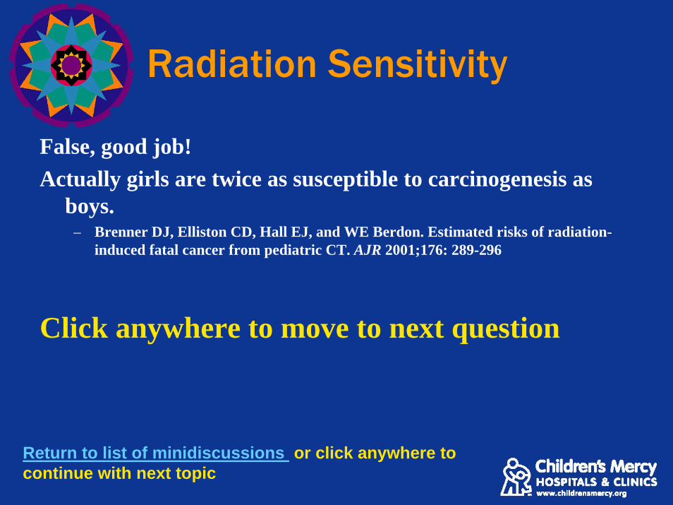
True or FalseBoys are twice as sensitive to radiation as girls.
Return to list of minidiscussions or click anywhere to continue with next topic

Radiation Sensitivity
True is not the correct answer, good try.
Move to correct answer
Return to list of minidiscussions or click anywhere to continue with next topic
Radiation Sensitivity
False, good job!Actually girls are twice as susceptible to carcinogenesis as
boys.– Brenner DJ, Elliston CD, Hall EJ, and WE Berdon. Estimated risks of radiation-
induced fatal cancer from pediatric CT. AJR 2001;176: 289-296
Click anywhere to move to next question
Return to list of minidiscussions or click anywhere to continue with next topic

Self Assessment Exam: Question #2
True or FalseInfants are up to 15 times more sensitive to radiation than adults.
Return to list of minidiscussions or click anywhere to continue with next topic

Radiation Sensitivity
False, nice try but incorrect.
Move to correct answer
Return to list of minidiscussions or click anywhere to continue with next topic

Radiation Sensitivity
True! Correct.“Because they have more rapidly dividing cells than adults
and have a longer life expectancy, the odds that children will develop cancer from x-ray radiation are higher than adults.”
– “One size does not fit all: Reducing Risks from Pediatric CT” ACR Bulletin February 2001 57(2): 20-23.
Click anywhere to move to next question
Return to newsletter page or click anywhere to continue with next topic

Self Assessment Exam: Question #3
True or False3D CT should be utilized as a screening test for children suspected of having craniosynostosis.
Return to list of minidiscussions or click anywhere to continue with next topic

3D CT
True, nice try, but incorrect.
Move to correct answer
Return to list of minidiscussions or click anywhere to continue with next topic

3D CT
False, good job!3 dimensional CT adds 3-10 times more radiation and is
mostly reserved for surgical planning.
Click anywhere to move to next question
Return to list of minidiscussions or click anywhere to continue with next topic

Self Assessment Exam: Question #4
True or FalseIt has been estimated that radiation from CT may cause up to 1:1,000 neoplasms.
Return to list of minidiscussions or click anywhere to continue with next topic

Cancer
False. Nice try!
Move to correct answer
Return to list of minidiscussions or click anywhere to continue with next topic

Cancer
True. It has been estimated that CT may cause up to 1 in 1,000
neoplasms– Brenner, DJ Estimating cancer risks from pediatric CT: going from the
qualitative to the quantitative. Pediatric Radiology 2002: 32: 228-231
Click anywhere to move to next question
Return to list of minidiscussions or click anywhere to continue to the final question

Self assessment #5
True or FalseMany pediatric radiologists estimate that greater than 30 percent of CT scans are unneeded.
Return to list of minidiscussions or click anywhere to continue with next topic

Need for scans
False. Incorrect, but good try.
Move to correct answer
Return to list of minidiscussions or click anywhere to continue with next topic

Need for scans
True. Nice job!“A poll might show that we consider 10% to 30% of our CT
examinations unnecessary. A nonscientific consensus at the ALARA conference was that about 30% of CT examinations in children were totally unnecessary or were readily replaceable by examinations not using radiation.”
– Slovis, Thomas L. Introduction to Seminar in Radiation Dose Reduction. Pediatric Radiology (2002) 32: 707-708.
Please click anywhere for references and conclusion.
Return to list of minidiscussions page or click anywhere to continue with next topic

Conclusion
• The newsletters in this presentation are available to the public on the internet and are not copyright protected
• They were distributed to 4,500 primary care physicians, nurse practitioners, and physician assistants in MO and KS along with our medical staff
Return to the BEGINNING of presentation by clicking here
Return to list of minidiscussions by clicking here

• This ends the presentation on our education campaign
• If you wish to go to the list of mini-discussion topics clickhere
• Return to beginning of presentation by clicking here
• Click anywhere for references
• Thank you for your attention!
Conclusions

References
1. Arch, Michael and Donald P. Frush. “Pediatric Body MDCT: A 5-year follow up survey of scanning parameters used by Pediatric Radiologists.” AJR 2008; 191: 611-617.
2. Brenner DJ, Hall EJ. Computed tomography: an increasing source of radiation exposure. N Engl J Med 2007; 357:2277-2284.
3. Brenner, DJ Estimating cancer risks from pediatric CT: going from the qualitative to the quantitative. Pediatric Radiology 2002: 32: 228-231
4. Brenner DJ, Elliston CD, Hall EJ, and WE Berdon. Estimated risks of radiation-induced fatal cancer from pediatric CT. AJR 2001;176: 289-296
5. Cohen, MM Jr. Epidemiology of Craniosynostosis. In: Cohen, MM Jr, ed Craniosynostosis: diagnosis, evaluation, and management, 2nd ed. New York: Oxford University Press, 2000: 112-118.
6. Do, Twee. Congenital Muscular Torticollis: Current Concepts and Review of Treatment. Current Opinion in Pediatrics 2006; 18:26-29
7. Dudkiewicz, I, Ganel, A, Blankstein, A. Congenital Muscular Torticollis in Infants: Ultrasound-Assisted Diagnosis and Evaluation. Journal of Pediatric Orthopaedics 2005; 25:812-814.
8. Goske MJ, et. al. The ‘Image Gently’ campaign: increasing CT radiation dose awareness through a national education and awareness program. Pediatr Radiol 2008 38:265-269.
9. The Image Gently Campaign: Working Together to Change Practice. AJR February 2007; 100:273-274.
10. Kriss, VM, et. al. Occult spinal dysraphism in neonates: assessment of high risk cutaneous stigmata on sonography. AJR 17 (6): 1687-92.

References
11. Kriss, VM, Kriss TC, Babcock, DS. The Ventriculus Terminalis of the Spinal Cord in the Neonate: A Normal Variant on Sonography. AJR 1995; 165: 1491-1493.
12. Lajeunie, E, Le Merrer, et al. Genetic study of nonsyndromic coronal craniosynostosis. Am J Med Genet 1995; 55: 500-504
13. Lee, CI, Haim, AH, Monico, EP et al. Diagnostic CT scans: assessment of patient, physician, and radiologist awareness of radiation dose and possible risks. Radiology 2004; 231: 393-398.
14. Lowe, LH, Johanek AJ, Moore, CW. Sonography of the Neonatal Spine: Part I, Normal Anatomy, Imaging Pitfalls, and Variations that May Simulate Disorders. AJR 2007; 188:733-738.
15. Medina, LS, R Richardson, and K Crone. Children with Suspected Craniosynostosis: A Cost Effectiveness Analysis of Diagnostic Strategies. AJR 2002; 179: 215-221.
16. “One size does not fit all: Reducing Risks from Pediatric CT” ACR Bulletin February 2001 57(2): 20-23.
17. Slovis, Thomas L. Introduction to Seminar in Radiation Dose Reduction. Pediatric Radiology (2002) 32: 707-708
18. Smith, R, et al. The value of head ultrasound in infants with macrocephaly. Pediatric Radiology. 1998 Mar; 28(3): 143-146.
19. Subach, BR, McLaughlin, MR, Albright, LA, Pollack, IF. Current Management of Pediatric Atlantoaxial Rotatory Subluxation. Spine 1998; 23:2174-2179.
Return to the BEGINNING of presentation by clicking here or ESC to exit to main screen

Imaging sacral dimples: US or MRI?
• Sacral dimples are common and usually of no significance
• US is the modality of choice in infants < 4-6 months age– Sedation risk is higher < 6 months age
• MRI is the modality of choice > 6 months age• If surgery is planned, it is useful to delay imaging until
surgery is imminent
• Caveat: MRI is the study of choice in a child of ANY age with a draining dimple
Return to list of minidiscussions or click anywhere to continue with next topic

Sacral Dimples: Imaging Low versus High Risk lesions
Low Risk High RiskMidline Off midline< 5mm > 5mm
Within 2.5 cm of anus > 2.5 cm from the anus
No cutaneous abnormalities Cutaneous abnormalities (i.e. hemangiomas, cutis aplasia, hairy patches, skin tags)
Return to list of minidiscussions or click anywhere to continue with next topic

Sacral Dimples with cutaneous stigmata: High risk
Hemangioma
Myelomeningocele with skin covering Hairy patch
Sacral dimple above gluteal crease
Return to list of minidiscussions or click anywhere to continue with next topic

2 children with dimples above the gluteal crease
US—Child less than 6 months of age
MR—Child greater than 6 months of age
Normal US—conus at L2
Dimple that extends to the dural but does violate it
Return to list of minidiscussions or click anywhere to continue with next topic

Craniosynostosis Imaging
Imaging is based on 3 categories:1. Low risk- Developmentally normal or posterior
flattening only– Plain films (4 view skull)
2. Intermediate risk - Healthy children with head deformity– CT head
3. High risk - Obviously misshapen head– 3D CT needed for surgical planning
Return to list of minidiscussions or click anywhere to continue with next topic

Craniosynostosis: Low vs. intermediate vs. high risk imaging
Risk Category Imaging modality recommended
Low risk—developmentally normal and posterior flattening only
4 view skull (plain films)
Intermediate risk—healthy children with head deformity
CT Head
High risk—developmentally abnormal children and/or children with obvious head deformity almost certainly needing surgery
3D Head CT
Return to list of minidiscussions or click anywhere to continue with next topic

Craniosynostosis: Low RiskHistory: Developmentally normal child with posterior flattening
Coronal
Diagnosis: Normal suturesSagittal Lambdoid
Return to list of minidiscussions or click anywhere to continue with next topic
Lambdoid

Craniosynostosis: Intermediate Risk
History: Developmentally delayed child with parieto-occipital flattening
Diagnosis: Plagiocephaly (parieto-occipital flattening, but no craniosynostosis
Return to list of minidiscussions or click anywhere to continue with next topic

Craniosynostosis: High Risk
Diagnosis: Sagittal synostosis
Diagnosis: Lambdoid synostosis
Return to list of minidiscussions or click anywhere to continue with next topic
History: Child with obvious abnormally shaped head

Imaging Torticollis: Based on age and history
• Infant with torticollis:– Most often due to fibromatosis coli (hematoma
of sternocleidomastoid muscle)– More common in forceps delivery– Sonography is diagnostic– Rx: Physical therapy or surgical release
• Beyond young infants the work up depends on history
Return to list of minidiscussions or click anywhere to continue with next topic

Torticollis: Beyond infants
• History of isolated torticollis (no trauma) – Usually rotatory subluxation (self-limiting, due to muscle spasm)– No imaging needed unless persistent after a week or 2 of
conservative treatment– Isolated, persistent – do CT then consider dynamic scan with head
in neutral, right, and left positions• History of trauma
– Plain films or CT without contrast• History of sore throat/signs of infection
– CT with contrast• History of torticollis with neurological signs
– MRI
Return to list of minidiscussions or click anywhere to continue with next topic

Infantile TorticollisHx: 6-week-male with torticollis and forceps delivery
Normal right sterno-cleidomastoid muscle(SCM)
Return to list of minidiscussions or click anywhere to continue with next topic
Hematoma left SCMDiagnosis: Fibromatosis coli

Isolated Torticollis in a child
History: 10-year-male awakened with neck stuck to left 1.5weeks ago; No response to conservative treatment
Return to list of minidiscussions or click anywhere to continue with next topic
Coronal image demonstrates asymmetry between C1 and C2
C2
C1C1
Notice abnormal widening between C1 and dens widens with
head turning to the left (toward torticollis)
Notice normalization of distance between C1
and dens with head turning to the right
(away from torticollis)
Diagnosis: Rotatory Subluxation

Isolated persistent torticollis
Return to list of minidiscussions or click anywhere to continue with next topic
History: 15-year-male with neck “stuck to the right” after “neck adjustment”
Normal alignment Abnormal alignment with head turning right
Note C1 is looking a different direction than C2-C7
C2C1
Diagnosis: Rotatory Fixation

Torticollis with feverHistory: 1-year-female with sore throat and difficulty swallowing
Return to list of minidiscussions or click anywhere to continue with next topic
Lateral view of the neck demonstrates prevertebral swelling of soft tissues
Axial CT neck demonstrates fluid pockets both on the right and at midline
Diagnosis: Peritonsillar and retro-
pharyngeal infection

Torticollis with neurological symptoms
Return to list of minidiscussions or click anywhere to continue with next topic
Diagnosis: Spinal cord astrocytoma
History: 1- yr-female with persistent torticollis & scratching of the left arm

Imaging torticollis based on history
Clinical history and age Initial imaging approach< 3 months; asymptomatic except for torticollis
Ultrasound
> 3 months; asymptomatic except for torticollis
No imaging; conservative Rx 1-2 weeks;Persistent symptoms –CT without contrast
Any age with history of trauma CT without contrast Any age with symptoms of infection CT with IV contrast
Any age with neurological symptoms MRI
Return to list of minidiscussions or click anywhere to continue with next topic

Imaging macrocephaly
Definition: Head greater than 95%– If the head circumference levels out—no imaging– If the head growth rate continues to increase or
developmental abnormality, do imagingImaging approach:• US is used if the child is less than 6 months or if
they still have an open fontanelle• CT is used if the child is greater than 6 months
and no fontanelle• MRI if associated neurological symptoms
Return to list of minidiscussions or click anywhere to continue with next topic

Imaging macrocephaly
Clinical presentation and age Imaging approach
•Developmentally normal infant/child with open fontanelle
Head ultrasound
•Developmentally normal infant/child with closed fontanelle
CT or MRI
•Developmentally abnormal infant/child with open or closed fontanelle
MRI
Return to list of minidiscussions or click anywhere to continue with next topic

Macrocephaly: developmentally normal child with open fontanelleHistory: Normal subarachnoid space contains vessels
Return to list of minidiscussions or click anywhere to continue with next topic
Diagnosis: Benign enlarged subarachnoid spaces

Benign enlarged subarachnoid spaces (BESS)
• Usually presents between 3 months and 3 years of age (esp. 6-18mo)
• Most common cause of macrocephaly in a developmentally normal child
• Parents often have big head• Resolves spontaneously
Return to list of minidiscussions or click anywhere to continue with next topic

Macrocephaly in a neurologically abnormal child• 4-month-male with macrocephaly and lethargy
Return to list of minidiscussions or click anywhere to continue with next topic
Diagnosis: Choroid plexus papilloma

Vomiting infant: Imaging
• 3 categories:• Bilious vomiting – EMERGENCY
– Confirm with NG tube & get UGI• Nonbilious vomiting since birth
– Not an emergency, and often gastroesophageal reflux– Do not need UGI– Rx: Anti-reflux meds or surgical consult
• Projectile vomiting first occurring after several weeks of life – Ultrasound for Pyloric stenosis
Return to list of minidiscussions or click anywhere to continue with next topic

Vomiting Infant: Bilious
Clinical Presentation Initial ApproachBilious Vomiting —Emergency, refer to pediatric facility
NG tube and abdomen radiographs:•If radiographs suggest proximal obstruction (few or no dilated bowel loops) request UGI as malrotation with midgut volvulus is most likely•If radiographs suggest distal obstruction (many dilated bowel loops) request contrast enema first•May need to do both UGI and enema•No need for small bowel follow through
Return to list of minidiscussions or click anywhere to continue with next topic

Vomiting Infant: Non-bilious
Non-bilious vomiting•With normal weight gain (often since birth)
•No imaging; GERD most likely•Treat conservatively with anti-reflux meds
Non-bilious vomiting•With airway symptoms (cough, stridor)
•Request UGI•Vascular ring is most likely•No need for small bowel follow through
Non-bilious vomiting•4 to 8 week old
•Pyloric ultrasound •Pyloric stenosis most likely•No need for UGI or small bowel follow through
Return to list of minidiscussions or click anywhere to continue with next topic

Vomiting Infant: Bilious
Return to list of minidiscussions or click anywhere to continue with next topic
Abdomen radiograph shows a nonspecific bowel gas pattern with paucity of gas
UGI reveals obstruction of contrast at the 2nd portion of the duodenum. The contrast column corkscrews down into a beak.
History: 6-day-male with bilious vomitingDiagnosis: Malrotation with midgut
volvulus

Vomiting Infant: Non-bilious
Return to list of minidiscussions or click anywhere to continue with next topic
History: 4-week-male with non-bilious vomiting
Diagnosis: Pyloric stenosis•US shows a thickened (>3mm), elongated pyloric channel(>15mm)

Imaging the hip: Infants
• If you suspect hip dysplasia:– Child age < 4-6 months—ultrasound– Child age > 4-6 months—plain radiograph
Return to list of minidiscussions or click anywhere to continue with next topic

Imaging the hip: Beyond infants
Clinical Presentation
Imaging Study based on degree of suspicion for septic hip
No signs of infection (fever, WBC, ESR) with limp
•Low suspicion-likely toxic synovitis•No imaging needed•If persistent consider plain radiographs for AVN
Signs of infection with mild hip pain or limp
•Intermediate suspicion - MR to evaluate for possible osteomyelitis
Signs of infection with severe pain, decreased ROM, and severe limp or refusal to walk
•High suspicion for septic hip — emergent hip joint tap indicated with or without ultrasound depending on surgeon preference
Return to list of minidiscussions or click anywhere to continue with next topic

Infant hip < 6 month age• History: Newborn infant with breech delivery• Ultrasound shows dislocation of both femoral
heads
RightLeft
Right Left
Femoral heads are not yet ossified and cannot be seen on hip radiographs
Return to list of minidiscussions or click anywhere to continue with next topic
Diagnosis: Hip dysplasia

Child: Intermediate suspicion for septic hip
• 4-year-male with fever, moderate hip pain & limp
• MRI: Enhancing femoral neck/head with associated small effusion
Return to list of minidiscussions or click anywhere to continue with next topic
Diagnosis: Osteomyelitis
T1+c

Child: High suspicion for septic hip
• 3-year-male with fever, severe hip pain and refusal to walk
Normal-no fluid
Abnormal-joint effusion
Diagnosis: Septic hip
Return to list of minidiscussions or return to start of presentation by clicking here