
Intro to procedural US - Agency for Clinical Innovation · Other Draining fluid Peritoneal Pleural...
97
1 Intro to procedural US Emergency Ultrasound Course Justin Bowra
Transcript of Intro to procedural US - Agency for Clinical Innovation · Other Draining fluid Peritoneal Pleural...

1
Intro to procedural US
Emergency Ultrasound CourseJustin Bowra

Indications
VesselsCollections
Nerve blocksForeign bodies
Lumbar puncture

Vessels Central venous access Safer Fewer attempts Variable anatomy Obese Standard of care?
Other vesselsPeripheral veinsArteries

Other Draining fluid
PeritonealPleuralPericardial Abscesses
Finding thingsForeign bodiesNerves
Inserting needles Bladder (SPC)LP
Equipment

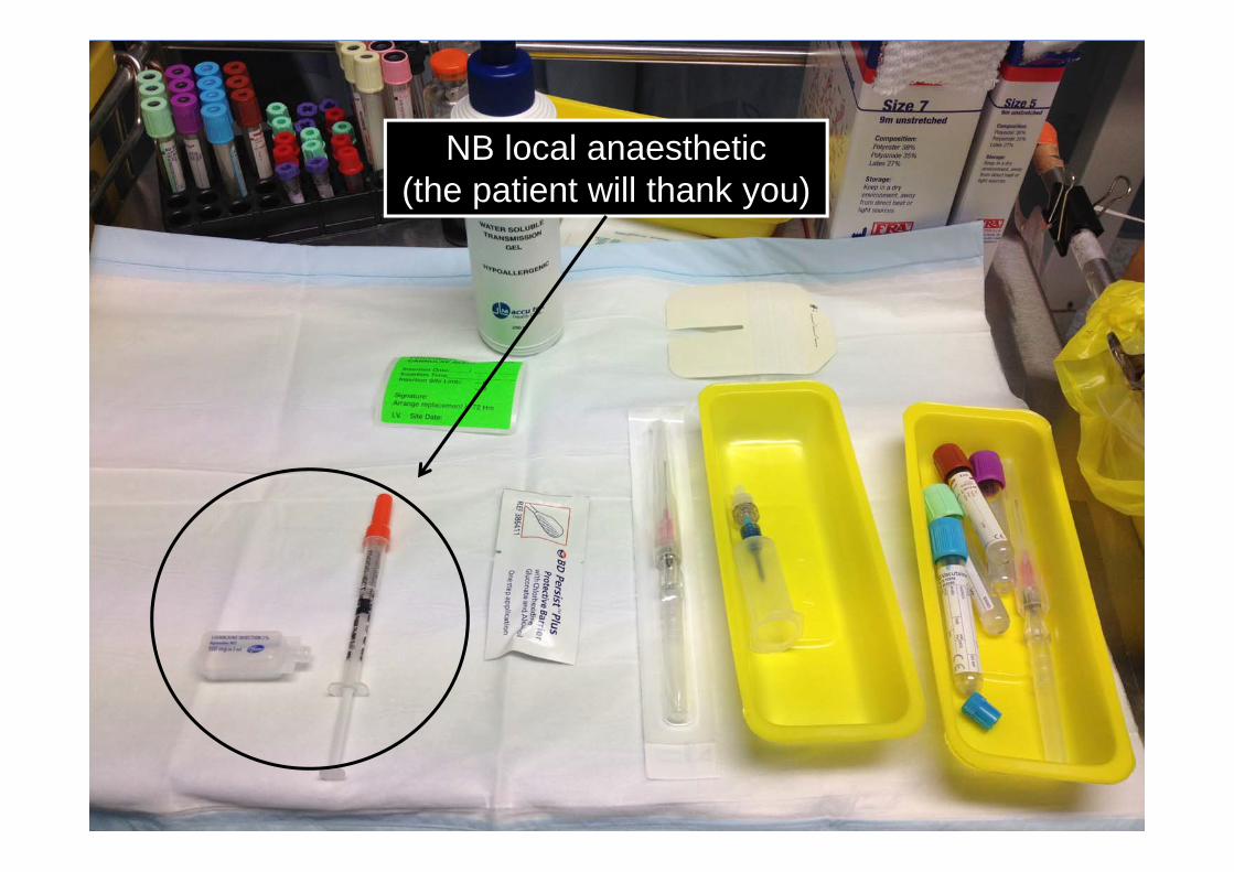
Prepare everything as usual…
7
NB local anaesthetic(the patient will thank you)

US machine
In line of sight Assistant drives the machine
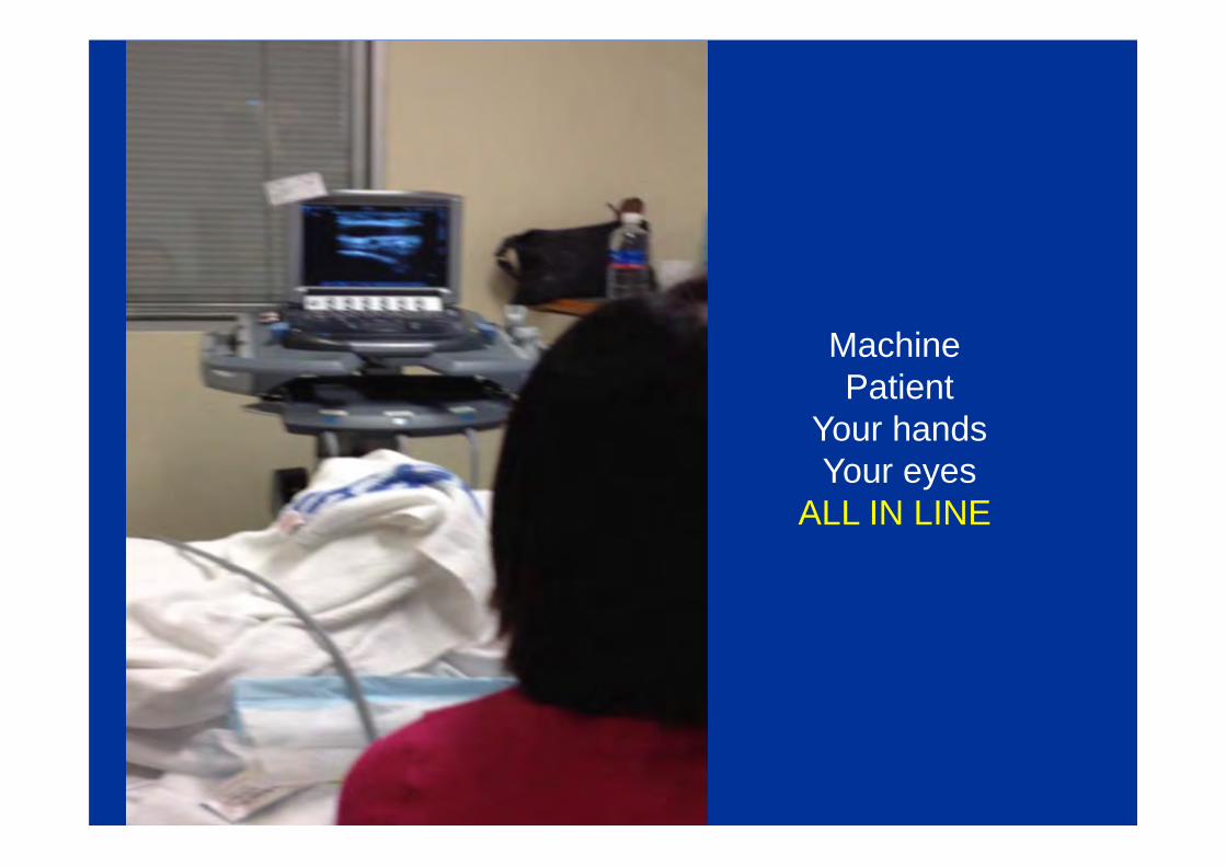
Machine Patient
Your handsYour eyes
ALL IN LINE
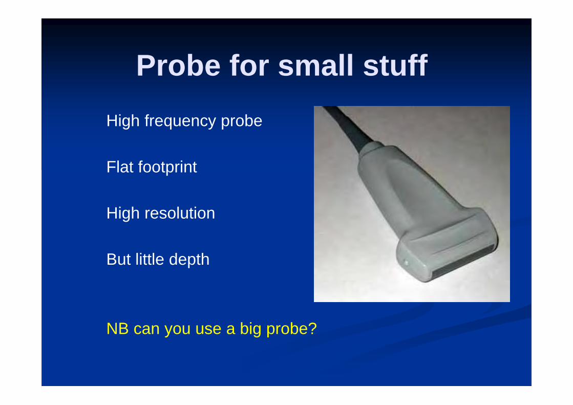
Probe for small stuffHigh frequency probe
Flat footprint
High resolution
But little depth
NB can you use a big probe?

11
Probe for big stuffStart with curved / sector probe
ID the anatomy esp diaphragm, organs
Switch to linear?
OR keep using curved / sector

Prepare sterile probe: traditional
Standard gel 1st
Condom/glove(avoid air bubbles)
Sterile gel

14
But it’s so messy!
Is there a better way?

For peripheral cannulas
Gel Occlusive dressing (eg Opsite 3000) Put it on tight! Then use antiseptic (eg chlorhexidine) or
sterile saline

16
1. Apply a layer of non-sterile gel

17
2. Sterile adherent dressing

18
3: Apply the dressing (Don’t squeeze out the gel!)

19

20
Do I need more gel outside the dressing to
create an image?

21
No. Pouring sterile saline on the skin will suffice.

Result
Don’t try this at home! For peripheral veins.

Needles in veins

24
Vein versus artery
What are the clues?

Larger But more variable Thinner walls Oval cross section Compressible Changes with
Valsalva Artery pulses (vein
too!) NB: Doppler?
Vein (cf artery)

26

27

NB: What if vein is not compressible?

What if vein is not compressible?
Thrombosed Distal obstruction Not a vein!
(eg lymph node)

Central veins

Technique for central line

Which technique?
StaticConfirm veinMark before sterile prepEasierNot as safe
Real time Sterile probe, gel Safer Harder!

Which REAL-TIME technique?
LONGIT./in-plane Harder Safer Better for central
veins
TRANSVERSE/ out of plane
Easier Esp for periph veins

Transverse: needle enters vein

Needle in vein (but where’s the tip?)

Why longitudinal is better
Needle tip seen as it enters the vein Safer

Why longitudinal is better

Top tip: use static then in-plane
Quick scout lookPrepare equipment, US, patient
US monitor in line of sightPrep & drape site…

ID the vein
Local anaesthetic
Advance needle slowly
Tenting
Flashback

Guidewire

41

42

Top tips
Longitudinal / in-plane is saferTransverse / out of plane easier
US monitor in line of sightAdvance needle slowly
Tip on screen at all timesFan probe if transverseCheck the guidewire too

Peripheral veins

Identify the vein in transverse and long axes.

46

47
Transverse (cross-sectional) view

48
Longitudinal view

A drop of local anaesthetic

50

Inserting the needle

52

53
Transverse (out-of-plane) approach

54
Longitudinal (in-plane) approach

55

Now for the cannula

57

58
Are you sure we are in?

59
Testing the cannulawith a saline flush

60
Flow seen inside vein

61

…and done.

Radial artery

64

65

Foreign bodies

Foreign bodies
• Linear probe• Bright: glass, metal, fresh wood• Dark: organic material• Look for shadow / reverberation• Measure depth• X marks the spot• Map it out if possible• Don’t use real time US (air obscures
things)

Abscesses

Abscesses
DDx: cellulitisDDx: vessels
Is there a collection?Can I reach it?Is it worth it?

Abscesses
Linear probeMeasure depth
X marks the spotMap it out if possible
Don’t use real time US (air obscures things)

71

72

73

74
Remember?
DDx: vessels
76
Draining effusions
PeritonealPleural
Pericardial

Draining effusions
Consent, equipment as usual
Patient position
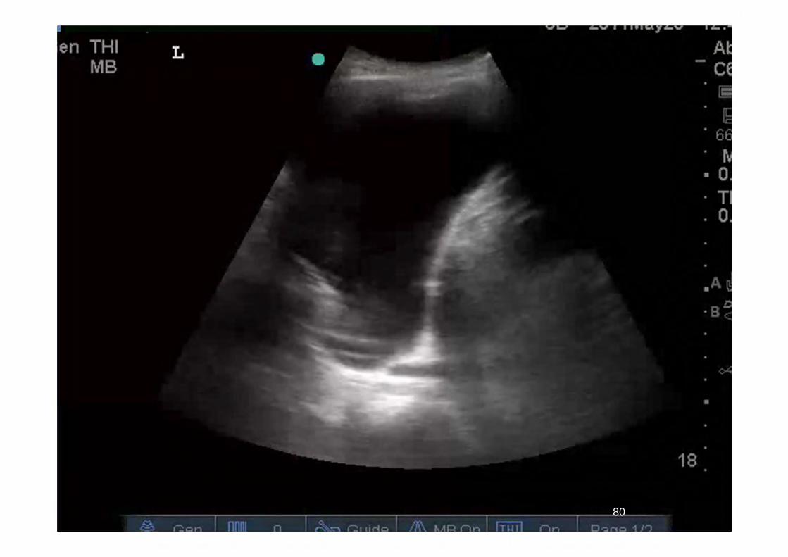
Which probe?

Low frequency (curved)
For big effusions
Get the big picture in 2 planesScan thru resp cycle
(Avoid nasty surprises)Check depth

79
80

High frequency (linear)
Can change to this
Allows you to see needleAs per CVC technique
In-plane/longitudinal is best
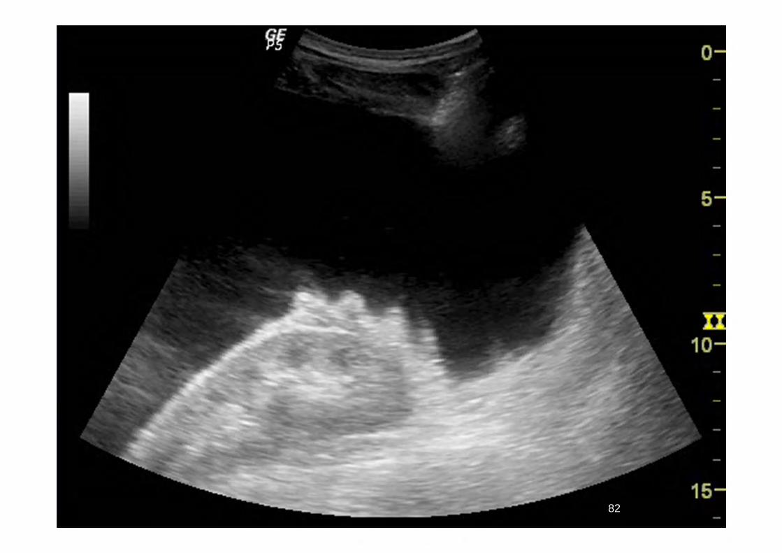
82

SPC insertion
Start with low frequency probe:Get the big picture in 2 planes
Check depth
Optional: switch to high frequency linear

84
Lumbar puncture
NB this is a static technique

Lumbar puncture
Consent, equipment as usual Plus surgical marking pen or indelible
marker Get the patient in the right position Start with linear probe Switch to curved if obese
74% chance of visualisation

Start with longitudinal position

ID the spinous processes

Centre the probe on the interspinous space
X

OBESE: interspinous space
X
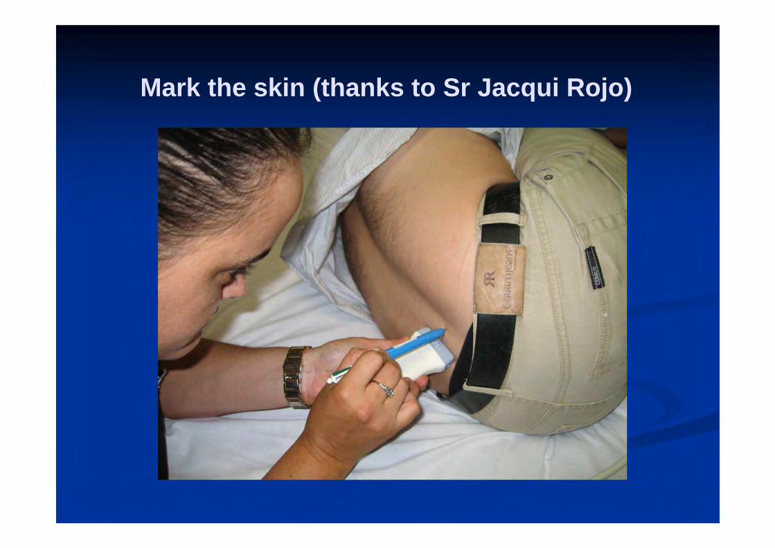
Mark the skin (thanks to Sr Jacqui Rojo)

Switch to transverse

Spinous process trans

OBESE: spinous process trans

Final patient markings (X marks the spot!)

95
Putting it together
http://www.acep.org/publications.aspx?id=33402

Summary
Everything lines up: machine, patient, you
Long cannula if you have one
Anchor your hands on the patient
If using transverse, keep angling probe back & forth to keep needle tip in view

Thanks
Dr Maggie ChungSr Jacqui Rojo