Intraoperative Cytology of CNS Lesions - · PDF fileIntraoperative Cytology of CNS Lesions...
27
1 Intraoperative Cytology of CNS Lesions Matthew A Zarka, M.D. Director of Cytopathology Department of Laboratory Medicine and Pathology Mayo Clinic Arizona Scottsdale, Arizona Gregory S. Moes, M.D. Department of Pathology Oakland Kaiser Medical Center Oakland, California
Transcript of Intraoperative Cytology of CNS Lesions - · PDF fileIntraoperative Cytology of CNS Lesions...
1
Intraoperative Cytology of CNS Lesions
Matthew A Zarka, M.D. Director of Cytopathology
Department of Laboratory Medicine and Pathology Mayo Clinic Arizona Scottsdale, Arizona
Gregory S. Moes, M.D. Department of Pathology
Oakland Kaiser Medical Center Oakland, California
2
Intraoperative Cytology of CNS Tumor Lesions
Cytologic methods for the diagnosis of CNS lesions have been utilized for about 50 years. However, many institutions base intraoperative diagnosis on the exclusive use of traditional frozen section. Although frozen section has the advantage of better architectural preservation, squash/scrape cytology often yields superior cytologic detail of cells and is devoid of freezing artifacts. The pathologist can often make a more confident diagnosis at time of an intraoperative evaluation of CNS lesions when adding cytology to his or her diagnostic armamentarium. A. Definition of Practical Pattern Based Approach to Intraoperative CNS Cytology A consistent approach to rapid CNS smear diagnosis is to categorize lesions that exhibit a one or more cytologic architectural patterns first at low to intermediate magnification (40x, 100x, 200x), and then confirm ones impression of a lesion at high magnification (400x-600x). I term this the practical pattern recognition approach to cytologic smear diagnosis. These patterns do not necessarily rely on the presence of a specific cell type, for instance, astrocytes in a case of an astrocytoma. For example, a cytologic pattern may include a specific type of architectural structure that the tumor cells and associated stromal cells or blood vessels exhibit, such as a papillary structure. Another pattern may encompass the interrelationship that clusters of neoplastic cells have with surrounding blood vessels, stromal tissue, extracellular matrix, or inflammatory cells. This approach first emphasizes grouping pathologic processes with an element or elements that they have in common with one another, and then subdividing these processes into their respective diagnostic categories based upon their unique cellular characteristics. This diagnostic approach is intuitive to many experienced cytopathologists; however, it is all too easy to rush to high power magnification when examining a case, carrying the risk of “missing the forest for the trees”. B. Method of CNS Squash Preparation Preparation of a squash preparation is simple. I prefer to prepare a smear slide by taking a small piece of tissue with a scalpel blade (less than 0.5 mm in diameter), placing the material on a slide, and subsequently smearing the specimen with another slide, holding the second slide at right angles to the first, and applying uniform pressure during smearing, similar to smearing a routine FNA specimen. If a stereotactic core biopsy is submitted, I will often remove a small piece of tissue from opposite ends of the core biopsy specimen and place both fragments of the same slide and subsequently and smear both pieces, in order to better evaluate the representative material that is present within that specific core specimen. The smear is immediately fixed in alcohol and stained with hematoxylin and eosin. Additional slides can be air dried and stained with a Wright stain.
3
The most common artifacts include crush and air drying with loss of cytologic detail. It is important not to smear too large a specimen, which may yield a slide too thick for optimal cytologic detail. Recognition of the fine fibrillary processes that often are associated with glial tumors is dependent on a thin specimen. C. Advantages and Limitations of Cytologic Smear Preparation Cytologic Smears: Advantages Speed Ease of preparation Simplicity Cytologic preservation Small sample size Cytologic Smears: Limitations Relies on tissue soft enough to smear Histologic architecture not apparent Relies on accurate localization by the surgeon
Normal Central Nervous System Constituents and Associated Pathologic Reactions
The various cellular elements of the CNS can be divided broadly into neuroectodermal and mesenchymal derivatives. Neurons and glia, to include: astrocytes, oligodendrocytes, ependyma and choroid plexus, are neuroectodermally derived elements. The vasculature, meninges and microglia (bone marrow derived monocytes) are of mesenchymal derivation. Neurons Neurons are large, polygonal cells with low nuclear to cytoplasmic ratios, a prominent centrally placed nucleolus and conspicuous rough endoplasmic reticulum (nissl substance). They reside in the gray matter of the cortices and deep cortical nuclei, but a few are present in the white matter of the temporal lobe. Another type of neuron is the granular neuron. These are smaller, naked nuclei by light microscopy and are seen in the cerebellum and hippocampal formation. Immunoperoxidase studies for synaptophysin (cytoplasmic/membranous) and NeuN (nuclear) will identify neurons. Pathologic Reactions: In acute ischemic events, such as infarction, the neuron undergoes specific identifiable changes; the so-called “red neuron”. These conspicuous neurons have bright, eosinophilic cytoplasm with concomitant loss of the nissl substance and dark, pyknotic nuclei in which typically prominent nucleoli are not discernable. The background likely will be infiltrated by numerous macrophages as well. Be very cautious in diagnosing a neoplasm in a background of numerous macrophages with scattered ischemic neurons. The red neuron is the sine qua non of ischemic/hypoxic change.
4
Astrocytes There are two types of astrocytes or “star cells”: the more common fibrous or fibrillary astrocyte, and the protoplasmic astrocyte. The fibrous type is present in the white matter and shows numerous and prominent cytoplasmic extensions compared to the protoplasmic astrocytes. Most astrocytic neoplasms arise from the fibrillary astrocyte and thus give rise to a fibrillary background on squash preparation technique. The protoplasmic astrocytes are present predominantly in the gray matter. Immunoperoxidase analyses for GFAP and S100 are useful to distinguish glial lineage, but are not specific for astrocytes per se. Pathologic Reactions: Gliosis is a response to CNS injury and consists of two components, hypertrophy and hyperplasia. The initial response is astrocytic hypertrophy, and consists of an increase in cell size and in cytoplasmic prominence. The observance of well-defined astrocytic cytoplasm by H&E with multiple cytoplasmic extensions usually indicates reactive gliosis. When the eosinophilic cytoplasm becomes exuberant with well-defined rounded edges these astrocytes are referred as gemistocytes or “stuffed” astrocytes. Chronic astrocytosis is characterized by dense fibrillary gliosis. An increase in astrocytic number may also follow insult to the CNS. Rare non-neoplastic mitoses may be observed, but a hyperplastic response is typified by the presence of mirror nuclei. These are astrocytic nuclei that occur in physically contiguous matched pairs. Creutzfeldt astrocytes not specific for, but are characteristic of demyelinating disease. These are reactive astrocytes with multiple, small nuclei (micronuclei). Their precursors with tiny chromatid bodies are termed “granular mitoses”. Rosenthal fibers Rosenthal fibers are brightly; eosinophilic, ropy, elongate cytoplasmic inclusions seen in astrocytes cell processes and are possibly accumulations of alpha-beta-crystallin. Rosenthal fibers are seen in association with neoplastic, reactive or metabolic processes and indicate chronicity. In neoplasia, Rosenthal fibers are associated with juvenile pilocytic astrocytoma. Rosenthal fibers may also be present normally around the pineal gland or any slow growing tumor, i.e., craniopharyngioma. Eosinophilic granular bodies (EGBs) These are multiple “granular” bright eosinophilic deposits (protein deposits) and share with Rosenthal fibers, immunoreactivity with GFAP, ubiquitin and alpha-beta crystallin. EGBs are associated with three tumor types, juvenile pilocytic astrocytoma, pleomorphic xanthoastrocytoma and ganglioglioma.
5
Corpora Amylacea Corpora Amylacea are intracytoplasmic glucose polymers (polygylcosan bodies) present in astrocytic cytoplasm. They are located around blood vessels and in subpial locations and may be a result of trauma, i.e., epilepsy or present as a part of normal aging. Corpora stain positively for PAS, GMS and alcian blue and care should be taken not to misinterpret these structures as fungal yeast forms. Oligodendrocytes Myelin producing glial cell associated with white matter. They are also seen in the gray matter, where they serve as “satellite cells” to neurons (astrocytes and microglia can also do this). Oligodendrocyte satellitosis may be physiologic or associated with neoplasia (oligodendroglioma) known as secondary structures of Scherer. Oligodendrocytes have fewer cytoplasmic processes compared to astrocytes and are seen as naked nuclei in the neuropil. Unlike astrocytomas, oligodendrogliomas invade neuropil without prominent disruption of myelin. Thus, the background squash of an oligodendroglioma may be less fibrillary and more granular with some areas of ropy myelin. The nuclei of oligodendrocytes are small, dark and uniformly round, whereas the nuclei of astrocytes are more oval or mildly irregular and less hyperchromatic. A characteristic morphologic feature of oligodendroglia as a result of delayed fixation is referred to as “perinuclear halo” or fried egg appearance is seen in normal and neoplastic oligodendrocytes. No specific immunoperoxidase marker is yet available that can distinguish between neoplastic and non-neoplastic oligodendrogliomas. Pathologic reactions: Oligodendrocytes demonstrate limited pathologic reaction and are mainly proliferation with satellitosis i.e., chronic seizures. Ependyma Ependymal cells line the surface of the ventricular system and vary from the robust ciliated cells of the fetal system to the flattened cuboidal cells of adults. Subependymal glias are glia present just beneath the ependymal layer and give rise to the subependymomas. Normal ependymal cells are GFAP negative and vimentin positive. PTAH can be used to identify the blepharoblasts of ependymal cells and may a useful adjunct in determining ependymal differentiation in tumors, short of electron microscopy. Tanycytes are applicable to several cell populations of the developing and adult nervous system and share a highly elongate shape that spans the entire surface of the ependyma from the ventricular system to the subependymal-CNS parenchyma. Tanycytes in adults are located in the floor of the third ventricle. Tanycytes are strongly GFAP positive and are thought to be precursors of the tanycytic ependymoma and astroblastoma.
6
Choroid plexus Choroid plexus are papillary tufts of epithelium covered by fibrovascular connective tissue that project into the ventricles. The cells are plumper and larger than ependymal cells and have a cobble stone surface. The largest masses of choroid plexus cells are found in the lateral ventricular atria and are called the glomerula choroidea. Choroid plexus cells are negative for GFAP and positive for S100 and transthyretin. Meningothelial rests and whorls are common inhabitants of normal choroid plexus and their location explains the occurrence of intraventricular meningiomas. Microglia/Monocytes/Macrophages Microglia are bone marrow derived monocytes that arrive via the blood stream and transform into indigenous microglia of the central nervous system. Resting microglia blend inconspicuously into the neuropil and consist of naked nuclei on H&E. Microglia show immunoreactivity for CD68 and HAM-56. Pathologic reactions include diffuse proliferation and infiltration of CNS parenchyma and can form microglial nodules that consist of microglia and astrocytes. These are seen in viral and Rickettsial infections. Proliferative microglia are seen as rod shaped haphazardly arranged nuclei. D. The Importance of Clinical History When examining an intraoperative CNS case, it is essential to know the age of the patient and the location of the lesion in question before the specimen arrives in the frozen section suite. Other useful information includes the type and duration of clinical symptoms. For example, a history of seizures is more in keeping with a slower growing lesion such as a low grade astrocytoma, as compared to a rapid growing lesion such as a glioblastoma
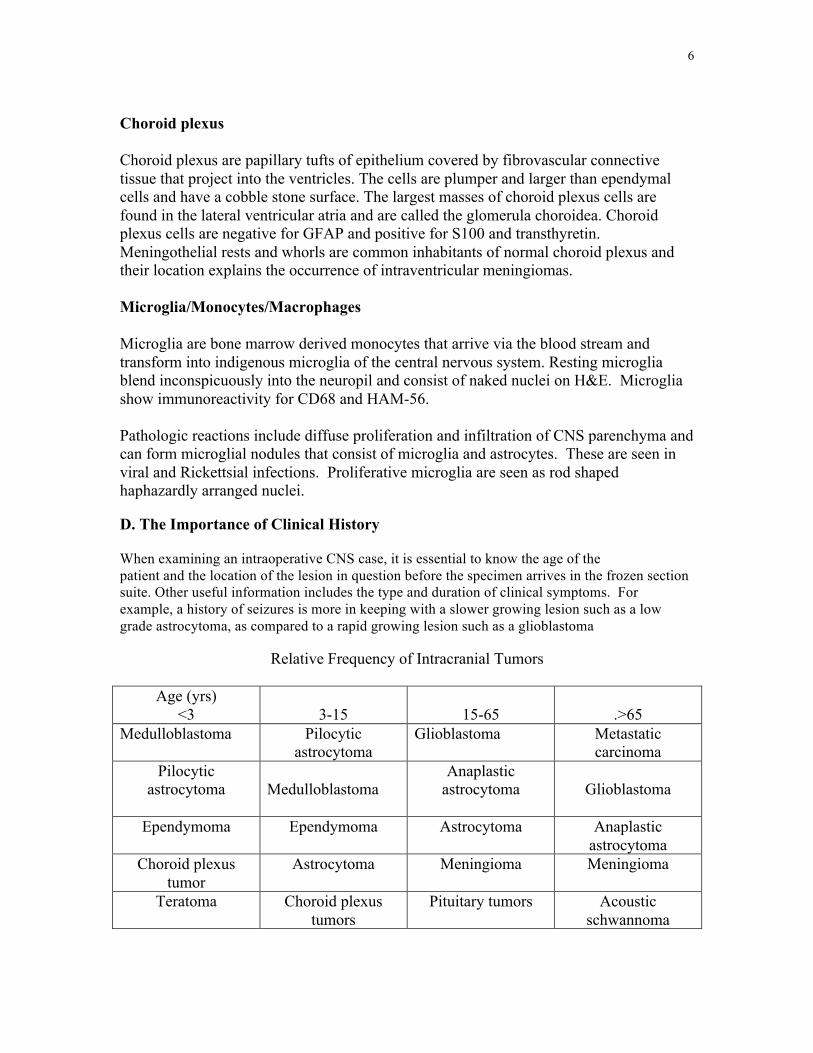
Relative Frequency of Intracranial Tumors
Age (yrs) <3
3-15
15-65
.>65
Medulloblastoma Pilocytic astrocytoma
Glioblastoma Metastatic carcinoma
Pilocytic astrocytoma
Medulloblastoma
Anaplastic astrocytoma
Glioblastoma
Ependymoma Ependymoma Astrocytoma Anaplastic astrocytoma
Choroid plexus tumor
Astrocytoma Meningioma Meningioma
Teratoma Choroid plexus tumors
Pituitary tumors Acoustic schwannoma
7
E. Practical Architectural Pattern Approach to CNS Smear Cytology:
- Basic Principles When evaluating a lesion on a cytologic smear, pay particular attention to the following: Relationship of Neoplastic Cells to Blood Vessels Blood Vessel Type Type of Background (felt-like vs., fibrillary) 1. Tumor distribution in relation to blood vessels Gliomas: especially astrocytomas, usually demonstrate aggregation of tumor cells close to
blood vessels; the concentration of tumor cells decreases the farther away the tumor cells are from a blood vessel: Perivascular Gradient Pattern
Lymphoma: can infiltrate blood vessel walls but are often dispersed in a discohesive fashion away from blood vessels also: Angiocentric and Diffuse Pattern
Metastatic carcinoma: clusters of malignant cells are distributed close to and away from blood vessels; variable with tumor type: Randomized Clusters With and Without Vascular Affinity
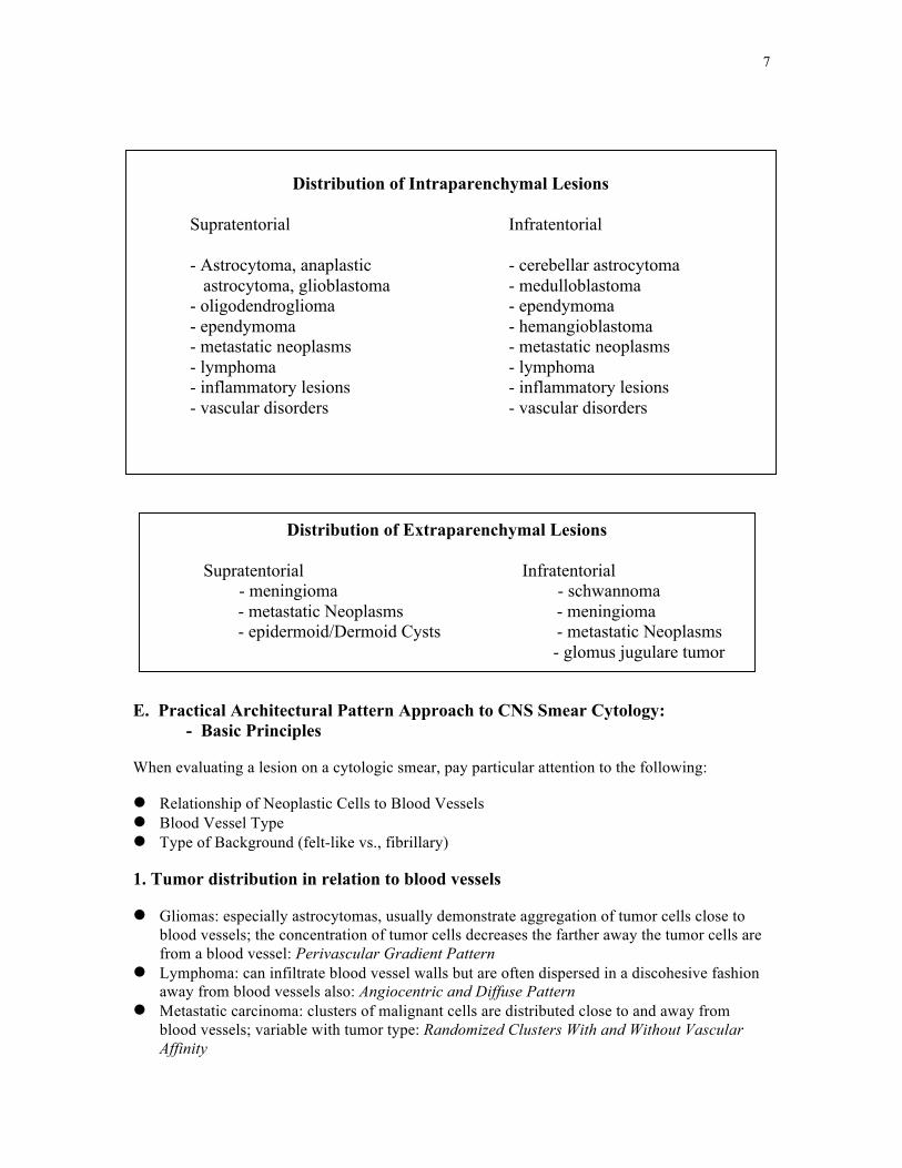
Distribution of Intraparenchymal Lesions
Supratentorial Infratentorial
- Astrocytoma, anaplastic - cerebellar astrocytoma astrocytoma, glioblastoma - medulloblastoma - oligodendroglioma - ependymoma - ependymoma - hemangioblastoma - metastatic neoplasms - metastatic neoplasms - lymphoma - lymphoma - inflammatory lesions - inflammatory lesions - vascular disorders - vascular disorders
Distribution of Extraparenchymal Lesions
Supratentorial Infratentorial - meningioma - schwannoma
- metastatic Neoplasms - meningioma - epidermoid/Dermoid Cysts - metastatic Neoplasms
- glomus jugulare tumor
8
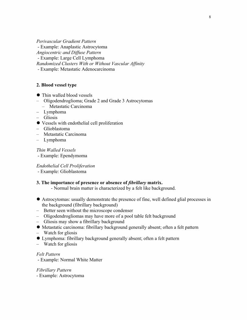
Perivascular Gradient Pattern - Example: Anaplastic Astrocytoma Angiocentric and Diffuse Pattern - Example: Large Cell Lymphoma Randomized Clusters With or Without Vascular Affinity - Example: Metastatic Adenocarcinoma 2. Blood vessel type Thin walled blood vessels – Oligodendroglioma; Grade 2 and Grade 3 Astrocytomas
– Metastatic Carcinoma – Lymphoma – Gliosis Vessels with endothelial cell proliferation – Glioblastoma – Metastatic Carcinoma – Lymphoma Thin Walled Vessels - Example: Ependymoma Endothelial Cell Proliferation - Example: Glioblastoma 3. The importance of presence or absence of fibrillary matrix. - Normal brain matter is characterized by a felt like background. Astrocytomas: usually demonstrate the presence of fine, well defined glial processes in
the background (fibrillary background) – Better seen without the microscope condenser – Oligodendrogliomas may have more of a pool table felt background – Gliosis may show a fibrillary background Metastatic carcinoma: fibrillary background generally absent; often a felt pattern – Watch for gliosis Lymphoma: fibrillary background generally absent; often a felt pattern – Watch for gliosis Felt Pattern - Example: Normal White Matter Fibrillary Pattern - Example: Astrocytoma
9
The patterns described above are general patterns useful for many intracranial processes. The following often associated with some specific CNS and peripheral nervous system lesions, however, can also apply to some metastatic lesions as well. Vascular corona pattern - Example: Ependymoma
- note some metastatic tumors such as metastatic melanoma, some adenocarcinomas, and metastatic renal cell carcinomas can also demonstrate this pattern, however, they do not show the extensive fibrillary matrix that ependymoma exhibits
Random dispersed pattern - Example: Pituitary Adenoma Syncytial aggregates with or without whorls - Example: Meningioma Old fish net pattern - Example: Schwannoma Randomized loose clusters and diffuse pattern - Example: Neuroblastoma Metastatic Small Cell Carcinoma Summary: Pattern Based Approach to CNS Intraoperative Smear Preparations
I. Evaluate the Slide at Low-Intermediate Magnification (40-200x) • What Is The Relationship of Neoplastic Cells to Blood Vessels? • What Type of Blood Vessels Are There? • What Type of Background: Fibrillary Versus Felt-Like? II. Evaluate the Slide at High Magnification (400-600x) • Look for cytologic features to confirm your low magnification impression
10
Cytologic Features of Specific Lesions Astrocytic Neoplasms A. Astrocytoma (WHO Grade 2)
1. Clinical features Usually arises in cerebral hemispheres of young adult, but can occur basically
anywhere within the CNS in all ages CT scan show ill defined low density lesion without contrast enhancement May be associated with a fluid filled cyst Intraoperatively, the neurosurgeon may have difficulty recognizing the tumor grossly;
the junction of tumor and normal tissue may be recognizable by a change in tissue texture
2. Smear cytology Can be difficult to diagnose Usually smear easily Smear shows low to moderate cellularity; neoplastic cells often arranged in irregular
clusters around blood vessels Fibrillary processes often abundant Nuclei round, oval, or elongated with irregular borders Mitotic figures and necrosis absent Thin walled blood vessels At the tumor margin, neoplastic astrocytes may be admixed among neurons and
oligodendrocytes with felt-like background, in contrast to the prominent fibrillary background when sampling in the center of the lesion
3. Differential diagnosis Reactive proliferation of astrocytes (gliosis): usually reactive astrocytes have more
abundant processes and a generous amount of cytoplasm (gemistocytic astrocytomas can have generous cytoplasm however). Reactive astrocytes generally have more uniform nuclei than neoplastic astrocytes
Microcysts discovered on frozen section usually are not a feature of reactive gliosis Infarction: may have atypical astrocytes, but often show the presence of
macrophages. Gliomas as a rule should not have macrophages Oligodendroglioma: cells in oligodendrogliomas have spherical nuclei, show less of a
fibrillary background, and smear more uniformly B. Anaplastic Astrocytoma (WHO Grade 3) 1. Clinical features Survival rate in between that for astrocytoma and glioblastoma; mean post-op
survival approximately 2 years. Patients usually middle aged
11
CT resembles astrocytoma; no contrast enhancement of evidence of necrosis 2. Smear cytology Variable presentation, however, as a general rule shows moderate to marked nuclear
and cytoplasmic pleomorphism and moderate to marked increased cellularity Fibrillary background Tumor cells often aggregated around blood vessels, a feature present in many
astrocytic neoplasms No endothelial proliferation Can see mitoses, however, by definition, should not see necrosis and/or endothelial
proliferation Keep in mind that the specimen evaluated intraoperatively is often limited in volume
and therefore may not be totally representative of the specimen evaluated on permanent sections (permanents may show features of glioblastoma)
3. Differential diagnosis Usually not difficult to distinguish anaplastic astrocytoma with reactive gliosis,
however don’t forget to consider progressive multifocal leucoencephalopathy (PML). In PML the astrocytes may show considerable pleomorphism. PML often shows the presence of atypical oligodendrocytes and macrophages. Remember that the presence of macrophages argues against a glioma.
Most common lesion in the differential is glioblastoma; look for endothelial proliferation and necrosis, features that favor glioblastoma. Possible that the diagnostic components were not included in the biopsy intraoperatively
C. Glioblastoma (WHO Grade 4) 1. Clinical features Most common astrocytic neoplasm. Most patients are middle aged or elderly with a
short duration of symptoms. Usually involves cerebral hemispheres Often arise de novo, or arise in patients with a previous history of a lower grade
astrocytoma , with prolonged history previous radiation therapy Radiographic studies often show a low density area with a central enhancing area of
necrosis with surrounding edema; may be confused with an abscess or metastasis clinically
Grossly the tumor is gray or brown with areas of necrosis or hemorrhage. May appear circumscribed intraoperatively, however, the tumor is always infiltrative microscopically
Poor prognosis, mean survival often less than one year 2. Smear cytology Usually easy to diagnose; the tumor demonstrates marked nuclear and cytoplasmic
pleomorphism, coupled with mitotic activity, endothelial proliferation, and/or tumor necrosis
12
Some cases are composed of neoplastic cells with a fair amount of cytoplasm and abundant fibrillary processes which aggregate near blood vessels, while other cases show very poor cohesion of cells with few cytoplasmic processes
Very poorly differentiated tumors may be difficult to differentiate between a metastatic carcinoma or lymphoma
True endothelial proliferation is rarely present in other tumors (exceptions: pilocytic astrocytoma, anaplastic oligodendroglioma, metastatic lung carcinoma)
Sometimes it is appropriate to render a diagnosis of high grade astrocytic neoplasm, await permanents when endothelial proliferation or necrosis is not present, when clinically the tumor is a glioblastoma
Giant cell glioblastoma variant: very large, bizarre cells, often multinucleated Gliosarcoma: contains both an astrocytic and mesenchymal components. Often
superficially located and may be confused clinically with a meningioma 3. Differential diagnosis Clinically a glioblastoma may resemble an abscess, although this is usually not
confused with a glioblastoma on smears or frozen section. A necrotic glioblastoma usually shows a paucity of inflammatory cells and does not show the presence of macrophages.
If the biopsy only shows necrosis, advise the surgeon for more material Do not confuse reactive gliosis adjacent to an abscess or metastasis with glioblastoma Occasionally, a glioblastoma with few astrocytic processes can be confused with
other neoplasms such as lymphoma, metastatic carcinoma, or melanoma. Look for glial processes. Look for endothelial proliferation to help distinguish glioblastoma from other neoplasms
Distinction between glioblastoma and anaplastic astrocytoma may be difficult due to sampling error
D. Pilocytic Astrocytoma (WHO Grade 1) 1. Clinical features Most frequently in children and young adults Cerebellum, optic nerves and chiasm, area of third ventricle, brain stem and spinal
cord are the usual locations; symptoms related to location, usually slow growth rate Most cases do not have the propensity to undergo anaplastic transformation Radiographic studies may show well circumscribed lesions which may contain cysts;
cerebellar tumors may cross midline and may involve leptomeninges Tumor is tough and rubbery, can have well defined margin, in contrast to other
astrocytomas 2. Smear cytology Astrocytic pattern with tumor cells aggregating around blood vessels Abundant cytoplasmic processes; often elongated nuclei with bipolar processes. Keep
in mind that the tumor cells may show atypia and that some tumors consist of cells with round nuclei and few processes
13
Rosenthal fibers may be present characterized by elongated or multilobated structures which are brightly eosinophilic on H and E. Their presence demonstrates that the tumor is slow growing/low grade (note that Rosenthal fibers may be seen in benign reactive conditions as well)
Mitotic figures and necrosis are not a feature of pilocytic astrocytoma Note: vascular proliferation may be present and may mimic endothelial proliferation
seen in glioblastomas Frozen section may show microcyst formation and tumor which has extended into the
subarachnoid space 3. Differential diagnosis The presence of atypia and vascular proliferation may lead one to make the incorrect
diagnosis of a high-grade astrocytoma. Keep the clinical history and location of the tumor in perspective.
Reactive process: can lead to the formation of Rosenthal fibers. The presence of Rosenthal fibers demonstrates that the lesion is low grade of slow growing, but does not distinguish between a neoplastic or reactive process.
E. Pleomorphic Xanthoastrocytoma (WHO Grade 2) 1. Clinical features Usually in children and young adults; often presents with seizures of less commonly,
symptoms of a space occupying lesion Most of these tumors are supratentorial; often in the temporal lobe and superficial,
may be adjacent or within the leptomeninges. Often associated with a cyst Often well circumscribed mass on the surface of the brain 2. Smear cytology Markedly pleomorphic astrocytic tumor; considerable variation in nuclear size and amount of cytoplasm Some cells often show vacuolated cytoplasm No mitosis, necrosis, or endothelial proliferation The tumor is occasionally infiltrated with lymphoid cells 3. Differential diagnosis Glioblastoma is the major lesion in the differential due to the degree of pleomorphism
common in PXA; however, glioblastoma is rare in the age typical of PXA. In addition, other features of a high-grade glioma (necrosis, endothelial proliferation, mitotic figures) are absent in PXA.
Oligodendroglioma 1. Clinical features Approximately 5% of intracranial gliomas, most frequently in the 4th and 5th decades
with slight predominance in males; can be seen in children however
14
Most common in white matter in the frontal and parietal regions, but may involve the corpus callosum and extend to contralateral hemisphere; infrequently in cerebellum; rare in spinal cord
Radiographic studies show a supratentorial lesion, 60% have calcification. MRI may show poorly enhancing peripheral margin in low-grade neoplasms. Cystic degeneration may be seen, especially in anaplastic oligodendrogliomas
Anaplastic oligodendrogliomas and mixed gliomas with an anaplastic astrocytoma component may display hemorrhage and ring enhancement
Patients present with symptoms of an infiltrating neoplasm; many experience a long history of seizures prior to an onset of headache or signs of increased intracranial pressure
Anaplastic oligodendrogliomas often present with an more aggressive course At surgery often are diffuse neoplasms of gelatin like consistency with irregular
margins Occasional there are gross calcifications or cystic change Occasionally can project into the 3rd or 4th ventricles with accompanying
hydrocephalus Anaplastic oligodendrogliomas may be hemorrhagic at surgery and more commonly
have cystic change. Can also show areas of necrosis 2. Smear cytology Usually smear easily and show a fine capillary network The tumor cells can aggregate around vessels, but the cells often spread evenly,
without cell clumping and little cohesion Calcification is common, but not specific for this lesion. May even notice a gritty
sensation when smearing Nuclear pleomorphism is not a usual feature; anaplastic oligodendrogliomas may
exhibit nuclear pleomorphism however Round, relatively uniform nuclei with faint speckled chromatin without prominent
nucleoli with pale staining scant cytoplasm. Nuclei are often eccentric Perinuclear halo (fried egg) pattern is absent on smears/frozen section, in contrast to
paraffin sections Mitotic figures are usually difficult to find Lack gliofibrillary matrix, however reactive gliosis may be present at the tumor
margin – don’t confuse this phenomenon with a mixed glioma In mixed gliomas, the oligo component often predominates and is mixed with
neoplastic astrocytic cells, the later which are associated with glial processes and vessels with endothelial hyperplasia
Keep in mind that anaplastic oligodendrogliomas have show vessels with endothelial hyperplasia, frequent hemorrhage, and cells with a significant amount of eosinophilic cytoplasm and nuclear pleomorphism. A small amount of necrosis may be present. Mitotic figures are often present in these tumors
Consider obtaining material for 1p/19q deletion studies (genetic/PCR) when dealing with an oligodendroglioma
3. Differential diagnosis
15
Astrocytomas usually demonstrate more obvious glial processes. Oligodendrogliomas usually have the characteristic round nuclei with speckled chromatin pattern and have less of a tendency to aggregate around blood vessels.
Oligodendrogliomas can resemble pituitary adenomas when confronted with a centrally located tumor. Pituitary adenomas often have multiple nucleoli and tend to form cohesive sheets, unlike oligos. Pituitary tumors often have branching vessels with thick walls.
Oligodendrogliomas may be confused with lymphomas, although most CNS lymphomas are high grade with considerable nuclear variation and abundant mitotic figures. Lymphomas are often accompanied with prominent vessels, which are infiltrated with neoplastic cells, unlike the thin walled vessels in oligos.
Ependymomas often demonstrate a gliofibrillary matrix and cells connected by processes to blood vessels, often thick walled
The distinction of oligos with metastatic carcinomas is usually easy. Oligodendrogliomas show uniform cellularity, a tendency to smear in a monolayer with little cohesion, and demonstrate the typical thin walled vessels, and absence of mitotic figures and necrosis. Metastatic carcinoma usually shows cohesive groups of cells with considerable pleomorphism, necrosis and mitotic activity.
Ependymal Neoplasms A. Ependymoma 1. Clinical Features Most common site is the 4th ventricle arising in the floor or roof, but can be seen
lining the lateral and 3rd ventricles, and in the spinal cord in the region of the cauda equina
Found in children and adults. Childhood tumors usually arise in the 4th ventricle presenting with obstructive hydrocephalus
Supratentorial tumors usually fill one or the other lateral ventricle and present with mass effect or seizures
Deep intracerebral tumors separate from the ventricular system are rare, usually large and rapidly growing
Spinal ependymomas usually present in adults with back pain, are often myxopapillary type, but can be classic ependymomas
At surgery, ependymomas are often firm, lobular masses with red-gray color. Usually there is a defined surgical plane. Tumors of the 4th ventricle can show calcification
2. Smear cytology Usually smear easily. An important feature is the presence of papillary groups around vessels. On cytologic
preparations, this may be seen as palisaded groups of tumor cells arranged on both sides of a long axis of a vessel. A narrow anucleate area of fibrillary processes may be seen between the palisaded nuclei and the vessel wall
A prominent fibrillary matrix is often present, adjacent to the blood vessels or at the edges of groups of tumor cells
Individual cells drift away larger groups of tumor cells
16
Tumor cell nuclei are usually oval, but can be round, with pale stippled chromatin and one or more nucleoli
Presence of more than a few mitotic figures may suggest an anaplastic lesion, especially in a child. Watch for hypercellular lesions with necrosis and vascular proliferation
3. Differential diagnosis Think of choroid plexus tumors if one is confronted with a tumor of the lateral or 4th
ventricle. Choroid plexus tumors usually show bright contrast enhancement on CT and arise from a choroid plexus site. These lesions are often very vascular. They usually show a more developed papillary configurations and do not show glial processes
A medulloblastoma is also a consideration of a tumor from the 4th ventricle. Medulloblastomas do not show an obvious fibrillary stroma. They often smear out into a diffuse monolayer, unlike ependymomas, which often show clumps of tumor and perivascular papillary formations.
B. Myxopapillary Ependymoma 1. Clinical features Almost exclusively found in the cauda equina region; comprises 50% of all
ependymal neoplasms; may also arise ectopically in the parasacral region Usually presents with history of back pain in young adults Radiographically well defined elongated mass with moderate enhancement Often have smooth surface, separate from the spinal cord. Cut section may show hemorrhage, but calcification is not as prominent as typical
ependymomas 2. Smear cytology Usually smear easily with numerous papillary structures, with multiple layers of
nuclei surrounding vascular structures In contrast to typical ependymomas, fibrillary stroma is replaced with an amorphous
myxoid matrix, which is strongly metachromatic particularly on toluidine blue, or wright air-dried stains. The matrix often is seen near vessels or at the edges of tumor masses
3. Differential diagnosis Chordomas can have a myxoid matrix and smear easily as well, but lack the papillary
structures. Chordomas usually show physalipherous cells and pleomorphism. Metastatic adenocarcinomas are also a consideration and may produce mucinous,
papillary structures. Carcinomas are usually more cohesive which usually smear as clumps of cells. Malignant nuclear features should be a warning not to make a diagnosis of myxopapillary ependymoma
17
Primitive Neuroectodermal Tumors of the Cerebellum (Medulloblastoma) 1. Clinical features PNETs usually occur in childhood, the most common lesion is within the cerebellum
(medulloblastoma) Medulloblastomas are the second most common brain tumors in children, after
pilocytic astrocytoma. They usually occur at 3-8 years, but 20% of them are in patients over 20 years of age
Patients often present with irritability, lethargy, and loss of appetite, followed by signs of increased intracranial pressure due to obstruction of the cerebral aqueduct or 4th ventricle, including headache, nausea, and vomiting. Abnormalities of gaze, nystagmus, or ataxia may follow
Most medulloblastomas in children arise in the vermis; in adults medulloblastomas tend to arise in the cerebellar hemispheres
Medulloblastomas can metastasize through the CSF pathway and occasionally present from symptoms from the metastatic lesion
On CT medulloblastomas usually present as a hyperdense mass with uniform enhancement with occasional hemorrhage or cystic change. Calcification is infrequent.
Peritumor edema is often present, well visualized on MRI At surgery are usually vascular and may be lobulated and occasionally invade the
subarachnoid space; ventricular invasion more common Supratentorial PNET are usually ill-defined infiltrative lesions involving the cortex
and surrounding white matter, with surrounding edema 2. Smear cytology Usually smear easily. Often large numbers of densely packed cells with oval or carrot shaped
hyperchromatic nuclei with scant cytoplasm, which may show nuclear molding. Naked nuclei often present
Tumor cells tend to spread widely from thin walled to large vessels with hyperplastic endothelium
Rare rosettes may be seen Mitotic figures common; occasional tumors show necrosis Occasional cells may show ganglionic differentiation Supratentorial PNETs generally have an identical appearance to medulloblastomas 3. Differential diagnosis Normal granular cell neurons of the cerebellum. These cells are small with scant
cytoplasm and uniform round nuclei. They are often admixed with Purkinje cell nuclei. In contrast medulloblastomas have larger nuclei with an elongated or carrot shape, variable chromatin, with nuclear molding and mitotic activity
18
Pilocytic astrocytomas can also be found in the cerebellum, but are readily distinguished form medulloblastomas by astrocytic differentiation, low cellularity, gliofibrillary matrix and lack of mitoses
Unlike ependymomas, medulloblastomas smear out in a diffuse manner, and lack the prominent fibrillary stroma and clumps of cells in perivascular palisaded arrangements. Anaplastic ependymomas may be more of a challenge on account of their high cellularity, mitotic activity, and focal necrosis, however, the glial nature and relation of tumor cells with blood vessels should help distinguish these tumors.
Medulloblastomas in adults can be confused with metastatic small cell carcinomas. Small cell carcinomas may show a small nucleolus and have a tendency to smear in aggregates; medulloblastomas smear in a diffuse monolayer
Meningiomas
1. Clinical features Meningiomas are neoplasms that arise from arachnoidal cells. Account for 13-26% of primary intracranial tumors A tumor of middle age and elderly patients. Females are affected more often than males. Meningiomas of the spinal cord predominantly occur in females and tend to be the
fibrous variant. Whereas anaplastic or malignant meningiomas occur predominantly in males. Meningiomas in children tend to be more aggressive.
Symptoms usually result from slow growing, compressive effects that may present with headaches or seizures.
Intracranial meningiomas tend to occur in the cerebral convexities associated with the falx cerebri. They may also arise in the orbits and intraventricular cavities, but can occur in almost any organ.
Intraventricular meningiomas tend to be aggressive or atypical. Atypical and anaplastic meningiomas occur mainly in the falx and lateral convexities.
Metastatic disease to a meningioma can occur (be aware). 2. Neuroimaging The MRI studies typically demonstrate an isodense dural mass that may be calcified
and show contrast enhancement. A characteristic feature is a “dural tail” seen adjacent to the main mass. Peritumoral edema is occasionally prominent-especially in the secretory variant. Microcystic meningiomas show poor enhancement.
3. Smear cytology These are rubbery, firm and well-demarcated tumors that have a dural attachment. Cytologic analysis is useful to exclude anaplastic carcinoma, which may metastasize
to the leptomeninges. Low power magnification demonstrates generally cohesive syncytial clusters of
meningothelial cells. Syncytial and meningotheliomatous meningiomas have slightly oval nuclei with
evenly dispersed fine to coarse chromatin and a small indiscreet nucleolus. Whorl formation is variable but almost always seen.
19
Single intact meningothelial cells are seen in the background and consist of eccentrically placed bland nuclei with abundant “tissue paper” or delicate, wispy appearing cytoplasm.
Considerable pleomorphism can be seen, but without other features of atypicality. Nuclear pseudoinclusions are diagnostically useful, especially when a prominent
finding, and are particularly easy to identify on the squash preparation. In the secretory variant of meningioma sometimes-bright eosinophilic
pseudopsammoma bodies are seen. True psammoma bodies are diagnostically useful, and a clue to their presence on
smear preparation is the “grinding” sensation during the squash. In an abundant necrotic background, consider pre-operative embolization therapy or
metastatic carcinoma.
4. Histologic subtypes Meningiomas are graded in the WHO grading schema as I-III. There are a wide variety of histopathological appearances, the most common WHO
grade I meningiomas are the meningothelial, fibrous and transitional types. Other less common patterns include psammomatous, angiomatous, microcystic, secretory (secrete CEA), lymphoplasmcyte-rich and metaplastic types.
WHO grade II meningiomas include chordoid, clear cell and atypical meningiomas (see below).
Papillary, rhabdoid and anaplastic (malignant) meningiomas are WHO grade III lesions.
5. Differential diagnosis Hemangiopericytoma Schwannomas (intraspinal and infratentorial locations) Metastatic carcinoma Gliomas Atypical Meningioma and Anaplastic Meningioma Any meningioma that demonstrates 4 to 20 mitoses/10 HPF is classified as atypical A meningioma with three of the following features is also classified as atypical:
prominent nucleoli, sheeting, small cells with increased nuclear to cytoplasmic ratios, increased cellularity, and necrosis.
Brain invasion, once thought to be a feature of malignant meningioma, is now generally considered a finding of atypical meningiomas.
An anaplastic meningioma shows features of frank cytologic malignancy or a high mitotic index of greater than 20 mitoses/10 HPF.
Proliferation index analysis with Mib-1 is particularly useful since the labeling index significantly increases from benign (3.8%) to atypical (7.2%) to anaplastic (14.7%).
Hemangioblastoma 1. Clinical features
20
Most common in the cerebellum, less frequently in the brainstem or spinal cord Often a fluid filled cyst with a contrast enhancing mural nodule Often have a leptomeningeal attachment May occur sporadically or in patients with von Hippel-Lindau disease Most patients are middle aged Tumor grossly is often a firm, dark red nodule in the wall of a cyst 2. Smear cytology Smear poorly into densely vascular clumps of cells Anastomosing network of blood vessels separated by cells with medium to large
nuclei and indistinct or foamy cytoplasm Hemosiderin laden macrophages may be present Mitotic figures absent Mast cells, although specific, may be present and may be a clue to the diagnosis 3. Differential diagnosis The surrounding area of gliosis may show the presence of Rosenthal fibers, leading to
the erroneous diagnosis of pilocytic astrocytoma May be confused with meningioma due to a possible meningeal attachment May be difficult to distinguish between a metastatic renal cell carcinoma
Pituitary Adenomas
1. Clinical features Functioning adenomas can present at any size because of tumor hormone secretion.
Corticotroph adenomas (Cushing’s disease), growth hormone cell adenomas (acromegaly or gigantism) and prolactin cell adenomas (amenorrhea or infertility) are the most common.
Larger adenomas (macroadenomas) produce mass effect, which may be the only sign of the neoplasm if they are non-functioning (silent). Tumors with suprasellar extension typically cause visual symptoms due to compression of the optic chiasm and headaches result of raised intracranial pressure.
2. Radiological features On plain skull X-rays, macroadenomas show ballooning and erosion of the bony
fossa caused by larger adenomas. On CT scans, microadenomas (< 10 mm) are not well visualized. Larger tumors are
more readily identified and show heterogeneous signal, stalk shift and elevation of the diaphragma.
MRI scans are the most useful exam. On T1-weighted MR images, the tumor tissue is hypo-intense compared to the surrounding gland and iso-intense to grey-matter, but they are diffusely hyper-intense on T2-weighted images. When contrast media is used, there is moderate to strong enhancement of larger adenomas.
3. Smear cytology The majority of the pituitary adenomas have soft texture and smear easily into a thin
film
21
In the monolayer, the cells do not mold against each other and show little or no cohesive tendency
Blood vessels are usually not prominent in the smears. In some cases, cuffs of cells clinging to blood vessels are present, forming papillary structures. Cell rosettes without central blood vessels may also be seen.
In higher magnification, there is a single population of cells with monotonous cytological features.
In typical cases, the nuclei are centrally placed, very uniform and rounded in shape, and they have finely speckled chromatin with no distinct nucleolus. The tumor cells are round and have no cytoplasmic processes.
Bare nuclei may be seen, in a granular or amorphous background, especially in poorly preserved or over-smeared specimens.
Pronounced anisonucleosis and multinucleation may be observed and do not indicate malignancy.
Following pituitary apoplexy (hemorrhage and tumor infarction), the smears show tumor cell necrosis, bloody background and neutrophilic infiltrate.
4. Differential diagnosis From normal anterior pituitary gland: adenomas are softer and easily smeared.
Adenomas show a single cell population, while normal gland smears show cytological heterogeneity
From metastasis: metastatic carcinoma is characterized by a cohesive pattern with clusters, acinar and glandular structures and cellular molding. Cytological features of malignancy are more common in metastases
From meningioma: these tend to smear much less thinly. Whorled and fibroblastic features characterize meningiomas. The nuclei in meningiomas are oval shape, vesicular, often with nuclear inclusions
From granular cell tumor: swollen cells with abundant, granular cytoplasm characterize these tumors.
Primary CNS Lymphoma
1. Clinical features Although rare (1%), the incidence of primary CNS lymphoma has increased in both
immunocompetent and immunocompromised patients, especially in AIDS and organ transplant patients (renal).
It can occur in any age group, but mostly in the 6th or 7th decades with a slight male predominance. In AIDS patients however, the ratio male: female is higher (17:1), and the median age is 38 years.
Most primary CNS lymphomas are supratentorial, involving most commonly the frontal and temporal lobes. The neoplasm can spread to the other hemisphere via the corpus callosum. The cerebellum can also be involved; however, the brain stem and spinal cord are seldom the primary location. The signs and symptoms reflect the location of the tumor. Personality changes, cerebellar signs of motor dysfunction and raised intracranial pressure are the most common presentation.
22
2. Radiological features The lesions are usually located in the periventricular white matter involving the
corpus callosum, thalamus and basal ganglia. Multifocal disease is observed in 45% of the cases. In contrast, secondary CNS lymphoma tends to involve subdural and subarachnoid spaces.
On non-enhanced CT scans, the lesion shows a typical high signal intensity (hyperdense) with a hypodense rim result of surrounding edema.
On MRI scans, T1-weighted images, CNS lymphomas are hypo-intense. They are iso-intense to hyper-intense on T2-weighted images with variable intense gadolinium enhancement.
3. Smear cytology Most primary CNS lymphomas are Diffuse Large B cell lymphomas. T-cell
lymphomas are exceedingly rare but may occur in the CNS. Hodgkin’s lymphoma is exceptional either as primary or secondary from systemic disease.
Grossly, the tissue is soft enough to make good smear preparations. On low power, the smears show no cellular cohesion. The neoplastic cells aggregate
around and spread away from prominent blood vessels that characterize a vascular network associated to these tumors. The endothelial proliferation and the gliofibrillary background of gliomas are not observed.
On high-power, there is cellular dispersion. The cells show a wide range of nuclear pleomorphism, with large immunoblasts and centroblasts. Most cells have distinct cell borders but scant cytoplasm, high nucleus/cytoplasmic ratio, irregular nuclear contours, granular chromatin and prominent nucleoli, sometimes multiple.
Tumor cell necrosis is often seen, as well as occasional mitosis. 4. Differential diagnosis From glioblastoma multiforme: Absence of vascular endothelial proliferation. Wide
dispersion of lymphoma cells away from blood vessels. Absence of gliofibrillary matrix
From oligodendroglioma: Absence of calcifications. No perivascular orientation of the tumor cells. Oligodendroglioma cells have no nucleoli
From metastatic small cell carcinoma: Discohesive cell pattern. Absence of cellular molding. Absence of elongated nuclei seen in carcinoma cells
From cerebellar granular neurons: These normal cells lack the nuclear pleomorphism of lymphoma cells. They show uniform chromatin. Lymphoma cells are associated with blood vessels
Metastatic neoplasms
1. Clinical features Solitary lesion in a patient without known history of malignancy may constitute a
diagnostic challenge. There is usually short history, with rapid progression of signs and symptoms relating to an expanding space occupying lesion. The clinical picture results of the effects of raised intracranial pressure secondary to peritumoral edema.
23
The most common metastases are from the lung, breast, gastrointestinal tract and skin melanoma.
2. Radiological features Most cerebral metastases are superficial located in arterial watershed zones. Cerebral
and cerebellar hemispheres are equally affected. Less common locations are ventricular cavities, cerebellopontine angle and pineal gland. The brainstem is rarely involved.
Both by CT and MRI scans, metastases show variable intensity signal. On CT and T1 weighted images, non-necrotic areas are iso or hypodense compared to the white matter. In T2 weighted images, mets are high density. A rim of contrast enhancement is observed in lesions with central necrosis.
3. Smear cytology Metastatic carcinoma and melanoma in the brain are usually soft enough to make
good smears Cellular cohesiveness is a striking feature in epithelial neoplasms Molding of adjacent cells in the clusters is a helpful feature The degree of pleomorphism and the cytological features vary in the many types of
tumors. Glandular differentiation and mucin production may be seen in adenocarcinomas. Clear cell features may characterize metastatic renal cell carcinoma
Melanoma cells are less cohesive and show a tendency to marked pleomorphism. They have prominent nucleoli and occasional intranuclear inclusions
Giant cells, multinucleated and bizarre cells are common in melanoma 4. Differential diagnosis • From glioblastoma multiforme: absence of glial fibrillary background; absence of
cellular arrangement around vessels Schwannoma 1. Clinical features Most intracranial schwannomas are cerebellopontine angle tumors arising in the
vestibular part of the 8th cranial nerve (acoustic neuroma); most common symptom is unilateral deafness
In the spine, schwannomas can be seen at all levels, most common in the lumbar region; usually cause symptoms of nerve root or spinal cord compression
At surgery, schwannomas are often tough, well circumscribed masses. In smaller tumors, the involved nerve root may be stretched over the mass; in larger tumors the involved nerve may not be recognizable
Grossly the tumor has a firm rubbery texture with a whorled appearance; cystic degeneration and old hemorrhage may be present
2. Smear cytology Often tough, but can often make a smear; try to avoid crush artifact cohesive, spindle cells in sweeping fascicles; sometimes alternating hyper and
hypocellular areas
24
elongated, ovoid, hyperchromatic nuclei, which have rounded ends; beware of degenerated changes: the nuclei may show considerable pleomorphism, but mitosis should be absent unless malignant degeneration is present
no true fibrillary background twisted rope pattern may see hyalinized vessels and evidence of old hemorrhage lymphocytic infiltration may give the false impression of hypercellularity 3. Differential diagnosis • In the region of the cerebellopontine angle, the two most important lesions to
remember are 1. Meningioma and 2. Metastatic tumor • Meningiomas are usually recognized by whorls and vesicular nuclei. Meningiomas
usually do not present with deafness; radiographically there is often dural attachment noted. Often contain psammoma bodies
• Metastasis are usually easily recognized microscopically, although they can mimic a schwannoma grossly
• In the spine, schwannomas can mimic neurfibromas. Neurofibromas do not demonstrate twisted ropes. The nuclei are elongated, often with pointed ends. Often a loose background undulating collagen fibers
25
References Bardales RH, Porter MC, Sawyer JR, Mrak RE, Stanley MW. Metastatic myxopapillary ependymoma: report of a case with fine-needle aspiration findings. Diagn Cytopathol. 1994;10(1):47-53. Bhargava P, McGrail KM, Manz HJ, Baidas S. Lung carcinoma presenting as metastasis to intracranial meningioma: case reportand review of the literature. Am J Clin Oncol. 1999 Apr;22(2):199-202. Bigner SH, Johnston WW. Cytopathology of the Central Nervous System. First Edition, 1994. ASCP Press. Chicago Bose S, Kapila K, Sarkar C, Verma K. Fine-needle aspiration cytology of meningiomas with unusual presentations. Diagn Cytopathol. 1988;4(3):258-61. Burger PC and Scheithauer BW. Atlas of Tumor Pathology. Tumors of the Central Nervous System. Armed Forces Institute of Pathology. Washington, DC 1994 Chandrasoma P: Intraoperative brain biopsy cytology. In Schmidt WA (ed): Cytopathology Annual 1993. Baltimore, Williams and Wilkens, 1993, p. 1. Crain BJ, Bigner SH, Johnston WW. Fine needle aspiration biopsy of deep cerebrum. A comparison of normal and neoplastic morphology. Acta Cytol. 1982 Nov-Dec;26(6):772-8. Ellison D, Love S, Chimelli L, Harding B, Lowe J, Roberts GW, Vinters HV. Neuropathology. London, Mosby.1998 Frias-Hidvegi D, Hessel G, Cajulis R, Yu G: Neurocytology. In DeMay RM (ed): The Art and Science of Cytopathology, vol 2 Aspiration Cytology, Chicago, ASCP Press, 1996, p.1177. Fuller GN and Goodman JC. Practical review of neuropathology Lippincott Williams and Wilkins, Philadelphia 2001 Girgis S, Ramzy I, Baer SC, Schwartz MR. Fine needle aspiration diagnosis of transitional cell carcinoma metastatic tothe brain. A case report. Acta Cytol. 1999 Mar-Apr;43(2):235-8. Graham DI and Lantos PL. Greenfield’s Neuropathology. 7th edition. Arnold, London 2002 Imlay SP, Snider TE, Raab SS. Clear-cell meningioma: diagnosis by fine-needle aspiration biopsy. Diagn Cytopathol. 1998 Feb;18(2):131-6.
26
Kieserman S, Linstrom C, McCormick S, Petschenik AJ. Choroid plexus papilloma of the cerebellopontine angle. Am J Otol. 1996 Jan;17(1):119-22. Kleihues P and Cavenee KW. World Health Organization Classification of tumors Pathology and Genetics. Tumors of the Nervous System. IARCPress Lyon, 2000 Kobayashi S. Meningioma, neurilemmoma and astrocytoma specimens obtained with the squashmethod for cytodiagnosis. A cytologic and immunochemical study. Acta Cytol. 1993 Nov-Dec;37(6):913-22. Kobayashi S, Haba R, Hirakawa E, Yamadori I, Miki H, Ohmori M. Cytology and immunohistochemistry of anaplastic meningiomas in squash preparations. A report of two cases. Acta Cytol. 1995 Jan-Feb;39(1):118-24. Kulesza P, Tihan T, Ali SZ. Myxopapillary ependymoma: cytomorphologic characteristics and differentialdiagnosis. Diagn Cytopathol. 2002 Apr;26(4):247-50. Loesel LS. Fine needle aspiration cytology of a cerebral ganglioglioma. Report of a case. Acta Cytol. 1988 May-Jun;32(3):391-4. Maier H et al. Proliferation and DNA fragmentation in meningioma subtypes. Neuropathol Appl Neurobiol 23: 496-506 Martinez AJ, Pollack I, Hall WA, Lunsford LD. Touch preparations in the rapid intraoperative diagnosis of central nervous system lesions. A comparison with frozen sections and paraffin-embedded sections. Mod Pathol. 1988 Sep;1(5):378-84 McLendon RE, Bigner DD, Bigner SH, Provenzale JM. Pathology of Tumors of the Central Nervous System. A guide to histologic diagnosis. First edition, 2000. Oxoford University Press, INC. NY Monabati A, Kumar PV, Kamkarpour A. Intraoperative cytodiagnosis of metastatic brain tumors confused clinically with brain abscess. A report of three cases. Acta Cytol. 2000 May-Jun;44(3):437-41. Moss TH, Nicoll JAR, Ironside JW. Intra-operative Diagnosis of CNS Tumours. London, Arnold, 1997. Okazaki H. Fundamentals of Neuropathology. Igaki-Shoin, New York 1989 Ortega L, Jimenez-Heffernan JA, Perna C. Squash cytology of cerebellar haemangioblastoma. Cytopathology. 2002 Jun;13(3):1 Ortega L, Jimenez-Heffernan JA, Sanz E, Ortega P. Squash cytology of intradural myxopapillary ependymoma. Acta Cytol. 2002 Mar-Apr;46(2):428-30.
27
Perry et al. “Malignancy” in meningiomas: a clinicopathologic study of 116 patients, with grading implications. Cancer 1999 May 1;85(9):2046-56 Reyes CV, Thompson KS, Jensen JD. Cytopathologic evaluation of lung carcinomas presenting as brain metastasis. Diagn Cytopathol. 1999 Jun;20(6):325-7. Riazmontazer N, Bedayat G. Cytodiagnosis of meningioma with atypical cytologic features. Acta Cytol. 1991 Sep-Oct;35(5):501-4. Roessler K, Dietrich W, Kitz K. High diagnostic accuracy of cytologic smears of central nervous system tumors. A 15-year experience based on 4,172 patients. Acta Cytol. 2002 Jul-Aug;46(4):667-74. Shah AB, Muzumdar GA, Chitale AR, Bhagwati SN. Squash preparation and frozen section in intraoperative diagnosis of central nervous system tumors. Acta Cytol. 1998 Sep-Oct;42(5):1149-54. Silverman JF, Timmons R, Harris LS. Fine needle aspiration cytology of primary epidermoid cyst of the brain. Acta Cytol. 1985 Nov-Dec;29(6):989-93. Silverman JF. Cytopathology of fine-needle aspiration biopsy of the brain and spinal cord. Diagn Cytopathol. 1986 Dec;2(4):312-9. Silverman JF, Timmons RL, Leonard JR 3rd, Hardy IM, Harris LS, O'Brien K, Norris HT. Cytologic results of fine-needle aspiration biopsies of the central nervous system.Cancer. 1986 Sep 1;58(5):1117-21 Silverman JF, Dabbs DJ, Leonard JR 3rd, Harris LS. Fine needle aspiration cytology of hemangioblastoma of the spinal cord. Report of a case with immunocytochemical and ultrastructural studies. Acta Cytol. 1986 May-Jun;30(3):303-8. Timperley WR: Cerebrospinal fluid examination and direct brain preparations. In Gray W (ed): Diagnositic Cytopathology. New York, Churchill Livingstone, 1995. P 901. Acknowledgements Special thanks to Dr. Diva Salomao, Dept of Pathology at Mayo Clinic Rochester for assistance in the preparation of this handout.