Interstitial Lung DiseaseInterstitial Lung DiseaseInterstitial Lung DiseaseInterstitial Lung Disease...
47
Interstitial Lung Disease Interstitial Lung Disease David J. Lederer, MD, MS Irving Assistant Professor of Clinical Medicine Division of Pulmonary Allergy and Critical Care Medicine Division of Pulmonary, Allergy, and Critical Care Medicine Columbia University College of Physicians and Surgeons
Transcript of Interstitial Lung DiseaseInterstitial Lung DiseaseInterstitial Lung DiseaseInterstitial Lung Disease...
Interstitial Lung DiseaseInterstitial Lung Disease
David J. Lederer, MD, MSIrving Assistant Professor of Clinical Medicine
Division of Pulmonary Allergy and Critical Care MedicineDivision of Pulmonary, Allergy, and Critical Care MedicineColumbia University College of Physicians and Surgeons
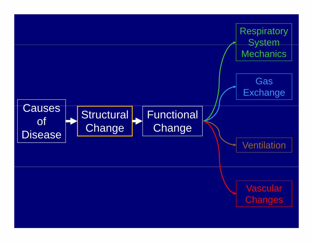
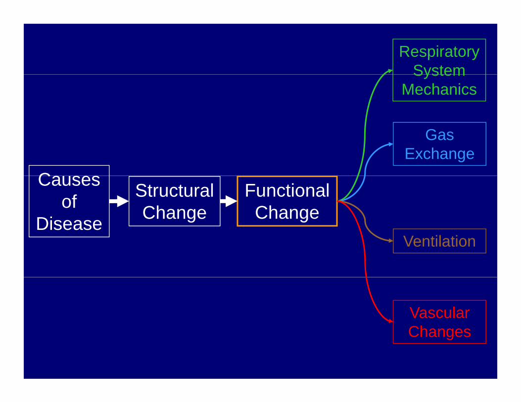
Respiratory SystemSystem
Mechanics
C
Gas Exchange
Causes of
Disease
Structural Change
FunctionalChangeDisease
Ventilation
VascularChChanges
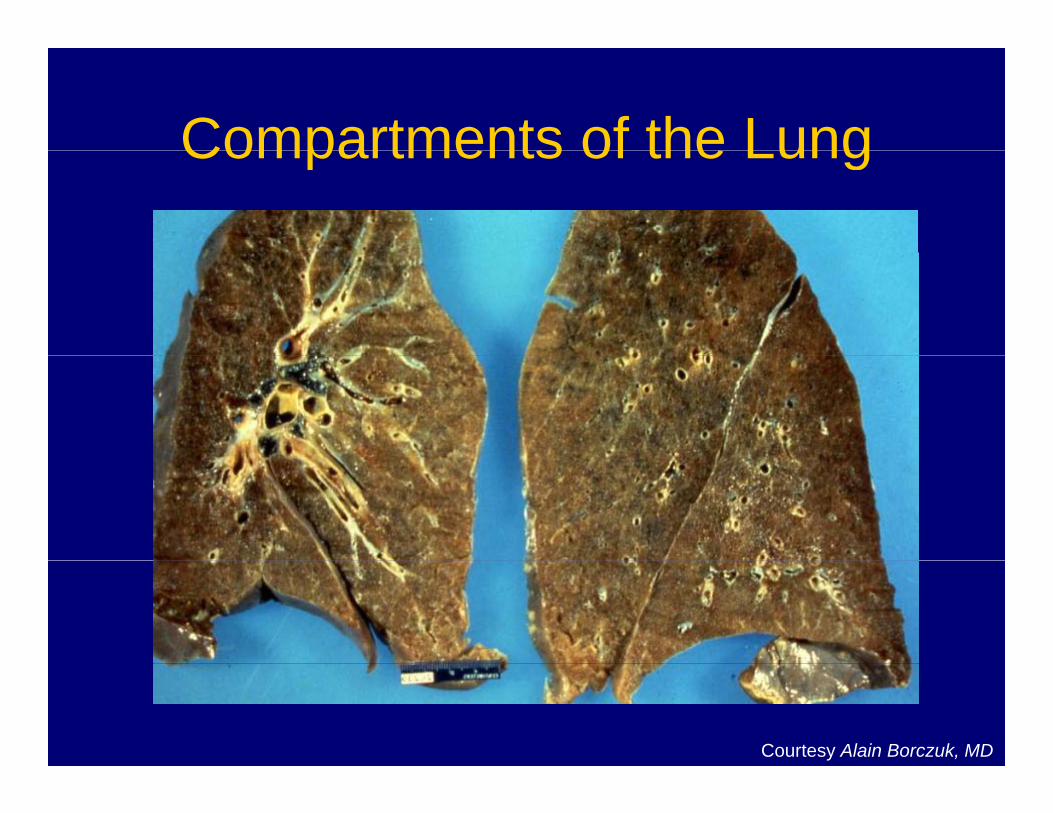
Compartments of the LungCompartments of the Lung
Courtesy Alain Borczuk, MD
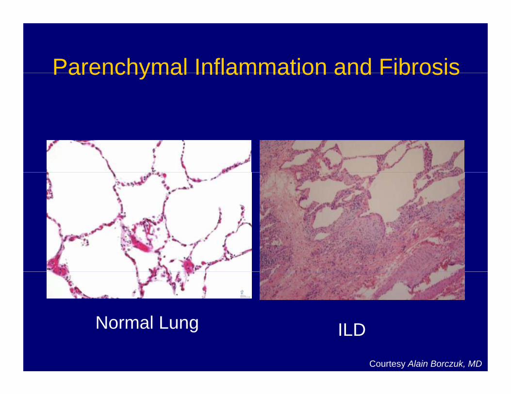
Parenchymal Inflammation and FibrosisParenchymal Inflammation and Fibrosis
Normal LungNormal Lung ILD
Courtesy Alain Borczuk, MD
OverviewOverview
• Terminology and classification schemeTerminology and classification scheme• Pathophysiology
Cli i l if t ti• Clinical manifestations• Pathogenesis• Management
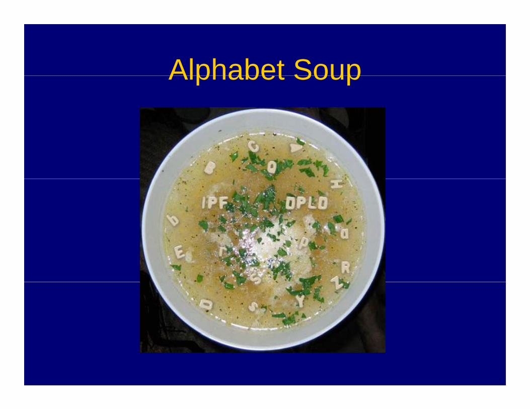
Alphabet SoupAlphabet Soup
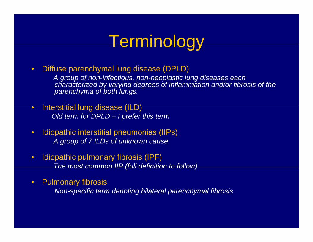
TerminologyTerminology• Diffuse parenchymal lung disease (DPLD)
A f i f ti l ti l di hA group of non-infectious, non-neoplastic lung diseases each characterized by varying degrees of inflammation and/or fibrosis of the parenchyma of both lungs.
Interstitial l ng disease (ILD)• Interstitial lung disease (ILD)Old term for DPLD – I prefer this term
• Idiopathic interstitial pneumonias (IIPs)p p ( )A group of 7 ILDs of unknown cause
• Idiopathic pulmonary fibrosis (IPF)The most common IIP (full definition to follow)The most common IIP (full definition to follow)
• Pulmonary fibrosisNon-specific term denoting bilateral parenchymal fibrosis
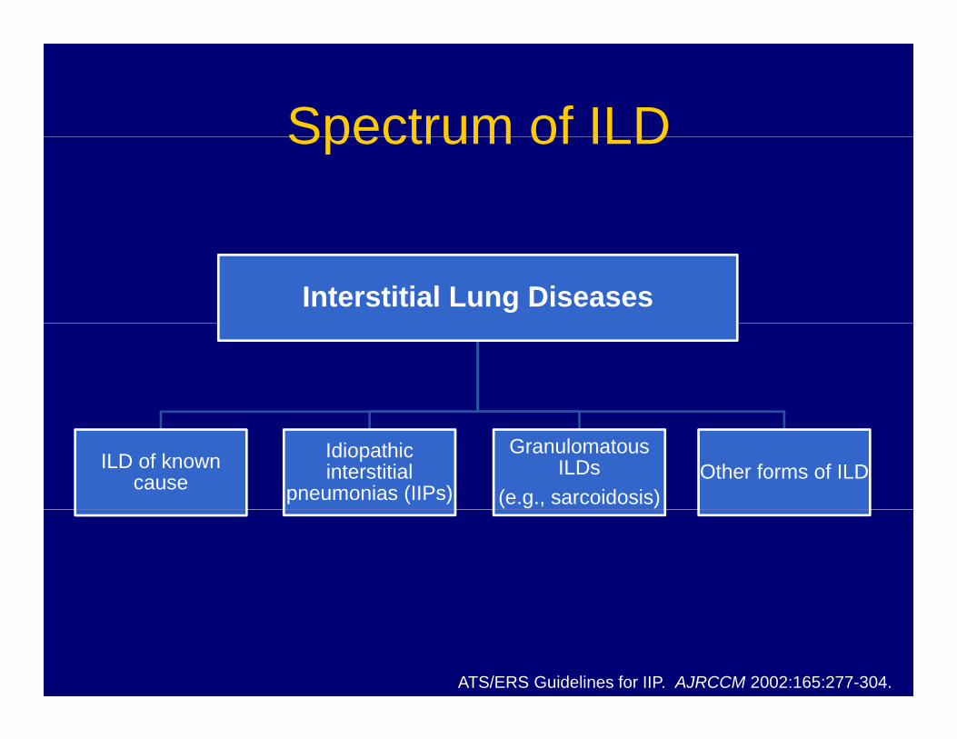
Spectrum of ILDSpectrum of ILD
Interstitial Lung Diseases
ILD of known cause
Idiopathic interstitial
pneumonias (IIPs)
Granulomatous ILDs
(e.g., sarcoidosis)Other forms of ILD
ATS/ERS Guidelines for IIP. AJRCCM 2002:165:277-304.
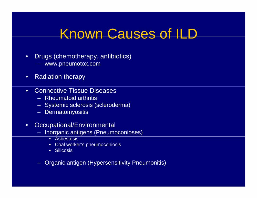
Known Causes of ILDKnown Causes of ILD• Drugs (chemotherapy, antibiotics)
t– www.pneumotox.com
• Radiation therapy
• Connective Tissue Diseases– Rheumatoid arthritis– Systemic sclerosis (scleroderma)
Dermatomyositis– Dermatomyositis
• Occupational/Environmental– Inorganic antigens (Pneumoconioses)
• Asbestosis• Coal worker’s pneumoconiosis• Silicosis
Organic antigen (H persensiti it Pne monitis)– Organic antigen (Hypersensitivity Pneumonitis)
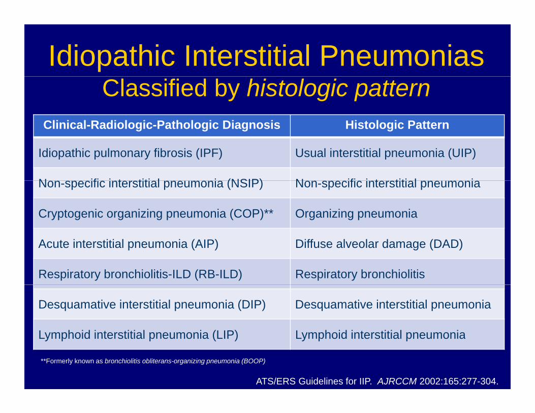
Idiopathic Interstitial PneumoniasClassified by histologic pattern
Clinical-Radiologic-Pathologic Diagnosis Histologic PatternClinical Radiologic Pathologic Diagnosis Histologic Pattern
Idiopathic pulmonary fibrosis (IPF) Usual interstitial pneumonia (UIP)
N ifi i t titi l i (NSIP) N ifi i t titi l iNon-specific interstitial pneumonia (NSIP) Non-specific interstitial pneumonia
Cryptogenic organizing pneumonia (COP)** Organizing pneumonia
Acute interstitial pneumonia (AIP) Diffuse alveolar damage (DAD)
Respiratory bronchiolitis-ILD (RB-ILD) Respiratory bronchiolitis
Desquamative interstitial pneumonia (DIP) Desquamative interstitial pneumonia
Lymphoid interstitial pneumonia (LIP) Lymphoid interstitial pneumoniaLymphoid interstitial pneumonia (LIP) Lymphoid interstitial pneumonia
**Formerly known as bronchiolitis obliterans-organizing pneumonia (BOOP)
ATS/ERS Guidelines for IIP. AJRCCM 2002:165:277-304.
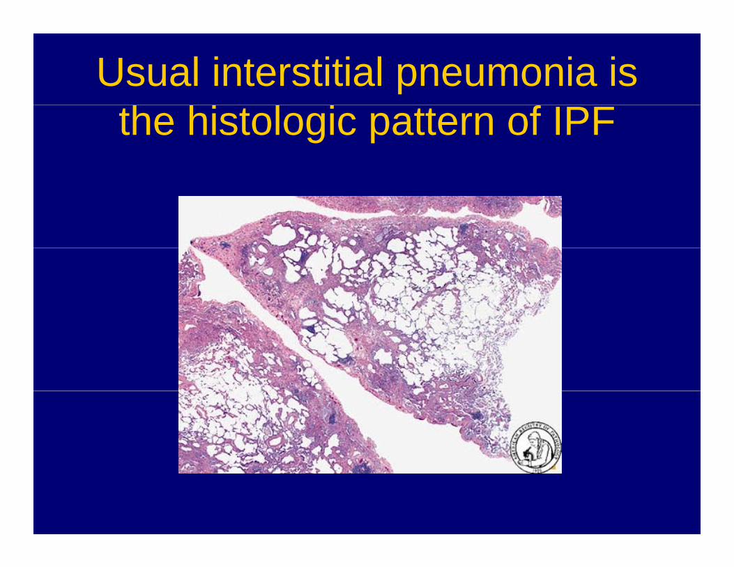
Usual interstitial pneumonia is the histologic pattern of IPF
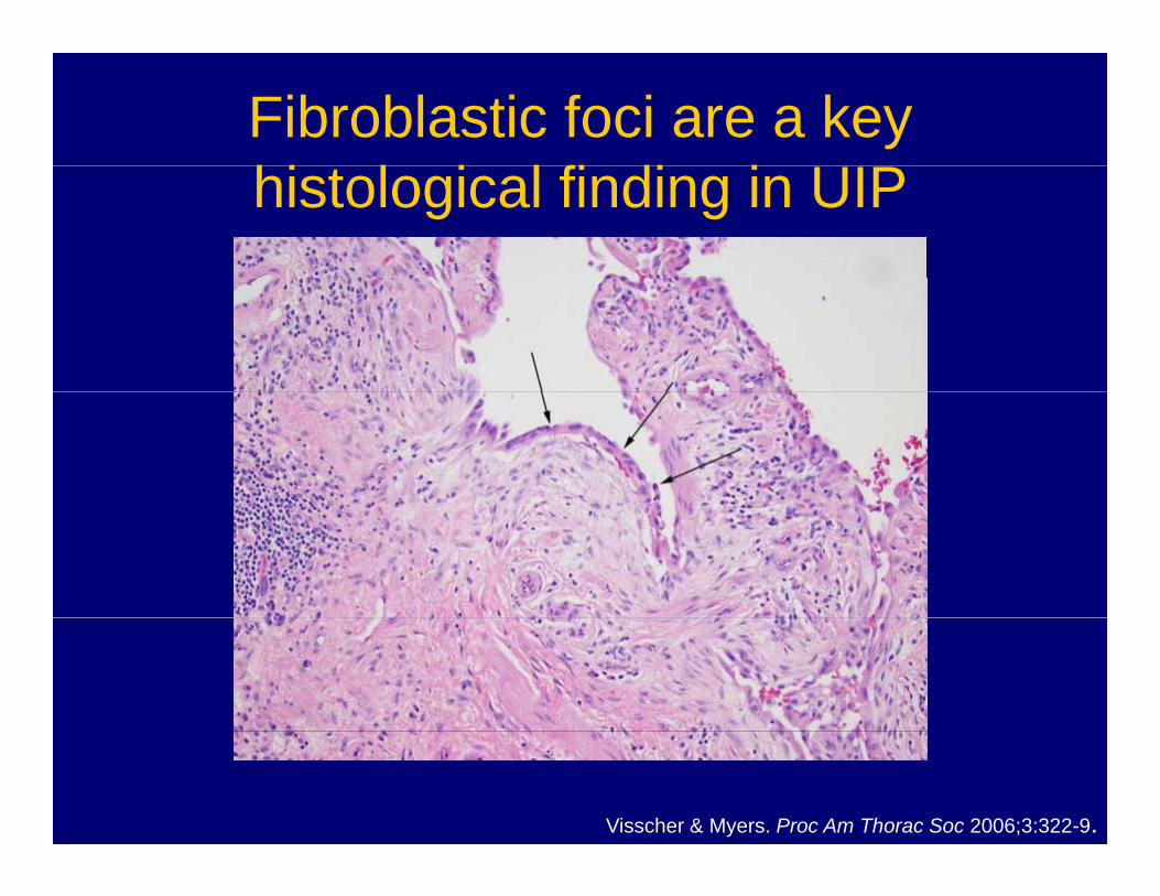
Fibroblastic foci are a key hi l i l fi di i UIPhistological finding in UIP
Visscher & Myers. Proc Am Thorac Soc 2006;3:322-9.
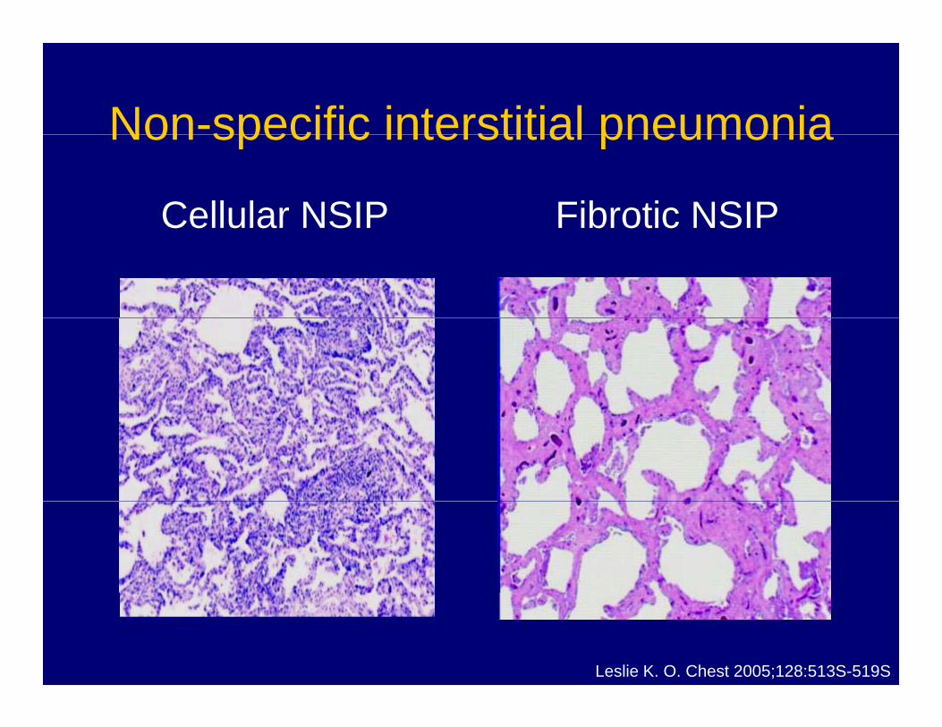
Non-specific interstitial pneumoniaNon specific interstitial pneumonia
Cellular NSIP Fibrotic NSIPCellular NSIP Fibrotic NSIP
Leslie K. O. Chest 2005;128:513S-519S
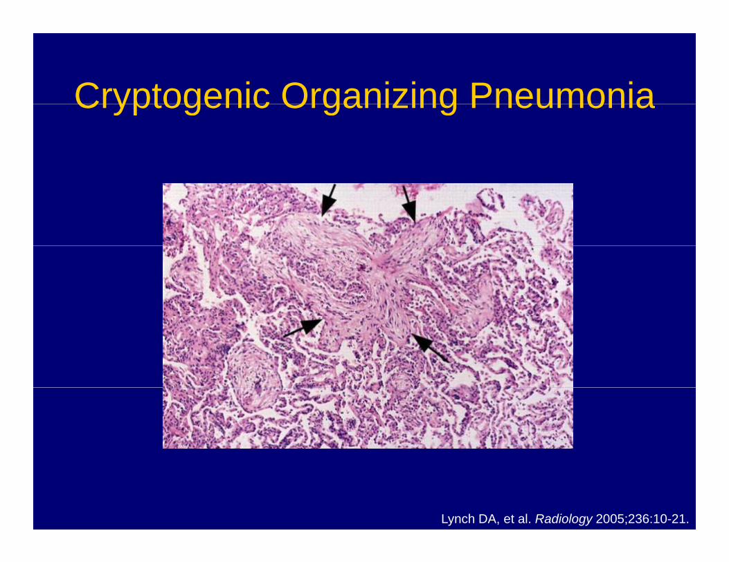
Cryptogenic Organizing PneumoniaCryptogenic Organizing Pneumonia
Lynch DA, et al. Radiology 2005;236:10-21.
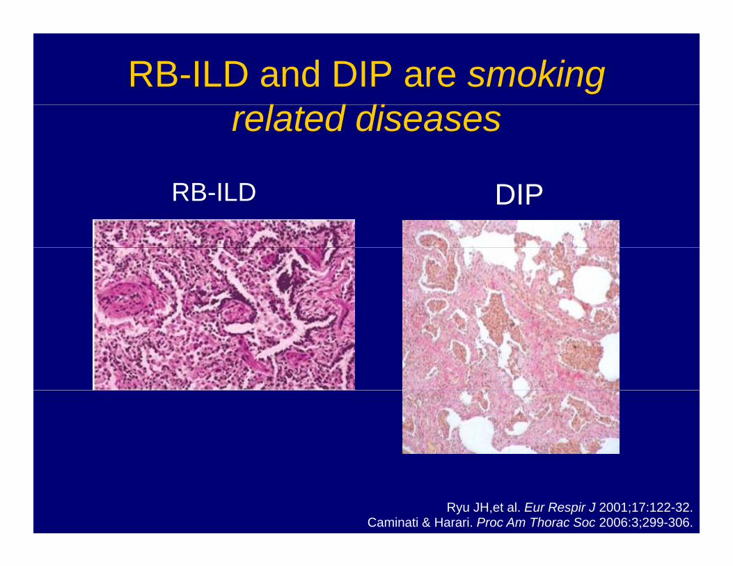
RB-ILD and DIP are smoking l d direlated diseases
RB-ILD DIP
Ryu JH,et al. Eur Respir J 2001;17:122-32.Caminati & Harari. Proc Am Thorac Soc 2006:3;299-306.
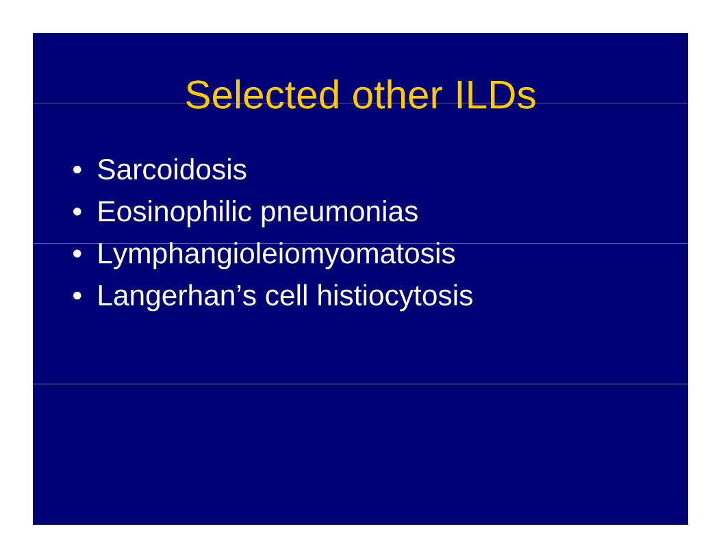
Selected other ILDsSelected other ILDs
• SarcoidosisSarcoidosis• Eosinophilic pneumonias
L h i l i t i• Lymphangioleiomyomatosis• Langerhan’s cell histiocytosis
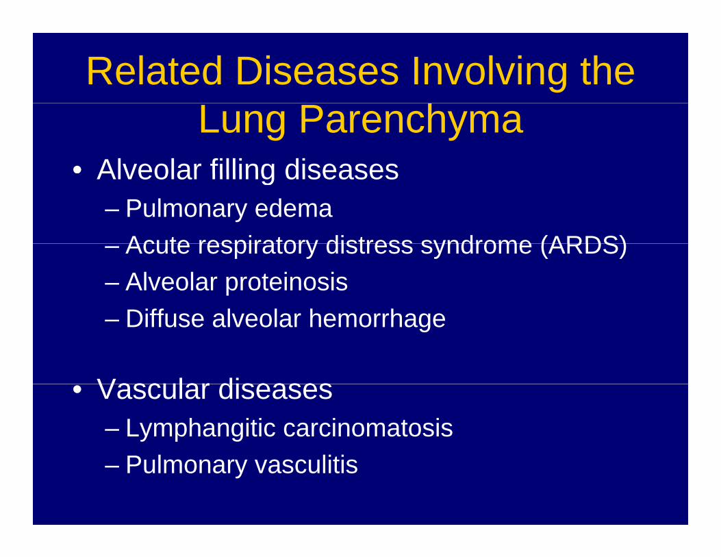
Related Diseases Involving the Lung Parenchyma
• Alveolar filling diseasesAlveolar filling diseases– Pulmonary edema
Acute respiratory distress syndrome (ARDS)– Acute respiratory distress syndrome (ARDS)– Alveolar proteinosis
Diffuse alveolar hemorrhage– Diffuse alveolar hemorrhage
Vascular diseases• Vascular diseases– Lymphangitic carcinomatosis– Pulmonary vasculitis
Respiratory SystemSystem
Mechanics
C
Gas Exchange
Causes of
Disease
Structural Change
FunctionalChangeDisease
Ventilation
VascularChChanges
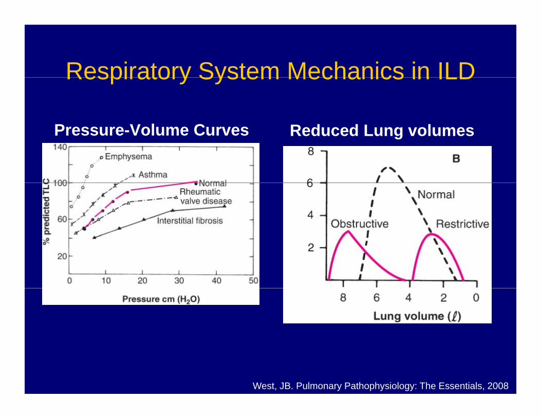
Respiratory System Mechanics in ILDRespiratory System Mechanics in ILD
Pressure Volume Curves Reduced Lung volumesPressure-Volume Curves Reduced Lung volumes
West, JB. Pulmonary Pathophysiology: The Essentials, 2008
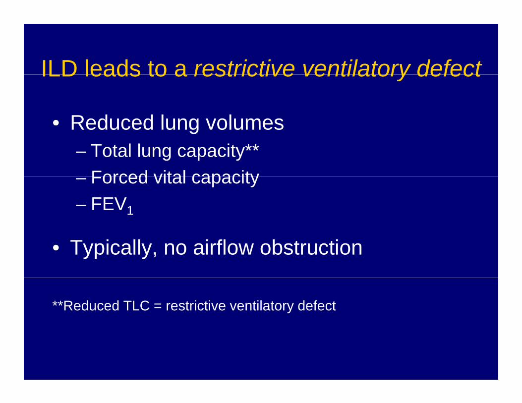
ILD leads to a restrictive ventilatory defectILD leads to a restrictive ventilatory defect
• Reduced lung volumesReduced lung volumes– Total lung capacity**
Forced vital capacity– Forced vital capacity– FEV1
• Typically, no airflow obstruction
**Reduced TLC = restrictive ventilatory defect
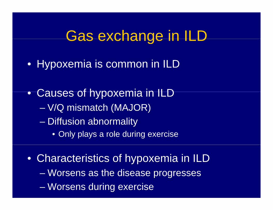
Gas exchange in ILDGas exchange in ILD
• Hypoxemia is common in ILDHypoxemia is common in ILD
• Causes of hypoxemia in ILD• Causes of hypoxemia in ILD– V/Q mismatch (MAJOR)
Diff i b lit– Diffusion abnormality• Only plays a role during exercise
• Characteristics of hypoxemia in ILDWorsens as the disease progresses– Worsens as the disease progresses
– Worsens during exercise
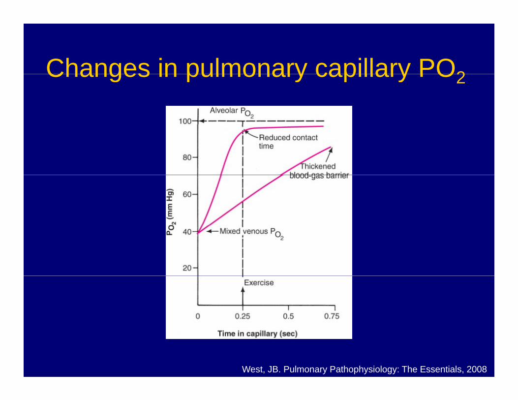
Changes in pulmonary capillary PO2Changes in pulmonary capillary PO2
West, JB. Pulmonary Pathophysiology: The Essentials, 2008
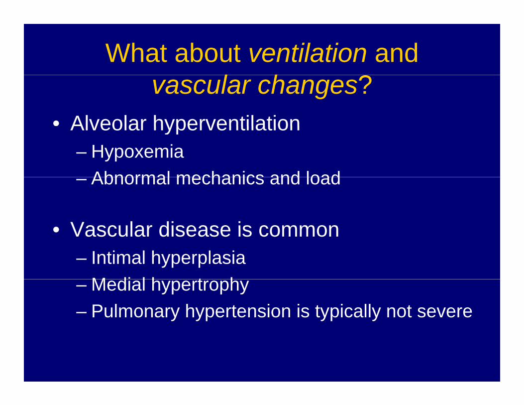
What about ventilation and l h ?vascular changes?
• Alveolar hyperventilationAlveolar hyperventilation– Hypoxemia
Abnormal mechanics and load– Abnormal mechanics and load
• Vascular disease is common• Vascular disease is common– Intimal hyperplasia
M di l h t h– Medial hypertrophy– Pulmonary hypertension is typically not severe
Clinical Manifestations of ILDClinical Manifestations of ILD
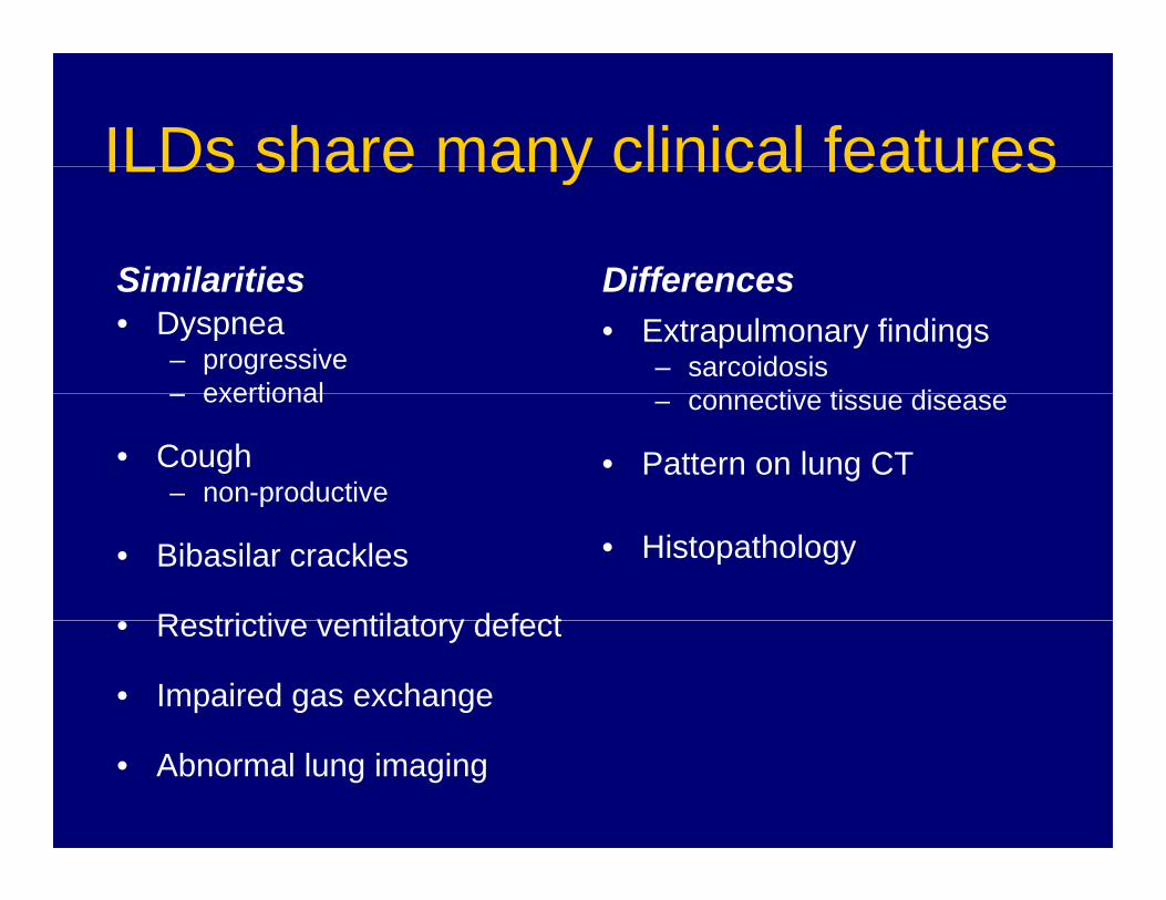
ILDs share many clinical featuresILDs share many clinical features
Similarities DifferencesSimilarities• Dyspnea
– progressiveexertional
Differences• Extrapulmonary findings
– sarcoidosisti ti di– exertional
• Cough– non-productive
– connective tissue disease
• Pattern on lung CTp
• Bibasilar crackles
Restrictive ventilatory defect
• Histopathology
• Restrictive ventilatory defect
• Impaired gas exchange
• Abnormal lung imaging
CaseCase
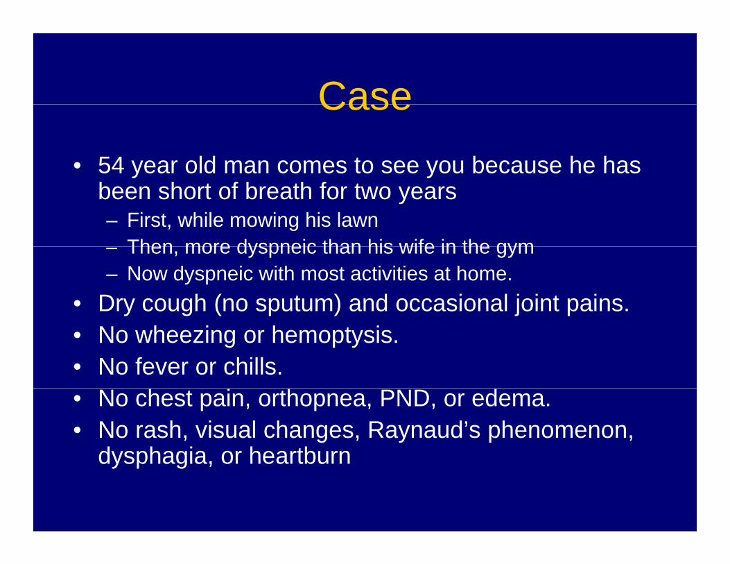
CaseCase• 54 year old man comes to see you because he has y y
been short of breath for two years– First, while mowing his lawn
Then more dyspneic than his wife in the gym– Then, more dyspneic than his wife in the gym– Now dyspneic with most activities at home.
• Dry cough (no sputum) and occasional joint pains. • No wheezing or hemoptysis.• No fever or chills. • No chest pain, orthopnea, PND, or edema. • No rash, visual changes, Raynaud’s phenomenon,
dysphagia or heartburndysphagia, or heartburn
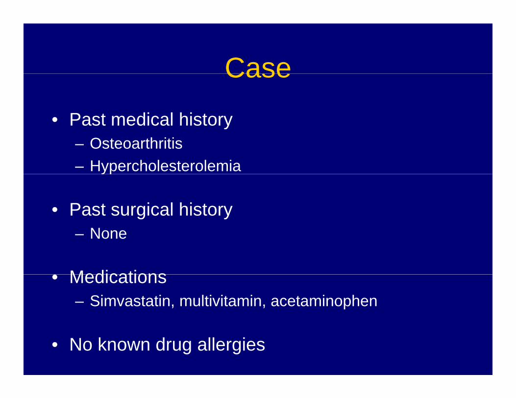
CaseCase
• Past medical historyPast medical history– Osteoarthritis– Hypercholesterolemiayp
• Past surgical history– None
• Medications• Medications– Simvastatin, multivitamin, acetaminophen
• No known drug allergies
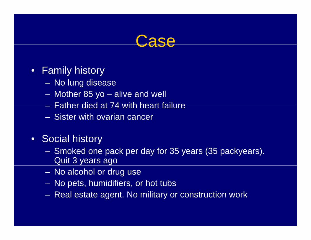
CaseCase• Family historyy y
– No lung disease– Mother 85 yo – alive and well
Father died at 74 with heart failure– Father died at 74 with heart failure– Sister with ovarian cancer
S i l hi t• Social history– Smoked one pack per day for 35 years (35 packyears).
Quit 3 years ago– No alcohol or drug use– No pets, humidifiers, or hot tubs– Real estate agent No military or construction workReal estate agent. No military or construction work
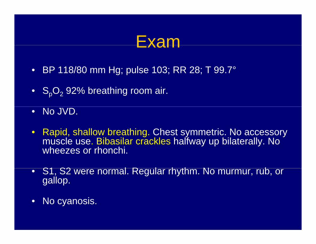
ExamExam• BP 118/80 mm Hg; pulse 103; RR 28; T 99.7°
• SpO2 92% breathing room air.
• No JVD.
• Rapid, shallow breathing. Chest symmetric. No accessory fmuscle use. Bibasilar crackles halfway up bilaterally. No
wheezes or rhonchi.
S1 S2 l R l h th N b• S1, S2 were normal. Regular rhythm. No murmur, rub, or gallop.
N i• No cyanosis.
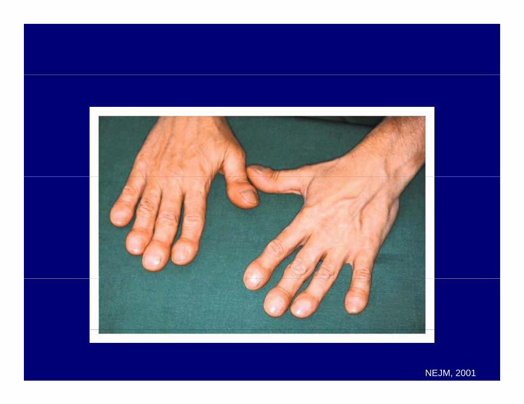
NEJM, 2001
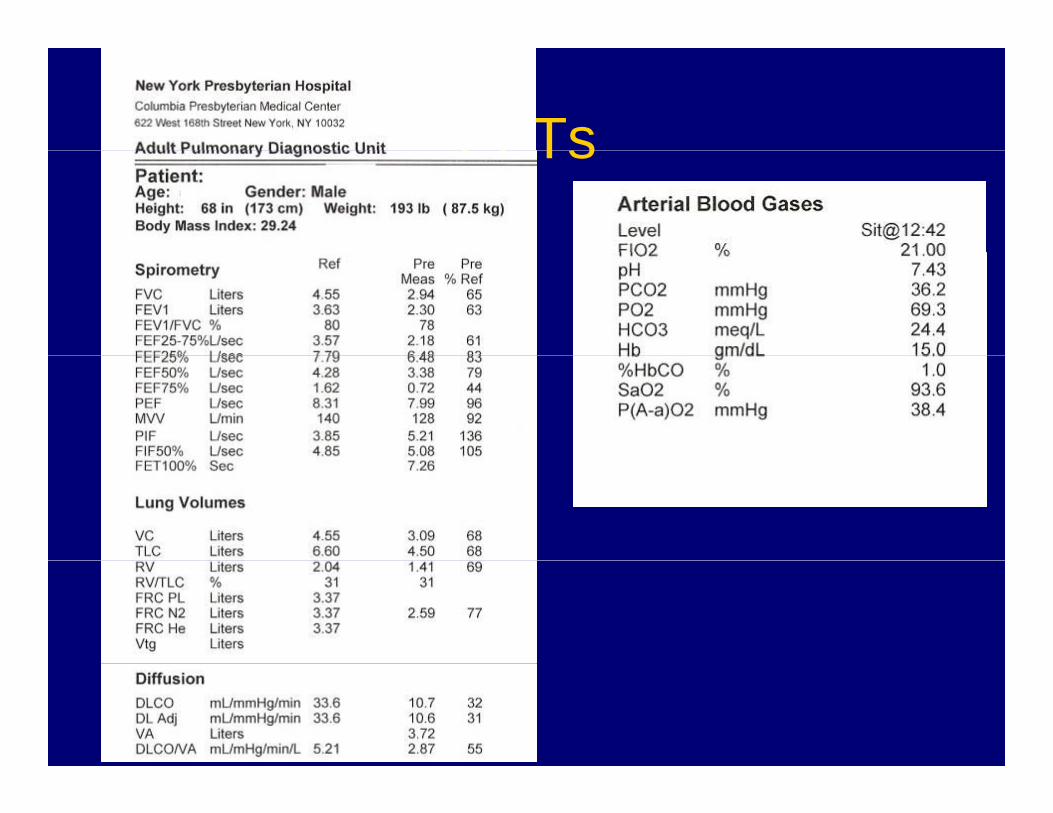
PFTsPFTs
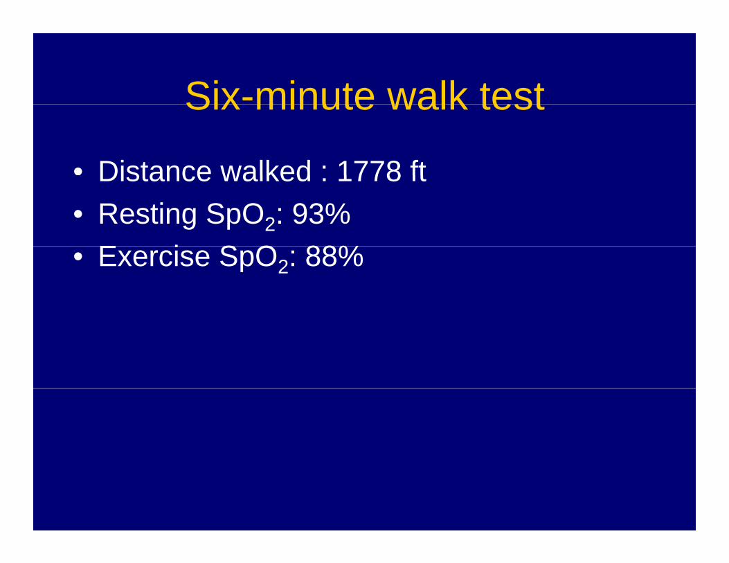
Six-minute walk testSix minute walk test
• Distance walked : 1778 ftDistance walked : 1778 ft • Resting SpO2: 93%
E i S O 88%• Exercise SpO2: 88%
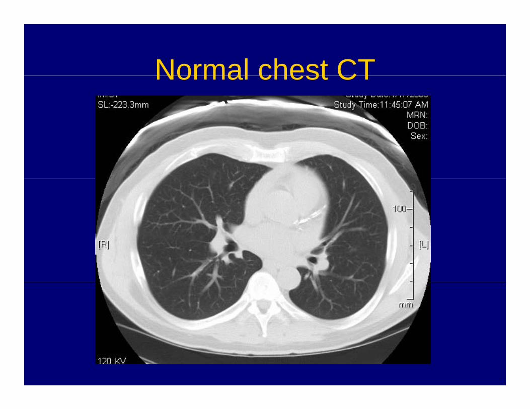
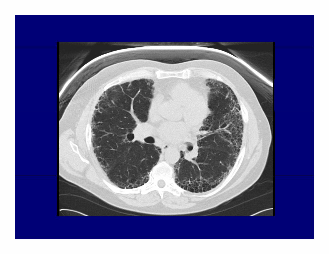
Normal chest CTNormal chest CT
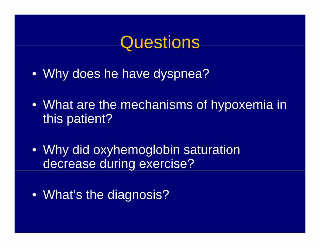
QuestionsQuestions
• Why does he have dyspnea?Why does he have dyspnea?
• What are the mechanisms of hypoxemia inWhat are the mechanisms of hypoxemia in this patient?
• Why did oxyhemoglobin saturation decrease during exercise?g
• What’s the diagnosis?g
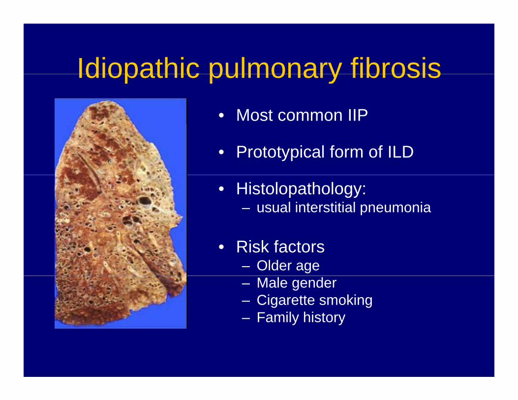
Idiopathic pulmonary fibrosisIdiopathic pulmonary fibrosis• Most common IIP
• Prototypical form of ILD
• Histolopathology:– usual interstitial pneumonia
• Risk factors– Older age– Male gender– Cigarette smoking– Family history
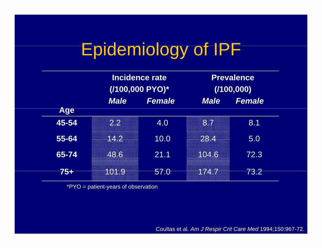
Epidemiolog of IPFEpidemiology of IPFIncidence rate Prevalence
A
Incidence rate (/100,000 PYO)*Male Female
Prevalence (/100,000)
Male FemaleAge
45-54 2.2 4.0 8.7 8.1
55-64 14 2 10 0 28 4 5 055-64 14.2 10.0 28.4 5.0
65-74 48.6 21.1 104.6 72.3
75+ 101 9 57 0 174 7 73 275+ 101.9 57.0 174.7 73.2
*PYO = patient-years of observation
Coultas et al. Am J Respir Crit Care Med 1994;150:967-72.
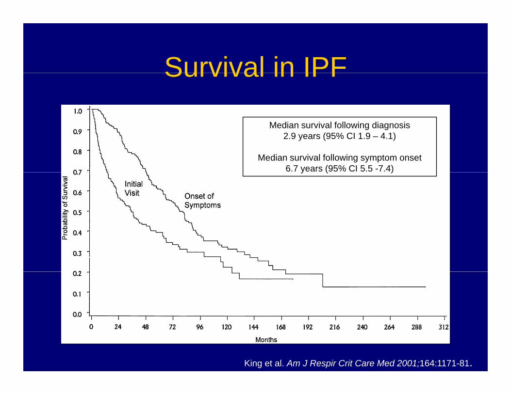
Survival in IPFSurvival in IPF
M di i l f ll i di iMedian survival following diagnosis2.9 years (95% CI 1.9 – 4.1)
Median survival following symptom onset6.7 years (95% CI 5.5 -7.4)y ( )
King et al. Am J Respir Crit Care Med 2001;164:1171-81.
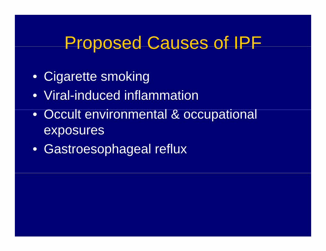
Proposed Causes of IPFProposed Causes of IPF
• Cigarette smokingCigarette smoking• Viral-induced inflammation
O lt i t l & ti l• Occult environmental & occupational exposures
• Gastroesophageal reflux
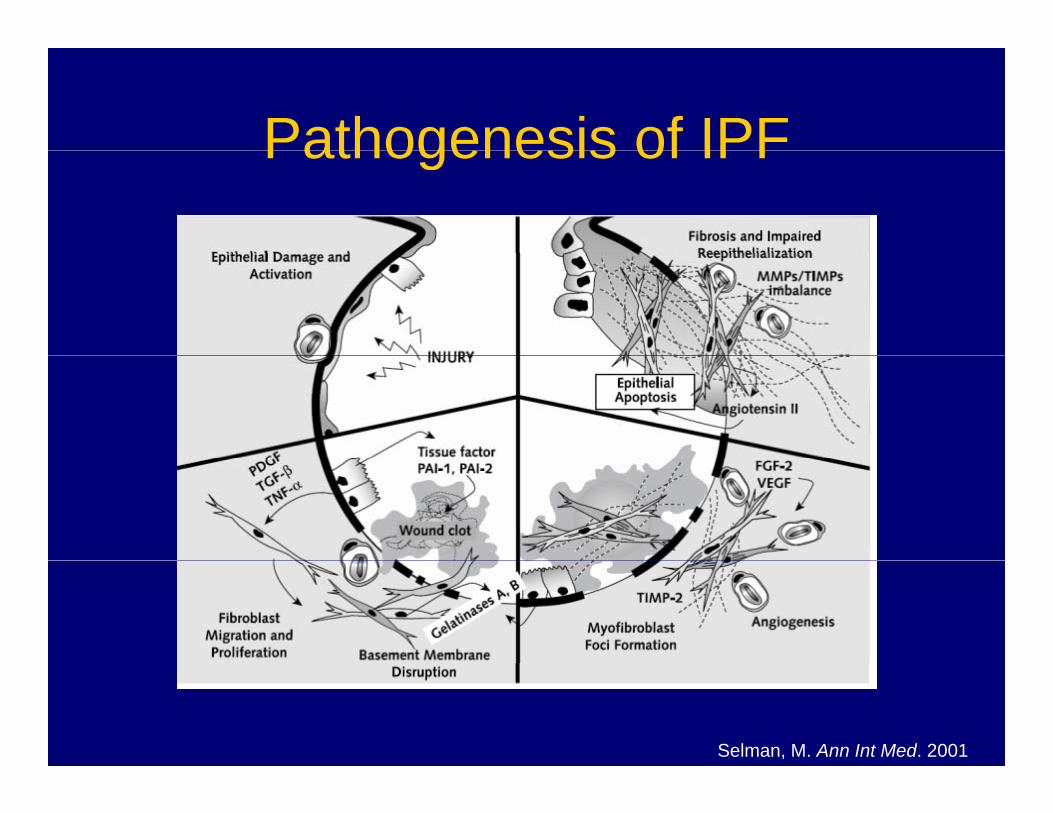
Pathogenesis of IPFPathogenesis of IPF
Selman, M. Ann Int Med. 2001
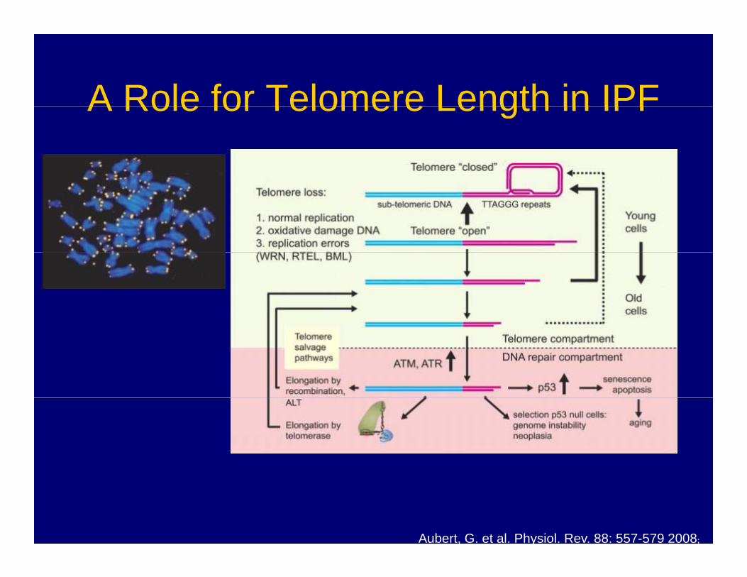
A Role for Telomere Length in IPFA Role for Telomere Length in IPF
Aubert, G. et al. Physiol. Rev. 88: 557-579 2008;
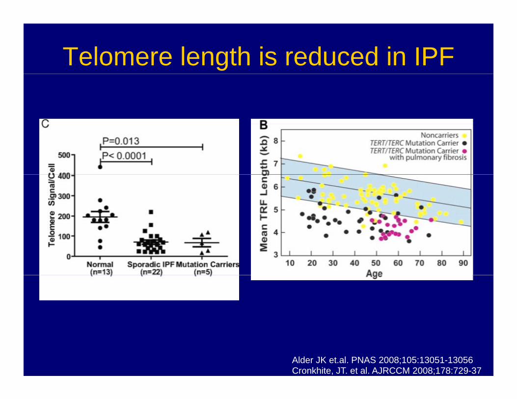
Telomere length is reduced in IPF
Alder JK et.al. PNAS 2008;105:13051-13056Cronkhite, JT. et al. AJRCCM 2008;178:729-37
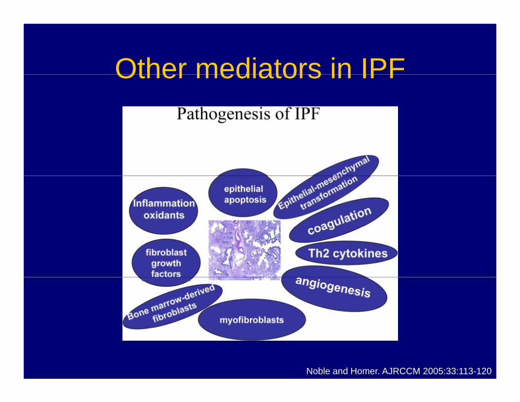
Other mediators in IPFOther mediators in IPF
Noble and Homer. AJRCCM 2005:33:113-120
What about other ILDs?What about other ILDs?
• Injurious triggersInjurious triggers– Autoimmune mediated inflammation
Drug induced injury– Drug-induced injury– Radiation-induced injury
Eosinophil degranulation– Eosinophil degranulation– Hypersensitivity reaction
Management of ILDManagement of ILD
• Biopsy often required to make a diagnosisBiopsy often required to make a diagnosis– Surgical lung biopsy (gold standard)
Transbronchial lung biopsy (less useful)– Transbronchial lung biopsy (less useful)
• Oxygen therapy• Oxygen therapy
P l h bilit ti• Pulmonary rehabilitation
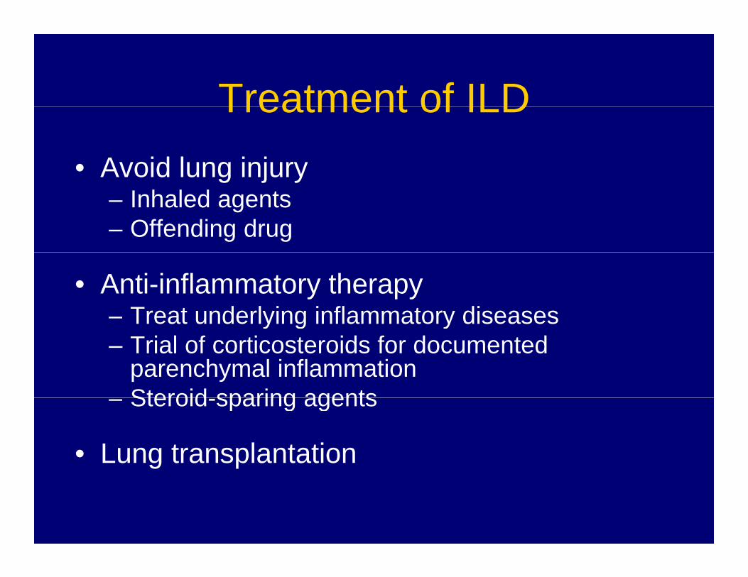
Treatment of ILDTreatment of ILD• Avoid lung injuryg j y
– Inhaled agents– Offending drug
• Anti-inflammatory therapy– Treat underlying inflammatory diseasesy g y– Trial of corticosteroids for documented
parenchymal inflammationSteroid sparing agents– Steroid-sparing agents
• Lung transplantation