Interstitial cystitis/bladder pain syndrome: The evolving ...
13
Review Article Interstitial cystitis/bladder pain syndrome: The evolving landscape, animal models and future perspectives Yoshiyuki Akiyama, 1,2 Yi Luo, 2 Philip M Hanno, 3 Daichi Maeda 4 and Yukio Homma 5 1 Department of Urology, Graduate School of Medicine, The University of Tokyo, Tokyo, Japan, 2 Department of Urology, University of Iowa, Iowa City, Iowa, 3 Department of Urology, Stanford University School of Medicine, Stanford, California, USA, 4 Department of Clinical Genomics, Graduate School of Medicine, Osaka University, Osaka, and 5 Japanese Red Cross Medical Center, Tokyo, Japan Abbreviations & Acronyms BPS = bladder pain syndrome CCL2 = C-C motif ligand 2 CNS = central nervous system DMSO = dimethyl sulfoxide EAC = experimental autoimmune cystitis ESSIC = International Society for the Study of BPS FSS = functional somatic syndrome GAG = glycosaminoglycan IC = interstitial cystitis IFN-c = interferon-gamma IL = interleukin MC = mast cell NGF = nerve growth factor OVA = ovalbumin TCR = T-cell receptor TNF = tumor necrosis factor TLR = Toll-like receptor UCPPS = urological chronic pelvic pain syndrome UPK = uroplakin Upo = unpublished observation VEGF = vascular endothelial growth factor WAS = water avoidance stress Correspondence: Yoshiyuki Akiyama M.D., Ph.D., Department of Urology, Graduate School of Medicine, The University of Tokyo, 7-3-1 Hongo, Bunkyo-ku, Tokyo 113- 0033, Japan. Email: [email protected] Received 28 November 2019; accepted 24 February 2020. Abstract: Interstitial cystitis/bladder pain syndrome is a debilitating condition of unknown etiology characterized by persistent pelvic pain with lower urinary tract symptoms and comprises a wide variety of potentially clinically useful phenotypes with different possible etiologies. Current clinicopathological and genomic evidence suggests that interstitial cystitis/bladder pain syndrome should be categorized by the presence or absence of Hunner lesions, rather than by clinical phenotyping based on symptomatology. The Hunner lesion subtype is a distinct inflammatory disease with proven bladder etiology characterized by epithelial denudation and enhanced immune responses frequently accompanied by clonal expansion of infiltrating B cells, with potential engagement of infection. Meanwhile, the non-Hunner lesion subtype is a non- inflammatory disorder with little evidence of bladder etiology. It is potentially associated with urothelial malfunction and neurophysiological dysfunction, and frequently presents with somatic and/or psychological symptoms, that commonly result in central nervous sensitization. Animal models of autoimmune cystitis and neurogenic sensitization might serve as disease models for the Hunner lesion and non-Hunner lesion subtypes, respectively. Here, we revisit the taxonomy of interstitial cystitis/bladder pain syndrome according to current research, and discuss its potential pathophysiology and representative animal models. Categorization of interstitial cystitis/bladder pain syndrome based on cystoscopy is mandatory to design optimized treatment and research strategies for each subtype. A tailored approach that specifically targets the characteristic inflammation and epithelial denudation for the Hunner lesion subtype, or the urothelial malfunction, sensitized/altered nervous system and psychosocial problems for the non-Hunner lesion subtype, is essential for better clinical management and research progress in this complex condition. Key words: animal model, autoimmune cystitis, bladder pain syndrome, interstitial cystitis, pathophysiology. Introduction IC/BPS refers to a symptom syndrome complex characterized by persistent pain perceived to be related to the urinary bladder in conjunction with urinary frequency and/or urgency, and presents with a wide variety of clinical phenotypes. 1–4 Although the whole picture of IC/BPS remains unclear, IC/BPS with Hunner lesions is currently a well-recognized clear clinical entity, characterized by the cystoscopic finding known as Hunner lesions, reddish mucosal lesions accompanied by abnormal capillary structures (Fig. 1). Past studies have shown that this entity presents with distinct clinicopathological and genomic characteristics compared with other forms of IC/BPS. 5–11 This evidence suggests that IC/BPS with Hunner lesions comprises a potentially distinct disease entity with different etiology in the IC/BPS umbrella. In the past, many clinical studies were carried out to develop promising therapies for IC/BPS. However, most failed to achieve positive results. Likewise, a wide range of basic studies on the pathogenesis of IC/BPS have been carried out, but the results were variable and inconsis- tent. These unexpected and confusing results are likely due to multiple clinical conditions (i.e. pathogenetically different entities) having been combined in a single (and small) cohort. 12 In © 2020 The Japanese Urological Association 1 International Journal of Urology (2020) doi: 10.1111/iju.14229
Transcript of Interstitial cystitis/bladder pain syndrome: The evolving ...
Review Article
Interstitial cystitis/bladder pain syndrome: The evolving landscape,animal models and future perspectivesYoshiyuki Akiyama,1,2 Yi Luo,2 Philip M Hanno,3 Daichi Maeda4 and Yukio Homma5
1Department of Urology, Graduate School of Medicine, The University of Tokyo, Tokyo, Japan, 2Department of Urology, University ofIowa, Iowa City, Iowa, 3Department of Urology, Stanford University School of Medicine, Stanford, California, USA, 4Department ofClinical Genomics, Graduate School of Medicine, Osaka University, Osaka, and 5Japanese Red Cross Medical Center, Tokyo, Japan
Abbreviations & AcronymsBPS = bladder painsyndromeCCL2 = C-C motif ligand 2CNS = central nervoussystemDMSO = dimethyl sulfoxideEAC = experimentalautoimmune cystitisESSIC = InternationalSociety for the Study of BPSFSS = functional somaticsyndromeGAG = glycosaminoglycanIC = interstitial cystitisIFN-c = interferon-gammaIL = interleukinMC = mast cellNGF = nerve growth factorOVA = ovalbuminTCR = T-cell receptorTNF = tumor necrosis factorTLR = Toll-like receptorUCPPS = urological chronicpelvic pain syndromeUPK = uroplakinUpo = unpublishedobservationVEGF = vascular endothelialgrowth factorWAS = water avoidancestress
Correspondence: YoshiyukiAkiyama M.D., Ph.D.,Department of Urology,Graduate School of Medicine,The University of Tokyo, 7-3-1Hongo, Bunkyo-ku, Tokyo 113-0033, Japan. Email:[email protected]
Received 28 November 2019;accepted 24 February 2020.
Abstract: Interstitial cystitis/bladder pain syndrome is a debilitating condition of
unknown etiology characterized by persistent pelvic pain with lower urinary tract
symptoms and comprises a wide variety of potentially clinically useful phenotypes with
different possible etiologies. Current clinicopathological and genomic evidence suggests
that interstitial cystitis/bladder pain syndrome should be categorized by the presence or
absence of Hunner lesions, rather than by clinical phenotyping based on
symptomatology. The Hunner lesion subtype is a distinct inflammatory disease with
proven bladder etiology characterized by epithelial denudation and enhanced immune
responses frequently accompanied by clonal expansion of infiltrating B cells, with
potential engagement of infection. Meanwhile, the non-Hunner lesion subtype is a non-
inflammatory disorder with little evidence of bladder etiology. It is potentially associated
with urothelial malfunction and neurophysiological dysfunction, and frequently presents
with somatic and/or psychological symptoms, that commonly result in central nervous
sensitization. Animal models of autoimmune cystitis and neurogenic sensitization might
serve as disease models for the Hunner lesion and non-Hunner lesion subtypes,
respectively. Here, we revisit the taxonomy of interstitial cystitis/bladder pain syndrome
according to current research, and discuss its potential pathophysiology and
representative animal models. Categorization of interstitial cystitis/bladder pain
syndrome based on cystoscopy is mandatory to design optimized treatment and
research strategies for each subtype. A tailored approach that specifically targets the
characteristic inflammation and epithelial denudation for the Hunner lesion subtype, or
the urothelial malfunction, sensitized/altered nervous system and psychosocial problems
for the non-Hunner lesion subtype, is essential for better clinical management and
research progress in this complex condition.
Key words: animal model, autoimmune cystitis, bladder pain syndrome, interstitial
cystitis, pathophysiology.
Introduction
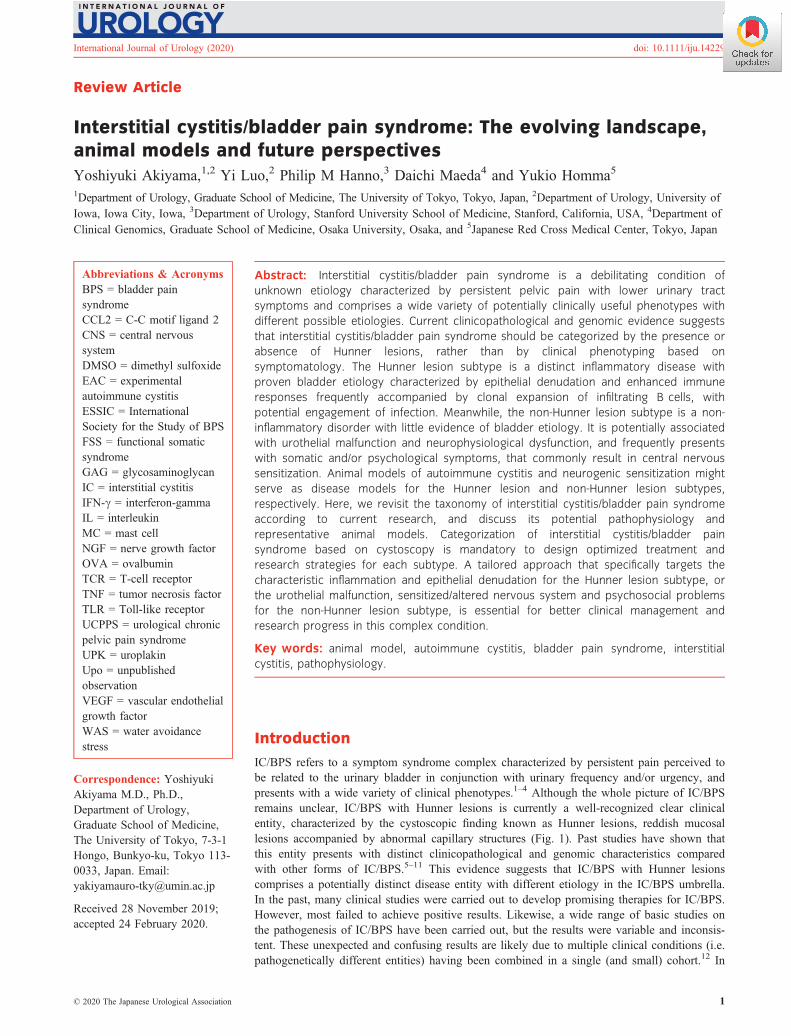
IC/BPS refers to a symptom syndrome complex characterized by persistent pain perceived tobe related to the urinary bladder in conjunction with urinary frequency and/or urgency, andpresents with a wide variety of clinical phenotypes.1–4 Although the whole picture of IC/BPSremains unclear, IC/BPS with Hunner lesions is currently a well-recognized clear clinicalentity, characterized by the cystoscopic finding known as Hunner lesions, reddish mucosallesions accompanied by abnormal capillary structures (Fig. 1). Past studies have shown thatthis entity presents with distinct clinicopathological and genomic characteristics comparedwith other forms of IC/BPS.5–11 This evidence suggests that IC/BPS with Hunner lesionscomprises a potentially distinct disease entity with different etiology in the IC/BPS umbrella.In the past, many clinical studies were carried out to develop promising therapies for IC/BPS.However, most failed to achieve positive results. Likewise, a wide range of basic studies onthe pathogenesis of IC/BPS have been carried out, but the results were variable and inconsis-tent. These unexpected and confusing results are likely due to multiple clinical conditions (i.e.pathogenetically different entities) having been combined in a single (and small) cohort.12 In
© 2020 The Japanese Urological Association 1
International Journal of Urology (2020) doi: 10.1111/iju.14229
addition, the unknown etiology, diverse clinical symptomsand varying histological manifestations also hinder clearlyshowing the landscape of IC/BPS. In this article, we reviewrecent evidence on IC/BPS categorization to describe theevolving taxonomy of IC/BPS, discuss the potential patho-physiologies and animal models, and suggest future strategiesto achieve better clinical management and research progressof IC/BPS based on these emerging concepts.
Categorization of IC/BPS
The workshop on IC/BPS phenotyping at the 4th Interna-tional Consultation on Interstitial Cystitis Japan meeting dis-cussed potential categorization of IC/BPS in terms of theirclinical characteristics, epidemiology and bladder histology.4,8
It was unanimously agreed that IC/BPS with Hunner lesionsis a proven, clinically relevant subtype that should beregarded as a distinct disease entity. However, other forms ofIC/BPS could not be clearly defined because of their obscurepathophysiology and histology, and their varied clinical mani-festations. Thus, IC/BPS can be categorized into two sub-types at present: (i) IC/BPS with Hunner lesions, which isalso known as the ESSIC BPS type 3; and (ii) IC/BPS with-out Hunner lesions, which includes the ESSIC BPS type 1and 2. These two subtypes, which are distinguished simplyby the cystoscopic presence or absence of Hunner lesions,present with different, but overlapping, clinical characteristicsand cannot be clinically differentiated in the absence of cys-toscopic findings (Table 1). IC/BPS with Hunner lesions ischaracterized by an older age at diagnosis, more severe blad-der-centric symptoms, reduced bladder capacity, fewercomorbid non-bladder conditions and more favorable out-comes on endoscopic treatment compared with IC/BPS with-out Hunner lesions.7,9 IC/BPS without Hunner lesions isfrequently accompanied by non-bladder symptoms, includingother common systemic pain problems (“bladder-beyond”pain), psychosocial health problems and affect dysregula-tion.13 Recent studies have shown that these clinical charac-teristics of IC/BPS without Hunner lesions strongly overlapwith those of widely known somatoform disorders or FSSs,
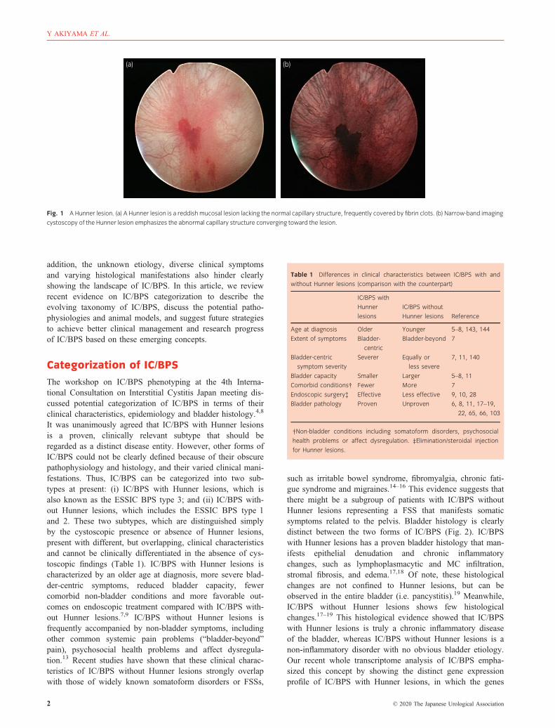
such as irritable bowel syndrome, fibromyalgia, chronic fati-gue syndrome and migraines.14–16 This evidence suggests thatthere might be a subgroup of patients with IC/BPS withoutHunner lesions representing a FSS that manifests somaticsymptoms related to the pelvis. Bladder histology is clearlydistinct between the two forms of IC/BPS (Fig. 2). IC/BPSwith Hunner lesions has a proven bladder histology that man-ifests epithelial denudation and chronic inflammatorychanges, such as lymphoplasmacytic and MC infiltration,stromal fibrosis, and edema.17,18 Of note, these histologicalchanges are not confined to Hunner lesions, but can beobserved in the entire bladder (i.e. pancystitis).19 Meanwhile,IC/BPS without Hunner lesions shows few histologicalchanges.17–19 This histological evidence showed that IC/BPSwith Hunner lesions is truly a chronic inflammatory diseaseof the bladder, whereas IC/BPS without Hunner lesions is anon-inflammatory disorder with no obvious bladder etiology.Our recent whole transcriptome analysis of IC/BPS empha-sized this concept by showing the distinct gene expressionprofile of IC/BPS with Hunner lesions, in which the genes
(a) (b)
Fig. 1 A Hunner lesion. (a) A Hunner lesion is a reddish mucosal lesion lacking the normal capillary structure, frequently covered by fibrin clots. (b) Narrow-band imaging
cystoscopy of the Hunner lesion emphasizes the abnormal capillary structure converging toward the lesion.
Table 1 Differences in clinical characteristics between IC/BPS with and
without Hunner lesions (comparison with the counterpart)
IC/BPS with
Hunner
lesions
IC/BPS without
Hunner lesions Reference
Age at diagnosis Older Younger 5–8, 143, 144
Extent of symptoms Bladder-
centric
Bladder-beyond 7
Bladder-centric
symptom severity
Severer Equally or
less severe
7, 11, 140
Bladder capacity Smaller Larger 5–8, 11
Comorbid conditions† Fewer More 7
Endoscopic surgery‡ Effective Less effective 9, 10, 28
Bladder pathology Proven Unproven 6, 8, 11, 17–19,
22, 65, 66, 103
†Non-bladder conditions including somatoform disorders, psychosocial
health problems or affect dysregulation. ‡Elimination/steroidal injection
for Hunner lesions.
2 © 2020 The Japanese Urological Association
Y AKIYAMA ET AL.
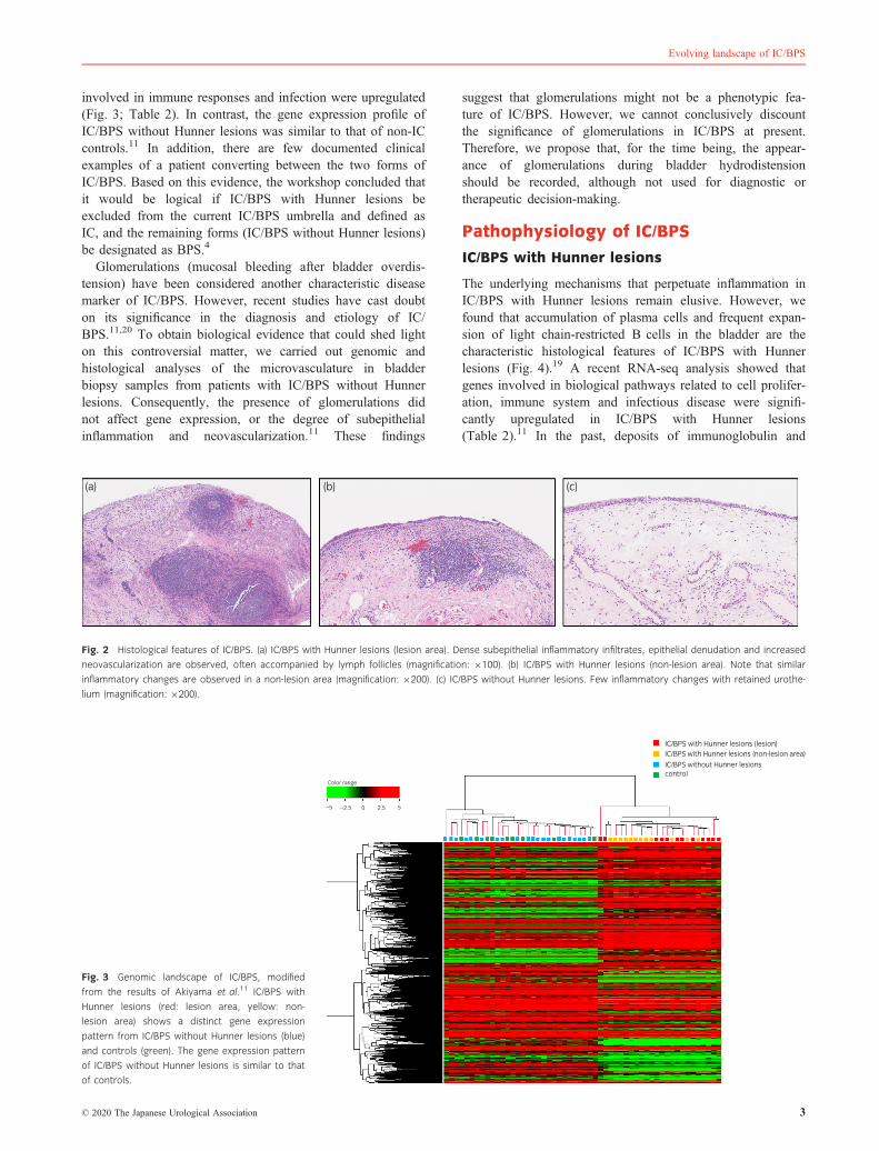
involved in immune responses and infection were upregulated(Fig. 3; Table 2). In contrast, the gene expression profile ofIC/BPS without Hunner lesions was similar to that of non-ICcontrols.11 In addition, there are few documented clinicalexamples of a patient converting between the two forms ofIC/BPS. Based on this evidence, the workshop concluded thatit would be logical if IC/BPS with Hunner lesions beexcluded from the current IC/BPS umbrella and defined asIC, and the remaining forms (IC/BPS without Hunner lesions)be designated as BPS.4
Glomerulations (mucosal bleeding after bladder overdis-tension) have been considered another characteristic diseasemarker of IC/BPS. However, recent studies have cast doubton its significance in the diagnosis and etiology of IC/BPS.11,20 To obtain biological evidence that could shed lighton this controversial matter, we carried out genomic andhistological analyses of the microvasculature in bladderbiopsy samples from patients with IC/BPS without Hunnerlesions. Consequently, the presence of glomerulations didnot affect gene expression, or the degree of subepithelialinflammation and neovascularization.11 These findings
suggest that glomerulations might not be a phenotypic fea-ture of IC/BPS. However, we cannot conclusively discountthe significance of glomerulations in IC/BPS at present.Therefore, we propose that, for the time being, the appear-ance of glomerulations during bladder hydrodistensionshould be recorded, although not used for diagnostic ortherapeutic decision-making.
Pathophysiology of IC/BPS
IC/BPS with Hunner lesions
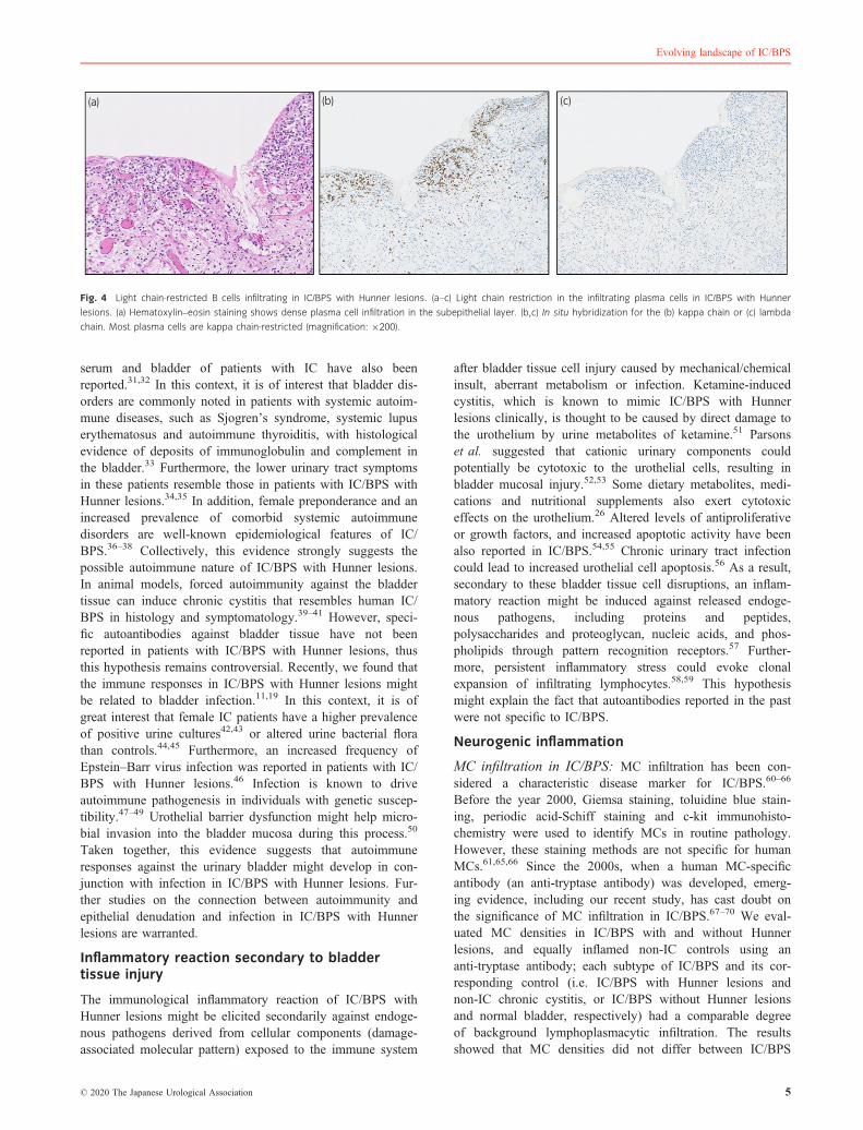
The underlying mechanisms that perpetuate inflammation inIC/BPS with Hunner lesions remain elusive. However, wefound that accumulation of plasma cells and frequent expan-sion of light chain-restricted B cells in the bladder are thecharacteristic histological features of IC/BPS with Hunnerlesions (Fig. 4).19 A recent RNA-seq analysis showed thatgenes involved in biological pathways related to cell prolifer-ation, immune system and infectious disease were signifi-cantly upregulated in IC/BPS with Hunner lesions(Table 2).11 In the past, deposits of immunoglobulin and
(a) (b) (c)
Fig. 2 Histological features of IC/BPS. (a) IC/BPS with Hunner lesions (lesion area). Dense subepithelial inflammatory infiltrates, epithelial denudation and increased
neovascularization are observed, often accompanied by lymph follicles (magnification: 9100). (b) IC/BPS with Hunner lesions (non-lesion area). Note that similar
inflammatory changes are observed in a non-lesion area (magnification: 9200). (c) IC/BPS without Hunner lesions. Few inflammatory changes with retained urothe-
lium (magnification: 9200).
IC/BPS with Hunner lesions (lesion)IC/BPS with Hunner lesions (non-lesion area)
IC/BPS without Hunner lesions control
–5
Color range
–2.5 0 2.5 5
Fig. 3 Genomic landscape of IC/BPS, modified
from the results of Akiyama et al.11 IC/BPS with
Hunner lesions (red: lesion area, yellow: non-
lesion area) shows a distinct gene expression
pattern from IC/BPS without Hunner lesions (blue)
and controls (green). The gene expression pattern
of IC/BPS without Hunner lesions is similar to that
of controls.
© 2020 The Japanese Urological Association 3
Evolving landscape of IC/BPS
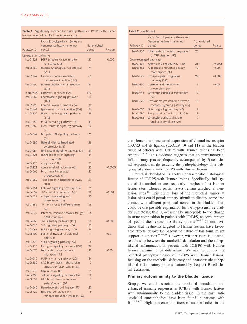
complement, and increased expression of chemokine receptorCXCR3 and its ligands (CXCL9, 10 and 11), in the bladdertissue of patients with IC/BPS with Hunner lesions has beenreported.21–23 This evidence suggests that an immunologicalinflammatory process frequently accompanied by B-cell clo-nal expansion might underlie the pathophysiology in a sub-group of patients with IC/BPS with Hunner lesions.
Urothelial denudation is another characteristic histologicalfeature of IC/BPS with Hunner lesions. Specifically, full lay-ers of the urothelium are frequently sloughed off at Hunnerlesion sites, whereas partial layers remain attached at non-lesion sites.18 This entire loss of the urothelial barrier atlesion sites could permit urinary stimuli to directly come intocontact with afferent peripheral nerves in the bladder. Thiscould be one possible explanation for the hypersensitive blad-der symptoms; that is, occasionally susceptible to the changein urine composition in patients with IC/BPS, as consumptionof specific diets exacerbate the symptoms.24–27 Clinical evi-dence that treatments targeted to Hunner lesions have favor-able effects, despite the pancystitic nature of this form, mightsupport this notion.9–10,28 However, whether there is a causalrelationship between the urothelial denudation and the subep-ithelial inflammation in patients with IC/BPS with Hunnerlesions remains to be determined. We next to discuss thepotential pathophysiologies of IC/BPS with Hunner lesions,focusing on the urothelial deficiency and characteristic subep-ithelial inflammatory process featured by frequent B-cell clo-nal expansion.
Primary autoimmunity to the bladder tissue
Simply, we could associate the urothelial denudation andenhanced immune responses in IC/BPS with Hunner lesionswith autoimmunity to the bladder tissue. In the past, anti-urothelial autoantibodies have been found in patients withIC.21,29,30 High incidence and titers of autoantibodies in the
Table 2 Significantly enriched biological pathways in IC/BPS with Hunner
lesions (selected results from Akiyama et al.11)
Pathway ID
Kyoto Encyclopedia of Genes and
Genomes pathway name (no.
genes)
No. enriched
genes P-value
Upregulated pathways
hsa01521 EGFR tyrosine kinase inhibitor
resistance (79)
37 <0.0001
hsa05163 Human cytomegalovirus infection
(225)
71
hsa05167 Kaposi sarcoma-associated
herpesvirus infection (186)
61
hsa05165 Human papillomavirus infection
(339)
85
map09020 Pathways in cancer (526) 120
hsa04062 Chemokine signaling pathway
(185)
54
hsa05220 Chronic myeloid leukemia (76) 30
hsa05169 Epstein–Barr virus infection (201) 56
hsa04722 Neurotrophin signaling pathway
(119)
38
hsa04150 mTOR signaling pathway (151) 41
hsa04662 B-cell receptor signaling pathway
(71)
27
hsa04664 Fc epsilon RI signaling pathway
(68)
23
hsa04650 Natural killer cell-mediated
cytotoxicity (131)
38
hsa04064 NF-kappa B signaling pathway (95) 29
hsa04621 NOD-like receptor signaling
pathway (168)
44
hsa04210 Apoptosis (138) 71
hsa05221 Acute myeloid leukemia (66) 22 <0.0005
hsa04666 Fc gamma R-mediated
phagocytosis (91)
27
hsa04660 T-cell receptor signaling pathway
(101)
29
hsa04151 PI3K-Akt signaling pathway (354) 75
hsa04659 Th17 cell differentiation (107) 28 <0.001
hsa04612 Antigen processing and
presentation (77)
22
hsa04658 Th1 and Th2 cell differentiation
(92)
25
hsa04672 Intestinal immune network for IgA
production (49)
16
hsa04668 TNF signaling pathway (110) 26 <0.005
hsa04620 TLR signaling pathway (104) 25
hsa04066 HIF-1 signaling pathway (100) 24
hsa05100 Bacterial invasion of epithelial
cells (74)
19 <0.01
hsa04370 VEGF signaling pathway (59) 16
hsa04915 Estrogen signaling pathway (137) 37
hsa04670 Leukocyte transendothelial
migration (112)
25 <0.05
hsa04010 MAPK signaling pathway (295) 54
hsa00532 GAG biosynthesis – chondroitin
sulfate/dermatan sulfate (20)
7
hsa04540 Gap junction (88) 19
hsa04350 TGF-beta signaling pathway (84) 18
hsa00534 GAG biosynthesis – heparan
sulfate/heparin (20)
7
hsa04640 Hematopoietic cell lineage (97) 20
hsa05120 Epithelial cell signaling in
Helicobacter pylori infection (68)
15
Table 2 (Continued)
Pathway ID
Kyoto Encyclopedia of Genes and
Genomes pathway name (no.
genes)
No. enriched
genes P-value
hsa04750 Inflammatory mediator regulation
of TRP channels (97)
20
Down-regulated pathways
hsa01521 AMPK signaling pathway (120) 28 <0.0005
hsa05163 Aldosterone-regulated sodium
reabsorption (37)
12 <0.001
hsa04072 Phospholipase D signaling
pathway (146)
29 <0.005
hsa00270 Cysteine and methionine
metabolism (45)
11 <0.05
hsa00564 Glycerophospholipid metabolism
(97)
19
hsa03320 Peroxisome proliferator-activated
receptor signaling pathway (72)
15
hsa04330 Notch signaling pathway (48) 11
hsa01230 Biosynthesis of amino acids (74) 15
hsa00563 Glycosylphosphatidylinositol-
anchor biosynthesis (25)
7
4 © 2020 The Japanese Urological Association
Y AKIYAMA ET AL.
serum and bladder of patients with IC have also beenreported.31,32 In this context, it is of interest that bladder dis-orders are commonly noted in patients with systemic autoim-mune diseases, such as Sjogren’s syndrome, systemic lupuserythematosus and autoimmune thyroiditis, with histologicalevidence of deposits of immunoglobulin and complement inthe bladder.33 Furthermore, the lower urinary tract symptomsin these patients resemble those in patients with IC/BPS withHunner lesions.34,35 In addition, female preponderance and anincreased prevalence of comorbid systemic autoimmunedisorders are well-known epidemiological features of IC/BPS.36–38 Collectively, this evidence strongly suggests thepossible autoimmune nature of IC/BPS with Hunner lesions.In animal models, forced autoimmunity against the bladdertissue can induce chronic cystitis that resembles human IC/BPS in histology and symptomatology.39–41 However, speci-fic autoantibodies against bladder tissue have not beenreported in patients with IC/BPS with Hunner lesions, thusthis hypothesis remains controversial. Recently, we found thatthe immune responses in IC/BPS with Hunner lesions mightbe related to bladder infection.11,19 In this context, it is ofgreat interest that female IC patients have a higher prevalenceof positive urine cultures42,43 or altered urine bacterial florathan controls.44,45 Furthermore, an increased frequency ofEpstein–Barr virus infection was reported in patients with IC/BPS with Hunner lesions.46 Infection is known to driveautoimmune pathogenesis in individuals with genetic suscep-tibility.47–49 Urothelial barrier dysfunction might help micro-bial invasion into the bladder mucosa during this process.50
Taken together, this evidence suggests that autoimmuneresponses against the urinary bladder might develop in con-junction with infection in IC/BPS with Hunner lesions. Fur-ther studies on the connection between autoimmunity andepithelial denudation and infection in IC/BPS with Hunnerlesions are warranted.
Inflammatory reaction secondary to bladdertissue injury
The immunological inflammatory reaction of IC/BPS withHunner lesions might be elicited secondarily against endoge-nous pathogens derived from cellular components (damage-associated molecular pattern) exposed to the immune system
after bladder tissue cell injury caused by mechanical/chemicalinsult, aberrant metabolism or infection. Ketamine-inducedcystitis, which is known to mimic IC/BPS with Hunnerlesions clinically, is thought to be caused by direct damage tothe urothelium by urine metabolites of ketamine.51 Parsonset al. suggested that cationic urinary components couldpotentially be cytotoxic to the urothelial cells, resulting inbladder mucosal injury.52,53 Some dietary metabolites, medi-cations and nutritional supplements also exert cytotoxiceffects on the urothelium.26 Altered levels of antiproliferativeor growth factors, and increased apoptotic activity have beenalso reported in IC/BPS.54,55 Chronic urinary tract infectioncould lead to increased urothelial cell apoptosis.56 As a result,secondary to these bladder tissue cell disruptions, an inflam-matory reaction might be induced against released endoge-nous pathogens, including proteins and peptides,polysaccharides and proteoglycan, nucleic acids, and phos-pholipids through pattern recognition receptors.57 Further-more, persistent inflammatory stress could evoke clonalexpansion of infiltrating lymphocytes.58,59 This hypothesismight explain the fact that autoantibodies reported in the pastwere not specific to IC/BPS.
Neurogenic inflammation
MC infiltration in IC/BPS: MC infiltration has been con-sidered a characteristic disease marker for IC/BPS.60–66
Before the year 2000, Giemsa staining, toluidine blue stain-ing, periodic acid-Schiff staining and c-kit immunohisto-chemistry were used to identify MCs in routine pathology.However, these staining methods are not specific for humanMCs.61,65,66 Since the 2000s, when a human MC-specificantibody (an anti-tryptase antibody) was developed, emerg-ing evidence, including our recent study, has cast doubt onthe significance of MC infiltration in IC/BPS.67–70 We eval-uated MC densities in IC/BPS with and without Hunnerlesions, and equally inflamed non-IC controls using ananti-tryptase antibody; each subtype of IC/BPS and its cor-responding control (i.e. IC/BPS with Hunner lesions andnon-IC chronic cystitis, or IC/BPS without Hunner lesionsand normal bladder, respectively) had a comparable degreeof background lymphoplasmacytic infiltration. The resultsshowed that MC densities did not differ between IC/BPS
(a) (b) (c)
Fig. 4 Light chain-restricted B cells infiltrating in IC/BPS with Hunner lesions. (a–c) Light chain restriction in the infiltrating plasma cells in IC/BPS with Hunner
lesions. (a) Hematoxylin–eosin staining shows dense plasma cell infiltration in the subepithelial layer. (b,c) In situ hybridization for the (b) kappa chain or (c) lambda
chain. Most plasma cells are kappa chain-restricted (magnification: 9200).
© 2020 The Japanese Urological Association 5
Evolving landscape of IC/BPS
and controls with equal background inflammation.70 Otherrecent studies have also suggested that increased MC den-sity might not be a specific histological feature of IC/BPS.67–69
Nerve–MC interaction axis: Enhanced release of neuropep-tides from the sensory and/or sympathetic nerves leads to per-sistent afferent nerve sensitization and local inflammatorychanges.71 These processes, referred to as neurogenic inflam-mation, are mediated by MCs. Neurotransmitters released byperipheral neurons, including vasoactive peptides, calcitoningene-related peptide, tachykinins or substance P, induce MCdegranulation and the release of pro-inflammatory mediators,such as histamine, serotonin, tryptase, TNF-a and NGF,resulting in local bladder inflammation. These inflammatorymediators act back on the afferent neurons in a positive feed-back loop, resulting in increased release of neuropeptides thatfurther exacerbate the MC degranulation and inflammatoryresponse (known as the nerve–MC interaction axis).72–79 Per-sistent afferent nerve stimulation results in altered neural plas-ticity and central nervous sensitization in the dorsal rootganglia and the upper spinal cord, contributing to symptompersistence in IC/BPS.80 Chitinase-like protein (YKL-40),another protein contained within MC granules, also promotesstromal edema and fibrosis.81 Thus, MCs play a role indiverse pathophysiological processes including the synthesisof pro-inflammatory cytokines, leukocyte recruitment, afferentnerve sensitization and vascular remodeling.82 However, asaforementioned, a skeptical view on the role of MCs in IC/BPS is emerging based on recent studies. Gamper et al.reported that not only MC counts, but also the degree of MCdegranulation did not differ significantly between patientswith IC/BPS and overactive bladder and non-IC controlswithout bladder pain, suggesting that the functional contribu-tion of MCs to the pathophysiology of IC/BPS might bequestionable as well.69 Given the disproportionately lownumbers of infiltrating MCs compared with other infiltratinginflammatory cells in IC/BPS with Hunner lesions, it mightwell be unlikely that MCs play a pivotal role in its pathogen-esis. Meanwhile, based on this current evidence, the role ofMCs in IC/BPS without Hunner lesions should not be com-pletely discounted, as MCs have been implicated in other dis-orders characterized by afferent hypersensitivity andneurogenic inflammation, that are known to frequently over-lap with IC/BPS without Hunner lesions.83–86 The role ofneurogenic inflammation and MC infiltration in IC/BPSshould be revisited in light of categorization of IC/BPS andproper controls.
Increased angiogenesis in the bladder mucosa
Increased angiogenesis is considered another pathogenic fea-ture of IC/BPS, as it could be associated with the formationof glomerulations in conjunction with urothelial defi-ciency.50,87,88 However, recent studies have cast doubt onthe specificity of glomerulations in IC/BPS.11,20 Our quanti-tative analysis of the subepithelial microvasculature showedincreased microvessel density in IC/BPS with Hunnerlesions, and showed that microvessel density did not differaccording to the presence or absence of glomerulations in
IC/BPS without Hunner lesions.11 Increased levels of VEGF,a pro-inflammatory growth factor that induces neovascular-ization, have also been reported in IC/BPS with Hunnerlesions.89 Given the robust chronic inflammatory changes inIC/BPS with Hunner lesions, increased angiogenesis mightresult from the subepithelial inflammation, rather than beingthe cause of the inflammation. Comparison with non-ICchronic cystitis, as we did in the MC assessment, might helpvalidate the specificity of VEGF in IC/BPS with Hunnerlesions.
Thus, the etiology of IC/BPS with Hunner lesions mightbe multifactorial, as the variable cystoscopic appearance ofHunner lesions implies. Given the evidence from clinical, epi-demiological and basic research studies described in thisreview, genetic, environmental and infectious factors mightall play a role in the pathogenesis of IC/BPS with Hunnerlesions.
IC/BPS without Hunner lesions
The key to understanding the pathophysiology of IC/BPSwithout Hunner lesions is exploring the mechanisms underly-ing nociceptive amplification in the absence of overt bladderpathology.
BPS as a somatoform disorder
Growing evidence suggests a potential connection betweenIC/BPS without Hunner lesions and somatoform disorders.Chen et al. showed that somatic symptoms could be linkedto biological pathways that increase the risk of IC/BPS with-out Hunner lesions.16 A study that carried out magnetic reso-nance imaging of the brain of patients with UCPPS andfibromyalgia, and pain-free controls showed similar abnormalbrain activity in patients with UCPPS and fibromyalgia.90
The relationship between specific dietary intake and symptomchanges is commonly seen in IC/BPS and FSSs,24-27,91-95 inwhich some dietary metabolites might act as excitatory neuro-transmitters, resulting in the central nervous sensitization.96,97
These findings suggest that IC/BPS without Hunner lesionsmight share its pathogenetic neurophysiological processaffecting the CNS with somatoform disorders or FSSs.98 Theunderlying pathophysiology of FSSs remains unclear, butaberrant neuroimmune or endocrine processes with certainstressors might be responsible for central nervous sensitiza-tion and systemic hypersensitivity.99
Urothelial alterations
Parsons et al. postulated that abnormalities of the GAG layermight be associated with the urothelial dysfunction of IC/BPS.100 The GAG layer, which consists of glycoproteins andproteoglycans, plays a pivotal role in protective barrier func-tion.101 Disruption of the GAG layer leads to increasedurothelial permeability. The use of conventional intravesicaltherapies with GAG family components, such as heparan sul-fate, chondroitin sulfate or hyaluronic acid, in IC/BPS isbased on the hypothesis that replenishment of the GAG layermight promote urothelial layer recovery.102 In contrast, Liuet al. reported that tight junction proteins, such as E-cadherinand zonula occludens-1, are downregulated in patients with
6 © 2020 The Japanese Urological Association
Y AKIYAMA ET AL.

IC/BPS without Hunner lesions compared with patients withstress urinary incontinence or overactive bladder syn-drome.103 Recurrent urinary tract infection could lead todownregulation of E-cadherin expression of the urothelium,56
which might explain the symptom exacerbations exposedafter bacterial infection in patients with IC/BPS.104 Theumbrella cells of patients with IC/BPS, including those with-out Hunner lesions, showed denudation, defects in integrityand microplicae, and severe pleomorphism by electron micro-scopy, and the degree of the umbrella cell defects correlatedwith symptom severity.50 These urothelial alterations impairits barrier function, resulting in increased permeability, whichenables urinary stimuli to gain access to the subepithelial tis-sue and stimulate the afferent neurons persistently, leading tocentral nervous amplification with subtle histological bladderdisruption undetectable by optical microscopy.105
Altered nociceptive sensory pathways
Neurogenic inflammation contributes to bladder afferenthypersensitivity and stromal fibrotic/edematous changesthrough nerve–MC interactions, as aforementioned. Kimet al. reported that fibrosis and MC counts are increased inIC/BPS without Hunner lesions, suggesting the potentialinvolvement of neurogenic inflammation in its pathophysiol-ogy.106 Elevated gene expression of NGF and transient recep-tor potential vanilloid type 2 in IC/BPS without Hunnerlesions was also reported.23 These findings might indicate thesensory pathways that are altered in patients with IC/BPSwithout Hunner lesions.
Animal models
Potential animal models of IC/BPS withHunner lesions
For many years, animal models induced by local or systemicadministration of noxious stimuli, such as hydrochloric acidand cyclophosphamide, have been used for IC/BPS research,in which induction of “chronic” cystitis requires repetitiveadministration and the inflamed bladder manifests few dense
lymphocytic infiltrates (e.g. lymphoid follicles) seen in IC/BPS with Hunner lesions.107,108 However, current evidencehas emphasized that an immunological inflammatory processaccompanied by frequent B-cell clonal expansion mightunderlie the pathophysiology of IC/BPS with Hunner lesions.In this context, EAC models can most closely mimic IC/BPSwith Hunner lesions. EAC models were first reported in theearly 1990s, and have continually emerged since then(Table 3). At present, EAC models can be categorized intofour groups: (i) EAC models induced by bladder tissuehomogenate; (ii) EAC models induced by bladder urothelialantigens; (iii) spontaneous EAC model; and (iv) transgenicEAC models. These models are capable of recapitulating oneor more key clinical correlates seen in patients with IC/BPS,offering a unique property for investigating certain specificaspects of human IC/BPS, such as bladder inflammation, pel-vic/bladder pain and voiding dysfunction.
EAC models induced by bladder tissue homoge-nate
During the early stage of EAC development, investigators usedhomogenized bladder tissues for immunization to induce EACin rodents. This EAC type has been developed in Balb/cAN,109,110 SWXJ(H-2q.s)111,112 and C57BL/6 mice,113,114 aswell as in Lewis rats.115,116 In these models, bladders histolog-ically showed thickened lamina propria, perivascular lympho-cytic infiltration, increased urothelial permeability, increasedvascularity and glomerulations, and detrusor MC accumulation.They also showed functional changes, such as increased uri-nary frequency and suprapubic pain behavior, and decreasedbladder capacity.113,114 These findings remained for severalmonths, and were transferable to na€ıve syngeneic animalsthrough adoptive splenocyte transfer.109,115 The successfulinduction of cystitis by adoptive transfer showed the capacityof primed immune cells to recognize and respond to normalbladder tissues. These EAC models have been applied in thera-peutic studies, including the use of angiotensin II type 1 recep-tor antagonist,110 anti-CXCL10 antibody112 and NK1R
Table 3 Animal models of autoimmune cystitis
Species Strain Induction of autoimmune cystitis Antigen
Bladder
inflammation [Ref]
Pelvic/bladder
pain
Voiding
dysfunction
MC
increment
Mouse Balb/cAN Immunize with syngeneic bladder homogenate Unknown 109, 110 109 109
Mouse SWXJ (H-2q.s) Immunize with syngeneic bladder homogenate Unknown 111, 112 111 111, 112
Rat Lewis Immunize with syngeneic bladder homogenate A 12-kDa protein 115, 116 115, 116
Mouse SWXJ (H-2q.s) Immunize with recombinant UPK II UPK II 40 40
Mouse Balb/c-Fcgr2b�/
�Pdcd1�/�Spontaneous UPK IIIa 120 120 120
Mouse Balb/c Immunize with UPK3A65-84 peptide UPK 3A 39, 119 39, 119 39, 119
Mouse Balb/cJ Immunize with UPK3A65-84 peptide UPK 3A 118 118 118
Mouse C57BL/6 Immunize with syngeneic bladder homogenate Unknown 113, 114 113, 114 113, 114 113, 114
Mouse C57BL/6 Immunize with TRPM8 T2 peptide TRPM8 117 117 117 117
Mouse URO-OVA Adoptive transfer of OVA-specific CD8+ T cells Transgenic OVA 41, 122–124 124 124 41
Mouse URO-OVA Adoptive transfer of OVA-specific CD4+ T cells Transgenic OVA 128
Mouse URO-OVA Adoptive transfer of OVA-specific T and B cells Transgenic OVA Figure 6 (Upo) (Upo)
Mouse URO-OVA/OT-I Spontaneous Transgenic OVA 41, 123, 127 127 127
© 2020 The Japanese Urological Association 7
Evolving landscape of IC/BPS
antagonist.114 All treatments showed favorable effects onreducing cystitis and associated symptoms in the EAC models.
EAC models induced by bladder urothelial anti-gens
Since the past decade, investigators have used known bladderurothelial antigens for immunization to induce EAC in mice.Altuntas et al. used recombinant mouse UPK II to induceEAC in mice.40 This model showed T-cell infiltration in theurothelium, upregulated gene expression of IFN-c, TNF-a,IL-17A and IL-1b, and voiding dysfunction. Zhang et al.used T2 peptide, an antigenic epitope of TRPM8 protein toinduce EAC in mice.117 The EAC mice showed extensive T-cell infiltration, edema and MC accumulation in the bladder,and showed increased suprapubic pain and urinary frequency.Izgi et al. used an UPK3A-derived immunogenic peptide(UPK3A 65–84) to induce EAC in mice.39 The mice showedantigen-specific lymph node cell proliferation, serum antibodyresponses, upregulated gene expression of IFN-c, TNF-a, andIL-1b, and increased suprapubic pain and voiding dysfunc-tion. The induced immune responses were characterized byselective activation of CD4+ T cells with a Th1-like pheno-type, whereas no CD8+ T-cell activation was observed. Biceret al. reported that the pelvic pain in the EAC mice wasmediated by chemokine CCL2-driven MCs in the bladder, astreatment with MC stabilizer cromolyn or histamine receptors1/2 antagonists inhibited pelvic pain.118 Also, mice defectivein CCL2 or chemokine C-C motif receptor 2 gene showedmarkedly reduced pelvic pain along with reduced MC accu-mulation after EAC induction.118 Recently, Li et al. reportedthat local treatment with low-energy shock wave improvedbladder inflammation and pain, and reduced levels of TNF-aand NGF in bladder and serum in the EAC mice.119
Spontaneous EAC model
Sugino et al. reported that double knockout mice in lowaffinity type IIb Fc receptor for immunoglobulin G and pro-grammed cell death 1 presented anti-urothelial autoantibodies,such as anti-UPKIIIa antibody, and therefore could sponta-neously develop EAC during the normal aging process.120
This model showed degenerated urothelium, such as poorerplaque formation and ablated umbrella cells, increased num-ber of c-kit-, CD4-, CD11c- and B220-positive cells, andupregulated gene expression of TNF-a and IL-1b, as well asvoiding dysfunction.
Transgenic EAC models
We have strived for developing transgenic EAC models since2004, and reported our first model URO-OVA in 2007.41 TheURO-OVA mice express the well-defined model antigen,OVA, as a “self” antigen on the urothelium, and developbladder inflammation after adoptive transfer of OVA-specificCD8+ T cells from OT-I mice that express the transgenicCD8+ TCR specific for the OVA257-264 epitope peptide (H2-Kb).121 The bladder shows profound cellular infiltration,including MCs, edema, mucosal hyperemia and epithelialhyperplasia, with a peak at 7–14 days, and upregulated geneexpression for TNF-a, NGF, substance P, IL-6, IFN-c and
MCP-1 after cystitis induction.41,122,123 In parallel with blad-der inflammation, the URO-OVA mice show increased pel-vic/bladder nociceptive responses and irritative voidingsymptoms.124 Thus, this model recapitulates the key patho-logical features of IC/BPS with Hunner lesions, providing anovel EAC model for IC/BPS research.
Our early studies showed the responsiveness of the URO-OVA model to intravesical treatment with DMSO,123 one ofthe mainstays in the pharmacological treatment of human IC/BPS. The DMSO administration significantly reduced bladderinflammation and gene expression for IL-6, TNF-a, NGF,MCP-1 and IFN-c. The studies also showed that DMSOimpaired effector CD8+ T-cell infiltration in vivo and viabilityin vitro.
The URO-OVA model presents altered TLR4 activation,124
which is consistent with our recent findings suggesting a sig-nificant role of TLR4 in IC/BPS pain.125 To determine therole of TLR4, we generated URO-OVATLR4�/� mice thatretained the urothelial OVA expression, but lacked TLR4expression.124 Interestingly, we observed that URO-OVATLR4�/� mice showed reduced pelvic/bladder nociceptiveresponses, although similar bladder inflammation and voidingdysfunction to URO-OVA mice were observed. We furthertreated the URO-OVA EAC mice with TAK-242, a well-de-fined TLR4 selective inhibitor,126 and observed significantpain relief in the treated mice.124 Our study showed the roleof TLR4 in autoimmune-associated pelvic/bladder pain.
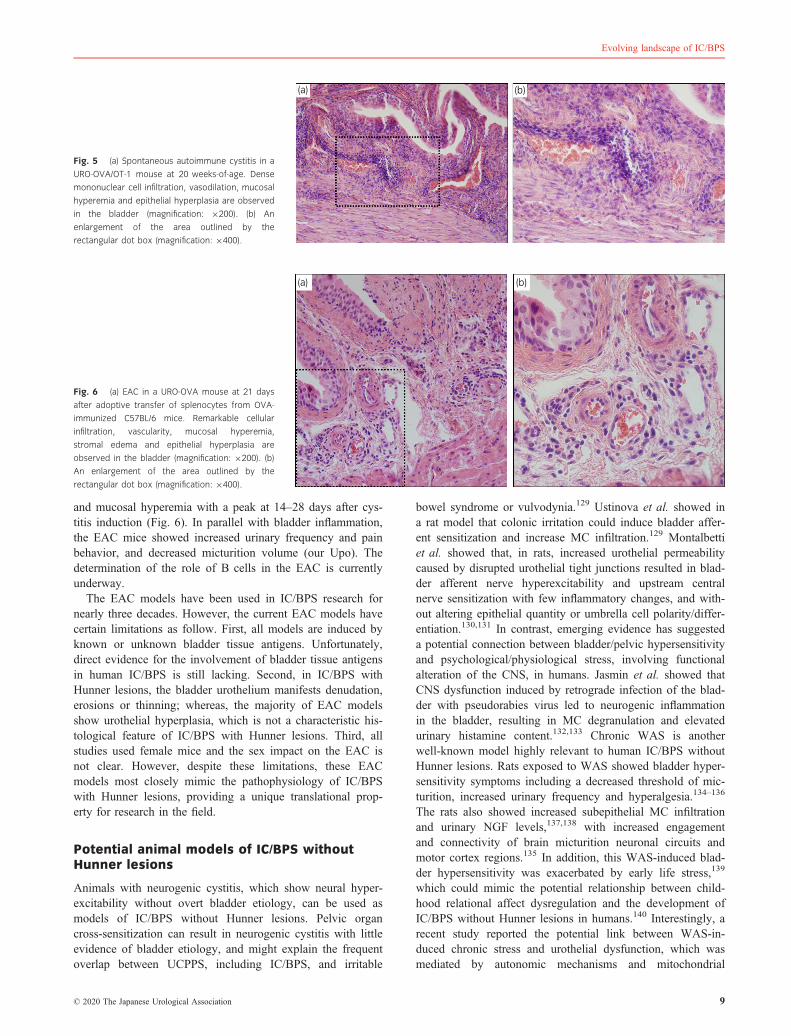
To develop a transgenic EAC model that can more closelymimic human IC/BPS, we generated URO-OVA/OT-I micethrough crossbreeding between URO-OVA and OT-I mice.The F1 generation acquires both urothelial OVA expressionand endogenous OVA-specific CD8+ T cells and, therefore,can spontaneously develop bladder inflammation over timeduring the normal aging process.41,123,127 The EAC initiatesat 4 weeks, and appears mild at 4 weeks, moderate at6 weeks and severe at 8 weeks that sustains up to 20 weekstested (Fig. 5). Like the URO-OVA mice, the URO-OVA/OT-I mice show pelvic/bladder pain and voiding dysfunctionafter EAC development, providing a valuable model for iden-tifying the mechanisms underlying the initiation, progression,and maintenance of chronic cystitis and associated symp-toms.127
In addition to CD8+ T cells, CD4+ T cells are capable ofinducing EAC in the URO-OVA mice. We observed thatadoptive transfer of OT-II CD4+ T cells that express thetransgenic TCR specific for the I-Ab/OVA323–339 epitope pep-tide induced EAC in the URO-OVA mice.128 Our studiesshowed that antigen-specific CD4+ T cells functioned asdirect effector cells and induced EAC independent of CD8+
T cells.In addition to T cells, B cells are also implicated in the
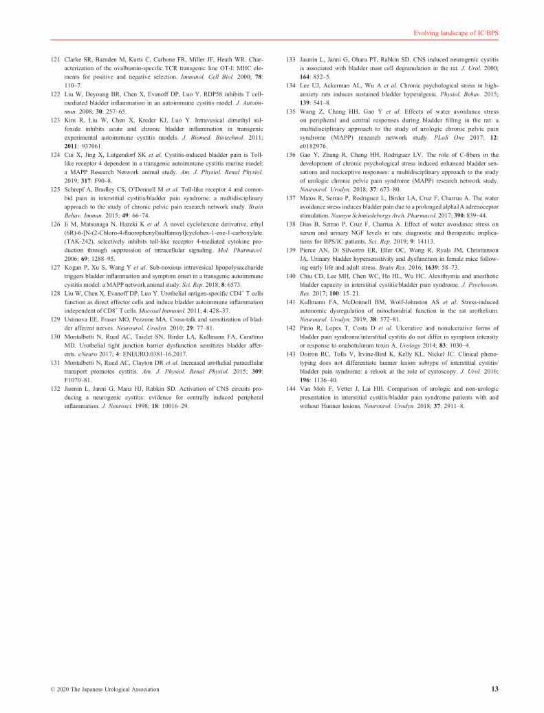
pathophysiology of IC/BPS with Hunner lesions, as describedin this review. To evaluate the role of B cells, we generatedantigen-specific B cells, along with antigen-specific T cells,through immunization of C57BL/6 mice with adjuvant-emul-sified OVA protein. We observed that URO-OVA micedeveloped EAC after adoptive transfer of splenocytes (a mix-ture of T and B cells) from the OVA-immunized mice. Thebladder showed remarkable cellular infiltration, vascularity
8 © 2020 The Japanese Urological Association
Y AKIYAMA ET AL.
and mucosal hyperemia with a peak at 14–28 days after cys-titis induction (Fig. 6). In parallel with bladder inflammation,the EAC mice showed increased urinary frequency and painbehavior, and decreased micturition volume (our Upo). Thedetermination of the role of B cells in the EAC is currentlyunderway.
The EAC models have been used in IC/BPS research fornearly three decades. However, the current EAC models havecertain limitations as follow. First, all models are induced byknown or unknown bladder tissue antigens. Unfortunately,direct evidence for the involvement of bladder tissue antigensin human IC/BPS is still lacking. Second, in IC/BPS withHunner lesions, the bladder urothelium manifests denudation,erosions or thinning; whereas, the majority of EAC modelsshow urothelial hyperplasia, which is not a characteristic his-tological feature of IC/BPS with Hunner lesions. Third, allstudies used female mice and the sex impact on the EAC isnot clear. However, despite these limitations, these EACmodels most closely mimic the pathophysiology of IC/BPSwith Hunner lesions, providing a unique translational prop-erty for research in the field.
Potential animal models of IC/BPS withoutHunner lesions
Animals with neurogenic cystitis, which show neural hyper-excitability without overt bladder etiology, can be used asmodels of IC/BPS without Hunner lesions. Pelvic organcross-sensitization can result in neurogenic cystitis with littleevidence of bladder etiology, and might explain the frequentoverlap between UCPPS, including IC/BPS, and irritable
bowel syndrome or vulvodynia.129 Ustinova et al. showed ina rat model that colonic irritation could induce bladder affer-ent sensitization and increase MC infiltration.129 Montalbettiet al. showed that, in rats, increased urothelial permeabilitycaused by disrupted urothelial tight junctions resulted in blad-der afferent nerve hyperexcitability and upstream centralnerve sensitization with few inflammatory changes, and with-out altering epithelial quantity or umbrella cell polarity/differ-entiation.130,131 In contrast, emerging evidence has suggesteda potential connection between bladder/pelvic hypersensitivityand psychological/physiological stress, involving functionalalteration of the CNS, in humans. Jasmin et al. showed thatCNS dysfunction induced by retrograde infection of the blad-der with pseudorabies virus led to neurogenic inflammationin the bladder, resulting in MC degranulation and elevatedurinary histamine content.132,133 Chronic WAS is anotherwell-known model highly relevant to human IC/BPS withoutHunner lesions. Rats exposed to WAS showed bladder hyper-sensitivity symptoms including a decreased threshold of mic-turition, increased urinary frequency and hyperalgesia.134–136
The rats also showed increased subepithelial MC infiltrationand urinary NGF levels,137,138 with increased engagementand connectivity of brain micturition neuronal circuits andmotor cortex regions.135 In addition, this WAS-induced blad-der hypersensitivity was exacerbated by early life stress,139
which could mimic the potential relationship between child-hood relational affect dysregulation and the development ofIC/BPS without Hunner lesions in humans.140 Interestingly, arecent study reported the potential link between WAS-in-duced chronic stress and urothelial dysfunction, which wasmediated by autonomic mechanisms and mitochondrial
(a) (b)
Fig. 5 (a) Spontaneous autoimmune cystitis in a
URO-OVA/OT-1 mouse at 20 weeks-of-age. Dense
mononuclear cell infiltration, vasodilation, mucosal
hyperemia and epithelial hyperplasia are observed
in the bladder (magnification: 9200). (b) An
enlargement of the area outlined by the
rectangular dot box (magnification: 9400).
(a) (b)
Fig. 6 (a) EAC in a URO-OVA mouse at 21 days
after adoptive transfer of splenocytes from OVA-
immunized C57BL/6 mice. Remarkable cellular
infiltration, vascularity, mucosal hyperemia,
stromal edema and epithelial hyperplasia are
observed in the bladder (magnification: 9200). (b)
An enlargement of the area outlined by the
rectangular dot box (magnification: 9400).
© 2020 The Japanese Urological Association 9
Evolving landscape of IC/BPS

malfunction.137,141 Taken together, these models might facili-tate the development of novel therapies and future researchon the pathogenesis of IC/BPS without Hunner lesions.
Summary and future perspectives
In this article, we emphasized the critical differences betweenIC/BPS with and without Hunner lesions. IC/BPS with Hun-ner lesions is an inflammatory disease of the urinary bladderpotentially associated with enhanced immune responses andinfection, whereas IC/BPS without Hunner lesions is a non-inflammatory disorder with little evidence of bladder etiology.Thus, the IC/BPS umbrella does not represent a series of onedisorder, but comprises a variety of “distinct” disorders.
On the basis of symptomatology, IC/BPS can be catego-rized into “bladder-centric” or “bladder-beyond” phenotypes.The former can be represented by IC/BPS with Hunner lesionsthat presents with more severe bladder/urethral pain, smallerbladder capacity, specific clinical prognosis and few comorbidsystemic conditions. The latter category is represented by IC/BPS without Hunner lesions that frequently presents withsomatic symptoms, and affects dysregulation, larger anatomi-cal bladder capacity and more comorbid systemic conditions.However, clinical phenotyping based on patient demograph-ics, the presence of other comorbid diseases and symptomseverity, as well as characteristics measured by validated scor-ing systems, such as UPOINT and the Interstitial CystitisSymptom Indices, cannot distinguish the presence or absenceof Hunner lesions.142–144 Meanwhile, bladder histology coulddifferentiate these two forms. IC/BPS with Hunner lesions hasa proven bladder histology that manifests epithelial denuda-tion and chronic inflammatory changes characterized by pan-cystitis and frequent expansion of infiltrating B cells, whereasIC/BPS without Hunner lesions shows little histologicalchanges in the bladder. Therefore, we propose that categoriza-tion of IC/BPS should be carried out based on cystoscopic(and histological) examination at initial diagnosis to ensurespecific clinical and investigational methodologies optimizedfor each subtype. For example, local fulguration and steroidinjections, intravesical instillation of DMSO, andcyclosporine A administration should be indicated to patientswith Hunner lesions. In contrast, a neuromodulation therapyand/or a multidisciplinary treatment strategy modeled on themanagement of related somatoform disorders and forms ofaffect dysregulation might be rationale for patients with IC/BPS without Hunner lesions. Taken together, a tailoredapproach that targets the chronic inflammation and epithelialdenudation for IC/BPS with Hunner lesions, or the sensitized/altered nervous system, urothelial malfunction and psychoso-cial problems for IC/BPS without Hunner lesions, might bereasonable, and could lead to better outcomes in clinical man-agement and future research of IC/BPS.
Acknowledgments
The authors are financially supported by the Japan Intractable Dis-eases Research Foundation (to YA), KAKENHI Grants-in-Aidfrom the Japanese Society for the Promotion of Science (JSPS)(grant numbers 19K18552 [to YA], 19K07433 [to DM]), a Health
Labor Sciences Research Grant from the Ministry of Health,Labor and Welfare (grant number 18060798 [to YH]), andNational Institute of Diabetes and Digestive and Kidney Diseases(grant number R01-DK-111396 [to YL]).
Conflict of interest
None declared.
References
1 Hanno PM, Erickson D, Moldwin R, Faraday MM. Diagnosis and treatmentof interstitial cystitis/bladder pain syndrome: AUA guideline amendment. J.Urol. 2015; 193: 1545–53.
2 Homma Y, Ueda T, Tomoe H et al. Clinical guidelines for interstitial cysti-tis and hypersensitive bladder updated in 2015. Int. J. Urol. 2016; 23: 542–9.
3 van de Merwe JP, Nordling J, Bouchelouche P et al. Diagnostic criteria,classification, and nomenclature for painful bladder syndrome/interstitialcystitis: an ESSIC proposal. Eur. Urol. 2008; 53: 60–7.
4 Akiyama Y, Hanno P. Phenotyping of interstitial cystitis/bladder pain syn-drome. Int. J. Urol. 2019; 26(Suppl 1): 17–9.
5 Peeker R, Fall M. Toward a precise definition of interstitial cystitis: further evi-dence of differences in classic and nonulcer disease. J. Urol. 2002; 167: 2470–2.
6 Logadottir Y, Fall M, Kabjorn-Gustafsson C, Peeker R. Clinical characteris-tics differ considerably between phenotypes of bladder pain syndrome/inter-stitial cystitis. Scand. J. Urol. Nephrol. 2012; 46: 365–70.
7 Peters KM, Killinger KA, Mounayer MH, Boura JA. Are ulcerative andnonulcerative interstitial cystitis/painful bladder syndrome 2 distinct dis-eases? A study of coexisting conditions. Urology 2011; 78: 301–8.
8 Whitmore KE, Fall M, Sengiku A, Tomoe H, Logadottir Y, Kim YH. Hun-ner lesion versus non-Hunner lesion interstitial cystitis/bladder pain syn-drome. Int. J. Urol. 2019; 26(Suppl 1): 26–34.
9 Chennamsetty A, Khourdaji I, Goike J, Killinger KA, Girdler B, PetersKM. Electrosurgical management of Hunner ulcers in a referral center’sinterstitial cystitis population. Urology 2015; 85: 74–8.
10 Hillelsohn JH, Rais-Bahrami S, Friedlander JI et al. Fulguration for Hunnerulcers: long-term clinical outcomes. J. Urol. 2012; 188: 2238–41.
11 Akiyama Y, Maeda D, Katoh H et al. Molecular taxonomy of interstitialcystitis/bladder pain syndrome based on whole transcriptome profiling bynext-generation RNA sequencing of bladder mucosal biopsies. J. Urol.2019; 202: 290–300.
12 Nickel JC, Moldwin R, Hanno P et al. Targeting the SHIP1 pathway failsto show treatment benefit in interstitial cystitis/bladder pain syndrome: les-sons learned from evaluating potentially effective therapies in this enigmaticsyndrome. J. Urol. 2019; 202: 301–8.
13 Warren JW, Howard FM, Cross RK et al. Antecedent nonbladder syn-dromes in case-control study of interstitial cystitis/painful bladder syndrome.Urology 2009; 73: 52–7.
14 Warren JW. Bladder pain syndrome/interstitial cystitis as a functionalsomatic syndrome. J. Psychosom. Res. 2014; 77: 510–5.
15 Chen IC, Lee M, Wu SL, Lin HH, Chang KM, Lin H. Somatic symptomsare sensitive in predicting interstitial cystitis/bladder pain syndrome. Int. J.Psychiatry Med. 2017; 52: 48–61.
16 Chen IC, Lee MH, Lin HH, Wu SL, Chang KM, Lin HY. Somatoform dis-order as a predictor of interstitial cystitis/bladder pain syndrome: Evidencefrom a nested case-control study and a retrospective cohort study. Medicine2017; 96: e6304.
17 Logadottir Y, Delbro D, Lindholm C, Fall M, Peeker R. Inflammation char-acteristics in bladder pain syndrome ESSIC type 3C/classic interstitial cysti-tis. Int. J. Urol. 2014; 21(Suppl 1): 75–8.
18 Akiyama Y, Homma Y, Maeda D. Pathology and terminology of interstitialcystitis/bladder pain syndrome: a review.Histol. Histopathol. 2019; 34: 25–32.
19 Maeda D, Akiyama Y, Morikawa T et al. Hunner-type (classic) interstitialcystitis: a distinct inflammatory disorder characterized by pancystitis, withfrequent expansion of clonal B-cells and epithelial denudation. PLoS One2015; 10: e0143316.
20 Wennevik GE, Meijlink JM, Hanno P, Nordling J. The role of glomerula-tions in bladder pain syndrome: a review. J. Urol. 2016; 195: 19–25.
10 © 2020 The Japanese Urological Association
Y AKIYAMA ET AL.

21 Mattila J, Linder E. Immunoglobulin deposits in bladder epithelium and vesselsin interstitial cystitis: possible relationship to circulating anti-intermediate fila-ment autoantibodies.Clin. Immunol. Immunopathol. 1984; 32: 81–9.
22 Akiyama Y, Morikawa T, Maeda D et al. Increased CXCR3 expression of infil-trating plasma cells in Hunner type interstitial cystitis. Sci. Rep. 2016; 6: 28652.
23 Homma Y, Nomiya A, Tagaya M et al. Increased mRNA expression ofgenes involved in pronociceptive inflammatory reactions in bladder tissue ofinterstitial cystitis. J. Urol. 2013; 190: 1925–31.
24 Koziol JA, Clark DC, Gittes RF, Tan EM. The natural history of interstitialcystitis: a survey of 374 patients. J. Urol. 1993; 149: 465–9.
25 Gillespie L. Metabolic appraisal of the effects of dietary modification onhypersensitive bladder symptoms. Br. J. Urol. 1993; 72: 293–7.
26 Friedlander JI, Shorter B, Moldwin RM. Diet and its role in interstitial cys-titis/bladder pain syndrome (IC/BPS) and comorbid conditions. BJU Int.2012; 109: 1584–91.
27 Bassaly R, Downes K, Hart S. Dietary consumption triggers in interstitialcystitis/bladder pain syndrome patients. Female Pelvic Med. Reconstr. Surg.2011; 17: 36–9.
28 Funaro MG, King AN, Stern JNH, Moldwin RM, Bahlani S. Endoscopicinjection of low dose triamcinolone: a simple, minimally invasive, andeffective therapy for interstitial cystitis with Hunner lesions. Urology2018; 118: 25–9.
29 Keay S, Zhang CO, Trifillis AL, Hebel JR, Jacobs SC, Warren JW. Urineautoantibodies in interstitial cystitis. J. Urol. 1997; 157: 1083–7.
30 Neal DE Jr, Dilworth JP, Kaack MB. Tamm-Horsfall autoantibodies ininterstitial cystitis. J. Urol. 1991; 145: 37–9.
31 Jokinen EJ, Alfthan OS, Oravisto KJ. Antitissue antibodies in interstitialcystitis. Clin. Exp. Immunol. 1972; 11: 333–9.
32 Ochs RL, Stein TW Jr, Peebles CL, Gittes RF, Tan EM. Autoantibodies ininterstitial cystitis. J. Urol. 1994; 151: 587–92.
33 Boye E, Morse M, Huttner I, Erlanger BF, MacKinnon KJ, Klassen J. Immunecomplex-mediated interstitial cystitis as a major manifestation of systemiclupus erythematosus. Clin. Immunol. Immunopathol. 1979; 13: 67–76.
34 Haarala M, Alanen A, Hietarinta M, Kiilholma P. Lower urinary tractsymptoms in patients with Sjogren’s syndrome and systemic lupuserythematosus. Int. Urogynecol. J. Pelvic Floor Dysfunct. 2000; 11:84–6.
35 Leppilahti M, Tammela TL, Huhtala H, Kiilholma P, Leppilahti K, Auvinen A.Interstitial cystitis-like urinary symptoms among patients with Sjogren’s syn-drome: a population-based study in Finland. Am. J. Med. 2003; 115: 62–5.
36 Peeker R, Atanasiu L, Logadottir Y. Intercurrent autoimmune conditions in classicand non-ulcer interstitial cystitis. Scand. J. Urol. Nephrol. 2003; 37: 60–3.
37 van de Merwe JP. Interstitial cystitis and systemic autoimmune diseases.Nat. Clin. Pract. Urol. 2007; 4: 484–91.
38 Wen JY, Lo TS, Chuang YC et al. Risks of interstitial cystitis amongpatients with systemic lupus erythematosus: A population-based cohortstudy. Int. J. Urol. 2019; 26: 897–902.
39 Izgi K, Altuntas CZ, Bicer F et al. Uroplakin peptide-specific autoimmunityinitiates interstitial cystitis/painful bladder syndrome in mice. PLoS One2013; 8: e72067.
40 Altuntas CZ, Daneshgari F, Sakalar C et al. Autoimmunity to uroplakin IIcauses cystitis in mice: a novel model of interstitial cystitis. Eur. Urol.2012; 61: 193–200.
41 Liu W, Evanoff DP, Chen X, Luo Y. Urinary bladder epithelium antigeninduces CD8+ T cell tolerance, activation, and autoimmune response. J.Immunol. 2007; 178: 539–46.
42 Keay S, Schwalbe RS, Trifillis AL, Lovchik JC, Jacobs S, Warren JW. Aprospective study of microorganisms in urine and bladder biopsies frominterstitial cystitis patients and controls. Urology 1995; 45: 223–9.
43 Haarala M, Kiilholma P, Lehtonen OP. Urinary bacterial flora of womenwith urethral syndrome and interstitial cystitis. Gynecol. Obstet. Invest.1999; 47: 42–4.
44 Siddiqui H, Lagesen K, Nederbragt AJ, Jeansson SL, Jakobsen KS. Alter-ations of microbiota in urine from women with interstitial cystitis. BMCMicrobiol. 2012; 12: 205.
45 Abernethy MG, Rosenfeld A, White JR, Mueller MG, Lewicky-Gaupp C,Kenton K. Urinary microbiome and cytokine levels in women with intersti-tial cystitis. Obstet. Gynecol. 2017; 129: 500–6.
46 Jhang JF, Hsu YH, Peng CW, Jiang YH, Ho HC, Kuo HC. Epstein–Barrvirus as a potential etiology of persistent bladder inflammation in humaninterstitial cystitis/bladder pain syndrome. J. Urol. 2018; 200: 590–6.
47 Sener AG, Afsar I. Infection and autoimmune disease. Rheumatol. Int.2012; 32: 3331–8.
48 Manfredo Vieira S, Hiltensperger M, Kumar V et al. Translocation of a gutpathobiont drives autoimmunity in mice and humans. Science 2018; 359:1156–61.
49 Minamitani T, Yasui T, Ma Y et al. Evasion of affinity-based selection ingerminal centers by Epstein–Barr virus LMP2A. Proc. Natl. Acad. Sci. USA2015; 112: 11612–7.
50 Jhang JF, Ho HC, Jiang YH, Lee CL, Hsu YH, Kuo HC. Electron micro-scopic characteristics of interstitial cystitis/bladder pain syndrome and theirassociation with clinical condition. PLoS One 2018; 13: e0198816.
51 Jhang JF, Hsu YH, Kuo HC. Possible pathophysiology of ketamine-relatedcystitis and associated treatment strategies. Int. J. Urol. 2015; 22: 816–25.
52 Parsons CL, Bautista SL, Stein PC, Zupkas P. Cyto-injury factors in urine:a possible mechanism for the development of interstitial cystitis. J. Urol.2000; 164: 1381–4.
53 Parsons CL, Shaw T, Berecz Z, Su Y, Zupkas P, Argade S. Role of urinarycations in the aetiology of bladder symptoms and interstitial cystitis. BJUInt. 2014; 114: 286–93.
54 Keay S, Kleinberg M, Zhang CO, Hise MK, Warren JW. Bladder epithelialcells from patients with interstitial cystitis produce an inhibitor of heparin-binding epidermal growth factor-like growth factor production. J. Urol.2000; 164: 2112–8.
55 Keay S, Seillier-Moiseiwitsch F, Zhang CO, Chai TC, Zhang J. Changes inhuman bladder epithelial cell gene expression associated with interstitialcystitis or antiproliferative factor treatment. Physiol. Genomics 2003; 14:107–15.
56 Chuang FC, Kuo HC. Increased urothelial cell apoptosis and chronicinflammation are associated with recurrent urinary tract infection in women.PLoS One 2013; 8: e63760.
57 Marshak-Rothstein A. Toll-like receptors in systemic autoimmune disease.Nat. Rev. Immunol. 2006; 6: 823–35.
58 Li Y, Uccelli A, Laxer KD et al. Local-clonal expansion of infiltrating Tlymphocytes in chronic encephalitis of Rasmussen. J. Immunol. 1997; 158:1428–37.
59 Pereira MI, Medeiros JA. Role of Helicobacter pylori in gastric mucosa-as-sociated lymphoid tissue lymphomas. World J. Gastroenterol. 2014; 20:684–98.
60 Kastrup J, Hald T, Larsen S, Nielsen VG. Histamine content and mast cellcount of detrusor muscle in patients with interstitial cystitis and other typesof chronic cystitis. Br. J. Urol. 1983; 55: 495–500.
61 Aldenborg F, Fall M, Enerback L. Proliferation and transepithelial migra-tion of mucosal mast cells in interstitial cystitis. Immunology 1986; 58:411–6.
62 Lynes WL, Flynn SD, Shortliffe LD et al. Mast cell involvement in intersti-tial cystitis. J. Urol. 1987; 138: 746–52.
63 Christmas TJ, Rode J. Characteristics of mast cells in normal bladder, bacte-rial cystitis and interstitial cystitis. Br. J. Urol. 1991; 68: 473–8.
64 Theoharides TC, Sant GR, El-Mansoury M, Letourneau R, Ucci AA,Meares EM. Jr. Activation of bladder mast cells in interstitial cystitis: alight and electron microscopic study. J. Urol. 1995; 153: 629–36.
65 Peeker R, Enerback L, Fall M, Aldenborg F. Recruitment, distribution andphenotypes of mast cells in interstitial cystitis. J. Urol. 2000; 163: 1009–15.
66 Yamada T, Murayama T, Mita H, Akiyama K. Subtypes of bladder mastcells in interstitial cystitis. Int. J. Urol. 2000; 7: 292–7.
67 Larsen MS, Mortensen S, Nordling J, Horn T. Quantifying mast cells inbladder pain syndrome by immunohistochemical analysis. BJU Int. 2008;102: 204–7.
68 Liu H-T, Jiang Y-H, Kuo H-C. Alteration of urothelial inflammation, apop-tosis, and junction protein in patients with various bladder conditions andstorage bladder symptoms suggest common pathway involved in underlyingpathophysiology. Low. Urin. Tract Symptoms 2015; 7: 102–7.
69 Gamper M, Regauer S, Welter J, Eberhard J, Viereck V. Are mast cells stillgood biomarkers for bladder pain syndrome/interstitial cystitis? J. Urol.2015; 193: 1994–2000.
70 Akiyama Y, Maeda D, Morikawa T et al. Digital quantitative analysis ofmast cell infiltration in interstitial cystitis. Neurourol. Urodyn. 2018; 37:650–7.
71 Geppetti P, Nassini R, Materazzi S, Benemei S. The concept of neurogenicinflammation. BJU Int. 2008; 101(Suppl 3): 2–6.
© 2020 The Japanese Urological Association 11
Evolving landscape of IC/BPS

72 Krumins SA, Broomfield CA. C-terminal substance P fragments elicit his-tamine release from a murine mast cell line. Neuropeptides 1993; 24: 5–10.
73 Frieling T, Cooke HJ, Wood JD. Serotonin receptors on submucous neuronsin guinea pig colon. Am. J. Physiol. 1991; 261: G1017–23.
74 Frieling T, Cooke HJ, Wood JD. Histamine receptors on submucous neu-rons in guinea pig colon. Am. J. Physiol. 1993; 264: G74–80.
75 Leon A, Buriani A, Dal Toso R et al. Mast cells synthesize, store, andrelease nerve growth factor. Proc. Natl. Acad. Sci. USA 1994; 91: 3739–43.
76 van Houwelingen AH, Kool M, de Jager SC et al. Mast cell-derived TNF-alpha primes sensory nerve endings in a pulmonary hypersensitivity reac-tion. J. Immunol. 2002; 168: 5297–302.
77 van der Kleij HP, Ma D, Redegeld FA, Kraneveld AD, Nijkamp FP,Bienenstock J. Functional expression of neurokinin 1 receptors on mastcells induced by IL-4 and stem cell factor. J. Immunol. 2003; 171: 2074–9.
78 De Jonge F, De Laet A, Van Nassauw L et al. In vitro activation of murineDRG neurons by CGRP-mediated mucosal mast cell degranulation. Am. J.Physiol. Gastrointest. Liver Physiol. 2004; 287: G178–91.
79 Ito A, Hagiyama M, Oonuma J. Nerve-mast cell and smooth muscle-mastcell interaction mediated by cell adhesion molecule-1, CADM1. J. SmoothMuscle Res. 2008; 44: 83–93.
80 Steers WD, Tuttle JB. Mechanisms of disease: the role of nerve growth fac-tor in the pathophysiology of bladder disorders. Nat. Clin. Pract. Urol.2006; 3: 101–10.
81 Richter B, Roslind A, Hesse U et al. YKL-40 and mast cells are associatedwith detrusor fibrosis in patients diagnosed with bladder pain syndrome/in-terstitial cystitis according to the 2008 criteria of the European Society forthe Study of Interstitial Cystitis. Histopathology 2010; 57: 371–83.
82 Anand P, Singh B, Jaggi AS, Singh N. Mast cells: an expanding pathophys-iological role from allergy to other disorders. Naunyn Schmiedebergs Arch.Pharmacol. 2012; 385: 657–70.
83 Weston AP, Biddle WL, Bhatia PS, Miner PB Jr. Terminal ileal mucosalmast cells in irritable bowel syndrome. Dig. Dis. Sci. 1993; 38: 1590–5.
84 O’Sullivan M, Clayton N, Breslin NP et al. Increased mast cells in the irri-table bowel syndrome. Neurogastroenterol. Motil. 2000; 12: 449–57.
85 Theoharides TC, Cochrane DE. Critical role of mast cells in inflamma-tory diseases and the effect of acute stress. J. Neuroimmunol. 2004; 146:1–12.
86 Santos J, Guilarte M, Alonso C, Malagelada JR. Pathogenesis of irritablebowel syndrome: the mast cell connection. Scand. J. Gastroenterol. 2005;40: 129–40.
87 Tamaki M, Saito R, Ogawa O, Yoshimura N, Ueda T. Possible mechanismsinducing glomerulations in interstitial cystitis: relationship between endo-scopic findings and expression of angiogenic growth factors. J. Urol. 2004;172: 945–8.
88 Kiuchi H, Tsujimura A, Takao T et al. Increased vascular endothelialgrowth factor expression in patients with bladder pain syndrome/interstitialcystitis: its association with pain severity and glomerulations. BJU Int.2009; 104: 826–31.
89 Lee JD, Lee MH. Increased expression of hypoxia-inducible factor-1alphaand vascular endothelial growth factor associated with glomerulation forma-tion in patients with interstitial cystitis. Urology 2011; 78: 971.e11–5.
90 Kutch JJ, Ichesco E, Hampson JP et al. Brain signature and functionalimpact of centralized pain: a multidisciplinary approach to the study ofchronic pelvic pain (MAPP) network study. Pain 2017; 158: 1979–91.
91 Heizer WD, Southern S, McGovern S. The role of diet in symptoms of irri-table bowel syndrome in adults: a narrative review. J. Am. Diet. Assoc.2009; 109: 1204–14.
92 Haugen M, Kjeldsen-Kragh J, Nordvag BY, Forre O. Diet and diseasesymptoms in rheumatic diseases – results of a questionnaire based survey.Clin. Rheumatol. 1991; 10: 401–7.
93 Arranz LI, Canela MA, Rafecas M. Fibromyalgia and nutrition, what do weknow? Rheumatol. Int. 2010; 30: 1417–27.
94 Alpay K, Ertas M, Orhan EK, Ustay DK, Lieners C, Baykan B. Diet restric-tion in migraine, based on IgG against foods: a clinical double-blind, ran-domised, cross-over trial. Cephalalgia 2010; 30: 829–37.
95 Logan AC, Wong C. Chronic fatigue syndrome: oxidative stress and dietarymodifications. Altern. Med. Rev. 2001; 6: 450–9.
96 Holton KF, Kindler LL, Jones KD. Potential dietary links to central sensiti-zation in fibromyalgia: past reports and future directions. Rheum. Dis. Clin.North Am. 2009; 35: 409–20.
97 Olney JW. Excitotoxins in foods. Neurotoxicology 1994; 15: 535–44.
98 Bourke JH, Langford RM, White PD. The common link between functionalsomatic syndromes may be central sensitisation. J. Psychosom. Res. 2015;78: 228–36.
99 Anderson G, Berk M, Maes M. Biological phenotypes underpin the physio-somatic symptoms of somatization, depression, and chronic fatigue syn-drome. Acta Psychiatr. Scand. 2014; 129: 83–97.
100 Parsons CL, Lilly JD, Stein P. Epithelial dysfunction in nonbacterial cystitis(interstitial cystitis). J. Urol. 1991; 145: 732–5.
101 Klingler CH. Glycosaminoglycans: how much do we know about their rolein the bladder? Urologia 2016; 83(Suppl 1): 11–4.
102 Janssen DA, van Wijk XM, Jansen KC, van Kuppevelt TH, Heesakkers JP,Schalken JA. The distribution and function of chondroitin sulfate and othersulfated glycosaminoglycans in the human bladder and their contribution tothe protective bladder barrier. J. Urol. 2013; 189: 336–42.
103 Liu HT, Shie JH, Chen SH, Wang YS, Kuo HC. Differences in mast cellinfiltration, E-cadherin, and zonula occludens-1 expression between patientswith overactive bladder and interstitial cystitis/bladder pain syndrome. Urol-ogy 2012; 80: 225.e13–18.
104 Stanford E, McMurphy C. There is a low incidence of recurrent bac-teriuria in painful bladder syndrome/interstitial cystitis patients fol-lowed longitudinally. Int. Urogynecol. J. Pelvic Floor Dysfunct. 2007;18: 551–4.
105 Zeng Y, Wu XX, Homma Y et al. Uroplakin III-delta4 messenger RNA asa promising marker to identify nonulcerative interstitial cystitis. J. Urol.2007; 178: 1322–7.
106 Kim A, Han JY, Ryu CM et al. Histopathological characteristics of intersti-tial cystitis/bladder pain syndrome without Hunner lesion. Histopathology2017; 71: 415–24.
107 Bjorling DE, Wang ZY, Bushman W. Models of inflammation of the lowerurinary tract. Neurourol. Urodyn. 2011; 30: 673–82.
108 Birder L, Andersson KE. Animal modelling of interstitial cystitis/bladderpain syndrome. Int. Neurourol. J. 2018; 22: S3–9.
109 Bullock AD, Becich MJ, Klutke CG, Ratliff TL. Experimental autoimmunecystitis: a potential murine model for ulcerative interstitial cystitis. J. Urol.1992; 148: 1951–6.
110 Phull H, Salkini M, Purves T, Funk J, Copeland D, Comiter CV. Angioten-sin II plays a role in acute murine experimental autoimmune cystitis. BJUInt. 2007; 100: 664–7.
111 Lin YH, Liu G, Kavran M et al. Lower urinary tract phenotype of experi-mental autoimmune cystitis in mouse: a potential animal model for intersti-tial cystitis. BJU Int. 2008; 102: 1724–30.
112 Singh UP, Singh NP, Guan H et al. The severity of experimental autoim-mune cystitis can be ameliorated by anti-CXCL10 Ab treatment. PLoS One2013; 8: e79751.
113 Jin XW, Liu BK, Zhang X, Zhao ZH, Shao Y. Establishment of a novelautoimmune experimental model of bladder pain syndrome/interstitial cysti-tis in C57BL/6 mice. Inflammation 2017; 40: 861–70.
114 Liu BK, Jin XW, Lu HZ, Zhang X, Zhao ZH, Shao Y. The effects of neu-rokinin-1 receptor antagonist in an experimental autoimmune cystitis modelresembling bladder pain syndrome/interstitial cystitis. Inflammation 2019;42: 246–54.
115 Luber-Narod J, Austin-Ritchie T, Banner B et al. Experimental autoimmunecystitis in the Lewis rat: a potential animal model for interstitial cystitis.Urol. Res. 1996; 24: 367–73.
116 Mitra S, Dagher A, Kage R, Dagher RK, Luber-Narod J. Experimentalautoimmune cystitis: further characterization and serum autoantibodies.Urol. Res. 1999; 27: 351–6.
117 Zhang L, Ihsan AU, Cao Y et al. An immunogenic peptide, T2 inducesinterstitial cystitis/painful bladder syndrome: an autoimmune mouse modelfor interstitial cystitis/painful bladder syndrome. Inflammation 2017; 40:2033–41.
118 Bicer F, Altuntas CZ, Izgi K et al. Chronic pelvic allodynia is mediated byCCL2 through mast cells in an experimental autoimmune cystitis model.Am. J. Physiol. Renal Physiol. 2015; 308: F103–13.
119 Li H, Zhang Z, Peng J et al. Treatment with low-energy shock wave allevi-ates pain in an animal model of uroplakin 3A-induced autoimmune intersti-tial cystitis/painful bladder syndrome. Investig. Clin. Urol. 2019; 60: 359–66.
120 Sugino Y, Nishikawa N, Yoshimura K et al. BALB/c-Fcgr2bPdcd1 mouseexpressing anti-urothelial antibody is a novel model of autoimmune cystitis.Sci. Rep. 2012; 2: 317.
12 © 2020 The Japanese Urological Association
Y AKIYAMA ET AL.
121 Clarke SR, Barnden M, Kurts C, Carbone FR, Miller JF, Heath WR. Char-acterization of the ovalbumin-specific TCR transgenic line OT-I: MHC ele-ments for positive and negative selection. Immunol. Cell Biol. 2000; 78:110–7.
122 Liu W, Deyoung BR, Chen X, Evanoff DP, Luo Y. RDP58 inhibits T cell-mediated bladder inflammation in an autoimmune cystitis model. J. Autoim-mun. 2008; 30: 257–65.
123 Kim R, Liu W, Chen X, Kreder KJ, Luo Y. Intravesical dimethyl sul-foxide inhibits acute and chronic bladder inflammation in transgenicexperimental autoimmune cystitis models. J. Biomed. Biotechnol. 2011;2011: 937061.
124 Cui X, Jing X, Lutgendorf SK et al. Cystitis-induced bladder pain is Toll-like receptor 4 dependent in a transgenic autoimmune cystitis murine model:a MAPP Research Network animal study. Am. J. Physiol. Renal Physiol.2019; 317: F90–8.
125 Schrepf A, Bradley CS, O’Donnell M et al. Toll-like receptor 4 and comor-bid pain in interstitial cystitis/bladder pain syndrome: a multidisciplinaryapproach to the study of chronic pelvic pain research network study. BrainBehav. Immun. 2015; 49: 66–74.
126 Ii M, Matsunaga N, Hazeki K et al. A novel cyclohexene derivative, ethyl(6R)-6-[N-(2-Chloro-4-fluorophenyl)sulfamoyl]cyclohex-1-ene-1-carboxylate(TAK-242), selectively inhibits toll-like receptor 4-mediated cytokine pro-duction through suppression of intracellular signaling. Mol. Pharmacol.2006; 69: 1288–95.
127 Kogan P, Xu S, Wang Y et al. Sub-noxious intravesical lipopolysaccharidetriggers bladder inflammation and symptom onset in a transgenic autoimmunecystitis model: a MAPP network animal study. Sci. Rep. 2018; 8: 6573.
128 Liu W, Chen X, Evanoff DP, Luo Y. Urothelial antigen-specific CD4+ T cellsfunction as direct effector cells and induce bladder autoimmune inflammationindependent of CD8+ T cells.Mucosal Immunol. 2011; 4: 428–37.
129 Ustinova EE, Fraser MO, Pezzone MA. Cross-talk and sensitization of blad-der afferent nerves. Neurourol. Urodyn. 2010; 29: 77–81.
130 Montalbetti N, Rued AC, Taiclet SN, Birder LA, Kullmann FA, CarattinoMD. Urothelial tight junction barrier dysfunction sensitizes bladder affer-ents. eNeuro 2017; 4: ENEURO.0381-16.2017.
131 Montalbetti N, Rued AC, Clayton DR et al. Increased urothelial paracellulartransport promotes cystitis. Am. J. Physiol. Renal Physiol. 2015; 309:F1070–81.
132 Jasmin L, Janni G, Manz HJ, Rabkin SD. Activation of CNS circuits pro-ducing a neurogenic cystitis: evidence for centrally induced peripheralinflammation. J. Neurosci. 1998; 18: 10016–29.
133 Jasmin L, Janni G, Ohara PT, Rabkin SD. CNS induced neurogenic cystitisis associated with bladder mast cell degranulation in the rat. J. Urol. 2000;164: 852–5.
134 Lee UJ, Ackerman AL, Wu A et al. Chronic psychological stress in high-anxiety rats induces sustained bladder hyperalgesia. Physiol. Behav. 2015;139: 541–8.
135 Wang Z, Chang HH, Gao Y et al. Effects of water avoidance stresson peripheral and central responses during bladder filling in the rat: amultidisciplinary approach to the study of urologic chronic pelvic painsyndrome (MAPP) research network study. PLoS One 2017; 12:e0182976.
136 Gao Y, Zhang R, Chang HH, Rodriguez LV. The role of C-fibers in thedevelopment of chronic psychological stress induced enhanced bladder sen-sations and nociceptive responses: a multidisciplinary approach to the studyof urologic chronic pelvic pain syndrome (MAPP) research network study.Neurourol. Urodyn. 2018; 37: 673–80.
137 Matos R, Serrao P, Rodriguez L, Birder LA, Cruz F, Charrua A. The wateravoidance stress induces bladder pain due to a prolonged alpha1A adrenoceptorstimulation. Naunyn Schmiedebergs Arch. Pharmacol. 2017; 390: 839–44.
138 Dias B, Serrao P, Cruz F, Charrua A. Effect of water avoidance stress onserum and urinary NGF levels in rats: diagnostic and therapeutic implica-tions for BPS/IC patients. Sci. Rep. 2019; 9: 14113.
139 Pierce AN, Di Silvestro ER, Eller OC, Wang R, Ryals JM, ChristiansonJA. Urinary bladder hypersensitivity and dysfunction in female mice follow-ing early life and adult stress. Brain Res. 2016; 1639: 58–73.
140 Chiu CD, Lee MH, Chen WC, Ho HL, Wu HC. Alexithymia and anestheticbladder capacity in interstitial cystitis/bladder pain syndrome. J. Psychosom.Res. 2017; 100: 15–21.
141 Kullmann FA, McDonnell BM, Wolf-Johnston AS et al. Stress-inducedautonomic dysregulation of mitochondrial function in the rat urothelium.Neurourol. Urodyn. 2019; 38: 572–81.
142 Pinto R, Lopes T, Costa D et al. Ulcerative and nonulcerative forms ofbladder pain syndrome/interstitial cystitis do not differ in symptom intensityor response to onabotulinum toxin A. Urology 2014; 83: 1030–4.
143 Doiron RC, Tolls V, Irvine-Bird K, Kelly KL, Nickel JC. Clinical pheno-typing does not differentiate hunner lesion subtype of interstitial cystitis/bladder pain syndrome: a relook at the role of cystoscopy. J. Urol. 2016;196: 1136–40.
144 Van Moh F, Vetter J, Lai HH. Comparison of urologic and non-urologicpresentation in interstitial cystitis/bladder pain syndrome patients with andwithout Hunner lesions. Neurourol. Urodyn. 2018; 37: 2911–8.
© 2020 The Japanese Urological Association 13
Evolving landscape of IC/BPS

![GLOBAL INTERSTITIAL CYSTITIS, BLADDER PAIN SOCIETY · Senior Consultant Urologist, ... New Delhi, India AUTHOR. Newsletter VOLUME 1, ISSUE 1 [APRIL 2019] Dear Colleagues, This year](https://static.fdocuments.us/doc/165x107/5f765bcb0f4e486c9a65d8ef/global-interstitial-cystitis-bladder-pain-society-senior-consultant-urologist.jpg)


![GLOBAL INTERSTITIAL CYSTITIS, BLADDER PAIN ......Newsletter “Registration Started” Visit GLOBAL INTERSTITIAL CYSTITIS, BLADDER PAIN SOCIETY VOLUME 2, ISSUE 1 [FEBRUARY 2020] GIBS](https://static.fdocuments.us/doc/165x107/5f0b648e7e708231d4304bc6/global-interstitial-cystitis-bladder-pain-newsletter-aoeregistration-starteda.jpg)