Interpretation of the velocity measurement ballistocardiogram
7
INTERPRETATION OF THE VELOCITY MEASUREMENT BALLISTOCARDIOGRAM WILLIAM BUCKINGHAM, M.D., GEORGE C. SUTTON, M.D., RAY RONDINELLI, B.S., AND DON C. SUTTON, M.D. CHICAGO, ILL. E ASE of construction and use have encouraged interest in portable velocity measurement ballistocardiographs. The status of such instruments and their records relative to other types of ballistic systems, the high-frequency un- damped machines, low-frequency critically damped machines, and portable displacement recorders, has been summarized recently by Scarborough and associates.’ A critical examination of the effect of change in network filters on frequency response of electromagnetic recorders has been made2 and methods for calibrating electromagnetic pickups have been proposed.3f4 These and other improvements will evolve ultimately a more standardized form of pickup and recording system than is employed now in numerous clinical and laboratory investigations. Variability in criteria for normality of records has been found from laboratory to laboratory. The absence of both standardized recording apparatus and of precise criteria for the normal condition has not delayed, how- ever, the widespread clinical use of these machines. There exists a need for knowl- edge concerning the diagnostic accuracy of the ballistocardiogram when analyzed by criteria now in use, and when recorded by a popular type coil, network, and filter system. Such information for the Starr table has recently been published.6 The machine chosen was of the type described by Dock and Taubman,” Mandel- baum and Mandelbaum’ and others, with the impression that the choice of this machine would facilitate comparison of results with those of other workers. The criteria for analysis were chosen because of widespread use and have been pub- lished previously by many authors,5,s using numerous types of machines. The establishment of more certain normal means and limits for the physical charac- teristics of such ballistocardiograms was first sought. A sizable group of healthy adults was studied with this as the central objective. A corollary of this analysis was an evaluation of the reliability of these criteria of the ballistocardiogram in indicating a clinically normal cardiovascular system. The work was then ex- tended to include the effect of primarily noncardiovascular diseaseon the ballis- tocardiogram and, finally, the effect of serious cardiovascular disease itself. From the Department of Medicine and Pmhle Laboratory of Nort,hwestern University Medical School, Cook County Hospital. Chicago, Ill. Aided by a grant from Mrs. Alfred Eustice. Received for publication May 19, 1953. 341
-
Upload
william-buckingham -
Category
Documents
-
view
214 -
download
0
Transcript of Interpretation of the velocity measurement ballistocardiogram

INTERPRETATION OF THE VELOCITY MEASUREMENT BALLISTOCARDIOGRAM
WILLIAM BUCKINGHAM, M.D., GEORGE C. SUTTON, M.D., RAY RONDINELLI, B.S., AND DON C. SUTTON, M.D.
CHICAGO, ILL.
E ASE of construction and use have encouraged interest in portable velocity measurement ballistocardiographs. The status of such instruments and
their records relative to other types of ballistic systems, the high-frequency un- damped machines, low-frequency critically damped machines, and portable displacement recorders, has been summarized recently by Scarborough and associates.’ A critical examination of the effect of change in network filters on frequency response of electromagnetic recorders has been made2 and methods for calibrating electromagnetic pickups have been proposed.3f4 These and other improvements will evolve ultimately a more standardized form of pickup and recording system than is employed now in numerous clinical and laboratory investigations. Variability in criteria for normality of records has been found from laboratory to laboratory. The absence of both standardized recording apparatus and of precise criteria for the normal condition has not delayed, how- ever, the widespread clinical use of these machines. There exists a need for knowl- edge concerning the diagnostic accuracy of the ballistocardiogram when analyzed by criteria now in use, and when recorded by a popular type coil, network, and filter system. Such information for the Starr table has recently been published.6 The machine chosen was of the type described by Dock and Taubman,” Mandel- baum and Mandelbaum’ and others, with the impression that the choice of this machine would facilitate comparison of results with those of other workers. The criteria for analysis were chosen because of widespread use and have been pub- lished previously by many authors,5,s using numerous types of machines. The establishment of more certain normal means and limits for the physical charac- teristics of such ballistocardiograms was first sought. A sizable group of healthy adults was studied with this as the central objective. A corollary of this analysis was an evaluation of the reliability of these criteria of the ballistocardiogram in indicating a clinically normal cardiovascular system. The work was then ex- tended to include the effect of primarily noncardiovascular disease on the ballis- tocardiogram and, finally, the effect of serious cardiovascular disease itself.
From the Department of Medicine and Pmhle Laboratory of Nort,hwestern University Medical School, Cook County Hospital. Chicago, Ill.
Aided by a grant from Mrs. Alfred Eustice. Received for publication May 19, 1953.
341
342 .\MERICAN HEART JOURNAL
MATERIALS .\ND METHODS
All ballistocardiograms taken during this study were made by a portable electromagnetic ballistocardiographic machine of a construction identical to that of Mandelbaum and Mandelbaum. 7 The recordings were made on a converted General Electric photographic electrocardiograph. All individuals rested on a rigid table for approximately ten minutes prior to registration of the ballisto- cardiogram and were from 1 to 3 hours postprandial.
A group of 327 normal individuals, men and women, were examined. Their ages ranged from 12 through 65 years (mean 32), with the majority under 30 years of age. This group consisted predominantly of high school and medical school students and professional hospital personnel, all with repeated physical examinations in the past and accurate medical histories. Inclusion in this group was based on a negative history and physical examination, chest roentgenogram, urinalysis in past and present, and the absence of any minor illness at the time of recording the ballistocardiogram.
A second group of seventy-three patients who were hospitalized with rela- tively severe, diverse, noncardiac diseases were studied, generally at a time when they were convalescing from the severe phases of their illnesses. The ages ranged from 14 to 100 years, averaged 35 years.
The third group studied consisted of 121 cardiac patients who were hos- pitalized for obvious heart disease and its complications. All in this group had cardiac abnormalities sufficiently severe to warrant hospitalization. The exist- ence of heart disease was further corroborated by means of history, physical examination, teleroentgenogram, electrocardiograms, and cardiac fluoroscopy. Pathology varied and consisted of the following types: congenital, rheumatic (mitral, mitral and aortic, aortic, and mitral, aortic and tricuspid valvular lesions combined), luetic (both aneurysm and aortic regurgitation, together and alone), hypertensive, arteriosclerotic, and car pulmonale lesions, combined with several miscellaneous causes as myxedema and idiopathic cardiac hypertrophy.
These patients were studied at various times during their course of hos- pitalization, but, in general, an attempt was made to record the ballistocardio- gram at the time cardiac failure had improved as much as could be expected. A total of 136 ballistocardiograms were taken on these 121 patients. No selection or exclusion of patients was made on the basis of factors now thought to influence the ballistocardiograms (such as bundle branch block). The group was one containing individuals with severe heart disease of many types.
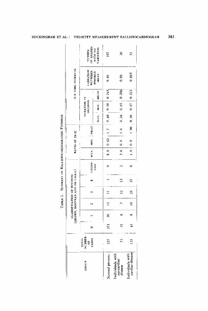
All tracings were subjected to the following analysis: (1) classification on the basis of the criteria of Brown, Hoffman and de Lalla;sa (2) measurement in millimeters of the depth of K and the height of J with subsequent calculation of JK:IJ ratio; (3) measurement to the nearest 0.01 second of the time interval from the apex of H to the nadir of the K wave in all cycles; (4) detection of the maximum variation between H:K interval in all cycles.
BUCKINGHAM ET AL. : VELOCITY MEASUREMENT BALLISTOCARDIOGRAM 343
i- B E
E; P- 2 ,m c 6 d -
I
0 cs 24 ;s: c’ d d
h \o 2? 3 3
i 3
G a a d d 0
m
N

344 AMERICAN HEART JOURNAL
RESULTS
Normal Group.-Analysis of the tracings of this group on the basis of the criteria of variation in magnitude and form of the tracing (Brown, Hoffman and de Lalla)8a yielded the following results: 271 records, Grade 0; twenty records, Grade 1 (slight variation); thirteen records, Grade 2 (moderate variation) ; eleven records classified as Grade 3 (marked variation); and three records were Grade 4 because the variation was so marked that systolic complexes could not be distinguished from diastolic components. In nine records artifacts, tremor, body motion, or amplitude variation so extreme that the tracing was not full\ recorded, prevented classification.
The relationship of K depth to J height expressed as a ratio had a mean value for the entire group of 1.7. The maximum ratio was 8.0 and the minimum was 0.6. It was noted that 94 per cent of the group had ratios between 1.1 and 2.3, or within 0.6 of the mean value of 1.7.
The mean of the duration of time from the nadir of H to the nadir of K was 0.245 second, and the range of the group was from 0.1 to 0.40 second. In any given individual tracing the variation in this H-K interval from cycle to cycle was too small to be measured in 107 tracings. The remaining records in which there were a sufficient number of identifiable ballistic cycles for comparison (121) revealed an average variation of 0.01 second and ranged from 0.0 to 0.07 second. Ninety-nine tracings possessed a sufficient number of bizarre cycles in the re- corded strip of film so that a comparison between H-K times was meaningless.
Individuals Convalescing From Serious Noncardiac Disease.-Applying the same four methods of analysis to this group of seventy-three individuals showed thirty-one records classified Grade 0; eight, Grade 1; seven, Grade 2; twelve, Grade 3; and thirteen, Grade 4. The mean of the JK:IJ ratio was 1.6 and the range of variation was from 0.9 to 2.6. The average H-K time interval was 0.206 second and ranged from 0.26 to 0.15 second. The variation in H-K time from cycle to cycle in the same graph was insignificant in twenty-six cases, and aver- aged 0.01 second in the remainder, with a maximum of 0.09 second.
Individuals With Clinically Obvious Heart Disease.--,4nalysis of this group of 121 patients revealed forty-one records classified Grade 0; eight, Grade 1; ten, Grade 2; twenty-five, Grade 3; and thirty-seven, Grade 4. The mean JK:I J ratio was 1.96 and the ratio varied from 0.9 to 4.0. The H-K time interval averaged 0.22 second, with a range of 0.17 to 0.36. The H-K time could not be measured in seventy-two graphs because of the large number of bizarre com- plexes. Variation of the H-K time in individual graphs was insignificant in thirty-one cases and, in the remainder, averaged 0.15 second with a maximum variation of 0.6 second.
A further breakdown of the group of cardiac patients was made according to the etiologic classification, as determined by available means, of the under- lying heart disease. In the study of the records by the classification of Brown and associatesEa the etiologic groups of car pulmonale, congenital, and miscel- laneous types contained too small a number of patients to be accurate. In the rheumatic, luetic, hypertensive, and arteriosclerotic groups, normal as well as all

BUCKINGHAM ET AL. : VELOCITY MEASUREMENT BALLISTOCARDIOGRAM 345
four grades of variation were present with no significant distribution. The JK:IJ ratio in the luetic group and in the hypertensive group was 2.6 and 2.2, respectively. These are substantially greater than that for the group of normal patients and exceed the ratio in the other cardiac groups. Two of the luetic patients had a JK:I J ratio of 1.6, while nine hypertensive patients had similar ratios. Although examples of early and late M complexes were seen in all three groups, the precise critetia.for determining these forms were not available and thus no statistical study could be made.
DISCUSSION
Ballistocardiography offers a potentially new method of gathering infor- mation about the cardiovascular status of individuals. The value of the informa- tion gained rests upon knowledge of normal distribution and physiologic varia- t ions. Five constituents of the ballistocardiograph were considered in the interpretation of the record obtained by a portable nonstandardized machine. These were: (1) forms of individual complexes, (2) degree of variation in regu- larity and definitiveness, (3) ratio of JK to IJ, (4) duration of H-K time, and (5) the variation in the H-K interval in a series of cycles.
The appearance of several different complex forms, such as early M and late M, has been described.‘eQ Easily recognizable examples of these waves were seen in the ptesent series in all three groups. Attempts to determine the criteria or ratios necessary for designation of such waves wete unsuccessful. The number of classical examples of each was too small in this series of over 500, and the num- ber of borderline cases too great for criteria to be drawn.
Variation in I J amplitude with respiration and of complex regularity and definitiveness has been used in classifying graphs. Insofar as the present series of 327 normal persons of diverse ages (mean 32 years) is a representative sample, it appears that an appreciable (14 per cent) percentage of such individuals will have a definite variation in IJ height and complex regularity, that is, will have graphs classified as 1, 2, 3, or 4. This is in keeping with the incidence of 25.2 per cent obtained by Scarborough and associates5 in a study of normal persons with an older mean age of 44. The higher incidence of abnormal wave form in older age groups is well substantiated and is probably responsible for this higher figure. There was an inverse relation between the degree of variation and the number of people having this variation; only three persons with normal cardio- vascular systems by the criteria used had 100 per cent bizarre complexes. The incidence of definite variation in the group of individuals with noncardiac disease increased to 54 per cent. This emphasizes the role of extracardiac factors in producing ballistocardiographic abnormality.
While 66 per cent (80 out of 121) of the individuals with frank cardiac disease had graphs with some degree of variation (1, 2, 3, or 4), only 31 per cent of the individuals had 100 per cent variation or completely bizarre, Grade 4, graphs. Furthermore, 55 per cent of the individuals who were convalescing from non- cardiac disease also had ballistocardiograms graded 1, 2, 3, or 4, and 18 per cent of this same group were Grade 4. These results emphasize that abnormality of the ballistocardiogram was not necessarily associated with cardiac disease. Since
346 AMERICAN HEART JOURNAL
34 per cent of the persons with frankly diseased hearts had normal records, the determination of the presence or absence of heart disease on the basis of the ballistocardiogram was difficult.
The JK:I J ratio of normal persons as recorded on a Starr or Nickerson table is about 1.0.6 Smith and Bryan,” using a low-frequency velocity measurement ballistocardiograph, obtained a ratio of approximately 1.3 in a study of 100 nor- mal people between 20 and 30 years of age. Barrera and associate9 have pre- viously called attention to this observation that records obtained with the electro- magnetic machines have a greater JK:IJ ratio than those from the Starr or Nickerson table. Results of the present study of a larger group of individuals and of a greater age range confirm this; the mean JK:IJ ratio of normal persons was 1.7, and there were numerous examples of ratios of 2.0 and 3.0 with an extreme of 8.0. These figures are substantially greater than the limits of 0.85 to 1.40 set by Scarborough for the Starr-type machine. The presence of noncardiac or diverse types of cardiac disease produced no change in the average JK:IJ ratio or in the extremes observed. The one exception to this was seen within the group of cardiac patients. The persons with hypertensive and luetic heart disease had higher average values for the ratio. This observation is in keeping with previous reports on such patients .l,13 Certain individual cases of these two types, however, had a normal JK:IJ ratio. The hypertensive and luetic group thus overlapped all other groups, giving a high JK:I J ratio limited in diagnostic value. This lack of specificity of a high JK:IJ ratio has previously been noted.’ It has been produced by certain drugs14 and is accentuated by use of the electromagnetic machines.
The present results confirm the observation that the H-K time of an indi- vidual is constant to about 0.01 second,iO and that there is a relatively great (fourfold) variation between different persons. The presence of either cardiac or noncardiac disease did not affect the mean H-K time, its extremes, or its varia- tion in the individual.
SUMMARY
1. A portable electromagnetic ballistocardiograph was used to study three groups of individuals : normal persons, patients confined to a hospital and convalescing from noncardiac disease, and patients with obvious cardiac disease.
2. The records were analyzed by four methods: (a) presence of so-called early M or late M pattern, (b) JK:IJ ratio, (c) H-K time interval and its varia- tion, (d) Brown, Hoffman, and de Lalla criteria.
3. Precise determination of the limits of the early and late M patterns could not be made.
4. Observations that the JK:IJ ratio is materially increased by use of the electromagnetic machine are confirmed.
5. The JK:IJ ratio and the H-K interval and its variation failed to dis- tinguish the three groups. Within the cardiac group those with hypertensive and luetic heart disease had a higher JK:IJ ratio, but some of these individuals had normal ratios.

UCKINGHAM ET .4L. : VELOCITY MEASUREMENT BAI,I~ISTOCAKDIOGK,~M 347
6. Records with some degree of variation (1, 2, 3, or 4) were found in 66 per cent of the individuals with frank cardiac disease, in 55 per cent of the pa- tients convalescing from noncardiac disease, and in 14 per cent of the normal persons. Grade 4 ballistocardiograms were found in 31 per cent, 18 per cent, and 0.9 per cent of these three groups, respectively. These results emphasize that abnormality of the ballistocardiogram was not necessarily associated with cardiac disease.
7. It is emphasized that 34 per cent of the individuals with frank heart disease had normal ballistocardiograms.
REFERENCES
1. Scarborough, W. R., Davis, F. W., Jr., Baker, B. M., Jr., Mason, R. E., and Singewald, M. L.: A Review of Ballistocardiography, AM. HEART J. 44:910, 1952.
2. Smith, J. E., and Bryan, S.: Studies of Frequency Response in Ballistocardiography, AM. HEART J. 45:40, 1953.
3. Smith, J. E.: A Calibrated Bar-Magnet Velocity Meter fo; Use in Ballistocardiography, AM. HEART J. 44:872, 1952.
1. Arbeit, S. R., and Lindner, N.: A New Full-Frequency Range Calibrated Ballistocardio- graph I, AM. HEART J. 45:52, 1953.
5. Scarborough, W. R., Davis, F. W., Jr., Baker, B. M., Jr.! Mason, R. E., Singewald, M. L., Lore, S. A., and Fox, L. M.: A Ballistocardiographlc Study of 369 Apparently Normal Persons, AM. HEART J. 45:161, 1953.
6. Dock, W., and Taubman, F.: Some Techniques for Recording the Ballistocardiogram Directly From the Body, Am. J. Med. 7:751, 1949.
7. Mandelbaum, H., and Mandelbaum, R. A.: Studies Utilizing the Portable Electromagnetic Ballistocardiograph, Circulation 3:663, 1951.
8a. Brown, H. R., Hoffman, M. J., and de Lalla, V.., Jr.: Ballistocardiographic Findings in Patients With Symptoms of Angina Pectorls, CircuIation 1:132, 1950.
b. Mandelbaum, H., and Mandelbaum, R. A.: Studies Utilizing the Portable Electromagnetic Ballistocardiograph II, Circulation 5:885, 19.52.
9. Starr, I., and Schroeder, H. A.: Ballistocardiogram II. Normal Standards, Abnormalities Commonly Found in Diseases of the Heart and Circulation, and Their Significance, J. Clin. Investigation 19:437, 1940.
10. Brown, H. R., de Lalla, V., Jr., Epstein, M. A., and Hoffman, M. J.:, Clinical Ballisto- cardiography, New York, 1952, The Macmillan Company.
11. Smith, J. EI and-Bryan, S.: The Low Frequency Velocity keasurement Ballistocardio- graph, Circulation 5:892, 1952.
12. Barrera, F., Leiro, A. R., Pons, R., and Kaufman, J.: El Balli stocardiograma Tornado Directamente de1 Cuerpo por el Mbtodo ElectromagCntico de Dock, Rev. Cubana Caidiol. 12:1, 1951.
13. Nickerson, J. L.: Some Observations on the Ballistocardiographic Pattern, With Special Reference to the H and K Waves, J. Clin. Investigation 28:369, 1949.
14. Buckingham, W. B., Wendel, G., Sutton, .G. C., and Sutton, D. C.: Studies in the Nature ;b;p Deep K Stroke Ballistocardlogram, Proc. Central Sot. Chn. Research 24:20,


![Ballistocardiogram Amplitude Modulation Induced by ...cinc.org/archives/2015/pdf/0313.pdfdiovascular reactivity [7–9]. Indeed, stepwise controlled breathings allow to separate precisely](https://static.fdocuments.us/doc/165x107/6040bd856524305e71135b84/ballistocardiogram-amplitude-modulation-induced-by-cincorgarchives2015pdf0313pdf.jpg)