INTERNATIONAL ISO STANDARD 7218 - smartjd.org · 9.1 Hygienic precautions during analysis ......
74
Reference number ISO 7218:2007(E) © ISO 2007 INTERNATIONAL STANDARD ISO 7218 Third edition 2007-08-15 Microbiology of food and animal feeding stuffs — General requirements and guidance for microbiological examinations Microbiologie des aliments — Exigences générales et recommendations –§fi•„‚‹ ²‹»fi²¿‹•–²¿· fi„¿²•ƒ¿‹•–² ”–fi ˝‹¿²…¿fi…•ƒ¿‹•–² —fi–“•…»… § ˝ «²…»fi ·•‰»²›» '•‹‚ ˝ –‹ ”–fi ˛»›¿·» – fi»fi–…«‰‹•–² –fi ²»‹'–fi•²„ »fi‡•‹‹»… '•‹‚–«‹ ·•‰»²›» ”fi–‡ ˝
Transcript of INTERNATIONAL ISO STANDARD 7218 - smartjd.org · 9.1 Hygienic precautions during analysis ......

Reference numberISO 7218:2007(E)
© ISO 2007
INTERNATIONAL STANDARD
ISO7218
Third edition2007-08-15
Microbiology of food and animal feeding stuffs — General requirements and guidance for microbiological examinations
Microbiologie des aliments — Exigences générales et recommendations
ݱ°§®·¹¸¬ ײ¬»®²¿¬·±²¿´ Ñ®¹¿²·¦¿¬·±² º±® ͬ¿²¼¿®¼·¦¿¬·±² Ю±ª·¼»¼ ¾§ ×ØÍ «²¼»® ´·½»²» ©·¬¸ ×ÍÑ
Ò±¬ º±® λ¿´»Ò± ®»°®±¼«½¬·±² ±® ²»¬©±®µ·²¹ °»®³·¬¬»¼ ©·¬¸±«¬ ´·½»²» º®±³ ×ØÍ
óóÀôôÀÀÀôôôôÀÀÀÀóÀóÀôôÀôôÀôÀôôÀóóó
ISO 7218:2007(E)
PDF disclaimer This PDF file may contain embedded typefaces. In accordance with Adobe's licensing policy, this file may be printed or viewed but shall not be edited unless the typefaces which are embedded are licensed to and installed on the computer performing the editing. In downloading this file, parties accept therein the responsibility of not infringing Adobe's licensing policy. The ISO Central Secretariat accepts no liability in this area.
Adobe is a trademark of Adobe Systems Incorporated.
Details of the software products used to create this PDF file can be found in the General Info relative to the file; the PDF-creation parameters were optimized for printing. Every care has been taken to ensure that the file is suitable for use by ISO member bodies. In the unlikely event that a problem relating to it is found, please inform the Central Secretariat at the address given below.
COPYRIGHT PROTECTED DOCUMENT
© ISO 2007 All rights reserved. Unless otherwise specified, no part of this publication may be reproduced or utilized in any form or by any means, electronic or mechanical, including photocopying and microfilm, without permission in writing from either ISO at the address below or ISO's member body in the country of the requester.
ISO copyright office Case postale 56 CH-1211 Geneva 20 Tel. + 41 22 749 01 11 Fax + 41 22 749 09 47 E-mail [email protected] Web www.iso.org
Published in Switzerland
ii © ISO 2007 – All rights reservedݱ°§®·¹¸¬ ײ¬»®²¿¬·±²¿´ Ñ®¹¿²·¦¿¬·±² º±® ͬ¿²¼¿®¼·¦¿¬·±² Ю±ª·¼»¼ ¾§ ×ØÍ «²¼»® ´·½»²» ©·¬¸ ×ÍÑ
Ò±¬ º±® λ¿´»Ò± ®»°®±¼«½¬·±² ±® ²»¬©±®µ·²¹ °»®³·¬¬»¼ ©·¬¸±«¬ ´·½»²» º®±³ ×ØÍ
óóÀôôÀÀÀôôôôÀÀÀÀóÀóÀôôÀôôÀôÀôôÀóóó
ISO 7218:2007(E)
© ISO 2007 – All rights reserved iii
Contents Page
Foreword............................................................................................................................................................. v
Introduction ....................................................................................................................................................... vi
1 Scope ..................................................................................................................................................... 1
2 Normative references ........................................................................................................................... 1
3 Premises ................................................................................................................................................ 23.1 General................................................................................................................................................... 23.2 Safety considerations........................................................................................................................... 23.3 Laboratory design................................................................................................................................. 23.4 Laboratory areas................................................................................................................................... 33.5 Layout and fittings of the premises .................................................................................................... 43.6 Cleaning and disinfection .................................................................................................................... 5
4 Staff ........................................................................................................................................................ 54.1 General................................................................................................................................................... 54.2 Competence .......................................................................................................................................... 64.3 Verification of on-going staff competence......................................................................................... 64.4 Hygiene .................................................................................................................................................. 6
5 Apparatus and equipment ................................................................................................................... 6
6 Preparation of glassware and other laboratory materials .............................................................. 286.1 Preparation .......................................................................................................................................... 286.2 Sterilization/decontamination............................................................................................................ 286.3 Disposable equipment and materials ............................................................................................... 286.4 Storage of clean glassware and materials ....................................................................................... 286.5 Management of sterile glassware and materials ............................................................................. 296.6 Use of decontamination and disinfection ........................................................................................ 296.7 Waste management ............................................................................................................................ 296.8 Washing ............................................................................................................................................... 30
7 Preparation and sterilization of culture media ................................................................................ 30
8 Laboratory samples............................................................................................................................ 308.1 Sampling.............................................................................................................................................. 308.2 Transport ............................................................................................................................................. 318.3 Receipt ................................................................................................................................................. 318.4 Storage................................................................................................................................................. 328.5 Test portion ......................................................................................................................................... 32
9 Examination......................................................................................................................................... 329.1 Hygienic precautions during analysis .............................................................................................. 329.2 Preparation of initial suspension and dilutions .............................................................................. 34
10 Enumeration ........................................................................................................................................ 3410.1 General................................................................................................................................................. 3410.2 Enumeration using a solid medium.................................................................................................. 3510.3 Calculation and expression of results obtained with solid media ................................................ 3710.4 Enumeration of yeasts and moulds .................................................................................................. 4310.5 Enumeration using a liquid medium................................................................................................. 44
11 Detection method (qualitative method) ............................................................................................ 4911.1 General................................................................................................................................................. 4911.2 Principle............................................................................................................................................... 5011.3 Measurement of uncertainty.............................................................................................................. 50
ݱ°§®·¹¸¬ ײ¬»®²¿¬·±²¿´ Ñ®¹¿²·¦¿¬·±² º±® ͬ¿²¼¿®¼·¦¿¬·±² Ю±ª·¼»¼ ¾§ ×ØÍ «²¼»® ´·½»²» ©·¬¸ ×ÍÑ
Ò±¬ º±® λ¿´»Ò± ®»°®±¼«½¬·±² ±® ²»¬©±®µ·²¹ °»®³·¬¬»¼ ©·¬¸±«¬ ´·½»²» º®±³ ×ØÍ
óóÀôôÀÀÀôôôôÀÀÀÀóÀóÀôôÀôôÀôÀôôÀóóó
ISO 7218:2007(E)
iv © ISO 2007 – All rights reserved
12 Confirmatory methods........................................................................................................................ 5012.1 General ................................................................................................................................................. 5012.2 Preparation of a pure culture............................................................................................................. 5012.3 Gram’s stain (modified Hucker technique)....................................................................................... 5012.4 Use of biochemical galleries for identification ................................................................................ 5212.5 Use of nucleic probes for identification ........................................................................................... 5212.6 Serological methods........................................................................................................................... 53
13 Test report............................................................................................................................................ 53
14 Validation of microbiological methods............................................................................................. 5414.1 Validation of reference methods ....................................................................................................... 5414.2 Validation of alternative methods...................................................................................................... 5414.3 Validation of in-house methods......................................................................................................... 54
15 Quality assurance of results/quality control of performance......................................................... 5415.1 Internal quality control ....................................................................................................................... 5415.2 Reference strains ................................................................................................................................ 5415.3 External quality assessment (proficiency testing) .......................................................................... 55
Annex A (informative) Properties of some disinfectants.............................................................................. 56
Annex B (normative) Determination of most probable number (MPN) ....................................................... 57
Bibliography ..................................................................................................................................................... 64
ݱ°§®·¹¸¬ ײ¬»®²¿¬·±²¿´ Ñ®¹¿²·¦¿¬·±² º±® ͬ¿²¼¿®¼·¦¿¬·±² Ю±ª·¼»¼ ¾§ ×ØÍ «²¼»® ´·½»²» ©·¬¸ ×ÍÑ
Ò±¬ º±® λ¿´»Ò± ®»°®±¼«½¬·±² ±® ²»¬©±®µ·²¹ °»®³·¬¬»¼ ©·¬¸±«¬ ´·½»²» º®±³ ×ØÍ
óóÀôôÀÀÀôôôôÀÀÀÀóÀóÀôôÀôôÀôÀôôÀóóó
ISO 7218:2007(E)
© ISO 2007 – All rights reserved v
Foreword
ISO (the International Organization for Standardization) is a worldwide federation of national standards bodies (ISO member bodies). The work of preparing International Standards is normally carried out through ISO technical committees. Each member body interested in a subject for which a technical committee has been established has the right to be represented on that committee. International organizations, governmental and non-governmental, in liaison with ISO, also take part in the work. ISO collaborates closely with the International Electrotechnical Commission (IEC) on all matters of electrotechnical standardization.
International Standards are drafted in accordance with the rules given in the ISO/IEC Directives, Part 2.
The main task of technical committees is to prepare International Standards. Draft International Standards adopted by the technical committees are circulated to the member bodies for voting. Publication as an International Standard requires approval by at least 75 % of the member bodies casting a vote.
Attention is drawn to the possibility that some of the elements of this document may be the subject of patent rights. ISO shall not be held responsible for identifying any or all such patent rights.
ISO 7218 was prepared by Technical Committee ISO/TC 34, Food products, Subcommittee SC 9, Microbiology, in collaboration with CEN Technical Committee CEN/TC 275, Food analysis — Horizontal methods.
This third edition cancels and replaces the second edition (ISO 7218:1996), which has been technically revised. It also incorporates the Amendment ISO 7218:1996/Amd.1:2001.
ݱ°§®·¹¸¬ ײ¬»®²¿¬·±²¿´ Ñ®¹¿²·¦¿¬·±² º±® ͬ¿²¼¿®¼·¦¿¬·±² Ю±ª·¼»¼ ¾§ ×ØÍ «²¼»® ´·½»²» ©·¬¸ ×ÍÑ
Ò±¬ º±® λ¿´»Ò± ®»°®±¼«½¬·±² ±® ²»¬©±®µ·²¹ °»®³·¬¬»¼ ©·¬¸±«¬ ´·½»²» º®±³ ×ØÍ
óóÀôôÀÀÀôôôôÀÀÀÀóÀóÀôôÀôôÀôÀôôÀóóó
ISO 7218:2007(E)
vi © ISO 2007 – All rights reserved
Introduction
When conducting microbiological examinations, it is especially important that
only those microorganisms which are present in the samples are isolated and enumerated;
the microorganisms do not contaminate the environment.
In order to achieve this, it is necessary to pay attention to personal hygiene and to use working techniques which ensure, as far as possible, exclusion of extraneous contamination.
Since, in this International Standard, it is possible to give only a few examples of the precautions to be taken during microbiological examinations, a thorough knowledge of the microbiological techniques and of the microorganisms involved is essential. It is important that the examinations are conducted as accurately as possible, including monitoring and recording aspects that may affect results and calculation of the number of microorganisms and the uncertainty of the results.
Ultimately, it is the responsibility of the head of the laboratory to judge whether the manipulations are safe and can be considered to be good laboratory practice.
A large number of manipulations can, for example, unintentionally lead to cross-contamination, and the analyst should always verify the accuracy of the results given by his or her technique.
In order to conduct the examinations correctly, it is necessary to take certain precautions when constructing and equipping the laboratory.
Certain precautions must be taken, not only for reasons of hygiene, but also to ensure good reproducibility of the results. It is not possible to specify all the precautions to be taken in all circumstances, but this International Standard at least provides the main measures to be taken when preparing, sterilizing, storing the media, and using the equipment.
If the guidance given in this International Standard is followed, this will also contribute towards maintaining the health and safety of personnel. Additional information on this subject is to be found in the literature listed in the Bibliography.
In order to distinguish the guidance in this International Standard, it has been printed in a different typeface (Times New Roman).
ݱ°§®·¹¸¬ ײ¬»®²¿¬·±²¿´ Ñ®¹¿²·¦¿¬·±² º±® ͬ¿²¼¿®¼·¦¿¬·±² Ю±ª·¼»¼ ¾§ ×ØÍ «²¼»® ´·½»²» ©·¬¸ ×ÍÑ
Ò±¬ º±® λ¿´»Ò± ®»°®±¼«½¬·±² ±® ²»¬©±®µ·²¹ °»®³·¬¬»¼ ©·¬¸±«¬ ´·½»²» º®±³ ×ØÍ
óóÀôôÀÀÀôôôôÀÀÀÀóÀóÀôôÀôôÀôÀôôÀóóó
INTERNATIONAL STANDARD ISO 7218:2007(E)
© ISO 2007 – All rights reserved 1
Microbiology of food and animal feeding stuffs — General requirements and guidance for microbiological examinations
1 Scope
This International Standard gives general requirements and guidance/options intended for three main uses:
implementation of ISO/TC 34/SC 9 or ISO/TC 34/SC 5 standards for detection or enumeration of microorganisms, named hereafter “specific standards”;
good laboratory practice for food microbiological laboratories (the purpose is not to detail them in this International Standard, manuals are available for that purpose);
guidance for accreditation of food microbiological laboratories (this International Standard describes the technical requirements according to Annex B of ISO/IEC 17025:2005 for the accreditation of a microbiological laboratory by national organizations).
The requirements of this International Standard supersede the corresponding ones of existing specific standards.
Additional instructions in the field of molecular biology examinations are specified in ISO 22174.
This International Standard covers examination for bacteria, yeasts and moulds and can be used if supplemented with specific guidance for prions, parasites and viruses. It does not cover the examination for toxins or other metabolites (e.g. amines) from microorganisms.
This International Standard applies to the microbiology of food, animal feeding stuffs, the food production environment and the primary production environment.
The purpose of this International Standard is to help to ensure the validity of food microbiology examinations, to assist in ensuring that the general techniques used for conducting these examinations are the same in all laboratories, to help achieve homogeneous results in different laboratories, and to contribute towards the safety of the laboratory personnel by preventing risks of infection.
2 Normative references
The following referenced documents are indispensable for the application of this document. For dated references, only the edition cited applies. For undated references, the latest edition of the referenced document (including any amendments) applies.
ISO 835 (all parts), Laboratory glassware — Graduated pipettes
ISO 6887 (all parts), Microbiology of food and animal feeding stuffs — Preparation of test samples, initial suspension and decimal dilutions for microbiological examination
ISO 8199, Water quality — General guidance on the enumeration of micro-organisms by culture
ISO 8261, Milk and milk products — General guidance for the preparation of test samples, initial suspensions and decimal dilutions for microbiological examination
ݱ°§®·¹¸¬ ײ¬»®²¿¬·±²¿´ Ñ®¹¿²·¦¿¬·±² º±® ͬ¿²¼¿®¼·¦¿¬·±² Ю±ª·¼»¼ ¾§ ×ØÍ «²¼»® ´·½»²» ©·¬¸ ×ÍÑ
Ò±¬ º±® λ¿´»Ò± ®»°®±¼«½¬·±² ±® ²»¬©±®µ·²¹ °»®³·¬¬»¼ ©·¬¸±«¬ ´·½»²» º®±³ ×ØÍ
óóÀôôÀÀÀôôôôÀÀÀÀóÀóÀôôÀôôÀôÀôôÀóóó
ISO 7218:2007(E)
2 © ISO 2007 – All rights reserved
ISO 8655-1, Piston-operated volumetric apparatus — Part 1: Terminology, general requirements and user recommendations
ISO/TS 11133 (all parts), Microbiology of food and animal feeding stuffs — Guidelines on preparation and production of culture media
ISO 16140, Microbiology of food and animal feeding stuffs — Protocol for the validation of alternative methods
ISO/TS 19036, Microbiology of food and animal feeding stuffs — Guidelines for the estimation of measurement uncertainty for quantitative determinations
ISO 22174, Microbiology of food and animal feeding stuffs — Polymerase chain reaction (PCR) for the detection of food-borne pathogens — General requirements and definitions
3 Premises
3.1 General
This clause gives general requirements, e.g. the principles of design and organization, for the layout of a microbiological laboratory.
Examination of primary production stage samples (especially for sample reception and sample preparation) shall be separated from examination of other samples to reduce the risks of cross-contamination.
3.2 Safety considerations
The laboratory design shall comply with safety requirements which will depend on the type of microorganism. To this end, microorganisms are classified in four risk categories:
Risk category 1 (no or very low risk to the individual and to the community).
A microorganism that is unlikely to cause human or animal disease.
Risk category 2 (moderate risk to the individual, low risk to the community).
A pathogen that can cause human or animal disease but is unlikely to be a serious hazard to laboratory workers, the community or the environment. Laboratory exposures may cause serious human infection, but effective treatment and preventive measures are available and the risk of spread of infection is limited.
Risk category 3 (high risk to the individual, low risk to the community).
A pathogen that usually causes serious human or animal disease but does not ordinarily spread from one infected individual to another. Effective treatment and preventive measures are available.
Risk category 4 (high risk to the individual and to the community).
A pathogen that usually causes serious human or animal disease and that can be readily transmitted from one individual to another, directly or indirectly. Effective treatment and preventive measures are not usually available.
WARNING — Refer to national regulations which will define, in particular, the risk category of the microorganisms encountered within the boundaries of the country concerned.
3.3 Laboratory design
The guidelines for laboratory layout described below cover examinations for the detection of microorganisms belonging to risk category 1, 2 and 3 for food microbiology.
ݱ°§®·¹¸¬ ײ¬»®²¿¬·±²¿´ Ñ®¹¿²·¦¿¬·±² º±® ͬ¿²¼¿®¼·¦¿¬·±² Ю±ª·¼»¼ ¾§ ×ØÍ «²¼»® ´·½»²» ©·¬¸ ×ÍÑ
Ò±¬ º±® λ¿´»Ò± ®»°®±¼«½¬·±² ±® ²»¬©±®µ·²¹ °»®³·¬¬»¼ ©·¬¸±«¬ ´·½»²» º®±³ ×ØÍ
óóÀôôÀÀÀôôôôÀÀÀÀóÀóÀôôÀôôÀôÀôôÀóóó
ISO 7218:2007(E)
© ISO 2007 – All rights reserved 3
It should be noted that additional safety measures may be necessary depending on local legislation.
3.4 Laboratory areas
3.4.1 General
The laboratory comprises areas associated with samples and testing (see 3.4.2) and general areas (see 3.4.3). These shall be separated.
3.4.2 Areas associated with samples and testing
It is considered good practice to have separate locations, or clearly designated areas, for the following:
receipt and storage of samples;
preparation of samples, particularly in the case of raw materials (e.g. powdered products containing a high number of microorganisms);
examination of samples (from the initial suspension), including incubation of microorganisms;
manipulation of presumptive pathogens;
storage of reference and other strains;
preparation and sterilization of culture media and equipment;
storage of culture media and reagents;
examination of foodstuffs for sterility;
decontamination;
cleaning of glassware and other equipment;
storage of hazardous chemicals, preferably kept in specially designated cabinets, cupboards, rooms or buildings.
3.4.3 General areas
Separate areas should be considered for the following:
entrances, corridors, stairways, lifts;
administrative areas (e.g. secretarial, offices, documentation rooms, etc.);
cloakrooms and toilets;
archive rooms;
stores;
rest rooms.
ݱ°§®·¹¸¬ ײ¬»®²¿¬·±²¿´ Ñ®¹¿²·¦¿¬·±² º±® ͬ¿²¼¿®¼·¦¿¬·±² Ю±ª·¼»¼ ¾§ ×ØÍ «²¼»® ´·½»²» ©·¬¸ ×ÍÑ
Ò±¬ º±® λ¿´»Ò± ®»°®±¼«½¬·±² ±® ²»¬©±®µ·²¹ °»®³·¬¬»¼ ©·¬¸±«¬ ´·½»²» º®±³ ×ØÍ
óóÀôôÀÀÀôôôôÀÀÀÀóÀóÀôôÀôôÀôÀôôÀóóó
ISO 7218:2007(E)
4 © ISO 2007 – All rights reserved
3.5 Layout and fittings of the premises
3.5.1 Objectives
The objective is to ensure that the environment within which the microbiological examinations are carried out does not affect the reliability of the test results.
Arrange the premises so as to avoid risk of cross-contamination. Ways to achieve this objective are, for example:
a) to construct the laboratory according to the “no way back” layout principle;
b) to carry out procedures in a sequential manner using appropriate precautions to ensure test and sample integrity (e.g. use of sealed containers);
c) to separate activities in time or space.
Avoid extreme conditions such as excess temperature, dust, humidity, steam, noise, vibration, etc.
Space should be sufficient to allow work areas to be kept clean and tidy. The space required should be commensurate with the volume of analyses handled and the overall internal organization of the laboratory. The space should be as required by national regulations, when such exist.
3.5.2 Fittings
The test premises should be constructed and equipped in the following ways in order to reduce the risk of contamination by dust and therefore by microorganisms (for risk category 3 microorganisms, refer to national regulations).
a) The walls, ceilings and floors should be smooth, easy to clean and resistant to detergents and disinfectants used in laboratories.
b) Floors should be slip-resistant.
c) Overhead pipes conveying fluids should not cross the premises unless they are hermetically enclosed. Any other overhead structures should be covered or readily accessible for regular cleaning.
d) Windows and doors should be able to be closed when conducting the tests in order to minimize draughts. Furthermore, they should be designed so as to avoid the formation of dust traps and thus facilitate their cleaning. The ambient temperature (18 °C to 27 °C) and air quality (microorganism content, dust spreading rate, etc.) should be compatible with carrying out the tests. A filter ventilation system for incoming air and for outgoing air is recommended for this purpose.
e) An adequate extraction system should be installed to prevent exposure to dust arising from handling of dehydrated culture media, and dusty or powdered samples.
f) When tests are to be conducted in a low-contamination atmosphere, the room should be specially equipped with a clean laminar airflow cabinet and/or a safety cabinet.
g) If necessary, the laboratory environment should be protected from the harmful effects of solar radiation by use of shutters or suitably treated glass panels. Internally installed blinds are not suitable as they may be difficult to clean and could become a source of dust.
3.5.3 Other points
The following points should be considered:
availability of water supply, of suitable quality for the intended use;
ݱ°§®·¹¸¬ ײ¬»®²¿¬·±²¿´ Ñ®¹¿²·¦¿¬·±² º±® ͬ¿²¼¿®¼·¦¿¬·±² Ю±ª·¼»¼ ¾§ ×ØÍ «²¼»® ´·½»²» ©·¬¸ ×ÍÑ
Ò±¬ º±® λ¿´»Ò± ®»°®±¼«½¬·±² ±® ²»¬©±®µ·²¹ °»®³·¬¬»¼ ©·¬¸±«¬ ´·½»²» º®±³ ×ØÍ
óóÀôôÀÀÀôôôôÀÀÀÀóÀóÀôôÀôôÀôÀôôÀóóó
ISO 7218:2007(E)
© ISO 2007 – All rights reserved 5
availability of electricity;
availability of gas (piped or bottled);
adequate light in every section of the laboratory;
laboratory bench tops and furniture manufactured in smooth, impermeable material that is easy to clean and disinfect;
laboratory furniture designed so as to facilitate cleaning the floors (e.g. movable furniture);
no furniture, documents or other items other than those strictly necessary for testing activities kept in the testing areas;
availability of storage facilities for storing documents used when manipulating the samples, culture media, reagents, etc.;
provision of hand wash-basins in each testing room and, if needed, in general areas, preferably near the door;
availability of an autoclave for destruction of contaminated waste materials and culture media, unless an appropriate system for removal of contaminated waste for incineration is in place;
provision of safety systems to cover fire, electrical emergency and emergency shower and eyewash facilities;
provision of first aid facilities.
3.6 Cleaning and disinfection
The following points should be checked.
a) The floors, walls, ceilings, laboratory bench tops, furniture, and junctions between these should be subjected to regular maintenance and repair in order to avoid cracks which may act as a source of contamination.
b) Regular cleaning and disinfection should be carried out in order to keep the premises in a condition suitable for conducting tests. Contaminated or potentially contaminated surfaces should be decontaminated using disinfectant known to be bactericidal and fungicidal.
NOTE 1 Rooms and equipment can be decontaminated by fumigation with formaldehyde vapour, if allowed by national regulations.
c) The ventilation systems and their filters should be regularly maintained and filters changed when necessary.
d) The microbiological quality of laboratory working surfaces, staff contact surfaces, and air should be monitored regularly (the frequency depends on the results of previous testing).
e) Surface contamination may be estimated by directly applying to the surface a contact plate containing suitable neutralizing agents against sanitizers (e.g. lecithin, sodium thiosulfate). The air quality may be examined by exposing for 15 min an open Petri dish containing a non-selective agar medium (e.g. plate count agar — PCA) or a selective agar appropriate for the target microorganism sought (e.g. mould).
NOTE 2 Other methods can also be used in order to estimate contamination of surfaces and the air. See ISO 18593.
4 Staff
4.1 General
General requirements on the competence of staff can be found in ISO/IEC 17025.
ݱ°§®·¹¸¬ ײ¬»®²¿¬·±²¿´ Ñ®¹¿²·¦¿¬·±² º±® ͬ¿²¼¿®¼·¦¿¬·±² Ю±ª·¼»¼ ¾§ ×ØÍ «²¼»® ´·½»²» ©·¬¸ ×ÍÑ
Ò±¬ º±® λ¿´»Ò± ®»°®±¼«½¬·±² ±® ²»¬©±®µ·²¹ °»®³·¬¬»¼ ©·¬¸±«¬ ´·½»²» º®±³ ×ØÍ
óóÀôôÀÀÀôôôôÀÀÀÀóÀóÀôôÀôôÀôÀôôÀóóó
ISO 7218:2007(E)
6 © ISO 2007 – All rights reserved
4.2 Competence
For each method or technique, objective criteria shall be defined for assessment of appropriate competence, both initially and on an ongoing basis.
The competence may be established within the laboratory by internal quality control (see 15.1.2).
NOTE One of the means of investigating the cause of poor performance (pipetting, poor homogeneity of the initial suspension, counting, etc.) in the case of enumerations by counting colonies is given in ISO 14461-1.
4.3 Verification of on-going staff competence
Verification of on-going staff competence should be evaluated regularly against objective parameters. This includes participation in internal quality assurance programmes, proficiency tests (see ISO/IEC Guide 43-1), the use of reference materials or by self-assessment tests for enumeration of microorganisms as described in ISO 14461-2.
4.4 Hygiene
The following personal hygiene precautions shall be taken in order to avoid contaminating the samples and culture media and to avoid the risk of infection of personnel.
a) Wear properly fastened laboratory clothing that is clean and in good condition, manufactured from a fabric which limits the risks of flammability. This clothing shall not be worn outside the work areas and, possibly, cloakrooms.
b) Wear protection for the hair and beard, if necessary for the integrity of the sample.
c) Keep nails clean and preferably short.
d) Wash hands thoroughly in lukewarm water, preferably delivered by a non-manually operated tap, before and after microbiological examinations and immediately after visiting the toilets. Use liquid or powder soap or, possibly a sanitizer, delivered preferably by a dispenser maintained in clean condition. For drying hands, use single-use paper or single-use cloth towels. These precautions are applicable both to laboratory staff and visitors.
e) When working with exposed samples, cultures, media, and when inoculating, avoid speaking, coughing, etc.
f) Persons having skin infections or illnesses shall take precautions where microorganisms from these are likely to contaminate samples and may invalidate results.
g) Do not eat or drink in the laboratory and do not put food for personal consumption in the laboratory refrigerators or freezers.
h) Mouth pipetting is prohibited.
5 Apparatus and equipment
5.1 General
In accordance with good laboratory practice, all apparatus and equipment should be kept clean and in good working condition. Before use, equipment should be verified as fit for the intended purpose and its performance monitored during use, where appropriate.
Where necessary, equipment and monitoring devices should be calibrated to traceable national standards, and recalibration and any necessary intermediate checks performed, and procedures and results documented.
ݱ°§®·¹¸¬ ײ¬»®²¿¬·±²¿´ Ñ®¹¿²·¦¿¬·±² º±® ͬ¿²¼¿®¼·¦¿¬·±² Ю±ª·¼»¼ ¾§ ×ØÍ «²¼»® ´·½»²» ©·¬¸ ×ÍÑ
Ò±¬ º±® λ¿´»Ò± ®»°®±¼«½¬·±² ±® ²»¬©±®µ·²¹ °»®³·¬¬»¼ ©·¬¸±«¬ ´·½»²» º®±³ ×ØÍ
óóÀôôÀÀÀôôôôÀÀÀÀóÀóÀôôÀôôÀôÀôôÀóóó
ISO 7218:2007(E)
© ISO 2007 – All rights reserved 7
Equipment should be regularly checked and maintained to ensure safety and fitness for use. Equipment should be monitored according to the working conditions and the accuracy demanded for the results.
The frequency of calibration and verification checks of each item of equipment is, in most cases, not specified in this International Standard, since it shall be determined by each laboratory, depending on the type of equipment and on the laboratory’s level of activity, and in accordance with the manufacturer’s instructions. In a limited number of cases, a frequency has been specified since it was considered to be essential.
Apparatus and equipment shall be constructed and installed to facilitate operation and to allow for ease of maintenance, cleaning, decontamination and calibration.
Any measurement uncertainties given in this clause relate to the apparatus and equipment concerned and not to the whole method of analysis.
Throughout this clause, requirements for accuracy of measuring of measuring equipment are given. These are based on the practical tolerance required to demonstrate suitable control of equipment in routine use. The accuracy stated is related to the metrological uncertainty of the device (see ISO Guide 99).
For temperature control equipment, check the stability and homogeneity of the temperature before initial use and after any repair or modification which might have an effect on the temperature control.
5.2 Protective cabinets
5.2.1 Description
A protective cabinet is a work station with horizontal or vertical laminar airflow to remove dust and other particles, such as microbes, from the air.
The maximum tolerable number of particles per cubic metre with a size greater than or equal to 0,5 µm represents the dust-spreading class of a safety cabinet. For cabinets used in food microbiology, the number of particles shall not exceed 4 000 per cubic metre.
Cabinets for use in food microbiology laboratories are of four types.
a) Class I safety cabinets are open-fronted exhaust-protective cabinets that are intended to protect the operator and the environment but will not protect the product from extraneous contamination. Potentially infected aerosols will be contained within the cabinet and trapped by impaction on the filter. The filtered air is normally discharged to the atmosphere; if this is not done, the air shall pass through two HEPA filters mounted in series. They are not recommended for work with risk category 3 pathogens because of the difficulties in maintaining and ensuring appropriate operator protection.
b) Class II safety cabinets protect the product, the operator and the environment. They recirculate some filtered air, exhaust some to the atmosphere and take in replacement air through the working aperture, thereby providing operator protection. They are suitable for work with risk category 3 pathogens.
c) Horizontal laminar outflow cabinets protect the work from contamination, but blow any aerosols generated into the operator’s face. Therefore they are not suitable for handling inoculated cultures or preparation of tissue culture.
d) Vertical laminar airflow cabinets protect the product by the use of vertical laminar flow of HEPA-filtered air. They also protect the operator by the use of internally recirculated air. They are particularly suitable for providing an aseptic environment for handling sterile products and for protecting the operator when handling powders.
Use protective cabinets for all work involving the handling of pathogens and contaminated powders, if required by national regulations.
ݱ°§®·¹¸¬ ײ¬»®²¿¬·±²¿´ Ñ®¹¿²·¦¿¬·±² º±® ͬ¿²¼¿®¼·¦¿¬·±² Ю±ª·¼»¼ ¾§ ×ØÍ «²¼»® ´·½»²» ©·¬¸ ×ÍÑ
Ò±¬ º±® λ¿´»Ò± ®»°®±¼«½¬·±² ±® ²»¬©±®µ·²¹ °»®³·¬¬»¼ ©·¬¸±«¬ ´·½»²» º®±³ ×ØÍ
óóÀôôÀÀÀôôôôÀÀÀÀóÀóÀôôÀôôÀôÀôôÀóóó
ISO 7218:2007(E)
8 © ISO 2007 – All rights reserved
The use of a gas burner or wire incinerator is not recommended in protective cabinets. If it is necessary, the gas burner should have a small flame so that the airflow is not disturbed. The use of disposable equipment (loops, pipettes, etc.) is a suitable alternative.
5.2.2 Use
Cabinets should be kept as free of equipment as possible.
Where practicable, place everything needed inside the cabinet before starting work to minimize the number of arm movements into and out of the working aperture. Position equipment and materials so as to minimize disturbance to the airflow at the working aperture.
Operators should be adequately trained in the correct use of cabinets to ensure their safety and the integrity of the product or culture.
5.2.3 Cleaning and disinfection
Clean and disinfect the working area after use with appropriate and non-corrosive disinfectant in accordance with the manufacturer’s instructions. Regularly examine wire grids protecting prefilters and wipe clean with a disinfectant-soaked cloth.
For laminar flow cabinets, the filter face should be vacuum cleaned regularly, taking care not to damage the filter medium.
Safety cabinets should be fumigated before filter changing or servicing.
After cleaning of the cabinets, UV lamps may be used for disinfection. UV lamps should be regularly cleaned and replaced in accordance with the manufacturer’s instructions.
5.2.4 Maintenance and inspection
Use protective cabinets that are appropriate for the intended application and environmental conditions in the laboratory.
The efficiency of a protective cabinet shall be checked by a qualified person on receipt and thereafter at regular intervals as recommended by the manufacturer, as well as after any repair or modification.
Periodic verification of freedom from any microbial contamination should be carried out by a check of the working surface and walls of the cabinet.
A periodic verification of the number of airborne microorganisms present should be carried out during operation of the filters using the usual equipment. For example, expose several open Petri dishes containing a non-selective agar culture medium (e.g. PCA) in each cabinet for 30 min. Other methods may be used.
5.3 Balances and gravimetric diluters
5.3.1 Use and measurement uncertainty
Balances are mainly used for weighing the test portion of the sample to be examined and the components of the culture media and reagents. In addition, they may be used for carrying out measurements of dilution fluid volumes by mass.
Gravimetric diluters are electronic instruments consisting of a balance and programmable liquid dispenser and are used during the preparation of initial sample suspensions; they function by adding diluent to a subsample at a set ratio. The subsample is then weighed to the tolerance specified in the application, and the diluter set to dispense sufficient diluent for the ratio required (e.g. 9 to 1 for decimal dilutions).
ݱ°§®·¹¸¬ ײ¬»®²¿¬·±²¿´ Ñ®¹¿²·¦¿¬·±² º±® ͬ¿²¼¿®¼·¦¿¬·±² Ю±ª·¼»¼ ¾§ ×ØÍ «²¼»® ´·½»²» ©·¬¸ ×ÍÑ
Ò±¬ º±® λ¿´»Ò± ®»°®±¼«½¬·±² ±® ²»¬©±®µ·²¹ °»®³·¬¬»¼ ©·¬¸±«¬ ´·½»²» º®±³ ×ØÍ
óóÀôôÀÀÀôôôôÀÀÀÀóÀóÀôôÀôôÀôÀôôÀóóó
ISO 7218:2007(E)
© ISO 2007 – All rights reserved 9
A food microbiology laboratory shall be equipped with balances of the required range and measurement uncertainty for the different products to be weighed.
Unless otherwise stated, the maximum permissible errors should be 1 % or better when weighing out test samples.
Place the equipment on a stable horizontal surface, adjusted as necessary to ensure that it is level and protected from vibration and draughts.
5.3.2 Cleaning and disinfection
Equipment should be cleaned and disinfected after use or following spillage during weighing with an appropriate and non-corrosive disinfectant.
5.3.3 Performance verification and calibration
The performance of the balance system shall be regularly verified during use and after cleaning with check weights by a trained person. Calibration shall be checked across the entire range by a qualified person at a frequency dependent on use.
Check weights may also be verified immediately after calibration of the balance.
5.4 Homogenizers, blenders and mixers
5.4.1 Description
This equipment is used to prepare the initial suspension from the test sample of non-liquid products.
The following apparatus may be used:
a peristaltic blender (stomacher) with sterile bags, possibly with a device for adjusting speed and time; or
a rotary homogenizer (blender), the notional speed of which is between 8 000 r/min and 45 000 r/min inclusive, with sterilizable glass or metals bowls equipped with covers; or
a vibrational mixer (pulsifier) with sterile bags; or
another homogenizing system with equivalent efficiency.
In certain cases, manual mixing may be carried out using sterile glass beads having an appropriate diameter (approximately 6 mm; see ISO 6887-2 to ISO 6887-4 and ISO 8261).
5.4.2 Use
The usual operating time of a peristaltic homogenizer is 1 min to 3 min (see ISO 6887-2 to ISO 6887-4 and ISO 8261 for specific foods).
Do not use this type of apparatus for certain foodstuffs, such as:
products which risk puncturing the bag (presence of sharp, hard or dry particles);
products which are difficult to homogenize because of their texture (e.g. salami-type sausage).
The rotary homogenizer shall operate for a duration such that the total number of revolutions is between 15 000 r/min and 20 000 r/min inclusive. Even with the slowest homogenizer, this time shall not exceed 2,5 min.
ݱ°§®·¹¸¬ ײ¬»®²¿¬·±²¿´ Ñ®¹¿²·¦¿¬·±² º±® ͬ¿²¼¿®¼·¦¿¬·±² Ю±ª·¼»¼ ¾§ ×ØÍ «²¼»® ´·½»²» ©·¬¸ ×ÍÑ
Ò±¬ º±® λ¿´»Ò± ®»°®±¼«½¬·±² ±® ²»¬©±®µ·²¹ °»®³·¬¬»¼ ©·¬¸±«¬ ´·½»²» º®±³ ×ØÍ
óóÀôôÀÀÀôôôôÀÀÀÀóÀóÀôôÀôôÀôÀôôÀóóó
ISO 7218:2007(E)
10 © ISO 2007 – All rights reserved
The vibrational mixer may be used for most foodstuffs, including hard or dry products. The usual operating time is 0,5 min to 1 min. If microorganisms are likely to be encountered deep inside cohesive structures, the sample should be cut into small pieces prior to processing.
Glass beads can be used for the preparation, by shaking, of the initial suspensions of certain viscous or thick products, in particular certain dairy products (see specific standards).
5.4.3 Cleaning and disinfection
Clean and disinfect peristaltic homogenizers and vibrational mixers regularly and after any bag spillage or leakage.
For rotary homogenizers, clean and sterilize the glass or metal bowl after each use.
5.4.4 Maintenance
Inspect and maintain equipment in accordance with the manufacturer’s instructions.
5.5 pH meter
5.5.1 Description
A pH meter is used to measure the potential difference, at a determined temperature, between a measuring electrode and a reference one, both electrodes being introduced into the product. It shall be capable of measuring to an accuracy of 0,05 pH units and its resolution shall be 0,01 pH units. The pH meter shall be equipped with either manual or automatic temperature compensation.
NOTE The measuring electrode and the reference electrode are usually grouped together in a combined electrode system.
5.5.2 Use
A pH meter is used to measure the pH value of culture media and reagents to check if adjustment is needed during preparation and as a quality check after sterilization.
It may also be used to measure the pH value of samples and sample suspensions. The use of a pH meter is discussed in the standard specific to the product to be analysed, in which the conditions for the determination of the pH value and for adjustment of the pH value are specified.
Adjust the pH meter as indicated in the manufacturer’s manual to measure the pH value at a standardized temperature, e.g. 25 °C. Read the pH value after stabilization has been reached. Record the value to two decimal places.
NOTE The reading may be considered stable when the pH value measured over a period of 5 s varies by not more than 0,02 pH units. Using electrodes in good condition, equilibrium is normally achieved within 30 s.
5.5.3 Verification and gauging
Verify the pH meter in accordance with the manufacturer’s instructions, using at least two, and preferably three, standard buffer solutions at least daily before use. Define maximum permissible errors for this verification, depending on the use.
The standard solutions shall have pH values specified to two decimal places at the measurement temperature (in general, pH 7,00 and pH 4,00 and/or pH 9,00 at 25 °C, in accordance with the manufacturer’s instructions). The standards used shall encompass the pH value to be measured.
After the verification of the pH meter with the two traceable standard buffer solutions, the pH should be checked by the use of a third buffer, namely a control buffer, e.g. pH 5 or 8.
Gauge the pH meter when the verifications give a result falling outside the maximum permissible errors and in accordance with the manufacturer’s instructions.
ݱ°§®·¹¸¬ ײ¬»®²¿¬·±²¿´ Ñ®¹¿²·¦¿¬·±² º±® ͬ¿²¼¿®¼·¦¿¬·±² Ю±ª·¼»¼ ¾§ ×ØÍ «²¼»® ´·½»²» ©·¬¸ ×ÍÑ
Ò±¬ º±® λ¿´»Ò± ®»°®±¼«½¬·±² ±® ²»¬©±®µ·²¹ °»®³·¬¬»¼ ©·¬¸±«¬ ´·½»²» º®±³ ×ØÍ
óóÀôôÀÀÀôôôôÀÀÀÀóÀóÀôôÀôôÀôÀôôÀóóó
ISO 7218:2007(E)
© ISO 2007 – All rights reserved 11
This gauging may be followed by a calibration which would allow the measurement uncertainty of the pH meter to be estimated.
5.5.4 Maintenance
Check and maintain the electrodes in accordance with the manufacturer’s instructions. It is necessary, in particular, to monitor regularly
the condition of the electrodes with respect to ageing and soiling, and
the response time and stability.
Rinse the electrodes with distilled or deionized water after each use. In order to take into account the soiling and ageing of the electrodes, regularly clean them more thoroughly in accordance with the manufacturer’s instructions.
Store the electrodes in accordance with the manufacturer’s instructions.
5.6 Autoclave
5.6.1 Description
An autoclave enables a saturated steam temperature to be attained in the chamber, and is used for the destruction of microorganisms.
The autoclave should be equipped with
at least one safety valve,
a drain cock,
a regulation device allowing the temperature in the chamber to be maintained to within 3 °C of the target temperature (to take into account the measurement uncertainty associated with the measuring thermocouple), and
a temperature probe or a recording thermocouple.
It should also be equipped with a timer and temperature recorder.
5.6.2 Use
With steam sterilization, all air is expelled prior to the pressure build-up. If the autoclave is not fitted with an automatic evacuation device, it is necessary to remove the air until a continuous jet of steam is emitted.
For the destruction of microorganisms, the saturated steam in the chamber shall be at a temperature of at least 121 °C.
During the same sterilization cycle, do not use the autoclave to sterilize clean equipment (and/or culture media) and at the same time to decontaminate used equipment (and/or used culture media).
It is preferable to use separate autoclaves for these two processes. After autoclaving, all materials and equipment should be allowed to cool within the autoclave before removal.
For safety reasons, do not remove the contents until the temperature has dropped below approximately 80 °C.
5.6.3 Maintenance
Clean the chamber, drain filter and door seals regularly. Check the door seals for integrity. Carry out draining operations and descaling, if necessary, at regular intervals. Follow the manufacturer’s recommendations.
ݱ°§®·¹¸¬ ײ¬»®²¿¬·±²¿´ Ñ®¹¿²·¦¿¬·±² º±® ͬ¿²¼¿®¼·¦¿¬·±² Ю±ª·¼»¼ ¾§ ×ØÍ «²¼»® ´·½»²» ©·¬¸ ×ÍÑ
Ò±¬ º±® λ¿´»Ò± ®»°®±¼«½¬·±² ±® ²»¬©±®µ·²¹ °»®³·¬¬»¼ ©·¬¸±«¬ ´·½»²» º®±³ ×ØÍ
óóÀôôÀÀÀôôôôÀÀÀÀóÀóÀôôÀôôÀôÀôôÀóóó
ISO 7218:2007(E)
12 © ISO 2007 – All rights reserved
5.6.4 Verification and calibration
The autoclave shall be kept in good operating condition and shall be regularly inspected by competent qualified personnel in accordance with the manufacturer’s instructions.
Keep the monitoring instruments in good working order and verify them regularly.
Initial validation should include performance studies for each operating cycle and each load configuration used in practice. This process should be repeated after significant repair or modification. Sufficient temperature sensors should be positioned within the load to demonstrate adequate heat penetration at all locations. Validation and revalidation should consider the suitability of heat-up and cool-down times as well as the sterilization temperature.
For each load, as a minimum, a process indicator should be included at the centre of the load to verify the heating process where a traceable record of process efficiency is not available.
5.7 Media preparator
5.7.1 Description
A media preparator is principally designed for the sterilization of large volumes of media ( 1 l). It consists of a heating vessel, water jacket and continuous stirring device. The equipment shall also be fitted with a temperature gauge, pressure gauge, timer and safety valve.
In addition, the unit should have a safety lock to prevent opening until a temperature of 80 °C is reached.
5.7.2 Use
Follow the manufacturer’s instructions at all times.
The entire production process takes place within the apparatus. After addition of all the ingredients, they are dissolved by stirring and heating. This is followed by sterilization.
5.7.3 Maintenance
Wash the preparator and rinse thoroughly with purified water between each media batch.
5.7.4 Verification
The preparator shall be kept in good working condition and inspected regularly by competent qualified personnel in accordance with the manufacturer’s instructions.
Keep the monitoring instruments in good working order and verify their performance regularly.
Initial validation should include performance studies for each operating cycle and each load size used in practice. This process should be repeated after significant repair or modification. Two temperature probes, one adjacent to the control probe and another remote from it, may be used to demonstrate uniform heating.
The temperature and duration of each cycle should be checked.
5.8 Incubator
5.8.1 Description
An incubator consists of an insulated chamber which enables the temperature to be kept stable and uniformly distributed to within the maximum permissible temperature error specified in the test method.
ݱ°§®·¹¸¬ ײ¬»®²¿¬·±²¿´ Ñ®¹¿²·¦¿¬·±² º±® ͬ¿²¼¿®¼·¦¿¬·±² Ю±ª·¼»¼ ¾§ ×ØÍ «²¼»® ´·½»²» ©·¬¸ ×ÍÑ
Ò±¬ º±® λ¿´»Ò± ®»°®±¼«½¬·±² ±® ²»¬©±®µ·²¹ °»®³·¬¬»¼ ©·¬¸±«¬ ´·½»²» º®±³ ×ØÍ
óóÀôôÀÀÀôôôôÀÀÀÀóÀóÀôôÀôôÀôÀôôÀóóó
ISO 7218:2007(E)
© ISO 2007 – All rights reserved 13
5.8.2 Use
Incubators shall be equipped with a regulation system that allows the temperature or other parameters to be kept even and stable over their entire working volume. Define the working volume to ensure that this is achieved.
If the ambient temperature is close to or higher than that of the incubator, it is necessary to fit a cooling system to the chamber.
The walls of incubators should be protected from sunlight.
If possible, incubators should not be completely filled in one single operation because the culture media will take a long time to come to temperature equilibrium, whatever type of incubator is used (forced-air convection or otherwise). Refrain from leaving the incubator door open for long periods.
When loading incubators, attention should be paid to air circulation (see 10.2.4).
5.8.3 Cleaning and sanitization
Clean and sanitize regularly the inner and outer walls of the incubator and, if appropriate, remove dust from the ventilation system.
5.8.4 Verification
Check the temperature stability and the homogeneity of the temperature distribution at the working temperature(s) throughout the working volume of the incubator through simultaneous use of a number of thermometers or thermocouples of known accuracy and appropriate temperature range.
Use the information to define the acceptable operating range of the incubator and the optimum position of the thermometer used to monitor working temperatures.
For example, to achieve a target temperature of 37 °C ± 1 °C when the profiling data shows a range of 36,8 °C to 37,3 °C across the incubator, then the operating range should be reduced to 36,2 °C to 37,7 °C in order to ensure all parts of the incubator achieve the target temperature of 37 °C. This process should be repeated after each significant repair or modification.
The temperature of operation should be checked with one or more maximum and minimum thermometers or recording thermocouples, for example.
The thermometer or recording thermocouple used for routine monitoring of the incubator shall be fixed in a position defined from the profiling data as achieving the target temperature.
Check the incubator temperature at least every working day. For this purpose, each incubator shall incorporate at least one working measurement device, whose bulb can be immersed in glycerol (or other appropriate heat sink) contained in a sealed bottle.
Other checking systems of equivalent performance may be used.
5.9 Refrigerator, cold-storage room
5.9.1 Description
These are chambers which allow maintenance of cold storage. For the conservation of food samples for analysis, the temperature shall be 3 °C 2 °C (maximum permissible errors), except for particular applications. For other uses, the temperature, unless otherwise specified, shall be 5 °C 3 °C.
ݱ°§®·¹¸¬ ײ¬»®²¿¬·±²¿´ Ñ®¹¿²·¦¿¬·±² º±® ͬ¿²¼¿®¼·¦¿¬·±² Ю±ª·¼»¼ ¾§ ×ØÍ «²¼»® ´·½»²» ©·¬¸ ×ÍÑ
Ò±¬ º±® λ¿´»Ò± ®»°®±¼«½¬·±² ±® ²»¬©±®µ·²¹ °»®³·¬¬»¼ ©·¬¸±«¬ ´·½»²» º®±³ ×ØÍ
óóÀôôÀÀÀôôôôÀÀÀÀóÀóÀôôÀôôÀôÀôôÀóóó
ISO 7218:2007(E)
14 © ISO 2007 – All rights reserved
5.9.2 Use
In order to avoid cross-contamination, use different chambers, or at least different containers, to achieve physical separation, for the storage of
uninoculated culture media and reagents,
test samples, and
microorganism cultures and incubated media.
Load refrigerators, chillers and cold-storage rooms in such a way that appropriate air circulation is maintained and the potential for cross-contamination is minimized.
5.9.3 Verification
Check the temperature of each chamber each working day using a thermometer or a permanently installed probe. The accuracy required of the temperature-monitoring device is dependent on the purpose for which the unit is used.
5.9.4 Maintenance and cleaning
Carry out the following maintenance operations at regular intervals to ensure proper operation:
removal of dust from the motor blades or from the external heat-exchange plates;
defrosting;
cleaning and sanitization of the inside of the chambers.
5.10 Freezer and deep freezer
5.10.1 Description
A freezer is a chamber which allows frozen storage to be guaranteed. The temperature, unless otherwise specified, shall be below 15 °C, preferably below 18 °C for food samples.
A deep freezer is a chamber which allows deep-frozen storage to be guaranteed. The temperature, unless otherwise specified, shall be below 70 °C.
5.10.2 Use
5.10.2.1 Freezer
Different chambers, or at least different containers, shall be available to achieve physical separation for the storage of
uninoculated reagents,
samples for analysis, and
microorganism cultures.
Load the freezer in such a way that a sufficiently low temperature is maintained, in particular when unfrozen products are introduced.
ݱ°§®·¹¸¬ ײ¬»®²¿¬·±²¿´ Ñ®¹¿²·¦¿¬·±² º±® ͬ¿²¼¿®¼·¦¿¬·±² Ю±ª·¼»¼ ¾§ ×ØÍ «²¼»® ´·½»²» ©·¬¸ ×ÍÑ
Ò±¬ º±® λ¿´»Ò± ®»°®±¼«½¬·±² ±® ²»¬©±®µ·²¹ °»®³·¬¬»¼ ©·¬¸±«¬ ´·½»²» º®±³ ×ØÍ
óóÀôôÀÀÀôôôôÀÀÀÀóÀóÀôôÀôôÀôÀôôÀóóó
ISO 7218:2007(E)
© ISO 2007 – All rights reserved 15
5.10.2.2 Deep freezer
The principle use is storage of microorganisms, reference and/or working cultures, and reagents.
Load the freezer in such a way that a sufficiently low temperature is maintained and cross-contamination between microorganisms and reagents is prevented.
5.10.3 Verification
Check the temperature of each chamber regularly using a suitable temperature-monitoring device.
5.10.4 Maintenance
Carry out regularly the following maintenance operations:
removal of dust from the motor blades and from the external heat-exchange plates (if accessible);
defrosting;
cleaning and sanitization of the inside of the chambers.
5.11 Thermostatically controlled bath
5.11.1 Description
A thermostatically controlled bath, filled with a liquid (water, ethylene glycol, etc.), with or without a fitted lid or other device to limit evaporation, is required to maintain a specified temperature. Temperature control is often more precise than an air incubator, enabling maximum permissible errors of 0,5 °C or better to be achieved. The working temperatures and required maximum permissible errors are stipulated in each individual application or method. A cooling system is necessary to maintain a temperature near or below ambient temperature.
5.11.2 Use
The main uses are as follows:
incubation at a constant temperature of inoculated culture media;
maintenance of sterile molten agar media during media preparation;
tempering of sterile molten agar media for use in specific methods;
preparation of initial sample suspensions or solutions at a controlled temperature;
heat treatment of initial sample suspensions at a controlled temperature (e.g. pasteurization).
Where precise temperature control is required, the bath shall be equipped with a circulating-water pump and an automatic temperature-regulation system. Any agitation of the liquid shall not cause droplet dispersal.
Lidded baths are preferable for precise or high-temperature usage. Sloping lids that allow condensate to drain should be used.
For incubation of inoculated media, maintain the liquid level so that the top of the test medium is at least 2 cm below the liquid level in the bath throughout the incubation.
Other containers should be placed within baths such that the level of their contents is below that of the liquid.
ݱ°§®·¹¸¬ ײ¬»®²¿¬·±²¿´ Ñ®¹¿²·¦¿¬·±² º±® ͬ¿²¼¿®¼·¦¿¬·±² Ю±ª·¼»¼ ¾§ ×ØÍ «²¼»® ´·½»²» ©·¬¸ ×ÍÑ
Ò±¬ º±® λ¿´»Ò± ®»°®±¼«½¬·±² ±® ²»¬©±®µ·²¹ °»®³·¬¬»¼ ©·¬¸±«¬ ´·½»²» º®±³ ×ØÍ
óóÀôôÀÀÀôôôôÀÀÀÀóÀóÀôôÀôôÀôÀôôÀóóó
ISO 7218:2007(E)
16 © ISO 2007 – All rights reserved
The depth of immersion shall preclude entry of water through the closure.
Devices to maintain stability of the containers may be required, for example racks.
All containers should be dried after removal from the bath and before further use.
5.11.3 Verification
Check the stability and homogeneity of the temperature throughout the bath before initial use and after any repair or modification having an effect on the temperature control.
Monitor each bath with a thermometer, thermocouple or temperature-recording device of suitable minimum measurement uncertainty (see 5.28.2), and independent of the automatic temperature-regulation system.
A digital display may also be used, provided that its accuracy and resolution are verified.
Monitor the temperature of the bath during each use and at least daily for periods of extended incubation.
5.11.4 Maintenance
Baths should be filled with liquid as recommended by the manufacturer. For incubation of cultures, distilled or deionized water should preferably be used.
Check regularly the level of the liquid to ensure the correct functioning of the bath and satisfactory immersion of items in the bath. The liquid level shall always cover the heating elements.
Baths should be emptied, cleaned, sanitized and refilled regularly and at a frequency depending on usage, or after a spillage occurs.
5.12 Steamers, including boiling-water baths
5.12.1 Description
Steamers and boiling-water baths consist of a heating element surrounded by water in a vessel with a close-fitting lid. In a steamer, this creates steam at atmospheric pressure; in a boiling-water bath this heats the water to a temperature at or close to the boiling point, with or without the production of steam.
5.12.2 Use
The main uses are as follows:
melting of agar media;
preparation of heat-labile media;
reduction of contamination of small items of equipment between use.
A safe and adequate level of water shall be present in the vessel to ensure that the heating elements are covered at all times.
An autoclave with a free-steaming facility may also be used.
5.12.3 Maintenance
Keep steamers and boiling water baths clean.
If necessary, regular descaling should be performed at a frequency dependent on local water hardness.
ݱ°§®·¹¸¬ ײ¬»®²¿¬·±²¿´ Ñ®¹¿²·¦¿¬·±² º±® ͬ¿²¼¿®¼·¦¿¬·±² Ю±ª·¼»¼ ¾§ ×ØÍ «²¼»® ´·½»²» ©·¬¸ ×ÍÑ
Ò±¬ º±® λ¿´»Ò± ®»°®±¼«½¬·±² ±® ²»¬©±®µ·²¹ °»®³·¬¬»¼ ©·¬¸±«¬ ´·½»²» º®±³ ×ØÍ
óóÀôôÀÀÀôôôôÀÀÀÀóÀóÀôôÀôôÀôÀôôÀóóó
ISO 7218:2007(E)
© ISO 2007 – All rights reserved 17
5.13 Sterilizing oven
5.13.1 Description
A sterilizing oven is a chamber that is capable of maintaining a temperature of 160 °C to 180 °C for the destruction of microorganisms by dry heat.
5.13.2 Use
Only robust equipment such as glass and metalware shall be sterilized in the sterilizing oven; do not use it for plastic and rubber items.
Before sterilization, clean all glassware and metalware to be sterilized in the oven.
If volumetric glassware is sterilized in the sterilizing oven, verify regularly the accuracy of marked volumes.
The temperature shall be uniform throughout the chamber. The oven shall be equipped with a thermostat and a thermometer or temperature-recording device of suitable accuracy.
It should be equipped with a duration indicator, programmer or timer.
Once the operating temperature is reached, the sterilizing procedure shall last for at least 1 h at 170 °C or an equivalent time/temperature combination.
After sterilization, to prevent cracking, glassware should be allowed to cool in the oven before removal.
5.13.3 Verification
Check the stability and homogeneity of the temperature throughout the oven before initial use and after any repair or modification which might have an effect on the temperature control.
The oven shall be fitted with a calibrated thermometer, thermocouple or temperature-recording device of suitable accuracy which is independent of the automatic temperature-regulation system. The monitoring device shall have a resolution of 1 °C or better at the oven temperature used.
The temperature of the oven should be monitored and recorded during each use.
5.13.4 Maintenance
Clean internal surfaces when required.
5.14 Microwave oven
5.14.1 Description
A microwave oven is a device that allows heating of items by microwave energy at atmospheric pressure.
ݱ°§®·¹¸¬ ײ¬»®²¿¬·±²¿´ Ñ®¹¿²·¦¿¬·±² º±® ͬ¿²¼¿®¼·¦¿¬·±² Ю±ª·¼»¼ ¾§ ×ØÍ «²¼»® ´·½»²» ©·¬¸ ×ÍÑ
Ò±¬ º±® λ¿´»Ò± ®»°®±¼«½¬·±² ±® ²»¬©±®µ·²¹ °»®³·¬¬»¼ ©·¬¸±«¬ ´·½»²» º®±³ ×ØÍ
óóÀôôÀÀÀôôôôÀÀÀÀóÀóÀôôÀôôÀôÀôôÀóóó
ISO 7218:2007(E)
18 © ISO 2007 – All rights reserved
5.14.2 Use
Use the equipment currently available only to heat liquids or melt agar culture media.
WARNING — Do not heat media containing heat-sensitive components in a microwave unless it has been verified that this way of heating has no effect on medium performance. No assessment has yet been made of the efficiency of microwaves for sterilizing culture media and microwave ovens shall not be used for this purpose.
The oven shall be capable of heating liquids and culture media in a controlled manner via a microwave emission cycle. The distribution of microwaves shall be homogeneous to avoid zones of overheating. Ovens fitted with a turntable or a stirrer for the microwaves give better heat distribution.
Do not use metal equipment, including metal closures. Loosen bottle caps or stoppers before heating.
Heating for longer periods at lower power ratings can give better heat distribution.
WARNING — Handle heated items with care. Contents can become super-heated and boil out or bottles can explode.
When melting agar media, a low power setting (e.g. defrost cycle) and a water heat sink (e.g. 50 ml to 100 ml of water in a microwaveable beaker) are recommended to aid control of the heating process.
A standing time of at least 5 min is recommended after the heating process before removal from the microwave oven.
5.14.3 Verification
Suitable heating times and power settings shall be established at initial commissioning for the different volumes of liquids and culture media routinely handled, to ensure optimum performance and avoid overheating of sensitive products.
5.14.4 Maintenance
Clean the oven immediately any spillage occurs, as well as at regular intervals dependent on usage.
Ovens door seals should be inspected for integrity and the oven checked for radiation leakage at regular intervals.
5.15 Glass washer
5.15.1 Description
Laboratory glass washers are electronically controlled machines for washing general laboratory glassware, which can be programmed for different washing cycles and rinses (e.g. distilled or deionized water or acid).
Devices for washing glass pipettes are special glass washers designed to clean the narrow bores of pipettes.
5.15.2 Use
Many types of glass washer are available, and these shall generally be installed and used following the manufacturer’s instructions.
5.15.3 Verification
Check the effectiveness of cleaning by visual inspection and, in critical applications, carry out tests to ensure that glassware is free from inhibitory substances.
ݱ°§®·¹¸¬ ײ¬»®²¿¬·±²¿´ Ñ®¹¿²·¦¿¬·±² º±® ͬ¿²¼¿®¼·¦¿¬·±² Ю±ª·¼»¼ ¾§ ×ØÍ «²¼»® ´·½»²» ©·¬¸ ×ÍÑ
Ò±¬ º±® λ¿´»Ò± ®»°®±¼«½¬·±² ±® ²»¬©±®µ·²¹ °»®³·¬¬»¼ ©·¬¸±«¬ ´·½»²» º®±³ ×ØÍ
óóÀôôÀÀÀôôôôÀÀÀÀóÀóÀôôÀôôÀôÀôôÀóóó
ISO 7218:2007(E)
© ISO 2007 – All rights reserved 19
Alkaline or acidic residues may be checked for by using a pH indicator solution; a pH within the range 6,5 to 7,3 should be achieved.
5.15.4 Maintenance
Progamme regular maintenance as specified by the manufacturer at a suitable frequency.
More frequent servicing may be required for heavily used equipment or in hard-water areas.
5.16 Optical microscope
5.16.1 Description
There are several different types of microscope: monocular, biocular, with a VDU, a camera or fluorescence equipment, etc., and with an internal or external light source. For bacteriological examinations, objectives with magnifications from 10 (dry lens) to about 100 (oil immersion with spring-loaded turret) are used to obtain an overall magnification of 100 to 1 000. Phase contrast microscopy is also invaluable for examination of “wet preparations”.
5.16.2 Use
Set up the optics of the microscope in accordance with the manufacturer’s instructions. The optical axis of the light from the high-intensity light bulb shall pass through the centre of the substage condenser, the slide and the object lens to the eyepiece so that spherical and chromatic aberrations do not occur.
5.16.3 Maintenance
Follow the manufacturer's instructions concerning storage, cleaning and servicing. Prevent condensation occurring where humidity is high as this may lead to deterioration of lens quality.
Each day or after use, remove oil from the immersion lenses and related parts using lens tissue. Use a solvent recommended by the manufacturer. Regularly remove grease caused by eyelashes from the eyepiece lens.
The optical systems can be easily damaged, and servicing, preferably by the manufacturer, is therefore desirable.
5.17 Gas burner or wire incinerator
5.17.1 Description
Gas (Bunsen) burners produce a narrow naked flame from either mains or bottled gas. Varying the amount of air mixed with the gas controls the degree of heat produced.
Wire incinerators use gas or electricity to achieve red heat without a flame for sterilizing loops and straight wires used for manipulating cultures.
5.17.2 Use
A gas burner is mainly used for sterilizing metal loops and straight wires by bringing them to red heat and for flame-sterilizing other small durable items of equipment.
The wire incinerator is used for sterilizing metal loops and straight wires and is preferred when handling pathogenic bacteria as it prevents splatter and avoids risk of cross-contamination.
Gas burners can produce much heat and air turbulence in the laboratory.
Aseptic techniques can be achieved without a gas burner by using disposable materials.
ݱ°§®·¹¸¬ ײ¬»®²¿¬·±²¿´ Ñ®¹¿²·¦¿¬·±² º±® ͬ¿²¼¿®¼·¦¿¬·±² Ю±ª·¼»¼ ¾§ ×ØÍ «²¼»® ´·½»²» ©·¬¸ ×ÍÑ
Ò±¬ º±® λ¿´»Ò± ®»°®±¼«½¬·±² ±® ²»¬©±®µ·²¹ °»®³·¬¬»¼ ©·¬¸±«¬ ´·½»²» º®±³ ×ØÍ
óóÀôôÀÀÀôôôôÀÀÀÀóÀóÀôôÀôôÀôÀôôÀóóó
ISO 7218:2007(E)
20 © ISO 2007 – All rights reserved
In protective cabinets, the use of gas burners should be avoided, because they may interfere unacceptably with the laminar airflow. In this case, use of sterile disposable equipment is recommended.
5.17.3 Maintenance
Regularly clean and distinfect burners and covers on wire incinerators, particularly if any microbial culture has been spilled on the devices.
5.18 Dispenser for culture media and reagents
5.18.1 Description
A dispenser is an instrument or device used to distribute culture media and reagents into tubes, bottles or Petri dishes. Such devices range from simple measuring cylinders, pipettes or manual syringes, through automatic syringes and peristaltic pumps to programmable electronically controlled devices with variable automated delivery.
5.18.2 Use
Clean equipment used for dispensing culture media and reagents shall be free of inhibitory substances. Use separate tubing for selective media to minimize leaching/carryover of such substances.
If aseptic distribution of sterile culture media and reagents is required, all parts of the dispensing equipment in contact with the product shall be sterile.
5.18.3 Verification
The measurement uncertainty of the instrument or apparatus shall be appropriate for the maximum permissible error in the volume to be dispensed, which shall not routinely exceed 5 %. The maximum permissible error in measuring volumes of dilution fluid used for preparing decimal dilutions is 2 %.
Check volumes dispensed before initial use, then regularly in accordance with a documented schedule, and always after any adjustments affecting the volume dispensed.
5.18.4 Cleaning and maintenance
Clean the outer surface of the dispenser after each use. Wash and rinse thoroughly all parts of the dispenser that come in contact with the product and sterilize them if required for use in dispensing sterile liquid. Do not use disinfectants on surfaces that come into contact with the product to be dispensed as they may impart inhibitory properties.
All automated dispensers shall be kept in good condition by regular servicing in accordance with the manufacturer’s instructions.
5.19 Vortex mixer
5.19.1 Description
This instrument facilitates the homogeneous mixing of liquid media (e.g. decimal dilutions and samples of liquid for testing) or suspensions of bacterial cells in a liquid.
Mixing is achieved by an eccentric rotational movement of the contents of the tube or container (producing a vortex).
ݱ°§®·¹¸¬ ײ¬»®²¿¬·±²¿´ Ñ®¹¿²·¦¿¬·±² º±® ͬ¿²¼¿®¼·¦¿¬·±² Ю±ª·¼»¼ ¾§ ×ØÍ «²¼»® ´·½»²» ©·¬¸ ×ÍÑ
Ò±¬ º±® λ¿´»Ò± ®»°®±¼«½¬·±² ±® ²»¬©±®µ·²¹ °»®³·¬¬»¼ ©·¬¸±«¬ ´·½»²» º®±³ ×ØÍ
óóÀôôÀÀÀôôôôÀÀÀÀóÀóÀôôÀôôÀôÀôôÀóóó
ISO 7218:2007(E)
© ISO 2007 – All rights reserved 21
5.19.2 Use
Press the base of the tube or container containing the liquid to be mixed against the mixer head. The speed of mixing is controlled by varying the speed of the motor or the angle of contact with the mixer head.
The operator should ensure that spillage does not occur during mixing by adjusting the speed as necessary and by holding the tube approximately one-third of its length below the top in order to be able to control the tube better and hence avoid the liquid rising too high in the tube.
Appropriate precautions should be taken to minimize the release of aerosols when opening vortexed containers.
5.19.3 Verification
Adequate mixing is evidenced by the appearance of a vortex throughout the depth of the liquid during the mixing operation.
5.19.4 Maintenance
Keep equipment clean. If spillage occurs, decontaminate the equipment using an appropriate laboratory disinfectant.
5.20 Colony-counting device
5.20.1 Description
Manual colony-counting devices use a pressure-actuated counting device and usually give an audible indication of each count and a digital readout of the overall count. They may be simple pen-like devices or may consist of an illuminated stage with a calibrated grid for the plate and a magnifying screen to aid colony detection. Automated electronic colony counters, incorporating image analysers, operate by a combination of hardware and software systems incorporating the use of a camera and a monitor.
5.20.2 Use
Follow the manufacturer’s instructions. Adjust the sensitivity of an automated counter to ensure that all target colonies are counted. Automated electronic colony counters also require separate programming when used with different types of agar and matrices, and for surface counts and pour plate counts to ensure adequate discrimination of target colonies.
5.20.3 Verification
Checks should be made manually on a regular basis to ensure that accurate counts are obtained using a colony counter.
In addition, automated colony counters should be checked every day of use with a calibration plate containing a known number of countable particles or colonies.
5.20.4 Maintenance
Keep equipment clean and free of dust; avoid scratching of surfaces that are an essential element of the counting process. Programme regular maintenance of electronic counters incorporating image analysers as specified by the manufacturer, at a suitable frequency.
ݱ°§®·¹¸¬ ײ¬»®²¿¬·±²¿´ Ñ®¹¿²·¦¿¬·±² º±® ͬ¿²¼¿®¼·¦¿¬·±² Ю±ª·¼»¼ ¾§ ×ØÍ «²¼»® ´·½»²» ©·¬¸ ×ÍÑ
Ò±¬ º±® λ¿´»Ò± ®»°®±¼«½¬·±² ±® ²»¬©±®µ·²¹ °»®³·¬¬»¼ ©·¬¸±«¬ ´·½»²» º®±³ ×ØÍ
óóÀôôÀÀÀôôôôÀÀÀÀóÀóÀôôÀôôÀôÀôôÀóóó
ISO 7218:2007(E)
22 © ISO 2007 – All rights reserved
5.21 Equipment for culture in a modified atmosphere
5.21.1 Description
This may be a jar that can be hermetically sealed or any other appropriate equipment which enables modified atmosphere conditions (e.g. for anaerobiosis) to be maintained for the total incubation time of the culture medium. Other systems of equivalent performance, such as anaerobic cabinets, may be used.
Follow the manufacturer’s instructions for installation and maintenance.
5.21.2 Use
The composition of the atmosphere required can be achieved by means of the addition of a gas mixture (e.g. from a gas cylinder) after evacuation of air from the jar, by displacement of the atmosphere in a cabinet or by any other appropriate means (such as commercially available gas packs).
In general, anaerobic incubation requires an atmosphere of less than 1 % oxygen, 9 % to 13 % carbon dioxide; microaerobic (capnaerobic) incubation requires an atmosphere of 5 % to 7 % oxygen and approximately 10 % carbon dioxide.
Conditions may need modification depending on the requirements of the specific microorganism.
5.21.3 Verification
Place a biological or chemical indicator for monitoring the nature of the atmosphere in each chamber during each use. Growth of the control strain or a change in colour of the chemical indicator verifies that appropriate incubation conditions have been achieved.
5.21.4 Maintenance
If a catalyst is fitted, regularly regenerate it in accordance with the manufacturer’s instructions. If valves are fitted, clean and lubricate them to ensure proper functioning and replace as necessary.
Regularly clean and sanitize the equipment.
5.22 Centrifuge
5.22.1 Description
Centrifuges are mechanical or electronically operated devices that use centrifugal force to separate suspended particles, including microorganisms, from fluids.
5.22.2 Use
In some applications, concentration of target microorganisms is achieved by centrifuging liquid samples to provide a deposit, which can be resuspended in liquid and subjected to further examination.
Take necessary precautions to prevent aerosol generation and cross-contamination, by correct operation of the equipment and the use of sealed and sterile centrifuge tubes or pots.
5.22.3 Verification
Where the speed of centrifuging is critical to or specified in the application, the speed indicator or settings against a calibrated and independent tachometer should be checked regularly and after significant repairs or modifications.
ݱ°§®·¹¸¬ ײ¬»®²¿¬·±²¿´ Ñ®¹¿²·¦¿¬·±² º±® ͬ¿²¼¿®¼·¦¿¬·±² Ю±ª·¼»¼ ¾§ ×ØÍ «²¼»® ´·½»²» ©·¬¸ ×ÍÑ
Ò±¬ º±® λ¿´»Ò± ®»°®±¼«½¬·±² ±® ²»¬©±®µ·²¹ °»®³·¬¬»¼ ©·¬¸±«¬ ´·½»²» º®±³ ×ØÍ
óóÀôôÀÀÀôôôôÀÀÀÀóÀóÀôôÀôôÀôÀôôÀóóó
ISO 7218:2007(E)
© ISO 2007 – All rights reserved 23
5.22.4 Maintenance
Clean and disinfect centrifuges regularly and after any spillage involving microbial cultures or potentially contaminated samples.
Centrifuges should be serviced regularly.
5.23 Hotplate and heating mantle
5.23.1 Description
Hotplates and heating mantles are thermostatically controlled heating devices. Some hotplates and heating mantles incorporate magnetic stirring systems.
5.23.2 Use
Hotplates and heating mantles equipped with magnetic stirring systems are used for heating relatively large volumes of liquid such as media.
Do not use hotplates and heating mantles without stirring systems for preparation of media.
5.23.3 Maintenance
Clean up any spillages as soon as the unit is cool.
5.24 Spiral plater
5.24.1 Description
A spiral plater is a dispenser that distributes a predetermined volume of liquid over the surface of a rotating agar plate. The dispensing arm moves from the centre of the plate towards the outside edge in an Archimedean spiral. The volume dispensed is decreased as the dispensing stylus moves from the centre to the edge of the plate, so that an inverse relationship exists between the volume deposited and the radius of the spiral. The volume of sample dispensed on any particular segment is known and is constant. A vacuum source is required for loading and dispensing of liquids.
5.24.2 Use
The equipment is used to dispense a liquid sample, sample homogenate or dilution on to an appropriate agar plate in order to determine a colony count. After incubation, colonies develop along the lines where the liquid was deposited. The number of colonies in a known area is counted using a counting grid supplied with the equipment and the count calculated.
The surface of agar plates to be used with the spiral plater shall be level and free of air bubbles.
Plates should be pre-dried before use to ensure that they are free of excess moisture.
The dispensing system should be sanitized and rinsed with sterile water before each sample and after use.
5.24.3 Verification
Check the stylus tip angle daily by using a vacuum to hold a cover slip against the face of the stylus. The cover slip should be parallel to and 1 mm from the surface of the agar.
The dispensing pattern should be verified by dispensing washable ink. The spiral plater pattern should be most dense near the centre of the plate where deposition begins and become steadily less dense to the point of stylus lift-off. The clear portion of the plate should be central and approximately 2,0 cm in diameter.
ݱ°§®·¹¸¬ ײ¬»®²¿¬·±²¿´ Ñ®¹¿²·¦¿¬·±² º±® ͬ¿²¼¿®¼·¦¿¬·±² Ю±ª·¼»¼ ¾§ ×ØÍ «²¼»® ´·½»²» ©·¬¸ ×ÍÑ
Ò±¬ º±® λ¿´»Ò± ®»°®±¼«½¬·±² ±® ²»¬©±®µ·²¹ °»®³·¬¬»¼ ©·¬¸±«¬ ´·½»²» º®±³ ×ØÍ
óóÀôôÀÀÀôôôôÀÀÀÀóÀóÀôôÀôôÀôÀôôÀóóó
ISO 7218:2007(E)
24 © ISO 2007 – All rights reserved
A daily check should be carried out to ensure that the stylus tip is at the correct angle to the agar surface by using the cover slip and level gauge provided with the instrument.
The sterility of the spiral plater should be verified by plating sterile water for each series of samples examined.
A gravimetric check of the volume dispensed should be performed regularly using distilled water. The mass obtained should be within a maximum permissible error of 5 % of the expected mass for the volume dispensed.
5.24.4 Maintenance
Disinfection of the dispensing tubing and stylus may be achieved by flushing with a solution containing 0,5 % to 1 % of free chlorine. This should be followed by flushing with sterile distilled or deionized water.
Blockages can be prevented by allowing any particles to settle before loading the sample suspension and using a portion of the supernatant liquid.
Any spillages should be removed immediately and the equipment cleaned on a regular basis.
The equipment should be serviced and verified according to use.
5.25 Stills, deionizers and reverse-osmosis units
5.25.1 Description
These devices are used to produce distilled or deionized/demineralized water of the required quality (see ISO/TS 11133) for preparation of microbiological culture media or reagents and for other laboratory applications.
5.25.2 Use
Install, commission and use equipment in accordance with the manufacturer’s instructions, with due regard to the location of laboratory water, waste and electrical services.
5.25.3 Verification
Water shall be checked regularly or when used after storage for satisfactory conductivity and shall be no more than 25 µS/cm (equivalent to a resistivity W 40 000 cm) for media and reagents preparation.
If water is stored before use or produced through an ion exchanger, suitable checks for microbial contamination should be conducted in accordance with ISO/TS 11133.
5.25.4 Maintenance
Stills should be cleaned and descaled at a frequency dependent on the input water hardness. Deionizers and reverse-osmosis units should be maintained in accordance with the manufacturer’s instructions.
5.26 Timers and timing devices
5.26.1 Description
Timers and integral timing devices are instruments that enable correct time periods to be used for many laboratory applications where the duration is specified and critical.
ݱ°§®·¹¸¬ ײ¬»®²¿¬·±²¿´ Ñ®¹¿²·¦¿¬·±² º±® ͬ¿²¼¿®¼·¦¿¬·±² Ю±ª·¼»¼ ¾§ ×ØÍ «²¼»® ´·½»²» ©·¬¸ ×ÍÑ
Ò±¬ º±® λ¿´»Ò± ®»°®±¼«½¬·±² ±® ²»¬©±®µ·²¹ °»®³·¬¬»¼ ©·¬¸±«¬ ´·½»²» º®±³ ×ØÍ
óóÀôôÀÀÀôôôôÀÀÀÀóÀóÀôôÀôôÀôÀôôÀóóó
ISO 7218:2007(E)
© ISO 2007 – All rights reserved 25
5.26.2 Use
Analog and digital handheld or bench timers used to monitor the duration of laboratory operations (e.g. application of stains to microbial films, homogenization of samples) shall be in good operating condition and capable of achieving the accuracy required.
Operate integral timers on laboratory equipment (e.g. autoclaves, centrifuges, homogenizers) in accordance with the manufacturer’s instructions. These timers shall be capable of achieving the accuracy required.
5.26.3 Verification
Check all timers used in laboratory operations where the duration is critical to the result against the national time signal regularly and after significant repairs.
5.26.4 Maintenance
Regularly clean and check timers for correct functioning.
Integral timing devices should be checked as part of the maintenance procedure for the instrument.
5.27 Pipettes and pipettors
5.27.1 Description
Pipettes are glass or disposable plastic devices used to deliver volumes of liquid or viscous materials; graduated pipettes deliver measured volumes with an accuracy which is dependent on the specification.
Automatic (mechanical) pipettors fitted with plastic tips are devices that dispense fixed or adjustable volumes of liquids, by manually or electrically operated piston action.
5.27.2 Use
Discard pipettes that are damaged or broken.
Sterile Pasteur or graduated pipettes and pipettor tips should be fitted with a non-absorbent cotton wool plug to prevent contamination when used to manipulate microbial cultures.
Do not perform mouth pipetting in microbiological facilities, except for non-contaminated liquids.
Bulbs used on Pasteur or graduated pipettes and the tips for pipettors shall be of the correct size to prevent leakage and ensure efficient operation.
5.27.3 Verification
Check graduated pipettes to confirm delivery of correct volumes if the manufacturer does not certify their accuracy (trueness and precision).
The calibration of pipettes/pipettors is described in ISO 835 (all parts) and ISO 8655-1.
Test new pipettors before use, and at regular intervals depending on the frequency and nature of use, to confirm that they respect the maximum permissible errors defined in ISO 8655-1. Perform intermediate gravimetric checks using distilled or deionized water to ensure that volumes dispensed remain within the maximum permissible errors.
Check new batches of graduated pipettes.
ݱ°§®·¹¸¬ ײ¬»®²¿¬·±²¿´ Ñ®¹¿²·¦¿¬·±² º±® ͬ¿²¼¿®¼·¦¿¬·±² Ю±ª·¼»¼ ¾§ ×ØÍ «²¼»® ´·½»²» ©·¬¸ ×ÍÑ
Ò±¬ º±® λ¿´»Ò± ®»°®±¼«½¬·±² ±® ²»¬©±®µ·²¹ °»®³·¬¬»¼ ©·¬¸±«¬ ´·½»²» º®±³ ×ØÍ
óóÀôôÀÀÀôôôôÀÀÀÀóÀóÀôôÀôôÀôÀôôÀóóó
ISO 7218:2007(E)
26 © ISO 2007 – All rights reserved
5.27.4 Maintenance
Decontaminate and clean/sterilize non-disposable pipettes and automatic pipettors as appropriate after each use.
If the barrels or pistons of automatic pipettors become contaminated in use, disassemble them for decontamination and cleaning. After re-assembly, recalibrate them. Where it is not possible for this to be done in the laboratory, return the pipettors to the manufacturer for re-assembly and recalibration.
5.28 Thermometers and temperature-monitoring devices, including automatic recorders
5.28.1 Description
Thermometers are devices of the mercury-in-glass or alcohol-in-glass type that are used to monitor temperatures across the range of laboratory activities.
Other temperature-monitoring devices include platinum resistance thermometers and instruments that use thermocouples to measure temperature and provide a visual, hardcopy or electronic record of temperature variation with time.
Reference thermometers and other temperature-monitoring devices shall be calibrated to national or international standards and certified as such. They shall be used for reference purposes only and shall not be used for routine monitoring.
Working thermometers and other temperature-recording devices shall be calibrated in a way that allows traceability to national or international standards.
Devices of adequate accuracy that conform to an appropriate international or national specification may also be used as working thermometers after verification of their performance.
5.28.2 Use
Thermometers and other temperature-monitoring devices shall be capable of measuring the temperature required by an application within specified maximum permissible errors.
The measurement uncertainty of the temperature-monitoring device should be four times smaller than the range of the requested maximum permissible error. For example, for a target maximum permissible error of 1 °C, the measurement uncertainty should be 0,25 °C; for a target maximum permissible error of 0,5 °C, the measurement uncertainty should be ± 0,125 °C. The measurement uncertainty of the reference thermometer calibration should also be taken into account when determining the operating temperature.
Thermometers or thermocouples placed in air incubators should be secured in suitable containers filled with glycerol, liquid paraffin or polypropylene glycol to buffer against heat loss when the door is opened and provide a stable reading.
Use total-immersion thermometers with only the bulb immersed.
Thermometers placed in water baths should be immersed in the water in accordance with individual specifications, e.g. partial-immersion thermometers should be immersed to the depth specified for that thermometer, for example 76 mm or 100 mm.
Do not use thermometers if the mercury or alcohol column is broken.
Mercury-in-glass thermometers are fragile and, if there is a risk of breakage, they should be placed inside protective cases that do not interfere with temperature measurements.
WARNING — Mercury is hazardous to health. Remove spillages in accordance with national regulations.
ݱ°§®·¹¸¬ ײ¬»®²¿¬·±²¿´ Ñ®¹¿²·¦¿¬·±² º±® ͬ¿²¼¿®¼·¦¿¬·±² Ю±ª·¼»¼ ¾§ ×ØÍ «²¼»® ´·½»²» ©·¬¸ ×ÍÑ
Ò±¬ º±® λ¿´»Ò± ®»°®±¼«½¬·±² ±® ²»¬©±®µ·²¹ °»®³·¬¬»¼ ©·¬¸±«¬ ´·½»²» º®±³ ×ØÍ
óóÀôôÀÀÀôôôôÀÀÀÀóÀóÀôôÀôôÀôÀôôÀóóó
ISO 7218:2007(E)
© ISO 2007 – All rights reserved 27
5.28.3 Verification
Reference thermometers shall be calibrated across the entire range against traceable national or international standards before initial use and at least every 5 years. Intermediate single-point (e.g. ice point) calibration shall be performed to verify performance.
Reference thermocouples shall be fully calibrated against traceable national or international standards before initial use and in accordance with manufacturer’s instructions. Intermediate checks shall be made against a reference thermometer to verify performance.
Other temperature-monitoring devices (such as radio wave receivers) shall be calibrated against traceable national or international standards in accordance with the manufacturer’s instructions.
Working thermometers and thermocouples should be checked at the ice point and/or against a reference thermometer in the working temperature range.
5.28.4 Maintenance
Maintain thermometers and thermocouples in a clean and sound condition.
Maintain other temperature-monitoring devices in accordance with the manufacturer’s instructions.
5.29 Immunomagnetic separator
5.29.1 Description
This equipment is used to separate and concentrate target microorganisms in liquid cultures by means of paramagnetic beads coated with an appropriate antibody.
Manual separators consist of a rotary mixer capable of 12 r/min to 20 r/min and a particle concentrator with a removable magnetic bar.
Automated separators use comb-like arrays of magnetic rods and tube racks. The magnetic particles are moved from tube to tube and permit the entire separation procedure, including washing stages, to be performed automatically in an enclosed environment.
5.29.2 Use and verification
Follow the manufacturer’s instructions for use and those given in specific standards (e.g. for E. coli O157).
For manual systems, check the speed of rotation of the mixer.
For manual and automated systems, verify that the system is able to isolate low levels of the target microorganism before putting it into routine use.
It is important to appreciate the potential for cross-contamination during manual separation procedures and to take appropriate steps to avoid this happening.
5.29.3 Maintenance
Inspect and maintain equipment in accordance with the manufacturer’s instructions.
5.30 Filtration system
The filtration system used shall be as described in ISO 8199.
ݱ°§®·¹¸¬ ײ¬»®²¿¬·±²¿´ Ñ®¹¿²·¦¿¬·±² º±® ͬ¿²¼¿®¼·¦¿¬·±² Ю±ª·¼»¼ ¾§ ×ØÍ «²¼»® ´·½»²» ©·¬¸ ×ÍÑ
Ò±¬ º±® λ¿´»Ò± ®»°®±¼«½¬·±² ±® ²»¬©±®µ·²¹ °»®³·¬¬»¼ ©·¬¸±«¬ ´·½»²» º®±³ ×ØÍ
óóÀôôÀÀÀôôôôÀÀÀÀóÀóÀôôÀôôÀôÀôôÀóóó
ISO 7218:2007(E)
28 © ISO 2007 – All rights reserved
5.31 Other equipment and software
Other equipment and its associated software shall be capable of achieving the accuracy required and shall comply with specifications relevant to the tests concerned. Calibration programmes shall be established for key quantities or values where these properties have a significant effect on the result. Before routine use, calibrate or check the equipment to establish that it meets the laboratory’s requirements and complies with the relevant standard specifications. Any reconfigurations or modifications made by the laboratory to the software shall be verified to ensure that the modified software gives the correct result.
6 Preparation of glassware and other laboratory materials
6.1 Preparation
Glassware and other laboratory materials used in microbiology shall be of suitable design, used properly and prepared in such a manner as to guarantee its cleanliness and/or sterility up until the time of use.
It should be designed to prevent or limit contact between the operator and infectious material.
Tubes and bottles should be stoppered by appropriate means. If necessary, glassware to be sterilized (e.g. pipettes) should be placed in special containers or wrapped in an appropriate material (special paper, aluminium foil, etc.). Glassware to be autoclaved empty should allow free access of steam, otherwise sterilization will not be achieved.
6.2 Sterilization/decontamination
6.2.1 General
The temperature and duration of sterilization/decontamination should be recorded. Sterilization indicators may be used to distinguish between sterilized and unsterilized materials.
6.2.2 Sterilization by dry heat
Heat glassware, etc., in a sterilizing oven for at least 1 h at 170 °C or equivalent.
6.2.3 Sterilization by moist heat (steam)
Moist steam under pressure is the most effective method of sterilization of laboratory glassware and materials. The temperature of the autoclave chamber shall remain at 121 °C for at least 15 min (see 5.6).
6.2.4 Decontamination with chemical compounds
Use chemical compounds (e.g. chlorine-based products, alcohols, quaternary ammonium compounds) at appropriate concentrations and for an appropriate contact time.
Ensure that chemical residues will not affect the recovery of microorganisms.
6.3 Disposable equipment and materials
Disposable equipment and materials may be used instead of re-usable equipment and materials (glassware, Petri dishes, pipettes, bottles, tubes, loops, spreaders, etc.) if the specifications are similar.
It is advisable to verify that such equipment is suitable for use in microbiology (in particular as regards its sterility) and that the material contains no substances that inhibit the growth of microorganisms (see ISO 9998).
6.4 Storage of clean glassware and materials
Protect clean glassware and materials against dust during storage, in conditions which will maintain its cleanliness.
ݱ°§®·¹¸¬ ײ¬»®²¿¬·±²¿´ Ñ®¹¿²·¦¿¬·±² º±® ͬ¿²¼¿®¼·¦¿¬·±² Ю±ª·¼»¼ ¾§ ×ØÍ «²¼»® ´·½»²» ©·¬¸ ×ÍÑ
Ò±¬ º±® λ¿´»Ò± ®»°®±¼«½¬·±² ±® ²»¬©±®µ·²¹ °»®³·¬¬»¼ ©·¬¸±«¬ ´·½»²» º®±³ ×ØÍ
óóÀôôÀÀÀôôôôÀÀÀÀóÀóÀôôÀôôÀôÀôôÀóóó
ISO 7218:2007(E)
© ISO 2007 – All rights reserved 29
6.5 Management of sterile glassware and materials
Store glassware and materials under conditions that ensure that they remain sterile. Store single-use equipment in accordance with the manufacturer's instructions, without any deterioration of the packaging. Store laboratory-prepared equipment in clean conditions.
When sterilizing equipment is intended for microbiology, put an expiry date (or date of manufacture) on each package.
6.6 Use of decontamination and disinfection
6.6.1 Decontamination of disposable equipment
Decontaminate disposable equipment prior to its disposal.
Besides the methods described in this clause, incineration may be used. If there is an incinerator on the premises, decontamination and disposal may be carried out in a single operation.
6.6.2 Decontamination of glassware and materials prior to use
In general, sterilization of equipment should be done by moist heat (see 6.2.3) or dry heat (see 6.2.2).
In certain situations (e.g. field sampling), chemical decontamination may be appropriate. After such treatment, the equipment should be free of inhibitory substances.
6.6.3 Decontamination of glassware and materials after use
Materials for decontamination and disposal should be placed in containers, e.g. autoclavable plastic bags. Autoclaving is the preferred method for all decontamination processes (at least 30 min at 121 °C). The autoclave should be loaded in a way that favours heat penetration into the load, (e.g. without overpacking) and taking care to loosen caps/lids and open bags.
Alternative methods, other than autoclaving, may be used if allowed by national regulations.
Autoclave all equipment which has been in contact with microbiological cultures (solid or liquid culture media), including re-usable containers prior to being washed.
During examination, decontamination by immersion in freshly prepared use-dilution disinfectant may be used for small-sized and corrosion-resistant equipment (e.g. pipettes).
Use Pasteur pipettes only once.
Most disinfectants (see Annex A) have some toxic effects. Wear gloves and eye protection when handling concentrated disinfectant.
6.7 Waste management
The correct disposal of contaminated materials does not directly affect the quality of sample analysis, but it is a matter of good laboratory management.
It should conform to national environmental or health and safety regulations.
A system for the identification and separation of contaminated materials and their containers should be established for
non-contaminated waste (e.g. uncultured food samples) that can be disposed of with general waste,
scalpels, needles, knives, broken glass,
ݱ°§®·¹¸¬ ײ¬»®²¿¬·±²¿´ Ñ®¹¿²·¦¿¬·±² º±® ͬ¿²¼¿®¼·¦¿¬·±² Ю±ª·¼»¼ ¾§ ×ØÍ «²¼»® ´·½»²» ©·¬¸ ×ÍÑ
Ò±¬ º±® λ¿´»Ò± ®»°®±¼«½¬·±² ±® ²»¬©±®µ·²¹ °»®³·¬¬»¼ ©·¬¸±«¬ ´·½»²» º®±³ ×ØÍ
óóÀôôÀÀÀôôôôÀÀÀÀóÀóÀôôÀôôÀôÀôôÀóóó
ISO 7218:2007(E)
30 © ISO 2007 – All rights reserved
contaminated material for autoclaving and recycling, and
contaminated material for autoclaving and disposal, or for disposal only if the material is to be incinerated (see, however, the special requirements for risk category 3 microorganisms below).
Incineration of contaminated materials and their containers should be carried out in accordance with national environmental or health and safety regulations.
Materials contaminated with risk category 3 microorganisms and their containers shall be autoclaved before they are incinerated.
6.8 Washing
Wash re-usable equipment only after it has been decontaminated. After washing, rinse all equipment with deionized water.
Specialized equipment may be used to facilitate cleaning operations (e.g. a pipette washer, dishwasher, ultrasonic trough).
After washing, re-usable equipment shall be free of residues that may affect the subsequent growth of microorganisms.
7 Preparation and sterilization of culture media
Prepare and sterilize culture media in accordance with ISO/TS 11133-1 and ISO/TS 11133-2.
8 Laboratory samples
8.1 Sampling
8.1.1 General
Although extremely important for the interpretation of the results, sampling and sampling plans are not a part of this International Standard. It is important that the laboratory receive a sample which is representative of the batch of product and has not been damaged or changed during transport and storage.
The sample should be protected against extraneous contamination by the air, the sample container, the sampling devices used and improper handling. A sample container should not be more than three-quarters full in order to avoid leakage and to allow proper mixing of the sample in the laboratory.
Identify samples clearly and completely, and record the sample information.
Frequently, the temperature at the time of collection and upon receipt is useful to the laboratory for the interpretation of results.
The sample should be submitted in the original, unopened container.
If the product is bulky or in a container too large for submission to the laboratory, transfer a portion aseptically to a sterile sample container.
The sterile sample container should be opened for just long enough to allow the sample to be transferred and closed immediately afterwards.
ݱ°§®·¹¸¬ ײ¬»®²¿¬·±²¿´ Ñ®¹¿²·¦¿¬·±² º±® ͬ¿²¼¿®¼·¦¿¬·±² Ю±ª·¼»¼ ¾§ ×ØÍ «²¼»® ´·½»²» ©·¬¸ ×ÍÑ
Ò±¬ º±® λ¿´»Ò± ®»°®±¼«½¬·±² ±® ²»¬©±®µ·²¹ °»®³·¬¬»¼ ©·¬¸±«¬ ´·½»²» º®±³ ×ØÍ
óóÀôôÀÀÀôôôôÀÀÀÀóÀóÀôôÀôôÀôÀôôÀóóó
ISO 7218:2007(E)
© ISO 2007 – All rights reserved 31
8.1.2 Sampling plan
Sampling is not a part of this International Standard. See the specific International Standard dealing with the product concerned, if available.
8.2 Transport
The mode of transportation of the samples to the laboratory shall ensure that they are kept under conditions which will minimize any alteration in the number of microorganisms present.
Deliver samples to the laboratory promptly with the original storage conditions maintained as nearly as possible.
The sample should be packed in such a way that breakage or spillage is avoided.
The product label should indicate whether refrigeration is required.
Samples not requiring refrigeration or freezing may be packed in a container using appropriate packing material to avoid breakage.
Do not use loose ice as this may cause product contamination if the container breaks or leaks.
Unless otherwise specified in specific standards (e.g. ISO 6887 and ISO 8261), the following temperatures during transport are recommended:
stable products: ambient temperature (below 40°C);
frozen or deep-frozen products: below 15 °C, preferably below 18 °C;
other products not stable at ambient temperature: 1 °C to 8 °C;
swab samples: see ISO 18593 and ISO 17604.
When no conditions are specified, it is recommended that the parties concerned agree on the duration and the temperature of transport.
8.3 Receipt
Check the condition of the samples on receipt.
If their condition is unsatisfactory or if the samples are insufficient, the laboratory should refuse the samples.
In special circumstances, they may be tested, after discussion and agreement with the client.
However, the test report shall include reservations about the validity of the results.
Document the samples admitted into the laboratory in such a manner that their progress through to the time of drafting of the test report can be monitored. The identity and coding of samples and records shall ensure traceability throughout all stages in the laboratory.
If necessary, the exterior surfaces of the containers should be disinfected using an appropriate disinfectant.
Check sample containers for evident physical defects.
Note the following information:
date (and time, if relevant) of receipt;
details of sampling (sampling date and time, if relevant and known, sample condition);
ݱ°§®·¹¸¬ ײ¬»®²¿¬·±²¿´ Ñ®¹¿²·¦¿¬·±² º±® ͬ¿²¼¿®¼·¦¿¬·±² Ю±ª·¼»¼ ¾§ ×ØÍ «²¼»® ´·½»²» ©·¬¸ ×ÍÑ
Ò±¬ º±® λ¿´»Ò± ®»°®±¼«½¬·±² ±® ²»¬©±®µ·²¹ °»®³·¬¬»¼ ©·¬¸±«¬ ´·½»²» º®±³ ×ØÍ
óóÀôôÀÀÀôôôôÀÀÀÀóÀóÀôôÀôôÀôÀôôÀóóó
ISO 7218:2007(E)
32 © ISO 2007 – All rights reserved
client's name and address.
On receipt of perishable samples, record the temperature of transport or the temperature of a simulated sample included for this purpose.
Examine the samples as soon as possible after receipt, preferably within 24 h, or as agreed with the parties concerned.
For highly perishable products (such as shellfish), testing should commence within 24 h of sampling. For perishable products (such as fish, raw milk), testing should commence within 36 h.
If the testing deadlines mentioned above cannot be respected, the samples may be frozen at below 15 °C, preferably 18 °C, provided that it has been demonstrated that recovery of the target microorganism(s) is not significantly impaired
with the sample matrix concerned.
8.4 Storage
Store samples awaiting examination under conditions which will minimize any alteration in the number of microorganisms present.
The following storage temperatures are recommended:
stable products: ambient temperature (18 °C to 27 °C);
frozen or deep-frozen products: below 15 °C, preferably below 18 °C;
other products not stable at ambient temperature, including spoiled foods: 3 °C 2 °C (see ISO 6887-2 to ISO 6887-4 or ISO 8261);
swab samples: see ISO 18593 and ISO 17604.
8.5 Test portion
8.5.1 Specific rules for taking test portions
Refer to the relevant part of ISO 6887, or ISO 8261, for specific rules for taking the test portion and preparing the homogenate and initial suspension.
8.5.2 Conservation and destruction of laboratory samples
Except in special cases, keep the laboratory samples until all the results have been obtained, or longer if necessary, pack them in a sterile container (e.g. a plastic bag) and restore them to their original storage temperature.
Perishable products should be frozen.
NOTE It is not normally accepted practice to retest samples, due to possible changes in microbial status.
9 Examination
9.1 Hygienic precautions during analysis
To avoid contamination of the environment and the test portions, handle (dehydrated) powdered products in a separate room or area or in a protective cabinet.
Prior to opening ordinary samples, swab the area around the intended opening point with 70 % (by volume) alcohol (or another equivalent product) and allow to evaporate. Prior to opening sterile packs, immerse the
ݱ°§®·¹¸¬ ײ¬»®²¿¬·±²¿´ Ñ®¹¿²·¦¿¬·±² º±® ͬ¿²¼¿®¼·¦¿¬·±² Ю±ª·¼»¼ ¾§ ×ØÍ «²¼»® ´·½»²» ©·¬¸ ×ÍÑ
Ò±¬ º±® λ¿´»Ò± ®»°®±¼«½¬·±² ±® ²»¬©±®µ·²¹ °»®³·¬¬»¼ ©·¬¸±«¬ ´·½»²» º®±³ ×ØÍ
óóÀôôÀÀÀôôôôÀÀÀÀóÀóÀôôÀôôÀôÀôôÀóóó
ISO 7218:2007(E)
© ISO 2007 – All rights reserved 33
area to be opened in a solution containing 100 ppm to 200 ppm free chlorine (or another suitable sterilant) for at least 10 min to destroy microorganisms that might contaminate the sample.
Any instrument which is used for opening the packaging and removing all or part of the sample (tin-opener, scissors, spoon, forceps, pipette, etc.) shall be sterile.
The surrounding work area should be cleaned and swabbed with an appropriate disinfectant before testing begins.
Hands should be washed immediately before beginning testing and again during testing if they become contaminated.
All instruments used should be sterile and protected from exposure to contamination before and during use.
All instruments and tools used should be placed in a suitable container for subsequent disposal or sterilization.
Take precautions so that the work is conducted, as far as possible, under aseptic conditions. For example:
a) make sure that the work area is clean, that all possible sources of contamination have been removed or reduced to the minimum and that there are no draughts (i.e. that the doors and windows are closed), and avoid unnecessary movement of personnel during the examination;
b) before and after the work, decontaminate the work surface with an appropriate disinfectant;
c) prior to starting, ensure that everything required for carrying out the work (and only that) is available;
d) carry out the work without delay;
e) separate “clean” and “dirty” activities by time or location (this is particularly important with high-risk samples such as raw meat and raw eggs);
f) use disposable equipment;
g) if the whole contents of a pack of disposable pipettes, Petri dishes, etc., is not used during the course of an examination, make sure that the pack is properly closed after removing the appropriate number of units;
h) immediately mop up any spillage by means of cotton pads or any other appropriate material impregnated with 70 % (by volume) alcohol or any other appropriate disinfectant 1), then clean and disinfect the work surface prior to continuing;
i) use a safety cabinet for the handling of products likely to contain pathogenic bacteria, if required by national regulations;
j) when removing a sterile pipette from a case, do not allow the tip to touch the outside surfaces of the pipettes remaining in the case because such surfaces are subject to contamination;
k) do not allow the pipette to touch the lips or necks of dilution bottles.
Aerosols are a major cause of environmental contamination and of infection. Aerosols can be formed for example:
when opening Petri dishes, tubes and bottles;
when using shakers, syringes, centrifuges, etc.;
when emptying pipettes;
1) When disinfectants other than 70 % (by volume) alcohol are used, appropriate contact times, in accordance with the manufacturer's instructions, are needed for effective disinfection.
ݱ°§®·¹¸¬ ײ¬»®²¿¬·±²¿´ Ñ®¹¿²·¦¿¬·±² º±® ͬ¿²¼¿®¼·¦¿¬·±² Ю±ª·¼»¼ ¾§ ×ØÍ «²¼»® ´·½»²» ©·¬¸ ×ÍÑ
Ò±¬ º±® λ¿´»Ò± ®»°®±¼«½¬·±² ±® ²»¬©±®µ·²¹ °»®³·¬¬»¼ ©·¬¸±«¬ ´·½»²» º®±³ ×ØÍ
óóÀôôÀÀÀôôôôÀÀÀÀóÀóÀôôÀôôÀôÀôôÀóóó
ISO 7218:2007(E)
34 © ISO 2007 – All rights reserved
when sterilizing wet inoculation loops or needles;
when opening ampoules containing freeze-dried cultures.
Their formation shall therefore be minimized.
For molecular methods, take additional precautions in accordance with ISO 22174.
9.2 Preparation of initial suspension and dilutions
9.2.1 General
Prepare the initial suspension and dilutions in accordance with the relevant part of ISO 6887, or ISO 8261. The time which elapses between the end of the preparation of the initial suspension and the moment the inoculum comes into contact with the culture medium shall not exceed 45 min, unless specifically mentioned in the relevant International Standard.
The initial suspension and dilutions steps may be followed by an enrichment step as described in specific standards.
9.2.2 Concentration
9.2.2.1 Centrifugation or membrane filtration
If the enumeration of low numbers of microorganisms is required, the enumeration may be improved, in terms of both sensitivity and precision, by introducing a test portion concentration step. This concentration may be achieved by centrifugation or membrane filtration.
If using centrifugation, resuspend the centrifuged deposit in a known volume of diluent and continue the analysis.
For each combination (food plus microorganism) considered, a study (see e.g. Reference [23]) shall previously be carried out to demonstrate whether the addition of a concentration step is necessary and valid. The filterability of food suspensions shall be evaluated.
The performance of the overall method, in terms of sensitivity, selectivity, linearity and repeatability, should be verified. If the contamination level is unknown, the standard method (without filtration) should be conducted in parallel.
9.2.2.2 Immunoseparation
If low numbers of target microorganisms are present in the sample, separation and concentration of microorganisms may be achieved with immunomagnetic beads coated with specific antibodies.
Spread the beads, together with the captured target microorganisms, directly on specific solid media in accordance with specific standards. However, verify that the immunomagnetic beads coated with the specific antibodies used for this concentration step are suitable, as shown by evaluation studies published in the international scientific literature, preferably relating to food microbiology. This verification is especially important if this procedure has not been validated in accordance with ISO 16140.
10 Enumeration
10.1 General
When assessing the microbiological quality and/or safety of food and feeding stuffs, it is often not enough to know only which microorganisms are present. In most cases, the quantitative aspect is equally important, which brings about the need to enumerate microorganisms. This may be achieved in various ways: through direct examination (microscopy), by inoculating solid or liquid media, with flow cytometry, by real time
ݱ°§®·¹¸¬ ײ¬»®²¿¬·±²¿´ Ñ®¹¿²·¦¿¬·±² º±® ͬ¿²¼¿®¼·¦¿¬·±² Ю±ª·¼»¼ ¾§ ×ØÍ «²¼»® ´·½»²» ©·¬¸ ×ÍÑ
Ò±¬ º±® λ¿´»Ò± ®»°®±¼«½¬·±² ±® ²»¬©±®µ·²¹ °»®³·¬¬»¼ ©·¬¸±«¬ ´·½»²» º®±³ ×ØÍ
óóÀôôÀÀÀôôôôÀÀÀÀóÀóÀôôÀôôÀôÀôôÀóóó
ISO 7218:2007(E)
© ISO 2007 – All rights reserved 35
polymerase chain reaction, etc. However, this International Standard will only cover enumeration using solid and liquid media.
Enumeration on solid media is based on the capacity of many microorganisms to produce colonies in or on agar media that can be recognized as such with the naked eye or with the aid of a simple magnifying glass. However, if the matrix contains many particles that may interfere with the detection of colonies, or if the level of bacteria is very low, this principle cannot be used without first separating the target microorganisms from the matrix (for example by filtration or immunoseparation). In such cases, enumeration with liquid media is often a suitable alternative.
10.2 Enumeration using a solid medium
10.2.1 General
The Petri dish should be labelled with the sample number, dilution, date and any other desired information.
Dilutions should be selected to ensure that plates containing the appropriate number of colonies are obtained (see 10.3.1) and to overcome any possible inhibitory properties.
Use a separate sterile pipette for transfers from each dilution, except if working from the highest dilution to the lowest dilution.
10.2.2 Number of Petri dishes per dilution
For enumeration techniques in food microbiology, one plate per dilution shall be used with at least two successive dilutions, for laboratories which operate under quality assurance according to the principles of ISO 17025. If only one dilution is performed or if a laboratory does not operate under quality assurance, then two plates shall be used according to ISO 8199.
10.2.3 Pour plate techniques
10.2.3.1 General
Withdraw the defined volumes of the dilution to be examined, touching the tip of the pipette against the side of the tube to remove excess liquid adhering to the outside. Lift the sterile Petri dish lid just high enough to insert the pipette, then dispense the contents. Pour molten agar medium at 44 °C to 47 °C into each Petri dish 2).Avoid pouring the molten medium directly on to the inoculum. Immediately mix the molten medium and the inoculum carefully so as to obtain a homogeneous distribution of the microorganisms within the medium. Allow to cool and solidify by placing the Petri dish on a cool horizontal surface (the solidification time of the agar shall not exceed 10 min).
After removing tempered agar medium from the water bath, blot the bottle dry with a clean towel to prevent water from contaminating the plates. Avoid spilling the medium on the outside of the container or on the inside of the plate lid when pouring.
This may require holding the bottle in a near horizontal position or refraining from setting down the bottle between pouring steps.
If the presence of spreading colonies (e.g. Proteus spp.) is expected in the product to be examined, overlay the solidified plates with sterile non-nutritive agar or agar identical to the culture medium used in the test 3), in order to prevent or minimize spreading.
2) Generally 18 ml to 20 ml of agar in 90 mm Petri dishes, to obtain at least 3 mm thickness.
3) Generally 5 ml of agar in 90 mm Petri dishes.
ݱ°§®·¹¸¬ ײ¬»®²¿¬·±²¿´ Ñ®¹¿²·¦¿¬·±² º±® ͬ¿²¼¿®¼·¦¿¬·±² Ю±ª·¼»¼ ¾§ ×ØÍ «²¼»® ´·½»²» ©·¬¸ ×ÍÑ
Ò±¬ º±® λ¿´»Ò± ®»°®±¼«½¬·±² ±® ²»¬©±®µ·²¹ °»®³·¬¬»¼ ©·¬¸±«¬ ´·½»²» º®±³ ×ØÍ
óóÀôôÀÀÀôôôôÀÀÀÀóÀóÀôôÀôôÀôÀôôÀóóó
ISO 7218:2007(E)
36 © ISO 2007 – All rights reserved
10.2.4 Surface inoculation
10.2.4.1 General
Methods of plating designed to produce only surface colonies on agar plates have certain advantages over the pour plate method. The morphology of surface colonies is easily observed, improving the analyst’s ability to distinguish between different types of colony.
Microorganisms are not exposed to the heat of the melted agar medium, so higher counts may be obtained.
Use pre-poured plates, of at least 3 mm thickness of the agar medium, that are level and free from air bubbles and surface moisture.
To facilitate uniform spreading, the surface of solidified agar should be dried in accordance with ISO/TS 11133 or as specified in the relevant International Standard so that the inoculum is absorbed within 15 min.
10.2.4.2 Spreading-spatula method
Using a sterile pipette, transfer the inoculum (usually 0,1 ml or 0,5 ml) of the liquid test sample or of the initial suspension in the case of other samples to the agar plate (90 mm or 140 mm in diameter, respectively). Repeat this step for the next decimal dilution (the colonies to be counted will then be present in a dilution step of 10–1 in the case of liquid sample material and 10–2 in the case of other sample material) and, if necessary, repeat for further decimal dilutions.
If it is necessary to detect low microbial counts in the case of certain products, the limit of detection may be increased by a factor of 10 by analysing 1,0 ml of the sample in the case of liquid products and 1,0 ml of the initial suspension in the case of other products. For this purpose, 1,0 ml of the inoculum is spread either over the surface of a large Petri dish (140 mm diameter) or over the surfaces of three small Petri dishes (90 mm diameter).
Using a spreading spatula made of glass, plastic or steel (for example made from a glass rod and shaped like a hockey stick about 3,5 mm in diameter and 20 cm long, bent at right angles at about 3 cm from one end and flattened at the ends by heating), spread the inoculum as quickly as possible evenly over the agar surface without touching the sidewalls of the Petri dish. Allow the inoculum to absorb with the lids in place for about 15 min at room temperature.
In certain cases (as stated in the relevant International Standard), the inoculum may be deposited on a membrane then spread as previously described.
10.2.4.3 Spiral-plate method
10.2.4.3.1 General
The spiral-plate method for determining the level of microorganisms has been tested in interlaboratory trials with milk and milk products and other foods.
The equipment used — the spiral plater — is described in 5.24.
10.2.4.3.2 Preparation of agar plate
An automatic dispenser with a sterile delivery system is recommended for the preparation of agar plates, to help ensure that the plates are level.
Pour the same quantity of agar into all the plates so that the same height of agar will be presented to the spiral plater stylus tip in order to maintain the correct contact angle.
Alternatively, commercially prepared ready-to-use agar plates may be used.
ݱ°§®·¹¸¬ ײ¬»®²¿¬·±²¿´ Ñ®¹¿²·¦¿¬·±² º±® ͬ¿²¼¿®¼·¦¿¬·±² Ю±ª·¼»¼ ¾§ ×ØÍ «²¼»® ´·½»²» ©·¬¸ ×ÍÑ
Ò±¬ º±® λ¿´»Ò± ®»°®±¼«½¬·±² ±® ²»¬©±®µ·²¹ °»®³·¬¬»¼ ©·¬¸±«¬ ´·½»²» º®±³ ×ØÍ
óóÀôôÀÀÀôôôôÀÀÀÀóÀóÀôôÀôôÀôÀôôÀóóó
ISO 7218:2007(E)
© ISO 2007 – All rights reserved 37
10.2.4.3.3 Plating procedure and counting
Decontaminate the stylus tip and tubing by drawing first sodium hypochlorite solution (see 5.24.4) and then sterile water through the system before drawing the liquid sample into the stylus.
Place a pre-poured agar plate in a Petri dish on the turntable and lower the stylus. The sample is differentially dispersed as the stylus tip rides on the surface of the rotating agar plate. Remove the inoculated plate and return the stylus to its starting position. Decontaminate the stylus and load for the inoculation of another plate.
After incubation, put the spiral-plate counting grid centrally in place. Use the counting rule of 20 for determining counts. Choose any wedge and begin counting colonies from the outer edge of the first segment towards the centre until 20 colonies have been counted. Complete by counting the remaining colonies in the segment containing the twentieth colony. Count a corresponding area on the opposite side of the plate and divide the number of colonies counted on both sides by the sample volume deposited in these two areas. The volumes of sample associated with each portion of the counting grid are given in the operating manual that accompanies each spiral plater.
10.2.5 Incubation
Unless otherwise stated in specific standards, immediately invert dishes once they have been inoculated, and place them quickly in the incubator set at the appropriate temperature. If excessive dehydration occurs (e.g. at 55 °C or in the event of strong air circulation), wrap the dishes loosely in plastic bags prior to incubation or use any similar system of equivalent efficiency.
During the incubation period, minor variations in the incubation temperature may be unavoidable and acceptable, for example during the usual operations of loading or unloading the incubator, but it is important that these periods are kept to a minimum. The duration of these variations should be monitored to ensure that they do not have a significant effect on the result.
NOTE In certain cases, it can be useful to make provision for storing inoculated dishes at 3 °C ± 2 °C for use in comparison withincubated inoculated dishes when counting, in order to avoid confusing particles of the product being examined with colonies. Abinocular magnifying glass can also be use to distinguish particles of product from the colonies.
In certain circumstances, it may be desirable for the organization of the work in the laboratories to refrigerate the inoculated dishes for a maximum of 24 h prior to incubation. If this is done, the laboratory shall ensure that this practice does not affect the resulting counts.
Generally, Petri dishes should not be stacked more than six high for aerobic incubation and should be separated from each other and from the incubator walls by at least 25 mm. However, higher stacks with less spacing may be acceptable in incubators fitted with air circulation systems; in this case, the temperature distribution should be verified.
After incubation, the dishes should normally be examined immediately. They may, however, be stored, unless otherwise specified in specific standards, for up to 48 h in the refrigerator. Refrigerated storage for longer periods is only acceptableif it has been shown to have no effect on the numbers, the appearance or the subsequent confirmation of the colonies. With certain media containing indicator dyes, refrigerated plates should be allowed to equilibrate at room temperature before examining, to ensure that the correct colour is regained.
10.3 Calculation and expression of results obtained with solid media
10.3.1 Counting of colonies
Following the period of incubation stated in the specific standard, count the colonies (total colonies, typical colonies or presumptive colonies) for each dish containing less than 300 colonies (or any other number stated in the specific standard).
When counting typical or presumptive colonies, the description of the colonies shall be as given in the specific standard.
ݱ°§®·¹¸¬ ײ¬»®²¿¬·±²¿´ Ñ®¹¿²·¦¿¬·±² º±® ͬ¿²¼¿®¼·¦¿¬·±² Ю±ª·¼»¼ ¾§ ×ØÍ «²¼»® ´·½»²» ©·¬¸ ×ÍÑ
Ò±¬ º±® λ¿´»Ò± ®»°®±¼«½¬·±² ±® ²»¬©±®µ·²¹ °»®³·¬¬»¼ ©·¬¸±«¬ ´·½»²» º®±³ ×ØÍ
óóÀôôÀÀÀôôôôÀÀÀÀóÀóÀôôÀôôÀôÀôôÀóóó
ISO 7218:2007(E)
38 © ISO 2007 – All rights reserved
In certain cases, it may be difficult to count the colonies (for example where spreading microorganisms are present). Consider spreading colonies as single colonies. If less than one-quarter of the dish is overgrown by spreading, count the colonies on the unaffected part of the dish and calculate from it the number for the entire dish. Deduct from it by extrapolation the theoretical number which should correspond to the entire dish. If more than one-quarter is overgrown, discard the count. Consider spreading colonies in the form of chains as one colony.
In the various methods of calculation given in 10.3.2, account shall be taken of dishes containing no colonies, where these dishes have been retained.
When a spiral plater has been used, the colony counting is as described in 10.2.4.3.3.
10.3.2 Expression of results
10.3.2.1 General
10.3.2.1.1 The cases dealt with in this subclause are general cases:
inoculation of one 90 mm-diameter Petri dish per dilution;
maximum number of counts for the total colonies present: 300 per dish;
maximum total number of colonies (typical and atypical) present on a dish when counting typical or presumptive colonies: preferably 300 per dish;
maximum number of counts for typical or presumptive colonies: 150 per dish;
number of presumptive colonies inoculated for identification or confirmation (see 10.3.2.3) in each dish retained: in general 5.
These figures are defined in the specific standards.
When dishes with a diameter different from 90 mm are used, the maximum number of colonies shall be increased or decreased in proportion to the surface area of the dishes (or membranes).
10.3.2.1.2 The methods of calculation given below are for the cases which occur most frequently when tests are carried out in accordance with good laboratory practice. Special cases may occasionally occur (for example, the ratio of the dilution factors used for two successive dilutions may be very different), and it is therefore necessary for the counting results obtained to be examined and interpreted by a qualified microbiologist and, if necessary, rejected.
10.3.2.2 Method of calculation: general case (counting of total colonies or typical colonies)
For a result to be valid, it is generally considered necessary to count the colonies on at least one dish containing at least 10 colonies [total colonies, typical colonies or colonies complying with identification criteria (see 10.3.2.3)].
Calculate the number N of microorganisms present in the test sample as a weighted mean from two successive dilutions using Equation (1):
11C
N =V , d
(1)
where
C is the sum of the colonies counted on the two dishes retained from two successive dilutions, at least one of which contains a minimum of 10 colonies;
ݱ°§®·¹¸¬ ײ¬»®²¿¬·±²¿´ Ñ®¹¿²·¦¿¬·±² º±® ͬ¿²¼¿®¼·¦¿¬·±² Ю±ª·¼»¼ ¾§ ×ØÍ «²¼»® ´·½»²» ©·¬¸ ×ÍÑ
Ò±¬ º±® λ¿´»Ò± ®»°®±¼«½¬·±² ±® ²»¬©±®µ·²¹ °»®³·¬¬»¼ ©·¬¸±«¬ ´·½»²» º®±³ ×ØÍ
óóÀôôÀÀÀôôôôÀÀÀÀóÀóÀôôÀôôÀôÀôôÀóóó
ISO 7218:2007(E)
© ISO 2007 – All rights reserved 39
V is the volume of inoculum placed in each dish, in millilitres;
d is the dilution corresponding to the first dilution retained [d 1 when the undiluted liquid product (test sample) is retained].
Round off the calculated result to two significant figures. When doing this, if the third figure is less than 5, do not modify the preceding figure; if the third figure is greater than or equal to 5, increase the preceding figure by one unit.
Express the result preferably as a number between 1,0 and 9,9 multiplied by the appropriate power of 10, or a whole number with two significant figures.
Report the result as the number N of microorganisms per millilitre (liquid products) or per gram (other products).
EXAMPLE Counting has produced the following results:
at the first dilution retained (10–2): 168 colonies;
at the second dilution retained (10–3): 14 colonies.
2
168 +14 182= = 16 545
1,1 0,0111×1,1×10
CN
V d
Rounding off the result as specified above, the number of microorganisms is 17 000 or 1,7 104 per millilitre or per gram of product.
10.3.2.3 Method of calculation: after identification
When the method used requires identification, a given number A (generally 5) of presumptive colonies is identified from each of the dishes retained for the colony counting. After identification, calculate, for each of the dishes, the number a of colonies complying with identification criteria, using Equation (2):
ba CA
(2)
where
b is the number of colonies complying with identification criteria among the identified colonies A;
C is the total number of presumptive colonies counted on the dish.
Round off the calculated result to the nearest whole number. When doing this, if the first figure after the decimal point is less than 5, do not modify the preceding figure; if the first figure after the decimal point is greater than or equal to 5, increase the preceding figure by one unit.
Calculate the number N of identified microorganisms present in the test sample by replacing C by a in the equation given in 10.3.2.2.
Round off the result as specified in 10.3.2.2.
Express the result as specified in 10.3.2.2.
ݱ°§®·¹¸¬ ײ¬»®²¿¬·±²¿´ Ñ®¹¿²·¦¿¬·±² º±® ͬ¿²¼¿®¼·¦¿¬·±² Ю±ª·¼»¼ ¾§ ×ØÍ «²¼»® ´·½»²» ©·¬¸ ×ÍÑ
Ò±¬ º±® λ¿´»Ò± ®»°®±¼«½¬·±² ±® ²»¬©±®µ·²¹ °»®³·¬¬»¼ ©·¬¸±«¬ ´·½»²» º®±³ ×ØÍ
óóÀôôÀÀÀôôôôÀÀÀÀóÀóÀôôÀôôÀôÀôôÀóóó
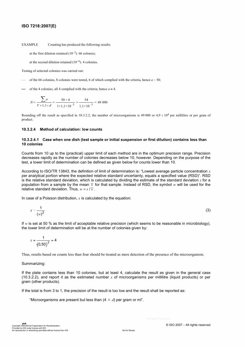
ISO 7218:2007(E)
40 © ISO 2007 – All rights reserved
EXAMPLE Counting has produced the following results:
at the first dilution retained (10–3): 66 colonies;
at the second dilution retained (10–4): 4 colonies.
Testing of selected colonies was carried out:
of the 66 colonies, 8 colonies were tested, 6 of which complied with the criteria, hence a 50;
of the 4 colonies, all 4 complied with the criteria; hence a 4.
3 3
50 + 4 54= = 49 090
1,1 1 × 1,1 × 10 1,1 × 10
aN = =
V d
Rounding off the result as specified in 10.3.2.2, the number of microorganisms is 49 000 or 4,9 104 per millilitre or per gram of product.
10.3.2.4 Method of calculation: low counts
10.3.2.4.1 Case when one dish (test sample or initial suspension or first dilution) contains less than 10 colonies
Counts from 10 up to the (practical) upper limit of each method are in the optimum precision range. Precision decreases rapidly as the number of colonies decreases below 10, however. Depending on the purpose of the test, a lower limit of determination can be defined as given below for counts lower than 10.
According to ISO/TR 13843, the definition of limit of determination is: “Lowest average particle concentration xper analytical portion where the expected relative standard uncertainty, equals a specified value (RSD)”. RSD is the relative standard deviation, which is calculated by dividing the estimate of the standard deviation s for a population from a sample by the mean x for that sample. Instead of RSD, the symbol w will be used for the relative standard deviation. Thus, w s / x .
In case of a Poisson distribution, x is calculated by the equation:
21
( )x
w(3)
If w is set at 50 % as the limit of acceptable relative precision (which seems to be reasonable in microbiology), the lower limit of determination will be at the number of colonies given by:
21 4
0,50x
Thus, results based on counts less than four should be treated as mere detection of the presence of the microorganism.
Summarizing:
If the plate contains less than 10 colonies, but at least 4, calculate the result as given in the general case (10.3.2.2), and report it as the estimated number x of microorganisms per millilitre (liquid products) or per gram (other products).
If the total is from 3 to 1, the precision of the result is too low and the result shall be reported as:
“Microorganisms are present but less than (4 d) per gram or ml”.
ݱ°§®·¹¸¬ ײ¬»®²¿¬·±²¿´ Ñ®¹¿²·¦¿¬·±² º±® ͬ¿²¼¿®¼·¦¿¬·±² Ю±ª·¼»¼ ¾§ ×ØÍ «²¼»® ´·½»²» ©·¬¸ ×ÍÑ
Ò±¬ º±® λ¿´»Ò± ®»°®±¼«½¬·±² ±® ²»¬©±®µ·²¹ °»®³·¬¬»¼ ©·¬¸±«¬ ´·½»²» º®±³ ×ØÍ
óóÀôôÀÀÀôôôôÀÀÀÀóÀóÀôôÀôôÀôÀôôÀóóó
ISO 7218:2007(E)
© ISO 2007 – All rights reserved 41
10.3.2.4.2 Case when the dish (test sample or initial suspension or first dilution) contains no colonies
If the dish containing the test sample (liquid products) or the initial suspension (other products) or the first dilution inoculated or retained does not contain any colonies, report the result as follows:
“less than 1/d microorganisms per millilitre” (liquid products) or “less than 1/d microorganisms per gram” (other products)
where d is the dilution factor of the initial suspension or of the first dilution inoculated or retained (d 100 1 where the directly inoculated test sample of liquid product is retained).
10.3.2.4.3 Special cases
10.3.2.4.3.1 General
These cases concern the counting of typical or presumptive colonies.
10.3.2.4.3.2 Case 1
If the number of typical and atypical colonies for the dish containing a first dilution d1 is greater than 300 (or any other number stated in the specific standard), with visible typical colonies or confirmed colonies, and if, the dish containing the subsequent dilution d2 contains less than 300 colonies (or any other number stated in the specific standard), and no typical or confirmed colony is visible, report the result as follows:
“less than 1/d2 and more than 1/d1 microorganisms per millilitre” (liquid products) or “less than 1/d2 and more than 1/d1 microorganisms per gram” (other products)
where d1 and d2 are the dilution factors corresponding to the dilution d1 and d2.
EXAMPLE Counting has produced the following results:
at the first dilution retained (10–2): more than 300 colonies on the dish, with typical or confirmed colonies present;
at the second dilution retained (10–3): 33 colonies, with no typical or confirmed colonies present.
The result, expressed in microorganisms, is less than 1 000 and more than 100 per millilitre or per gram of product.
10.3.2.4.3.3 Case 2
If the number of typical and atypical colonies for the dish containing a first dilution d1 is greater than 300 (or any other number stated in the specific standard), without visible typical colonies or confirmed colonies, and if the dish containing the subsequent dilution d2 contains less than 300 colonies (or any other number stated in the specific standard) and no typical or confirmed colonies are visible, report the result as follows:
“less than 1/d2 microorganisms per millilitre” (liquid products) or “less than 1/d2 microorganisms per gram” (other products)
where d2 is the dilution factor corresponding to the dilution d2.
EXAMPLE Counting has produced the following results:
at the first dilution retained (10–2): more than 300 colonies on the dish, with no typical or confirmed colonies present;
at the second dilution retained (10–3): 33 colonies, with no typical or confirmed colonies present;
The result, expressed in microorganisms, is less than 1 000 per millilitre or per gram of product.
ݱ°§®·¹¸¬ ײ¬»®²¿¬·±²¿´ Ñ®¹¿²·¦¿¬·±² º±® ͬ¿²¼¿®¼·¦¿¬·±² Ю±ª·¼»¼ ¾§ ×ØÍ «²¼»® ´·½»²» ©·¬¸ ×ÍÑ
Ò±¬ º±® λ¿´»Ò± ®»°®±¼«½¬·±² ±® ²»¬©±®µ·²¹ °»®³·¬¬»¼ ©·¬¸±«¬ ´·½»²» º®±³ ×ØÍ
óóÀôôÀÀÀôôôôÀÀÀÀóÀóÀôôÀôôÀôÀôôÀóóó
ISO 7218:2007(E)
42 © ISO 2007 – All rights reserved
10.3.2.5 Method of calculation: special cases
10.3.2.5.1 When the number of colonies counted (total colonies, typical colonies or presumptive colonies) is greater than 300 (or any other number stated in the specific standard) for the dish containing a first dilution d1,with a number of colonies (total colonies, typical colonies or colonies complying with identification criteria) of less than 10 for the dish containing the subsequent dilution d2:
if the number of colonies for the dish containing the dilution d1 is within the 334 to 300 interval (the upper part of the confidence interval for a weighted mean equal to 300), use the calculation method for general cases (see 10.3.2.2);
if the number of colonies for the dish containing the dilution d1 is greater than 334 (the upper limit of the confidence interval for a weighted mean equal to 300), only take account of the result of the count of dilution d2 and calculate an estimated count (see 10.3.2.4), except, when a maximum of 300 has been set for the colony count, if this estimated count is less than 8 (lower limit of the confidence interval for a weighted mean equal to 10), since the difference between the two dilutions is then unacceptable.
The figures corresponding to confidence intervals shall be adapted to the maximum number stated for the colony counts.
EXAMPLE 1 Counting has produced the following results:
at the first dilution retained (10–2): 310 colonies;
at the second dilution retained (10–3): 8 colonies.
Use the method of calculation for general cases using the dishes for the two dilutions retained.
EXAMPLE 2 Counting has produced the following results:
at the first dilution retained (10–2): more than 334 colonies in the dish;
at the second dilution retained (10–3): 9 colonies.
Report an estimated count on the basis of the colonies counted in the dish for the 10–3 dilution.
EXAMPLE 3 Counting (when a maximum number of 300 has been set for the counting of colonies) has produced the following results:
at the first dilution retained (10–2): more than 334 colonies in the dish;
at the second dilution retained (10–3): 7 colonies.
The result of this counting is unacceptable.
EXAMPLE 4 Counting (when a maximum number of 150 has been set for the counting of colonies) has produced the following results:
at the first dilution retained (10–2): more than 167 colonies in the dish (upper limit of the confidence interval with a weighted mean equal to 150);
at the second dilution retained (10–3): 7 colonies.
Report an estimated count on the basis of the colonies counted in the dish for the 10–3 dilution.
10.3.2.5.2 Where the counting of colonies (total colonies, typical colonies or presumptive colonies) for each one of the dishes for all inoculated dilutions produces a number greater than 300 (or any other number stated in the specific standard), report the result as follows:
“more than 300/d ” (in the case of total colonies or typical colonies) or “more than 300 b/A 1/d ” (in the case of confirmed colonies), expressed in microorganisms per millilitre (liquid products) or microorganisms per gram (other products)
ݱ°§®·¹¸¬ ײ¬»®²¿¬·±²¿´ Ñ®¹¿²·¦¿¬·±² º±® ͬ¿²¼¿®¼·¦¿¬·±² Ю±ª·¼»¼ ¾§ ×ØÍ «²¼»® ´·½»²» ©·¬¸ ×ÍÑ
Ò±¬ º±® λ¿´»Ò± ®»°®±¼«½¬·±² ±® ²»¬©±®µ·²¹ °»®³·¬¬»¼ ©·¬¸±«¬ ´·½»²» º®±³ ×ØÍ
óóÀôôÀÀÀôôôôÀÀÀÀóÀóÀôôÀôôÀôÀôôÀóóó
ISO 7218:2007(E)
© ISO 2007 – All rights reserved 43
where
d is the dilution of the last inoculated dilution;
b is the number of colonies complying with identification criteria among the presumptive colonies A.
10.3.2.5.3 Where the dish containing the last inoculated dilution contains more than 10 colonies and less than 300 (or any other number stated in the specific standard) colonies (total colonies, typical colonies or presumptive colonies), calculate the number N of microorganisms present using Equation (4):
cN'V d
(4)
where
c is the number of colonies counted in the dish;
V is the volume of the inoculum used in each dish, in millilitres;
d is the dilution corresponding to the dilution retained.
Round off the result as specified in 10.3.2.2.
Report the result as the number N of microorganisms per millilitre (liquid products) or per gram (other products).
EXAMPLE Counting has produced the following results:
at the last dilution inoculated (10–4): 120 colonies.
Thus 4
1201 200 000
1 10N'
Rounding off the result as specified in 10.3.2.2, the number N' of microorganisms is 1 200 000, or 1,2 106 per millilitre or per gram of product.
10.3.2.6 Measurement of uncertainty
See ISO/TS 19036 for quantitative determinations.
10.4 Enumeration of yeasts and moulds
10.4.1 General
Yeasts and moulds should usually be enumerated either by a pour-plate technique which allows easier enumeration or by a surface spread-plate technique which provides maximum exposure of the cells to atmospheric oxygen and avoids heat stress from molten agar. Pre-poured agar plates should be dried before being inoculated (see ISO/TS 11133).
Some yeasts and moulds can be infectious or can elicit allergic responses, sometimes even in healthy individuals. Thus it is important to be reasonably cautious when working with them. Ideally, plates should be kept in incubators, not in an open room. Plate lids should be removed as infrequently as possible, normally only for essential purposes such as the preparation of a slide for microscopic examination. Flamed needles should be cooled before making transfers, to avoid dispersal of conidia and other cells. Work benches and incubators should be disinfected routinely.
Petri dishes should be incubated in an upright position and not disturbed until the plates are ready to be counted, as movement can result in the release of mould conidia or spores and the subsequent development of satellite colonies, giving an overestimate of the population.
ݱ°§®·¹¸¬ ײ¬»®²¿¬·±²¿´ Ñ®¹¿²·¦¿¬·±² º±® ͬ¿²¼¿®¼·¦¿¬·±² Ю±ª·¼»¼ ¾§ ×ØÍ «²¼»® ´·½»²» ©·¬¸ ×ÍÑ
Ò±¬ º±® λ¿´»Ò± ®»°®±¼«½¬·±² ±® ²»¬©±®µ·²¹ °»®³·¬¬»¼ ©·¬¸±«¬ ´·½»²» º®±³ ×ØÍ
óóÀôôÀÀÀôôôôÀÀÀÀóÀóÀôôÀôôÀôÀôôÀóóó
ISO 7218:2007(E)
44 © ISO 2007 – All rights reserved
10.4.2 Counting of colonies for yeasts and moulds
Plates with 10 to 150 colonies are usually counted. If the mycoflora consists primarily of moulds, select dishes containing counts in the lower population range; if the mycoflora consists primarily of yeasts, dishes containing counts up to the upper limit may be selected for counting.
If the identity of the colonies is in doubt, examine wet mounts or stains of cells from at least 5 colonies per sample to confirm that bacteria are not present.
10.5 Enumeration using a liquid medium
10.5.1 Principle
Test portions are inoculated into a liquid medium that is designed to support the growth of a particular microorganism or a group of microorganisms, and often inhibits proliferation of non-target microorganisms.
To determine whether growth of the target microorganisms has occurred, various criteria can be used, e.g. visual detection of turbidity, gas production, colour changes, subsequent isolation of the microorganisms on a selective agar medium. The composition of the growth medium and the criteria for discriminating between a positive and a negative result are defined in the corresponding standards.
Using this approach, only a qualitative value can be attributed to each test portion, i.e. the result is either positive or negative. To obtain an estimate of the quantity of microorganisms that is present, it is necessary to examine several test portions and use statistical procedures to determine the most probable number (MPN).
10.5.2 Inoculation
10.5.2.1 General
If a selective growth medium is used, the addition of the test portion should not reduce its selective properties (thereby allowing the growth of non-target microorganisms). In most standards, information about the compatibility of a specific matrix and the liquid medium is described in the scope, but care should be taken with matrices like spices, cocoa, bouillon, etc., as they may contain growth-inhibiting substances which require the addition of neutralizing compounds, the use of higher dilution factors, centrifugation, filtration or immunomagnetic separation to separate the target microorganisms from the matrix, even though this is not always specifically defined in the corresponding standards. Incompatibility can also be due to the biological composition of the matrix: heavily contaminated environmental samples, fermented products or products with probiotic bacteria obviously represent a bigger challenge for the analytical microbiologist than samples which contain only very few microorganisms. For these problematic matrices, spiking experiments using representative microorganisms should be performed to verify that the method is indeed compatible with the matrix.
10.5.2.2 Procedure
Unless stated otherwise in the corresponding standards, test portion volumes of less than, or equal to, 1 ml are normally added to five to ten times the volume of single-strength media. Test portions between 1 ml and 100 ml are normally added to equal volumes of double-strength media.
For volumes greater than 100 ml, more concentrated media may be used. For special purposes, sterile dehydrated media may be dissolved in the cold (or pre-warmed to 30 °C) sample to be analysed.
Unless stated otherwise, the time lapse between preparing the first dilution of a sample and inoculation of the last tube, multiwell plate or bottle should be less than 15 min.
A new sterile pipette shall be used for each dilution.
ݱ°§®·¹¸¬ ײ¬»®²¿¬·±²¿´ Ñ®¹¿²·¦¿¬·±² º±® ͬ¿²¼¿®¼·¦¿¬·±² Ю±ª·¼»¼ ¾§ ×ØÍ «²¼»® ´·½»²» ©·¬¸ ×ÍÑ
Ò±¬ º±® λ¿´»Ò± ®»°®±¼«½¬·±² ±® ²»¬©±®µ·²¹ °»®³·¬¬»¼ ©·¬¸±«¬ ´·½»²» º®±³ ×ØÍ
óóÀôôÀÀÀôôôôÀÀÀÀóÀóÀôôÀôôÀôÀôôÀóóó
ISO 7218:2007(E)
© ISO 2007 – All rights reserved 45
10.5.3 Choice of inoculation system
The essence of the MPN method is the dilution of a sample to such a degree that inocula will sometimes but not always contain viable microorganisms. The “outcome”, i.e. the number of inocula producing growth at each dilution, will give an estimate of the initial concentration of bacteria in the sample. In order to obtain estimates over a broad range of possible concentrations, microbiologists use serial dilutions, incubating several tubes (or plates, etc.) at each dilution. Themost probable number (MPN) of microorganisms present in the original sample, and the precision of the estimate, can be calculated by statistical procedures on the basis of the numbers of positive and negative tubes observed after incubation.
Make a choice from the various MPN configurations available according to
the expected number of microorganisms in the sample under investigation,
regulatory requirements,
the precision needed, and
any other practical considerations.
The measurement uncertainty depends on the number of positive test portions observed in a roughly similar way as the measurement uncertainty of a colony count depends on the number of colonies on a plate. Measurement uncertainty increases as a function of the square root of the number of tubes used. The number of tubes has to be quadrupled to halve the measurement uncertainty. When systems having only a few replicate tubes are used, the measurement uncertainty is low.
Depending on their size, test portions can be inoculated into tubes or bottles containing the required amount of liquid medium. For small test portions, multiwell plates can also be used.
10.5.3.1 Single-dilution system
When the expected concentration of microorganisms is small or expected to vary only moderately, the most appropriate inoculation system is a single series of equal test portions. Where the expected ratio between the maximum and minimum number of microorganisms is less than about 25, ten parallel test portions is the smallest number expected to function; with 50 parallel tubes, a ratio of 200 is the limit. Examples of single-dilution MPNs are given in Annex B, Tables B.1 to B.4.
10.5.3.2 Multiple-dilution system
When the concentration of microorganisms in the sample is unknown, or if great variation is anticipated, it may be necessary to inoculate series of tubes from several dilutions. Inoculate a sufficient number of dilutions to ensure a system with both positive and negative results. The number of dilutions also depends on the calculation method used for estimating the MPN value. If tables need to be used, then results from three dilutions must be available, and the configurations of the systems are restricted to those available in tables. With computer programs, the numbers of dilutions and parallel tubes are unrestricted.
10.5.3.3 Symmetric dilution system
The most commonly applied symmetric MPN system uses three or five parallel tubes per dilution. The precision obtained with this system declines rapidly with lower numbers of tubes per dilution. Results from a three-tube design are hardly more than indications of the order of magnitude of the concentration. If more precision is required, it is recommended that five or more parallel tubes are chosen. Examples of a three-tube MPN and a five-tube MPN are given in Annex B, Tables B.5 and B.7, respectively.
10.5.3.4 Non-symmetric dilution system
In non-symmetric systems, the different dilution levels do not have the same number of tubes. Use these systems only to estimate numbers of microorganisms within a well-defined range. For examples, refer to ISO 8199.
ݱ°§®·¹¸¬ ײ¬»®²¿¬·±²¿´ Ñ®¹¿²·¦¿¬·±² º±® ͬ¿²¼¿®¼·¦¿¬·±² Ю±ª·¼»¼ ¾§ ×ØÍ «²¼»® ´·½»²» ©·¬¸ ×ÍÑ
Ò±¬ º±® λ¿´»Ò± ®»°®±¼«½¬·±² ±® ²»¬©±®µ·²¹ °»®³·¬¬»¼ ©·¬¸±«¬ ´·½»²» º®±³ ×ØÍ
óóÀôôÀÀÀôôôôÀÀÀÀóÀóÀôôÀôôÀôÀôôÀóóó
ISO 7218:2007(E)
46 © ISO 2007 – All rights reserved
10.5.4 Incubation
Incubate the inoculated tubes, flasks or bottles in an incubator or in a water bath. Place multiwell plates in an incubator.
Choose the duration and the temperature of incubation after reference to a specific standard method, as they depend on the microorganism or group of microorganisms sought.
For some microorganisms, a two-stage incubation procedure and/or a confirmation step may be necessary. Refer to the specific standards for details.
10.5.5 Interpretation of results
The criteria that distinguish positive from negative results vary with each microorganism or group of microorganisms and are defined in the corresponding standards. Using these criteria, count and record the number of positive results obtained with all the test portions derived from one sample.
10.5.6 Determination of MPN values
There are three different possibilities for determining the MPN value: calculation with mathematical formulae, consultation of MPN tables, or utilization of specific computer programs. Provided that they are based on the same statistical considerations, they are equally valid. These three methods are detailed below.
10.5.6.1 Mathematical formulae
10.5.6.1.1 Approximate formula for all cases
The approximate MPN values for any number of dilutions and parallel tubes are derived by application of the following equation (adapted from Reference [36]):
p r
s tMPN
Z m
m m
where
Zp is the number of positive tubes;
mr is the sample reference mass, in grams;
ms is the total mass, in grams, of sample in all tubes with negative reactions;
mt is the total mass, in grams, of sample in all tubes.
MPN is expressed per sample reference mass in grams (usually 1 g, sometimes 100 g).
10.5.6.1.2 "Exact" solution for one series of tubes
The MPN value for a single series of tubes is derived from the formula:
r
m pMPN ln
m nm n z
where
mr is the sample reference mass, in grams;
ݱ°§®·¹¸¬ ײ¬»®²¿¬·±²¿´ Ñ®¹¿²·¦¿¬·±² º±® ͬ¿²¼¿®¼·¦¿¬·±² Ю±ª·¼»¼ ¾§ ×ØÍ «²¼»® ´·½»²» ©·¬¸ ×ÍÑ
Ò±¬ º±® λ¿´»Ò± ®»°®±¼«½¬·±² ±® ²»¬©±®µ·²¹ °»®³·¬¬»¼ ©·¬¸±«¬ ´·½»²» º®±³ ×ØÍ
óóÀôôÀÀÀôôôôÀÀÀÀóÀóÀôôÀôôÀôÀôôÀóóó
ISO 7218:2007(E)
© ISO 2007 – All rights reserved 47
mm is the mass, in grams, of sample in each tube of the series;
ln is the natural logarithm;
n is the number of tubes in the series;
zp is the number of tubes with a positive reaction.
10.5.6.1.3 Precision estimates single-dilution assays
The 95 % confidence limits of the MPN estimate can be calculated approximately using the equation:
r
m nn
ln
2
m nxm z n z
zn
where
x is the upper or lower 95 % confidence limit;
mr is the sample reference mass, in grams;
mm is the mass, in grams, of sample in each tube of the series;
ln is the natural logarithm;
n is the number of tubes in the series;
zn is the number of tubes with a negative reaction.
The plus sign is connected with the lower limit and the minus sign with the upper limit. The approximation is not very good when most of the tubes are negative (sterile) but improves when the proportion of positive tubes increases.
10.5.6.1.4 Precision estimates symmetrical multiple-dilution assays
The log10 standard uncertainty of a symmetrical multiple-dilution MPN system can be obtained from Cochran’s approximate equation [28]:
SE 0,58 10log fn
where
SE is the standard error of log10MPN;
f is the dilution factor between consecutive dilutions (mostly 10);
n is the number of tubes per dilution.
The upper and lower 95 % confidence limits can be approximated respectively by multiplying and dividing the MPN estimate by the antilogarithm of 2 SE. This procedure tends to exaggerate the upper confidence limit.
ݱ°§®·¹¸¬ ײ¬»®²¿¬·±²¿´ Ñ®¹¿²·¦¿¬·±² º±® ͬ¿²¼¿®¼·¦¿¬·±² Ю±ª·¼»¼ ¾§ ×ØÍ «²¼»® ´·½»²» ©·¬¸ ×ÍÑ
Ò±¬ º±® λ¿´»Ò± ®»°®±¼«½¬·±² ±® ²»¬©±®µ·²¹ °»®³·¬¬»¼ ©·¬¸±«¬ ´·½»²» º®±³ ×ØÍ
óóÀôôÀÀÀôôôôÀÀÀÀóÀóÀôôÀôôÀôÀôôÀóóó
ISO 7218:2007(E)
48 © ISO 2007 – All rights reserved
10.5.6.2 MPN tables
10.5.6.2.1 Tables for single-dilution systems
Tables B.1 to B.4 (Annex B) give the MPN values and the 95 % confidence intervals per test portion for 10, 15, 20 and 25 parallel tubes [each tube is inoculated with the same (single) dilution].
To express the outcome per sample reference mass (or volume for liquid samples), multiply the MPN and the 95 % limit values by the ratio (reference mass)/(test portion mass). Do not multiply the logarithmic standard uncertainty. The reference mass in food microbiology is usually 1 g. The test portion mass corresponds to the amount of sample (in grams) that is present in the volume used to inoculate the tubes, e.g. 0,1 g if 1 ml of the 10 1 homogenate has been used.
EXAMPLE (Reference [30])
Twenty tubes of double-strength broth were inoculated with 5 ml aliquots of a tenfold-diluted sample (0,1 g/ml). After incubation, 16 of the tubes showed visible growth. What was the most probable bacterial density (organisms per gram) in the sample? Table B.3 gives 1,61 as the most probable number of organisms per tube, with a lower 95 % limit of 0,93 and an upper 95 % limit of 2,77.
Each tube received a test portion of 5 ml, which corresponds to 0,5 g of sample. Therefore, the most probable number of microorganisms in 1 g of sample is given by:
MPN 1, 610, 5
per gram 3,2 per gram
with the 95 % confidence interval ranging from
lower 95 % limit 0,930,5
per gram 1,9 per gram;
upper 95 % limit 2,770,5
per gram 5,5 per gram.
10.5.6.2.2 Tables for multiple-dilution systems: three successive dilutions
With symmetrical systems, it is common practice to use three successive dilutions with either three (Table B.5) or five (Table B.7) replicates. Record the number of positive results for each set of tubes and, from the MPN table for the inoculation system used, read the most probable number of microorganisms present in the reference volume of the sample.
Some combinations of positive tubes are more likely to occur than others. For example, a combination of positive results 0, 0, 3 is much less likely to occur than the combination 3, 2, 1. To quantify this probability, all combinations of positive results have been attributed with a category, ranging from 0 to 3. A category 1 result is a result with higher probability, whereas a category 3 result is rare and may not be reproduced easily. The worst cases are category 0 results; they should be considered with great suspicion. Assuming that the results of the analysis are correct, one would expect that 95 % of the observed combinations would fall in category 1, 4 % in category 2, 0,9 % in category 3 and only 0,1 % in category 0. Categories are further explained in Table B.6.
In the case where more than three dilutions are made, the selection of the “right” combination of three consecutive dilutions is not always very clear. However, this can be easily done by recording all possible combinations of positive tubes and reading the corresponding category from Table B.5.
Thereafter, apply the following rules:
1) Select the combination of three consecutive dilutions having a category 1 profile to obtain the MPN index. If more than one combination having a category 1 profile is obtained, use the one with the highest number of positive tubes.
ݱ°§®·¹¸¬ ײ¬»®²¿¬·±²¿´ Ñ®¹¿²·¦¿¬·±² º±® ͬ¿²¼¿®¼·¦¿¬·±² Ю±ª·¼»¼ ¾§ ×ØÍ «²¼»® ´·½»²» ©·¬¸ ×ÍÑ
Ò±¬ º±® λ¿´»Ò± ®»°®±¼«½¬·±² ±® ²»¬©±®µ·²¹ °»®³·¬¬»¼ ©·¬¸±«¬ ´·½»²» º®±³ ×ØÍ
óóÀôôÀÀÀôôôôÀÀÀÀóÀóÀôôÀôôÀôÀôôÀóóó
ISO 7218:2007(E)
© ISO 2007 – All rights reserved 49
2) If no combination having a category 1 profile is available, use the one having a category 2 profile. If more than one combination having a category 2 profile is obtained, use the one with the highest number of positive tubes.
3) If no combination having a category 2 profile is available, use the one having a category 3 profile. If more than one combination having a category 3 profile is obtained, use the one with the highest number of positive tubes.
Some examples are shown in Table 1.
Table 1 — Examples of the selection of positive results for the calculation of the MPN
MPN b
Sample Number of positive tubes obtained from three incubated tubes for the following amounts of sample inoculated per tube a Liquid
product (ml–1)
Other products
(g–1)
Liquid product: 10 ml 1 ml 10–1 ml 10–2 ml 10–3 ml
Other products: 1 g 10–1 g 10–2 g 10–3 g 10–4 g
1 3 3 2 1 0 1,1 101 1,1 102
2 3 3 3 0 2,4 101 2,4 102
3 2 2 1 1 0 7,4 7,4 101
4 3 3 0 0 0 2,4 2,4 101
5 2 2 0 1 0 2,1 10–1 2,1 a Underlining indicates combination selected. b Calculated using the MPN index (see Table B.5).
10.5.6.3 Computer programs
The most versatile computer programs pose no restrictions regarding numbers of dilutions and parallel tubes or symmetry of the MPN system. MPN Assay Analyzer is a freely available program based upon a previous program (see Reference [29]).
10.5.7 Expression of results
From the MPN index read from table B.5 [according to the combination of three (or five) consecutive dilutions retained], determine the most probable microorganisms in the reference volume.
Report results as the most probable number of microorganisms (or specific group of microorganisms) per gram or millilitre. The mass or the reference volume can be different from g or ml (for example 100 g or 100 ml).
11 Detection method (qualitative method)
11.1 General
A detection method is a method that determines the presence or absence of particular microorganisms in a given quantity of product.
ݱ°§®·¹¸¬ ײ¬»®²¿¬·±²¿´ Ñ®¹¿²·¦¿¬·±² º±® ͬ¿²¼¿®¼·¦¿¬·±² Ю±ª·¼»¼ ¾§ ×ØÍ «²¼»® ´·½»²» ©·¬¸ ×ÍÑ
Ò±¬ º±® λ¿´»Ò± ®»°®±¼«½¬·±² ±® ²»¬©±®µ·²¹ °»®³·¬¬»¼ ©·¬¸±«¬ ´·½»²» º®±³ ×ØÍ
óóÀôôÀÀÀôôôôÀÀÀÀóÀóÀôôÀôôÀôÀôôÀóóó
ISO 7218:2007(E)
50 © ISO 2007 – All rights reserved
11.2 Principle
Unless otherwise stated in the relevant International Standard, mix (liquid products) or homogenize (other products) a quantity P of the product to be examined with 9 P ml or 9 P g of an elective and/or a selective broth.
To facilitate the recovery of stressed microorganisms in foods, samples are usually pre-enriched in a non-selective broth followed by selective enrichment and isolation on selective/differential agar media. The use of two different enrichment broths, as well as two or more selective agar media, increases method sensitivity.
After incubation, spread a loop of the culture obtained over the surface of a selective agar medium in such a manner as to obtain isolated colonies. Unless otherwise stated, the incubated enrichment broths may only be refrigerated after evaluation of the impact of refrigeration on the results and only if clearly stipulated in the test report.
A number (generally five per agar plate) of the colonies obtained after incubation is then identified using appropriate confirmation techniques.
The selection of colonies for confirmation should cover representative suspect colony types.
11.3 Measurement of uncertainty
The estimation of the measurement uncertainty of qualitative determinations is being investigated by ISO/TC 34/SC 9.
12 Confirmation methods
12.1 General
Use only pure cultures for biochemical and serological confirmation.
The reference confirmation tests are described in the specific standards. As an alternative to biochemical tests described in these specific standards, confirmation methods described in this clause (biochemical galleries, nucleic probes) may be used under the conditions described in the clause, unless otherwise stated in specific standards.
12.2 Preparation of a pure culture
Begin the preparation of a pure culture by the selection of a single colony on or in an agar medium. Then inoculate the selected colony onto a non-selective agar medium. After incubation, select a well-isolated colony for subsequent confirmation tests. Repeat the operation if necessary.
If possible, the confirmation tests should be carried out using cells from a single colony. If there is insufficient cell material in one colony, it should first be subcultured in a liquid medium or on an agar slant medium, after which the subculture can be used for the tests to be performed.
12.3 Gram’s stain (modified Hucker technique)
12.3.1 General
This way of staining bacterial cells allows description of the morphology of the bacteria and classification of them into two groups as a function of whether or not they are capable of retaining the crystal violet stain (Gram +) under the test conditions. This division results mainly from differences in the structure of the cell walls of the two groups and it correlates with other major differences between the two groups.
A satisfactory alternative to Gram staining is the use of 3 % potassium hydroxide (KOH) solution. A loopful of the bacterial growth is stirred in 2 drops of the KOH solution. Gram-negative bacteria will cause the solution to become very viscous and mucoid within 30 s, with a string of the mixture following the loop when raised.
ݱ°§®·¹¸¬ ײ¬»®²¿¬·±²¿´ Ñ®¹¿²·¦¿¬·±² º±® ͬ¿²¼¿®¼·¦¿¬·±² Ю±ª·¼»¼ ¾§ ×ØÍ «²¼»® ´·½»²» ©·¬¸ ×ÍÑ
Ò±¬ º±® λ¿´»Ò± ®»°®±¼«½¬·±² ±® ²»¬©±®µ·²¹ °»®³·¬¬»¼ ©·¬¸±«¬ ´·½»²» º®±³ ×ØÍ
óóÀôôÀÀÀôôôôÀÀÀÀóÀóÀôôÀôôÀôÀôôÀóóó
ISO 7218:2007(E)
© ISO 2007 – All rights reserved 51
There are a number of ways of conducting a Gram’s stain, but all follow the sequences given below.
12.3.2 Solutions
12.3.2.1 General
Commercially available solutions may be used.
In this case, follow the manufacturer’s recommendations.
12.3.2.2 Crystal violet solution
12.3.2.2.1 Composition
Crystal violet 2,0 g Ethanol (95 %) 20 ml Ammonium oxalate (C2H8N2O4) 0,8 g Water 80 ml
12.3.2.2.2 Preparation
Dissolve the crystal violet in the ethanol, and the ammonium oxalate in the distilled water. Mix the two solutions and allow the mixture to stand for 24 h prior to use.
12.3.2.3 Iodine solution
12.3.2.3.1 Composition
Iodine 1,0 g Potassium iodide (KI) 2,0 g Water 100 ml
12.3.2.3.2 Preparation
Dissolve the potassium iodide in 10 ml of water and add the iodine in fractions. After dissolving, make up to 100 ml in a volumetric flask.
12.3.2.4 Safranin solution
12.3.2.4.1 Composition
Safranin 0,25 g Ethanol (95 %) 10 ml Water 100 ml
12.3.2.4.2 Preparation
Dissolve the safranin in the ethanol then mix with the distilled water.
12.3.3 Staining technique
After flame-fixing a bacterial film on a microscope slide, prepared from an 18 h to 24 h culture or when the broth is turbid, cover the film with the crystal violet. Allow to react for 1 min.
Gently rinse the inclined slide with water for a few seconds.
ݱ°§®·¹¸¬ ײ¬»®²¿¬·±²¿´ Ñ®¹¿²·¦¿¬·±² º±® ͬ¿²¼¿®¼·¦¿¬·±² Ю±ª·¼»¼ ¾§ ×ØÍ «²¼»® ´·½»²» ©·¬¸ ×ÍÑ
Ò±¬ º±® λ¿´»Ò± ®»°®±¼«½¬·±² ±® ²»¬©±®µ·²¹ °»®³·¬¬»¼ ©·¬¸±«¬ ´·½»²» º®±³ ×ØÍ
óóÀôôÀÀÀôôôôÀÀÀÀóÀóÀôôÀôôÀôÀôôÀóóó
ISO 7218:2007(E)
52 © ISO 2007 – All rights reserved
Cover the slide with the iodine solution. Allow to react for 1 min.
Gently rinse the inclined slide with water for a few seconds.
Pour gently and continuously a film of ethanol (95 %) onto the inclined slide over a period of not more than 30 s until no more violet colour is washed out.
Gently rinse the inclined slide with water in order to eliminate the ethanol. Cover the slide with the solution of safranin for 10 s. Gently rinse the inclined slide with water.
Dry the slide.
12.3.4 Interpretation
Examine the slide under the high-power oil objective of a microscope. Those bacterial cells which appear blue or violet are termed Gram-positive (Gram +); those which are coloured dark pink to red are termed Gram-negative (Gram ).
For a pure culture of certain bacteria types, both Gram-positive and Gram-negative cells can be obtained in the same microscope field.
NOTE Densely packed cells can give an uncharacteristic response.
12.4 Use of biochemical galleries for identification
Available biochemical galleries may be used for identification of isolated colonies.
Verify that the galleries are suitable, as shown by evaluation studies published in the international scientific literature, preferably relating to food microbiology4). This verification is especially important if the manufacturer does not have validation data on those galleries.
The laboratory should obtain a control certificate for each batch, with an indication of the test strains.
The manufacturer shall also specify control strains that the laboratory may use to verify the preservation of the galleries' performance.
The galleries shall include, as a minimum, the biochemical tests described in specific standards or be supplemented by other tests.
12.5 Use of nucleic probes for identification
Currently available nucleic probes may be used for identification of isolated colonies.
However, verify that the nucleic probes used for confirmation are suitable, as shown by evaluation studies published in the international scientific literature, preferably relating to food microbiology (see e.g. Reference [23]). This verification is especially important if the manufacturer does not have validation data on those probes.
The laboratory should obtain a control certificate for each batch, with an indication of the test strains.
The manufacturer shall also specify control strains that the laboratory may use to verify the maintenance of probe performance.
4) Requests for information should be addressed to the national, regional or international reference centre indicated for each microorganism.
ݱ°§®·¹¸¬ ײ¬»®²¿¬·±²¿´ Ñ®¹¿²·¦¿¬·±² º±® ͬ¿²¼¿®¼·¦¿¬·±² Ю±ª·¼»¼ ¾§ ×ØÍ «²¼»® ´·½»²» ©·¬¸ ×ÍÑ
Ò±¬ º±® λ¿´»Ò± ®»°®±¼«½¬·±² ±® ²»¬©±®µ·²¹ °»®³·¬¬»¼ ©·¬¸±«¬ ´·½»²» º®±³ ×ØÍ
óóÀôôÀÀÀôôôôÀÀÀÀóÀóÀôôÀôôÀôÀôôÀóóó
ISO 7218:2007(E)
© ISO 2007 – All rights reserved 53
12.6 Serological methods
12.6.1 General
When serological confirmation is needed, perform it after biochemical identification of isolated colonies.
12.6.2 Slide agglutination tests
Antigen-antibody reactions cause bacterial cells to clump together and form flocculent masses or dense granules. In the case of bacteria of the family Enterobacteriaceae, the reaction between the “H” (i.e. flagellar) antigen and its homologous antiserum results in flocculent clumping, whereas the reaction involving the “O” (i.e. somatic) antigen results in more dense, granular clumping.
Before agglutination with antisera, a test should be carried out to determine whether the bacterial cells agglutinate in sodium chloride solution [3 % (by mass)]. If the bacterial cells agglutinate, the strain is autoagglutinable and should not be agglutinated with antisera.
Commercially available antisera are of two types: polyvalent antisera which react with microorganisms of a particular genus or with groups of serovars and which are suitable for preliminary screening, and specific monoclonal antibodies, the use of which allows identification of a particular serovar.
The laboratory should obtain a control certificate for each batch of antisera, with an indication of the test strains.
Verify that the slide agglutination tests are suitable, as shown by evaluation studies published in the international scientific literature, preferably relating to food microbiology5).
When using the reagents, suitable positive and negative controls should be used.
12.6.3 Latex agglutination test
A more rapid method is commercially available, employing latex particles coated with group-specific antibodies (e.g. Escherichia coli O157, see specific standard ISO 16654, or Staphylococcus aureus in ISO 6888). The antigen in the extract is tested against a range of latex reagents.
Verify that the latex agglutination tests are suitable, as shown by evaluation studies published in the international scientific literature, preferably relating to food microbiology5).
The laboratory should obtain a control certificate for each batch, with an indication of the test strains.
When using the reagents, suitable positive and negative controls should be used.
13 Test report
The test report shall specify the method used, the temperature of incubation, if necessary, and the results obtained. It shall also mention any operating details not specified in this International Standard, as well as those regarded as optional, together with details of any incidents that may have influenced the results.
State also in the test report whether further tests are to be carried out by a reference laboratory or, if such tests have been carried out, what the results were.
The test report should include all information necessary for the complete identification of the sample. It is also appropriate to include all information necessary for the interpretation of the test results.
5) Requests for information should be addressed to the national, regional or international reference centre indicated for each microorganism.
ݱ°§®·¹¸¬ ײ¬»®²¿¬·±²¿´ Ñ®¹¿²·¦¿¬·±² º±® ͬ¿²¼¿®¼·¦¿¬·±² Ю±ª·¼»¼ ¾§ ×ØÍ «²¼»® ´·½»²» ©·¬¸ ×ÍÑ
Ò±¬ º±® λ¿´»Ò± ®»°®±¼«½¬·±² ±® ²»¬©±®µ·²¹ °»®³·¬¬»¼ ©·¬¸±«¬ ´·½»²» º®±³ ×ØÍ
óóÀôôÀÀÀôôôôÀÀÀÀóÀóÀôôÀôôÀôÀôôÀóóó
ISO 7218:2007(E)
54 © ISO 2007 – All rights reserved
If necessary, the measurement of uncertainty, determined in accordance with ISO/TS 19036, should be included in the test report.
14 Validation of microbiological methods
14.1 Validation of reference methods
The validation of reference methods is being investigated by ISO/TC 34/SC 9.
14.2 Validation of alternative methods
Refer to ISO 16140 for the technical protocol for the validation of alternative methods against reference methods.
14.3 Validation of in-house methods
The validation of in-house methods is being investigated by ISO/TC 34/SC 9.
15 Quality assurance of results/quality control of performance
15.1 Internal quality control
15.1.1 Internal quality control consists of all procedures undertaken by a laboratory for the continuous evaluation of its work. The main objective is to ensure the consistency of results on a day-to-day basis and their conformity with well-defined criteria.
15.1.2 A programme of periodic checks is necessary to demonstrate that variability (between analysts, between equipment or between materials) is under control. All tests included in the laboratory’s scope of activity need to be covered.
The programme may involve:
the use of spiked samples, with variable contamination levels, including target and background flora;
the use of spiked/naturally contaminated samples from a range of matrices;
the use of reference materials (including proficiency testing scheme materials);
replicate testing;
replicate evaluation of test results.
The interval between these checks is influenced by the nature of the tests carried out by the laboratory and the frequency at which the tests are carried out.
It is recommended that, where possible, tests should incorporate controls to monitor performance.
15.1.3 In special cases, a laboratory may perform a particular test only rarely. It is recognized that, in such cases, an ongoing internal quality control programme may be inappropriate and that a scheme for demonstrating satisfactory performance which is carried out in parallel with the testing may be more suitable.
15.2 Reference strains
Refer to ISO 11133 for the maintenance of reference strains.
ݱ°§®·¹¸¬ ײ¬»®²¿¬·±²¿´ Ñ®¹¿²·¦¿¬·±² º±® ͬ¿²¼¿®¼·¦¿¬·±² Ю±ª·¼»¼ ¾§ ×ØÍ «²¼»® ´·½»²» ©·¬¸ ×ÍÑ
Ò±¬ º±® λ¿´»Ò± ®»°®±¼«½¬·±² ±® ²»¬©±®µ·²¹ °»®³·¬¬»¼ ©·¬¸±«¬ ´·½»²» º®±³ ×ØÍ
óóÀôôÀÀÀôôôôÀÀÀÀóÀóÀôôÀôôÀôÀôôÀóóó

ISO 7218:2007(E)
© ISO 2007 – All rights reserved 55
15.3 External quality assessment (proficiency testing)
Laboratories should regularly participate in proficiency testing schemes which are relevant to their scope of activity. Preference should be given to proficiency testing schemes which use appropriate matrices.
Laboratories should use external quality assessment not only to assess laboratory bias but also to check the validity of their overall quality system.
ݱ°§®·¹¸¬ ײ¬»®²¿¬·±²¿´ Ñ®¹¿²·¦¿¬·±² º±® ͬ¿²¼¿®¼·¦¿¬·±² Ю±ª·¼»¼ ¾§ ×ØÍ «²¼»® ´·½»²» ©·¬¸ ×ÍÑ
Ò±¬ º±® λ¿´»Ò± ®»°®±¼«½¬·±² ±® ²»¬©±®µ·²¹ °»®³·¬¬»¼ ©·¬¸±«¬ ´·½»²» º®±³ ×ØÍ
óóÀôôÀÀÀôôôôÀÀÀÀóÀóÀôôÀôôÀôÀôôÀóóó
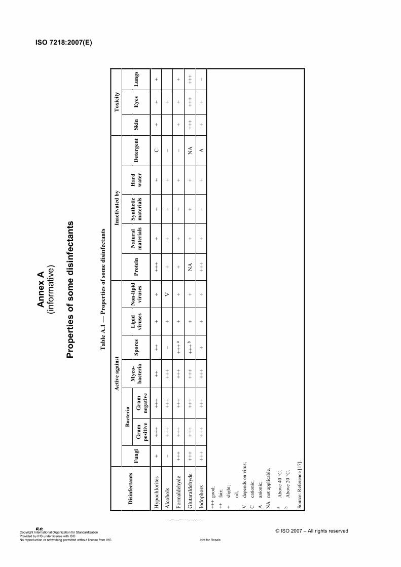
Ann
ex A
(info
rmat
ive)
Prop
ertie
s of
som
e di
sinf
ecta
nts
Tab
le A
.1 —
Pro
pert
ies o
f som
e di
sinf
ecta
nts
Act
ive
agai
nst
Inac
tivat
ed b
y T
oxic
ity
Bac
teri
a D
isinf
ecta
nts
Fung
i G
ram
po
sitiv
e G
ram
ne
gativ
e
Myc
o-ba
cter
ia
Spor
es
Lipi
d vi
ruse
s N
on-li
pid
viru
ses
Prot
ein
Nat
ural
m
ater
ials
Synt
hetic
m
ater
ials
H
ard
wat
er
Det
erge
nt
Skin
Ey
es
Lung
s
Hyp
ochl
orite
s +
+++
+++
++
++
+ +
+++
+ +
+ C
+
+ +
Alc
ohol
s –
+++
+++
+++
– +
V
+ +
+ +
–
+
Form
alde
hyde
++
+ ++
+ ++
+ ++
+ ++
+ a+
+ +
+ +
+ –
+ +
+
Glu
tara
ldeh
yde
+++
+++
+++
+++
+++ b
+ +
NA
+
+ +
NA
++
+ ++
+ ++
+
Iodo
phor
s ++
+ ++
+ ++
+ ++
+ +
+ +
+++
+ +
+ A
+
+ –
+++
good
; ++
fa
ir;
+ sli
ght;
– ni
l; V
de
pend
s on
viru
s;
C
catio
nic;
A
an
ioni
c;
NA
no
t app
licab
le.
a A
bove
40 °C
. b
Abo
ve 2
0 °C
.
Sour
ce: R
efer
ence
[17]
.
ISO 7218:2007(E)
56 © ISO 2007 – All rights reservedݱ°§®·¹¸¬ ײ¬»®²¿¬·±²¿´ Ñ®¹¿²·¦¿¬·±² º±® ͬ¿²¼¿®¼·¦¿¬·±² Ю±ª·¼»¼ ¾§ ×ØÍ «²¼»® ´·½»²» ©·¬¸ ×ÍÑ
Ò±¬ º±® λ¿´»Ò± ®»°®±¼«½¬·±² ±® ²»¬©±®µ·²¹ °»®³·¬¬»¼ ©·¬¸±«¬ ´·½»²» º®±³ ×ØÍ
óóÀôôÀÀÀôôôôÀÀÀÀóÀóÀôôÀôôÀôÀôôÀóóó
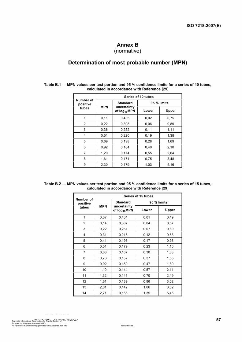
ISO 7218:2007(E)
© ISO 2007 – All rights reserved 57
Annex B(normative)
Determination of most probable number (MPN)
Table B.1 — MPN values per test portion and 95 % confidence limits for a series of 10 tubes, calculated in accordance with Reference [29]
Series of 10 tubes
95 % limits Number of
positive tubes MPN
Standarduncertainty of log10MPN Lower Upper
1 0,11 0,435 0,02 0,75
2 0,22 0,308 0,06 0,89
3 0,36 0,252 0,11 1,11
4 0,51 0,220 0,19 1,38
5 0,69 0,198 0,28 1,69
6 0,92 0,184 0,40 2,10
7 1,20 0,174 0,55 2,64
8 1,61 0,171 0,75 3,48
9 2,30 0,179 1,03 5,16
Table B.2 — MPN values per test portion and 95 % confidence limits for a series of 15 tubes, calculated in accordance with Reference [29]
Series of 15 tubes
95 % limits Number of
positive tubes MPN
Standarduncertainty of log10MPN Lower Upper
1 0,07 0,434 0,01 0,49
2 0,14 0,307 0,04 0,57
3 0,22 0,251 0,07 0,69
4 0,31 0,218 0,12 0,83
5 0,41 0,196 0,17 0,98
6 0,51 0,179 0,23 1,15
7 0,63 0,167 0,30 1,33
8 0,76 0,157 0,37 1,55
9 0,92 0,150 0,47 1,80
10 1,10 0,144 0,57 2,11
11 1,32 0,141 0,70 2,49
12 1,61 0,139 0,86 3,02
13 2,01 0,142 1,06 3,82
14 2,71 0,155 1,35 5,45
ݱ°§®·¹¸¬ ײ¬»®²¿¬·±²¿´ Ñ®¹¿²·¦¿¬·±² º±® ͬ¿²¼¿®¼·¦¿¬·±² Ю±ª·¼»¼ ¾§ ×ØÍ «²¼»® ´·½»²» ©·¬¸ ×ÍÑ
Ò±¬ º±® λ¿´»Ò± ®»°®±¼«½¬·±² ±® ²»¬©±®µ·²¹ °»®³·¬¬»¼ ©·¬¸±«¬ ´·½»²» º®±³ ×ØÍ
óóÀôôÀÀÀôôôôÀÀÀÀóÀóÀôôÀôôÀôÀôôÀóóó
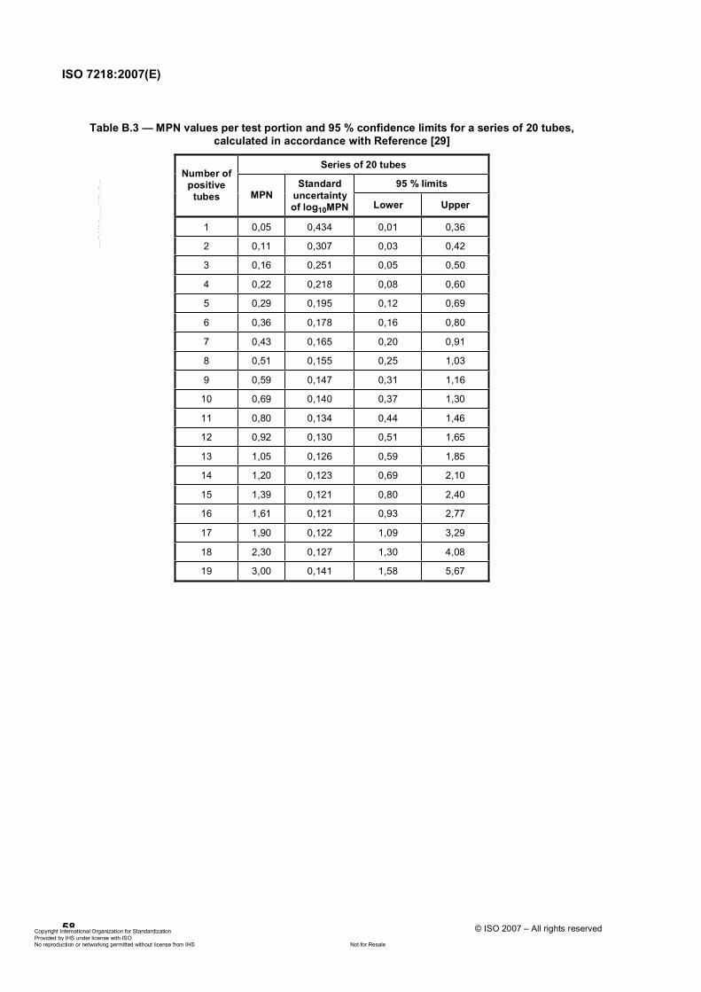
ISO 7218:2007(E)
58 © ISO 2007 – All rights reserved
Table B.3 — MPN values per test portion and 95 % confidence limits for a series of 20 tubes, calculated in accordance with Reference [29]
Series of 20 tubes
95 % limits Number of
positive tubes MPN
Standarduncertainty of log10MPN Lower Upper
1 0,05 0,434 0,01 0,36
2 0,11 0,307 0,03 0,42
3 0,16 0,251 0,05 0,50
4 0,22 0,218 0,08 0,60
5 0,29 0,195 0,12 0,69
6 0,36 0,178 0,16 0,80
7 0,43 0,165 0,20 0,91
8 0,51 0,155 0,25 1,03
9 0,59 0,147 0,31 1,16
10 0,69 0,140 0,37 1,30
11 0,80 0,134 0,44 1,46
12 0,92 0,130 0,51 1,65
13 1,05 0,126 0,59 1,85
14 1,20 0,123 0,69 2,10
15 1,39 0,121 0,80 2,40
16 1,61 0,121 0,93 2,77
17 1,90 0,122 1,09 3,29
18 2,30 0,127 1,30 4,08
19 3,00 0,141 1,58 5,67
ݱ°§®·¹¸¬ ײ¬»®²¿¬·±²¿´ Ñ®¹¿²·¦¿¬·±² º±® ͬ¿²¼¿®¼·¦¿¬·±² Ю±ª·¼»¼ ¾§ ×ØÍ «²¼»® ´·½»²» ©·¬¸ ×ÍÑ
Ò±¬ º±® λ¿´»Ò± ®»°®±¼«½¬·±² ±® ²»¬©±®µ·²¹ °»®³·¬¬»¼ ©·¬¸±«¬ ´·½»²» º®±³ ×ØÍ
óóÀôôÀÀÀôôôôÀÀÀÀóÀóÀôôÀôôÀôÀôôÀóóó
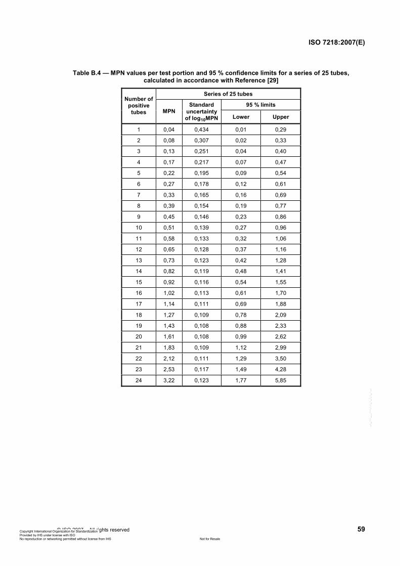
ISO 7218:2007(E)
© ISO 2007 – All rights reserved 59
Table B.4 — MPN values per test portion and 95 % confidence limits for a series of 25 tubes, calculated in accordance with Reference [29]
Series of 25 tubes
95 % limits Number of
positive tubes MPN
Standarduncertainty of log10MPN Lower Upper
1 0,04 0,434 0,01 0,29
2 0,08 0,307 0,02 0,33
3 0,13 0,251 0,04 0,40
4 0,17 0,217 0,07 0,47
5 0,22 0,195 0,09 0,54
6 0,27 0,178 0,12 0,61
7 0,33 0,165 0,16 0,69
8 0,39 0,154 0,19 0,77
9 0,45 0,146 0,23 0,86
10 0,51 0,139 0,27 0,96
11 0,58 0,133 0,32 1,06
12 0,65 0,128 0,37 1,16
13 0,73 0,123 0,42 1,28
14 0,82 0,119 0,48 1,41
15 0,92 0,116 0,54 1,55
16 1,02 0,113 0,61 1,70
17 1,14 0,111 0,69 1,88
18 1,27 0,109 0,78 2,09
19 1,43 0,108 0,88 2,33
20 1,61 0,108 0,99 2,62
21 1,83 0,109 1,12 2,99
22 2,12 0,111 1,29 3,50
23 2,53 0,117 1,49 4,28
24 3,22 0,123 1,77 5,85
ݱ°§®·¹¸¬ ײ¬»®²¿¬·±²¿´ Ñ®¹¿²·¦¿¬·±² º±® ͬ¿²¼¿®¼·¦¿¬·±² Ю±ª·¼»¼ ¾§ ×ØÍ «²¼»® ´·½»²» ©·¬¸ ×ÍÑ
Ò±¬ º±® λ¿´»Ò± ®»°®±¼«½¬·±² ±® ²»¬©±®µ·²¹ °»®³·¬¬»¼ ©·¬¸±«¬ ´·½»²» º®±³ ×ØÍ
óóÀôôÀÀÀôôôôÀÀÀÀóÀóÀôôÀôôÀôÀôôÀóóó
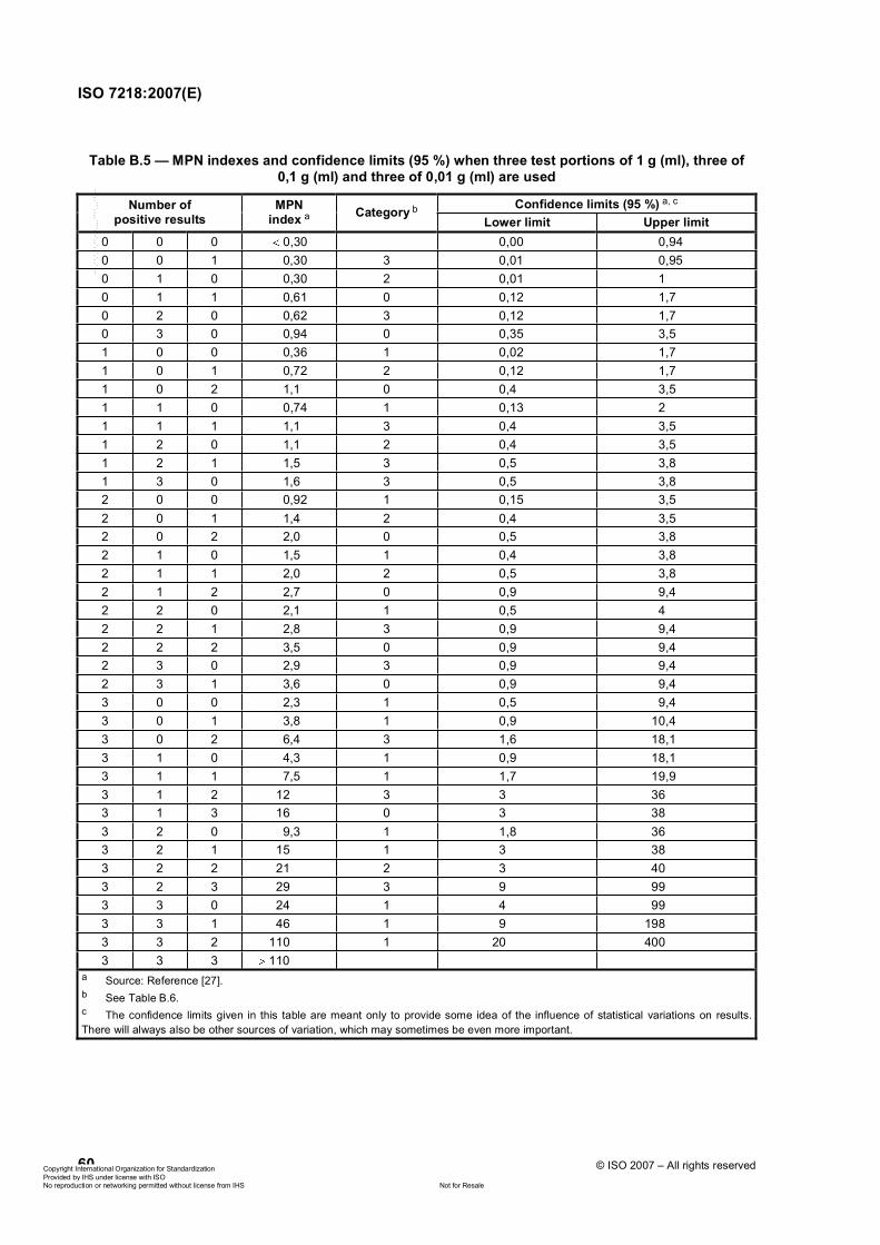
ISO 7218:2007(E)
60 © ISO 2007 – All rights reserved
Table B.5 — MPN indexes and confidence limits (95 %) when three test portions of 1 g (ml), three of 0,1 g (ml) and three of 0,01 g (ml) are used
Confidence limits (95 %) a, cNumber of positive results
MPNindex a Category b
Lower limit Upper limit0 0 0 0,30 0,00 0,94 0 0 1 0,30 3 0,01 0,95 0 1 0 0,30 2 0,01 1 0 1 1 0,61 0 0,12 1,7 0 2 0 0,62 3 0,12 1,7 0 3 0 0,94 0 0,35 3,5 1 0 0 0,36 1 0,02 1,7 1 0 1 0,72 2 0,12 1,7 1 0 2 1,1 0 0,4 3,5 1 1 0 0,74 1 0,13 2 1 1 1 1,1 3 0,4 3,5 1 2 0 1,1 2 0,4 3,5 1 2 1 1,5 3 0,5 3,8 1 3 0 1,6 3 0,5 3,8 2 0 0 0,92 1 0,15 3,5 2 0 1 1,4 2 0,4 3,5 2 0 2 2,0 0 0,5 3,8 2 1 0 1,5 1 0,4 3,8 2 1 1 2,0 2 0,5 3,8 2 1 2 2,7 0 0,9 9,4 2 2 0 2,1 1 0,5 4 2 2 1 2,8 3 0,9 9,4 2 2 2 3,5 0 0,9 9,4 2 3 0 2,9 3 0,9 9,4 2 3 1 3,6 0 0,9 9,4 3 0 0 2,3 1 0,5 9,4 3 0 1 3,8 1 0,9 10,4 3 0 2 6,4 3 1,6 18,1 3 1 0 4,3 1 0,9 18,1 3 1 1 7,5 1 1,7 19,9 3 1 2 12 3 3 36 3 1 3 16 0 3 38 3 2 0 9,3 1 1,8 36 3 2 1 15 1 3 38 3 2 2 21 2 3 40 3 2 3 29 3 9 99 3 3 0 24 1 4 99 3 3 1 46 1 9 198 3 3 2 110 1 20 400 3 3 3 110
a Source: Reference [27]. b See Table B.6. c The confidence limits given in this table are meant only to provide some idea of the influence of statistical variations on results. There will always also be other sources of variation, which may sometimes be even more important.
ݱ°§®·¹¸¬ ײ¬»®²¿¬·±²¿´ Ñ®¹¿²·¦¿¬·±² º±® ͬ¿²¼¿®¼·¦¿¬·±² Ю±ª·¼»¼ ¾§ ×ØÍ «²¼»® ´·½»²» ©·¬¸ ×ÍÑ
Ò±¬ º±® λ¿´»Ò± ®»°®±¼«½¬·±² ±® ²»¬©±®µ·²¹ °»®³·¬¬»¼ ©·¬¸±«¬ ´·½»²» º®±³ ×ØÍ
óóÀôôÀÀÀôôôôÀÀÀÀóÀóÀôôÀôôÀôÀôôÀóóó
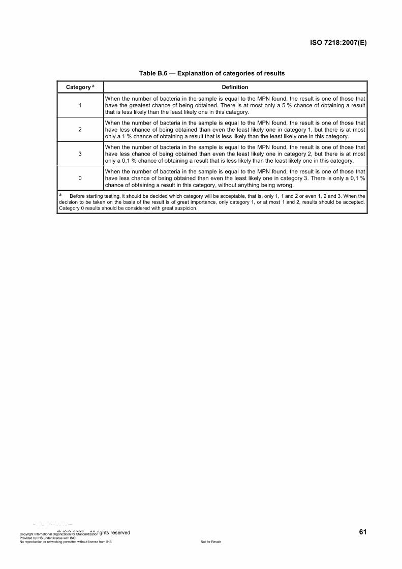
ISO 7218:2007(E)
© ISO 2007 – All rights reserved 61
Table B.6 — Explanation of categories of results
Category a Definition
1When the number of bacteria in the sample is equal to the MPN found, the result is one of those that have the greatest chance of being obtained. There is at most only a 5 % chance of obtaining a result that is less likely than the least likely one in this category.
2When the number of bacteria in the sample is equal to the MPN found, the result is one of those that have less chance of being obtained than even the least likely one in category 1, but there is at most only a 1 % chance of obtaining a result that is less likely than the least likely one in this category.
3When the number of bacteria in the sample is equal to the MPN found, the result is one of those that have less chance of being obtained than even the least likely one in category 2, but there is at most only a 0,1 % chance of obtaining a result that is less likely than the least likely one in this category.
0When the number of bacteria in the sample is equal to the MPN found, the result is one of those that have less chance of being obtained than even the least likely one in category 3. There is only a 0,1 % chance of obtaining a result in this category, without anything being wrong.
a Before starting testing, it should be decided which category will be acceptable, that is, only 1, 1 and 2 or even 1, 2 and 3. When the decision to be taken on the basis of the result is of great importance, only category 1, or at most 1 and 2, results should be accepted. Category 0 results should be considered with great suspicion.
ݱ°§®·¹¸¬ ײ¬»®²¿¬·±²¿´ Ñ®¹¿²·¦¿¬·±² º±® ͬ¿²¼¿®¼·¦¿¬·±² Ю±ª·¼»¼ ¾§ ×ØÍ «²¼»® ´·½»²» ©·¬¸ ×ÍÑ
Ò±¬ º±® λ¿´»Ò± ®»°®±¼«½¬·±² ±® ²»¬©±®µ·²¹ °»®³·¬¬»¼ ©·¬¸±«¬ ´·½»²» º®±³ ×ØÍ
óóÀôôÀÀÀôôôôÀÀÀÀóÀóÀôôÀôôÀôÀôôÀóóó
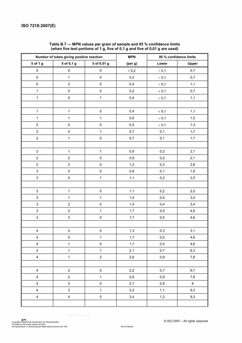
ISO 7218:2007(E)
62 © ISO 2007 – All rights reserved
Table B.7 — MPN values per gram of sample and 95 % confidence limits (when five test portions of 1 g, five of 0,1 g and five of 0,01 g are used)
Number of tubes giving positive reaction MPN 95 % confidence limits
5 of 1 g 5 of 0,1 g 5 of 0,01 g (per g) Lower Upper
0 0 0 0,2 0,1 0,7
0 1 0 0,2 0,1 0,7
0 2 0 0,4 0,1 1,1
1 0 0 0,2 0,1 0,7
1 0 1 0,4 0,1 1,1
1 1 0 0,4 0,1 1,1
1 1 1 0,6 0,1 1,5
2 0 0 0,5 0,1 1,3
2 0 1 0,7 0,1 1,7
2 1 0 0,7 0,1 1,7
2 1 1 0,9 0,2 2,1
2 2 0 0,9 0,2 2,1
2 3 0 1,2 0,3 2,8
3 0 0 0,8 0,1 1,9
3 0 1 1,1 0,2 2,5
3 1 0 1,1 0,2 2,5
3 1 1 1,4 0,4 3,4
3 2 0 1,4 0,4 3,4
3 2 1 1,7 0,5 4,6
3 3 0 1,7 0,5 4,6
4 0 0 1,3 0,3 3,1
4 0 1 1,7 0,5 4,6
4 1 0 1,7 0,5 4,6
4 1 1 2,1 0,7 6,3
4 1 2 2,6 0,9 7,8
4 2 0 2,2 0,7 6,7
4 2 1 2,6 0,9 7,8
4 3 0 2,7 0,9 8
4 3 1 3,3 1,1 9,3
4 4 0 3,4 1,2 9,3
ݱ°§®·¹¸¬ ײ¬»®²¿¬·±²¿´ Ñ®¹¿²·¦¿¬·±² º±® ͬ¿²¼¿®¼·¦¿¬·±² Ю±ª·¼»¼ ¾§ ×ØÍ «²¼»® ´·½»²» ©·¬¸ ×ÍÑ
Ò±¬ º±® λ¿´»Ò± ®»°®±¼«½¬·±² ±® ²»¬©±®µ·²¹ °»®³·¬¬»¼ ©·¬¸±«¬ ´·½»²» º®±³ ×ØÍ
óóÀôôÀÀÀôôôôÀÀÀÀóÀóÀôôÀôôÀôÀôôÀóóó
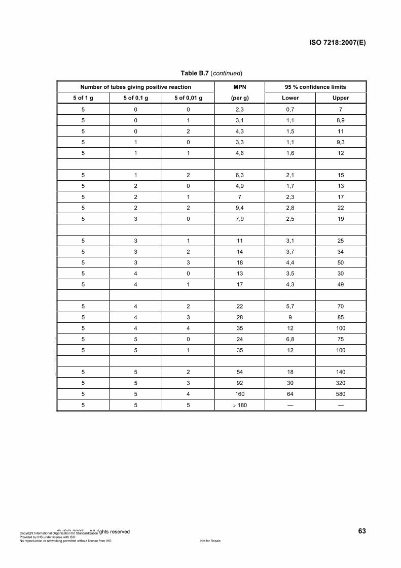
ISO 7218:2007(E)
© ISO 2007 – All rights reserved 63
Table B.7 (continued)
Number of tubes giving positive reaction MPN 95 % confidence limits
5 of 1 g 5 of 0,1 g 5 of 0,01 g (per g) Lower Upper
5 0 0 2,3 0,7 7
5 0 1 3,1 1,1 8,9
5 0 2 4,3 1,5 11
5 1 0 3,3 1,1 9,3
5 1 1 4,6 1,6 12
5 1 2 6,3 2,1 15
5 2 0 4,9 1,7 13
5 2 1 7 2,3 17
5 2 2 9,4 2,8 22
5 3 0 7,9 2,5 19
5 3 1 11 3,1 25
5 3 2 14 3,7 34
5 3 3 18 4,4 50
5 4 0 13 3,5 30
5 4 1 17 4,3 49
5 4 2 22 5,7 70
5 4 3 28 9 85
5 4 4 35 12 100
5 5 0 24 6,8 75
5 5 1 35 12 100
5 5 2 54 18 140
5 5 3 92 30 320
5 5 4 160 64 580
5 5 5 180 — —
ݱ°§®·¹¸¬ ײ¬»®²¿¬·±²¿´ Ñ®¹¿²·¦¿¬·±² º±® ͬ¿²¼¿®¼·¦¿¬·±² Ю±ª·¼»¼ ¾§ ×ØÍ «²¼»® ´·½»²» ©·¬¸ ×ÍÑ
Ò±¬ º±® λ¿´»Ò± ®»°®±¼«½¬·±² ±® ²»¬©±®µ·²¹ °»®³·¬¬»¼ ©·¬¸±«¬ ´·½»²» º®±³ ×ØÍ
óóÀôôÀÀÀôôôôÀÀÀÀóÀóÀôôÀôôÀôÀôôÀóóó
ISO 7218:2007(E)
64 © ISO 2007 – All rights reserved
Bibliography
[1] BELL, C. NEAVEs, P. and WILLIAMS, A.P. Food microbiology and laboratory practice, Blackwell Science Ltd, Oxford, UK (2005)
[2] ISO 9001, Quality management systems — Requirements
[3] EA-Guidelines for the use of computers and computer systems in accredited laboratories, European cooperation for Accreditation (1998)
[4] EA-4/10, Accreditation for microbiological laboratories, European cooperation for Accreditation (2002)
[5] Food microbiology program requirements, based upon the FLAWG document United States accreditation criteria for laboratories performing food microbiological testing, American Association for Laboratory Accreditation (A2LA) (1998)
[6] AS 1766, Australian standard methods for the microbiological examination of food — Part 1: General techniques and procedures update
[7] BUTTIAUX, R., BEERENS, H. and TACQUET, A. Manuel de techniques bactériologiques, Éditions Médicales Flammarion (4th edn.)
[8] APHA Technical Committee on Microbiological Methods for Foods. Compendium of methods for the microbiological examination of foods, 2nd edition, Speck, M.L., editor. Intersociety/Agency Committee on Microbiological Methods for Foods, American Public Health Association, Washington, DC, 1984
[9] CRUICKSHANK et al., Medical microbiology, Vol. 2, 12th edn., Churchill-Livingstone, Edinburgh (1975)
[10] D’AOUST, J.Y. Psychrotrophy and foodborne Salmonella. Int. J. Food. Microbiol. 1991, 13, pp. 207-16
[11] D’AOUST, J.Y., SEWELL, A.M. and MCDONALD, C. Recovery of Salmonella spp. from refrigerated pre-enrichment cultures of dry food composites, J. AOAC Int. 1995, 78, pp. 1322-7
[12] D’AOUST, J.Y., SEWELL, A.M. and GRECO, P. Detection of Salmonella in dry foods using refrigerated pre-enrichment and enrichment broth cultures: summary of collaborative study. J. AOAC. Int. 1994, 77,pp. 1490-1
[13] D’AOUST, J.Y., SEWELL, A.M. and GRECO, P. Detection of Salmonella in dry foods using refrigerated pre-enrichment and enrichment broth cultures — Interlaboratory study, J. AOAC. Int. 1993, 76,pp. 814-21.
[14] DALSGAARD, A., GUARDABASSI, L., LUND, C., BAGGE, L. and GRAVESEN, J. Opbevaring af badevands-og drikkevandsprøver ved 0-5 °C i et døgn medfører en signifikant reduktion i antal Escherichia coli og kimtal, Dansk. Vet. 2002 17, pp. 1-9
[15] HARREWIJIN, G.A., and HARTOG, B.J. Guidelines to perform microbiological analyses of food and food products (“Good laboratory practice”), De Ware(n)-Chemicus 1979, 9, pp. 1-11
[16] MUIR, G.D., ed. Hazards in the chemical laboratory, Royal Institute of Chemistry, London (1971)
[17] Laboratory biosafety manual, WHO, Geneva, 1993
[18] LIGHTFOOT, N.F., MAIER, E.A. (eds). Microbiological analysis of food and water — Guidelines for quality assurance. Elsevier, Amsterdam, Netherlands (1998)
[19] HARRIGAN, W.F. and MCCANCE, M.E. Laboratory methods in food and dairy microbiology, Academic Press (1976)
ݱ°§®·¹¸¬ ײ¬»®²¿¬·±²¿´ Ñ®¹¿²·¦¿¬·±² º±® ͬ¿²¼¿®¼·¦¿¬·±² Ю±ª·¼»¼ ¾§ ×ØÍ «²¼»® ´·½»²» ©·¬¸ ×ÍÑ
Ò±¬ º±® λ¿´»Ò± ®»°®±¼«½¬·±² ±® ²»¬©±®µ·²¹ °»®³·¬¬»¼ ©·¬¸±«¬ ´·½»²» º®±³ ×ØÍ
óóÀôôÀÀÀôôôôÀÀÀÀóÀóÀôôÀôôÀôÀôôÀóóó

ISO 7218:2007(E)
© ISO 2007 – All rights reserved 65
[20] Laboratory safety at the Centers for Disease Control (CDC), NHEW Publication No. CDC 79-8118, Atlanta, 1979
[21] Laboratory safety at the Centers for Disease Control, US Dept of Health, Education and Welfare (Public Health Service), Atlanta, 1979
[22] GERHARDT, P., MURRAY, R.G.E., COSTILLOW, R.N., NESTER, E.W., KRIEG, N.R. and PHILLIPS, G.B. eds. Manual of methods for general bacteriology. American Society for Microbiology, Washington, DC 20006 (1981)
[23] GNANOU BESSE, N., AUDINET, N., BEAUFORT, A., COLIN, P., CORNU, M. and LOMBARD, B. A contribution to the improvement of Listeria monocytogenes enumeration in cold-smoked salmon. Int. J. Food Microbiol. 2004, 91, pp. 119-27
[24] Microbiological testing laboratory accommodation guidelines, National Association of Testing Authorities, Australia
[25] Microorganisms in foods — 1: Their significance and methods of enumeration, ICMSF, University of Toronto Press (1968 update)
[26] SHAPTON, D.A., BOARD, R.G. and HAUSTER, W.J. eds. Safety in microbiology, Society for Applied Bacteriology Technical Series No. 1 and No. 6, Academic Press (1972)
[27] DE MAN, J.C. MPN tables (corrected), Eur. J. Appl. Biotechnol. 1983, 17, pp. 301-5
[28] COCHRAN, W.G. Estimation of bacterial densities by means of the “Most Probable Number”. Biometrics1950, 6, pp. 105-16
[29] HURLEY, M.A. and ROSCOE, M.E. Automated statistical analysis of microbial enumeration by dilution series, J. Appl. Bacteriol. 1983, 55, pp. 159-64
[30] NIEMELA, S. Statistical evaluation of results from quantitative microbiological examinations, Nordic Committee on Food Analysis (NMKL) Report No. 1, 2nd edition (1983)
[31] TAYLOR, J. The estimation of numbers of bacteria by tenfold dilution series, J. Appl. Bacteriol. 1962, 25,pp. 54-61
[32] HOLT, J.G., KRIEG, N.R., SNEATH, P.H.A., STALEY, J.T. and WILLIAMS, S.T. Bergey’s manual of determinative bacteriology, 9th edn., Williams & Wilkins, Baltimore, MD, USA (1994)
[33] KRIEG, N.R. and HOLT, J.G. Bergey’s manual of systematic bacteriology — Volume 1, Williams & Wilkins, Baltimore, MD, USA (1984)
[34] SNEATH, P.H.A., MAIR, N.S., SHARPE, M.E. and HOLT, J.G. Bergey’s manual of systematic bacteriology — Volume 2, 2nd edition, Springer, New York, NY, 2001
[35] KREGER-VAN RIJ, N.J.W. The yeasts — A taxonomic study, 3rd edn., North-Holland Publishing Co., Amsterdam, Netherlands
[36] THOMAS, H.A. Bacterial densities from fermentation tube tests, J. Am. Water Works Assoc. 1942, 34,pp. 572-6
[37] ISO/IEC Guide 43-1, Proficiency testing by interlaboratory comparisons — Part 1: Development and operation of proficenciency testing schemes
[38] ISO 6888 (all parts), Microbiology of food and animal feeding stuffs — Horizontal method for the enumeration of coagulase-positive staphylococci (Staphylococcus aureus and other species)
ݱ°§®·¹¸¬ ײ¬»®²¿¬·±²¿´ Ñ®¹¿²·¦¿¬·±² º±® ͬ¿²¼¿®¼·¦¿¬·±² Ю±ª·¼»¼ ¾§ ×ØÍ «²¼»® ´·½»²» ©·¬¸ ×ÍÑ
Ò±¬ º±® λ¿´»Ò± ®»°®±¼«½¬·±² ±® ²»¬©±®µ·²¹ °»®³·¬¬»¼ ©·¬¸±«¬ ´·½»²» º®±³ ×ØÍ
óóÀôôÀÀÀôôôôÀÀÀÀóÀóÀôôÀôôÀôÀôôÀóóó
ISO 7218:2007(E)
66 © ISO 2007 – All rights reserved
[39] ISO 9998, Water quality — Practices for evaluating and controlling microbiological colony count media used in water quality tests
[40] ISO/TR 13843, Water quality — Guidance on validation of microbiological methods
[41] ISO 14461-1, Milk and milk products — Quality control in microbiological laboratories — Part 1: Analyst performance assessment for colony counts
[42] ISO 14461-2, Milk and milk products — Quality control in microbiological laboratories — Part 2: Determination of the reliability of colony counts of parallel plates and subsequent dilution steps
[43] ISO 16654, Microbiology of food and animal feeding stuffs — Horizontal method for the detection of Escherichia coli O157
[44] ISO/IEC 17025:2005, General requirements for the competence of testing and calibration laboratories
[45] EN 12469, Biotechnology — Performance criteria for microbiological safety cabinets
[46] ISO 17604, Microbiology of food and animal feeding stuffs — Carcass sampling for microbiological analysis
[47] ISO 18593, Microbiology of food and animal feeding stuffs — Horizontal method for sampling techniques from surfaces using contact plates and swab methods
[48] ISO Guide 99:1996, International vocabulary of basic and general terms in metrology
[49] ISO/TC 34/SC 9 N852, Supporting document on the change from two to one plate per dilution for colony-count techniques (revision of ISO 7218), June 2007, Marie Cornu
ݱ°§®·¹¸¬ ײ¬»®²¿¬·±²¿´ Ñ®¹¿²·¦¿¬·±² º±® ͬ¿²¼¿®¼·¦¿¬·±² Ю±ª·¼»¼ ¾§ ×ØÍ «²¼»® ´·½»²» ©·¬¸ ×ÍÑ
Ò±¬ º±® λ¿´»Ò± ®»°®±¼«½¬·±² ±® ²»¬©±®µ·²¹ °»®³·¬¬»¼ ©·¬¸±«¬ ´·½»²» º®±³ ×ØÍ
óóÀôôÀÀÀôôôôÀÀÀÀóÀóÀôôÀôôÀôÀôôÀóóó

ݱ°§®·¹¸¬ ײ¬»®²¿¬·±²¿´ Ñ®¹¿²·¦¿¬·±² º±® ͬ¿²¼¿®¼·¦¿¬·±² Ю±ª·¼»¼ ¾§ ×ØÍ «²¼»® ´·½»²» ©·¬¸ ×ÍÑ
Ò±¬ º±® λ¿´»Ò± ®»°®±¼«½¬·±² ±® ²»¬©±®µ·²¹ °»®³·¬¬»¼ ©·¬¸±«¬ ´·½»²» º®±³ ×ØÍ
óóÀôôÀÀÀôôôôÀÀÀÀóÀóÀôôÀôôÀôÀôôÀóóó

ISO 7218:2007(E)
ICS 07.100.30 Price based on 66 pages
© ISO 2007 – All rights reserved ݱ°§®·¹¸¬ ײ¬»®²¿¬·±²¿´ Ñ®¹¿²·¦¿¬·±² º±® ͬ¿²¼¿®¼·¦¿¬·±² Ю±ª·¼»¼ ¾§ ×ØÍ «²¼»® ´·½»²» ©·¬¸ ×ÍÑ
Ò±¬ º±® λ¿´»Ò± ®»°®±¼«½¬·±² ±® ²»¬©±®µ·²¹ °»®³·¬¬»¼ ©·¬¸±«¬ ´·½»²» º®±³ ×ØÍ
óóÀôôÀÀÀôôôôÀÀÀÀóÀóÀôôÀôôÀôÀôôÀóóó


![IS 1885-34 (1972): Electrotechnical vocabulary, Part 34: Cinematography · Cinematography [ETD 1: Basic Electrotechnical Standards] ‘is:.m~~ (&tXXXIV)-1972 Indian Standard ELECTROTECHNICAL](https://static.fdocuments.us/doc/165x107/5fe030e6cbc55d4a9b147853/is-1885-34-1972-electrotechnical-vocabulary-part-34-cinematography-cinematography.jpg)