


J Glaucoma Volume 20, Number 5, June/July 2011 R1 何元輝 2011/09/15

VOLUME 21-22021
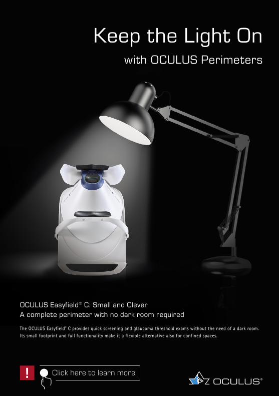
OCULUS Easyfield® C: Small and Clever A complete perimeter with no dark room required
The OCULUS Easyfield® C provides quick screening and glaucoma threshold exams without the need of a dark room. Its small footprint and full functionality make it a flexible alternative also for confined spaces.
Keep the Light On with OCULUS Perimeters
Click here to learn more!

INTERNATIONAL GLAUCOMA REVIEWA Quarterly JournalVolume 21 no. 2
Chief Editor Robert N. WeinrebContributing EditorsChristopher Leung (HK), Kaweh Mansouri (Switzerland), Arthur Sit (US)Associate EditorsMakoto Araie (JP), Jonathan Crowston (AU), Ki Ho Park (KR), Jeffrey Liebmann (US), Remo Susanna (BR)
Society EditorsEllen Ancker (SAGS), Makoto Araie (JGS and APGS), Anne M. Brooks (ANZGIG), Seng Kheong Fang(APGS), Christopher Girkin (AGS), Francesco Goñi (EGS), Rodolfo Perez Grossman (LAGS), Rajul Parikh (GSI), Marcello Nicolela (CanGS), Mike Patella (OGS), Tarek Shaarawy (ISGS), Patricio Schlottmann (PAGS), Fotis Topouzis (EGS), Moustafa Yaqub (MEAGS), Ningli Wang (ChinGS)
Board of EditorsMakoto Aihara (JP), Tadamichi Akagi (JP), Lee Alward (US), Alfonso Anton (SP), Leon Au (UK), Tin Aung (SG), Augusto Azuara Blanco (UK), Keith Barton (UK), Christoph Baudouin (FR), Eytan Blumenthal (IS), Andreas Boehm (DE), Rupert Bourne (UK), Chris Bowd (US), Andrew Camp (US), Subho Chakrabarthi (IN), Jack Cioffi (US), Anne Coleman (US), Tanuj Dada (IN), Gustavo DeMoraes (US), Robert Fechtner (US), Robert Feldman (US), Murray Fingeret (US), David Friedman (US), Jiang Ge (CN), Chris Girkin (US), Ivan Goldberg (AU), David Greenfield (US), Franz Grehn (DE), Neeru Gupta (CA), Alon Harris (US), Mingguang He (CN), Paul Healey (AU), Esther Hoffman (DE), Gabor Holló (HU), Alex Huang (US), Henry Jampel (US), Chris Johnson (US), Jost Jonas (DE), Malik Kahook (US), Kenji Kashiwagi (JP), Tae Woo Kim (KR), Dennis Lam (HK), George Lambrou (GR), Fabian Lerner (AR), Christopher Leung (HK), Shan Lin (US), John Liu (US), Nils Loewen (US), Steve Mansberger (US), Keith Martin (UK), Eugenio Maul (CL), Stefano Miglior (IT), Sasan Moghimi (IR), Sameh Mosaed (US), Kouros Nouri-Madhavi (US), Paul Palmberg (US), Louis Pasquale (US), Norbert Pfeiffer (DE), Luciano Quaranta (IT), Pradeep Ramulu (US), Harsha Rao (IN), Tony Realini (US), Doug Rhee (US), Prin RojanaPongpun (TH), Joel Schuman (US), Tarek Shaarawy (CH), Takuhei Shoji (JP), Kuldev Singh (US), Arthur Sit (US), George Spaeth (US), Min Hee Suh (US), Ernst Tamm (DE), Hidenobu Tanihara (JP), Andrew Tatham (UK), Fotis Topouzis (GR), Anja Tuulonen (FI), Rohit Varma (US), Ningli Wang (CN), Derek Welsbie (US), Tina Wong (SG), Benjamin Xu (US), Yeni Yücel (CA), Linda Zangwill (US)
Abstract EditorGeorge Lambrou (GR)
ISSN 1566-1040

Information on the member Glaucoma Societies of the WGA can be found in the WGA Global Directory of Glaucoma Societies at www.wga.one/wga/directory-of-glaucoma-societies
RegistrationAccess to IGR Online is complimentary for all members of glaucoma societies affiliated to the WGA. As of 2018, access to IGR is arranged through WGA#One; see next page for details. Should you have any questions, please contact us at [email protected]
Find us on Facebook: www.facebook.com/worldglaucomaFind us on Twitter: www.twitter.com/WorldGlaucomaFind us on LinkedIn: www.linkedin.com/company/world-glaucoma-associationWGA#One FAQ: www.wga.one/faq
ISSN 1566-1040
Contact InformationAll correspondence on copies, supplements, content, advertising, etc. should be directed to:WGA Executive Officec/o Schipluidenlaan 41062 HE AmsterdamThe NetherlandsTel: +31 20 570 9600E-mail: [email protected]
Published by Kugler Publications, P.O. Box 20538, 1001 NM Amsterdam, The Netherlands, on behalf of the World Glaucoma Association.Cover design: Cees van Rutten, The Hague, The NetherlandsTypesetting: 3bergen, www.3bergen.com
© 2021. World Glaucoma AssociationNo part of this publication may be reproduced, stored in a retrieval system, or transmitted in any form by any means, electronic, mechanical, photocopying or otherwise, without the prior consent of the copyright owners.
facebook TWITTER LINKEDIN

WGA#OneWGA#One is the name of the World Glaucoma Association’s customer relationship management system. With WGA#One we are moving forward towards one platform, and hence one user profile, for all our services.WGA#One is facilitating our communications about and access to our services, offers and initiatives. Therefore it’s very important to keep your WGA#One profile updated. See below for details on how to activate your account for the first time.Communicating effectively is key, and thus we extended our basic user profile with the option to activate different information preferences:
þ 1 - Monthly newsletter A concise monthly digest of all WGA activities, such as congresses, publications, courses, projects, gover-nance, scientific content, awareness activities etc. Find the archive here to get a taste: www.wga.one/wga/newsletter-archive
þ 2 - Glaucoma awareness initiatives Information on awareness activities, such as World Glaucoma Week
þ 3 - Educational & scientific content For example: Consensus statements/publications, International Glaucoma review, Journal of Glaucoma, recorded WGC session/enduring materials, etc.
In just a few clicks you’ll be ensured to stay in touch and receive the latest news according to your own preferences. We never share your information with third parties.
Your privacy is very important to us, so please see our privacy policy atwww.wga.one/terms-and-conditions
How to activate your WGA#One profile1. Please visit www.wga.one/activate to activate your WGA#One profile.2. Enter your email address (use the address where you are currently receiving our
communications).3. You will receive an email with an activation link (if not received, check your spam
folder first before contacting [email protected]).4. Click on the link, create a new password, and update your WGA#One profile.If none of your email addresses is found in the system you can either contact us at [email protected], or subscribe to our newsletter at: www.wga.one/wga/subscribe-to-newsletter

5
IGR 21-2 Table of Contents
Table of ContentsFrom the WGA Executive Office 8Spotlight Article 11Your Special Attention For 20Editor’s Selection, with contributions by Dong Feng Chen, David Crabb, Gustavo de Moraes, Crawford Downs, Ross Ethier, Alex Huang, Thomas Johnson, Pete Jones, Shan Lin, Steve Mansberger, Sameh Mosaed, Toru Nakazawa, Kouros Nouri-Mahdavi, Joel R. Palko, Louis Pasquale, Vincent Michael Patella, Dorota Skowronska-Krawczyk, Daniel Stamer, Yang Sun, Remo Susanna Jr, Ningli Wang and Diya Yang 22Journal of Glaucoma 58News Flashes 60
All abstracts are available online in the classified IGR searchable glaucoma databasewww.e-IGR.com
The affiliations of the contributors to this issue can be found on www.e-IGR.com.
www.e-IGR.com

The world glaucomacongress 2021 is going beyond borders!
Organized by
Virtually hosted by
9th WORLD GLAUCOMA E-CONGRESSBEYOND BORDERSJUNE 30 - JULY 3, 2021
www.worldglaucomacongress.org
8
IGR 21-2 From the WGA Executive Office
From the WGA Executive OfficeDear IGR readers,All of us at the Executive Office of the World Glaucoma Association hope that you and your loved ones are doing well and staying healthy during this COVID-19 pandemic. The introduction of new vaccines brings much needed hope during this period of global uncertainty. With the challenges presented by the pandemic, there have also been opportunities to reach out to more of our members through educational webinars, videos, and online resources. We are very proud to inform you about our exciting new WGA Global Webinars and update you about the World Glaucoma Congress 2021.The inaugural WGA Global Webinar on October 10, 2020, drew over 9,000 views; and the recent one on December 19, 2020, had over 13,000 views through YouTube, the internet, and Facebook. Depending on where you are in the world, the next Global Webinar will take place on February 12-13, 2021, and will cover the topic of Angle-Closure Glaucoma. Please join Drs. Robert Weinreb and Fabian Lerner who will lead a panel of worldwide experts to update you on the latest diagnostic techniques and treatments for this aggres-sive form of glaucoma. There will also be extensive discussion of the many controversies related to the management of ACG. Keep an eye out for the WGA announcements about how to register for the webinar.We wish to announce that the World Glaucoma Congress in 2021 will be all-virtual and will be hosted together with the Japanese Glaucoma Society. Since we are no longer limited by travel and in-person restrictions due to the epidemic, we have decided to move the meeting back to our traditional period. The World Glaucoma Congress 2021 will take place June 30-July 3, 2021. Although we will not be meeting in person in Japan, our first virtual WGC will have a Japanese theme, lectures and sessions held in conjunction with the JGS, and ‘social’ activities infused with a Japanese flavor. Registration opens February 2021. See you virtually at the WGC!Our many committees continue to work hard for the benefit of our members and our patients. In fact, we have expanded our committee activities during this difficult period of COVID to provide greater access to our educational materials. An example is the recent translations of our educational materials for patients into Malay. This is now available at www.glaucomapatients.org.Please stay safe and healthy into 2021. Our prayers to you and our entire world for a return to normalcy and the privilege of being able to meet once again in person, so that we may catch up with one another and continue to collaborate.

9
IGR 21-2 From the WGA Executive Office
GET TO KNOW US!Carlien Turkstra
Since September 2020 Carlien Turkstra has been involved with the WGA as a Community Coordinator, working closely together with Irene Koomans, the WGA Executive General Manager, and Marije de Graaf, Operations Manager. The past months have been filled with learning moments about WGA, our operations, our core purpose & values, and most importantly, our commu-nity. As Community Coordinator of WGA, Carlien is the liaison between the societies and WGA: she is the main point of contact for our society representatives. Carlien is looking forward to optimizing our glaucoma community and assisting our society members in getting the most out of their membership.
WGA website: www.wga.oneWGA on Facebook: www.facebook.com/worldglaucoma
WGA on Twitter: www.twitter.com/WorldGlaucomaWGA on LinkedIn: www.linkedin.com/company/world-glaucoma-association
facebook TWITTER LINKEDIN

Practical articlesExpert interviewsNews and insights
View | Download | SubcribeFREE
Touch Medical Media Limited is a private limited company registered in England - registered number 08197142
touchOPHTHALMOLOGY.com
Peer reviewed | Free-to-access | Concise | Multimedia

11
IGR 21-2 Spotlight Article
Spotlight Article86863 Reprogramming to recover youthful epigenetic information and restore vision. Lu Y, Brommer B, Tian X, Krishnan A, Meer M, Wang C, Vera DL, Zeng Q, Yu D, Bonkowski MS, Yang JH, Zhou S, Hoffmann EM, Karg MM, Schultz MB, Kane AE, Davidsohn N, Korobkina E, Chwalek K, Rajman LA, Church GM, Hochedlinger K, Gladyshev VN, Horvath S, Levine ME, Gregory-Ksander MS, Ksander BR, He Z, Sinclair DA. Nature 2020; 588(7836):124-129
pencil-alt Comment by Keith Martin, Melbourne, Australia
There has been much recent interest in the idea that an accumulation of epigenetic changes contributes to the effects of aging, including reduction in resistance to injury and loss of regenerative capacity in older animals. But can this process be reversed in order to boost injury resistance and regeneration in the optic nerve and other tissues?Previous work by Ocampo et al. (Ocampo, A. et al. Cell 2016;167:1719-1733) explored the effects of expressing four genes (encoding MYC, OCT4, SOX2 and KLF4 ‒ the so-called Yamanaka transcription factors) in mice genetically engineered to exhibit accelerated ageing. Turning these genes on for a few days, then turning them off again, led to mice which seemed to age more slowly with epigenetic features expected in much younger animals. However, multiple studies have reported an increased risk of tumor forma-tion when the Yamanaka factors are used for cellular reprogramming.In the current study, Lu et al. used nicely engineered AAV vectors to deliver Yamanaka factors to the eye by intravitreal injection. They argued that MYC was the most likely factor to cause tumors and was not necessary for the reprogramming effect and therefore used only three transcription factors (OSK) in their experiments. Interestingly, the results seemed to support this idea strongly, with no tumors observed in long-term experiments in mice extending over 15 months.Expression of the OSK factors in inner retinal cells could be switched on or off by exposure to an antibiotic in a clever use of a conditional expression vector system. The authors interpret their findings as evidence that AAV-OSK promotes axon regeneration after optic nerve injury, and improves some measures of visual function in a mouse model of glaucoma (microbeads injected into the anterior chamber) and in aged mice. The beneficial effects of AAV-OSK seemed to require the DNA demethylases TET1 and TET2. The authors suggest that their experiments indicate that mammalian tissues retain a record of youthful epigenetic information that can be accessed to improve optic nerve function and promote regeneration in vivo.

12
IGR 21-2 Spotlight Article
As is often the case with strong science, the experiments raise at least as many questions as they answer
These findings, published in Nature, are certainly of considerable interest and, as is often the case with strong science, the experiments raise at least as many questions as they answer. The magnitude of the observed regenerative effect after optic nerve crush, with some regenerating axons reaching the optic chiasm but little sign of functional improve-ment, was similar to or less than what has previously been reported using several other approaches. However, the reported effect in the microbead glaucoma model should be of particular interest to readers of IGR. In this model, IOP is elevated for about three weeks and then returns to baseline, with around 20-30% RGC loss observed during this time. When administered four weeks after IOP elevation, the authors found that their AAV-OSK seemed to improve optic nerve axonal density to normal (without RGC proliferation) and also improve some measures of optic nerve function (PERG and optokinetic responses). The apparent efficacy of a treatment delivered weeks after induction of IOP elevation certainly catches the attention, although it is not clear from the data presented how the suggested reversal of axonal density reduction in particular was mediated. In the authors’ defense, whereas axons regenerating beyond an optic nerve crush site are relatively easy to identify, it is much more difficult to identify regenerating axons in glaucoma models. This is an important technical problem in this field and, until we solve it, we should be cautious in attributing any improvements in axon counts and some measures of visual function in glaucoma models to regeneration rather than survival and delayed recovery.
We should be cautious in attributing any improvements in axon counts and some measures of visual function in glaucoma models to regeneration rather than survival and delayed recovery
Overall, this is a challenging and important study that should stimulate even more activity in this exciting field. From the glaucoma perspective, it would be great to see the key findings replicated in other labs and using other injury models and outcome measures. As ever, the relevance to human disease remains to be seen at this stage, but there are plenty of intriguing possibilities.

13
IGR 21-2 Spotlight Article
pencil-alt Comment by Harry Quigley, Baltimore, MD, USA
Lu et al. state ‘Compared with glaucomatous eyes that received either PBS or AAVs with no OSK induction (−OSK), the OSK-treated glaucomatous eyes (+OSK) presented with a restored axon density equivalent to that in the non-glaucomatous eyes’ and ‘the optomotor response assay indicated that half of the visual acuity lost from increased intraocular pressure was restored.’ Careful inspection of their data as presented do not support these statements.Mice had bead-induced glaucoma for four weeks, were then intravitreally injected with a viral vector overexpressing three factors (OSK) and followed four more weeks. Retinal ganglion cell (RGC) bodies in retina and axons in the optic nerve were counted and two functional tests were performed. Regarding RGC density, the loss of RGC somas at four weeks (Fig. 9b) is shown for only five eyes with values differing by < 5% among them. This remarkable lack of variation is inconsistent with either normal variation as seen in their control RGC densities (and our mouse studies) or with the typical variability in RGC cell loss among mouse glaucoma eyes. More importantly, Figure 9d shows that despite injection of OSK vector, there was significant loss of RGC bodies at eight weeks that was not significantly different from the control vector or saline injection controls. In fact, there was no ‘rescue or restoration’ of RGC somas.Assessment of axons was given as ‘density’ and not total axon number, the latter being the definitive method for axon counting. Various treatments (such as +OSK) may induce changes in axon density, but not in overall axon count (or vice versa), due to alterations in nerve astrocytes (Schaub et al. IOVS 2017) which affects optic nerve cross-sectional area. In addition, axon counts cannot be carried out in the same eyes at four weeks (prior to vector injection) and at eight weeks, so the axonal loss in different eyes is being compared, in as few as six per group, far smaller than needed to assure that variability has been accounted. In conducting bead glaucoma studies in > 1,000 mice, we find samples < 20 eyes per group are insufficient reliably to detect treatment effects.Individual IOP exposure may be responsible for observed differences and are not presented. While the authors show one IOP graph in non-vector, bead-treated eyes (Fig. 3b), they do not account for IOP as a covariate in RGC axon and soma data. Perhaps animals with OSK injections, studied at eight weeks, had lower IOP exposure than the four week group, so any ‘recovery’ could simply be failure to achieve as much damage. Indeed, the investigators removed ‘mice that had […] clouding or an oedematous cornea.’ IOP increase in mice causes axial elongation, corneal steepening and edema, which are greater with higher induced IOP. The investigators may have systematically removed higher IOP eyes, minimizing damage at eight weeks. How many eyes were not included and was this more common in +OSK vector groups?

14
IGR 21-2 Spotlight Article
Therefore, these data require replication with the addition of numerous critical controls
They state: ‘the optomotor response assay indicated that half of the visual acuity lost from increased intraocular pressure was restored (Fig. 3c,d).’ While precise descriptive statistics are not provided, from Figure 3, control mice scored ~37 cycles/degree (cpd), while mice after four weeks IOP elevation scored ~25 cpd. The same +OSK group at eight weeks was only ~27 cpd, a 16% increase in the 12 cpd lost at four weeks; not ‘half restored’ (as claimed) and still 27% below baseline control values. In optomotor data, one should compare the acuity loss at four weeks to its value at eight weeks for each eye, not as group data. Again, individual IOP was not included in appropriate statistical models for acuity and ERG analysis. Furthermore, it would be informative to show correlative RGC soma and axon loss in the functional study animals. Presumably, mice could see from both eyes during the optomotor testing. Since only one eye had experimental glaucoma, its true effect on function is problematic. If one eye were closed (e.g., lid suture) we could determine that vision effects were actually due to change in the glaucoma eye.Therefore, these data require replication with the addition of numerous critical controls.
pencil-alt Comment by Dorota Skowronska-Krawczyk, Irvine, CA, USA
Age is one of the most relevant clinical traits in predicting disease risk, mental and physical performance, mortality, and other important health issues. Given the increased lifespan and decreased fertility, the average population age is anticipated to significantly increase in the next few decades, bringing the wealth of interest in studying aging and improving quality of life in advanced age individuals. On a molecular level, aging is associated with a gradual decline in the efficiency and accuracy of molecular processes, including changes in gene expression and epigenetics, leading to a deterioration of cell functions and regenerative capacity. Epigenetic aging of tissues and organs has been tightly correlated with global genome hypomethylation accompanied by specific regions, called CpG islands, hypermethylation. The rates of methylation changes at subsets of affected sites were calculated and used to determine the cellular ‘epigenetic’ age, which generally well correlates with chronological age and therefore allows to assess biological aging in a quantitative manner,1,2 feature beautifully used in the reviewed work of Lu et al.

15
IGR 21-2 Spotlight Article
In this work, David Sinclair’s team seeks to restore the youthful epigenetic landscape of retinal ganglion cells (RGCs) to increase their regenerative capability. In a series of elegant experiments, the group has shown that concomitant overexpression of Oct4, Sox2, and Klf4 (OSK) pluripotency factors in RGCs allows restoring vision in several mouse models:1. After the optic nerve crush injury, overexpression of OSK factors in the retina was able to induce robust axon regrowth in the optic nerve without inducing the cell division. This ability was dependent on the presence of DNA demethylation enzymes, Tet proteins, which expression is upregulated upon OSK expression.2. In the microbeads-induced mouse model of glaucoma, OSK overexpression four weeks after the beads injection, is able to restore vision by increasing the number of healthy axons, again without inducing the RGC proliferation.3. In 12 months old animals, overexpression of OSK factors improved optomotor response, visual acuity, and partially restored transcriptional program as seen in younger animals.Several points were not fully described and will require further studies. As shown in several figures, there is a set (or sets?) of CpGs that are demethylated in the given model but are re-methylated after the OSK factors overexpression (e.g., Fig. 2e, Extended Data Fig. 5j). What are the genes that have this specific dynamic of methylation? Is it a partic-ular group of genes? Are they close to repeats or non-coding RNAs? What is the mecha-nism of the re-methylation after OSK overexpression? The team has several interesting avenues to pursue in the future.Finally, the authors ask: ‘how cells encode and store youthful epigenetic information’, suggesting that there is a particular program that can be stored by the cell. Although intriguing, this is not the only way to explain the results observed by the group. Similar to what happens in age-related methylation patterns, where sets of the same sites are methylated in aging in different tissues and organisms (which allowed the generation of ‘methylation clocks’), the same sites are demethylated in the experiments presented in the discussed paper. These two sets of observations might, in fact, suggest that there are sites more susceptible to epigenetic changes and that undirected approaches are preferentially modifying the same sites. Interestingly, this might explain why the overex-pression of other chromatin modifying enzymes also have a specific effect on the cell.3
The presented approach to treat age-related neurodegenerative diseases still requires adjustments before being brought to the clinic
The work of Lu and colleagues brings a fresh perspective on a current dogma of the inability of neurons to regenerate. The direct, quantitative measurement of aging through the methylation clock allows one to work on improvements and describing further the mechanism of the regenerative process. Pointing to the specific enzymes that could be involved in the process of rejuvenation of neurons further expands potential future applications. The presented approach to treat age-related neurodegen-erative diseases still requires adjustments before being brought to the clinic, for example

16
IGR 21-2 Spotlight Article
the design and use of safe overexpression system in human eye. Still, this work provides solid data increasing confidence in potential rejuvenating treatments for age-related eye conditions.
References1. Hannum G, Guinney J, Zhao L, et al. Genome-wide Methylation Profiles Reveal
Quantitative Views of Human Aging Rates. Mol Cell. 2013;49:359-367. doi:10.1016/j.molcel.2012.10.016.
2. Horvath S. DNA methylation age of human tissues and cell types. Genome Biology. 2013;14:R115. doi:10.1186/gb-2013-14-10-r115.
3. Chen B, Cepko CL. HDAC4 regulates neuronal survival in normal and diseased retinas. Science. 2009;323:256-259, doi:10.1126/science.1166226.
pencil-alt Comment by Derek Welsbie, La Jolla, CA, USA
For patients who have already lost vision from glaucoma, options are very limited and usually center around consultation with a low vision specialist. While dramatic lowering of the intraocular pressure (IOP) may lead to a very modest improvement in some patients, for the vast majority there are no treatments to restore vision. Several groups have shown that retinal ganglion cells (RGCs) injured in glaucoma enter a phase of axonal degeneration that precedes cell death, indicating the presence of injured-but-not-yet-dead cells. Thus, in order to restore vision, there have been a number of strategies devel-oped (with limited effectiveness) to regenerate axons in an attempt to reconnect these injured cells. In this article, Lu et al., from the labs of Bruce Ksander, Meredith Gregory-Ksander, Zhigang He and David Sinclair, demonstrate that epigenetic reprogramming of RGCs can lead to robust axon regeneration and partial restoration of vision, including in a mouse model of glaucoma.It is well-known that aging is a key risk factor for the development and worsening of glaucoma. Moreover, while developing, immature RGCs can extend axons, this capacity is greatly reduced in adult neurons. The question that Lu et al. addressed was whether RGCs could be partially reprogrammed, such that they de-age and increase their regenerative capacity, but not totally de-differentiate and lose their RGC identity. To test this, they turned to the Yamanaka factors, Myc, Oct4, Sox2 and Klf4, which convert cells into induced pluripotent stem cells (iPSCs). To avoid complete reprogramming (and to avoid the use of a potent oncogene), the authors excluded Myc and expressed Oct4, Sox2 and Klf4 cDNAs together using adeno-associated virus (AAV) and a tetracycline-regulated system. The viruses were injected intravitreally and the effect on RGC cell death and axon regeneration was tested using the mouse optic nerve crush (ONC) model. Typically, there

17
IGR 21-2 Spotlight Article
is profound cell death by two weeks and no meaningful axon regeneration. However, in RGCs expressing Oct4, Sox2 and Klf4, the team saw improved RGC survival coupled with axons regeneration at least to the level of the chiasm. Interestingly, by giving tetracycline on different schedules and altering the timing of expression, they found that the three genes had to be expressed after injury, suggesting that the effect was to reverse injury-in-duced changes.Since Yamanaka factors are known to change the epigenetic marks regulating gene expression, the authors measured DNA methylation across the genome. In response to ONC, they saw a change in the pattern of methylation, including increased methylation of ribosomal DNA, which indicates accelerated aging. In contrast, RGCs expressing Oct4, Sox2 and Klf4 had a nearly complete normalization of the DNA methylation pattern. Moreover, consistent with the model that partial reprogramming was removing the injury-induced methylation, the phenotype was dependent on the presence of cellular demethylation enzymes like TET1 and TET2. The expression of Oct4, Sox2 and Klf4 even reversed normal age-related vision loss in mice and was associated with a reversal of the methylation aging clock.
Clinically, it will be important to determine the abundance of injured-but-not-yet-dead RGCs that might be amenable to such a strategy and to figure out the timing of expression in RGCs at different stages of injury and regeneration
Finally, the authors turned to the mouse microbead model of glaucoma. After four weeks of elevated IOP, there was typical RGC cell death, axon loss and decreased vision (as measured by optomotor responses). The authors then injected the virus after the injury and showed unprecedented improvement of axon density and a partial restoration of visual function. Paradoxically, there was not a concomitant increase in RGC survival, leaving open the question how axon density increased. Clinically, it will be important to determine the abundance of injured-but-not-yet-dead RGCs that might be amenable to such a strategy and to figure out the timing of expression in RGCs at different stages of injury and regeneration.
Note: The corresponding author was sent the comments for him or the co-authors to respond. At this time, he was unable to respond. Their comments would be welcomed in the future.

132021
FEBRUARY
3rd WGA Global Webinar:Join us
Diagnosis and management of Angle Closure
Register now www.worldglaucoma.org
Organized by Hosted by Sponsored by

World Glaucoma WeekMarch 7 - 13, 2021
WGWeek | worldglaucomaweek | #GlaucomaWeek
www.worldglaucomaweek.org
THE WORLD IS BRIGHT, SAVE YOUR SIGHT

20
IGR 21-2 Your Special Attention For
Your Special Attention ForAcupuncture for glaucomaLaw SK, Wang L, Li TCochrane Database of Systematic Reviews 2020; 2: CD006030abstract no. 86119
Optical coherence tomography angiography in glaucomaRao HL, Pradhan ZS, Suh MH, Moghimi S, Mansouri K, Weinreb RNJournal of Glaucoma 2020; 29: 312-321abstract no. 86167
Macular imaging with optical coherence tomography in glaucomaMohammadzadeh V, Fatehi N, Yarmohammadi A, Lee JW, Sharifipour F, Daneshvar R, Caprioli J, Nouri-Mahdavi KSurvey of Ophthalmology 2020; 65: 597-638abstract no. 86508
Trends in authorship of original scientific articles in Journal of Glaucoma: An analysis of 25 years since the initiation of the journalChien JL, Wu BP, Nayer Z, Grits D, Rodriguez G, Gu A, Ghassibi MP, Chien GF, Oliveira C, Stamper RL, Van Tassel SH, Muylaert S, Belyea DAJournal of Glaucoma 2020; 29: 561-566abstract no. 86580
Matrix metalloproteinases and glaucoma treatmentWeinreb RN, Robinson MR, Dibas M, Stamer WDJournal of Ocular Pharmacology and Therapeutics 2020; 36: 208-228abstract no. 86600
Ginkgo Biloba extract in ophthalmic and systemic disease, with a focus on normal-tension glaucomaLabkovich M, Jacobs EB, Bhargava S, Pasquale LR, Ritch RAsia-Pacific Journal of Ophthalmology (Philadelphia, Pa.) 2020; 9: 215-225abstract no. 86688
Functional assessment of glaucoma: Uncovering progressionHu R, Racette L, Chen KS, Johnson CASurvey of Ophthalmology 2020; 65: 639-661abstract no. 86848

Order online and use discount code WGA1 to get
a 10% discount at www.kuglerpublications.com
Order online at www.kuglerpublications.com

22
IGR 21-2 Editor’s Selection • Quality of Life
Editor’s SelectionWith the multitude and variety of publications it seems almost impossible for the ophthal-mologist to intelligently read all the relevant subspecialty literature. Even the dedicated glaucomatologist may have difficulty to absorb 1200+ yearly publications concerning his/her favorite subject. An approach to this confusing situation may be a critical selection and review of the world literature.
Robert N. Weinreb, Chief Editor
Quality of LifeVR may help better understand patients’ needs
Comment by Pete Jones and David Crabb, London, UK86492 Use of virtual reality simulation to identify vision-related disability in patients with glaucoma; Lam AKN, To E, Weinreb RN, Yu M, Mak H, Lai G, Chiu V, Wu K, Zhang X, Cheng TPH, Guo PY, Leung CKS; JAMA ophthalmology 2020; 138: 490-498
What everyday challenges is my patient likely to face? Even in 2021 we remain remarkably ill-equipped to answer this question. Clinical measures of basic visual function (acuity, visual fields, etc.) are surprisingly poor at predicting quality of life, and, historically, there has been no practical way to observe ‘real-world’ task performance directly. Virtual reality (VR) may provide a solution: allowing us to quantify patients’ ability to perform the sorts of real-world tasks they really care about, in simulated environments that are safe and replicable. But is VR really capable of delivering clinically meaningful insights?
Is VR really capable of delivering clinically meaningful insights?

23
IGR 21-2 Editor’s Selection • Quality of Life
To address this question, Lam et al. gave 98 glaucoma patients, and 50 controls, five simu-lated tasks to perform (identifying products on a supermarket shelf, navigating a street at night, etc.). They measured performance in terms of completion time and number of errors.As one might predict, glaucoma patients performed significantly more poorly than controls; for example taking 15 seconds (34%) longer to identify ten products on a supermarket shelf, and making more collisions in the navigation task. There were also encouraging associations with more basic visual function measures (e.g., navigation times increasing by 8.4 seconds for each 1 dB decrease in binocular visual field sensitivity), and a modest-but-respectable association with patient-reported quality of life (VFQ-25: R2 = 0.21).These findings are consistent with ‒ and substantively extend ‒ previous findings from independent research groups. For example, Goh et al (TVST, 2018), who used a smart-phone-based virtual reality device to similarly assess activity limitation in glaucoma, and Jones et al (NPJ Digital Medicine, 2020), where we used augmented reality to assess the ‘real-world’ impact of simulated glaucoma.Overall, Lam et al.’s work represents an exciting proof-of-principle. It suggests that new digital technologies may indeed be capable of providing novel and meaningful insights into the challenges a particular patient may face, as well as into the effects of sight loss more generally. However, key practical hurdles remain, such as the fact that current-gen-eration VR headsets are bulky, and not particularly comfortable to wear. In that respect, it is perhaps telling that the mean patient age in the present study was just 49.8 years, and that 16% of participants reported motion sickness when using the device.

24
IGR 21-2 Editor’s Selection • Basic Science
Basic ScienceIs lymphatic drainage decline linked to age-related eye disease?
Comment by Alex Huang, Los Angeles. CA, USA and Jong Yeon Lee, Seongnam, Incheon, South Korea86629 Age-related decline of lymphatic drainage from the eye: A noninvasive in vivo photoacoustic tomography study; Yücel YH, Cheng F, Cardinell K, Zhou X, Irving H, Gupta N; Experimental Eye Research 2020; 194: 108029
The authors of this paper previously described the presence of intraocular luminal pathways expressing lymphatic markers in the uveal tract1 as well as a photoacoustic method2 to follow lymphatic delivery of intraocular injected tracer (QC1:albumin). Here, the authors use mice and nicely demonstrate ~64% reduced delivery of intraoc-ular injected QC1:albumin to ipsilateral cervical lymph nodes of older (~13.5 months; n = 13) compared to younger (~2.5 months; n = 10) mice. While the photoacoustic imaging is described as non-invasive, this overall approach is still invasive as the tracer must be directly injected into the anterior chamber of the eyes.The mechanism of what is happening is of great interest. Uveoscleral outflow is long-known to be decreased with age.3 The authors described the uveolymphatic pathway, and this pathway may share initial portions with the uveoscleral outflow pathway. Thus, any common age-related outflow decrease in these two pathways may help localize age-related changes to the shared proximal portions. Alternatively, these findings could be due to age-related changes in distal lymphatic outflow along the cervical chain leading to the lymph nodes themselves. Lastly, some tracer could have also moved through conventional outflow, leaked out, and been picked up by the subconjunctival lymphatics as another way for intraocular QC1:albumin to reach cervical lymph nodes. Age-related changes here could be relevant as well.Overall, ocular lymphatic biology in the eye is a rapidly growing area of research. Lymphatics in the conjunctiva,4 cornea (post-stimulatory), and intraocular likely play a role in fluid homeostasis and immune surveillance. Even Schlemm’s canal has a partial molec-ular lymphatic identity.5 Thus, potential clinical benefit exists for the future. Promotion of lymphatic pathways could improve native aqueous humor outflow or outflow after glaucoma surgery. Alternatively, limiting lymphatics could assist in developing better drug delivery solutions for the eye. What is clear at this point is that to achieve all of this requires considerably more research into the structure and function of ocular lymphatics.

25
IGR 21-2 Editor’s Selection • Basic Science
Promotion of lymphatic pathways could improve native aqueous humor outflow or outflow after glaucoma surgery.
References1. Yücel YH, Johnston MG, Ly T, et al. Identification of lymphatics in the ciliary
body of the human eye: a novel “uveolymphatic” outflow pathway. Exp Eye Res. 2009;89:810-819.
2. Yücel YH, Cardinell K, Khattak S, et al. Active Lymphatic Drainage From the Eye Measured by Noninvasive Photoacoustic Imaging of Near-Infrared Nanoparticles. Invest Ophthalmol Vis Sci. 2018;59:2699-2707.
3. Toris CB, Yablonski ME, Wang YL, Camras CB. Aqueous humor dynamics in the aging human eye. Am J Ophthalmol. 1999;127:407-412.
4. Akiyama G, Saraswathy S, Bogarin T, et al. Functional, structural, and molecular identification of lymphatic outflow from subconjunctival blebs. Exp Eye Res. 2020;196:108049.
5. Park DY, Lee J, Park I, et al. Lymphatic regulator PROX1 determines Schlemm’s canal integrity and identity. J Clin Invest. 2014;124:3960-3974.
World Glaucoma AssociationThe Global Glaucoma Network
www.worldglaucoma.org

26
IGR 21-2 Editor’s Selection • Basic Science
Cross-species cell types implicated in glaucoma
Comment by Daniel Stamer, Durham, NC, USA and Ross Ethier, Atlanta, GA, USA86822 Cell atlas of aqueous humor outflow pathways in eyes of humans and four model species provides insight into glaucoma pathogenesis; van Zyl T, Yan W, McAdams A, Peng YR, Shekhar K, Regev A, Juric D, Sanes JR; Proceedings of the National Academy of Sciences of the United States of America 2020; 117: 10339-10349
The prominent cell ‘type’ of the conventional pathway is the trabecular meshwork (TM) cell, displaying at least two different morphologies (TM vs. juxtacanalicular, JCT)
The architecture of conventional outflow tissues is unique, with resident cells having specialized responsibilities and relationships that together determine IOP. The prominent cell ‘type’ of the conventional pathway is the trabecular meshwork (TM) cell, displaying at least two different morphologies (TM vs. juxtacanalicular, JCT) that correspond to their anatomical location (inner versus outer TM) and physiological responsibility (biological filter vs. resistance generator).1 Due to their extensive connectivity, separation of these outflow cells by dissection is extremely difficult. As a result, only bulk RNA sequencing studies of conventional outflow tissues have been performed to genetically profile resident cells.2-5 With the recent advent of high throughput single cell RNA sequencing, tran-scriptomic profiles of resident cell ‘types’ in the conventional outflow is now possible. Using this powerful technology, two recent groundbreaking studies were conducted in parallel, generating cell atlases of conventional outflow pathway and surrounding tissues.6
Due to their extensive connectivity, separation of these outflow cells by dissection is extremely difficult
While both studies provide foundational data sets, this review focuses on the work of van Zyl et al., who identified individual transcriptomic signatures from 19 (!) different cell types in human outflow tissues. Remarkably, seven different cell types were identified in the conventional outflow pathway. In the filtering region, transcriptomic signatures for JCT cells, resident macrophages (CD63+/LYVE1+), SC cells and two types of TM cells were discov-ered. In the non-filtering region, one TM cell type (also known as insert or Schwalbe’s line
27
IGR 21-2 Editor’s Selection • Basic Science
cells) was identified. Lastly, a distinct expression pattern for endothelia distal, but contin-uous with SC (i.e., collector channel/intrascleral venous plexus/aqueous veins) was also identified.All three TM cell ‘types’ in the filtering region expressed high levels of known markers (MYOC, MGP, and PDPN). The two different TM ‘beam’ cell types were distinguished by expression of the markers FABP4 and TMEFF2, but they did not segregate to specific TM regions in sagittal sections. It would be interesting to learn whether these two beam types corre-spond to high versus low flow regions. By comparison, JCT cells differentially expressed several genes, CH13L1, ANGPTL7, RSPO4, FMOD and NELL2. SC cells displayed an expression pattern of both blood and lymphatic endothelia, confirming genetic lineage tracing studies in mice.7 Surprisingly, there was an abundance of macrophages in TM, having the second highest cellular representation in the conventional tract.In terms of glaucoma-associated genes, there was differential expression by TM cell types (MYOC, FOXC1, PITX2, CYP1B1, LOXL1, ANGPT1, EFEMP1) versus by SC cells (CAV1, CAV2, TEK, PRSS23, ANGPT2). Moreover, there was clear evidence for differential expression of glauco-ma-associated genes involving elevated IOP vs. genes associated with IOP-independent glaucoma: the former showed preferential expression in conventional outflow cells, whereas the latter were more highly expressed by retinal ganglion cells. Future work needs to focus on expression profiles of outflow cells in ocular hypertensive versus normotensive eyes, and in eyes over a range of ages (mean eye donors age here was 67 years old).An important feature of this study was the comparison of human transcriptomic profiles to those from four different model species (two monkeys, mouse and pig). In general, there was good conservation of expression patterns and markers across species, with the greatest source of variability being in the expression patterns of TM cells. Interestingly, despite the anatomical differences between the continuous SC of human, monkey and mouse, the transcriptome of pig angular aqueous plexus cells was similar to SC cells. All four model species also contained abundant CD63+/LYVE1+ macrophages in their conven-tional outflow pathway, suggesting an important physiological role. A limitation to the analyses of mouse eyes was that profiling was performed only on albino CD1s. Future work needs to compare the profile of CD1 with that of pigmented mice such as the commonly used C57Bl/6.
It is now clear that generation and regulation of IOP likely involves a complex interplay between many cell types in the outflow pathway
In summary, it is now clear that generation and regulation of IOP likely involves a complex interplay between many cell types in the outflow pathway. This atlas of conventional outflow pathway cells provides a valuable resource that will guide many future studies attempting to better understand the molecular basis for IOP homeostasis in heath, and dysregulation resulting in ocular hypertension.

28
IGR 21-2 Editor’s Selection • Basic Science
References1. Stamer WD, Clark AF. The many faces of the trabecular meshwork cell. Exp Eye Res.
2017;158:112-123. PMID: 27443500.2. Y Liu, RR Allingham, X Qin, D Layfield, AE Dellinger, J Gibson, J Wheeler, AE
Ashley-Koch, WD Stamer, MA Hauser. Gene expression profile in human trabecular meshwork from patients with primary open-angle glaucoma. Invest Ophthalmol Vis Sci. 2013;54(9):6382-6389. PMID: 24003086.
3. Sathiyanathan P, Tay CY, Stanton LW. Transcriptome analysis for the identification of cellular markers related to trabecular meshwork differentiation. BMC Genomics. 2017;18(1):383. PMID: 28514956
4. Carnes MU, Allingham RR, Ashley-Koch A, Hauser MA. Transcriptome analysis of adult and fetal trabecular meshwork, cornea, and ciliary body tissues by RNA sequencing. Exp Eye Res. 2018;167:91-99. PMID: 27914989.
5. Liton PB, Luna C, Challa P, Epstein DL, Gonzalez P. Genome-wide expression profile of human trabecular meshwork cultured cells, nonglaucomatous and primary open angle glaucoma tissue. Mol Vis. 2006;12:774-790. PMID: 16862071.
6. Patel G, Fury W, Yanga H, et al. Molecular taxonomy of human ocular outflow tissues defined by single cell transcriptomics. Proc Nat Acad Sci USA. 2020;117(23):12856-12867. PMID: 32439707.
7. Kizhatil K, Ryan M, Marchant JK, Henrich S, John SWM. Schlemm’s canal is a unique vessel with a combination of blood vascular and lymphatic phenotypes that forms by a novel developmental process PLoS Biol. 2014;12(7):e1001912. PMID: 25051267
Cross-species cell types implicated in glaucoma
Comment by Yang Sun, Palo Alto, CA, USA86822 Cell atlas of aqueous humor outflow pathways in eyes of humans and four model species provides insight into glaucoma pathogenesis; van Zyl T, Yan W, McAdams A, Peng YR, Shekhar K, Regev A, Juric D, Sanes JR; Proceedings of the National Academy of Sciences of the United States of America 2020; 117: 10339-10349
Single-cell transcriptomic studies are powerful methods of identifying the unique messenger RNA composition of complex tissue. In Cell Atlas of Aqueous Humor Flow, van Zyl et al. used a high-throughput single-cell RNA sequencing approach to identify the cell types involved in aqueous outflow. They examined the genes that are expressed in the major cell types in humans and in four model species, including cynomolgus macaque, rhesus macaque, pig, and mouse. Prior to this study, trabecular outflow

29
IGR 21-2 Editor’s Selection • Basic Science
studies had not detailed the cell types implicated in aqueous outflow, including both conventional and uveoscleral outflow tracts. Using the human tissues derived from normal postmortem eyes, the investigators dissected the trabecular meshwork and subjected these cells for high-throughput single-cell transcriptomic analyses.The major findings of the study include the identification of 19 major cell types. Eight cell types belonged to the conventional outflow pathway, seven to the uveal scleral pathway, and four immune cell types. High expression levels of MYOC, MGP, and PDPN were found as markers for TM cells. The authors further distinguished two populations of beam cells (Beam A and Beam B) with preferential expression of FABP4 and TMEFF2, respectively. Beam B cells were closer in proximity to juxtacanalicular tissue (JCT). Histological anal-ysis suggests that Beam A and B are intermingled layers of uveal and corneoscleral tissue rather than separated into discrete layers.
A key finding of the study is the conservation of gene expression across humans and macaques, with a surprising note that lymphatic markers are reduced in primates as compared to pigs and mice
A key finding of the study is the conservation of gene expression across humans and macaques, with a surprising note that lymphatic markers are reduced in primates as compared to pigs and mice. Detailed analysis of the cell type-specific analysis of glauco-ma-associated Mendelian genes (MYOC, FOXC1, PITX2, CYP1B1) showed strong expression within Beam A, Beam B, and JCT cells. Surprisingly, cells in the uveoscleral pathway also express a number of these genes. Finally, genes encoding the complement factors were selectively expressed in the conventional outflow pathway, including C1Q, suggesting an immunological ‘sink’ for resident antigen presenting cells that may egress via the SC and venous system.
World Glaucoma AssociationThe Global Glaucoma Network
www.worldglaucoma.org

30
IGR 21-2 Editor’s Selection • Basic Science
Circadian translaminar pressure in awake monkeys
Comment by Joel R. Palko, Morgantown, WV, USA86280 Diurnal Cycle of Translaminar Pressure in Nonhuman Primates Quantified With Continuous Wireless Telemetry; Jasien JV, Samuels BC, Johnston JM, Downs JC; Investigative Ophthalmology and Visual Science 2020; 61: 37
Biomechanical insults to the optic nerve head are thought to contribute to the devel-opment and progression of glaucoma. Significant attention has been placed on under-standing the IOP-induced deformations within the lamina cribrosa and peripapillary sclera. However, the forces from IOP alone insufficiently characterize the mechanical environment of the lamina cribrosa. Ex vivo and in vivo studies have shown that the acute mechanical strains and pore diameters of the lamina cribrosa are influenced by the inter-action between IOP and cerebrospinal fluid pressure (CSFP), or the translaminar pressure (TLP = IOP - CSFP). Increasing clinical evidence also suggests that a lower CSFP, measured via lumbar puncture, increases the risk of primary open-angle and normal-tension glau-coma. Understanding the mechanistic role TLP plays in glaucomatous optic neuropathy requires methods for long-term, continuous and accurate measurement of its constituent pressures in vivo.Jasien, Downs and colleagues have leveraged their previous experience with contin-uous telemetric IOP monitoring to engineer and validate an implantable telemetry system capable of simultaneous measurement of IOP, intracranial pressure (ICP as a surrogate for CSFP) and arterial blood pressure. Their approach utilized piezoelectric transducers to continuously capture 15 seconds of pressure data every 150 seconds at 200 Hz to measure the diurnal TLP cycle in four young adult nonhuman primates (NHPs) over relatively long intervals (22 to 281 days). Results show that their NHPs had a 4.2 mm Hg (56%) mean increase in TLP during waking hours compared to sleeping hours and that this increase was largely dictated by a highly consistent decrease in ICP during waking hours. The greater nocturnal ICP seen in NHPs, despite sleeping upright, matches the ICP increase seen in humans when supine during sleeping hours, providing important evidence of the fidelity of their model for future studies investigating the role TLP has in glaucoma patho-genesis. Specifically, the capability of their system to measure these pressures over clini-cally relevant time intervals has the potential to help unravel the complexities between the TLP gradient (TLP/laminar thickness), laminar remodeling, and glaucoma susceptibility.
The greater nocturnal ICP seen in NHPs, despite sleeping upright, matches the ICP increase seen in humans when supine during sleeping hours

31
IGR 21-2 Editor’s Selection • Basic Science
Beyond OCT-A: imaging the deep eye vasculature
Comment by Ningli Wang and Diya Yang, Beijing, China86280 Diurnal cycle of translaminar pressure in nonhuman primates quantified with continuous wireless telemetry; Jasien JV, Samuels BC, Johnston JM, Downs JC; Investigative Ophthalmology and Visual Science 2020; 61: 37
It has been a decade since the first prospective and retrospective clinical studies1,2 have suggested that glaucoma patients with normal intraocular pressure have significantly lower CSF pressure (CSFp) and a higher trans-lamina cribrosa pressure difference (TLPD) in comparison with normal subjects. More interestingly, with the chronic lowering of CSFp (resulting in increased TLPD) in non-human primates, a glaucoma-like optic neuropathy was induced in those monkeys.3 Assuming that an elevated TLPD is important for glau-comatous optic nerve damage, attempts have been made to quantify the TLPD in human (non-invasively) or in animal studies.4,5
Jasien, Downs and coworkers quantified the TLPD in real time with an implantable wireless telemetry pressure transducer and analyzed the diurnal cycle of TLPD in four rhesus monkeys. Results show that CSFp is significantly higher by an average of 4.8 ± 0.8 mmHg during sleeping hours (P < 0.01). IOP showed a small but significant nocturnal elevation (0.7-1.9 mmHg) in two of the four animals despite the monkeys slept in upright position (P < 0.05). TLPD was significantly lower during sleep (7.1 ± 0.6 mmHg; P < 0.01) than when the animals were awake and active (11.0 ± 0.9 mmHg), driven primarily by the large increase in ICP during sleep.
Given the fact that monkeys slept in a standing position, it is interesting and unexpected to find more significant elevation of CSFp than IOP, thus a significant lowering of TLPD during sleeping hours. The result matches the increase of CSFp reported in humans who slept in the supine position.
This study is important because it showed us a continuous recording of TLPD dynamics in diurnal cycles. Given the fact that monkeys slept in a standing position, it is interesting and unexpected to find more significant elevation of CSFp than IOP, thus a significant lowering of TLPD during sleeping hours. The result matches the increase of CSFp reported in humans who slept in the supine position. As a nocturnal elevation of IOP has been proven in healthy human subjects,6 it may give a plausible hypothesis that CSFp elevation may act

32
IGR 21-2 Editor’s Selection • Basic Science
as a counter pressure to alleviate the optic nerve head (reduce TLPD) from increased IOP while sleeping. Hence, for glaucoma patients, a deficient CSFp elevation during sleep may also contribute to the pathogenesis of glaucomatous optic neuropathy.
For glaucoma patients, a deficient CSFp elevation during sleep may also contribute to the pathogenesis of glaucomatous optic neuropathy.
Overall, this study given us insights to the physiology of 24-hour IOP, CSFp and TLPD rhythm patterns. In order to improve current glaucoma management, further continuous non-invasive measurement of human TLPD would be taken into exploration.
References1. Ren R, Jonas JB, Tian G, et al. Cerebrospinal fluid pressure in glaucoma: a
prospective study. Ophthalmology. 2010;117:259-266.2. Berdahl JP, Allingham RR, Johnson DH. Cerebrospinal Fluid Pressure Is Decreased
in Primary Open-angle Glaucoma. Ophthalmology. 2008;115:763-768.3. Yang D, Fu J, Hou R, et al. Optic neuropathy induced by experimentally reduced
cerebrospinal fluid pressure in monkeys. Invest Ophthalmol Vis Sci. 2014; 55(5): 3067-3073.
4. Wang N, Xie X, Yang D, et al. Orbital cerebrospinal fluid space in glaucoma: the Beijing intracranial and intraocular pressure (iCOP) study. Ophthalmology. 2012;119(10):2065-2073.e1.
5. Hou R, Zhang Z, Yang D, et al. Intracranial pressure (ICP) and optic nerve subarachnoid space pressure (ONSP) corre- lation in the optic nerve chamber: the Beijing Intracra- nial and Intraocular Pressure (iCOP) study. Brain Res. 2016;1635:201-208.
6. Liu JH, Bouligny RP, Kripke DF, Weinreb RN. Nocturnal elevation of intraocular pressure is detectable in the sitting position. Invest Ophthalmol Vis Sci. 2003;44(10):4439-4442.

33
IGR 21-2 Editor’s Selection • Basic Science
Beyond OCT-A: imaging the deep eye vasculature
Comment by Toru Nakazawa, Sendai, Japan86174 In Vivo Visualization of Eye Vasculature Using Super-Resolution Ultrasound Microvessel Imaging; Qian X, Kang H, Li R, Lu G, Du Z, Shung KK, Humayun MS, Zhou Q; IEEE Transactions on Bio-Medical Engineering 2020; 67: 2870-2880
Atrophy of the optic nerve caused by biomechanical changes and/or abnormalities in blood flow, deep inside the optic disc is believed to be the origin of glaucoma.1 Reduction in capillary density at the fovea is also linked with glaucoma.2 90% of blood flow for nour-ishment of the retina originates from the choroidal vessels and choriocapillaris. Therefore, non-invasive imaging of blood flow abnormalities at the lamina cribrosa, and the retinal and choroidal capillaries are important for early detection/understanding of ocular diseases. Present optical techniques face difficulties in providing fine details of deep tissue structures inside the eye.3 Vitreous opacity and cataract make imaging further challenging. Techniques like MRI and ultra-sound have low resolution for ocular imaging.Imaging at high resolution deep inside the eye is difficult to achieve unless the interac-tion of light/US is tissue/fluid selective. For example, US imaging with microbubbles as a contrasting agent can achieve ~10 times higher resolution than the diffraction limit. Imaging of vessels as small as 20 mm at a depth > 8 mm in the rat’s brain has been reported.4 Qian and coworkers applied this technique successfully to image the posterior pole of a rabbit eye at a depth of ~ 14-18mm. An 18 MHz linear array transducer with compounding plane wave imaging technique was used. Microvasculature structure was reconstructed by deconvoluting the centroid intensity detected from the resonating microbubbles. The authors detected an increase in vessel density from the retina to choroid, and fine choroid vessels branching from ciliary artery. They also successfully imaged the retrobulbar vessels beyond the sclera. However, the axial resolution (~ 100-120 mm) is not high enough to distinguish fine vessels between the retina and choroid. Nevertheless, the results are encouraging, and have room for further improvements (theoretical resolu-tion limit ~ 1.7 mm) e.g., by using monodisperse microbubbles.5
References1. Nakazawa T. Ocular bloodflow and influencing factors for glaucoma. Asia Pac J
Ophthalmol. 2016;5:38-44.2. Liu K, Xu H, Jiang H, et al. Macular vessel density and foveal avascular zone
parameters in patients after acute primary angle closure determined by OCT angiography. Sci Rep. 2020;10:18717.

34
IGR 21-2 Editor’s Selection • Basic Science
3. Upputuri PK, Sivasubramanian K, Mark CSK, et.al. Recent developments in vascular imaging techniques in tissue engineering and regenerative medicine. BioMed Res Int. 2015; 783983.
4. Errico C, Pierre J, Pezet S, et al. Ultrafast ultrasound localization for deep super-resolution vascular imaging. Nature. 2015;527:499.
5. Segers T. Appl Phys Lett. 2020;116:173701.
Neuroprotection 2
Comment by Dong Feng Chen, Boston, MA, USA86864 AIBP protects retinal ganglion cells against neuroinflammation and mitochondrial dysfunction in glaucomatous neurodegeneration; Choi SH, Kim KY, Perkins GA, Phan S, Edwards G, Xia Y, Kim J, Skowronska-Krawczyk D, Weinreb RN, Ellisman MH, Miller YI, Ju WK; Redox Biol. 2020; 27;37:101703. Epub ahead of print
Emerging evidence supports intricate association between Müller glial activation/neuroinflammation and mitochondrial dysfunction as main contributors of retinal ganglion cell death in glaucoma; however, the molecular signals that connect these events are obscure. Now, reporting in this paper, Choi et al. show that apolipoprotein A-I binding protein (AIBP) may represent such a signal and play a critical role in suppressing Müller glial activation and protecting RGCs against glia-driven neuroinflammation and mitochondrial dysfunction in glaucomatous neurodegeneration.
Apolipoprotein A-I binding protein (AIBP) may play a critical role in suppressing Müller glial activation and protecting RGCs against glia-driven neuroinflammation and mitochondrial dysfunction in glaucomatous neurodegeneration.
The authors used a mouse model of acute elevation of intraocular pressure (IOP) by cannulation of the anterior chamber of the eye, as well as DBA/2J mice, which develop glaucoma in response to a spontaneous elevation of IOP. In both models, they found that elevation of IOP caused a significant decrease in AIBP levels in RGCs. Importantly, using AIBP knockout mice (Apoa1bp-/-), they showed that AIBP deficiency not only exacerbated

35
IGR 21-2 Editor’s Selection • Basic Science
RGC loss to elevated IOP, but naïve Apoa1bp-/- mice also developed compromised visual acuity or decreased spatial frequency measured by optomotor response when compared to wild-type control mice – indicating a role for AIBP in maintaining normal visual function.Next, the authors showed that the decrease of AIBP expression in the retinas of experi-mental models of glaucoma as well as in human patients was associated with increased levels of toll-like receptor-4 activation and interleukin 1β (IL-1β) production in Müller glial endfeet. These are key signals associated with activated glial cells and retinal neuroinflam-mation. Consistently, AIBP deficiency resulted in mitochondrial fragmentation, reduced ATP production and impaired mitochondrial dynamics in the retina. In contrast, adminis-tration of AIBP by intravitreal injection promoted RGC survival and inhibited inflammatory responses in the high IOP mouse model.Collectively, these results suggest that elevated IOP-induced decrease of AIBP expression compromised mitochondrial network and function in RGCs and Müller glia, leading to reactive gliosis and exacerbated RGC vulnerability to cell death. Administration of recombinant AIBP prevented RGC death and inhibited inflammatory responses and cytokine production in Müller glia in vivo. Yet, we still do not know how elevated IOP signals Müller glia and RGCs to downregulate AIBP, and if AIBP expres-sion in Müller glia and RGC contribute equally to the pathogenesis of glaucoma. In any case, these findings suggest a possibility of utilizing recombinant AIBP as a thera-peutic agent for glaucoma through maintaining mitochondrial activity and function and suppressing glial activation.
Can stem cells restore trabecular meshwork function?
Comment by Thomas Johnson, Baltimore MD, USA86646 Adipose-derived stem cells integrate into trabecular meshwork with glaucoma treatment potential; Zhou Y, Xia X, Yang E, Wang Y, Marra KG, Ethier CR, Schuman JS, Du Y; FASEB Journal 2020; 34: 7160-7177
Zhou et al.1 are to be commended for their comprehensive report of human adipose-de-rived stem cell (ADSC) differentiation into a trabecular meshwork (TM) phenotype. As they note, reduced cellularity of TM tissue and pathological changes in extracellular struc-ture have been documented in human glaucoma patients and suggest that restoration of normal aqueous outflow might be achieved through a cell replacement approach. This idea is not necessarily new, and the authors themselves are responsible for some important prior work in transplantation of primary human TM cells.2 Of course, obtaining a scalable source of transplantable cells is necessary. Primary human TM cell isolation is

36
IGR 21-2 Editor’s Selection • Basic Science
both invasive in general and problematic for autologous use in glaucoma specifically. This has driven development of protocols to obtain TM cells from human induced pluripotent stem cells (iPSCs),3 for instance. The differentiation of ADSCs into TM-like cells contributes an additional potential source.The in-vitro characterization of ADSC-TM cells was well-designed with appropriate masking, control groups, and multimodal assays. While three potential differentiation techniques were tested, two ultimately performed best in achieving: (1) expression of two TM-related genes (CHI3L1 and AQP1); (2) phagocytosis of inactive S. Aureus particles; (3) dexametha-sone-induced formation of cross-linked actin networks; and (4) dexamethasone-induced upregulation of myocilin. Importantly, both protocols required primary human TM cells to achieve differentiation – one relied on non-contact co-culture exposure and the other required secreted extracellular matrix and conditioned media from human TM cells. Therefore, obtaining ADSC-TM without needing primary human TM samples will require further identification of the specific signals that drive TM differentiation.The authors conducted in-vivo transplantation studies in which ADSC, ADSC-TM, or human fibroblasts (as a negative control) were injected intracamerally into healthy, non-immuno-suppressed mice. They identified minimal inflammation and stable IOP and aqueous outflow facility following transplantation of the two ADSC types. However, there was persistent inflammation and ocular hypertension following fibroblast injection. This is purported to demonstrate maintenance of aqueous outflow physiology by ADSC-TM cells. However, this might be better characterized as a lack of IOP dysregulation following ADSC transplantation into normal eyes – i.e., while these data are consistent with lack of harm from the transplant, any benefit of treatment has yet to be shown.
While these data are consistent with lack of harm from the transplant, any benefit of treatment has yet to be shown
On the other hand, fibroblast injection into the anterior chamber causes inflammation, TM dysfunction, and ocular hypertension (could this have a role as an experimental glaucoma model?). As the authors note in the final sentence of their discussion, ‘further studies to discover the effectiveness of stem cell transplantation in an animal model of ocular hyper-tension are needed.’ I completely agree.The authors conclude their paper with several interesting experiments investigating the molecular pathways that might guide ADSC-TM homing, based on their qualitative obser-vation that these cells seemed to preferentially localize to the TM when intracamerally injected. Some caution, however, is needed in interpreting these data and I think this is one area where further control experiments will be critical to the future of this work. The photoreceptor transplantation field was shaken by the 2016 discovery that the majority of purported donor cell integration actually represented an artifactual misidentification donor cells due to of donor-to-host intercellular transfer of donor cell label (i.e., material transfer).4-6 As such, the present work would benefit from more robust methods to assure that the DiO label from injected ADSCs, ADSC-TMs, or fibroblasts was not simply trans-ferred to or phagocytosed by endogenous host TM cells. While the authors’ identification of CHI3L1 and AQP1 transcripts in the eyes of recipient mice using human-specific qPCR

37
IGR 21-2 Editor’s Selection • Basic Science
primers suggests that some donor cells survived at the timepoints tested, the number and location of human cells responsible for those transcripts remains unclear. As is now standard in the retinal cell transplantation field, additional controls including (1) immuno-histochemical detection of human-specific antigens in the donor cells; (2) transplantation into pan XFP-expressing recipients and demonstration of XFP exclusion from purported donor cells; and/or (3) sex-mismatched donor/recipient experiments with sex-chromo-somal fluorescence in situ hybridization could help clarify if this point.In summary, this paper highlights new possibilities for IOP reduction in glaucoma through cell transplantation. However, we await confirmatory results showing that donor human ADSC-TM cells truly integrate following transplantation and additional experiments that demonstrate a beneficial therapeutic effect in an ocular hyperten-sive model of glaucoma.
References1. Zhou Y, Xia X, Yang E, et al. Adipose-derived stem cells integrate into trabecular
meshwork with glaucoma treatment potential. Faseb J. 2020;34(5):7160-7177.2. Yun H, Wang Y, Zhou Y, et al. Human stem cells home to and repair laser-damaged
trabecular meshwork in a mouse model. Commun Biol. 2018;1:216.3. Kumar A, Cheng T, Song W, et al. Two-step induction of trabecular meshwork cells
from induced pluripotent stem cells for glaucoma. Biochem Biophys Res Commun. 2020;529(2):411-417.
4. Pearson RA, Gonzalez-Cordero A, West EL, et al. Donor and host photoreceptors engage in material transfer following transplantation of post-mitotic photoreceptor precursors. Nat Commun. 2016;7:13029.
5. Santos-Ferreira T, Llonch S, Borsch O, Postel K, Haas J, Ader M. Retinal transplantation of photoreceptors results in donor-host cytoplasmic exchange. Nat Commun. 2016;7.
6. Singh MS, Balmer J, Barnard AR, et al. Transplanted photoreceptor precursors transfer proteins to host photoreceptors by a mechanism of cytoplasmic fusion. Nat Commun. 2016;7.
IGR

38
IGR 21-2 Editor’s Selection • Basic Science
Neuroprotection 1
Comment by Dorota Skowronska-Krawczyk, Irvine, CA, USA86200 The neurosteroid allopregnanolone protects retinal neurons by effects on autophagy and GABRs/GABA receptors in rat glaucoma models; Ishikawa M, Takaseki S, Yoshitomi T, Covey DF, Zorumski CF, Izumi Y; Autophagy 2020; 0: 1-18
Numerous laboratories focus their research on finding the strategies to protect RGCs from death. Despite the wealth of preclinical studies showing efficacy for drugs targeting these pathways, almost all failed translation to the clinic, so effective treatment remains a ther-apeutic challenge.Several pieces of evidence show that dysfunctional autophagy recurs in neurodegenera-tive diseases making this process an attractive venue for neuroprotective drug discovery. Autophagy is a lysosome-mediated degradation system. Physiological levels of autophagy are essential for the maintenance of cellular homeostasis, and it is rapidly upregulated during various stress conditions. However, excessive, or uncontrolled levels of autophagy are able to induce autophagic cell death.In the paper of Ishikawa et al.1 the authors investigated the role of allopregnano-lone (AlloP) in protecting RGCs, focusing on the effect of this natural neurosteroid on autophagy. While studies agree that autophagy is induced in RGCs in response to injury, autophagy has been found to either protect or promote cell death depending on the experimental model used.2 In the ex-vivo and in-vivo glaucoma models in this study, the team measured the neurofilament layer thickness and number of damaged RGCs upon administering the AlloP or known factors that induce autophagy. The results of the study show that factors inducing autophagy, such as rapamycin and torin-2, are able to protect RGCs from death, but AlloP was more efficient. However, this activity was depen-dent on an intact ability to activate GABRs/GABAA receptors. This suggests that GABAergic signaling may have a modulatory role and may enhance the neuroprotective effect of AlloP. The work of Ishikawa et al. contributes to our understanding of neurodegenerative signals and the role of autophagy in the neuroprotection.
Autophagy is a lysosome-mediated degradation system. Physiological levels of autophagy are essential for the maintenance of cellular homeostasis, and it is rapidly upregulated during various stress conditions.

39
IGR 21-2 Editor’s Selection • Basic Science
References1. Ishikawa M, Takaseki S, Yoshitomi T, et al. The neurosteroid allopregnanolone
protects retinal neurons by effects on autophagy and GABRs/GABAA receptors in rat glaucoma models. Autophagy 2020, 10.1080/15548627.2020.1731270, 1-18, doi:10.1080/15548627.2020.1731270.
2. Hirt J, Porter K, Dixon A, et al. Contribution of autophagy to ocular hypertension and neurodegeneration in the DBA/2J spontaneous glaucoma mouse model. Cell Death Discov 2018, 4, 14, doi:10.1038/s41420-018-0077-y.
40
IGR 21-2 Editor’s Selection • Clinical Examination Methods
Clinical Examination Methods24-hour IOP monitoring 1
Comment by Crawford Downs, Birmingham, AL, USA86206 First-in-human continuous 24-hour measurement of intraocular pressure and ocular pulsation using a novel contact lens sensor; Wasilewicz R, Varidel T, Simon-Zoula S, Schlund M, Cerboni S, Mansouri K; British Journal of Ophthalmology 2020; 0:
IOP is incredibly dynamic, and recent evidence suggests that transient IOP fluctuations comprise 10-15% of the IOP-related mechanical energy that the eye must absorb during waking hours. IOP is a principal risk factor for glaucoma, and yet we know relatively little about which aspects of IOP dynamics drive glaucomatous pathophysiology. This gap in knowledge stems primarily from the lack of continuous IOP measurement technologies in human patients. Current commercially available contact lens sensors read in arbitrary units and cannot be calibrated to an individual’s IOP, which limits their use to detecting when a patient’s IOP is high or low, although they cannot discern the magnitude of the IOP change. In addition, current CLS systems read in bursts, and cannot read continuously over long periods. In the present study, Wasilewicz, Mansouri and colleagues acquired IOP and OPA values with a new the pressure measuring contact lens (PMCL) device in one eye of eight patients, wherein PMCL values at the beginning of the measurement were compared with tonometry values (Goldman applanation tonometry (GAT) and dynamic contour tonom-etry (DCT)) in the same eye just before PMCL placement. Furthermore, IOP and OPA values measured with PMCL on the study eye during a water drinking test (WDT) were compared with DCT values in the fellow eye. In almost 90% of eyes, the PMCL mean IOP readings were within ± 5 mmHg of the GAT and DCT values, with an average mismatch of 0.18 mmHg, and IOP elevation from WDT were detectable. While this represents substantial variance in mean IOP from gold standard tonometry, OPA with PMCL and DCT matched very well. Overall, this preliminary study shows that a new noninvasive contact lens-based IOP sensor with continuous readings once per second and bursts of 50 measurements per second every three minutes is on the horizon. Most importantly, the PMCL measures IOP in mmHg, and accurately captures transient IOP fluctuations accurately over 24-hour periods, which could represent a huge step forward in achieving accurate, continuous IOP telemetry in patients.

41
IGR 21-2 Editor’s Selection • Clinical Examination Methods
24-hour IOP monitoring 2
Comment by Crawford Downs, Birmingham, AL, USA86572 Highly Transparent and Sensitive Graphene Sensors for Continuous and Non-invasive Intraocular Pressure Monitoring; Xu J, Cui T, Hirtz T, Qiao Y, Li X, Zhong F, Han X, Yang Y, Zhang S, Ren TL; ACS applied materials & interfaces 2020; 12: 18375-18384
IOP is incredibly dynamic, and recent evidence suggests that transient IOP fluctuations comprise 10-15% of the IOP-related mechanical energy that the eye must absorb during waking hours. IOP is a principal risk factor for glaucoma, and yet we know relatively little about which aspects of IOP dynamics drive glaucomatous pathophysiology. This gap in knowledge stems primarily from the lack of continuous IOP measurement technologies in human patients. One of the current commercially available IOP sensors are based on contact lenses that measure the circumlimbal stretch (strain) in the cornea to estimate the IOP change in the eye. These sensors read in arbitrary units and cannot be calibrated to an individual’s IOP, which limits their use to detecting when a patient’s IOP is high or low, although they cannot discern the magnitude of the IOP change. In the present study, Xu and colleagues describe a new graphene based strain gauge system that would purport-edly improve the resolution and sensitivity of contact lens-based ‘IOP’ telemetry systems, and also possibly decrease measurement drift over time.
Corneal strain based systems cannot be calibrated to true IOP and so do not measure IOP directly.
They test the new sensor in a contact lens placed on a mock silicone model of the eye, and vary pressure in the mock eye at rates up to 0.8 mmHg/s. The new sensor was linear with pressure increase, performed well in tracking pressure variations up to 0.8 mmHg/s, and was stable over a 3-month testing interval. Further testing will be required to determine if the new sensor is capable of tracking strain changes at faster rates typical of OPA (~3 mmHg/s) or blink and saccade (up to ~40 mmHg/s). Integration of this improved sensor into current contact lens telemetry systems could improve measurement accuracy and performance. That said, corneal strain based systems cannot be calibrated to true IOP and so do not measure IOP directly. Hence, their utility in glaucoma management is limited and improvements to that approach are also limited.

42
IGR 21-2 Editor’s Selection • Clinical Examination Methods
Can water-drinking be a substitute for the diurnal IOP curve?
Comment by Remo Susanna Jr, São Paulo, Brazil and Gustavo de Moraes, New York, NY, USA86520 Correlation and Agreement Between Water Drinking Test and Modified Diurnal Tension Curve in Untreated Glaucoma Patients in Nigeria; Olatunji OP, Olawoye O, Ajayi B; Journal of Glaucoma 2020; 29: 498-503
This study compared the intraocular pressure (IOP) peak, mean and fluctuation during the water drinking test to a modified diurnal tension curve (mDTC). Although IOP is the most important risk factor for development and progression of glaucoma, it remains poorly explored. In this study, 50 untreated primary open-angle glaucoma (POAG) patients received a mDTC with measurements every two hours from 7:00AM to 3:00 PM. The WDT was performed thereafter.The average peak IOP was 27.8 ± 4.0 mmHg during the WDT and 24.9 ± 3.1 mmHg during the mDTC (P < 0.001). The average mean IOP was 25.8 ± 3.6 mmHg (WDT) and 22.3 ± 2.4 mmHg (mDTC). The average IOP fluctuation was 6.6 ± 2.9 mmHg (WDT) and 4.7 ± 2.0 mmHg (mDTC). There was limited agreement between mDTC and WDT IOP values due to the higher IOP values from WDT compared to the mDTC.
IOP peaks triggered by the WDT may reveal instability of IOP inconsistent with controlled glaucoma, in a similar fashion as a cardio stress test may reveal coronary ischemia not seen in physiologic states
Higher IOP values during the WDT compared to IOP measurements during a steady state situation (physiologic situation such as mDTC) is expected from a stress test such as the WDT. Stress tests have been widely employed in medicine to assess changes in physiolog-ical systems when stressed. Clinicians can detect signs that allow a more accurate estima-tion of future events.1 During the WDT, eyes with worse outflow facility tend to experience higher IOP peaks and fluctuation than eyes with normal outflow. Higher mean, peak and greater fluctuation in IOP are known to be associated with incidence and worsening of OAG.2,3 IOP peaks triggered by the WDT may reveal instability of IOP inconsistent with controlled glaucoma, in a similar fashion as a cardio stress test may reveal coronary isch-emia not seen in physiologic states. Also, 30% of normotensive glaucoma patients have IOP peaks greater than 21 mmHg during the WDT 4.

43
IGR 21-2 Editor’s Selection • Clinical Examination Methods
Besides the author’s suggestions, another possible explanation for the moderate agree-ment between both tests is that only one mDTC was done in this study. Higher IOP values may occur in different days. POAG patients do not manifest a repeatable diurnal IOP pattern from day to day. Measurement of single-day IOP variation can poorly characterize the short-term IOP variation.5 On the other hand, IOP peak and mean during WDT are quite reproducible in different days and months.6,7
Measurement of single-day IOP variation can poorly characterize the short-term IOP variation. On the other hand, IOP peak and mean during WDT are quite reproducible in different days and months
The authors were accurate in concluding that peak and mean IOP can be estimated from the WDT, which is quicker, compared with the mDTC. Reducing the time during office examinations is also important due to the COVID-19 pandemic outbreak constraints.
References1. Morrow DA. Cardiovascular risk prediction in patients with stable and unstable
coronary heart disease. Circulation. 2010; 121:2681-26912. Nouri-Mahdavi K, Hoffman D, Coleman A, et al. Predictive factors for glaucomatous
visual field progression in the Advanced Glaucoma Intervention Study. Ophthalmol. 2004; 111:1627-1635.
3. De Moraes CG, Susanna R, Sakata LM, Hatanaka M. Predictive Value of the Water Drinking Test and the Risk of Glaucomatous Visual Field Progression. J Glaucoma. 2017. doi:10.1097/IJG.0000000000000725
4. Sakata R,Aihara M, MurataH et al, Intraocular pressure change over a habitual 24-hour period after changing posture or drinking water and related factors in normal tension glaucoma Invest Ophthalmol Vis Sci. 2013;54(8):5313-5320.
5. Realini T, Weinreb RN, Wisniewski S . Short-term repeatability of diurnal intraocular pressure patterns in glaucomatous individuals. Ophthalmology. 2011;118(1):47-51.
6. Hatanaka M, Alencar LM Dr Moraes CG, Susanna Jr R. Reproducibility of intraocular pressure peak and fluctuation of the water-drinking test. Clin Exp Ophthalmol. 2013;41(4):355-359.
7. Babic M, De Moraes G , Hatanaka M, et al. Reproducibility of the water drinking test in treated glaucomatous patients. Clinical and Experimental Ophthalmology. 2015;43:228-233.

44
IGR 21-2 Editor’s Selection • Clinical Examination Methods
Caffeine may cancel out IOP-lowering effect of low-intensity exercise
Comment by Elaine Han and Louis Pasquale, New York, NY, USA86564 Effects of caffeine consumption on intraocular pressure during low-intensity endurance exercise: A placebo-controlled, double-blind, balanced crossover study; Vera J, Redondo B, Bardón A, Pérez-Castilla A, García-Ramos A, Jiménez R; Clinical and Experimental Ophthalmology 2020; 48: 602-609
Even modest intraocular pressure (IOP) elevations are thought to increase optic nerve damage. Many previous studies on the IOP-modifying effect of caffeine consumption have observed modest acute post-ingestion IOP increases over a one to four-hour period, ranging from 0-4 mmHg. There is also evidence of beneficial IOP reduction from low-intensity phys-ical activity. The authors hypothesize that the consumption of caffeine prior to exercise coun-teracts the IOP reduction from low-intensity endurance exercise, and thus caffeine intake prior to exercise should be discouraged in patients performing low-intensity exercise. This double-masked, placebo controlled cross-over study is the first to assess whether caffeine consumption modified the effect of low-intensity endurance exercise on IOP.The authors measured IOPs of eighteen subjects after ingesting either a caffeine pill (~4mg /kg) or placebo at various time points before, during, and after low-intensity endurance exercise. After 30 minutes of cycling, baseline caffeine dosing was associated with a 1.8 mmHg higher IOP compared to placebo. The authors found that the caffeine-related increase in IOP during low-intensity aerobic exercise was independent of heart rate response.Although the data seems to confirm the author’s hypothesis that caffeine consumption adversely modifies the IOP-reducing effect of low-intensity endurance exercise, there are factors that limit the generalizability and clinical significance of the results. Specifically, it is important to address the presence or absence of family history of glaucoma in the subjects, as well as their habitual caffeine consumption. Prior studies suggest these attributes could confound the study results.1,2 Furthermore, study participants were young and healthy, which limits study results applicability to glaucoma patients. Despite study limitations, which the authors give due consideration, this work raises awareness about the need to understand environment X environment interactions impacting IOP.
References1. Kang JH, Willett WC, Rosner BA, Hankinson SE, Pasquale LR. Caffeine consumption
and the risk of primary open-angle glaucoma: a prospective cohort study. Invest Ophthalmol Vis Sci. 2008;49(5):1924-1931. doi: 10.1167/iovs.07-1425.
2. Chandrasekaran S, Rochtchina E, Mitchell P. Effects of caffeine on intraocular pressure: the Blue Mountains Eye Study. J Glaucoma. 2005;14(6):504-507.

45
IGR 21-2 Editor’s Selection • Clinical Examination Methods
An algorithm to detect visual field progression
Comment by Kouros Nouri-Mahdavi, Los Angeles, CA, USA86675 Detection of Progression With 10-2 Standard Automated Perimetry: Development and Validation of an Event-Based Algorithm; De Moraes CG, Paula JS, Blumberg DM, Cioffi GA, Al-Aswad LA, Girkin CA, Weinreb RN, Zangwill LM, Ritch R, Susanna R, Hood DC, Liebmann JM; American Journal of Ophthalmology 2020; 216: 37-43
De Moraes and colleagues introduce a long-awaited algorithm for carrying out pointwise event analyses (PEA) on longitudinal central 10-2 visual fields (VF) to detect change. They validated this algorithm using a cohort of patients from the ADAGES study. Only patients with established glaucoma were included and variability data were defined based on three rings of eccentricity rather than single locations.The investigators provided evidence that progressing eyes based on the PEA showed faster rates of change compared to stable eyes, displayed low improvement rates and that the proportion of deteriorating eyes increased as a function of follow-up time. The specificity of the above criteria was estimated based on the number of tests locations displaying improvement relative to the limits of variability. While using this approach is not infrequent in the perimetry literature, it is based on the assumption that the amount of noise is symmetric for both deterioration and improvement, which may not necessarily be true. Similar to the Humphrey Field Analyzer’s Guided Progression Analysis (GPA) software criteria, a diagnosis of progression was established when 3 or more test locations were found to be deteriorating.Validation of this approach in a larger longitudinal cohort with longer follow-up and ideally in a longitudinal series of normal subjects to establish true specificity would be desirable and would pave the way for clinical implementation of this approach. My wish list, when that happens, includes as much versatility as possible for the clinician using this algorithm. I would encourage incorporating a total deviation based approach in addition to the current pattern deviation based approach and the ability to choose the number of locations required to meet worsening or improvement criteria. While a minimum of 3 worsening locations for the 24-2 strategy reflects a fairly large area of deterioration, given the much denser grid of the 10-2 VF, flagging progression with a larger number of dete-riorating locations may be desirable with 10-2 VF. Highlighting the number of improving points would be an advantage; the current GPA for 24-2 does not provide this.As the authors mentioned, the small number of eyes with more severe visual field loss at baseline could be a limitation for detection of progression in more advanced glaucoma and enlarging the database before finalizing the algorithm would be advantageous.

46
IGR 21-2 Editor’s Selection • Clinical Examination Methods
The authors should be commended for addressing one of the unmet needs in the realm of glaucoma diagnostics; I look forward to a full clinical implementation of this algorithm without the usual constraints imposed by perimetry manufacturers!
Identifying highly progressing patients
Comment by Vincent Michael Patella, Iowa City, IA, USA86606 Pointwise Methods to Measure Long-term Visual Field Progression in Glaucoma; Salazar D, Morales E, Rabiolo A, Capistrano V, Lin M, Afifi AA, Yu F, Nouri-Mahdavi K, Caprioli J; JAMA ophthalmology 2020; 138: 536-543
Salazar et al. retrospectively compared three pointwise methods of detecting visual field (VF) progression in 729 eyes of 567 POAG patients that had at least six reliable VFs and at least three years follow-up. Methods evaluated were the Early Manifest Glaucoma Trial progression event analysis (EMGT), Pointwise Linear Progression (PLR) and their recently-described Glaucoma Rate Index (GRI).1-5
The authors describe the GRI as being a trend-based method to measure VF progression, in which an exponential regression is performed at individual test point locations. Pointwise rate of change was expressed as the fraction of each test point’s entire perimetric range that is lost or gained per year, corrected for age and location. An overall rate index is generated by summing all statistically significant pointwise rates of change, which were then normalized relative to the maximum possible rates of decay or improvement.
On the other hand, one might hypothesize that considering eyes progressing so markedly as to be detected by the AGIS method may not provide a realistic assessment of sensitivity to subtle change. Similarly, eyes having visual fields that are so unvarying that they are found to be stable in a PoPLR analysis may be presenting a test of specificity that is unrealistically undemanding.
The main outcome measures of this study were the proportion of VF series detected as progressing, estimates of relative specificities, time to detect progression, and agreement among measures. The authors found the GRI to be a sensitive and specific method that can detect long-term visual field progression events in glaucoma earlier than pointwise linear regression and the EMGT method.

47
IGR 21-2 Editor’s Selection • Clinical Examination Methods
Perhaps the most novel aspect of this study is the manner in which the authors compared the specificity and sensitivity of each of the studied methods. The authors compared findings from the three methods to findings using the highly specific AGIS scoring system.5,6 Eyes progressing according to the AGIS method were defined as a reference group with likely progression that could then be used to find a surrogate measurement for sensitivity. Similarly, the authors used a highly sensitive progression analysis, PoPLR7 to establish a reference group with few false-negative findings as a surrogate measurement for specificity. While this approach may not provide accurate estimates of true sensitivity or specificity, it does allow preliminary comparison of the three methods. On the other hand, one might hypothesize that considering eyes progressing so markedly as to be detected by the AGIS method may not provide a realistic assessment of sensitivity to subtle change. Similarly, eyes having visual fields that are so unvarying that they are found to be stable in a PoPLR analysis may be presenting a test of specificity that is unrealistically undemanding. Certainly, this idea deserves further assessment.We look forward to learning how GRI’s estimations of progression rates compare to current methods, and how GRI might be used when making Quality of Life-based therapeutic decisions.
References1. Heijl A, Leske MC, Bengtsson B, et al. Measuring visual field progression in the
Early Manifest Glaucoma Trial. Acta Ophthalmol Scand. 2003;81(3):286-293. doi:10.1034/j.1600-0420. 2003.00070.x
2. Nouri-Mahdavi K, Caprioli J, Coleman AL, et al. Pointwise Linear Regression for Evaluation of Visual Field Outcomes and Comparison with the Advanced Glaucoma Intervention Study Methods. Arch Ophthalmol. 2005;123:193-199.
3. Caprioli J, Mohamed L, Morales E, et al. A method to measure the rate of glaucomatous visual field change. Transl Vis Sci Technol. 2018;7(6):14. doi:10.1167/tvst.7.6.14/
4. Rabiolo A, Morales E, Mohamed L, et al. Comparison of methods to detect glaucomatous visual field progression. Transl Vis Sci Technol. 2019;8(5):2. doi:10.1167/tvst.8.5.2
5. Vesti E, Johnson CA, Chauhan BC. Comparison of Different Methods for Detecting Glaucomatous Visual Field Progression. Invest Ophthalmol Vis Sci. 2003;44(9):3873-3879.doi: 10.1167/iovs.02-1171.
6. Katz J, Congdon N, Friedman DS. Methodological variations in estimating apparent progressive visual field loss in clinical trials of glaucoma treatment. Arch Ophthalmolol. 1999;117(9):1137-1142. doi: 10.1001/archopht.117.9.1137.
7. O’Leary N, Chauhan BC, Artes PH. Visual field progression in glaucoma: estimating the overall significance of deterioration with permutation analyses of pointwise linear regression (PoPLR). Invest Ophthalmol Vis Sci. 2012;53(11):6776-6784. doi:10.1167/iovs.12-10049

48
IGR 21-2 Editor’s Selection • Medical Treatment
Medical Treatment24-hour IOP monitoring 3
Comment by Sameh Mosaed, Irvine, CA, USA86243 Correlation Between Office-Hour and Peak Nocturnal Intraocular Pressure in Patients Treated with Prostaglandin Analogs; Yang D, Liu JHK, Wang N, Weinreb RN; American Journal of Ophthalmology 2020; 215: 112-117
This retrospective study by Yang et al. is another in a series of rigorous 24-hour IOP studies originating from the Hamilton Glaucoma Center at the University of California at San Diego. In this particular study, the authors aimed to compare the correlation between office-hour IOP and peak nocturnal IOP in patients with open- angle glaucoma or ocular hypertension that were treated with any one of three prostaglandin analogues (PGAs), (Latanoprost, Bimatoprost, or Travoprost) compared to their untreated baseline correlation. Similarly-designed studies have previously established a robust relationship between office-hour IOP and peak nocturnal IOP in both healthy untreated adults, as well as untreated patients with POAG, normal-tension glaucoma, or ocular hypertension.1-3 However, this correla-tion has not been reliably reproduced with patients on various topical antihypertensive treatments. In fact, published studies by other groups have failed to show a correlation in patients on beta blockers, PGAs, or carbonic anhydrase inhibitors.4-6 In this study, the investigators analyzed data from the laboratory database where PGA monotherapy was employed, and included records from both eyes of 51 patients. The authors found that while a correlation still exists between peak nocturnal IOP and average office-hour IOP, the strength of this correlation is weaker when the patients were treated with a PGA (r = 0.373) as compared to untreated baseline (r = 0.517). The study also found that the correlations (both in treated and untreated patients) are stronger when the average diurnal IOP is used as opposed to an individual reading. In the real world, the strength of these correlations is likely limited by several different factors, including baseline IOP levels, the class of medication used, whether single office- hour IOP is used versus the average of a diurnal curve, whether or not prior trabeculoplasty or other surgical interven-tion was previously employed, the subset/mechanism of glaucoma, the use of concom-itant systemic medications with cardiovascular effects, the age of the patient, etc. The real value in this study is mainly to underscore that the formulae we have established to predict peak 24-hour IOP from office-visit data are not applicable to all patients in a typical glaucoma clinic, and that further studies are warranted analyzing individual factors that impact 24-hour IOP profiles.

49
IGR 21-2 Editor’s Selection • Medical Treatment
The real value in this study is mainly to underscore that the formulae we have established to predict peak 24-hour IOP from office-visit data are not applicable to all patients in a typical glaucoma clinic.
References1. Mosaed S, Liu JH, Weinreb RN. Correlation between office and peak nocturnal
intraocular pressures in healthy subjects and glaucoma patients. Am J Ophthalmol. 2005;139(2):320-324.
2. Nakakura S, Nomura Y, Ataka S, Shiraki K. Relation between office intraocular pressure and 24-hour intraocular pressure in patients with primary open-angle glaucoma treated with a combination of topical antiglaucoma eye drops. J Glaucoma. 2007;16(2):201-204.
3. Agnifili L, Mastropasqua R, Frezzotti P, et al. Circadian intraocular pressure patterns in healthy subjects, primary open angle and normal tension glaucoma patients with a contact lens sensor. Acta Ophthalmol. 2015;93(1):e14-e21.
4. Colombo L, Fogagnolo P, Montesano G, De Cilla` S,Orzalesi N, Rossetti L. Strategies to estimate the characteristics of 24-hour IOP curves of treated glaucoma patients during office hours. BMC Ophthalmol. 2016;16:15.

50
IGR 21-2 Editor’s Selection • Surgical Treatment
Surgical TreatmentTube-versus-trabeculectomy
Comment by Vincent Michael Patella, Iowa City, IA, USA86795 Visual Field Outcomes in the Tube Versus Trabeculectomy Study; Swaminathan SS, Jammal AA, Kornmann HL, Chen PP, Feuer WJ, Medeiros FA, Gedde SJ; Ophthalmology 2020; 127: 1162-1169
The Tube versus Trabeculectomy (TVT) Study was a multicenter randomized clinical trial that compared the safety and efficacy of tube shunt surgery and trabeculectomy with mitomycin C in glaucomatous eyes that had previously undergone cataract surgery or glaucoma surgery.Over the past fifteen years, the Tube Versus Trabeculectomy Study Group has published 14 papers describing the design, execution and findings of their study. Similar IOP reduc-tions and use of medical therapy were observed with both procedures after five years of follow-up. Early postoperative complications occurred more frequently after trabeculec-tomy, but both procedures had similar rates of late postoperative complications and serious complications at five years. No significant difference in visual outcomes was seen.In this most recent paper, the TVT Study Group evaluated Visual Field (VF) findings in 122 eyes of 122 patients, with 61 eyes in each treatment group. A total of 724 Humphrey 24-2 VFs were taken during the study. However, 140 VFs were excluded because they did not satisfy the study’s criteria for FP and FN rates. An additional 148 VFs were excluded on the basis of central visual acuity levels or changes in VA compared to baseline, leaving 436 VFs covering more than three years of follow-up in the two treatment groups together – and an average of 3.6 visual fields per eye. Both treatment groups had average baseline MDs of approximately -13 dB.The authors found MD rates of change of -0.60 dB/year in the tube group and - .38 dB/year in the trabeculectomy group, which were not significantly different (P = 0.34). This naturally raises two questions: What progression rate difference was this study powered to detect, and what difference might be deemed clinically important? The authors point out that VFs were a secondary outcome measure, and thus power calculations were not prospectively completed. However, as a post hoc analysis, the authors determined that, ‘[…] if there were to be a significant difference, we can assert with 95% confidence that the difference is no more than 0.2 dB/year faster in the trabeculectomy group and, similarly, no more than 0.7 dB/year faster in the tube group.’ I asked the TVT Study team for clarification, and here is their reply: ‘While a 95% confidence interval suggests the per year difference between treatment groups in MD progression rates is < 1 dB, the potential

51
IGR 21-2 Editor’s Selection • Surgical Treatment
differences become clinically significant when extrapolated out to longer follow up. For example, after five years, the MD progression rate could be 3.5dB faster in the tube group or, alternatively, 1dB faster in the trabeculectomy group.’1
In this study, cut-off values for perimetric reliability parameters were based on a recent paper by Yohannan et al.2 On the basis of that paper, the TVT authors quite reasonably chose to reject tests having false positive rates > 20% or false-negative rates > 35%, and to not consider FL rates. However, use of these limits led to rejection of 19.3% of available visual fields. Three papers, when considered together, suggest to me that reliability param-eters may be the least reliable metrics produced by the Humphrey perimeter,3,4,5 leading to the conclusion that perimetric test results should seldom be discarded solely on the basis of reliability parameters.6 Given that this paper’s most important finding may regard the difference between tube and trabeculectomy rates of perimetric progression and not the absolute rates found in each group, and noting that patients really have been randomized in this study, should we not consider how treatment group rates of progression compare when reliability limits are not imposed? Similar discussions might also be productive, regarding the 20.4% of fields that were discarded due to visual acuity requirements.We congratulate the TVT Study Group on fifteen years of key contributions to glaucoma surgical care and look forward to further discussions on this important aspect of their work.
Reliability parameters may be the least reliable metrics produced by the Humphrey perimeter,3,4,5 leading to the conclusion that perimetric test results should seldom be discarded solely on the basis of reliability parameters
References1. Personal communication from William J. Feuer, Swarup S. Swaminathan, and
Steven J. Gedde, for the Tube Versus Trabeculectomy Study Group, December 31, 2020, used with permission.
2. Yohannan J, Wang J, Brown J, et al. Evidence-based Criteria for Assessment of Visual Field Reliability. Ophthalmology. 2017;124:1612-1620.
3. Bengtsson B, Heijl A. False-Negative Responses in Glaucoma Perimetry: Indicators of Patient Performance or Test Reliability? Invest Ophthalmolol Vis Sci. 2000;41:2201-2204.
4. Bengtsson B. Reliability of computerized perimetric threshold tests as assessed by reliability indices and threshold reproducibility in patients with suspect and manifest glaucoma. Acta Ophthalmol. Scand. 2000: 78: 519-522.
5. Asaoka R, Fujino Y, Aoki S, et al. Estimating the Reliability of Glaucomatous Visual Field for Accurate Assessment of Progression Using the Gaze-Tracking and Reliability Indices. Ophthalmology Glaucoma 2019;2:111-119.
6. Heijl A, Patella VM & Bengtsson B. The Field Analyzer Primer: Excellent Perimetry, Fifth Edition, in Press. Copyright 2021.

52
IGR 21-2 Editor’s Selection • Surgical Treatment
Ab externo microshunt 1
Comment by Steve Mansberger, Portland, OR USA86439 Intermediate Outcomes of a Novel Standalone Ab Externo SIBS Microshunt With Mitomycin C; Schlenker MB, Durr GM, Michaelov E, Ahmed IIK; American Journal of Ophthalmology 2020; 215: 141-153
Clinicians have many surgical options for lowering IOP. While trabeculectomy and glau-coma tube shunts are a mainstay of surgical treatment of glaucoma, glaucoma patients would benefit from surgeries that have a short learning curve, excellent efficacy, and low risk of complications. The Preserflo MicroShunt (Santen, Miami, Florida, USA) may be a new option for these goals. It incorporates a commonly-used, biocompatible, synthetic polymer known as SIBS to create a filtering bleb through an ab externo approach at the limbus.Schlenker et al. use a retrospective case series to investigate the efficacy and risk factors for failure of the Preserflo MicroShunt with MMC in a large cohort (N = 164) of open-angle glaucoma (OAG) patients with an IOP above target and/or progressing on maximally toler-ated medical therapy and that had at least one year of follow-up. Surgical technique was the same in all eyes but the concentrations of MMC varied in the study: 31% of patients received 0.2 mg/mL, 56% received 0.4 mg/mL, and 13% received 0.5 mg/mL. Median IOP decreased from 20 mmHg (range, 16.5-26mmHg) to 12 mmHg (range, 10-15mmHg). The paper seems to suggest that the number of glaucoma medication decreased from a median of 4.0 (range 3-4) to 0 postoperatively with a range of (0-0). However, the range should incorporate more than 0, and this may be a typographical error.Complete success (defined as an IOP of 6 to 17 mmHg and at least a 20% IOP reduc-tion) at one year was achieved in 76.9% of cases, and qualified success (with medi-cations) was 91.8%. Lower concentrations of MMC (0.2 mg/mL) and primary open angle glaucoma diagnosis were associated with a higher risk of failure (hazard ratio, 2.51; 95% CI, 1.12–5.65 and 2.51; 95% CI 1.01-6.23, respectively). Table 2 suggests that 8.5% of eyes required needling and 3% required anterior chamber reformation. Other complications are similar to trabeculectomy and glaucoma tube placement with 6.7% with choroidal detachment and 5.5% with shallow anterior chamber. However, the rate of postoperative complications is lower than those reported in a recent trabeculectomy and glaucoma tube placement study.1 The authors highlight that patients may require a higher dose of mito-mycin C (≥ 0.4mg/ml) for improved success.The authors should be congratulated on providing the results of Preserflo MicroShunt on a large cohort of patients with glaucoma. The study had a small proportion lost to follow-up, rigorous outcome measures, and detailed analysis. This surgery requires some of the same

53
IGR 21-2 Editor’s Selection • Surgical Treatment
skills as trabeculectomy and glaucoma tube surgery including conjunctival incision, place-ment of mitomycin-C, scleral tunnel creation, and insertion of the shunt with the scleral tunnel track. However, the presumed benefits of this new device is a more standardized surgery with a posterior filtering bleb when compared to standard trabeculectomy, less postoperative visits, and less postoperative interventions. The study is retrospective and non-comparative, but may suggest high potential as a useful device in the surgical treatment of patients with glaucoma.
Reference1. Gedde SJ, Feuer WJ, Shi W, et al; Primary Tube Versus Trabeculectomy Study
Group. Treatment Outcomes in the Primary Tube Versus Trabeculectomy Study after 1 Year of Follow-up. Ophthalmology. 2018;125(5):650-663. doi: 10.1016/j.ophtha.2018.02.003. Epub 2018 Feb 21. PMID: 29477688.
Ultrasound cycloplasty
Comment by Shan Lin, San Francisco, CA, USA86555 High-intensity Focused Ultrasound Treatment in Moderate Glaucoma Patients: Results of a 2-Year Prospective Clinical Trial; Leshno A, Rubinstein Y, Singer R, Sher I, Rotenstreich Y, Melamed S, Skaat A; Journal of Glaucoma 2020; 29: 556-560
Leshno et al. report the results of their observational study on the safety and efficacy of ultrasound cycloplasty (UCP) using high-intensity focused ultrasound (HIFU) for the treat-ment of moderate glaucoma.1 This study enrolled 15 subjects with open-angle glaucoma (nine were primary open-angle glaucoma and six were pseudoexfoliation glaucoma) and tracked the effects of UCP-HIFU for two years. Success (defined as intraocular pres-sure [IOP] reduction of 20% or greater and at least 5 mmHg) was achieved in 87% of eyes at their last follow-up. Among the 11 patients who were able to complete the two-year follow-up, 10 (91%) had achieved surgical success. IOP reduction was achieved in all subjects and was maintained over the various time points, with a mean reduction of 31%. The number of medications decreased from 2.5 ± 0.8 to 2.2 ± 1.0 at the two-year visit although this was not statistically significant (P = 0.502). There were no major adverse events noted in any of the treated eyes. Most eyes (75%) had mild inflammation on the first postoperative day and one eye had transient hypotony.

54
IGR 21-2 Editor’s Selection • Miscellaneous
The field of glaucoma has seen numerous laser and surgical procedures come and go despite early results which appeared favorable, but when analyzed on a more extended basis, turned out to be not as effective or safe as originally thought.
Even though there was no control group of non-UCP-HIFU treated eyes or an alternative treatment method such as laser trabeculoplasty or one of many available minimally inva-sive glaucoma surgeries (MIGS), this study is still helpful in understanding the long-term effects of this novel non-invasive procedure. The field of glaucoma has seen numerous laser and surgical procedures come and go despite early results which appeared favor-able, but when analyzed on a more extended basis, turned out to be not as effective or safe as originally thought. The results of the present study provide useful data for current clinical use and a foundation for further expanded studies with larger cohorts and perhaps comparison with traditional and emerging surgical procedures.
MiscellaneousVisual field restoration through electrotherapy
Comment by Kouros Nouri-Mahdavi, Los Angeles, CA, USA86620 Reversibility of visual field defects through induction of brain plasticity: vision restoration, recovery and rehabilitation using alternating current stimulation; Sabel BA, Gao Y, Antal A; Neural Regeneration Research 2020; 15: 1799-1806
Visual improvement in glaucoma eyes has been a topic of interest ever since Dr. George Spaeth proposed decades ago that it would be the ideal outcome to aim for in the treat-ment of glaucoma. Over the last decade multiple publications, including one from our group, have shown that with significant and consistent IOP reduction, improvement of visual field can be demonstrated in some glaucoma patients. The working hypothesis here has been that moribund or partially dormant retinal ganglion cells (RGC) can recover with significant IOP reduction. In addition, recent OCT imaging studies have also demon-strated that reproducible and consistent anterior movement of the lamina cribrosa can be observed after marked IOP reduction with surgery.As ophthalmologists, we have often assumed that this improvement is a result of redis-tribution of mechanical forces or improved blood flow to the optic nerve head and other modalities would be unlikely to be helpful. Sabel et al. summarize decades of animal and human evidence that such improvement can occur due to changes in the brain and propose that alternating current stimulation (ACS) can reliably achieve it in

55
IGR 21-2 Editor’s Selection • Miscellaneous
some patients. The proposed mechanisms, in addition to resurrection of unhealthy RGCs or ‘silent survivors’, consist of synchronization of neuronal oscillations and their temporal organization (‘coherence’), and reorganization and amplification of spatial neural networking. Sabel et al. also propose that the high variability observed in damaged areas of the visual field is due to the fact that these regions are more susceptible to envi-ronmental and physiological influences and are functioning at their metabolic limits. ACS is hypothesized to affect brain plasticity and synaptic connections across the brain and optimize desirable neural networks in the brain. In a randomized trial on ACS, the best evidence available so far on this matter, Gall et al. demonstrated a modest improvement in visual field threshold sensitivity (about 10%) and other outcome measures immediately and two months after a ten-day course of ACS. It is important to note that the results are from patients with all types of neuropathy including glaucoma.While the data provided in this review are promising, longer term findings would be essen-tial to generalize these findings. It stands to reason that the results may not be permanent after a single ten-day course of treatment and that repeating the ACS would be necessary to maintain effect. The findings need also to be confirmed by other groups and in other patient populations.
Results may not be permanent after a single ten-day course of treatment and that repeating the ACS would be necessary

www.glaucomapatients.org
At this educational website, brought to you by the World Glaucoma
Association, you will find out who is at risk, what are the symptoms, and how
glaucoma can be treated. Our aim is to offer information about glaucoma,
using easy accessible language in a user-friendly platform. We sincerely
hope you will find useful information about glaucoma here.
You will learn what are the exams used for glaucoma diagnosis and
follow-up, get useful information on how to best perform in such exams and
how frequent they should be repeated. There are also some considerations
about glaucoma and driving, how to treat it during pregnancy, and if glau-
coma patients can undergo refractive surgery. Finally, some advice on how
to live with this disease and how relatives can help the glaucoma patient.
Available in English, Spanish, Malay and more coming!

CLINICAL EXAMINATION OF THE OPTIC NERVEMichael Coote, Jonathan CrowstonExamining the ONH is a key skill of ophthal-mologists, optometrists and other eye care professionals.
GONIOSCOPYAnton Hommer, Tanuj Dada, Pooja Shah, Talvir SidhuGonioscopy is an important diagnostic test in ophthalmology to correctly diagnose and properly treat each individual patient.In this module, you will learn about the princi-ples of Gonioscopy, its importance, the type of lenses and classification systems.
INTRAOCULAR PRESSUREEmily P. Jones, Robert Kinast, David Simons, Steven L. MansbergerIntraocular pressure (IOP) is the pressure of the fluid inside the eye.
STANDARD AUTOMATED PERIMETRYAnders Heijl, Balwantray ChauhanFunctional status in glaucoma is best evaluated with perimetry; Visual acuity is insufficient, since it usually remains normal until very late in the process of glaucomatous disease.
Basic Course in GlaucomaThis course consists of 4 modules that address basic aspects of glaucoma diagnosis:
All modules were written by world renowned experts in the field, and reviewed by members of the WGA Education Committee. They are intended for ophthalmologists and other eye-care providers.
All texts, pictures and videos were adapted to an online platform by a team of e-learning experts. This will allow you to have a pleasant learning experi-ence. At the end of each module there is a multiple choice test that will auto correct once the exam is completed. You will also be able to download a Certificate of Completion.
Available in Chinese, Portuguese and Spanish
Access Course

58
IGR 21-2 Free access to the Journal of Glaucoma
Free access to the Journal of GlaucomaAs the official journal of the WGA, online access to the Journal of Glaucoma is provided for free to all individual members of our affiliated Glaucoma Societies, including all ophthal-mologists from sub-Saharan countries and glaucoma fellows worldwide.
How to access JOG?To access JOG, login to your WGA#One Dashboard via www.wga.one/dashboard and go to Publications.No access? Perhaps your membership is not registered in your profile, which you can check in two easy steps:1. At your WGA#One Dashboard check your profile in the tab relationships.2. Is your membership not registered in your profile? Please contact the WGA Executive
office at [email protected] after having your membership registered in your WGA#One profile, you can access JOG online for free.If you are not affiliated to any Glaucoma Society yet, but are interested, please contact your local or regional glaucoma society for membership opportunities. An overview of all WGA affiliated Glaucoma Societies can be found in the WGA Directory.
About the Journal of GlaucomaJournal of Glaucoma is currently the only scientific journal devoted to glaucoma that is both indexed and has an impact factor, giving the Journal a unique posi-tion in the glaucoma community. In conjunction with the World Glaucoma Association, the publisher will also be offering special rates for hard copy subscriptions. The Journal of Glaucoma boasts an impact factor of 1.661, ranking in the 3rd quartile of ranked journals in the field of ophthalmology. Accepted articles are published online ahead of print within two weeks of acceptance and published in final issues online within eight weeks. The journal website receives more than 12,000 visits per month and 20,000 page views per month.

59
IGR 21-2 Glaucoma Industry Members
Glaucoma Industry MembersWe acknowledge the unrestricted educational grants of our industry members.For more information about our Glaucoma Industry Members, please click below on the company names.
PLATINUM
Gold
Silver
Bronze

60
IGR 21-2 News Flashes
News Flashes★ Is VR really capable of delivering clinically meaningful insights?★ Promotion of lymphatic pathways could improve native aqueous humor outflow or
outflow after glaucoma surgery★ The prominent cell ‘type’ of the conventional pathway is the trabecular meshwork
(TM) cell, displaying at least two different morphologies (TM vs. juxtacanalicular, JCT)★ Due to their extensive connectivity, separation of these outflow cells by dissection is
extremely difficult★ It is now clear that generation and regulation of IOP likely involves a complex interplay
between many cell types in the outflow pathway★ A key finding of the study is the conservation of gene expression across humans and
macaques, with a surprising note that lymphatic markers are reduced in primates as compared to pigs and mice
★ Given the fact that monkeys slept in a standing position, it is interesting and unex-pected to find more significant elevation of CSFp than IOP, thus a significant lowering of TLPD during sleeping hours. The result matches the increase of CSFp reported in humans who slept in the supine position
★ For glaucoma patients, a deficient CSFp elevation during sleep may also contribute to the pathogenesis of glaucomatous optic neuropathy
★ Apolipoprotein A-I binding protein (AIBP) may play a critical role in suppressing Müller glial activation and protecting RGCs against glia-driven neuroinflammation and mito-chondrial dysfunction in glaucomatous neurodegeneration
★ While these data are consistent with lack of harm from the transplant, any benefit of treatment has yet to be shown
★ Autophagy is a lysosome-mediated degradation system. Physiological levels of auto-phagy are essential for the maintenance of cellular homeostasis, and it is rapidly upregulated during various stress conditions
★ Corneal strain based systems cannot be calibrated to true IOP and so do not measure IOP directly
★ IOP peaks triggered by the WDT may reveal instability of IOP inconsistent with controlled glaucoma, in a similar fashion as a cardio stress test may reveal coronary ischemia not seen in physiologic states
★ Measurement of single-day IOP variation can poorly characterize the short-term IOP variation5 On the other hand, IOP peak and mean during WDT are quite reproducible in different days and months
★ On the other hand, one might hypothesize that considering eyes progressing so mark-edly as to be detected by the AGIS method may not provide a realistic assessment of sensitivity to subtle change. Similarly, eyes having visual fields that are so unvarying that they are found to be stable in a PoPLR analysis may be presenting a test of speci-ficity that is unrealistically undemanding

61
IGR 21-2 News Flashes
★ The real value in this study is mainly to underscore that the formulae we have estab-lished to predict peak 24-hour IOP from office-visit data are not applicable to all patients in a typical glaucoma clinic
★ Reliability parameters may be the least reliable metrics produced by the Humphrey perimeter,3,4,5 leading to the conclusion that perimetric test results should seldom be discarded solely on the basis of reliability parameters
★ The field of glaucoma has seen numerous laser and surgical procedures come and go despite early results which appeared favorable, but when analyzed on a more extended basis, turned out to be not as effective or safe as originally thought
★ Results may not be permanent after a single ten-day course of treatment and that repeating the ACS would be necessary
★ As is often the case with strong science, the experiments raise at least as many ques-tions as they answer
★ We should be cautious in attributing any improvements in axon counts and some measures of visual function in glaucoma models to regeneration rather than survival and delayed recovery
★ Therefore, these data require replication with the addition of numerous critical controls
★ The presented approach to treat age-related neurodegenerative diseases still requires adjustments before being brought to the clinic
★ Clinically, it will be important to determine the abundance of injured-but-not-yet-dead RGCs that might be amenable to such a strategy and to figure out the timing of expression in RGCs at different stages of injury and regeneration