International Conference on - CDC
216
International Conference on Emerging Infectious Diseases 2002 March 24 – 27, 2002 Hyatt Regency Atlanta, Georgia, USA Program and Abstracts Book Council of State and Territorial Epidemiologists • Which infectious diseases are emerging? • Whom are they affecting? • Why are they emerging now? • What can we do to prevent and control them? www.cdc.gov/iceid
Transcript of International Conference on - CDC

International Conference onEmerging Infectious Diseases 2002
March 24 – 27, 2002
Hyatt Regency
Atlanta, Georgia, USA
Program and Abstracts Book
Council of State andTerritorial Epidemiologists
• Which infectious diseases are emerging?
• Whom are they affecting?
• Why are they emerging now?
• What can we do to prevent and control them?
www.cdc.gov/iceid

FUNDING PartnersThe organizers and cosponsors of the
2002 International Conference on EmergingInfectious Diseases wish to thank the follow-ing Funding Partners for their financial sup-port of the conference:
Corporate and Non-Profit:
Gold LevelPfizer Inc.
Silver LevelBristol-Myers Squibb
Bronze LevelAbbott LaboratoriesILSI N.A. Technical Committee on Food MicrobiologyWyeth Lederle Vaccines
Federal Agency:
Centers for Disease Control and Prevention
National Institutes of HealthNational Institute of Allergy and Infectious Diseases Fogarty International Center
Food and Drug Administration
United States Agency for International Development
United States Department of DefenseGlobal Emerging Infections System
Goal:To bring together colleagues to share thelatest information about emerging infectiousdiseases, to build relationships, and plan thenext steps in disease surveillance, response,research and prevention strategies.
Objectives:1. Exchange scientific information on
emerging infectious disease issues in theUnited States and other countries.
2. Discuss programs and activities that havebeen implemented to address emerginginfectious disease threats.
3. Identify program gaps that need to beaddressed.
4. Increase awareness in the public healthand scientific communities of issues relat-ing to emerging infectious diseases.
5. Encourage and enhance partnerships toaddress emerging infectious diseases.
Prerequisite Skillsand Knowledge:Participants should have a medical/sciencebackground. Participants may beresearchers, clinicians, laboratorians, veteri-narians, and other health professionals.

Organized by the Centers for Disease Control and Prevention (CDC), the Council of State and Territorial Epidemiologists (CSTE), the CDC Foundation (CDCF),the Association for Public Health Laboratories (APHL),and the World Health Organization (WHO)The American Society for Microbiology (ASM)
International Conference onEmerging Infectious Diseases 2002
Program and Abstracts Book
March 24 – 27, 2002
Hyatt Regency
Atlanta, Georgia, USA

I C E I D 2 0 0 2 P R O G R A M a n d A B S T R A C T Spage ii
Centers for Disease Control and Prevention (CDC)
Council of State and Territorial Epidemiologists (CSTE)
CDC Foundation (CDCF)
Association of Public Health Laboratories (APHL)
World Health Organization (WHO)
American Society for Microbiology (ASM)
Alliance for the Prudent Use of Antibiotics
American Academy of Pediatrics
American Medical Association
American Public Health Association
American Sexually Transmitted Diseases Association
American Society for Clincal Pathology
American Veterinary Medical Association
Association of State and Territorial Health Officials
College of American Pathologists
Emory University School of Medicine, Division of Infectious Diseases
Fogarty International Center
Food and Drug Administration
ILSA N.A. Technical Committee on Food Microbiology
International Society for Infectious Diseases
International Union of Microbiological Societies
National Association of County and City Health Officials
National Foundation for Infectious Diseases
National Institute of Allergy and Infectious Diseases
Pan American Health Organization
The Robert Wood Johnson Foundation
Rollins School of Public Health at Emory University
U.S. Agency for International Development
U.S. Department of Defense, Global Emerging Infections System
The World Bank
ICEID PARTNERS 2002

March 24 – 27, 2002 • Hyatt Regency • Atlanta, Georgia, USA
page1
Steering Committee . . . . . . . . . . . . . . . . . . . . . . . . . . . . . . . . . . . . . . . . . . . 2
Scientific Program Committee . . . . . . . . . . . . . . . . . . . . . . . . . . . . . . . . . . . . 3
Plenary Sessions at-a-Glance . . . . . . . . . . . . . . . . . . . . . . . . . . . . . . . . . . . . 5
Invited Panels at-a-Glance . . . . . . . . . . . . . . . . . . . . . . . . . . . . . . . . . . . . . . 6
Slide Sessions at-a-Glance . . . . . . . . . . . . . . . . . . . . . . . . . . . . . . . . . . . . . . 7
General Information. . . . . . . . . . . . . . . . . . . . . . . . . . . . . . . . . . . . . . . . . . . 8
Satellite Partnership Meetings . . . . . . . . . . . . . . . . . . . . . . . . . . . . . . . . . . . 10
Scientific Program
Sunday, March 24 . . . . . . . . . . . . . . . . . . . . . . . . . . . . . . . . . . . . . . . 11
Monday, March 25. . . . . . . . . . . . . . . . . . . . . . . . . . . . . . . . . . . . . . . 12
Tuesday, March 26 . . . . . . . . . . . . . . . . . . . . . . . . . . . . . . . . . . . . . . . 18
Wednesday, March 27 . . . . . . . . . . . . . . . . . . . . . . . . . . . . . . . . . . . . 26
Exhibitors . . . . . . . . . . . . . . . . . . . . . . . . . . . . . . . . . . . . . . . . . . . . . . . . . 28
Abstracts in Session Order . . . . . . . . . . . . . . . . . . . . . . . . . . . . . . . . . . . . . 31
Author and Presenter Index . . . . . . . . . . . . . . . . . . . . . . . . . . . . . . . . . . . 189
Exhibit Floor Plan . . . . . . . . . . . . . . . . . . . . . . . . . . . . . . . . Inside Back Cover
Hotel Floor Plan . . . . . . . . . . . . . . . . . . . . . . . . . . . . . . . . . . . . . . Back Cover
Table of Contents

I C E I D 2 0 0 2 P R O G R A Mpage 2
Charles Schable, ChairNational Center for Infectious DiseasesCenters for Disease Control and PreventionAtlanta, GA
Larry AndersonProgram CommitteeNational Center for Infectious DiseasesCenters for Disease Control and PreventionAtlanta, GA
Scott BeckerAdministrative CommitteeAssociation of Public Health LaboratoriesWashington, DC
Duke BellAdministrative CommitteeNational Center for Infectious Diseases Centers for Disease Control and PreventionAtlanta, GA
Carol CrawfordHealth Communications CommitteeNational Center for Infectious DiseasesCenters for Disease Control and PreventionAtlanta, GA
Valerie CurryContinuing Education CommitteeNational Center for Infectious DiseasesCenters for Disease Control and PreventionAtlanta, GA
Lori FeinmanAdministrative CommitteeAmerican Society for MicrobiologyWashington, DC
Christy FinnAdministrative CommitteeAmerican Society for MicrobiologyWashington, DC
Lois FosterSecretary, Steering CommitteeNational Center for Infectious DiseasesCenters for Disease Control and PreventionAtlanta, GA
Diane HolleyAdministrative CommitteeNational Center for Infectious DiseasesCenters for Disease Control and PreventionAtlanta, GA
James HughesCo-Sponsorship CommitteeNational Center of Infectious DiseasesCenters for Disease Control and PreventionAtlanta, GA
Harold JaffeProgram CommitteeNational Center for Infectious DiseasesCenters for Disease Control and PreventionAtlanta, GA
Kimberly LanumResources CommitteeCDC FoundationAtlanta, GA
Patrick McConnonResources CommitteeNational Center for Infectious DiseasesCenters for Disease Control and PreventionAtlanta, GA
Barbara ReynoldsMedia Relations CommitteeCenters for Disease Control and PreventionAtlanta, GA
Thomas SkinnerMedia Relations Committee National Center for Infectious DiseasesCenters for Disease Control and PreventionAtlanta, GA
Carol SnareyEditorial CommitteeNational Center for Infectious DiseasesCenters for Disease Control and PreventionAtlanta, GA
Robert TauxeProgram CommitteeNational Center for Infectious DiseasesCenters for Disease Control andPreventionAtlanta, GA
Melody TravisResources CommitteeCDC FoundationAtlanta, GA
STEERING COMMITTEE

March 24 – 27, 2002 • Hyatt Regency • Atlanta, Georgia, USA
page3
Larry Anderson, Co-chairCenters for Disease Control andPreventionAtlanta, GA
Harold Jaffe, Co-chairCenters for Disease Control andPreventionAtlanta, GA
Robert Tauxe, Co-chairCenters for Disease Control andPreventionAtlanta, GA
Fred AnguloCenters for Disease Control andPreventionAtlanta, GA
Ray ArthurWorld Health OrganizationGeneva, Switzerland
Timothy BarrettCenters for Disease Control andPreventionAtlanta, GA
Ermias BelayCenters for Disease Control andPreventionAtlanta, GA
David BellCenters for Disease Control andPreventionAtlanta, GA
Paul BergerEnvironmental Protection AgencyWashington, DC
Carolyn BlackCenters for Disease Control andPreventionAtlanta, GA
Joel BremanFogarty International Center, NationalInstitutes of HealthBethesda, MD
Jay ButlerCenters for Disease Control andPreventionAnchorage, AK
Mary ChamberlandCenters for Disease Control andPreventionAtlanta, GA
Louisa ChapmanCenters for Disease Control andPreventionAtlanta, GA
Stephen CorberPan American Health OrganizationWashington, DC
Nancy CoxCenters for Disease Control andPreventionAtlanta, GA
Georgia DashAssociation for Professionals in InfectionControlPhiladelphia, PA
Scott DowellCenters for Disease Control andPreventionAtlanta, GA
Cherie DrenzekNational Association of State PublicHealth VeterinariansAtlanta, GA
Peter DrotmanCenters for Disease Control andPreventionAtlanta, GA
Joel GaydosU.S. Department of DefenseSilver Springs, MD
Irene GlowinskiNational Institutes of HealthBethesda, MD
Patricia GriffinCenters for Disease Control andPreventionAtlanta, GA
Gail HansenNational Association of State PublicHealth VeterinariansTopeka, KS
Meredith HicksonCenters for Disease Control andPreventionAtlanta, GA
Allen HightowerCenters for Disease Control andPreventionAtlanta, GA
Scott HolmbergCenters for Disease Control andPreventionAtlanta, GA
Christopher HustonAmerican Society for Tropical Medicineand HygieneCharlottesville, VA
William JarvisCenters for Disease Control andPreventionAtlanta, GA
Patrick KelleyU.S. Department of DefenseWashington, DC
Ali KhanCenters for Disease Control andPreventionAtlanta, GA
Altaf LalCenters for Disease Control andPreventionAtlanta, GA
James LeDucCenters for Disease Control andPreventionAtlanta, GA
William LevineCenters for Disease Control andPreventionAtlanta, GA
Deborah LevyCenters for Disease Control andPreventionAtlanta, GA
Stuart LevyTufts University School of MedicineBoston, MA
Art LiangCenters for Disease Control andPreventionAtlanta, GA
Jairam LingappaCenters for Disease Control andPreventionAtlanta, GA
Brian MahyCenters for Disease Control andPreventionAtlanta, GA
continued on next page
PROGRAM COMMITTEE

I C E I D 2 0 0 2 P R O G R A Mpage 4
Susan MaloneyCenters for Disease Control andPreventionAtlanta, GA
Alison MawleCenters for Disease Control andPreventionAtlanta, GA
Scott McNabbCenters for Disease Control andPreventionAtlanta, GA
Janet McNichollCenters for Disease Control andPreventionAtlanta, GA
Andrea MeyerhoffFood and Drug AdministrationRockville, MD
Steve MonroeCenters for Disease Control andPreventionAtlanta, GA
Dale MorseCouncil of State and TerritorialEpidemiologistsAlbany, NY
Stephen MorseCenters for Disease Control andPreventionAtlanta, GA
Steve ObersteCenters for Disease Control andPreventionAtlanta, GA
Ida OnoratoCenters for Disease Control andPreventionAtlanta, GA
Michael OsterholmInfectious Diseases Society of AmericaEden Prairie, MN
Christopher PaddockCenters for Disease Control andPreventionAtlanta, GA
Marguerite PappaioanouCenters for Disease Control andPreventionAtlanta, GA
Lyle PetersenCenters for Disease Control andPreventionFort Collin, CO
William PetriAmerican Society of Tropical Medicineand HygieneCharlottesville, VA
Robert PinnerCenters for Disease Control andPreventionAtlanta, GA
John RidderhoffCenters for Disease Control andPreventionAtlanta, GA
José RigauCenters for Disease Control andPreventionSan Juan, PR
Lisa Rotz Centers for Disease Control andPreventionAtlanta, GA
Abigail SalyersAmerican Society for MicrobiologyWashington, DC
Charles SchableCenters for Disease Control andPreventionAtlanta, GA
Stephanie SchragCenters for Disease Control andPreventionAtlanta, GA
Anne SchuchatCenters for Disease Control andPreventionAtlanta, GA
Michael SkeelsAssociation of Public Health LaboratoriesPortland, OR
Harrison SpencerAssociation of Schools of Public HealthWashington, DC
Richard SteketeeCenters for Disease Control andPreventionAtlanta, GA
Larry StrausbaughSociety of Health Care Epidemiology ofAmericaPortland, OR
Balasubra SwaminathanCenters for Disease Control andPreventionAtlanta, GA
John WardCenters for Disease Control andPreventionAtlanta, GA
Melinda WhartonCenters for Disease Control andPreventionAtlanta, GA
Michael ZeilingerU. S. Agency for InternationalDevelopmentWashington, DC
PROGRAM COMMITTEE continued

March 24 – 27, 2002 • Hyatt Regency • Atlanta, Georgia, USA
page5
Sunday, March 24
5:00 p.m. – 7:00 p.m.
14 ICEID 2002 Keynote SessionPatty Stonesifer, TommyThompson, John H. MarburgerCentennial Ballroom
Monday, March 25
8:30 a.m. – 10:00 a.m.
16 Bioterrorism Preparedness:Lessons, Challenges, andOpportunitiesJames Hughes
Emerging and ReemergingSTDsGail A. BolanCentennial Ballroom I/II
17 The Impact of InfectiousAgents on Farming andHuman HealthPeter J. Walker
The Interface of Animal andHuman HealthCorrie Brown Centennial Ballroom III/IV
Tuesday, March 26
10:00 a.m. – 11:30 a.m.
64 Foot and Mouth DiseaseChristopher Bostock
Integrated Surveillance andControl of EmergingFoodborne Diseases—TheSuccessful Danish ExperienceHenrik C. WegenerCentennial Ballroom I/II
65 Trypanosomiasis as aReemerging InfectionAnne Moore
New Strategies on the OldIdeas: Malaria ControlWithout Insecticides andCommunity Participation inAedes aegypti Control, MexicoExperiencesJorge F. Méndez GalváánCentennial Ballroom III/IV
Wednesday, March 27
8:30 a.m. – 10:00 a.m.
85 Global Drug Resistance: TheCase of Streptococcus pneumoniaeKeith Klugman
Use of Antiretroviral Agents inDeveloping CountriesPeter MugyenyiCentennial Ballroom I/II
86 Virology of 1918 Flu PandemicAnn Reid
Genetic Susceptibility toInfectious DiseasesJanet McNicoll Centennial Ballroom III/IV
PLENARY SESSIONS AT-A-GLANCE

I C E I D 2 0 0 2 P R O G R A Mpage 6
Monday, March 25
1:00 p.m. – 2:30 p.m.
39 Zoonotic DiseasesWilliam D. Hueston, PeterDaszak, Kennedy F. Shortridge,Charles E. RupprechtCentennial Ballroom I
40 Innovative SurveillanceSystemsRichard Platt, FarzadMostashari, Michael Catchpole,Balasubra SwaminathanCentennial Ballroom II
41 Foodborne/WaterborneDiseaseAndrea Ellis, Christine Moe,Carol Tucker ForemanCentennial Ballroom III
42 Public Health Policy/LawLeslie Kux, Richard Hoffman,Wilfredo Lopez, David FidlerCentennial Ballroom IV
43 Disease EradicationWalter Dowdle, Ernesto Ruiz-Tiben, Eric Ottesen, GabrielSchmunis, Peter SchantzRegency Ballroom V
Tuesday, March 26
1:00 p.m. – 2:30 p.m.
69 Pathogen DiscoveryDavid A. Relman, Carol A.Glaser, Donald Hunt, AlbertOsterhausCentennial Ballroom I
70 Emerging Issues in HealthcareSettingsBarry Farr, Leonard Mermel,Julie Gerberding, Trish PerlCentennial Ballroom II
71 Anthrax 2001: Lessons ThatStunned UsSteve Wiersma, MarcelleLayton, James L. Hadler,Michele L. PearsonCentennial Ballroom III
72 Preventing Infectious Diseasethrough Behavior Change Emily Zielinski-Gutierrez,Carmen L. Perez-Guerra, Philip S. Makutsa, Rafael Mazin Centennial Ballroom IV
73 The World and Its MovingParts: Implications forEmerging Infectious DiseasesMartin Cetron, DanieleGrondin, Michael Osterholm,C.J. PetersRegency Ballroom V
Wednesday, March 27
10:30 a.m. – 12:00 noon
89 Emerging Vaccines forEmerging DiseasesRajiv Bahn, Gustavo Sierra,Margaret Ann Liu, ArthurFriedlanderCentennial Ballroom I
90 Anatomy of EmergingInfectious DiseasesDavid Bolton, Dagmar Heim,Robert Will, Ermias BelayCentennial Ballroom II
91 Antimicrobial ResistanceAbigail Salyers, Gail H. Cassell,Matthew Samore, SayompornSirinavinCentennial Ballroom III
92 Infectious Diseases in AgingPopulationsSteven Castle, Richard Miller,Thomas Yoshikawa, BradleyBenderCentennial Ballroom IV
93 Foreign Policy and InfectiousDiseasesBill Steiger, Jack Chow, Ok PannenborgRegency Ballroom V
INVITED PANELS AT-A-GLANCE

March 24 – 27, 2002 • Hyatt Regency • Atlanta, Georgia, USA
page7
Monday, March 25
10:30 a.m. – 11:30 a.m.
33 Foodborne Illness andAntibiotic ResistanceS. J. O’Brien, A. Khalakdina, J.M. Counts, I. S. FisherCentennial Ballroom I
34 Vectorborne Diseases IH. El Sakka, A. E. Platonov, J.D. Callahan, C. BlackmoreRegency Ballroom V
35 Molecular Diagnostics andEpidemiology IP. J. Hoover, C. J. Palmer, A.Sánchez Fauquier, B. I. RosenCentennial Ballroom III
36 Latebreakers ICentennial Ballroom IV
37 Smallpox ResponseCentennial Ballroom II
3:00 p.m. – 4:30 p.m.
44 Emerging Zoonoses IZ. Duprey, T. L. Cromeans, Y.T. Van Duynhoven, Sr., J. G.Wright, P. Srikantiah, F.AndersonRegency Ballroom V
45 BioterrorismD. R. Mayo, K. Musser, D. A.Pettit, D. B. Levin, K. Kishore,D. DasCentennial Ballroom II
46 Chronic DiseasesS. Pitlik, T. St. Louis, P.Toivanen, H. H. Chang, M.Starr, Y. Goguet Centennial Ballroom III
47 Foodborne and WaterborneIllness IM. Widdowson, F. X. Hanon, S.A. Seys, O. Alvseike, L. Indar,G. K. AdakCentennial Ballroom IV
48 Antimicrobial Resistance IJ. Bartkus, S. A. Wang, P. J.Guerin, I. A. Gillespie, J.McClellan, J. M. BesserCentennial Ballroom I
Tuesday, March 26
2:45 p.m. – 4:15 p.m.
74 Emerging Zoonoses IIS. R. Telford, III, D. R.O’Leary, M. T. Jay, J. H.McQuiston, M. J. Leslie, M. A.GuerraCentennial Ballroom I
75 Foodborne and WaterborneIllness IIE. Swanson, Q. Phan, C. R.Braden, M. H. Kennedy, J. A.Crump, J. ScheftelCentennial Ballroom II
76 Antimicrobial Resistance IID. R. Croft, K. Gay, K. Molbak,J. K. Varma, S. Rossiter, M. C.EvansCentennial Ballroom III
77 Vectorborne Diseases IIP. Sen, J. Costa, F. Dantoine, A.Ammon, P. Effler, G. ComachCentennial Ballroom IV
78 Prevention and ControlStrategiesC. Dentinger, B. Bolstorff, L.M. Hammond, Y. T. vanDuynhoven, T. E. Bertrand, G.A. MummaRegency Ballroom V
4:30 p.m. – 6:00 p.m.
79 Molecular Diagnostics andEpidemiology IIB. Slater, M. Youssef, C.Fitzgerald, S. B. Hunter, E. J.Threlfall, S. K. ShuklaCentennial Ballroom I
80 Surveillance and InformationSystemsJ. Lee, P. F. Smith, M. G.Baker, A. Naleway, S. Afifi, M. KoopmansCentennial Ballroom II
81 InfluenzaF. G. UytdeHaag, D. E.Salman, M. Kacica, C. Viboud,L. C. Canas, M. StotoRegency Ballroom V
82 Global Health and GISM. Yazdanpanah, M. L. Stone,P. Backenson, J. H. McQuiston,WHO Global Salm-Surv SouthAmerica Working Group, G. M.Ruiz-PalaciosCentennial Ballroom IV
83 Latebreakers IICentennial Ballroom III
SLIDE SESSIONS AT-A-GLANCE
Americans with Disabilities Act ComplianceThe Hyatt Regency Atlanta is in compliance with theAmericans with Disabilities Act to the extent of the law. Ifspecial accommodations would enhance your enjoyment ofthe conference, please visit the Headquarters Office on theBallroom Level. We will make all reasonable accommoda-tions to ensure your comfort at the meeting.
AudiotapesAudiotapes of most invited oral sessions will be availablefor purchase in the Ballroom Level Lobby of the Hyatt.Please consult your Program Addendum for a complete list of sessions available for sale.
Business CenterExecutive Express/Business Center is located in theMain Lobby of the Hyatt Regency Atlanta. Hours areposted.
Continuing Education Instructions for Physicians, Nurses, Health Educators,Laboratorians, and OthersAll conference participants are eligible for ContinuingEducation credit. Award is contingent upon completion of an evaluation for every session for which you arerequesting Continuing Education and completion of theOverall Conference Evaluation.
Continuing Education credits provided:
Continuing Nursing Education (CNE)
Continuing Medical Education (CME) for physicians and nonphysicians
Continuing Education Units (CEU) available for other professions
Certified Health Education Specialist (CHES) for health educators
Continuing Medical Education (CME) This activity has been planned and implemented in accor-dance with the Essential Areas and policies of theAccreditation Council for Continuing Education (ACCME)through the joint sponsorship of Centers for DiseaseControl and Prevention (CDC) and the Association forPublic Health Laboratories (APHL) and the Council forState and Territorial Epidemiologists (CSTE). CDC isaccredited by the ACCME to provide continuing medical
education for physicians and takes responsibility for thecontent, quality, and scientific integrity of this CME activity.
CDC designates this educational activity for a maximum of 20 hours in category 1 credit towards the AMAPhysician’s Recognition Award. Each physician shouldclaim only those hours of credit that he/she actually spentin the educational activity.
Continuing Nursing Education (CNE)This activity for 24 contact hours is provided by CDC,which is accredited as a provider of continuing educationin nursing by the American Nurses Credentialing Center’sCommission on Accreditation.
Continuing Education Units (CEU)CDC awards 2.0 Continuing Education Units (CEUs).CDC is an authorized CEU Sponsor of the InternationalAssociation for Continuing Education and Training.
Certified Health Education Specialist (CHES)CDC has been designated as a provider of continuing edu-cation contact hours in health education by the NationalCommission for Health Education Credentialing, Inc. Thisconference is a designated event and has been approvedfor 20 Category 1 Continuing Education Contact Hours(CECH) for Certified Health Education Specialist.
Attendance Record and CertificateAttendees will have two methods for recording ICEID session attendance and generating their CE AttendanceRecord.
• You may generate your CE Attendance Record on-siteby visiting the Cyber Café in the Hanover Room.
• You may generate your CE Attendance Record on-lineuntil June 1, 2002, by visiting www.cdc.gov/iceid .
ExhibitsExhibits will be held in the Grand Hall East (ExhibitLevel) of the Hyatt Regency on Sunday, March 24 throughTuesday, March 26. Don’t miss the Opening Receptionwith food, drink, and camaraderie on Sunday, March 24,following the Opening Session. Additional Exhibit Hallhours are from 12:00 noon to 5:00 p.m. on Sunday, March24, 10:00 a.m. to 6:00 p.m. on Monday, March 25, and 8:30 a.m. to 1:00 p.m. on Tuesday, March 26.
I C E I D 2 0 0 2 P R O G R A M page 8
GENERAL INFORMATION

Headquarters OfficeThe ICEID Headquarters Office, located on the BallroomLevel, will be open and staffed during registration hours.Feel free to come to us with questions or concerns.
Cyber CaféThe CDC is providing internet access for your convenienceduring registration hours in the Hanover Room.
Presentations Available OnlineYou will be able to hear the audio and see the slides ofselected plenary, invited panel, and slide sessions on theWeb (www.cdc.gov/iceid) beginning April 1, 2002. You mayreceive continuing education credits by watching presenta-tions on line at www.cdc.gov/iceid.
Press RoomPress representatives are welcome at the ICEID. Pleasecheck in at the Press Room, located in the Fairlie Room.
Program AddendumIn addition to this program, registrants receive a ProgramAddendum, which incorporates all Late-Breaker Abstracts,additions to the Program, and changes made after thisProgram went to press.
RegistrationTo maintain the quality and size of the conference toensure meaningful scientific exchange, registration at theICEID is limited to 2,500 attendees. On-site registration isonly available if fewer than 2,500 individuals preregister.Registration will be open during the following times toanswer your questions and provide information on themeeting and Atlanta:
Sunday, March 24 . . . . . . . . . 12:00 noon – 5:30 p.m.
Monday, March 25 . . . . . . . . 8:00 a.m. – 6:00 p.m.
Tuesday, March 26 . . . . . . . . 8:00 a.m. – 6:00 p.m.
Wednesday, March 27 . . . . . . 8:00 a.m. – 12:00 noon
Speaker Ready RoomSpeakers may preview materials in the Speaker ReadyRoom, located in the Greenbriar Room. The room is openduring registration hours Sunday, Monday, and Tuesday.Speakers should come by the Speaker Ready Room at theirearliest convenience to ensure presentations are in order.
Professional audiovisual consultants will be available forassistance. Both Mac and PC platforms will be available.Presenters will have the opportunity to review, update, ormake changes on computers in the Speaker Ready Room.Presenters using PowerPoint must visit the Speaker ReadyRoom at least 30 minutes prior to their session in order toupload their presentation.
Slide trays and caramates will be available for those pre-senters not using PowerPoint. Also provided will be blanklabels to write name, and session number to identify yourslide carousel. Speakers using slides should take theirlabeled slide carousel to the projectionist stationed in thesession room.
Verification of AttendanceConference attendees requiring a letter verifying theirattendance should visit the Headquarters Office duringregistration hours. We will be happy to provide you withthe needed documentation.
GE
NE
RA
L INFO
RM
ATIO
N
March 24 – 27, 2002 • Hyatt Regency • Atlanta, Georgia, USA
page9

Saturday, March 23
8:00 a.m. – 4:30 p.m.
Southern Cone and Amazon Network SurveillanceMeeting on Emerging/Reemerging InfectiousDiseasesContact: Eric MintzRoom:Courtland
Sunday, March 24
8:00 a.m. – 4:30 p.m.
Southern Cone and Amazon Network SurveillanceMeeting on Emerging/Reemerging InfectiousDiseasesContact: Eric MintzRoom:Courtland
9:00 a.m. – 12:00 noon
Pandemic Planning Discussion Contact: Ann MoenRoom: Baker
12:00 noon – 4:00 p.m.
Asia Pacific Economic CooperationInfectious Disease Strategy DiscussionContact: Melinda MooreRoom: Auburn
12:00 noon – 5:00 p.m.
Biothreat Reduction MeetingContact: Chris RobinsonRoom: Chicago CD
Monday, March 25
5:30 p.m. – 7:30 p.m.
DTRA ReceptionOpportunities for U.S. Scientists with DefenseThreat Reduction Agency’s Cooperative BiodefenseResearch Program.Contact: Caroline SaumRoom: Courtland
6:00 p.m. – 9:00 p.m.
WHO Global Salm-Surv Meeting (open) Contact: Beth ImhoffRoom: Chicago AB
7:00 p.m. – 9:00 p.m.
IOM Report on Microbial Threats to HealthContact: Mark SmolinskyRoom: Regency Ballroom V
Tuesday, March 26
6:00 p.m. – 9:00 p.m.
International Food-Net MeetingContact: Malinda KennedyRoom: Chicago AB
I C E I D 2 0 0 2 P R O G R A M page 10
SATELLITE PARTNERSHIP MEETINGS

Sunday, March 24
Session 1
Poster SessionBioterrorismSunday, March 24, 12:00 noon – 5:00 p.m.; Authorspresent 3:30 p.m. – 5:00 p.m.Grand Hall EastFor a complete listing of poster titles and abstracts, see pages 31-35.
Session 2
Poster SessionTuberculosisSunday, March 24, 12:00 noon – 5:00 p.m.; Authorspresent 3:30 p.m. – 5:00 p.m.Grand Hall EastFor a complete listing of poster titles and abstracts, see pages 35-36.
Session 3
Poster SessionDisease EradicationSunday, March 24, 12:00 noon – 5:00 p.m.; Authorspresent 3:30 p.m. – 5:00 p.m.Grand Hall EastFor a complete listing of poster titles and abstracts, see pages 36-37.
Session 4
Poster SessionEmerging Nosocomial Infections Sunday, March 24, 12:00 noon – 5:00 p.m.; Authorspresent 3:30 p.m. – 5:00 p.m.Grand Hall EastFor a complete listing of poster titles and abstracts, see pages 37-40.
Session 5
Poster SessionWaterborne Infections ISunday, March 24, 12:00 noon – 5:00 p.m.; Authorspresent 3:30 p.m. – 5:00 p.m.Grand Hall EastFor a complete listing of poster titles and abstracts, see pages 40-42.
Session 6
Poster SessionEmerging Zoonoses ISunday, March 24, 12:00 noon – 5:00 p.m.; Authorspresent 3:30 p.m. – 5:00 p.m.Grand Hall EastFor a complete listing of poster titles and abstracts, see pages 42-47.
Session 7
Poster SessionEvolutionary Potential of VirusesSunday, March 24, 12:00 noon – 5:00 p.m.; Authorspresent 3:30 p.m. – 5:00 p.m.Grand Hall EastFor a complete listing of poster titles and abstracts, see page 48.
Session 8
Poster SessionFood Safety ISunday, March 24, 12:00 noon – 5:00 p.m.; Authorspresent 3:30 p.m. – 5:00 p.m.Grand Hall EastFor a complete listing of poster titles and abstracts, see pages 48-57.
Session 9
Poster SessionGIS and Remote SensingSunday, March 24, 12:00 noon – 5:00 p.m.; Authorspresent 3:30 p.m. – 5:00 p.m.Grand Hall EastFor a complete listing of poster titles and abstracts, see pages 57-58.
Session 10
Poster SessionHealth Department ActivitiesSunday, March 24, 12:00 noon – 5:00 p.m.; Authorspresent 3:30 p.m. – 5:00 p.m.Grand Hall EastFor a complete listing of poster titles and abstracts, see pages 58-59.
Session 11
Poster SessionInfectious Causes of Chronic Diseases Sunday, March 24, 12:00 noon – 5:00 p.m.; Authorspresent 3:30 p.m. – 5:00 p.m.Grand Hall EastFor a complete listing of poster titles and abstracts, see pages 60-61.
Session 12
Poster SessionPrevention and Control ProgramsSunday, March 24, 12:00 noon – 5:00 p.m.; Authorspresent 3:30 p.m. – 5:00 p.m.Grand Hall EastFor a complete listing of poster titles and abstracts, see pages 61-63.
March 24 – 27, 2002 • Hyatt Regency • Atlanta, Georgia, USA
page
11
SCIENTIFIC PROGRAM
Session 13
Poster SessionSurveillance and Information Systems TechnologySunday, March 24, 12:00 noon – 5:00 p.m.; Authorspresent 3:30 p.m. – 5:00 p.m.Grand Hall EastFor a complete listing of poster titles and abstracts, see pages 63-68.
Session 14
ICEID 2002 Keynote SessionSunday, March 24, 5:00 p.m. – 7:00 p.m. Centennial BallroomPATTY STONESIFER, Co-chair and President, Bill andMelinda Gates Foundation, Seattle, WA
TOMMY THOMPSON, Secretary, U.S. Department of Healthand Human Services, Washington, DC
JOHN H. MARBURGER, Director, White House Office ofScience and Technology Policy, Washington, DC
Monday, March 25
Session 15
Meet-the-ExpertsMonday, March 25, 7:30 a.m. – 8:15 a.m.Regency Ballroom VI/VIIEmerging Vectorborne InfectionsDUANE GUBLER, CDC, Fort Collins, Co
BARRY BEATY, Colorado State University, Fort Collins, CO
Laboratory CapacityRAY ARTHUR, World Health Organization, Geneva, Switzerland
Session 16
Plenary Session IMonday, March 25, 8:30 a.m. – 10:00 a.m.Centennial Ballroom I/IIModerators:CLAIRE BROOME, CDC, Atlanta, GA
RICHARD GUERRANT, University of Virginia Hospital,Charlottesville, VA
Bioterrorism Preparedness: Lessons, Challenges,and OpportunitiesJAMES HUGHES, CDC, Atlanta, GA
Emerging and Reemerging STDsGAIL A. BOLAN, California Department of Health Services,Berkeley, CA
Session 17
Plenary Session IIMonday, March 25, 8:30 a.m. – 10:00 a.m.Centennial Ballroom III/IVModerators:BRIAN MAHY, CDC, Atlanta, GA
MORRIS POTTER, Food and Drug Administration, Atlanta, GA
The Impact of Infectious Agents on Farming andHuman HealthPETER J. WALKER, CSIRO Livestock Industries, Indooroopilly,Australia
The Interface of Animal and Human HealthCORRIE BROWN, College of Veterinary Medicine, University ofGeorgia, Athens, GA
Session 20
Poster SessionAntimicrobial Resistance IMonday, March 25, 10:00 a.m. – 6:00 p.m.; Authorspresent 4:30 p.m. – 6:00 p.m.Grand Hall EastFor a complete listing of poster titles and abstracts, see pages 69-77.
Session 21
Poster SessionSyndromes and Diagnosis IMonday, March 25, 10:00 a.m. – 6:00 p.m.; Authorspresent 4:30 p.m. – 6:00 p.m.Grand Hall EastFor a complete listing of poster titles and abstracts, see page 77.
Session 22
Poster SessionBioterrorism IIMonday, March 25, 10:00 a.m. – 6:00 p.m.; Authorspresent 4:30 p.m. – 6:00 p.m.Grand Hall EastFor a complete listing of poster titles and abstracts, see pages 77-81.
Session 23
Poster SessionVectorborne Diseases IMonday, March 25, 10:00 a.m. – 6:00 p.m.; Authorspresent 4:30 p.m. – 6:00 p.m.Grand Hall EastFor a complete listing of poster titles and abstracts, see pages 81-83.
I C E I D 2 0 0 2 P R O G R A Mpage 12

Session 24
Poster SessionWaterborne Infections IIMonday, March 25, 10:00 a.m. – 6:00 p.m.; Authorspresent 4:30 p.m. – 6:00 p.m.Grand Hall EastFor a complete listing of poster titles and abstracts, see page 84.
Session 25
Poster SessionEmerging Opportunistic InfectionsMonday, March 25, 10:00 a.m. – 6:00 p.m.; Authorspresent 4:30 p.m. – 6:00 p.m.Grand Hall EastFor a complete listing of poster titles and abstracts, see pages 84-86.
Session 26
Poster SessionEmerging Aspects of HIV and STDsMonday, March 25, 10:00 a.m. – 6:00 p.m.; Authorspresent 4:30 p.m. – 6:00 p.m.Grand Hall EastFor a complete listing of poster titles and abstracts, see pages 86-88.
Session 27
Poster SessionEmerging Zoonoses IIMonday, March 25, 10:00 a.m. – 6:00 p.m.; Authorspresent 4:30 p.m. – 6:00 p.m.Grand Hall EastFor a complete listing of poster titles and abstracts, see pages 88-93.
Session 28
Poster SessionFood Safety IIMonday, March 25, 10:00 a.m. – 6:00 p.m.; Authorspresent 4:30 p.m. – 6:00 p.m.Grand Hall EastFor a complete listing of poster titles and abstracts, see pages 93-103.
Session 29
Poster SessionMalariaMonday, March 25, 10:00 a.m. – 6:00 p.m.; Authorspresent 4:30 p.m. – 6:00 p.m.Grand Hall EastFor a complete listing of poster titles and abstracts, see pages 103-105.
Session 30
Poster SessionMolecular Epidemiology I Monday, March 25, 10:00 a.m. – 6:00 p.m.; Authorspresent 4:30 p.m. – 6:00 p.m.Grand Hall EastFor a complete listing of poster titles and abstracts, see pages 105-108.
Session 31
Poster SessionNew or Rapid Diagnostics IMonday, March 25, 10:00 a.m. – 6:00 p.m.; Authorspresent 4:30 p.m. – 6:00 p.m.Grand Hall EastFor a complete listing of poster titles and abstracts, see pages 108-109.
Session 32
Poster SessionEnvironmental ChangesMonday, March 25, 10:00 a.m. – 6:00 p.m.; Authorspresent 4:30 p.m. – 6:00 p.m.Grand Hall EastFor a complete listing of poster titles and abstracts, see pages 109-110.
Session 33
Slide SessionFoodborne Illness and Antibiotic ResistanceMonday, March 25, 10:30 a.m. – 11:30 a.m. Centennial Ballroom IModerators:JANET NICHOLSON, CDC, Atlanta, GA
DEBORAH LEVY, CDC, Atlanta, GA
Campylobacter coli – What’s the Big Deal? S. J. O’Brien, G. K. Adak, S. M. Long, I. A. Gillespie, J. A. Frost
Public Health Laboratory Service, London, UNITED KINGDOM
Is Drinking Water a Risk Factor for EndemicCryptosporidiosis in the ImmunocompetentGeneral Population of the San Francisco Bay Area? A. Khalakdina1,2,3, D. J. Vugia2,4, J. Nadle2, J. M. Colford, Jr.1,2
1Division of Public Health Biology and Epidemiology, Centers forFamily & Community Health and Occupational & EnvironmentalHealth, School of Public Health, University of California at Berkeley,Berkeley, CA, 2California Emerging Infections Program, Oakland, CA,3Community Health Epidemiology and Disease Control, SanFrancisco Department of Public Health, San Francisco, CA, 4Divisionof Communicable Disease Control, California Department of HealthServices, Berkeley, CA
Washington Clinical Laboratory Initiative –Assessment of Laboratory Practice J. M. Counts
University of Washington, Seattle, WA
SC
IEN
TIF
IC P
RO
GR
AM
March 24 – 27, 2002 • Hyatt Regency • Atlanta, Georgia, USA
page
13
Session 33 continuedReal-Time International Surveillance ofAntimicrobial Resistance by the Enter-netSurveillance NetworkI. S. Fisher1, N. Gill1, B. Reilly2, H. Smith3, J. Threlfall3, on behalfof the Enter-net participants4
1PHLS Communicable Disease Surveillance Centre, London,UNITED KINGDOM, 2Scottish Centre for Infection andEnvironmental Health, Glasgow, UNITED KINGDOM, 3PHLS Central Public Health Laboratory, London, UNITEDKINGDOM, 4Enter-net Surveillance Hub, London, UNITEDKINGDOM
Session 34
Slide Session Vectorborne Diseases IMonday, March 25, 10:30 a.m. – 11:30 a.m. Regency Ballroom VModerators:BARRY BEATY, Colorado State University, Ft. Collins, CO
LYLE PETERSEN, CDC, Ft. Collins, CO
The Emergence of Rift Valley Fever in the ArabianPeninsula, 2000 H. El Sakka1, R. Graham1, E. Mohareb1, D. Salma1, S. Lewis1, A.El Kholy2, M. Ibrahim2, F. Mahoney1,3
1U.S. Naval Medical Research Unit No. 3, Cairo, EGYPT, 2FieldEpidemiology Training Program, Egyptian Ministry of Health andPopulation, Cairo, EGYPT, 3CDC, Atlanta, GA
Microheterogenicity of the Volgograd Clone ofWest Nile Virus A. E. Platonov1, L. S. Karan1, S. B. Yazyshina1, K. O. Mironov1, E.M. Krasnova2, V. V. Lazorenko2, N. V. Rusakova2, A. N. Zhukov2, V.A. Antonov3, I. L. Obukhov4, G. A. Shipulin1
1Central Research Institute of Epidemiology, Moscow, RUSSIANFEDERATION, 2State Center for Sanitary and Epidemic Control,Volgograd, RUSSIAN FEDERATION, 3Anti-Plague Institute,Volgograd, RUSSIAN FEDERATION, 4State Institute for Controlof Veterinary Products, Moscow, RUSSIAN FEDERATION
Rapid Screening and Identification of West NileVirus in Captive and Wild Birds Using Non-Invasive Environmental Samples and a PortableTaqMan RT-PCRJ. D. Callahan1, T. S. McNamara2, A. L. Glaser3, M. Turell4, K.Gaffney1, S. Ellis1, W. M. Nelson1
1Tetracore, Inc, Gaithersburg, MD, 2Wildlife Conservation Society,New York, NY, 3Cornell University, Ithaca, NY, 4US Army MedicalResearch Institute of Infectious Diseases, Fort Detrick, MD
West Nile Virus First Transmission Season inFlorida, 2001 – More than 400 Horse Cases and170 Chicken Seroconversions but Only SporadicHuman DiseaseC. Blackmore1, L. M. Stark2, R. L. Oliveri3, L. A. Conti3, S. T.Wiersma3
1Florida Department of Health, Jacksonville, FL, 2FloridaDepartment of Health, Tampa, FL, 3Florida Department ofHealth, Tallahassee, FL
Session 35
Slide SessionMolecular Diagnostics and Epidemiology IMonday, March 25, 10:30 a.m. – 11:30 a.m. Centennial Ballroom IIIModerators:JAMIE MAGUIRE, CDC, Atlanta, GA
STEVE MORSE, CDC, Atlanta, GA
A Molecular Approach to the Epidemiology ofGiardia duodenalis in a Peruvian Shantytown P. J. Hoover1, C. Reed1, G. D. Sturbaum1, R. H. Gilman2, C. R. Sterling1
1University of Arizona, Tucson, AZ, 2Johns Hopkins University,Baltimore, MD
Increasing Detection of Malaria in U.S. HospitalsUsing the OptiMAL Rapid Diagnostic Test C. J. Palmer1, J. A. Bonilla1, D. A. Bruckner2, E. Barnett3, N. S.Miller4, J. Masci5, M. A. Haseeb6
1University of Florida, Gainesville, FL, 2UCLA Medical Center,Los Angeles, CA, 3Boston Medical Center, Boston, MA,4Washington Hospital Center, Washington, DC, 5Elmhurst HospitalCenter, Elmhurst, NY, 6Kings County Hospital, Brooklyn, NY
Incidence and Type Distribution of AstrovirusAmong Spanish Children R. M. Dalton1, E. Roman2, N. Negredo1, I. Wilhelmi3, A. Sánchez Fauquier1
1Centro Nacional de Microbiología, Madrid, SPAIN, 2Servicio dePediatría, Hospital Severo Ochoa, Leganés, Madrid, SPAIN,3Servicio de Microbiología, Hospital Severo Ochoa, Leganés,Madrid, SPAIN
Detection and Typing of Enterovirus inCerebrospinal Fluid B. I. Rosen, B. Slater, M. Dupuis, R. Hull, R. Ferrera, C. Huang
New York State Department of Health, Slingerlands, NY
Session 36
Slide SessionLatebreakers IMonday, March 25, 10:30 a.m. – 11:30 a.m.Centennial Ballroom IVModerators:STEVE OSTROFF, CDC, Atlanta, GA
JAMIE MAGUIRE, CDC, Atlanta, GA
Session 37
Slide SessionSmallpox ResponseMonday, March 25, 10:30 a.m. – 11:30 a.m. Centennial Ballroom IIModerators:D. A. HENDERSON, U.S. Department of Health and Human Services
HAROLD MARGOLIS, CDC, Atlanta, GA
I C E I D 2 0 0 2 P R O G R A Mpage 14
Session 38
Bring-Your-LunchFirst Encounters with New Diseases: TheClinician’s PerspectiveMonday, March 25, 11:30 a.m. – 1:00 p.m.Regency Ballroom VI/VIIModerator:MERLE A. SANDE, University of Utah, Salt Lake City, UT
AIDS in Rural Tennessee: The Search for Meaningin a Medical LifeABRAHAM VERGHESE, Texas Tech University Health SciencesCenter, El Paso, TX
Session 39
Invited PanelZoonotic DiseasesMonday, March 25, 1:00 p.m. – 2:30 p.m. Centennial Ballroom IConveners/Moderators: JAMIE CHILDS, CDC, Atlanta, GA
CHERIE DRENZEK, National Association of Public HealthVeterinarians, Atlanta, GA
Surveillance of Livestock for Zoonotic Diseases andVeterinary Bio-threat AgentsWILLIAM D. HUESTON, College of Veterinary Medicine,University of Minnesota, St. Paul, MN
The Emergence of Infectious Diseases AmongWildlife and the Origin of Human ZoonosesPETER DASZAK, Consortium for Conservation Medicine,Palisades, NY
Shared Animal and Human Influenza Viruses: ARole in the Next Pandemic? KENNEDY F. SHORTRIDGE, University of Hong Kong, QueenMary Hospital, HONG KONG
Preventing Human Disease by ControllingPathogen Transmission in the Animal Reservoir CHARLES E. RUPPRECHT, CDC, Atlanta, GA
Session 40
Invited PanelInnovative Surveillance SystemsMonday, March 25, 1:00 p.m. – 2:30 p.m. Centennial Ballroom IIConveners/Moderators:ROBERT PINNER, CDC, Atlanta, GA
DALE MORSE, New York State Department of Health, Albany, NY
Syndromic Surveillance in a Managed Care SettingRICHARD PLATT, Harvard Medical School, Boston, MA
Bioterrorism Surveillance in New York CityFARZAD MOSTASHARI, New York City Department of Health,New York, NY
CD Electronic Surveillance in the United Kingdomand European UnionMICHAEL CATCHPOLE, PHLS Communicable DiseaseSurveillance Centre, London, UNITED KINGDOM
PULSENET and BeyondBALASUBRA SWAMINATHAN, CDC, Atlanta, GA
Session 41
Invited PanelFoodborne/Waterborne DiseaseMonday, March 25, 1:00 p.m. – 2:30 p.m.Centennial Ballroom IIIConveners:PAUL BERGER, Environmental Protection Agency,Washington, DC
DEBORAH LEVY, CDC, Atlanta, GA
ART LIANG, CDC, Atlanta, GA
Moderators:PAUL BERGER, ART LIANG
Public Health and Policy Implications of theRecent Large Drinking Water Outbreaks in CanadaANDREA ELLIS, Center for Infectious Diseases Prevention andControl, Guelph, Ontario, CANADA
The Risk of Food and Water Contamination fromAnimal ManureCHRISTINE MOE, Rollins School of Public Health, EmoryUniversity, Atlanta, GA
Food Safety: Perceived Risk and Public TrustCAROL TUCKER FOREMAN, Consumer Federation ofAmerica, Washington, DC
Session 42
Invited PanelPublic Health Policy/LawMonday, March 25, 1:00 p.m. – 2:30 p.m. Centennial Ballroom IVConveners/Moderators:RICK GOODMAN, CDC, Atlanta, GA
JOEL GAYDOS, U.S. Department of Defense, Washington, DC
Foodborne DiseaseLESLIE KUX, U.S. Food and Drug Administration, Rockville, MD
BioterrorismRICHARD HOFFMAN, Colorado Department of Public Healthand Environment, Denver, CO
West Nile Virus Outbreak in New York CityWILFREDO LOPEZ, New York City Department of Health, NewYork, NY
Global Health and International RegulationsDAVID FIDLER, School of Law, Indiana University,Bloomington, IN
SC
IEN
TIF
IC P
RO
GR
AM
March 24 – 27, 2002 • Hyatt Regency • Atlanta, Georgia, USA
page
15
Session 43
Invited PanelDisease EradicationMonday, March 25, 1:00 p.m. – 2:30 p.m.Regency Ballroom VConveners:CAROLYN BLACK, CDC, Atlanta, GA
WALTER DOWDLE, Task Force for Child Survival andDevelopment, Atlanta, GA
FRANK RICHARDS, CDC, Atlanta, GA
Moderators: FRANK RICHARDS, CAROLYN BLACK
OverviewWALTER DOWDLE, Task Force for Child Survival andDevelopment, Atlanta, GA
Eradication of DracunculiasisERNESTO RUIZ-TIBEN, CDC, Atlanta, GA
Eradication of Lymphatic Filariasis andOnchocerciasisERIC OTTESEN, Rollins School of Public Health, EmoryUniversity, Atlanta, GA
Eradication of Chagas DiseaseGABRIEL SCHMUNIS, Pan American Health Organization,Washington, DC
Eradication of T. solium CysticercosisPETER SCHANTZ, CDC, Atlanta, GA
Session 44
Slide SessionEmerging Zoonoses IMonday, March 25, 3:00 p.m. – 4:30 p.m.Regency Ballroom VModerators:FREDERICK A. MURPHY, School of Veterinary Medicine,University of California, Davis, CA
SHERIF ZAKI, CDC, Atlanta, GA
Emergence of Canine Visceral Leishmaniasis inDogs in North AmericaZ. Duprey1, P. M. Schantz1, F. Steurer1, J. Jackson2, J. Rooney3, E.Rowton4, M. Gramiccia5, E. Breitschwerdt6
1Division of Parasitic Diseases, NCID, CDC, Atlanta, GA, 2WalterReed Army Institute of Research, Silver Spring, MD, 3VirginiaState Department of Health, Richmond, VA, 4Walter Reed ArmyInstitute of Research, Washington, D.C., DC, 5Institute of PublicHealth, Rome, ITALY, 6College of Veterinary Medicine, NorthCarolina State University, Raleigh, NC
Detection and Genetic Analysis of Swine HepatitisE Virus in Farm Waste T. L. Cromeans1,2, A. S. Foust1,2, A. E. Fiore1, M. D. Sobsey2, B. P.Bell1, H. S. Margolis1, B. H. Robertson1
1CDC, Atlanta, GA, 2University of North Carolina, Chapel Hill, NC
Enhanced Laboratory-based Surveillance of Shiga Toxin-Producing Escherichia coli O157, the NetherlandsY. T. Van Duynhoven, Sr.1, C. M. De Jager1, A. E. Heuvelink, Sr.2,W. K. Van der Zwaluw1, H. M. Maas1, W. Van Pelt, Sr.1, W. J.Wannet, Sr.1
1National Institute of Public Health and the Environment,Bilthoven, NETHERLANDS, 2Inspectorate for Health Protectionand Veterinary Public Health, Zutphen, NETHERLANDS
Outbreaks of Multidrug-Resistant SalmonellaSerotype Typhimurium Infections Associated withSmall Animal Veterinary Facilities in Idaho,Minnesota and Washington, 1999 J. G. Wright1, K. E. Smith2, L. Tengelsen3, J. Grendon4, D.Boxrud2, B. Holland1, A. D. Anderson1
1CDC, Atlanta, GA, 2Minnesota Department of Health,Minneapolis, MN, 3Idaho Department of Health and Welfare,Boise, ID, 4Washington State Department of Health, Olympia, WA
An Outbreak of Salmonella Javiana Associated withAmphibian Contact – Mississippi, 2001 P. Srikantiah1, J. C. Lay1, J. A. Crump1, S. Hand2, J. Campbell2, V.Janakiraman1, H. Fletemier1, R. Middendorf1, S. Van Duyne1, M.Currier2, P. S. Mead1, K. Molbak1
1CDC, Atlanta, GA, 2Mississippi State Department of Health,Jackson, MS
Outbreaks of Salmonellosis at Elementary SchoolsAssociated with Dissection of Owl Pellets F. Anderson1, C. Medus2, F. Leano2, J. Adams2, K. Smith2
1Washington County Department of Public Health andEnvironment, Stillwater, MN, 2Minnesota Department of Health,Minneapolis, MN
Session 45
Slide SessionBioterrorismMonday, March 25, 3:00 p.m. – 4:30 p.m. Centennial Ballroom IIModerators:RONALD ATLAS, University of Louisville, Louisville, KY
EDWARD EITZEN, U. S. Army Medical Research Institute ofInfectious Diseases, Fort Detrick, MD
Public Health Laboratory Response to an AnthraxIncident in Connecticut D. Barden, T. Brennan, G. Budnick, E. Gaynor, A. Kinney, D. R.Mayo, M. Ridley, K. Kelley
Connecticut State Health Department, Hartford, CT
Rapid Molecular Identification of B. anthracis inNew York State in Response to RecentBioterrorism Incidents K. Musser, M. Kelly, S. Davis, C. Egan, N. Dumas, A. Waring, T.Halse, P. Maupin, K. Hechemy, J. Hibbs, D. Morse, R. Limberger
New York State Department of Health, Albany, NY
I C E I D 2 0 0 2 P R O G R A Mpage 16

Laboratory Response in the Commonwealth of Virginia to the Intentional Release of Bacillus anthracisD. A. Pettit, J. V. Carroll, E. M. Basinger, M. R. Ettinger, S. S.Walker, T. L. York, J. L. Pearson
Division of Consolidated Laboratory Services, Richmond, VA
Monitoring of Human Exposure to Bacillusthuringiensis after Aerial Applications for Insect ControlD. B. Levin, G. V. de Amorim
University of Victoria, Victoria, BC, CANADA
Threat Letter Menace: The Fiji Experience K. Kishore1, E. Buadromo2, S. Singh2
1Fiji School of Medicine, Suva, FIJI, 2Colonial War MemorialHospital, Suva, FIJI
Enhanced Emergency Department SurveillanceSystem Following the World Trade Disaster – NewYork City, September 14 to October 10, 2001 D. Das1, D. Weiss1, S. Balter1, T. Treadwell2, J. McQuiston2, L.Hutwagner2, A. Karpati2,1, F. Mostashari1, K. Bornschlegel1, B.Cherry1, S. Mathew2, A. Fine1, M. Layton1
1New York City Department of Health, New York, NY, 2CDC,Atlanta, GA
Session 46
Slide SessionChronic DiseasesMonday, March 25, 3:00 p.m. – 4:30 p.m. Centennial Ballroom IIIModerators:SIOBHAN O’CONNOR, CDC, Atlanta, GA
CLADD STEVENS, New York Blood Center, New York, NY
Failure to Detect C. pneumoniae in AtheroscleroticSpecimens from Major Arteries of 93 PatientsJ. Bishara1, S. Pitlik1, A. Vojdani2, A. Kazakov1, G. Sahar1, M.Haddad1, S. Rosenberg3, Z. Samra3
1Rabin Medical Center; Beilinson Campus, Petach-Tikva, ISRAEL,2Immunosciences Lab, Inc., Drew University School of Medicineand Science, Los Angeles, CA, 3Chlamydia and MycoplasmaNational Center, Department of Microbiology. Rabin MedicalCenter, Beilinson Campus, Petach-Tikva, ISRAEL
Prevalence of Hepatitis C and Other Chronic LiverDisease Etiologies in Primary Care Practices T. St. Louis1, V. Navarro1, A. Sofair1, B. Bell2
1Connecticut Emerging Infections Program, New Haven, CT,2CDC, Atlanta, GA
Intestinal Anaerobic Bacteria in Early Rheumatoid Arthritis R. Manninen1, S. Vartiainen1, J. Jalava1, R. Luukkainen2, T.Möttönen3, E. Eerola1, P. Toivanen1
1Department of Medical Microbiology, Turku University, Turku,FINLAND, 2Satalinna Hospital, Harjavalta, FINLAND, 3Divisionof Rheumatology, Department of Medicine, Turku UniversityCentral Hospital, Turku, FINLAND
Postdiarrheal Hemolytic Uremic Syndrome in New York State B. Tserenpuntsag, H. H. Chang, M. Kacica, P. F. Smith, D. L. Morse
New York State Department of Health, Albany, NY
Outbreak of Cryptococcus neoformans var. gattii in a Restricted Zone in British Columbia M. Starr1, M. Fyfe1, W. Black1, P. Kibsey2, L. MacDougall1, S.Mak1, M. Pearce3, J. Isaac-Renton1, M. Romney1, L. Stein1, C.Stephen4, D. Patrick1
1University of British Columbia Centre for Disease Control,Vancouver, BC, CANADA, 2Victoria General Hospital, Victoria,BC, CANADA, 3Capital Health Region, Victoria, BC, CANADA,4Centre for Coastal Health, Nanaimo, BC, CANADA
Tuberculosis Gene Deletion Typing, Not YATM(“Yet Another Typing Method”) Y. Goguet
Stanford University, Stanford, CA
Session 47
Slide SessionFoodborne and Waterborne Illness IMonday, March 25, 3:00 p.m. – 4:30 p.m. Centennial Ballroom IVModerators:ROGER GLASS, CDC, Atlanta, GA
MARGUERITE NEILL, Memorial Hospital of Rhode Island,Providence, RI
Outbreak of Viral Gastroenteritis and an Ill BakerWho Should Have Known Better: NovelApplication of Email for Rapid Investigation M. Widdowson1, M. A. de Wit2, H. Vennema2, M. Koopmans2
1CDC, Atlanta, GA, 2National Institute of Public Health and theEnvironment, Bilthoven, NETHERLANDS
Challenges in the Interpretation of Classical and Molecular Epidemiology Results; TwoCalicivirus Outbreaks Due to Oysters: Denmark at New Year 2000 F. X. Hanon1, S. Corbet1, B. Böttiger1, A. C. Schultz2, P. Saadbye2,A. Perge2, K. Mølbak1
1Statens Serum Institut, Copenhagen, DENMARK, 2NationalFood Agency, Søborg, DENMARK
Coordinating Environmental Public HealthPractice with Epidemiology and LaboratoryAnalysis: A Waterborne Outbreak of “Norwalk-likeVirus” in the Big Horn Mountains of Wyoming S. A. Seys1, H. M. Mainzer2, A. G. Heryford1, A. D. Anderson3,4, S.S. Monroe4, G. S. Fout5, J. P. Sarisky2, K. J. Musgrave1
1Wyoming Department of Health, Cheyenne, WY, 2NationalCenter for Environmental Health, CDC, Atlanta, GA,3Epidemiology Program Office, CDC, Atlanta, GA, 4NationalCenter for Infectious Diseases, CDC, Atlanta, GA, 5NationalExposure Research Laboratory, U.S. Environmental ProtectionAgency, Cincinnati, OH
SC
IEN
TIF
IC P
RO
GR
AM
March 24 – 27, 2002 • Hyatt Regency • Atlanta, Georgia, USA
page
17

Session 47 continuedSesame-Seed Paste Caused an InternationalOutbreak Due to Salmonella Typhimurium DT104 – The Investigation in Norway O. Alvseike1, P. J. Guerin1,2, T. Stavnes1, P. Aavitsland1
1Norwegian Institute of Public Health, Oslo, NORWAY, 2EPIET(European Programme for Intervention Epidemiology Training),Oslo, NORWAY
Changing Epidemiological Patterns of SalmonellaSerotype Enteritidis in Barbados: Implications forTourism and Trade L. Indar1, P. N. Levett2, R. Knight3, M. Ingram3, E. Commissiong1,G. Baccus-Taylor1, P. Prabhakar4, J. Hospedales4
1University of the West Indies, St.Augustine, TRINIDAD ANDTOBAGO, 2University of the West Indies, Bridgetown, BARBA-DOS, 3Ministry of Health, Bridgetown, BARBADOS, 4CAREC,Port of Spain, TRINIDAD AND TOBAGO
Health Impact of the Salmonella enterica SerotypeEnteritidis Phage Type 4 Epidemic G. K. Adak, S. M. Long, S. J. O’Brien, L. R. Ward
Public Health Laboratory Service, London, UNITED KINGDOM
Session 48
Slide SessionAntimicrobial Resistance IMonday, March 25, 3:00 p.m. – 4:30 p.m. Centennial Ballroom IModerators:JULIE GERBERDING, CDC, Atlanta, GA
ROBERT WEINSTEIN, Cook County Hospital, Chicago, IL
Antibiotic Susceptibility and the Mechanisms ofMacrolide Resistance in Invasive Group BStreptococcus, Minnesota, 1998 and 2000J. Bartkus, C. Morin, S. Vetter, E. Thompson, A. Glennen, R. Lynfield
Minnesota Department of Health, Minneapolis, MN
Multi-Drug Resistant Neisseria gonorrhoeae withDecreased Susceptibility to Cefixime, Hawaii, 2001 S. A. WANG1, M. V. Lee2, C. J. Iverson1, N. O’Connor3, R. G.Ohye2, J. A. Hale4, J. S. Knapp1, P. V. Effler2, H. S. Weinstock1
1CDC, Atlanta, GA, 2Hawaii Dept of Health, Honolulu, HI,3Hawaii Dept of Health State Laboratory, Pearl City, HI, 4SeattleGISP Laboratory, Seattle, WA
Shigella dysenteriae Serotype 1 in West Africa:Intervention Strategy for an Outbreak in SierraLeone, 1999-2000 P. J. Guerin1,2,3, C. Brasher4, E. Baron4, D. Mic4, F. Grimont5, M.Ryan6, P. Aavitsland3, D. Legros1
1Epicentre, Paris, FRANCE, 2EPIET (European Programme forIntervention Epidemiology Training), Oslo, NORWAY, 3NorwegianInstitute of Public Health, Oslo, NORWAY, 4Médecins SansFrontières, Paris, FRANCE, 5Centre National de Référence deTypage Moléculaire Entérique. Unité Biodiversité des BactériesPathogènes Emergentes. Institut Pasteur, Paris, FRANCE,6Department of Communicable Disease Surveillance andResponse, World Health Organisation, Geneva, SWITZERLAND
The Acquisition of Ciprofloxacin Resistance in Travel-Associated and Home-AcquiredCampylobacter jejuni Infection: A Case-Case ComparisonI. A. Gillespie1, S. J. O’Brien2, J. A. Frost2
1Public Health Laboratory Service, London, UNITED KING-DOM, 2Public Health laboratory Service, London, UNITEDKINGDOM
Prevalence and Consequences of Fluoroquinolone-Resistant Campylobacter Infections: NARMS1997-2000 J. McClellan, S. Rossiter, K. Joyce, K. Stamey, A. D. Anderson,and the NARMS Working Group
CDC, Atlanta, GA
High Prevalence of Antibiotic Resistance inEnterotoxigenic E. coli (ETEC); Minnesota 2000 - 2001 J. M. Besser, K. Smith, C. Taylor, P. Gahr, C. Medus
Minnesota Department of Health, Minneapolis, MN
Tuesday, March 26
Session 49
International Health PanelTuesday, March 26, 7:30 a.m. – 8:30 a.m.Regency Ballroom VI/VIIModerators:RAY ARTHUR, World Health Organization, Geneva,SWITZERLAND
JAMES LEDUC, CDC, Atlanta, GA
CDC-PAHO Collaboration: Surveillance ofEmerging/Reemerging Diseases in the Amazon andthe Southern Cone RegionGABRIEL SCHMUNIS, Pan American Health Organization,Washington, DC
Eradication of Chagas’ DiseaseGABRIEL SCHMUNIS, Pan American Health Organization,Washington, DC
Emerging Infectious Diseases – IndiaN.K. GANGULY, Indian Council of Medical Research, NewDelhi, INDIA
Protecting the Nation’s Health in an Era ofGlobalization: CDC’s Global Infectious DiseaseStrategySCOTT DOWELL, CDC , Atlanta, GA
Session 50
Poster SessionAntimicrobial Resistance IITuesday, March 26, 8:30 a.m. – 1:00 p.m.; Authorspresent 8:30 a.m. – 10:00 a.m.Grand Hall EastFor a complete listing of poster titles and abstracts, see pages 127-136.
I C E I D 2 0 0 2 P R O G R A Mpage 18

Session 51
Poster SessionSyndromes and Diagnosis IITuesday, March 26, 8:30 a.m. – 1:00 p.m.; Authorspresent 8:30 a.m. – 10:00 a.m.Grand Hall EastFor a complete listing of poster titles and abstracts, see page 137.
Session 52
Poster SessionTravelers’ HealthTuesday, March 26, 8:30 a.m. – 1:00 p.m.; Authorspresent 8:30 a.m. – 10:00 a.m.Grand Hall EastFor a complete listing of poster titles and abstracts, see page 137.
Session 53
Poster SessionBlood SafetyTuesday, March 26, 8:30 a.m. – 1:00 p.m.; Authorspresent 8:30 a.m. – 10:00 a.m.Grand Hall EastFor a complete listing of poster titles and abstracts, see pages 137-138.
Session 54
Poster SessionDetection of Novel AgentsTuesday, March 26, 8:30 a.m. – 1:00 p.m.; Authorspresent 8:30 a.m. – 10:00 a.m.Grand Hall EastFor a complete listing of poster titles and abstracts, see pages 138-140.
Session 55
Poster SessionVaccinesTuesday, March 26, 8:30 a.m. – 1:00 p.m.; Authorspresent 8:30 a.m. – 10:00 a.m.Grand Hall EastFor a complete listing of poster titles and abstracts, see pages 140-142.
Session 56
Poster SessionVectorborne Diseases IITuesday, March 26, 8:30 a.m. – 1:00 p.m.; Authorspresent 8:30 a.m. – 10:00 a.m.Grand Hall EastFor a complete listing of poster titles and abstracts, see pages 143-149.
Session 57
Poster SessionGlobal Climate ChangeTuesday, March 26, 8:30 a.m. – 1:00 p.m.; Authorspresent 8:30 a.m. – 10:00 a.m.Grand Hall EastFor a complete listing of poster titles and abstracts, see page 149.
Session 58
Poster SessionHealth CommunicationTuesday, March 26, 8:30 a.m. – 1:00 p.m.; Authorspresent 8:30 a.m. – 10:00 a.m.Grand Hall EastFor a complete listing of poster titles and abstracts, see pages 150-151.
Session 59
Poster SessionInfluenzaTuesday, March 26, 8:30 a.m. – 1:00 p.m.; Authorspresent 8:30 a.m. – 10:00 a.m.Grand Hall EastFor a complete listing of poster titles and abstracts, see pages 151-154.
Session 60
Poster SessionInternational CooperationTuesday, March 26, 8:30 a.m. – 1:00 p.m.; Authorspresent 8:30 a.m. – 10:00 a.m.Grand Hall EastFor a complete listing of poster titles and abstracts, see pages 154-156.
Session 61
Poster SessionMolecular Epidemiology IITuesday, March 26, 8:30 a.m. – 1:00 p.m.; Authorspresent 8:30 a.m. – 10:00 a.m.Grand Hall EastFor a complete listing of poster titles and abstracts, see pages 157-160.
Session 62
Poster SessionNew or Rapid Diagnostics IITuesday, March 26, 8:30 a.m. – 1:00 p.m.; Authorspresent 8:30 a.m. – 10:00 a.m.Grand Hall EastFor a complete listing of poster titles and abstracts, see pages 160-164.
SC
IEN
TIF
IC P
RO
GR
AM
March 24 – 27, 2002 • Hyatt Regency • Atlanta, Georgia, USA
page
19

Session 63
Poster SessionPrions and Public HealthTuesday, March 26, 8:30 a.m. – 1:00 p.m.; Authorspresent 8:30 a.m. – 10:00 a.m.Grand Hall EastFor a complete listing of poster titles and abstracts, see page 164.
Session 64
Plenary Session IIITuesday, March 26, 10:00 a.m. – 11:30 a.m. Centennial Ballroom I/IIModerators:BRIAN MAHY, CDC, Atlanta, GA
ROBERT TAUXE, CDC, Atlanta, GA
Foot and Mouth DiseaseCHRISTOPHER BOSTOCK, Institute of Animal Health,Compton, UNITED KINGDOM
Integrated Surveillance and Control of EmergingFoodborne Diseases—The Successful DanishExperienceHENRIK C. WEGENER, Danish Zoonosis Institute, DENMARK
Session 65
Plenary Session IVTuesday, March 26, 10:00 a.m. – 11:30 a.m. Centennial Ballroom III/IVModerators:ENRIQUETA C. BOND, The Burroughs Wellcome Fund,Research Triangle Park, NC
DUANE GUBLER, CDC, Fort Collins, CO
Trypanosomiasis as a Reemerging InfectionANNE MOORE, CDC, Atlanta, GA
New Strategies on the Old Ideas: Malaria ControlWithout Insecticides and Community Participationin Aedes aegypti Control, Mexico ExperiencesJORGE F. MÉNDEZ GALVÁÁN, Ministry of Health, MexicoCity, MEXICO
Session 68
Bring-Your-LunchFirst Encounters with New Diseases: TheClinician’s PerspectiveTuesday, March 26, 11:30 a.m. – 1:00 p.m.Regency Ballroom VI/VIIModerator:RIMA KHABBAZ, CDC, Atlanta, GA
HAROLD JAFFE, CDC, Atlanta, GA
AIDSMICHAEL S. GOTTLIEB, Los Angeles, CA
West Nile Virus InfectionDEBORAH ASNIS, Brooklyn, NY
Hantavirus Pulmonary SyndromeBRUCE TEMPEST, Gallup, NM
Session 69
Invited PanelPathogen DiscoveryTuesday, March 26, 1:00 p.m. – 2:30 a.m. Centennial Ballroom IConveners: LARRY ANDERSON, CDC, Atlanta, GA
CHRISTOPHER HUSTON, American Society of TropicalMedicine and Hygiene
Moderators:CHRISTOPHER HUSTON
IAN LIPKIN, Mailman School of Public Health, ColumbiaUniversity, New York, NY
Multiple Array Technology To Look for mRNAPatterns Associated with Disease and PathogensDAVID A. RELMAN, Stanford University, Stanford, CA
Clusters of Clinical Syndromes in Patients withUnexplained EncephalitisCAROL A. GLASER, California Department of Health Services,Berkeley, CA
Proteomics and Pathogen DiscoveryDONALD HUNT, University of Virginia, Charlottesville, VA
The New Human MetapneumovirusALBERT OSTERHAUS, University of Rotterdam, Rotterdam,NETHERLANDS
Session 70
Invited PanelEmerging Issues in Healthcare SettingsTuesday, March 26, 1:00 p.m. – 2:30 p.m. Centennial Ballroom IIConveners/Moderators: WILLIAM JARVIS, CDC, Atlanta GA
GEORGIA DASH, Association for Professionals in InfectionControl and Epidemiology, Washington, DC
Controlling Antimicrobial ResistanceBARRY FARR, University of Virginia Hospital, Charlottesville, VA
Prevention of Device-Associated InfectionsLEONARD MERMEL, Rhode Island Hospital, Providence, RI
Patient Safety: Applying Industrial Quality Modelsin Healthcare Settings To Improve OutcomesJULIE GERBERDING, CDC, Atlanta, GA
The Role of Healthcare Systems in BioterrorismPreparednessTRISH PERL, Johns Hopkins Hospital, Baltimore, MD
Session 71
Invited PanelAnthrax 2001: Lessons That Stunned UsTuesday, March 26, 1:00 p.m. – 2:30 p.m. Centennial Ballroom IIIConveners:RIMA KHABBAZ, CDC, Atlanta, GA
BRADLEY PERKINS, CDC, Atlanta, GA
I C E I D 2 0 0 2 P R O G R A Mpage 20

Moderators:ANDREA MEYERHOFF, Food and Drug Administration,Washington, DC
BRADLEY PERKINS
Inhalation Anthrax in FloridaSTEVE WIERSMA, Florida Department of Health, Tallahassee, FL
Cutaneous Anthrax in New York CityMARCELLE LAYTON, New York City Department of Health,New York, NY
Inhalation Anthrax of Unknown SourceJAMES L. HADLER, Connecticut Department of Public Health,Hartford, CT
Anthrax in the U.S. Postal SystemMICHELE L. PEARSON, CDC, Atlanta, GA
Session 72
Invited PanelPreventing Infectious Disease through BehaviorChangeTuesday, March 26, 1:00 p.m. – 2:30 p.m. Centennial Ballroom IVConveners:STEPHEN CORBER, Pan American Health Organization,Washington, DC
MEREDITH HICKSON, CDC, Atlanta, GA
BOBBIE PERSON, CDC, Atlanta, GA
Moderators:MITCH COHEN, CDC, Atlanta, GA
MEREDITH HICKSON
The Essential Role of Behavioral and SocialSciences in the Prevention of Emerging Infectious DiseasesEMILY ZIELINSKI-GUTIERREZ, CDC, Fort Collins, CO
Bringing Dengue Prevention Down to Earth:Evaluation of a Head Start Project in Puerto RicoCARMEN L. PEREZ-GUERRA, CDC, San Juan, PR
Increasing Water Vessel Use in KenyaPHILIP S. MAKUTSA, CARE International in Kenya, Homa Bay,KENYA
Sex Workers as Targets of and Agents for HIV/STIPreventionRAFAEL MAZIN, Pan American Health Organization,Washington, DC
Session 73
Invited PanelThe World and Its Moving Parts: Implications forEmerging Infectious DiseasesTuesday, March 26, 1:00 p.m. – 2:30 p.m.Regency Ballroom VConveners:SUSAN MALONEY, CDC, Atlanta, GA
MICHAEL OSTERHOLM, University of Minnesota AcademicHealth Center, Minneapolis, MN
Moderators:MICHELE BARRY, Yale University School of Medicine, NewHaven, CT
SUSAN MALONEY
The World and Its Moving Parts: An OverviewMARTIN CETRON, CDC, Atlanta, GA
Global Migration and Emerging InfectiousDiseasesDANIELE GRONDIN, International Organization of Migration,Geneva, SWITZERLAND
The Gobalization of Our Food Supply: Implicationsfor Emerging Infectious DiseasesMICHAEL OSTERHOLM, University of Minnesota AcademicHealth Center, Minneapolis, MN
Emerging Vectorborne Disease: The Case ofYellow FeverC.J. PETERS, University of Texas Medical Branch, Galveston, TX
Session 74
Slide SessionEmerging Zoonoses IITuesday, March 26, 2:45 p.m. – 4:15 p.m. Centennial Ballroom IModerators:JANE KOEHLER, University of California, San Francisco, CA
TOM KSIAZEK, CDC, Atlanta, GA
Emerging Rickettsioses of the Thai-MyanmarBorder P. Parola1,2,3, R. S. Miller2, P. McDaniel4, P. E. Fournier3, D.Raoult3, S. R. Telford, III1, C. Wongsrichanalai2
1Harvard School of Public Health, Boston, MA, 2Armed ForcesInstitute of Medical Sciences (AFRIMS), Bangkok, THAILAND,3Unite des Rickettsies, Marseille, FRANCE, 4Kwai River ChristianHospital, Sangkhlaburi, THAILAND
Clinical Management and Outcomes of LymeDisease in Wisconsin D. R. O’Leary1, E. Belongia2, K. Orloski1, P. Chyou 2, C. Gale2, M.Finkel3, E. B. Hayes1
1CDC, Fort Collins, CO, 2Marshfield Medical Research Foundation,Marshfield, WI, 3Mayo-Midelfort Clinic, Eau Claire, WI
A Neighborhood Outbreak of Q Fever Linked to aGoat Ranch in California M. T. Jay1, J. Douglas2, K. Carter1, J. D. Miller3, J. McQuiston3, G.Rishwain4, E. Schneider4, H. Thompson3, D. Kelaita2
1California Department of Health Services, Sacramento, CA,2Calaveras Public Health Department, San Andreas, CA, 3CDC,Atlanta, GA, 4Stockton Medical Groups, Stockton, CA
Q fever in the United States: Experience ofInfectious Disease Consultants and Comparison toNational Reporting during 2000 J. H. McQuiston1, L. J. Strausbaugh2, D. B. Jernigan1, L. A.Liedtke2, J. E. Childs1, H. A. Thompson1
1CDC, Atlanta, GA, 2VA Medical Center, Portland, OR
SC
IEN
TIF
IC P
RO
GR
AM
March 24 – 27, 2002 • Hyatt Regency • Atlanta, Georgia, USA
page
21

Session 74 continuedEmerging Zoonoses: A Novel Epizootic of SkunksInfected with a Bat Variant of Rabies M. J. Leslie1, C. Hanlon2, J. Smith2, R. Rohde3, R. Cheshier1, C.Rupprecht2
1Arizona Department of Health Services, Phoenix, AZ, 2CDC,Atlanta, GA, 3Texas Department of Health, Austin, TX
Epidemiology of Raccoon and Skunk Rabies in the Eastern United States M. A. Guerra, A. T. Curns, W. Ivy, III, C. A. Hanlon, C. E.Rupprecht, J. W. Krebs, J. E. Childs
CDC, Atlanta, GA
Session 75
Slide SessionFoodborne and Waterborne Illness IITuesday, March 26, 2:45 p.m. – 4:15 p.m. Centennial Ballroom IIModerators:PATRICIA GRIFFIN, CDC, Atlanta, GA
LEE RILEY, University of California–Berkeley, School ofPublic Health, Berkeley California
Three Outbreaks of E. coli O157 Infections Due toRetail Ground Beef in Minnesota, 2000: Detection,Investigation, and Characteristics K. Smith, E. Swanson, E. Wagstrom, F. Leano, D. Boxrud, J.Adams, J. Besser, R. Danila, H. F. Hull
Minnesota Department of Health, Minneapolis, MN
Epidemiology of Shiga Toxin–ProducingEscherichia coli (STEC)Infections in Connecticut,February 1, 2000–January 31, 2001 Q. Phan1, T. McCarthy2, P. Mshar1, C. Welles3, R. Howard3, T.Rabatsky-Ehr4, J. L. Hadler1
1Connecticut Department of Public Health, Epidemiology Section,Hartford, CT, 2CDC, Atlanta, GA, 3Connecticut Department ofPublic Health, Laboratory Division, Hartford, CT, 4ConnecticutEmerging Infections Program, Yale University, New Haven, CT
Microbiologic Testing to Identify Shiga Toxin-producing E. coli in HUS Patients: FoodNet 1997-2001 C. R. Braden1, J. C. Lay1, E. Boothe2, D. J. Vugia3, N. Dumas4, B.Shiferaw5, E. Wagstrom6, S. Tong7, S. Burnite8, S. M. Thomas9, S.Hurd10, and the EIP FoodNet Working Group1
1CDC, Atlanta, GA, 2Tennessee Department of Health, Nashville,TN, 3California Department of Health Services, Berkeley, CA,4New York State Department of Health, Albany, NY, 5OregonDepartment of Human Services, Portland, OR, 6MinnesotaDepartment of Health, Minneapolis, MN, 7Maryland Departmentof Health and Mental Hygiene, Baltimore, MD, 8ColoradoDepartment of Public Health and Environment, Denver, CO,9Georgia Division of Public Health, Baltimore, MD, 10ConnecticutEmerging Infections Program, Yale University, New Haven, CT
Risk Factors for Sporadic Escherichia coli O157Infections in the United States: A Case-controlStudy in FoodNet Sites, 1999-2000 M. H. Kennedy1, T. Rabatsky-Ehr2, S. M. Thomas3, S. Lance-Parker3, J. Mohle-Boetani4, K. Smith5, W. Keene6, P. Sparling7, F. P.Hardnett1, P. S. Mead1, and the EIP FoodNet Working Group1
1CDC, Atlanta, GA, 2Connecticut Emerging Infections Program,Yale University, New Haven, CT, 3Georgia Division of PublicHealth, Atlanta, GA, 4California Department of Health Services,San Francisco, CA, 5Minnesota Department of Health,Minneapolis, MN, 6Oregon Department of Human Services,Portland, OR, 7USDA, Food Safety and Inspection Service,Washington, DC
Re-Estimating the Global Burden of Typhoid Fever J. A. Crump, S. P. Luby, E. D. Mintz
CDC, Atlanta, GA
Yersinia enterocolitica Surveillance in Minnesota J. Scheftel1, J. Bender2, F. Leano1, D. Boxrud1, K. Smith1
1Minnesota Department of Health, Minneapolis, MN, 2Universityof Minnesota College of Veterinary Medicine, Saint Paul, MN
Session 76
Slide SessionAntimicrobial Resistance IITuesday, March 26, 2:45 p.m. – 4:15 p.m. Centennial Ballroom IIIModerators:ANNE SCHUCHAT, CDC, Atlanta, GA
FRED TENOVER, CDC, Atlanta, GA
Randomized Trial of Day Care Staff Education toImprove Parent Knowledge and AttitudesRegarding Appropriate Antibiotic UseD. R. Croft1, E. Belongia2, M. Knobloch2, P. Chyou2, R. Besser3, J.P. Davis4
1Wisconsin Division of Public Health and CDC, Madison, WI,2Marshfield Medical Research Foundation, Marshfield, WI, 3CDC,Atlanta, GA, 4Wisconsin Division of Public Health, Madison, WI
Quinupristin/Dalfopristin-Resistant Enterococcusfaecium Isolated from Human Stools, RetailChicken, and Retail Pork: EIP Enterococci Project K. Gay, K. Joyce, J. E. Stevenson, F. J. Angulo, T. Barrett, and theNARMS Working Group
CDC, Atlanta, GA
Antimicrobial Resistance in Salmonella SerotypeTyphimurium, R-Type ACSSuT, is Associated withBacteremia: NARMS 1996-2000 K. Molbak1, J. K. Varma2, S. Rossiter2, J. C. Lay2, K. Joyce2, K.Stamey2, F. J. Angulo2, and the NARMS Working Group2
1Statens Serum Institut, Copenhagen, DENMARK, 2CDC,Atlanta, GA
I C E I D 2 0 0 2 P R O G R A Mpage 22

Antimicrobial Resistance in Salmonella IsAssociated with Increased Hospitalization:NARMS 1996-2000 J. K. Varma1, K. Mølbak2, S. Rossiter1, M. A. Hawkins3, T. F.Jones4, S. H. Mauvais5, T. Rabatsky-Ehr6, S. Stenzel7, D. J. Vugia8,M. Park9, K. Joyce1, K. Stamey1, H. Chang10, F. J. Angulo1, and theEIP FoodNet Working Group1
1CDC, Atlanta, GA, 2Staten Serum Institut, Copenhagen, DEN-MARK, 3University of Maryland School of Medicine, Baltimore,MD, 4Tennessee Department of Health, Nashville, TN, 5OregonDepartment of Human Services, Portland, OR, 6ConnecticutEmerging Infections Program, Yale University, New Haven, CT,7Minnesota Department of Health, Minneapolis, MN, 8CaliforniaDepartment of Health Services, Berkeley, CA, 9Georgia Division ofPublic Health, Atlanta, GA, 10New York State Department ofHealth, Albany, NY
Emerging Fluoroquinolone Resistance among Non-Typhoidal Salmonella in the United States:NARMS 1996-2000 S. Rossiter, J. McClellan, T. Barrett, K. Joyce, A. D. Anderson, andthe NARMS Working Group
CDC, Atlanta, GA
Prevalence of Salmonella spp. and Campylobacterspp. Following the Discontinued Use ofAntimicrobial Growth Promoters in Broilers and Swine in Denmark M. C. Evans, H. C. Wegener
Danish Zoonosis Centre, Danish Veterinary Institute, Copenhagen,DENMARK
Session 77
Slide SessionVectorborne Diseases IITuesday, March 26, 2:45 p.m. – 4:15 p.m. Centennial Ballroom IVModerators:C. J. PETERS, University of Texas Medical Branch, Galveston, TX
BOB SWANEPOEL, National Institute for Virology,Sandringham, SOUTH AFRICA
A Greenhouse Study to Model Potential Field Useof Genetically Modified Bacterial Symbionts forChagas Disease Control P. Sen1,2, E. M. Dotson1, A. Betz1, J. Anderson1, G. Groner1, O.Kruglov3, R. V. Durvasula3, C. B. Beard1
1CDC, Atlanta, GA, 2Association of Public Health Laboratories,Washington, DC, 3Yale University School of Medicine, NewHaven, CT
Ecological Niche Modeling and Differentiation ofPopulations of Triatoma brasiliensis Neiva, 1911,the Most Important Chagas Disease Vector inNortheastern Brazil (Hemiptera, Reduviidae,Triatominae) J. Costa1, A. Townsend Peterson2, C. Ben Beard1
1CDC, Atlanta, GA, 2The University of Kansas, Lawrence, KS
In vivo Sensitivity of Plasmodium falciparum toChloroquine and Sulfadoxine/PyrimethamineDuring an Outbreak of Malaria in Burundi, 2001 F. Dantoine
EPICENTRE, Paris, FRANCE
Risk Factors for Lyme Borreliosis: A German Case-Control Study J. Fitzner1, A. Ammon1, I. Baumann2, T. Talaska3, A. Schönberg4,K. Stoebel4, V. Fingerle5, B. Wilske5, L. R. Petersen6
1Robert Koch Institute, Berlin, GERMANY, 2GesundheitsamtOder-Spree, Beeskow, GERMANY, 3Beratergruppe LymeBorreliose Oder-Spree, Lindow, GERMANY, 4Federal Institute forHealth Protection of Consumers and Veterinary Medicine, Berlin,GERMANY, 5Max v. Pettenkofer Institute, Ludwigs-Maximilians-Universität, Munich, GERMANY, 6CDC, Fort Collins, CO
Dengue Fever Outbreak in Hawaii – 2001 P. Effler1, L. Pang2, P. Kitsutani3, M. Nakata1, K. Mills2, V.Vorndam4, K. Street2, J. Elm1, T. Tom1, P. Reiter4, J. Rigau4, J. M.Hayes4, D. Sasaki1, M. Napier5, G. Clark4, D. Gubler6
1State of Hawaii Department of Health, Honolulu, HI, 2MauiDistrict Health Office, Wailuku, HI, 3CDC, Atlanta, GA, 4CDC,San Juan, PR, 5Pacific Disaster Center, Kihei, HI, 6CDC, FortCollins, CO
Re-Introduction of Dengue 3 in Aragua, Venezuela:Clinical, Epidemiological and Laboratory Featuresof a New Outbreak G. Comach1, M. Jimenez1,2, M. De Quintana1,2, D. Camacho1,2, M.Salcedo1, A. Chiarello1,2, M. Bracho1, M. Alvarez1, M. Soler1, G.Sierra1, E. Sanchez1, I. Villalobos3, J. Duarte3, E. Rojas2, F.Herrera1, L. Urdaneta1, R. Barrios1, N. Zoghbi1, Y. Rubio4, N.Uzcategui5, R. Cuello De Uzcategui6, E. C. Holmes7, E. A. Gould5,F. Liprandi8, A. P. Goncalvez8
1LARDIDEV-BIOMED, Universidad de Carabobo, Maracay,VENEZUELA, 2Corposalud Aragua, Maracay, VENEZUELA,3Hospital Central de Maracay, Maracay, VENEZUELA, 4IAESP-MSDS, Maracay, VENEZUELA, 5Centre of Ecology andHydrology (CEH), Oxford, UNITED KINGDOM, 6CentroMicrobiologia y Biologia Celular, IVIC, Caracas, VENEZUELA,7Department of Zoology, University of Oxford, Oxford, UNITEDKINGDOM, 8Laboratorio de Biologia de Virus, IVIC, Caracas,VENEZUELA
Session 78
Slide SessionPrevention and Control StrategiesTuesday, March 26, 2:45 p.m. – 4:15 p.m.Regency Ballroom VModerators:LEIGH SAWYER, NIH, Bethesda, MD
LARRY SCHONBERGER, CDC, Atlanta, GA
An Evaluation of an Educational Videotape toPrevent Botulism Among Alaska Natives C. Dentinger1, A. Horn2, L. Chiou3, D. Dahlberg2, T. Hennessy1, J.Butler1
1CDC, Anchorage, AK, 2Bristol Bay Area Health Corporation,Dillingham, AK, 3University of Washington, School of Medicine,Seattle, WA
SC
IEN
TIF
IC P
RO
GR
AM
March 24 – 27, 2002 • Hyatt Regency • Atlanta, Georgia, USA
page
23

Session 78 continuedActive Laboratory Surveillance in Massachusetts B. Bolstorff, G. Conidi, J. Daniel, L. Glenn, G. Hamilton, D.Heisey, T. LaPorte, J. Nsubuga, B. Matyas
Massachusetts Department of Public Health, Jamaica Plain, MA
The Emergence of Serogroup Y Disease and theEpidemiology of Invasive Meningococcal Diseasein Colorado, 1997 - 2001 L. M. Hammond, K. A. Gershman
Colorado Department of Public Health and Environment, Denver, CO
Transmission of the Main Viral Pathogens CausingGastroenteritis, NLV, SLV and Rotavirus M. A. de Wit, M. P. Koopmans, Y. T. van Duynhoven
National Institute of Public Health and the Environment,Bilthoven, NETHERLANDS
Improving Influenza and PneumococcalVaccination Rates for People 65+ in Rhode Islandthrough Coalition Building Efforts T. E. Bertrand
Rhode Island Department of Health, Providence, RI
Evidence of Effectiveness of Egg Quality AssurancePrograms, Mandatory Refrigeration, and TracebackInvestigations To Mitigate Egg-AssociatedSalmonella enteritidis Infections in the United States G. A. Mumma, P. M. Griffin, M. I. Meltzer, C. R. Braden, R. V. Tauxe
CDC, Atlanta, GA
Session 79
Slide SessionMolecular Diagnostics and Epidemiology IITuesday, March 26, 4:30 p.m. – 6:00 p.m. Centennial Ballroom IModerators:MARY GILCHRIST, Iowa State Health Department, IowaCity, IA
BALASUBR A. SWAMINATHAN, CDC, Atlanta, GA
Detection of La Crosse Virus in CerebrospinalFluid and Tissues by Reverse Transcription-Polymerase Chain Reaction B. Slater1, K. Bloch2, T. F. Jones3, G. Woodlief4, T. McPherson4, C.Huang1
1New York State Department of Health, Slingerlands, NY,2Vanderbilt University Medical Center, Nashville, TN, 3TennesseeDepartment of Health, Nashville, TN, 4North Carolina StateLaboratory of Public Health, Raleigh, NC
Real-time Fluorescence PCR Assays for theDetection and Characterization of Heat-labile andHeat-stable Enterotoxin Genes fromEnterotoxigenic Escherichia coliM. Youssef1, N. Strockbine1, N. Lehn2, U. Reischl2
1CDC, Atlanta, GA, 2Institute of Medical Microbiology andHygiene, University of Regensburg, Regensburg, GERMANY
Development and Evaluation of PCR-basedDiagnostics for Identification of Salmonella OAntigens Based on the rfb Locus R. Sherwood, C. Fitzgerald, L. Gheesling, P. I. Fields
CDC, Atlanta, GA
PulseNet Experience: Software Changes andImprovements to Online E. coli National Database S. B. Hunter, K. A. Kubota, E. M. Ribot, B. Swaminathan
CDC, Atlanta, GA
Molecular Characterisation of a MultiresistantStrain of Salmonella enterica SerotypeTyphimurium DT204b Responsible for anInternational Outbreak of Salmonellosis E. J. Threlfall1, E. A. Lindsay1, A. J. Lawson1, R. A. Walker1, L. R.Ward1, H. R. Smith1, F. W. Scott1, S. J. O’Brien2, I. S. T. Fisher2, P.D. Crook2, D. Wilson3, D. J. Brown4, H. Hardardottir5, W. J. B.Wannet6, H. Tschäpe7
1Central Public Health Laboratory, London, UNITED KING-DOM, 2PHLS Communicable Diseases Surveillance Centre,London, UNITED KINGDOM, 3County Durham and DarlingtonHealth Authority, Durham, UNITED KINGDOM, 4NorthGlasgow Univeristy Hospitals NHS Trust, Glasgow, UNITEDKINGDOM, 5Landspitali University Hosptial, Reykjavik, ICE-LAND, 6National Institute of Public Health and the Environment,Bilthoven, NETHERLANDS, 7Robert-Koch Institut,Werningerode/Harz, GERMANY
Determination of Allelic Diversity in the mecOperon of Methicillin-Resistant Staphylococcusaureus in Wisconsin S. K. Shukla1, M. E. Stemper1, J. M. Conradt1, R. A. Reich2, S. V.Ramaswamy2, E. A. Graviss2, K. D. Reed1
1Marshfield Medical Research Foundation, Marshfield, WI,2Baylor College of Medicine, Houston, TX
Session 80
Slide SessionSurveillance and Information SystemsTuesday, March 26, 4:30 p.m. – 6:00 p.m. Centennial Ballroom IIModerators:JAMES GIBSON, South Carolina Department of Health,Columbia, SC
MICHAEL GREGG, Private Consultant in Epidemiology,Guilford, VT
Analysis of a Health Indicator Surveillance System:Its Ability To Detect Annual Influenza Activity forthe 1999-2000 and 2000-2001 Seasons Comparedto Traditional Surveillance Systems J. Lee, J. Pavlin, Y. Elbert, P. Kelley
Department of Defense Global Emerging Infections System,Silver Spring, MD
Lessons Learned from Implementing ElectronicLaboratory Reporting, New York StateP. F. Smith, M. Davisson, J. M. Fuhrman, H. Chang, I. J. Gotham,G. Birkhead, D. L. Morse
New York State Department of Health, Albany, NY
I C E I D 2 0 0 2 P R O G R A Mpage 24

Outbreak Surveillance: An Important Tool forControlling Communicable Diseases M. G. Baker, C. N. Thornley
ESR, Wellington, NEW ZEALAND
Lyme Disease Incidence in Wisconsin: AComparison of State Reported Rates with Ratesfrom a Population-Based Cohort A. Naleway1, E. Belongia1, J. Kazmierczak2, R. Greenlee1, J. Davis2
1Marshfield Medical Research Foundation, Marshfield, WI,2Wisconsin Division of Health, Madison, WI
Surveillance for Patients with Acute Febrile Illnessin Egypt S. Afifi1, M. A. Azab1, F. G. Youssef1, H. El Sakka1, K. Earhart1, S.El Oun2, F. Mahoney1,3
1U.S. Naval Medical Research Unit No. 3, Cairo, EGYPT, 2Ministryof Health and Population, Cairo, EGYPT, 3Centers for DiseaseControl and Prevention, Atlanta, GA
Foodborne Viruses in Europe: Web-BasedTechnologies for Investigation of TransnationalOutbreaks of Viral GastroenteritisM. Koopmans1, B. A. Lopman2, H. Vennema1, M. Reacher2, J.Carrique-Mas3, Y. van Duynhoven4, F. Hanon5, D. W. Brown2
1Research Laboratory for Infectious Diseases, National Institutefor Public Health and the Environment (RIVM), Bilthoven,NETHERLANDS, 2Public Health Laboratory Service, London,UNITED KINGDOM, 3Swedish Institute for Infectious DiseaseControl, Solna, SWEDEN, 42Research Laboratory for InfectiousDiseases, National Institute for Public Health and theEnvironment (RIVM), Bilthoven, NETHERLANDS, 5StatensSerum Institute, Copenhagen, DENMARK
Session 81
Slide SessionInfluenzaTuesday, March 26, 4:30 p.m. – 6:00 p.m.Regency Ballroom VModerators:JANE SIEGEL, University of Texas Southwestern MedicalCenter, Dallas, TX
ROBERT WEBSTER, St. Jude Children’s Research Hospital,Memphis, TN
PER.C6: A Human Designer Cell Line Providing aPandemic Proof Platform for the Manufacturing ofSafe Influenza Vaccines M. Pau, C. Ophorst, M. Oerlemans, A. Vooys, J. Pasma, R.Lonsdale, G. Marzio, C. Tuijn, F. G. UytdeHaag
Crucell Holland BV, Leiden, NETHERLANDS
Acute Respiratory Virus Surveillance in Cairo andAlexandria, Egypt, July 2000 to June 2001 D. E. Salman1, S. Lewis1, E. Aoun2, C. Fayez1, H. M. Esmat2, B.Botros1, A. K. Soliman1, G. D. Chapman1, R. R. Graham1
1U. S. Naval Medical Research Unit No. 3, Cairo, EGYPT,2Ministry of Health and Population, Cairo, EGYPT
Influenza Surveillance in New York State, 1998-2001M. Kacica, V. Randle
New York State Department of Health, Albany, NY
Geographical Coherence of Influenza Epidemics inthe US, France and Australia: 1978-98C. Viboud1, A. Flahault1,2
1INSERM U444, Paris cedex 12, FRANCE, 2Centre-HospitalierSaint-Antoine, Paris cedex 12, FRANCE
A DoD Global Influenza Surveillance Program L. C. Canas, J. S. Neville, A. R. Krull, M. J. Rodriguez, L. T.Daum
Brooks Air Force Base, San Antonio, TX
Using CUSUM Techniques To Identify InfluenzaOutbreaksV. Foster1, J. Matheson1, M. Stoto2
1George Washington University School of Public Health & HealthServices, Washington, DC, 2The RAND Corporation, Arlington, VA
Session 82
Slide SessionGlobal Health and GISTuesday, March 26, 4:30 p.m. – 6:00 p.m. Centennial Ballroom IVModerators:HARRISON SPENCER, Association of Schools of PublicHealth, Washington, DC
MARY WILSON, Harvard University, Boston, MA
Tuberculosis Status Among Iranian and Afghanpatients Admitted to the National ResearchInstitute of Tuberculosis and Lung Disease (1998-2000) M. Yazdanpanah, M. R. Masjedi, H. Masjedi, M. Hosseini, A.Velayati
National Research Institute of Tuberculosis and Lung Disease,Tehran, IRAN
Modeling Tuberculosis Dissemination in HarrisCounty, Texas, 1995-1998, with Spatial Analysis andGeographic Information Systems (GIS) M. L. Stone1, E. A. Graviss2, L. Teeter2
1University of Texas-Houston, School of Public Health, Houston,TX, 2Baylor College of Medicine, Houston, TX
Mapping of West Nile Virus Risk in the NortheastUnited States Using Multi-TemporalMeteorological Satellite Data P. Backenson1, D. J. White1, M. Eidson1, P. F. Smith1, L. D.Kramer1, D. L. Morse1, C. J. Tucker2, M. F. Myers3, S. I. Hay4, S.E. Randolph4, D. J. Rogers4
1New York State Department of Health, Albany, NY, 2NASA,Greenbelt, MD, 3International Research Partnership for InfectiousDiseases, Greenbelt, MD, 4Oxford University, Oxford, UNITEDKINGDOM
SC
IEN
TIF
IC P
RO
GR
AM
March 24 – 27, 2002 • Hyatt Regency • Atlanta, Georgia, USA
page
25

Session 82 continuedInvestigation of Q Fever in Bosnia-Herzegovina,2000: An Example of International Cooperation J. H. McQuiston1, W. L. Nicholson1, R. Velic2, R. V. Gibbons1, L.Castrodale1, S. H. Wainwright3, T. J. Vannieuwenhoven3, E. W.Morgan4, L. Arapovic2, A. Delilic2, P. Puvacic5, T. Bajrovic2
1CDC, Atlanta, GA, 2Veterinary Faculty, Sarajevo, BOSNIA ANDHERZEGOVINA, 3USDA-APHIS, Riverdale, MD, 4US Army -SFOR, Sarajevo, BOSNIA AND HERZEGOVINA, 5Ministry ofHealth, Sarajevo, BOSNIA AND HERZEGOVINA
A WHO Global Salm-Surv Retrospective StudyExamining Salmonella Serotypes in South America,2000: Dominance of Salmonella Serotype EnteritidisWHO Global Salm-Surv South America Working Group1,WHOGlobal Salm-Surv2
1Instituto INEI-ANLIS “Carlos G Malbran”, Buenos Aires,ARGENTINA, 2World Health Organization, Geneva,SWITZERLAND
Recurrent Histoplasmosis Outbreaks in Acapulco,MexicoY. A. Roldán, L. E. Soto-Ramírez, G. Rosales, G. R. Pena, J.Sifuentes-Osornio, L. Taylor, G. M. Ruiz-Palacios
National Institute of Medical Science and Nutrition, Mexico City,MEXICO
Session 83
Slide SessionLatebreakers IITuesday, March 26, 4:30 p.m. – 6:00 p.m. Centennial Ballroom IIIModerators:PHILIP BRACHMAN, Emory University, Atlanta, GA
JOHN EISOLD, Attending Physician, U.S. Congress,Washington, DC
Wednesday, March 27
Session 84
Meet-the-ExpertsWednesday, March 27, 7:30 a.m. – 8:15 a.m. Regency Ballroom VI/VIIBehind the Scenes at the Emerging InfectiousDisease JournalPETER DROTMAN, POLY POTTER, BRIAN MAHY, CDC,Atlanta, GA
Prevention EffectivenessMARTIN MELTZER, CDC, Atlanta, GAANNE HADDIX, Emory University, Atlanta, GA
Session 85
Plenary Session VWednesday, March 27, 8:30 a.m. – 10:00 a.m. Centennial Ballroom I/IIModerators:HAROLD JAFFE, CDC, Atlanta, GA
JOHN McGOWAN, Rollins School of Public Health, EmoryUniversity, Atlanta, GA
Global Drug Resistance: The Case of StreptococcuspneumoniaeKEITH KLUGMAN, Emory University, Atlanta, GA
Use of Antiretroviral Agents in Developing CountriesPETER MUGYENYI, Joint Clinical Research Center, Kampala,UGANDA
Session 86
Plenary Session VIWednesday, March 27, 8:30 a.m. – 10:00 a.m. Centennial Ballroom III/IVModerators:NANCY COX, CDC, Atlanta, GA
ALTAF LAL, CDC, Atlanta, GA
Virology of 1918 Flu PandemicANN REID, Armed Forces Institute of Pathology, Rockville, MD
Genetic Susceptibility to Infectious DiseasesJANET MCNICOLL, CDC, Atlanta, GA
Session 89
Invited PanelEmerging Vaccines for Emerging DiseasesWednesday, March 27, 10:30 a.m. – 12:00 noon Centennial Ballroom IConveners/Moderators: IRENE GLOWINSKI, NIH, Bethesda, MD
MELINDA WHARTON, CDC, Atlanta, GA
Rotavirus Vaccines in DevelopmentRAJIV BAHN, All India Institute of Medical Sciences, New Delhi,INDIA
Group B Meningococcal VaccinesGUSTAVO SIERRA, Finlay Institute, Habana, CUBA
New Technologies and Vaccine DevelopmentMARGARET ANN LIU, Bill and Melinda Gates Foundation,Seattle, WA
Anthrax VaccinesARTHUR FRIEDLANDER, U.S. Army Medical ResearchInstitute of Infectious Diseases, Fort Detrick, MD
I C E I D 2 0 0 2 P R O G R A Mpage 26

Session 90
Invited PanelAnatomy of Emerging Infectious DiseasesWednesday, March 27, 10:30 a.m. – 12:00 noon Centennial Ballroom IIConveners/Moderators:LINDA DETWILER, U.S. Department of Agriculture,Robbinsville, NJ
RIMA KHABBAZ, CDC, Atlanta, GA
The Agent: Prions as Emerging Infectious ParticlesDAVID BOLTON, New York State Institute for Basic Research,Staten Island, NY
The Reservoir: Bovine Spongiform Encephalopathy(BSE) and What’s Been Done About ItDAGMAR HEIM, Swiss Federal Veterinary Office, Bern,SWITZERLAND
The Unfolding Variant Creutzfeldt-Jakob Disease(vCJD) EpidemicROBERT WILL, Western General Hospital, Edinburgh, UNIT-ED KINGDOM
Impact of the BSE/vCJD Outbreak: U.S. Concernsfor TSEsERMIAS BELAY, CDC, Atlanta, GA
Session 91
Invited PanelAntimicrobial ResistanceWednesday, March 27, 10:30 a.m. – 12:00 noon Centennial Ballroom IIIConveners/Moderators:DAVID BELL, CDC, Atlanta, GA
ABIGAIL SALYERS, American Society for Microbiology,Washington, DC
Implications of Gene Transfer and InducibleResistance for Successful TherapyABIGAIL SALYERS, American Society for Microbiology,Washington, DC
Prospects for New Antimicrobial DrugDevelopment: Industry Perspective GAIL H. CASSELL, Eli Lilly and Company, Indianapolis, IN
Computerized Decision Support for AppropriateAntimicrobial Drug PrescribingMATTHEW SAMORE, University of Utah, Salt Lake City, UT
Promoting Appropriate Antimicrobial Drug Use inDeveloping CountriesSAYOMPORN SIRINAVIn, Mahidol University, Bangkok, THAI-LAND
Session 92
Invited PanelInfectious Diseases in Aging Populations
Wednesday, March 27, 10:30 a.m. – 12:00 noon Centennial Ballroom IVConveners/Moderators:JAY BUTLER, CDC, Anchorage, AK
WILLIAM ERSHLER, Institute for Advanced Studies in Agingand Geriatric Medicine, Washington, DC
Biology of AgingSTEVEN CASTLE, University of California at Los Angeles, LosAngeles, CA
Biology of Immune SenescenceRICHARD MILLER, University of Michigan, Ann Arbor, MI
Impact of Aging on the Epidemiology of Infectionsin AdultsTHOMAS YOSHIKAWA, Charles R. Drew University of Medicineand Science, Los Angeles, CA
HIV in the Older PatientBRADLEY BENDER, University of Florida, Gainesville, FL
Session 93
Invited PanelForeign Policy and Infectious DiseasesWednesday, March 27, 10:30 a.m. – 12:00 noon Regency Ballroom VConvener/Moderator:STEPHEN B. BLOUNT, CDC, Atlanta, GA
Global Fund to Fight HIV/AIDS, TB and Malaria(New Political/Economic Model for AddressingPriority Infectious Diseases)BILL STEIGER, U.S. Department of Health and HumanServices, Washington DC
Infectious Diseases and U.S. Foreign Policy from aState Department PerspectiveJACK CHOW, U.S. Department of State, Washington, DC
Infectious Diseases: The World Bank PerspectiveOK PANNENBORG, The World Bank, Washington, DC
SC
IEN
TIF
IC P
RO
GR
AM
March 24 – 27, 2002 • Hyatt Regency • Atlanta, Georgia, USA
page
27

The ICEID Exhibit Hall opens on Sunday, March 24 from 12:00 noon –5:00 p.m., poster presentations are available for viewing all day, authors willbe standing at their posters from 3:30 – 5:00 p.m. The Opening Reception,at which substantial hors d’oeuvres and refreshments will be served, will beheld in the Exhibit Hall from 7:00 – 9:00 p.m., Late Breaker poster pre-sentations will be held at that time. The Exhibit Hall will also open onMonday, March 25, from 10:00 a.m. to 6:00 p.m. (poster authors available4:30 – 6:00 p.m.), and Tuesday, March 26, from 8:30 a.m. to 1:00 p.m.(poster authors available 8:30 – 10:00 a.m.).
Academia Book Exhibits booth 87700 Cerro Gordo RoadGainesville, VA 20144-1929703/753-2261acadbkex.com
Academia Book Exhibits organizes and arranges professional combinedbook and journal displays.
Air Sea Atlanta, Inc. booth 71234 Logan CircleAtlanta, GA 30318404/351-8600www.airseaatlanta.com
Air Sea Atlanta, Inc., is a world-wide supplier of a most comprehensiverange of United Nations–certified packaging for transport of infectious ordiagnostic specimens, with or without dry ice or cool packs, to the pharma-ceutical and biomedical industries. All items are available for immediateshipment, with no minimum order quantity.
American Society for Microbiology booth 121752 N Street, NWWashington, DC 20036202/737-3600www.asmusa.org
The American Society for Microbiology is the oldest and largest single lifescience membership organization in the world, representing over 42,000microbiologists throughout the world. ASM represents 25 disciplines ofmicrobiological specialization plus a division for microbiology educators.ASM is comprised of five boards—Education and Training, Meetings,Membership, Public and Scientific Affairs, and Publications, plus theAmerican Academy of Microbiology. ASM Programs represented atICEID include Meetings and Publications.
Applied Maths, Inc. booth 18512 East 11th StreetSuite 207Austin, TX 78701512/482-9700www.Applied-Maths.com
Applied Maths develops innovative software solutions for the biosciences.Areas of specialization are pattern-matching algorithms, clustering andidentification methods, and data-mining tools for massive datasets such asmicroarrays and gene chips. Since 1991, Applied Maths has set the trendfor the analysis of fingerprints with the renowned GelCompar package.Today the company continues to be a pioneer in bioinformatics, withBioNumerics, GelCompar II, GeneMaths, and GeneBase.
Armed Forces Institute of Pathology—Public Affairs Office booth 196825 16th Street, NWBldg 54, Room 1106Washington, DC 20306-6000www.afip.org
The AFIP, a tri-service Institute of the U.S. Department of Defense (DoD)in Washington,DC, presents material highlighting its DoD readiness sup-port. Included are AFIP efforts to identify emerging infections and diseasepatterns, such as bioterrorism agent detection and collaborative researchand surveillance, and sophisticated technology to track environmental toxinsand adverse drug reactions. Programs featured include Persian Gulf WarIllness studies, expanded DNA research and applications, molecular biologyand genetics research programs, and veterinary pathology work.
U.S. Army Healthcare Recruiting booth 311568 Hood AvenueForest Park, GA 30297-5104
Career and educational opportunities with the Medical Department of theU..S Army and U.S. Army Reserve.
Association of Public Health Laboratories booth 142025 M Street, NWSuite 550Washington, DC 20036-3320202/822-5227www.aphl.org
The Association of Public Health Laboratories (APHL) is a professional,non-profit organization representing national, state, and local public healthlaboratories. APHL collaborates with partners in private and public sectors,advocating and formulating sound public health and environmental healthpolicies, and providing technical assistance and training.
Blackhawk BioSystems, Inc. booth 912945 Alcosta BlvdSan Ramon, CA 94583800/866-0305www.blackhawkbiosystems.com
Blackhawk BioSystems is a leading worldwide manufacturer of quality con-trol products for use in laboratories that perform infectious disease testing.Products available include the complete line of VIROTROL® independentquality assurance reagents that are used with commercially available testingprocedures for hepatitis, HIV, sexually transmitted diseases, and other testprocedures.
Centers for Disease Control and Prevention booth 51600 Clifton Road, NEAtlanta, GA 30333www.cdc.gov
In 1995, the Centers for Disease Control and Prevention (CDC) launcheda national campaign to reduce antimicrobial resistance through the promo-tion of more judicious antibiotic use. This campaign is comprehensive andextensive, encompassing public media, professional education, and appliedresearch and surveillance activities. CDC’s National Campaign forAppropriate Antibiotic Use has two objectives:
1. Reduce inapppropriate antibiotic use
2. Reduce the spread of resistance to antibiotics
To accomplish these objectives, the campaign has several major areas of activity:
• Developing strategies and materials that will lead to changes in risk behaviors related to antibiotic use
• Serving as a resource to groups implementing and
I C E I D 2 0 0 2 P R O G R A Mpage 28
EXHIBITORS LISTING
evaluating campaigns and intervention programs
• Forming partnerships to harness the resources of collaborating organizations
• Assessing the impact on antibiotic use, resistance, and patient/provider satisfaction.
Centers for Disease Control and Prevention booth 2Morbidity and Mortality Weekly Report1600 Clifton Road, NEAtlanta, GA 30333404/639-3636www.cdc.gov/mmwr
The office of Scientific and Health Communications (OSHC) mission isto conduct effective scientific and health communications, develop newinformation concepts and systems such as Multimedia MMWREpidemic Information Exchange (Epi-X), and provide consultation andtraining programs.
Centers for Disease Control and Prevention booth 13National Center for Infectious Diseases1600 Clifton Road, NEAtlanta, GA 30333404/639-3311www.cdc.gov
CDC’s vision for the future is a world in which all available tools are usedto combat today’s diseases and prevent those of tomorrow. PreventingEmerging Infectious Diseases: A Strategy for the 21st Century describes astrategy for moving towards the fulfillment of this vision. This strategyand other informational and educational literature regarding infectiousdiseases are available at the exhibit booth.
Cereplex booth 221124 Sorrel Ridge LaneOakton, VA 22124www.cereplex.com
Cereplex provides information technologies to hospitals and clinics to helpthem automate disease detection and reporting (both routine and for bioter-rorism detection), address nosocomial infections and antibiotic resistance,and improve the quality of care they provide.
U.S. Department of Agriculture booth 30Animal and Plant Health Inspection Service555 South Howes StreetFort Collins, CO 80521www.aphis.usda.gov
The U.S. Department of Agriculture’s (USDA) Animal and Plant HealthInspection Service (APHIS) provides leadership in ensuring the health andcare of animals and plants. APHIS safeguards resources from exotic inva-sive pests and diseases, monitors and manages agricultural pests and dis-eases existing in the United States, resolves and manages trade issues relat-ed to animal or plant health, and ensures the humane care and treatmentof animals.
U.S. Department of Defense booth 26Global Emerging Infections System503 Robert Grant AvenueSilver Springs, MD 20910-7500301/319-9072www.geis.ha.osd.mil
The mission of the DoD-GEIS is to implement the Presidential Directiveon emerging infections through an international, coordinated, joint ServiceProgram focused on timely recognition and control of emerging and re-emerging infections. The means include systematic surveillance (especiallylaboratory-based), research, response, training, and capacity building. Thismission is executed in two primary settings: the DoD’s unique overseas lab-oratory network and the service-specific hubs.
ESRI booth 1380 New York StreetRedlands, CA 92373909/793-2853www.esri.com/industry/health
ESRI develops computer software that maps health data. ArcView, ESRI’smost well known geographic information systems (GIS) software is used byover 5,000 health organizations world wide, in areas such as epidemiology,in-home service routing, marketing, research and Internet mapping. VisitESRI on the web at www.esri.com/industry/health.
EXAKT Technologies, Inc. booth 167416 N Broadway ExtensionSuite EOklahoma City, OK 73116-9066405/848-5800www.exaktpak.com
Whether you’re shipping vials, jars, Petri dishes, or even medical devices,EXAKT offers infectious substance and diagnostic specimen packaging tai-lored to fit your specific shipping needs. Our packaging approach gives usthe flexibility to provide a customized packaging solution to best matchyour unique shipping requirements. Visit our website at www.exaktpak.comand take our 5Q test, or contact us by e-mail at [email protected] orcall toll-free 800/866-7172.
Focus Technologies booth 28-2910703 Progress WayCypress, CA 90630
At Focus Technologies, formerly MRL, our world revolves around onething—infectious disease. Our full-service infectious disease laboratoryoffers over 1200 assays. Our diagnostics group sells serological productsworldwide for rare and emerging diseases, as well as type-specific kits forHSV-1 and HSV-2, HerpeSelect™. Focus is also a leader in antimicrobialdrug resistance surveillance with TSN—the world’s largest antimicrobialsusceptibility database.
Fogarty International Center booth 25National Institutes of Health31 Center DriveMSC 2220Bethesda, MD 20892-2220
The Fogarty International Center has been a critical component of theNIH international effort since 1968. The Center’s mission is to reduce glob-al health disparities by supporting and promoting research and to preparethe current and future generation of international and U.S. scientists tomeet global health needs.
Gen-Probe booth 2310210 Genetic Center DriveSan Diego, CA 92121800/523-5001www.gen-probe.com
Gen-Probe, the recognized world leader in the development, manufacture,and commercialization of genetic probe tests for diagnosing human disease,has been a pioneer in bringing DNA probe assays to the clinical laboratory.Its advanced amplification technologies continue to lead the industry.Representatives will be available to discuss the new APTIMA® Combo 2Assay, PACE Systems for STDs, and assays for AMPLIFIED™ MTD andGASDirect™.
EX
HIB
ITO
RS
March 24 – 27, 2002 • Hyatt Regency • Atlanta, Georgia, USA
page
29
MIDI, Inc. booth 17125 Sandy DriveNewark, DE 19713302/737-4297www.midi-inc.com
MIDI, Inc. is dedicated to developing microbial identifcation products andservices for research and diagnostic purposes. MIDI develops, manufac-tures, and sells the Sherlock Microbial Identificaton System worldwide.The Sherlock System identifies bacteria and yeast based on gas chromato-graphic analysis of their whole cell fatty acid content. Additionally, MIDIprovides a Bioterrorism database with each system capable of identifyingkey agents of biological warfare, including Bacillus anthracis.
National Institute of Allergy booth 24and Infectious Diseases31 Center Drive, Bldg 31, Room 7A-50MSC 2520Bethesda, MD 20892-2520www.niaid.nih.gov
Institute staff will provide materials and information for health care profes-sionals and the general public on NIAID biomedical research areas. Theseareas include allergic, immunologic, and infectious diseases.
Packard Instrument Company booth 15800 Research PkwyMeriden, CT 06450
Packard Instrument Company is a subsidiary of Packard BioscienceCompany, a leading developer, manufacturer, and marketer of analyticalinstruments and related products and services for use in the drug discoveryand molecular biology segments of the life sciences industry and in thenuclear instrumentation industry. Primary products incude the following:automated liquid handling and sample preparation systems, microwell platereaders and plate imaging systems, drug screening and detection reagents,biochip systems, and bioanalytical scintillation instrumentation.
PANBIO, Inc. booth 69075 Guilford RoadColumbia, MD 21406410/381-8550www.panbio.com.au
Established in 1987, PANBIO, Inc. is a globally focused Australian biotech-nology company which specializes in the development, manufacturing, andmarketing of diagnostic kits for infectious diseases. With the acquisition ofIntegrated Diagnostics, Inc. USA (INDX) in 1999 and Stellar Bio Systems,Inc. in 2001, PANBIO’s product range includes more than 60 diagnosticassays with a core focus on arbovirus and tick-borne diseases and high qual-ity mouse serum and mouse serum proteins. It has an FDA-approved facil-ity in Columbia, MD.
Pfizer, Inc. booth 11
Public Health Foundation booth 331220 L Street, NWSuite 350Washington, DC 20005202/898-5600www.phf.org
The Public Health Foundation is a national nonprofit organization, whichworks in partnership with the Centers for Disease Control and Prevention,to provide training and resource materials to public health and clinical pro-fessionals. Topics include bioterrorism and emergency preparedness,appropriate antibiotic use, epidemiology, immunizations, influenza, travelmedicine, and infectious diseases. PHF also has a free on-line clearing-house of distance learning courses at www.TrainingFinder.org.
Purified div Germfree Labs booth 107435 NW 41st StreetMiami, FL 33166800/888-5357www.purified.com
Purified microEnvironments a Division of Germfree Laboratories, Inc., hasbeen manufacturing biological and chemical safety equipment for over 40years. We specialize in transportable high containment class III gloveboxesand related NBC filtration systems. New products include rapid contain-ment systems for the first responder.
Response Biomedical Corporation booth 218855 Northbrook CourtBurnaby, BC, Canada V5J 5J1604/681-4101www.responsebio.com
Response Biomedical develops diagnostic tests for use with its FDA clearedRAMP Reader for use in clinical, STAT-lab, and point-of-care applications,as well as rapid on-site environmental testing for biological agents. TheRAMP System delivers accurate and reliable results in less than fifteenminutes, and has the potential to be used with more than 250 medical andnon-medical tests currently performed in laboratories.
Thermo Forma booth 32P.O. Box 649Marietta, OH 45750800/848-3080www.thermoforma.com
Thermo Formo is a leading manufacturer of controlled-environmentequipment for the educational, biomedical, pharmaceutical, clinical, andindustrial research markets. Product lines include cell culture incubators,ultra-low temperature freezers, biological safety cabinets, orbital shakers,LN2 cryopreservation equipment, laboratory refrigerators/freezers, bloodbank equipment, and custom-designed products. Stop by our booth forcomplete details.
Saunders/Mosby/Churchill booth 20Atlanta, GA 770/449 9437
Medical books, journals, and multimedia
Wyeth Pharmaceuticals booth 27150 Radnor Chester RoadSt. Davids, PA 19087
ZOSYN is an everyday empiric antibiotic for moderate to severe infectionsdue to its broad specturm. Its spectrum covers gram-positive, gram-nega-tive, and anaerobic pathogens, and ZOSYN is not associated withClostridium difficile disease, emergence of vancomycin-resistant entero-coccus, and gram-negative multi-drug resistance.
I C E I D 2 0 0 2 P R O G R A Mpage 30
page
31March 24 – 27, 2002 • Hyatt Regency • Atlanta, Georgia, USA
1 BioterrorismSunday, March 24, 12:00 noon Grand Hall East
Board 1. Boston’s Early Warning Surveillance System forBioterrorism: September 11 - November 11 J. E. Gunn1, V. McKenna1, K. H. Brinsfield2, K. S. Dyer2, M. A.Barry1, Surveillance Task Force1
1Boston Public Health Commission, Boston, MA, 2Boston EmergencyMedical Services, Boston, MA
Objective: Traditional surveillance systems based in thereporting of specific diseases have limited potential for early detec-tion of mass casualty events such as bioterrorism or pandemicinfluenza. Prior to 9/11, an electronic real time volume based sur-veillance system was developed to identify a potential bioterrorismrelated event in Boston. We report our experience with this sur-veillance system from 9/11-11/11. Methods: Every 24 hours vol-ume data from Boston hospitals was sent electronically to a secureweb site. Volume data included urgent care visits and pediatric(PED) and adult (AED) emergency department visits. Using 2000data, monthly averages of site specific volume were obtained.Adjustments for day of the week were done based on site specificusage patterns. A binomial distribution was used to calculate 99%confidence intervals (CI). The upper 99% CI was compared withdaily volume in 2000 to detect excess activity. On days exceedingthreshold, rapid on site follow-up was done to identify potentialinfectious disease clusters. Results: From 9/11-11/11 daily volumedata was obtained from 9 hospitals which included 11 sites (9 AED,2 PED, and 1 urgent care center). For all sites a total of 17 daysexceeded threshold with at least 2 sites exceeding threshold on 3days. Combined volume data identified 4 sites exceeding thresholdon 10/15 (3 AED and 1 PED). This correlated with reports ofanthrax exposures in FL, DC and NYC. Follow-up with sites iden-tified persons seeking nasal swabs and antibiotics for anthrax as thecause of increased activity despite the fact that no anthrax cases oranthrax contaminated environmental specimens had been identi-fied in Massachusetts. Conclusions: Volume based surveillance isa feasible method for the early identification of mass morbidityevents. In Boston, information from this system was used inOctober to design public health messages for the general public. Arapid follow-up system is a critical component to understand initialsignals. Additional research is needed to define the sensitivity ofthe individual or combined measures being used and the optimalcombination to detect significant activity.
Board 2. The Electronic Surveillance System for the EarlyNotification of Community-based Epidemics (ESSENCE): GISModeling in an Early Detection Surveillance System forBioterrorist and Natural Disease Threats C. S. Polyak, Y. Elbert, J. A. Pavlin, P. W. KelleyDepartment of Defense Global Emerging Infections System (DoD-GEIS), Washington, DC
Introduction: Current infectious disease surveillance sys-tems are usually not automated, often depend on laboratory con-firmation and rely on passive information. ESSENCE, theElectronic Surveillance System for the Early Notification ofCommunity-based Epidemics, is an early detection system for
bioterrorist and natural disease threats using syndromic surveil-lance in the greater Washington, DC area. This automated systemprovides earlier indication of disease outbreaks in military treat-ment facilities (MTF) and defines geographically aberrant clinicalpatterns at the community level allowing for rapid epidemiologic-based targeting of limited response assets. Several agents likely tobe used in a bioterrorist attack, such as the causative agents ofanthrax, plague and smallpox, initially present as a non-specific flu-like syndrome. In order to assess ESSENCE’s ability to geograph-ically track the community-wide patterns of non-specific flu-likesyndrome in the greater Washington, DC area, we evaluated dataduring two influenza outbreaks in 1999 and 2000. Geospatial char-acteristics of the respiratory syndrome group were used to trackthe spread of flu-like illness through the community and to assessthe utility of incorporating GIS into ESSENCE.
Methods: All military personnel, retirees and beneficiarieslocated within a 50-mile radius of the National Capital Area (NCA)were included in the study population. Population densities weremapped for those eligible for care at MTFs by residence zip codeusing ArcView®, a GIS program. The study period incorporatesJanuary 1999 to November 2001, including two influenza outbreakperiods, defined as October through April of each year (1999/2000;2000/1). Outpatient clinical information, collected from each clin-ic and matched to existing demographic databases, was categorizedby ICD9 diagnosis into seven separate syndrome groups.
Results & Discussion: A total of 26 MTF’s with 106 pri-mary care or emergency clinics serve a population of 400,000 with-in the NCA. There were a total of 485, 965 respiratory syndromecases in the study period with a significantly higher number ofcases (p<0.001) during the influenza outbreak periods. Respiratorysyndrome data was examined by day, week, month and season andcombined with NCA population data which was classified by zipcode, community and city. Rates of respiratory illness were calcu-lated for the combinations and mapped using ArcView®. Theaverage rate of respiratory illness per zip code parcel was greater inthe 2000/1 flu season (214/1000 population) than in the 1999/2000season (154/1000 population). Preliminary analyses determinedthat clustering based on merged zip code parcels, which dependedon population density, have the greatest accuracy. Automaticgeospatial analyses, including GIS maps, are a valuable analysistool and will be incorporated into ESSENCE.
Board 3. A Mathematical Model of the Potential Spread ofSmallpox via Air Travel R. Freeman1, J. H. Ellis1, G. E. Glass2
1Johns Hopkins University, Baltimore, MD, 2Bloomberg School ofPublic Health, Johns Hopkins University, Baltimore, MD
Of potential pathogens likely to be used as biologicalweapons, smallpox (variola) may pose one of the greatest risks.Many experts now believe that there are supplies of smallpox out-side the confines of the CDC and the Russian State ResearchCenter of Virology and Biotechnology. Although the probability ofan intentional release of smallpox is small, if it were to occur, theresults would potentially be disastrous.
If a release of the virus were to occur in a US city, smallpoxwould likely spread both within the city and the US via the trans-portation network. Over one million travelers board planes everyday within the continental US magnifying the potential threat ofdisease spread. Thus, quarantine of an urban area where smallpoxwas detected may be a key aspect of disease control strategies.However, the cessation of air travel on September 11 significantlyimpacted the airline industry and the nation’s economy. Stopping
ICEID 2002 ABSTRACTS

air travel within the US for an extended period to contain thespread of smallpox would have extraordinary consequences.Mathematical models of the potential spread of smallpox may pro-vide insight for preparedness plans and disaster management.Simulations can help identify the possible lag time for interventionand areas where more refined information may improve publichealth preparedness.
The objective of this research was to explore the potentialspread of smallpox via air travel within the US. The purpose is todetermine the likelihood of inter-city dispersal of smallpox follow-ing secondary infection within Washington, D.C. and the feasibili-ty of air travel quarantine in disease containment strategies. Wedeveloped a compartmental model on the basis of previousresearch concerning the spread of influenza via air travel. 116 UScities were examined in the analysis with a time horizon of oneyear. We present the results of a series of simulations examiningdifferences in assumptions concerning population susceptibility,transmission rate, initial city of release, airport closure policies andseason of virus release.
The results of our simulation suggest that: 1) there is aclear benefit to stopping air travel in and out of cities after one caseof the disease is reported; 2) forecast epidemic peak and total casemagnitude depends upon the initial city of release in addition topopulation size; 3) the time lag for action after one case has beenreported is very short; 4) season of release plays a potentiallyimportant role in epidemic spread and expected cases; and 5) theresults of these simulations also highlight the importance of refin-ing estimates concerning the number of secondary cases producedfrom a single index case and the percentage of persons susceptible.
Board 4. Laboratory-Confirmed Influenza Illness in a HighlyImmunized Population K. M. McNeill1, B. L. Watts2, M. B. Goldenbaum2, Y. Li3, R. K.Poropatich4, J. C. Gaydos5
1Bureau of Disease Control, SC Department of Health andEnvironmental Control, Columbia, SC, 2Division of PreventiveMedicine, Walter Reed Army Institute of Research, Silver Spring, MD,3Allied Technology Group, Inc., Rockville, MD, 4Department ofMedicine, Walter Reed Army Medical Center, Washington, DC,5Department of Defense Global Emerging Infections Surveillance andReponse System, Walter Reed Army Institute of Research, SilverSpring, MD
Background: The Advisory Committee on ImmunizationPractices (ACIP) estimates the efficacy of influenza vaccine in pre-venting influenza in healthy individuals under 65 years of age to beabout 70-90%. The possibility of respiratory illness in individualswho are currently immunized against influenza is important to con-sider in assessing the potential value of influenza immunization inthe reduction of influenza-like illness (ILI) that might be confusedclinically with inhalational anthrax. The US military population isan excellent group in which to study breakthrough illness due to itshigh rate of influenza immunization. A recent study of febrile acuterespiratory disease (ARD) in young, healthy soldiers providedinformation on breakthrough influenza. Methods: During April 1,1997-March 31, 1999, virus cultures were performed on 340 Armysoldier trainees (median age 19 years, 80.3% males) hospitalizedwith ARD at Fort Gordon, GA. Febrile ARD was defined as fever(100.5˚ F or higher) with one or more symptoms or signs of a res-piratory infection (predominantly cough and sore throat). All indi-viduals had received the current influenza vaccine at least 10 weeksprior to the onset of their illness. Pharyngeal swabs were submittedfor virus culture on each patient. Results: A virus was culturedfrom 164 (48.2%) of the soldiers. A total of 29 (8.5%) individualshad influenza virus isolates, including 23 with isolates of influenzaA and six with isolates of influenza B. Influenza was second only toadenovirus (98 isolates, 28.8%) as a cause of febrile ARD. Age,gender and temperature were similar for patients with an influen-za virus isolate and others hospitalized with ARD. Conclusion:
Among 340 otherwise healthy young soldiers who had been immu-nized for influenza and subsequently hospitalized for ARD, 8.5%had an influenza A or B virus isolated; 39.7% had other viruses iso-lated. For purposes of comparison, the case definition of febrileARD used in this study closely mirrors that of the CDC ILI casedefinition (fever of 100˚ F or higher with cough and/or sore throat).Even though influenza vaccine is highly valued as a public healthintervention in the control of influenza, cases of ARD or ILI mayoccur in highly immunized populations. This situation may resultbecause the vaccine is less than 100% efficacious and many natu-rally occurring microbes other than influenza are capable of caus-ing ARD or ILI. Objective clinical and laboratory expertise shouldbe used in timely fashion to establish the correct diagnoses. Thesedata support the CDC position that influenza vaccination shouldnot be advocated for the general population solely to reduce theincidence of ILI that might be confused clinically with inhalation-al anthrax. More intensive laboratory-based surveillance for break-through influenza should be performed in military populations tomonitor vaccine effectiveness.
Board 5. Modularized Service-Oriented Guidelines in aDistributed Web-Based Environment : A Data Model andArchitecture for Managing Bioterrorism and Its ContinuousSurveillance A. Kumar, S. Quaglini, P. Ciccarese, M. StefanelliUniversity of Pavia, Pavia, ITALY
The immediate management of the bioterrorism has to befollowed by a long term planning in terms of continuous surveil-lance for bioterrorism and predetermined management methods.The amount of human, material and financial resources, utilized inthe management of the current emergencies in bioterrorism clear-ly determine that a long term expenditure of resources at the cur-rent scale is either unfeasible or extremely exhaustive at the nation-al and international levels. The paper defines a new architecturebased on knowledge management concepts, for representing andimplementing clinical practice guidelines, taking the anthrax emer-gency as the bench-test. They would help in providing a commonplatform not only for interaction among different pathologies, butalso between various individuals and institutions involved in thehealth care in general, and infectious disease based bioterrorism inparticular.
Health Risk Assessments have always been based onfocused questionnaires which neither provide the complete healthassessment nor such common platform. The classical view of look-ing at the guidelines is pathology based. For example, usually theguidelines depict the management of Hepatitis, Anthrax etc. orcertain predetermined combinations.
In the proposed architecture we have divided the knowl-edge into reusable objects called services. This takes into accountthe conventional medical classification into symptoms, signs, labo-ratory tests, treatment etc., as well as the universal standards ofobject orientation in the sector of information technology.
In the non-emergency situations, various reusable guidelinemodules would run at the level of different institutions involved inthe health care. In this case the guidelines become the object flowmodels, inside which the non-atomic tasks(activities) are dividedinto reusable atomic actions.
Further, in this scenario, the role of federal bodies, includ-ing CDC and ODP, will be to maintain certain guideline reposito-ries and to define the legal level for the compulsion of the imple-mentation of a particular guideline by the health care providers ina particular situation. The emergency guidelines can be releasedfor the nation-wide use in the situations of certain triggers, whichin turn, could come from different sources, like general practition-ers, hospitals etc.
To mention the main points, this paper deals with:I. The basis for the system architecture: conventional view
of guidelines compared with the modular architecture.
I C E I D 2 0 0 2 A B S T R A C T Spage 32

II. The federal action: CDC’s and ODP’s current approachIII. The new system architecture and data model applica-
tion in the present setup, taking the anthrax emergency as thebench-test.
Board 6. Tularemia Surveillance - United States, 1990-2000 S. L. Marshall, K. A. Feldman, D. T. Dennis, E. B. HayesCenters for Disease Control and Prevention, Fort Collins, CO
Background: Tularemia is a zoonotic disease, caused bythe gram-negative coccobacillus Francisella tularensis. Althoughtularemia was removed from the list of nationally notifiable dis-eases in 1995, it was reinstated in 2000, primarily because of thepotential for F. tularensis to be used as a bioterrorism agent.However, many states reported tularemia without interruption.This report summarizes the tularemia cases reported to CDC from1990 - 2000.
Methods: Reported tularemia cases were submitted elec-tronically by state health departments to CDC through theNational Electronic Telecommunications System for Surveillance.As of 2000, tularemia was reportable in 46 states. For surveillance,confirmed and probable tularemia cases are clinically compatibleillnesses with confirmatory (culture of F. tularensis from a clinicalspecimen or 4-fold change in serum antibody titer) or presumptive(antigen detection by fluorescent assay or single elevated titer) lab-oratory results, respectively. Reported data included county andstate of residence, age, sex, race, date of illness onset, and case sta-tus. Incidence rates were calculated using 1995 population esti-mates. Analysis was performed in Epi-Info.
Results: From 1990-2000, 1368 tularemia cases (average124 cases annually) were reported from 44 states. Case status wasconfirmed for 59% of cases, probable for 6%, and unknown for35%. The incidence did not decrease substantially during 1995-1999, but an increase was noted in 2000 when notifiable status wasrestored. More than 50% of all cases were reported from fourstates (AR, MO, OK, SD). Seventeen percent of U.S. countiesreported at least one case of tularemia during 1990-2000.
Males had a higher incidence than females in all age cate-gories. The age distribution of reported cases is bimodal, with thehighest incidence in persons aged 5-9 years and 75-84 years.Incidence was highest among Native Americans (0.50/100,000),compared with 0.04/100,000 among whites, 0.01/100,000 amongblacks, and <0.01 in Asians and Pacific Islanders. Seventy percentof cases reported onset of illness in the late spring and summermonths.
Discussion: Although tularemia occurs throughout theU.S., it is consistently reported in greater numbers from AR, MO,
OK, and SD. Most cases occur in the summer months and are asso-ciated with tick bites. The bimodal age distribution and prepon-derance of males may be due to differential exposure to infectiveanimals or ticks, differential use of personal protective measures,or to reporting bias. The high incidence among Native Americansmay be due to an increased exposure risk. By understanding theoccurrence of tularemia in the U.S. over the past decade, cliniciansand public health practitioners will be better able to recognizeunusual occurrences of tularemia including possible bioterroristevents.
Board 7. Albany County Emergency DepartmentSurveillance: a Pilot Study Using State and Local Public HealthSystems for Rapid Detection and Notification of BioterrorismEvents and Disease Outbreak L. H. Le1, R. G. Westphal1, I. Gotham1, C. Stetson1, T. Q. Nguyen2, J.Crucetti31New York State Department of Health, Albany, NY, 2SUNY atAlbany School of Public Health, Albany, NY, 3Albany CountyDepartment of Health, Albany, NY
Recent infectious disease and bioterrorism events in NewYork and elsewhere point out the critical need to strengthen pub-lic health surveillance systems to provide for rapid detection of dis-ease outbreaks and timely notification of public health agenciesand health care providers. We have developed an ongoing collabo-rative project between the New York State Department of Health,the Albany County Department of Health and the hospitals inAlbany County, instituting a pilot electronic surveillance systemusing daily passive reporting of emergency department visit datavia a secure website. Based on signal detection theory, this systemprovides for rapid detection and notification of disease outbreaks,which initiates a hierarchical active surveillance system for morecomplete investigation of the triggering event. Experiences fromthis pilot study are being used to improve regional and state-widesurveillance systems and to develop a more robust, multiply-regressed mathematical model for event detection.
Board 8. Rapid Influenza-like Illness Syndromic Surveillanceis Possible Using ICD-9 Codes from a Large HealthMaintenance OrganizationH. Kassenborg1, M. Hadidi1, J. Nordin2, G. Amundson2, B. Miller1,R. Danila1
1Minnesota Department of Health (MDH), Minneapolis, MN, 2HealthPartners Medical Group (HPMG), Minneapolis, MN
Issue: Potential bioterrorism agents will likely first presentas an influenza-like illness. Conventional infectious disease surveil-lance mechanisms that rely on passive reporting are too slow andinsensitive to rapidly detect a large scale infectious disease out-break; the time from patient presentation to specific disease diag-nosis to reporting of the diagnosis to MDH currently takes days toweeks. In the event of a large-scale bioterrorism event, the windowof opportunity for initiating effective post-exposure prophylaxis isextremely short. In order to minimize the damage to public health,rapid, real-time, syndrome-based surveillance mechanisms areneeded. In order to set up such a system in Minnesota, three cri-teria needed to be met in order for such a surveillance system to bepractical and effective. First, due to resource limitations, the sur-veillance data of interest must be collected for reasons other thanbioterrorism and exist in a digital format. Second, the surveillancedata need to be entered into a database in a timely fashion (within24-72 hours). Third, the data and the method for analysis need tobe capable of detecting an outbreak. Program: HealthPartners isa large health maintenance orginazation with Health PartnersMedical Group (HPMG) delivering primary care to over 240,000persons in the Minneapolis/St. Paul metropolitan area. Ninety-eight percent of patient encounter data including ICD-9 codingare entered into HPMG database within 24 hours of clinic visit.
Non-emergencyMultiple specificGuidelines
Dynamic triggers
Patient Management
specificationManager
Emergency guidelinesadded to the alreadyfunctioning guidelines
The non-emergency guidelines already function in a modularised form divided into signs, symptoms, tests, treatment, alternate guideline modules and the module specific to that particular guideline. When triggered, emergency guidelines are released and added to the already functioning guidelines, with a predetermined legal level for implementation.
Geographic Information System
Modularised Output
historyManagersignManager
clintestManagertreatmentManageralguideManager
Guideline Deflation
Deduced Notification
Guideline Repository
Determination of legal level
AB
STR
AC
TS
March 24 – 27, 2002 • Hyatt Regency • Atlanta, Georgia, USA
page
33
Thirty-one ICD-9 codes that could code for influenza-like illnesswere chosen from “check-off” sheets used within HPMG clinics.Outcome: Non-identifying demographic and ICD-9 data fromencounters that include one of the relevant ICD-9 codes are sentto MDH after posting to HPMG central database. Each evening,this data is automatically extracted and sent to MDH FileTransfer Protocol server where the data file is picked up andprocessed to analyze the ICD-9 codes and demographic informa-tion in relation to data from the same year and previous years.Lessons learned: This system demonstrates that rapid, nearreal-time syndromic surveillance can be accomplished usingexisting data. Aberration detection methods for outbreak detec-tion need development and refinement.
Board 9. Differentiation of Bacterial Agents of Bioterrorismfrom Routine Clinical Isolates Using Cellular Fatty Acid Analysis G. Osterhout1, C. Allan2, S. Redus2, J. Teska3, M. Romagnoli4, J.Dick4, M. Sasser1
1MIDI, Inc., Newark, DE, 2Anteon Corp., Frederick, MD, 3USAMRIID,Frederick, MD, 4Johns Hopkins Hospital, Baltimore, MD
Specimens submitted to the clinical microbiology laborato-ry generally may not be accompanied with a high index of suspicionfor containing potentially contagious/dangerous agents. Routinemicrobiology screening protocols including various staining tech-niques and use of selective/differential media provide preliminaryimpressions suggestive of groups of organisms. Rapid biochemicaltests can provide further narrowing of possible bacterial identifica-tions but this frequently requires extensive workup for confirma-tion. Fastidious or infrequently encountered bacteria often areunreactive in these conventional test systems or present with nodiscernible pattern. Additionally, many nonpathogenic species, orspecies of low pathogenicity may exhibit very similar reactions inconventional test systems. We developed a database based on cel-lular fatty acid (CFA) content that can quickly screen for both thepresumptive bacterial agents of bioterrorism (BA) and other lessvirulent taxonomic relatives. Reference strains of Bacillusanthracis (60), Burkholderia mallei (12), B. pseudomallei (16),Brucella melitensis (22), Francisella tularensis (20), and Yersiniapestis (12) as well as species genetically related to each agent (e.g.B. cereus, Y. pseudotuberculosis), were cultured on appropriatemedia. Cells were processed to generate methyl esters of fattyacids, derived from cellular membranes, for analysis by gas liquidchromatography. Chromatographic peaks were quantified andidentified using pattern recognition software. Composite entrieswere generated and a library entry created for each species.Routine clinical isolates and additional reference strains of eachdatabase entry were processed in an identical manner and used tochallenge the database. All challenge strains of the agents of bioter-rrorism were successfully identified by the database. Overlap wasnoted for several strains of the genetically related species with thecorresponding BA. Additional strains of nondatabase entries wereproperly designated as either “No Match” or an unacceptably lowmatch to the Bioterrorism library. The system allows the simulta-neous searching of additional libraries consisting of hundreds ofother common clinical species. When searched with the Sherlock®Microbial Identification Clinical database, these isolates were mostcommonly identified as B. megaterium, B. licheniformis, B. sub-tilis, or B. circulans. Batches of specimens can be automaticallyanalyzed with a run time for each specimen being ca. 20 minutes.The same protocol can be applied to all samples minimizing sam-ple handling and potential exposure. This methodology representsan effective approach for simultaneous identification of BA andrelated organisms.
Board 10. Modeling Results of Exposure to WeaponizedBacterial and Viral Pathogens During Bioterrorism Attack G. H. YoakumBioTechnologies, Inc., Windermere, FL
Modeling the effects of Biological Warfare Agents (BWA)will assist in strategic defense planning and tactical evaluation, ifpublic health systems are engaged by BWA events. We have devel-oped a matrix-analysis model of BWA events based on standardvaccination evaluation data. The Centers for Disease Control(CDC) and The Johns Hopkins Center For Civilian BiodefenseStudies reviewed the status of preparations for an attack withaerosol of anthrax spores or smallpox virus as Biological WarfareAgents. The U.S. has a protein-based vaccine that protects mon-keys against aerosol anthrax challenge between 8 and 38 weeks fol-lowing vaccination. This vaccine has been shown to provide exper-imental animals 88% protection after 100 weeks. A limited supplyof this vaccine is now available for military use. Public health plansare underway to prepare material for vaccination against smallpox.Current planning is based on public health data that assess theresponse of populations to natural diseases. The protective effectof immunization results from a decrease in the number of daysneeded during secondary response to a disease agent, an increasein the titer of total antibody bound when challenged by diseaseagent, and the length of time the immune system responds to anew challenge. Natural exposure to pathogens is different fromexpectations of BWA events. BWA munitions exposure will fre-quently involve many more infective particles per exposure, andmay involve enhanced chemical or genetic factors that increasepathogenic activity. The matrix-model presented here predicts asubstantial decriment of protection by primary immunization, ifpopulations are exposed to BWA munitions in these tactical cir-cumstances. This model also develops and applies Q-factors toidentify biologically distinct BWA munitions in the field, and assistin developing effective defensive measures. Model results indicatethat detections of BWA, at or before exposure, is an important partof a BWA defense strategy.
Board 11. Rapid Identification of Bacillus anthracis Directlyfrom Simulated Positive Blood Culture Bottles by PNA FISH H. Stender1, G. Haase2, K. Oliveira1, J. J. Hyldig-Nielsen1, J. Coull11Applied Biosystems, Bedford, MA, 2University Hospital RWTHAachen, Aachen, GERMANY
The definitive diagnosis of anthrax is based on identificationof Bacillus anthracis e.g. in positive blood cultures. However, stan-dard methods require subculture, overnight incubation and aseries of analysis for phenotypic characteristics and thus take atleast 24-48 hours. Fluorescence in situ hybridization (FISH) usingpeptide nucleic acid (PNA) probes targeting rRNA is a novelmethod for identification of organisms directly from simulated pos-itive blood culture bottles. In this study, we applied fluorescein-labeled PNA probes targeting 23S rRNA of Bacillus anthracis toPNA FISH to identify B. anthracis directly from simulated positiveblood cultures with gram-positive rods. The PNA probe was addedto smears made directly from the blood culture bottle and incu-bated for 10 min at 80*C and subsequently for 90 min at 55*C.Unhybridized PNA probe was removed by washing (30 min) andthe smears were examined by fluorescence microscopy and thusprovided results within 2.5 hours. Evaluation using referencestrains representing eleven Bacillus species, including all othertype strains of the B. cereus complex, i.e. B. cereus, B. thuringien-sis, B. mycoides, B. weihenstephaniensis and B. pseudomycoides,and four other bacterium and yeast species commonly found inblood cultures showed 100% sensitivity and specificity. Studiesusing clinical isolates are ongoing. Potentially, PNA FISH will be avaluable and powerful tool in the bioterrorism preparedness ofclinical microbiology laboratories used for both rapid diagnosis ofanthrax and rapid screening of environmental specimens for spores
I C E I D 2 0 0 2 A B S T R A C T Spage 34

of B. anthracis, since also these morphological forms can be visu-alized by PNA FISH.
Board 12. Rapid International Assessment of LaboratoryCapacity to Test for Bacillus anthracis via WHO Global Salm-Surv, 2001 B. C. Imhoff1, E. D. Mintz1, M. Jouan2, P. Braam3, H. C. Wegener4,F. J. Angulo1, WHO Global Salm-Surv3
1Centers for Disease Control and Prevention, Atlanta, GA, 2InstitutPasteur, Paris, FRANCE, 3World Health Organization, Geneva,SWITZERLAND, 4Danish Veterinary Institute, Copenhagen, DEN-MARK
Background: Initiated in January 2000, World HealthOrganization (WHO) Global Salm-Surv is a network of more than400 individuals from over 100 countries involved in Salmonellasurveillance. Members include representatives from national andregional public health, veterinary, and food reference laboratories.Several free services aimed at enhancing laboratory capacity,including training, an external quality assurance system, andenrollment in a moderated electronic discussion group, are avail-able for WHO Global Salm-Surv members. In response to WHOGlobal Salm-Surv member inquiries regarding information aboutanthrax, we sent a message listing resources for information aboutanthrax (e.g. anthrax laboratory protocol, facts about anthrax) tothe electronic discussion group in October 2001. Also in October2001, WHO requested WHO Global Salm-Surv conduct a rapidassessment of its members to identify laboratories that are willingand able to test environmental specimens for the presence of B.anthracis. Methods: A ten-question questionnaire was sent toWHO Global Slam-Surv members via the electronic discussiongroup on October 31, 2001. This message requested informationabout laboratory capacity to isolate B. anthracis, the methodsused to test for B. anthracis, whether the laboratory was a nation-al reference laboratory for B. anthracis, and whether the labora-tory was willing to test samples from countries that do not havethe capacity to test for B. anthracis. Results: Within 23 days, wereceived responses from 50 WHO Global Salm-Surv membersfrom 40 countries; we received most responses within 7 days.Eighty percent (n=40) of responding laboratories reported capa-bility to isolate and identify B. anthracis using culture methods.These 40 laboratories use conventional phenotypic methods(motility and hemolysis) to identify suspect B. anthracis strains.Only 66% (n=33) use India ink stain and 32% (n=16) use PCR.Laboratories from the following regions reported having thecapacity to isolate and identify B. anthracis: 3/3 (100%) fromAfrica, 3/3 (100%) from the Eastern Mediterranean, 17/23 (74%)from Europe, 10/12 (83%) from the Americas, 2/2 (100%) fromSouth-East Asia, and 5/7 (71%) from the Western Pacific. Of the40 responding laboratories with the capacity to test for B.anthracis, 25 (63%) are national reference laboratories foranthrax testing and 21 (53%) are willing to perform testing forcountries that do not have the capacity to isolate and identify B.anthracis. Conclusion: The WHO Global Salm-Surv electronicdiscussion group proved to be a valuable means to rapidly identi-fy laboratories willing and able to perform B. anthracis testing.These data will be used to create bioterrorism preparedness plansand response systems for regional WHO offices. More informa-tion is needed to understand these laboratories’ capacities to per-form confirmatory testing for B. anthracis.
2 TuberculosisSunday, March 24, 12:00 noon Grand Hall East
Board 13. Evaluation of Pulmonary Tuberculosis Status inIran: Bijar Pilot 1999-2000 M. R. Masjedi, M. Yazdanpanah, H. Masjedi, M. Hosseini, S. Salek,R. Taghizadeh, A. VelayatiNational Research Institute of Tuberculosis and Lung Disease, Tehran, IRAN
Objectives: According to the annual reports of theMinistry of Health of Iran the incidence of Tuberculosis and smearpositive pulmonary TB in 2000, were 18.7 per 100,000 and 8.46 per100,000 respectively. However, WHO has estimated the incidenceof different types of TB in Iran to be 54 per 100000 and the inci-dence of smear positive pulmonary TB, 31 per 100000.Accordingly, Iran was evaluated to have a relatively high preva-lence of TB! Having different reports on the incidence rate of TBin Iran, and also in order to initiate systematic study of TB in Iran,a pilot survey was conducted. Method: A survay was conducted inurban population of Bijar, Kordestan, in March 2000. Cough con-dition of everyone in the household were asked by visiting all thehouseholds in the city. Then cases with cough ≥ 3 weeks referredto the city hospital. With these two groups were matched on thebasis of sex, age and neighbourhood, who had no cough or cough ≤3 weeks. All the referred cases were examined; PPD test, sputumsmear (3 times), chest X-ray and sputum culture were carried out.Results:In this household survey in Bijar, initially 45,834 wereasked for any cough. 50.9% were male and 49.1% females.Amongst people who referred to the city hospital, after clinicalexamination, PPD test, chest X-ray, sputum smear and sputum cul-ture, 3 new cases were diagnosed as smear positive pulmonary TB.And 2 cases as culture positive. Conclusions: According to thereports of health care system of Bijar, 4 new cases were diagnosedas smear positive by March 2000. With 3 new cases who werefound in this study, 7 new cases were diagnosed as smear positivepulmonary TB (March 1999- March 2000). So, the incidence rateof smear positive pulmonary TB was estimated to be 15.5 per100,000. Since, there are places with higher and lower incidence ofTB in Iran, it seems that the incidence rate of smear positive pul-monary TB is about 18-20 per 100,000. And then the rate of dif-ferent types of TB is estimated to be about 40 per 100,000 nationwide. Thus the present study reveals that Iran has a relatively lowprevalence of TB.
Board 14. Species Determination of Mycobacteria Isolatedfrom AIDs Patients in India by 16s rRNA Sequencing S. ShahdadAll India Institute of Medical Sciences, New Delhi, INDIA
In developing countries the most common mycobacterialinfection as per reports till now, in persons with AIDS is reportedto be tuberculosis as oppposed to MAI infections in the developedcountries. The diagnosis of tuberculosis in developing countries ispredominantly made after clinical and radiological examination;microbiology services are often underutilized. Smear examinationalone can only indicate that acid fast bacilli are present and cannotgive any indication of the species of mycobacteria. Since patientswith AIDS may suffer from mycobacterial infection different fromM. tuberculosis, it is essential that the species identification shouldbe attempted whenever possible. Determination of Species identi-fication is necessary if we are to avoid misidentification and misla-beling of cases. Phenotypic methods have proved to be unreliablewhile sequencing of 16s rRNA has been shown to give definitive
AB
STR
AC
TS
March 24 – 27, 2002 • Hyatt Regency • Atlanta, Georgia, USA
page
35
and unambiguous results and be the method of choice, at least inthe reference centers.
Fourteen strains from patients testing positive for HIV anti-bodies were investigated on this study. Clinically all patients weresuspected to be suffering from tuberculosis and put on antituber-culosis treatment. PCR amplification of 1 Kb region of 16s rRNAwas positive for all 14 strains. After sequencing of hypervariableregion A and region B using primer 244 and 259 respectively, 7 iso-lates were identified as M. tuberculosis complex, 3 strains as MAI,2 strains as M. fortuitum, 1 as M. smegmatis, and one isolate as M.smegmatis.
Board 15. The Emergence of Tuberculous Meningitis inEgypt: Clinical and Epidemiological Features F. G. Youssef1, M. A. Azab1, S. Afifi1, H. El Sakka1, K. Earhart1, S. ElOun2, F. Mahoney1,3
1U.S. Naval Medical Research Unit No. 3, Cairo, EGYPT, 2Ministryof Health and Population, Cairo, EGYPT, 3Centers for Disease Controland Prevention, Atlanta, GA
Introduction: Mycobacterium tuberculosis meningitis(TB-M) has emerged as a significant cause of meningitis in Egyptwith a high rate of complications and death. We report the featuresof TB-M in a cohort of Egyptian patients. Methods: TB-M wasstudied as part of ongoing surveillance of meningitis in a networkof twelve infectious disease hospitals in Egypt. Patients suspectedof having TB meningitis received lumbar puncture for analysis ofcell count, glucose, protein, and culture on Lowenstein Jensen’smedia. Clinical data from patients with TB-M was compared withdata from patients with other causes of culture positive bacterialmeningitis. Results: Of the 6868 patients evaluated in the surveil-lance network, 691 had culture confirmed bacterial meningitisincluding 106 patients with TB-M. TB-M occurred in all agegroups with a mean age of 22 years (range 5 months to 56 yrs).60/106 cases (57%) occurred in patients greater than 20 years. Inpatients >20 years the causes of bacterial meningitis were S. pneu-moniae (31.7%), Mycobacterium tuberculosis (28%), Neisseriameningitides (22%) and all other bacteria (18%). The case fatalityratio (CFR) for patients with TB meningitis (46.2%) was higherthan that for all other causes of bacterial meningitis (21.7%).Among patients who survived, the mean length of hospitalizationwas 27.5 days (range 1-78) compared to 12 days (range 1 - 71) forpatients with other bacterial causes. Characteristic CSF findingsincluded elevated CSF WBC count (mean 360 WBC /mm3, range0-2830), decreased glucose (mean 31.6 mg/ml, range 0 - 100), ele-vated protein (mean 135 mg/ml, range 15-650) and percent ofWBCs that were lymphocytes (mean 29% range 0-92). Prodromeprior to admission was much longer in the TB-M cases (mean 13days, range 1-95). Conclusions: Three clinical and laboratory fea-tures proved to be most prevalent in cases of TB-M when com-pared with the non-TB-M culture positive cases: 1) prodromeinterval ≥ 5 days (OR=7.3), 2) CSF leukocyte ≤ 1000 cells/mm3(OR 10.3), and 3) lymphocytes ≥ 50% (OR=6.8). With enhancedsurveillance and diagnostic capabilities TB-M has emerged as a sig-nificant cause of bacterial meningitis in Egypt. In adults over age20, TB-M is second only to pneumococcus as the leading cause ofbacterial meningitis in Egypt.
Board 16. Evaluation of an Automated Method for IS6110RFLP Fingerprinting of Mycobacterium tuberculosisK. Dupnik1, J. Hansen2, C. Campus1, T. Dambaugh2, D. Toney1
1Division of Consolidated Laboratory Services, Richmond, VA,2Dupont Qualicon, Wilmington, DE
Molecular typing of Mycobacterium tuberculosis (Mtb) iso-lates using IS6110 RFLP is a useful tool for the epidemiologictracking of tuberculosis clusters in the community and is currentlyconsidered the “gold standard” for DNA typing of Mtb.Widespread use by public health laboratories has been limited due
to the considerable time and expertise required to perform thetesting and issues of pattern reproducibility and standardization.The availability of an automated method for IS6110 RFLP wouldeliminate many of the problems associated with manual IS6110RFLP and would make it more plausible for public health labora-tories to provide this testing as a routine service for state and localtuberculosis control programs. The current study investigated theuse of the QualiconTM RiboPrinter® Microbial CharacterizationSystem for IS6110-RFLP analysis of M. tuberculosis. Modificationof the RiboPrinter® platform to include PvuII digestion andIS6110 probe hybridization allowed for the generation of a RFLPfingerprint pattern from DNA in 8 hours as compared to an aver-age of 3-4 days using manual IS6110 RFLP. Automated typing of75 clinical Mtb isolates resulted in the generation of 51 extractablepatterns, 43 of which represented unique DNA fingerprints.Failure to extract patterns resulted from either insufficient sampleDNA concentrations or MW marker inconsistencies which pre-vented the automated extraction of fingerprint data. RepeatedRiboPrinter® typing of 7 Mtb isolates on multiple independentbatch runs (n=3-6) resulted in 100% pattern reproducibility.Automated typing of Mtb isolates from selected epidemiologically-linked clusters of tuberculosis generated indistinguishable RFLPfingerprint patterns for each cluster, strongly supporting the epi-demiologic findings. Manual IS6110 typing of the same clustersperformed at a reference laboratory also generated an indistin-guishable fingerprint pattern for each cluster. Comparison of man-ual and RiboPrinter® IS6110 fingerprint patterns from 15 Mtb iso-lates resulted in the generation of patterns with the same numberof bands and ratios of distances between bands; however,RiboPrinter®-generated fingerprints possessed less band separa-tion and differentiation, especially at the lower molecular weights.Limitations of this system currently under evaluation include theinability of the software to allow for user modification of data col-lection and analysis. The results of this study suggest that automat-ed IS6110 RFLP of Mtb using the RiboPrinter® System couldprovide a fast, efficient, and practical tool for public health labora-tories to identify, track, and compare clusters of tuberculosis with-in the community.
3 Disease EradicationSunday, March 24, 12:00 noon Grand Hall East
Board 17. Prevalence of 1999 Laboratory-ConfirmedNeurosyphilis Among All 1999 High Titer Syphilis Cases in aSoutheastern Public Hospital SystemE. S. Safran, J. R. Drayton, P. KlodaMorehouse School of Medicine, Atlanta, GA
Neurosyphilis is usually considered a disease of the past.However, in the largest public hospital system in Atlanta, in 1999,there were 11 CSF VDRL+ cases. According to the 1998 CDCguidelines for STDs, a reactive VDRL in CSF is considered diag-nostic of neurosyphilis. In the same year, there were 184 high titerlaboratory-confirmed syphilis cases — namely, all cases age oneyear or older where the RPR titer is greater or equal to 32, andthere is a documented treponemal IgG positive test. Therefore, theannual prevalence for VDRL+ neurosyphilis in 1999 within thishospital system is 11/184 or 5.98%. Eight out of the 11 (73%)VDRL+ neurosyphilis cases were documented HIV+, while theremaining three cases did not have any HIV testing recorded. Ofthese eleven cases, there were three cases of RPR=32, three casesof RPR=64, four cases of RPR=128 and one case of RPR=256.
I C E I D 2 0 0 2 A B S T R A C T Spage 36
Board 18. Efficacy of Balanites aegyptiaca(L.) DELBalanitaceae as Anthelminthic and Molluscicid Used byTraditional Healers in Burkina FasoT. H. Hamidou1, H. Kabore1, O. Ouattara1,2, S. Ouédraogo2, I.PGuissou2, L. Sawadogo3
1Institute for Environmental and Agricultural Research, Ouagadougou,BURKINA FASO; 2Institute for Health Sciences Research,Ouagadougou, BURKINA FASO; 3University of Ouagadougou,Ouagadougou, BURKINA FASO
Fast growing prices of modern veterinary drugs, moreresistance occurring frequently to current medicinal molecules,commonly used by veterinary practitioners and persistence ofmajor diseases as constrains to animal production in rural areas inmost of underdeveloped countries, particularly in Burkina Faso(West Africa), explains the importance given nowadays to indige-nous knowledge systems about pharmacopoeia both for humansand animals. The recipes, generally based on plants, are foundaround the village or watershed area, very well known by healers,less expensive, efficient and culturally accepted and integrated bypopulations into their day-to-day life. Balanites aegyptiaca is asahelian tree of six to nine meters high. Its organs (ie: leaves, bark,kernel) are widely used in Burkina Faso by vet-healers and live-stock owners for animal parasitic diseases treatments. The aim ofthe present study was to experimentally assess the anthelminthicand molluscicidal activities of its kernel powder. The in vivo exper-iment on goats (in village flocks and on - station animals) with dosesof 30 mg/kg, 40 mg/kg and 60 mg/kg showed a partial reduction ofparasitic eggs out-pout in faeces. The in vitro pharmacologicalstudy of acetonic, ethanolic and aqueous extracts of powdered ker-nels led to a complete helminth eggs development inhibition. Totallarvae mortality was obtained and LC50 values reached 27.24; 5.82and 129.02 ppm for acetonic, ethanolic and aqueous extracts,respectively. It also showed a complete lethality on freshwatersnails of Biomphalaria pfeifferi. LC50 values were 60 ppm; 172,40ppm and 84,15 ppm for crude kernels powder, acetonic andethanolic extracts, respectively. Further studies will address confir-mation of efficacy and therapeutic doses, which are recommendedat village level uses.
Board 19. A Combination of Lamivudine and IFN Adoptinga Different Timing-Schedule in the Treatment of B ChronicHepatitis E Antigen-Negative G. Tarantino, P. Conca, P. Sorrentino, P. Ragucci, A. Perrella, E.D’AvanzoDpt of Clinical and Experimental Medicine Federico II UniversityMedical School, Naples, ITALY
B chronic hepatitis e antigen-negative (BCHe-) amongother HBV-related liver diseases is common in our area and is gen-erally poor responsive to canonical IFN therapy. Recently, lamivu-dine (L) has been found to be an useful tool in treating virus Binfection alone or in combination, taking in account the possibilityof the so called YMDD variant. To assess the efficacy of a com-bined regimen, we selected 2 cohorts of BCHe- pts with high viralreplication, non responder (NR) or relapser (R) to a previous IFNcycle. Fourteen pts (Group A, 7 females, 12 NR) were randomlyenrolled after a histological evaluation (sec. Knodell, grading 6-10and sec. Scheuer, staging 1-3) and treated by L (100 mg/day) for 6mths, followed by natural leukocyte IFN alfa-n3 6 MU s. c. at alter-nate day lasting 12 mths. Group B comprehended 13 pts (8 males,10 NR), grading (7-9), staging (1-2) and received simultaneously Land IFN (for 6 mths), followed by a twelve mth-course of IFN, atthe same previously described doses. The response rate was signif-icantly superior in group A (10/14 versus 2/13) at chi-square, bymeasuring the undetectability of HBV DNA (PCR) at the end oftherapy cycle. Noteworthily, at 6 mths serum HBV DNA in groupA was absent in 12/14, meanwhile in group B only 6 out 13 did notshowed a positivity of HBV one. At log regression the negativity of
HBV DNA during the first 6 mths was an independent factor inforeseeing the complete response (persistently normal ALT levels,undetectable HBV DNA). None was characterized by a loss ofHbsAg. Significant life-style changes in examined cohorts were notobserved during treatment, nor severest side effects (excludingsome degrees of leukopenia) were faced. Three out 10 respondersunderwent successive liver biopsy evidencing a decrease of theirprevious grading score (minus 50%). It is our opinion that an abat-ing of viraemia after L makes IFN, successively administered,more active because it deals with a very low viraemic levels that lastalso in the successive course thanks to an immuno- modulatingaction of IFN. During the entire treatment (18 mths) unfortunate-ly some pts (group A: 2 versus group B: 4), after having had a viralclearance, displayed a presence of HBV at low-medium levelsmore pronounced in group B. YMDD variants were not discoveredprobably due to few pts studied.
4 Emerging Nosocomial InfectionsSunday, March 24, 12:00 noon Grand Hall East
Board 20. Two Epidemiological Patterns of Norwalk-LikeVirus Outbreaks: England and Wales, 1992 - 2000 B. A. Lopman, M. Reacher, G. K. AdakPublic Health Laboratory Service, London, UNITED KINGDOM
Introduction: Norwalk-like viruses (NLVs) cause out-breaks of acute gastroenteritis via a number of routes includingfoodborne and person-to-person transmission. Outbreaks occur ina range of settings, though outbreaks in healthcare institutions maybe particularly common. NLVs are the number one reported causeof hospital outbreaks in England and Wales. Methods: Data fromthe national surveillance for outbreaks of infectious intestinal dis-ease in England and Wales will be presented. NLV outbreaks thatoccurred in healthcare settings (hospitals and residential facilities)were compared with outbreaks that occurred in other settings.Results: From 1992 to 2000, there were 1877 outbreaks reportedthat were caused by NLV, the majority of which occurred in hospi-tals (40%) and residential facilities (39%), affecting 13195 and15545 persons, respectively. The remaining 21% of outbreaksoccurred in a range of settings including schools (6%), hotels (8%)and food outlets (6%). Pronounced wintertime seasonality wasobserved in healthcare institution outbreaks whereas no such peakwas seen in outbreaks in other settings (p < 0.0001). Person-to-per-son transmission was more common in healthcare institutions andoutbreaks were smaller, more prolonged and had different attackrates compared to outbreaks in other settings. Conclusions: Thereare two distinct epidemiological patterns of NLV outbreaks: thosethat occur in semi-closed healthcare institutions and those thatoccur in more open settings. Underlying reasons may include clin-ical and virological influences. The population at-risk in healthcareinstitutions has an annual increase in number in wintertime due toinfluenza. And, certain strains of virus may be particularly adaptedto causing infection in semi-closed settings.
AB
STR
AC
TS
March 24 – 27, 2002 • Hyatt Regency • Atlanta, Georgia, USA
page
37

Board 21. Emergence of Resistant Pathogens in Egypt:Antimicrobial Evaluation of Community and Hospital AcquiredInfections K. Earhart1, M. Wasfy1, T. Ismail1, M. A. Maksoud1, F. Mahoney1,2
1U.S. Naval Medical Research Unit No. 3, Cairo, EGYPT, 2Centersfor Disease Control and Prevention, Atlanta, GA
Background: Identifying the emergence of resistantpathogens is a key component of disease surveillance. Since 1998,NAMRU-3 has collaborated with the Egyptian Ministry of Healthto establish a surveillance network of infectious disease hospitals toevaluate patients with meningitis, acute febrile illness, and diar-rheal disease. More recently, the laboratory has supported the sur-veillance for patients with hospital-acquired infections. Data fromthese studies is reviewed to evaluate antimicrobial resistance pat-terns throughout Egypt. Methods: Patients with meningitis, acutefebrile illness, and diarrheal disease undergo a standardized clini-cal and laboratory evaluation. Bacterial isolates are archived andcharacterized further at NAMRU-3. Surveillance efforts have beenrecently expanded to include support in evaluating nosocomialpathogens from intensive care units. Respiratory, Intestinal, andnosocomial pathogens collected are culture confirmed and testedfor antimicrobial resistance by Kirby-Bauer disk diffusion and E-test methods using standard NCCLS criteria. Strains were classi-fied as extended spectrum beta lactamase (ESBL) producingorganisms based on a disk diffusion method comparing betweendisks with and without clavulanic acid. Results: Antimicrobial sus-ceptibility data indicates moderate levels of resistance of respirato-ry and enteric pathogens to commonly used antibiotics for com-munity-acquired infections (Table 1). Data on nosocomial-acquired bloodstream infections (BSI) reveals high levels of resist-ance to broad spectrum antibiotics and the emergence of extendedspectrum beta lactamase producing organisms (ESBL) (Table 2).ESBLs were present in >30% of K. pneumoniae isolates.Conclusion: Antimicrobial resistance is emerging as an importantpublic health problem in Egypt. Surveillance efforts are being usedto guide antimicrobial therapy. The high degree of resistance innosocomial pathogens suggests that more accurate molecularassays at detecting ESBLs would yield even greater presence. Theemergence of ESBLs warrants further investigation and institutionof infection control practices.
Board 22. Outbreak of Pseudomonas aeruginosa Associatedwith a Design Change in Specific Models of Bronchoscopes D. L. Kirschke1, T. F. Jones2, A. S. Craig2, P. Chu3, G. Mayernick4, P.Patel4, W. Schaffner3
1CDC/DAPHT/EPO/EIS/State Branch/Tennessee, Nashville, TN,2Tennessee Department of Health, Nashville, TN, 3VanderbiltUniversity School of Medicine, Nashville, TN, 4Skyline MedicalCenter, Nashville, TN
Introduction: Pseudomonas species are commonly impli-cated in endoscope-related nosocomial outbreaks because theythrive in moist hospital environments. We investigated a series ofreports of Pseudomonas aeruginosa obtained during bronchoscopyat a community hospital. Methods: We reviewed hospital recordsof patients undergoing bronchoscopy at the community hospitalduring June-October 2001. Environmental samples were obtainedfrom bronchoscopes and the hospital endoscopy suite. Pulsed-fieldgel electrophoresis (PFGE) was performed on isolates of P. aerug-inosa. Results: Two new bronchoscopes began use on July 13 andJuly 24, respectively; both were associated with positive cultureresults on the second day of use. During the period that thesebronchoscopes were used, between July 13 and August 14, 2001,bronchoscopy was performed on 32 patients; 18 (56%) had speci-mens that grew P. aeruginosa. Six (33%) specimens with P. aerugi-nosa also grew Serratia marcescens. Among the 26 procedures per-formed with the new bronchoscopes, 18 (69%) were associatedwith positive cultures for P. aeruginosa versus none of six proce-
dures performed with older models of bronchoscopes during thisperiod (p<0.01). One case-patient developed pneumonia eightdays after bronchoscopy and the sputum grew P. aeruginosa. P.aeruginosa and S. marcescens were cultured from a biopsy port ofone of the new bronchoscopes before and after routine disinfectionprocedures. P. aeruginosa was cultured from a vacuum trap andcleaning solution in an endoscopy cleaning room. PFGE patternsof isolates from the bronchoscopes, patients, and environmentalsamples were indistinguishable, and different from strains of P.aeruginosa from patients elsewhere in the same hospital. InSeptember, the implicated bronchoscopes were replaced withidentical models. Shortly thereafter, three endoscopy proceduresresulted in positive P. aeruginosa cultures, with a strain matchingthe initial cluster. The caps of the biopsy ports on all implicatedbronchoscopes were easily removed by investigators, despite infor-mation from the manufacturer indicating that they should be per-manently affixed. The manufacturer had made design changes in1998 to make that cap easier to remove during repair.Conclusions: This outbreak of P. aeruginosa, associated withnumerous positive cultures and at least one clinical illness, result-ed from contaminated bronchoscopes. Environmental contamina-tion within the endoscopy suite likely led to seeding of the bron-choscopes. A design change in the bronchoscopes appeared toallow persistence of contamination in an area not readily amenableto disinfection.
Board 23. Three Laboratory-Associated Infections ofEscherichia coli O157:H7, New York State 1999-2000 N. L. Spina1, S. M. Zansky1, N. B. Dumas2, S. F. Kondracki11New York State Dept. of Health, Albany, NY, 2New York State Dept.of Health, Wadsworth Center, Albany, NY
Background: Transmission of shiga-toxin producingEscherichia coli O157:H7 (EC) occurs mainly by the ingestion ofundercooked beef, raw milk or contaminated water. Laboratoryacquired infection of EC was first documented in 1993. The smallinfectious dose (as few as 10 organisms) of EC and its prolongedsurvival on environmental surfaces may be contributing factors tolaboratory-acquired infections. This report summarizes three labo-ratory-associated cases of EC in New York State within a one-yearperiod. Methods: The investigation included: 1) a review of labprocedures, 2) case interviews to identify potential sources of expo-sure and 3) comparison of Pulsed-Field Gel Electrophoresis(PFGE) results of the case isolates to laboratory isolates temporal-ly associated with their exposures. Results: Two cases occurred inMedical Technologists (MT) and a third in a child of a lab worker.Each case was associated with a different Microbiology laboratory.Two were temporally related to a large county fair outbreak. MT#1developed bloody diarrhea 3-4 days after initial exposure to work-ing with an outbreak patient’s EC isolate. MT#1’s PFGE patternmatched the isolate of a fair attendee identified in this lab. MT#2did not become infected herself, but her one-year old son devel-oped symptoms of fever and diarrhea 7 days following the mother’swork exposure. The child’s PFGE pattern matched one of the mainoutbreak strains tested by this laboratory. No household memberhad visited the fair but the child attended a facility-related daycarewhere his mother nursed him twice daily. No other childrenattending the day care were symptomatic. The mother was asymp-tomatic and a stool specimen submitted for culture was negative.MT#3 developed bloody diarrhea 4 days after working with EC iso-lates. An isolate with a matching PFGE pattern was tested in thislaboratory prior to onset of symptoms. A review of laboratory pro-cedures showed no obvious breeches in technique. Neither MT#1or MT# 2 had frequent work exposure to EC prior to the outbreak.Conclusions: The PFGE patterns of the laboratory-associatedcases of EC were indistinguishable from isolates handled in thelaboratory 3-7 days prior to the onset of illness. We were unable toidentify breeches in lab procedures to explain the infections. Thesecases reinforce the need for meticulous adherence to precautions,
I C E I D 2 0 0 2 A B S T R A C T Spage 38
particularly among lab workers who handle large volumes of ECcultures as in an outbreak situation. Further studies are needed toidentify protocols that decrease the likelihood of transmission inthe laboratory setting.
Board 24. Outpatient Candidemia: A Population-BasedStudy in Connecticut S. M. Huie1, T. Im1, A. N. Sofair1, R. A. Hajjeh2, M. E. Brandt21Yale University School of Medicine, Emerging Infections Program,New Haven, CT, 2Mycotic Diseases Branch, Centers for DiseaseControl and Prevention, Atlanta, GA
Candidemia is currently ranked as the fourth most commoninpatient bloodstream infection in the United States. However,candidemia as an outpatient infection is not well described. Weconducted a population-based laboratory surveillance study toidentify incident cases of candidemia in Connecticut from October1, 1998 through September 30, 2000. We were able to distinguishpatients who had outpatient candidemia (defined as a positiveblood culture for Candida at or within 48 hours of admission to ahospital, or a positive blood culture for Candida without a hospitaladmission) from those who had inpatient candidemia. During twoyears of surveillance, 464 cases of candidemia were identified, foran incidence rate of 7.2/100,000. Outpatient candidemia infectionoccurred in 28.7% (133/464) of cases. The species of Candida weredifferent in proportion for outpatient versus inpatient cases.Among outpatient cases the species were C. albicans 37%, versusnon-albicans 63%. The speciation among inpatient cases were C.albicans 57% versus non-albicans 43% (p <0.01). There were nodifferences in age, race and gender. Among cases, 62% of outpa-tients and 43% inpatients had required hospitalization in the threemonths prior to the diagnosis of candidemia. Although outpatientcandidemia cases were less likely than inpatient cases to have anindwelling catheter in place at the time of candidemia the propor-tion was still large (69% versus 96%). Antecedent use of immuno-suppressive therapy was documented among 32% of outpatientand 50% of inpatient cases. Pulmonary disease was present in56% and 81% of outpatient and inpatient cases respectively. Asimilar proportion of outpatient candidemia cases and inpatientcandidemia cases had co-existing malignancy, diabetes mellitus,cardiac, neurological, renal, or liver disease. The median lengthof stay for outpatient cases was 11 days (range 0-98 days) com-pared to 36 days (range 5-497 days) for inpatient cases. The crudemortality rate was 26% for outpatient and 51% for inpatient cases(p <0.01). Outpatient candidemia cases are more likely to be non-albicans, many of which are known to be azole resistant. Althoughcandidemia does occur among outpatients, our findings (the largeproportions of persons with catheters, underlying illness andprior hospitalizations) indicate that these infections are stillhealth-care related. Clinicians need to maintain a high index ofsuspicion for candidemia among high risk outpatients.Additionally, infection control methods that are usually applied inhospitals need to be applied in the outpatient setting in order toprevent these infections.
Board 25. Mycobacterial Spondylitis After IntravesicularBacille Calmette-GuÈrin Treatment of Bladder Transitional CellCarcinoma D. S. Smith1, R. Wilson2, M. Burgos3
1Kaiser Permanente, Redwood City, CA, 2Kaiser Permanente MedicalGroup, Redwood City, CA, 3Stanford University, Stanford, CA
We report an unusual case of iatrogenic mycobacterialspondylitis (Pott’s disease) related to the treatment of bladder can-cer with BCG. A 79-year-old man presented to the urology clinic inSeptember 1996 with gross hematuria. A diagnostic cystoscopyrevealed papillary transitional cell carcinoma. A cystoscopictransurethral resection of the bladder tumor (TURBT) was per-formed in October. In January 1997 cystoscopy showed tumor
recurrence. In March he began a six-week regimen of intravesicu-lar Bacille Calmette-GuÈrin (BCG) therapy for a total of 18 treat-ments. The tissue sent in July 1997 revealed mild epithelial hyper-plasia and small non-caseating granulomas. Special stains of thebladder tissue for AFB were negative and the granulomas wereattributed to the recent BCG therapy. 15 months after the lastBCG wash, the patient presented to his physician with back paindiagnosed as radiculopathy and sciatica. He went to physical ther-apy but had focal weakness, so was referred to neurosurgery clinic.MRI in March 1999 showed L4-5 disc space obliteration andextensive scar tissue formation. A CT-guided needle biopsy of alesion of L4 and L5 vertebral bodies was consistent with abscess.The pathology of this tissue sample demonstrated granulomas,though AFB and Gram stains were negative. A boosted PPD testwas recorded at 15mm. The patient was discharged home on IVcefazolin, pending results of AFB culture. The AFB preliminaryculture report was positive in May 1999. An AccuProbe test (Gen-Probe Inc. San Diego, CA) identified Mycobacteria tuberculosiscomplex one week later and an anti-tuberculosis regimen with fourdrugs was begun. Susceptibility patterns (pyrazinamide resistance)suggested M. bovis. High Performance Liquid Chromatography(HPLC) confirmed M. bovis, BCG strain in July 1999. After com-pleting almost one year of ethambutol, rifampin, and isoniazid hemade an excellent recovery and is now ambulatory with a cane.BCG therapy has not been associated with Pott’s disease, however,the cause of our patient’s disease state appears related to the use ofBCG in the bladder washes for the following four reasons: 1) thespondylitis was temporally related to the exposure, developingafter the bladder treatments, 2) granulomatous changes wereobserved in the bladder consistent with pathology induced bymycobacteria, and later this was seen in the tissue biopsies fromthe spine, 3) the organism was isolated and cultured from theaffected tissue, and 4) the patient responded clinically to a drugregimen designed to treat the disease state. Thus, bone lesions inthe setting of a history of intravesicular BCG therapy should raisesuspicions of M. bovis osteomyelitis.
Board 26. Secondary Transmission of Invasive Group AStrep in a Group Home for the Disabled S. Kondracki1, B. Wallace1, S. Zansky1, D. Schoonmaker-Bopp2
1New York State Department of Health, Albany, NY, 2WadsworthLab, New York State Department of Health, Albany, NY
Background: Although 200 - 300 cases of invasive GroupA Streptococcal disease (GAS) are reported in New York Stateeach year, secondary transmission is rare. In February 2001, agroup home with 10 residents and 14 employees reported 2 casesof invasive GAS (one resident, one employee). This reportdescribes the epidemiologic investigation and use of chemopro-phylaxis to prevent additional cases. Methods: A site visit was con-ducted by representatives of the New York State Department ofHealth (NYSDOH) to review patient/employee records and inter-view care providers. Throat cultures were collected from clientsand staff. Positive cultures from the clients and the two invasiveGAS cases were sent to the NYSDOH laboratory for pulse field gelelectrophoresis (PFGE). Results: Two cases of invasive GASmeeting the CDC case definition were found. Case 1 involved a 40year old profoundly retarded male who had onset of fever onFebruary 7th and pneumonia on February 10th, at which time hewas hospitalized. He expired on February 13th due to GAS sepsis.The patient’s exposure history was unremarkable except for minordental work on February 7th. Case 2 involved an otherwise healthy47 year-old visiting nurse, who assisted in collecting throat cul-tures of other residents at the group home on February 12th. OnFebruary 13th, she had fever, chills, shoulder pain, and subse-quently was hospitalized and diagnosed with necrotizing fasciitis.The wound site was positive for GAS. She recovered andreturned to work. Throat cultures from four (44%) of nine con-sumers and 0 of 14 employees were positive for GAS. The PFGE
AB
STR
AC
TS
March 24 – 27, 2002 • Hyatt Regency • Atlanta, Georgia, USA
page
39

patterns of the throat cultures matched the two invasive cases.Control Measures: All residents and employees were treatedprophylactically with penicillin G benzathine and rifampin orwith azithromycin. No additional cases occurred. Conclusion:Secondary transmission of invasive GAS occurred at a grouphome for the disabled involving one resident and one employee.A follow-up identified four additional residents with throat colo-nization. All cultures matched by PFGE. No cases occurred fol-lowing prophylaxis.
5 Waterborne Infections ISunday, March 24, 12:00 noon Grand Hall East
Board 27. An Outbreak of Leptospirosis following aHouseboat Vacation in California E. Meites1, S. Deresinski2, M. Jay3, D. Smith4
1Stanford University School of Medicine, Stanford, CA, 2Division ofInfectious Diseases, Stanford University Medical Center, StanfordUniversity School of Medicine and Santa Clara Valley MedicalCenter, Stanford, CA, 3California Department of Health Services,Sacramento, CA, 4Department of Medicine, Division of ID, KaiserPermanente Redwood City, Redwood City, CA
We describe an outbreak of human leptospirosis thatoccurred after a spring houseboating vacation on the New MelonesLake, a popular recreational reservoir in California. This outbreakrepresents the largest reported common-source outbreak of lep-tospirosis in California in recent years. Only 3 of 8 men who sharedthe houseboat reported swimming to a remote cove to hike along acreek that drained into the main reservoir, where they wereexposed to muddied waters after an overnight thundershower.Each of these 3 men presented to separate hospitals in RedwoodCity 10-15 days later with a constellation of signs and symptomsincluding fever, headache, myalgias, nausea and vomiting. Eachdeveloped the salient common clinical feature of renal failure man-ifested by elevated creatinine levels that in one case requiredhemodialysis. Renal pathology in one patient showed acute tubu-lointerstitial nephritis.
Serologic studies for a panel of Leptospira serovars per-formed at the CDC showed the highest titers (1:400 or greater) forLeptospira serovars georgia, pomona and javanica in two of thepatients. All 3 men received supportive care including antibiotics inhospital settings, and recovered without sequelae. None of theother 5 houseboaters became ill. The following month, a multidis-ciplinary team conducted an epidemiologic and environmentalinvestigation to determine the source of infection and prevent fur-ther exposures. Water specimens were collected from the reservoir
and creek using the Moore Swab Technique. Darkfield microscopyof samples from the area where the patients swam was positive forspirochetes, though attempts to culture pathogenic leptospiresfrom these samples were unsuccessful, most likely due to the hotand dry weather conditions at the time of collection. Overall, epi-demiologic and environmental evidence strongly suggested thatthe tributary had been the likely source of the contamination, withpossible precipitating factors including the recent rain and warmweather facilitating snowmelt draining through nearby cattle graz-ing pastures. Public health authorities notified local health careproviders, but no further definitive cases were identified.Leptospirosis is the most widespread zoonosis in the world, thoughit is likely under-diagnosed in the continental United States. Theaverage incidence in California was 4 reported cases per year in1955-1964, compared to an average of only 1 reported case peryear in 1985-1994, when leptospirosis ceased to be a reportable ill-ness. Salient and unusual features in this California outbreakincluded high fever with uniform renal impairment and only mildhepatitis. Because leptospirosis can progress rapidly if untreated,this re-emerging infection deserves consideration in febrilepatients with a history of freshwater exposure, even in states with alow reported incidence of infection.
Board 29. An Increase in Gastrointestinal Illness Associatedwith Historic Flooding of the Mississippi River: DiseaseSurveillance from a Randomized Trial of In-Home DrinkingWater Treatment T. J. Wade1, S. K. Sandhu1, C. C. Wright2, S. Lee3, L. M. Katz4, S. E.Shaw5, M. W. LeChevallier6, D. A. Levy3, J. M. Colford, Jr.1
1University of California Berkeley, School of Public Health and theCalifornia Emerging Infections Program, Berkeley, CA, 2University ofCalifornia Berkeley, School of Public Health, Berkeley, CA, 3Centersfor Disease Control and Prevention, Division of Parasitic Diseases,Atlanta, GA, 4Scott County (Iowa) Health Department, Davenport, IA,5United States Environmental Protection Agency, Office of Water,Washington, DC, 6American Water Works Service Company,Vorhees, NJ
Background: Recent studies have suggested an associationbetween extreme precipitation and outbreaks of waterborne dis-ease. Since the most frequently observed manifestation of water-borne disease outbreaks, gastrointestinal illness, is relatively com-mon, moderate increases may be difficult to identify. Severe flood-ing occurred during the spring of 2001 along the Mississippi River.Although finished water continued to meet all treatment standards,untreated sewage and wastewater were discharged directly into theriver upstream from the vicinity of Davenport, Iowa for approxi-mately one month during the flood. Peak total and fecal coliformlevels in the source water increased at least 10-fold during thisperiod. Since November 2000 we have been collecting daily self-reported GI illness symptoms from 1296 individuals enrolled in arandomized, controlled trial of an in-home drinking water treat-ment (WET study) in this same area. Methods: Rates of illnesswere determined for the flood period as well as for prior and sub-sequent seasons. We considered two illness outcomes: “highlycredible gastrointestinal illness” (HCGI), a composite of several GIsymptoms published previously, and “diarrhea” (three or moreloose stools in one day). To determine the impact of the flood ill-ness rates, adjusted for the effect of season, Poisson regression wasused. Generalized estimating equations (GEE) were used toaccount for repeated measures within individuals and the correla-tion of individuals within households. The predictor variables werethe presence of flooding and season. Results: The river was aboveflood stage (>15 feet) between April 14 - May 23, 2000. Based on519 person-years of observation, the overall rate of HCGI was 2.52episodes per person-year; and the overall rate of diarrhea was 0.75episodes per person-year. The highest rates of both HCGI (3.28)and diarrhea (0.95) were in the winter (November-February).
I C E I D 2 0 0 2 A B S T R A C T Spage 40
After controlling for season, the flood period was associated(p=0.006) with elevated levels of HCGI (rate ratio=1.23; 95%CI[1.06-1.45]). Diarrhea rates were also elevated during the floodperiod (rate ratio=1.29; 95% CI[0.98-1.72]), but of borderline sta-tistical significance (p=0.074). Conclusion: These data suggestthat a significant increase in GI illness occurred during this severeflood. They do not, however, identify the source of this excess ill-ness. We are continuing our examination of the effect of the flood-ing on GI illness using other community data such as hospitalrecords and a concurrently conducted random digit dialing surveyof the population.
Board 30. Spores of UV- Repair Defective Bacillus subtilusStrains Exhibit UV Inactivation Kinetics Similar toCryptosporidium parvum and Encephalitozoon spp.: Potentialfor their Use as a Biodosimetry Assay for Evaluation of UVDisinfection Systems M. M. MarshallUniverstiy of Arizona, Tucson, AZ
Recent reports of protozoan sensitivity to low pressure UVhas generated great interest in the North American water indus-try. UV has many advantages in that it is a proven technology forwater and wastewater, and is used widely in Europe for drinkingwater disinfection. Furthermore, issues related to DBPs do notappear to be a concern for UV. Recent inactivation studies ofCryptosporidium parvum oocysts have shown that the oocysts offive different strains are sensitive to low pressure UV light. TheIowa, Moredun, Maine, TAMU and Glasgow strains treated withUV light at 10 mJ/cm2 provided oocyst inactivation at 4 logs.Studies on Encephalitozoon intestinalis, E. cuniculi and E. hellemspores treated with UV light at 20 mJ/cm2 also provided inactiva-tion at 4 logs. Monitoring of UV disinfection systems for optimalinactivation of protozoan parasites relies on a rapid and repro-ducible bioassay system. In this study we present the develop-ment of a UV biodosimeter based on spores of Bacillus subtilusmutants defective in the repair of UV damage to DNA, whichexhibit UV inactivation kinetics similar to Cryptosporidiumparvum oocysts and Encephalitozoon species spores and whichcan be thus used as a surrogate system. These non-pathogenicorganisms have the following advantages for the water industry:(1) They can be used to evaluate low-pressure UV disinfectionsystems in situ without the risk of pathogen contamination; (2)They provide a shorter turn around time of < 24 hours as com-pared to animal infectivity which takes 3-4 weeks for results orcell culture RT-PCR which takes 4-5 days for results; and (3) theyare more cost-efficient and rely on standard bacteriological tech-niques and equipment already available at many existing facili-ties. Therefore, spores of B. subtilis UV-Repair mutants can pro-vide a highly accurate, standardized and reproducible biodosime-try system suitable for municipal-scale applications.
Board 31. Waterborne Epidemic Infections in Ukraine at Last DecadeL. V. Parkhomenko, V. V. Alexeenko, E. V. MurashkoInstitute of Epidemiology and Infectious Diseases, Kiev, UKRAINE
Under influences of different ecologic and social factorsduring the last ten years there is aggravation of epidemiological sit-uation in Ukraine in regards to many infections including water-borne diseases. In August 1991 emerged water outbreak of cholerain towns Vilkovo and Kiliya, situated on river Dunay, where it wasrevealed 41 patients with illness and 115 - of vibrio-carriers. Afterthat cholera was spread on set of other territories of Nykolaev andKhersoh regions and all around in Ukraine it was revealed 106patients with cholera and 179 - of vibrio-carriers. Outbreak begunas result of spreading of agents with water of Dunay during freshetfrom Romania, where cholera was observed. During 1992 - 93years outbreaks of cholera in Ukraine were not observed. In 1994
year appeared most prominent epidemic of cholera at last 75 years.At that time 845 patients were hospitalized with illness and 600vibrio-carriers were revealed. By leading factor of infection trans-mission was water of river South Bug in Nykolaev region, that waspolluted by sewage water. Most probable the carry the infectionwas from Dagestan, where in 1994 year was epidemic of choleraduring which 2500 patients were revealed. This epidemic begunone month earlier than in Ukraine and carry the infection was bytransport way through Rostov region. In 1995 year there were 525patients with illness of cholera and 399 of vibrio-carriers and thatwas a continuation of previous epidemic. After that the cases ofcholera were only sporadic, however were connected with deep-seated infection. At that time, there were also continuous out-breaks of other waterborne infectious diseases such as typhoidfever - 6, Flexneri dysentery - 10, Sonne dysentery 3, as well as out-breaks of gastroenterocolitis caused by opportunistic bacteria.Serious problem for Ukraine represents virus hepatitis A and E.During last five years emerged 8 outbreaks of hepatitis A, duringwhich 2972 patients were revealed. All cases were connected withdrink water - water-supplies, from shaft wells and reservoirs. Abovedata evidence about serious danger of emergency of waterborneinfection epidemics in Ukraine.
Board 32. Antibodies Against Hepatitis E Virus (HEV)Proteins in Acute Hepatitis and Blood Donors in IndiaS. K. Mohanty, S. K. Panda, S. K. AcharyaAll India Institute of Medical Sciences, New Delhi, INDIA
Background: Hepatitis E virus (HEV), the major cause ofviral hepatitis is transmitted by contaminated food and water. Itcauses large epidemics and sporadic form of disease through outthe world. HEV infection is endemic in India. In such an endemicarea the diagnostic accuracy and kinetics of IgM anti HEV need tobe evaluated. The significance of IgG anti HEV in the endemicpopulation has not been studied. The magnitude of HEV infectionamong patients with sporadic acute viral hepatitis (AVH) has notbeen studied as well by sensitive assays for IgM anti HEV detec-tion. Therefore we developed a sensitive ELISA to detect IgM andIgG anti HEV and evaluated its efficacy among healthy blooddonors , patients with acute hepatitis in the sporadic and epidemicset up. Methods: Development of ELISA for detection of IgG andIgM anti HEV against the 3 recombinant proteins of HEV [fulllength open reading frame 2 encoded protein (pORF2), full lengthpORF3 and C terminal end of pORF1 protein]. Results: The val-idation of the test was seen by the high degree of correlationbetween presence of IgM anti HEV and HEV RNA in serumamong the icteric patients (72% and 50% in sporadic and epidem-ic hepatitis respectively with in the first 2 weeks). The IgM antiHEV was detected against all the 3 proteins in maximum numberof cases i.e. 19 of 200 in healthy blood donors, 185 of 266 in spo-radic hepatitis, 114 of 221 in epidemic hepatitis and also in 67 of124 contacts in the epidemic area. The kinetics of IgM anti HEVwas studied from the serial samples of 21 patients with acute HEVinfection. The IgM antibodies last for about 8 weeks. The IgG antiHEV was found in 30% of normal healthy blood donors and in spo-radic cases and 70% in epidemics. Conclusion: The IgM antiHEV against all the three proteins of HEV have maximum sensi-tivity and usually last for 8 weeks. IgG antiHEV may also be shortlasting. A significant number of subclinical infections is also seen.In India 45% of acute viral hepatitis is caused due to HEV.
Board 33. Diarrheal Pathogens Other than E. coli in Egypt H. Mansour1, M. Wasfy1, T. Ismail1, M. A. Maksoud1, F. G. Youssef1,F. Mahoney1,2
1U.S. Naval Medical Research Unit No. 3, Cairo, EGYPT, 2Centersfor Disease Control and Prevention, Atlanta, GA
Background: Diarrheal disease is a leading cause of mor-bidity and mortality in the developing world. Previous reports
AB
STR
AC
TS
March 24 – 27, 2002 • Hyatt Regency • Atlanta, Georgia, USA
page
41

have documented changing patterns of antibiotic resistance, par-ticularly a resurgence of sensitivity to chloramphenicol (C), tetra-cycline (TCN) and sulfamethoxazole-trimethoprim (SXT). A sur-veillance system initiated by the Egyptian Ministry of Health &Population and NAMRU-3 monitors the prevalence of bloody andwatery diarrhea at a number of infectious disease hospitalsthroughout the country. We report the laboratory findings fromtwo hospitals during JAN-JUL 2001. Methods: Stool samples orrectal swabs were collected from patients admitted with diarrhea.Samples were cultured on MacConkey, S-S, Hektoen Enteric,TCBS and Campylobacter blood agars. Sensitivity testing was per-formed to standard antibiotics using the disk diffusion method.Results: Of 397 stool cultures processed, 80 (20%) were positivefor enteric pathogens other than E.coli. Etiologies were Shigellaspp. (51.2%) and Salmonella spp. (48.8%). Shigella isolates wereidentified as Shigella flexneri (71 %), S. sonnei (23 %), S. dysente-riae (2.6%) and S. boydii (2.6 %). Salmonellae isolates were iden-tified Salmonella serogroups B (37%), D (37%), C1 (13%), C2(8%) and as S. typhi (5%). No V. cholera or Campylobacter iso-lates were recovered. S. flexneri were resistant to C (63%), ampi-cillin (AM) (63%), SXT (63%) and TCN (79%). 10% of S. sonneiwere resistant to both C and AM, while most S. dysenteriae and Sboydii were sensitive to all antibiotics tested. While S. typhi wassensitive to the antibiotic panel used, 21% of the other salmonel-lae were resistant to C, AM and SXT. No resistance tociprofloxacin and ceftriaxone was detected. Conclusion: Shigellaand Salmonella spp are the most common enteric pathogens otherthan E. coli in patients presenting to hospitals with diarrhea. S.flexneri harbors the greatest antibiotic resistance.
6 Emerging Zoonoses ISunday, March 24, 12:00 noon Grand Hall East
Board 34. Human Exposure to Herpes B SeropositiveMacaques in Bali, Indonesia G. A. Engel1, L. Jones-Engel1, M. A. Schillaci1, K. Suaryana2, A.Putra2, A. Fuentes3, R. Henkel41University of New Mexico, Albuquerque, NM, 2UniversitasUdayana, Denpasar, INDONESIA, 3Central Washington University,Ellensburg, WA, 4Herpes B Reference Laboratory, Atlanta, GA
Herpes B virus is implicated as the cause of nearly fortycases of meningoencephalitis affecting people in direct or indirectcontact with laboratory macaques. However, the threat of HerpesB in non-laboratory settings worldwide remains to be addressed.The present study investigates the potential for exposure to HerpesB virus among workers at a “monkey forest” in Bali, Indonesia. Asurvey eliciting information regarding contact with macaques wasadministered to 105 workers at the Sangeh monkey forest inCentral Bali in July, 2000. Nearly half of those interviewed hadeither been bitten or scratched by a macaque (M. fascicularis).Additionally, serum obtained from 31 of 38 Sangeh macaques con-tained antibodies to Herpes B virus. We conclude that workerscoming into contact with macaques at the Sangeh monkey forestare at risk for exposure to Herpes B virus. Public health implica-tions of these findings are discussed.
Board 35. Cryptosporidiosis in Veterinary TechnicianStudents — An Annual Rite of Passage A. S. Craig1, B. Wanamaker2, D. Gibbs3, J. Sanders4, D. Lampley2,W. Schaffner5
1Tennessee Department of Health, Nashville, TN, 2Columbia StateCommunity College, Columbia, TN, 3South Central Regional Office,Tennessee Department of Health, Columbia, TN, 4Maury CountyHealth Department, Columbia, TN, 5Vanderbilt University School ofMedicine, Nashville, TN
Background: Cryptosporidium parvum has emerged as animportant cause of human diarrheal disease in the past decade.Cryptosporidiosis has been reported as a cause of commonlyoccurring “scours” (diarrhea) in calves. Direct zoonotic transmis-sion of disease has been reported though uncommonly. We reportan outbreak among veterinary technician students at a local collegewho participated in a livestock management course. Student activ-ities included feeding, grooming, and taking rectal temperatureson calves three or more days per week. The principal instructorreported that similar outbreaks occurred each fall among studentsin his class shortly after the arrival of the calves. Methods:Students were interviewed regarding gastrointestinal illness andexposures to calves cared for by the class. Human and bovine stoolswere cultured for Salmonella, Shigella, Campylobacter, and E. coliO157. All stools were also examined for parasites by standardmicroscopic techniques and for Cryptosporidium parvum andCyclospora cayetenesis by direct fluorescent antibody (DFA).Three calves had been replaced due to severe diarrhea before theycould be cultured. Results: Twenty-one (95%) of 22 class mem-bers were interviewed. Fifteen (71%) of 21 students reported diar-rhea. The average duration of diarrhea was 8 days. Six sought med-ical attention for their illness; none were hospitalized. Stool cul-tures were collected from 10 (67%) of 15 ill students and 9 (64%)of 14 calves. All cultures for bacterial pathogens were negative.Three students were DFA positive for Cryptosporidium parvum.One calf was DFA positive for Cryptosporidium parvum. Fivecalves were DFA positive for Giardia lamblia. There was no asso-ciation between diarrhea and eating or drinking in the barn orhandwashing habits. Conclusions: An outbreak of cryptosporidio-sis occurred among veterinary technician students at a local col-lege. The students were exposed to at least one calf with cryp-tosporidiosis and several with giardiasis. It is likely the studentscontracted Cryptosporidium infection via the fecal-oral route dueto poor hygiene. Recommendations to prevent this annual rite ofpassage for students include wearing coveralls, boots and gloves,particularly when caring for ill calves and adhering to meticuloushandwashing after class. Future investigations might include followup of future classes and an assessment of the risk in other veteri-nary schools.
Board 36. VTalumnNET (VTA) of Virginia Tech: A Program ofDistributing Information on Emerging Infectious Diseases (EID)to Virginia Tech Alumni and the General Public T. E. Toth1, S. Turner2, M. Howard1, J. Phillips2, C. Zamboni21VA-MD Regional College of Veterinary Medicine, Virginia Tech,Blacksburg, VA, 2Institute of Distance and Distributed Learning,Virginia Tech, Blacksburg, VA
VTA is a newly initiated program by the President ofVirginia Tech providing an online virtual gateway to lifelong learn-ing. VTA allows easy distribution of knowledge through a wide vari-ety of subjects, asynchronous and instructor led courses to VTalumni and members of the public at large. EID, a one-creditcourse, is the first on-line (Spring 2001), asynchronous course ofthe VMRCVM, given by 14 faculty of the VMRCVM and wasadapted to the VTA program (Fall 2001). EID defines and dis-criminates between emerging and other infectious diseases,defines spatial and temporal determinants, host and agent charac-teristics and risk factors, analyzes social, economical and interna-
I C E I D 2 0 0 2 A B S T R A C T Spage 42
tional trade changes, improper uses of antibiotics, multi-drugresistant infectious agents as factors in emerging diseases ofhumans and animals. Selected emerging food-borne, bacterial,viral and zoonotic diseases of animals and humans are discussed. Inall lectures, whether they discuss human or animal diseases, thethree-way interaction among the infectious agents and thesehosts are emphasized. Content is primarily managed and deliv-ered through the Blackboard course management system and aReal streaming server. The lectures (Powerpoint slides narratedby the instructors) are available by streaming video on RealPlayerin two versions of audio and image delivery for high-, or lowbandwidth reception and audio only for the worst reception con-ditions. In the VTA program, the lectures of EID have beenassigned to two main types of self-paced delivery modules:Knowledge Units (1-1.5 hours of content) which contain selectedsingle lectures and Mini Courses (4-8 hours of content) whichcontain groups of lectures organized to meet pedagogical logicand by presumed interest groups, such as Public Health workers,people interested in horses, swine, poultry, and wild animals.Experience with the reaction of the public to this concept will beincluded and analyzed at the presentation of the poster.VTalumnNET URL: http://alumni. iddl.vt.edu General PublicDirect Link: http://www.vto.vt. edu/vetmed.
Board 37. Prognostic Factors in Moderate to SevereLeptospirosis: An Experience from Kerala, IndiaD. Unnikrishnan, R. Pisharody, N. VijayalekshmiMedical College, Trivandrum, Kerala, INDIA
Background: Leptospirosis, a worldwide zoonosis, hasincreasing incidence in the state of Kerala, South India. Often mildand self-limited, severe disease can be fatal. Methods: 92 patients(74 males, 18 females) with moderate-severe leptospirosis, admit-ted to Trivandrum Medical College, Kerala, during Jun 1995 - Dec1996 were studied. Diagnosis was by clinical and laboratory fea-tures, and confirmed by IgM ELISA. Demographic, clinical, labo-ratory and treatment details were analyzed in relation to outcome(mortality). Results: 14 of the 92 patients (11 males, 3 females)died, mortality rate 15.2%. Higher mortality was noted in thoseabove age 50 (27.7% vs 12.2%). No difference in mortality wasseen when grouped by sex, at-risk occupation or rodent/pet expo-sure. No significant difference was noted between day of startingantibiotics after initial symptoms and mortality (mean 7.60 +_ 2.95days in survivors, 8.57 +_ 3.08 in dead). 12 patients had symptomsof Jarish-Herxeimer reaction with penicillin, one died. 46 patientshad acute renal failure, 11 underwent hemodialysis. 34 patientshad electrocardiographic (EKG) changes: ectopics (24 patients),sinus tachycardia (7), T wave changes (17), atrial fibrillation (3),first-degree block (1), sinus bradycardia (1) and hyperkalemia (1).Clinical, EKG and echocardiographic features of myocarditis waspresent in 9 patients, 4 of whom died. Those with myocarditis andhypotension had 75% mortality. Abnormal chest X-rays includednodular (12 patients), reticular (7), ground glass (4), consolidation(5) and effusion (6). 3 out of 4 patients with respiratory distresssyndrome died. Significant difference (p<0.05) among survivorsand non-survivors was found in serum CPK (mean 246.21 vs877.71 u/L), serum sodium (mean 138 vs 127 mEq/L), potassium(mean 4.22 vs 5.21 mEq/L) and platelet count (mean 145,026 vs80,000/mm3). No significant difference was found in serum biliru-bin (mean 8.29 vs 14.92 mg/dL), ALT (98.97 vs 175.86 u/L) or cre-atinine (2.98 vs 4.04 mg/dL). In logistic regression analysis oforgan system dysfunction, bleeding (hemoptysis, gastrointestinal,epistaxis) [OR 17.45, p=0.003], cardiac (myocarditis, EKGchanges) [OR 15.56, p=0.02] and neurological (seizures, alteredmentation) [OR 22.12, p=0.0003] complications were significantpredictors of mortality. Among laboratory variables, CPK >500u/L (p=0.0002) and serum sodium < 130 mEq/L (p=0.0059) weresignificant as predictors of mortality. Conclusion: High mortalityreflects severe illness in hospitalized patients. Higher numbers of
young male patients results from high occupational risk. Bleeding,cardiac, neurologic complications, hyponatremia and high CPKspredict poor outcome. Poor prognostic factors identify patientswho need intensive monitoring and treatment. With efficientrenal support therapy, other organ dysfunctions account for mostdeaths in the disease.
Board 38. Acute West Nile Virus Infection of Reptiles and Amphibians K. KlenkCDC, Fort Collins, CO
Due to the seasonality of the most important West Nilevirus vector, the mosquito, it is presently unknown how the viruscan survive periods of mosquito inactivity. One theory suggests rep-tiles and amphibians may serve as West Nile virus hosts by sustain-ing infectious viremia for long periods during the winter until mos-quito activity resumes in the warmer months. Some reptiles andamphibians are known to develop arbovirus viremia and onespecies, Rana ridibunda, can produce high viremic titers of WestNile virus capable of re-infecting mosquitoes. This present studyfocuses on the acute viremia titers of three North American speciesof reptiles and one North American species of amphibian afterexperimental infection with West Nile virus. The representativespecies include: Rana catesbeiana (bullfrog), Iguana iguana (greeniguana), Thamnophis radix (garter snake), and Trachemys scriptaelegans (red ear slider). Twenty-one individuals of each specieswere infected with West Nile virus (NY99) followed by the sacri-fice of three individuals of each species each day for viremia deter-mination during 7 consecutive days. Additional animals were main-tained longer for evaluation of persistent viremia and/or infectionof tissues. These data will be presented and discussed.
Board 40. Pogosta Disease — A Potentially Chronic ArthritisCaused by Virus A. Toivanen5, M. Laine5, R. Luukkainen5, J. Oksi5, R. Vainionpää5, P.Kuusisto5
1Department of Medicine, Turku University, Turku, FINLAND,2Departments of Medicine and Medical Microbiology, TurkuUniversity, Turku, FINLAND, 3Satalinna Hospital, Harjavalta, FIN-LAND, 4Department of Virology, Turku University, Turku, FINLAND,5Ilomantsi Health Center, Ilomantsi, FINLAND
A disease characterised by arthritis, rash and fever wasdescribed in the Northern Karelia in Finland 1974 by a local physi-cian and named, acccording to the region, Pogosta disease. Whenanalysing the clinical picture during a later outbreak we found that93% of the patients had joint inflammation, and 40% of them hadpolyarthritis. In 8% of the cases the arthritis was severe. Rash wasseen in 88% of patients, and 23% had fever. It has been supposedthat the disease is mild and self-limiting, but in a follow-up study itturned out that 50% of the patients suffered of chronic joint andmuscle pains even more than 2.5 years. Three out of 26 patientshad fibromyalgia, six had occasional arthralgia and two had chron-ic arthritis. One patient developed an erosive disease closelyresembling rheumatoid arthritis. There have been several out-breaks of Pogosta disease in the Northern Karelia, where it is con-sidered endemic. The outbreaks seem to occur every seven years.Pogosta disease resembles closely the Ockelbo disease reportedfrom Sweden, and Karelian fever described in Russia. All thesethree diseases are attributed to Sindbis-related arboviruses. So farthe causative virus has not been isolated from Pogosta patients.The vector spreading the infection are mosquitoes, and the viruscausing Ockelbo disease was isolated from Culiseta late summermosquitoes. It has been assumed hat Pogosta disease is locallyrestricted as described for Ockelbo disease and Karelian fever.These diseases affect mainly young to middle aged adults. In anepidemiological study we analysed antibodies in 2250 serum sam-ples, using a semi-purified Sindbis virus as antigen. It turned out
AB
STR
AC
TS
March 24 – 27, 2002 • Hyatt Regency • Atlanta, Georgia, USA
page
43

that the prevalence of the disease was much higher than expected,and that positive sera originated from all parts of Finland. Eleven% were positive for IgG class and 0.6% for IgM antibodies. To oursurprise, antibodies were frequently found in individuals aged 1 to20 years. This may indicate that the disease is spreading in an unex-pected manner. Pogosta disease is an example of a viral disease therole of which has been underestimated. Pogosta disease may leadto both acute and chronic arthritis or fatigue symptoms. It’s occur-rence has not been studied outside Scandinavia and Karelia, butmosquitoes may spread it also elsewhere. Pogosta disease shouldbe considered as a differential diagnostic alternative at least inpatients with joint symptoms.
Board 41. Wildlife Tuberculosis, Bovine Tuberculosis andZoonotic Tuberculosis ñ Potential Threats to Human Health inRural African Communities? A. MichelOnderstepoort Veterinary Institute, Onderstepoort, SOUTH AFRICA
Following the implementation of a National TuberculosisControl and Eradication Scheme in 1969, the prevalence of tuber-culosis in South African commercial cattle herds has droppedsharply to around 0.0003%. This does, however, not include cattleherds belonging to rural communities and subsistence farmers. Itfurther excludes the tuberculosis epidemics currently experiencedin South Africa’s two major game reserves, the Kruger NationalPark (KNP) and Hluhluwe-Umfolozi Park (HUP). It is estimatedthat bovine tuberculosis spilled over from cattle to African buffalo(Syncerus caffer) in the KNP via the wildlife-livestock interfaceapproximately 40 years ago. It has since spread among buffaloherds in the southern and central regions of KNP and was morerecently also diagnosed in the northern areas. With increasingprevalence rates in buffalo, which are regarded as maintenancehosts for tuberculosis, spillover to other animal species such as lion(Panthera leo), leopard (Panthera pardus), Cheetah (Acinonyxjubatus) hyena (Crocuta crocuta), chacma baboon (Papio ursinus),greater kudu (Tragelaphus strepsiceros), warthog ( )and honeybadger ( ) became evident. If uncontrolled, this epidemic is expect-ed to spread over the entire areas of both the KNP and HUP,affecting many more species over time. Both parks are mostly bor-dered by communal farmland of resource poor farming communi-ties and only few commercial farms. Despite the erection of ade-quate game fencing over the full length of both parks, this barriercan only partially prevent contact between wild animals anddomestic livestock. Under these conditions the risk of tuberculosisspillover into domestic livestock outside the parks is increasing andcannot be ignored.As mentioned, communal cattle herds are notregularly tested for bovine tuberculosis. Therefore, once present ina herd, the infection is allowed to progress into the advanced stagesof disease, with animals developing open lesions and sheddingbacilli mainly in milk and aerosols. These are at the same time themain routes of transmitting zoonotic tuberculosis, in particularbecause milk is primarily consumed unpasteurised. The high inci-dence of HIV/AIDS in South Africa is a another complicating fac-tor potentially contributing to the transmission of zoonotic tuber-culosis. Immunocompromised patients, especially children, aremore likely to contract M. bovis infection than healthy individuals.Presently the role of zoonotic tuberculosis in humans in SouthAfrica is unknown, but both the raging HIV/AIDS and wildlifetuberculosis epidemics urge for a joint veterinary/medicalapproach to assess the current situation.
Board 42. Paleoepidemiology of Hantaviruses E. J. BassettUniversity of Utah, Salt Lake City, UT
Hantaviruses are rodent-borne zoonotic agents with agenetic diversity indicative of significant antiquity. AlthoughHantavirus Pulmonary Syndrome (HPS) has been described as a
re-emergent disease, little is known of past exposure levels. Oversix years, a series of replicative maize storage and processingexperiments were conducted to evaluate the risk prehistoric agri-culturalists living in the North American southwest would havefaced from virus-contaminated aerosols of rodent excreta. Twenty-eight traditional, maize pit storage units were constructed andmonitored for a year each. Rodents (mostly Peromyscus sp.)infested six of the units (21.4%) with excreta quantified throughfeces counts and Nessler’s reagent for the presence of urine; maizestored by traditional methods would have carried a high load ofhantavirus-laden rodent feces, urine, and saliva. Also, areas ofreuse were more likely to be reinfested in subsequent years androdent populations increased over time near storage units, possi-bly due to an increase in both food and predator cover. A series ofreplicative maize grinding experiments were conducted to evalu-ate the potential for aerosolization during maize processing.Sterilized maize rehydrated to average storage moisture wasground with prehistoric groundstone to meal consistency, duringwhich a personal particulate monitor was worn with pre-weighed,low-ash PVC canister filters. Mean inhalable dust was 36 mg/m3(range 1.1 to 232 mg/m3). During the initial grinding, most parti-cles belonged to the extrathoracic fraction but most later dust wasin the respirable fraction with much fugitive dust produced.Historical data indicates ground maize constituted as much as70% of the diet over two millennia and that adult females spent upto 6 hours a day in its preparation. Similar traditions are discussedfor other known areas of HPS, for HFRS in Asia and EasternEurope, and for arenaviruses, including Lassa fever. These resultssuggest that among prehistoric maize farmers, HPS exposurewould have been very different from the pattern documented dur-ing modern times, being common and systematic rather than rareand random; domestic rather than extradomestic; affecting adultfemales and children more than adult males; and influenced moreby cultural practices than by natural or climatic episodes. Highermortality by reproductive-age females within small breeding pop-ulations could have had significant demographic consequencesand/or resulted in differential resistance. The clinical recognitionof the Sin Nombre strain among non-Native Americans andamong Navajos (circa. A.D. 1500 non-agricultural arrivals to theSouthwest) and its apparent absence from modern NativeAmericans who traditionally practiced maize agriculture should beinvestigated in the light of these conclusions.
Board 43. Anthrax In Kazakhstan L. Y. Lukhnova, G. A. Temiraliyeva, A. M. AikimbayevKazakh Scientific Center of Quarantine and Zoonotic Diseases,Almaty, KAZAKHSTAN
During 1995-2001, about 150 human anthrax cases wereregistered, of which over 70% happened in the South Kazakhstan,Jambyl and East Kazakhstan oblasts. These oblasts are known forits cattle raising farms. The South Kazakhstan oblast ‡t present thenumber of cattle was 264,000, 64% of these were covered withanthrax vaccinations. Sheep and goats numbered 1.·5 million, 29%of them vaccinated, 65,000 horses (31% vaccinated), over 3,000camels (29% vaccinated) and 3,500 pigs (100% vaccinated).Numerous observations point that man can easily get infected withanthrax through tiny scratches, lesions, etc. This may happen whileslaughtering, dressing and skinning anthrax-infected animals, orwhen wearing fur clothes infected with anthrax spores. There arerecorded instances of human infection through the bites of gad-flies, flies and mosquitoes, which were in contact with animalsinfected with anthrax . Basically human anthrax is registered inspring-summer period from May to November with peak July-August. That humans are highly susceptible to infection withanthrax through damaged skin is evidenced by an analysis of anoutbreak of the disease in the South Kazakhstan Oblast during2000-2001. In 74% of the cases the source of infection was cattle,in 14% of the cases it was sheep and goats. 3% of the cases seemed
I C E I D 2 0 0 2 A B S T R A C T Spage 44
to get infected through contaminated soil. 9% of the cases had con-tacts with infected products of animal slaughter. An analysis of dataon the outbreaks of anthrax in the SKO in the summer of 2000-2001 allows maintaining: that humans are highly susceptible to thecausative agent of anthrax when infected through damaged skin.All people participating in a forced slaughter of farm animals con-tracted anthrax. They were infected as a result of direct contactwith the flesh of anthrax-infected animals. It is necessary to takenote of a possibility of getting infected through the transmissionway. In the course of the anthrax outbreak one of the patients whodied of septicaemic anthrax seem to have contracted infectionwhen using undercooked meat. The possibility of one patient get-ting infected through the contact with anthrax spores-contaminat-ed soil may not be ruled out.
Board 44. Epidemiological Features of Tularemia Incidence in Kazakstan T. N. Kunitsa, A. M. Aikimbayev, T. V. Meka-Mechenko, L. Y.Lukhnova, G. A. Temiraliyeva, O. Y. AimanovaKazakh Scientific Center of Quarantine and Zoonotic Diseases,
Almaty, KAZAKHSTANTularemia is natural-foci zoonotic disease, the wide distri-
bution of that in Kazakhstan is conditioned by the landscape-geo-graphic peculiarities favourable for the causal agent striking root inthe natural conditions. The landscape complex of focusing is rep-resented by different types: steppe, flood-lands-marsh, foothills-stream and tugai. Tularemia is registered almost in all the regionsof Kazakhstan except South-Kazakhstan and Mangistau oblasts.For the first time tularemia cases were registered in Kazakhstan in1928 in Uralsk province during the outbreak on the Urals riverconnected with the purveyance of water vole (Arvicola terrestris)skins. From the May 1 till June 9 105 cases in main of bubonic andulcerous-bubonic forms were registered. Tularemia etiology of thegiven disease was established by Golov D.A., Kniazevski A.N.,Berednikov V.A. and Tiflov V.Y. In summer 1930 a transmissivetularemia outbreak in Karatalsk region of the Taldi-Kurgan oblast(st.Ush-Tobe) was registered. In 1942 16 settlements in the deltaof the Volga were enveloped in tularemia. Especially great out-break was registered in Gvardeiski region of the Taldi-Kurganoblast in 1947 when 658 people fell ill. Contamination took placeas a result of using water from the reservoir infected by water volessecretions and their corpses. The following great outbreak was reg-istered in Karatalsk region in July-October 1949, - 152 humancases were registered. The same year the great outbreak was reg-istered in Norst-Kazakhstan oblast (200 people). In 1954 the great-est outbreak took place in Kazakhstan in Pavlodar oblast in theIrtish flood-lands, 1791 human cases were registered. By its natureit was mixed type; it was started as the trade one, then it becameof the transmissive character and took place as a water outbreaktype with the considerable number of people who fell ill as a resultof the contacts with contaminated water. The increase of the sick-ness rate in Tselinograd (now Akmolinsk), Karaganda andSemipalatinsk oblast, registered in 1954, 1958-1959 years, wasconditioned by the development of long-fallow lands, the migra-tion of large quantity of the not-immunized against tularemia pop-ulation from the tularemia favourable places. Then (in 1968, 1972)the mass cases were conditioned by stings of blood-suckingDiptera (gnats). The decrease of the sickness rate for some timepast was conditioned by the conducted complex of sanitary-hygienic and prophylactic measures. Over all in Kazakhstan sincethe beginning of the century due to the not total data more than6000 tularemia cases were registered.
Board 45. Epidemiological Grounds in Russia for VaccineProphylaxis of HFRS E. A. Tkachenko1, F. P. Filatov2, C. Schmaljohn3, R. V. Borovick2
1Chumakov Institute of Poliomyelitis and Viral Encephalites RAMS,Moscow, RUSSIAN FEDERATION, 2Research Center for Toxicologyand Hygienic Regulation of Biopreparations MPH, Serpukhov,Moscow region, RUSSIAN FEDERATION, 3USAMRIID, Ft. Detrick, MD
Among zoonotic virus infections and all other natural focusdiseases for humans, HFRS with its morbidity rate leads in Russia.A total of 135,222 cases were registered from 1978 to 2000 from 71out of 89 administrative regions involving 131,192 cases from theEuropean part of Russia. Serological and genetic typing of han-tavirus strains as well as wild rodent lung tissue and HFRS patientblood in-dicated the circulation of at least 7 hantavirus types on theterritory of Russia: Hantaan, Puumala, Seoul, Dobrava/Belgrade,Tula, Khabarovsk, and Topografov. The first four of them causeHFRS dis-ease in humans. HFRS cases in 52 European regions areetiologically conditioned mainly by Puumala virus type and makeup 97% of total number of HFRS cases registered in Russia while3% have been registered in 4 Far-Eastern regions where HFRScases are caused mainly by Hantaan and less by Seoul virus types.The sporadic HFRS cases caused by Dobrava/Belgrade virus typewere recently detected in European and West Siberian regions ofRussia. The highest rate of annual HFRS morbidity occurs in theSouth East of the European part of Russia, mainly on the territoryof Bashkiria with annual average morbidity rate of 58.3 per 100,000population. Human epidemics are characterized by cycles with afrequency of 3 to 4 years. Periodical and massive reproduction ofrodents, with the forming epizootics among them, is the main anddeterminative factor that influences HFRS epidemics in humans.The prevention of the HFRS disease mainly includes measuresaimed at reducing exposure to live rodents and their excreta.However, rodent control measures are expensive and difficult tomaintain over long periods because it is practically impossible toeradicate the rodent hosts of the hantaviruses from nature. Henceit is obvious that the most prospective and effective measure fordecreasing HFRS morbidity in endemic regions of Russia could bebut regular and massive vaccine prophylaxis against the disease.HFRS morbidity may be used as an indicator to estimate therequired HFRS vaccine doses for vaccination in Russia: 20European regions with population of about 45 mln as well as 4 Far-Eastern regions with population of about 5 mln. So, approximately25% of population of these regions (12.5 mln people) potentiallyneed the vaccination against HFRS.
Board 46. Dysphagia Presenting as an Early NeurologicComplaint in a Fatal Case of West Nile Virus Encephalopathy R. W. Hampton1, R. Bajaj2, H. K. Walker2, H. M. Blumberg1
1Division of Infectious Diseases, Department of Medicine, EmoryUniversity School of Medicine, Atlanta, GA, 2Department ofMedicine, Emory University School of Medicine, Atlanta, GA
Cases of West Nile virus (WNV) encephalopathy recentlyreported in the northeastern United States detail numerous neu-rologic complaints, the most common being weakness, headacheand altered mental status. Photophobia, slurred speech andseizures were observed less frequently. Dysphagia was notdescribed. We report the first case of WNV disease in Georgiawhich occurred in August 2001 in a 71 year old female whoresided in inner-city Atlanta. Unlike prior cases, this patient com-plained of dysphagia early in her clinical course. Interestingly, dys-phagia preceded decline in the patient’s mental status by approxi-mately 1 day. The patient subsequently developed an aspirationpneumonia which undoubtedly contributed to her eventualdemise on hospital day 12. An abnormal barium swallow and EEGconfirmed our clinical impression of initial dysphagia and subse-quent encephalopathy. WNV diagnosis was confirmed based onestablished CDC criteria using sera and CSF (EIA and plaque
AB
STR
AC
TS
March 24 – 27, 2002 • Hyatt Regency • Atlanta, Georgia, USA
page
45

reduction neutralization assay). The prominence of dysphagia inthis case is remarkable because it provides clinical correlation withWNV neuropathologic findings. Specifically, involvement of thebrainstem, an area of the brain which is also involved in the swal-low reflex, has been noted in multiple WNV cases. Secondly, pure-ly from a clinical viewpoint, this case argues that, in patients inwhom one has a high clinical suspicion of WNV encephalopathy,early aggressive airway management is important, especially in theface of unexplained dysphagia.
Board 48. Humoral Immune Responses to Hantavirus Andesin Humans N. Tischler1,2, H. Galeno3, J. Mora3, E. Villagra3, J. Fernández3, M.Rosemblatt1,2, P. Valenzuela1,2
1Fundación Ciencia para la Vida, Santiago, CHILE, 2Instituto Mileniode Biología Fundamental y Aplicada, Santiago, CHILE, 3Instituto deSalud Pública de Chile, Santiago, CHILE
The absence of an effective treatment of HantavirusPulmonary Syndrome causes mortality rates between 30 and 50%.A number of reports have shown that the passive transfer of neu-tralizing monoclonal antibodies protects rodents from Hantavirusinfection. This has lead several groups to investigate the humoralimmune response in patients with different clinical developmentsuggesting that the survival of patients seems to be strongly associ-ated with the titer of neutralizing antibodies. In this work, serafrom patients infected with Hantavirus Andes were tested for theirneutralizing activity by focus reduction neutralizing tests andassayed for their ability to react with linear epitopes of viral pro-teins. These epitopes were identified by the use of 120 peptides,each of 13 amino acids in length representing the entire nucleo-protein and the two envelop glycoproteins of the HantavirusAndes, Chilean isolate CHI99-7913. Analysis of these sera revealedantibodies reacting with epitopes covering the whole nucleopro-tein with three immunodominant regions approximately located ataa66 - aa78; aa248 - aa260 and aa326 - aa338. The glycoprotein G1was highly immunogenic in the N-terminal half which could bedivided in four main regions (aa14 - aa26; aa66 - aa104; aa196 -aa234 and aa287 - aa338) whereas in the glycoprotein G2, theregions from aa27 - aa65 and aa79 - aa117 showed strong reactivi-ty with these sera. The production of monoclonal antibodiesagainst these epitopes is in progress.
Board 49. A Single Genotype of Encephalitozoon intestinalisInfects Free-Ranging Gorillas and People Sharing TheirHabitats, Uganda T. K. Graczyk1, J. Bosco-Nizeyi2, R. B. Innocent2, A. J. DaSilva3, N. J.Pieniazek3, A. H. Lindquist4, M. R. Cranfield5
1Johns Hopkins University, Baltimore, MD, 2Makerere University,Kampala, UGANDA, 3Centers for Disease Control and Prevention,Atlanta, GA, 4U.S. Environmental Protection Agency, Cincinnati, OH,5The Baltimore Zoo, Baltimore, MD
For conservation purposes and due to ecotourism free-ranging gorillas of Uganda have been habituated to humans, andmolecular epidemiology evidence indicates that this habituationmight have enhanced transmission of anthropozoonotic pathogens.Microsporidian spores have been detected by modified trichomeand calcofluor stains in fecal samples of 3 gorillas and 2 peoplesharing gorilla habitats. All spore isolates have been identified bypolymerase chain reaction (PCR) with species-specific primers andfluorescent in situ hybridization (FISH) to be Encephalitozoonintestinalis. Sequencing analysis of the full length SSUrRNAamplified from all isolates were identical to E. intestinalisSSUrRNA GenBank SIU09929. In addition sequences generatedfrom the intergenic spacer (ITS) of these isolates were identical toGenBank sequence Y11611 from E. intestinalis isolated fromEuropean HIV-positive individuals. A single genotype in twogenetically distant but geographically united host groups indicates
anthropozoonotic transmission of E. intestinalis. It is unlikely thatinfections with genetically identical pathogens were acquired inde-pendently, and it is much more likely that one of these two hostgroups initiated infection of the other group. This may also indicatethat ITS region is not infromative for strain differentiation.
Board 50. A Case of Plague Successfully Treated withCiprofloxacin and Sympathetic Blockade for Gangrene T. Kuberski, L. Robinson, A. SchurginJohn C. Lincoln Hospital (Deer Valley), Phoenix, AZ
Background: Approximately 1800 cases of human plagueare reported to the World Health Organization each year world-wide. Roughly 10 cases yearly occur in the southwestern U.S. Therecommended antibiotic for this infection, streptomycin, is diffi-cult to obtain in the U.S. Ciprofloxacin has shown good activityagainst Yersinia pestis in vitro, but has not been systematicallyevaluated in humans with Y. pestis infections. This report detailsthe use of ciprofloxacin in a patient with septicemic plague, andsympathetic blockade for gangrene of his lower extremities.Presentation: A 45 year old white male presented with a 2 day his-tory of increasing fever, myalgias, rigors, headache and weakness.He had been camping in an area of Arizona known to be endem-ic for plague, but had no history of rodent exposure or insect bites.Physical examination revealed an acutely ill man with blood pres-sure of 85/57 mm Hg, respiratory rate 26, heart rate 117 and tem-perature 38.5∞C. He had acrocyanosis, severe myalgias and a pur-puric rash with evolving gangrene of the nose, ears and extremi-ties. There were no sores, lymphadenopathy or pneumonia. Bloodcultures were negative; however, Gram stain of a peripheral bloodsmear showed Gram-negative rods within leukocytes. Direct fluo-rescent antibody stains of organisms on the peripheral smear bythe Centers for Disease Control were positive for Y. pestis. Thisfinding was confirmed by seroconversion for Y. pestis using thepassive hemagglutination test, with titers rising from negative to1:1024. Treatment and Results: The patient was treated empir-ically for 15 days with ciprofloxacin because of its spectrum ofactivity and ability to penetrate leukocytes. He survived with sup-portive treatment and this antibiotic regimen. He also had pro-gressive gangrene of the extremities. On the fourth hospital day,before the gangrene had completely demarcated, a lumbarepidural infusion catheter was placed. This sympathetic blockadeof the lower extremities brought about almost complete resolutionof the gangrene involving his feet. He sustained gangrenous dam-age to his nose and fingers, however, which required plastic sur-gery reconstruction. Discussion: Septicemic plague can be fulmi-nant and patients often are critically ill when diagnosed; there is arisk of mortality even when appropriate antibiotic treatment isstarted. Our diagnosis was aided by the observation that peripher-al gangrene is more likely to be associated with septicemia due toplague than with other organisms. Ciprofloxacin proved well-tol-erated and effective in treating this patient, warranting furtherinvestigation in septicemic plague. Sympathetic blockade shouldbe considered for cases of evolving gangrene in patients with goodperipheral blood supply.
Board 51. Detection of West Nile Virus RNA in HumanClinical Specimens M. Dupuis, B. Slater, R. Hull, R. Ferrera, B. Rosen, C. HuangNew York State Department of Health, Slingerlands, NY
West Nile virus (WNV), a member of the Flaviviruses, wasresponsible for an outbreak of encephalitis in New York City in1999. Human WNV infections can be diagnosed by animmunoglobulin M capture enzyme-linked immunosorbent assay.Because of antigenic cross reactivity among members of theFlaviviruses, a plaque reduction neutralization assay is required toconfirm infections by WNV. Reverse transcription-PCR (RT-PCR)assays have been developed for the selective detection of several
I C E I D 2 0 0 2 A B S T R A C T Spage 46
RNA viruses, including WNV. We have assessed two molecularapproaches, a standard RT-PCR and a real-time (TaqMan) RT-PCR, with clinical specimens from confirmed human cases fromthe New York City outbreak. Thirty-two cerebrospinal fluid (CSF)samples and 4 brain tissue samples from 23 confirmed cases wereanalyzed. WNV sequences were detected in 12 (52%) and 8 (35%)of patients by two-step RT-PCR and TaqMan assays, respectively.Analyses of CSF samples collected from 4 fatal cases detectedWNV in only 1 person who was ill and in all 4 persons postmortemby both standard RT-PCR and TaqMan assays. Analyses of braintissue samples from the same 4 fatal cases by standard RT-PCR andTaqMan assays detected WNV in all samples with both assays. Thequantity of WNV RNA in the brain tissue samples, as determinedby the TaqMan assay with in vitro transcribed WNV RNA, rangedfrom 4.8 x 10⁴ to 6.2 x 10⁶ copies per milliliter of 10% brain sus-pensions. The quantity of WNV RNA in CSF samples from fataland nonfatal cases ranged from <50 to 3.1 x 10⁴ copies per milli-liter. In general, the quantities of WNV RNA in the CSF of nonfa-tal cases were lower than the quantities detected in fatal cases. Ourresults independently corroborate the results of other investigatorsconcerning a lower success in the detection of WNV in CSF by RT-PCR and TaqMan compared with serologic detection methods.
Board 52. Clinical and Molecular Investigation of aSalmonella Typhimurium DT104 Outbreak Due to Consumptionof Raw Beef V. Agmon1, D. Zamir2, J. Kopelowitz1, R. Yishai1, E. Khavalevsky1, S.Scharf2,3, N. Azor3, M. Gdalevich3
1Government Central Laboratories, Public Health Services, Ministry ofHealth, Jerusalem, ISRAEL, 2Barzilai Medical Center, Ashkelon,ISRAEL, 3Ashkelon District Health Office, Public Health Services,Ministry of Health, Ashkelon, ISRAEL
Background: Salmonella enterica serotype Typhimurium,definite phage type (DT)104 is a zoonotic pathogen. Since 1990 ithas become an increasingly common cause of human gastroenteri-tis worldwide. In Israel the first human cases were reported in1994. This strain is frequently associated with multidrug resistanceto antimicrobial agents. The common R-type in Israel is ACST (A.ampicillin; C. chloramphenicol; S. streptomycin; T. Tetracycline).Objectives: To determine the epidemiological, clinical and molec-ular characteristics of an outbreak of severe gastroenteritis in aclose community of East Asian workers. Materials and Methods:A comprehensive epidemiological investigation was carried out bythe Ashkelon District Health Office employing interviews and sur-vey of medical records. Salmonella spp. isolated from clinicalsources and from suspected beef and cow blood samples wereidentified and tested by classical laboratory methods (serotyping,phagetyping and antibiotic resistance typing). The epidemiologicaland serological data were complemented by molecular investiga-tion, using pulse field gel electrophoresis (PFGE). Results: In July2001, fifty nine individuals of an East Asian group of workersdeveloped severe diarrhea. Twenty five were hospitalized. Twopatients developed secondary bacteremia. Salmonella typhimuri-um DT104 was isolated from the stool of all the 25 patients and thetwo blood samples. The epidemiological investigation revealed asuspected common source: Raw beef eaten by all on the day beforeonset of symptoms. The estimated attack rate in the exposed pop-ulation was 100%. The median incubation time was 18h. The sameS. typhimurium DT104 was isolated from samples of the suspect-ed beef and cow blood. Application of PFGE with the restrictionenzymes XbaI and AvrII to all isolates revealed the same PFGEpattern for all human and food isolates. The clonality was con-firmed by a common drug resistance pattern (ACSTN; N. nalidix-ic acid). Conclusions: The main characteristic of the reported out-break is its definite focal pattern. We assume that this food borneoutbreak is correlated with the specific food handling practice.Similar events have been reported worldwide, emphasizing theglobal importance of Salmonella typhimurium DT104.
Board 53. Simian Foamy Virus (SFV) Infection inOccupationally Exposed Humans: Preliminary Results from aLong-term Follow-up Study R. S. Boneva, W. M. Switzer, T. Spira, V. Shanmugam, J. E.Cummins, V. B. Bhullar, W. Heneine, T. M. Folks, L. E. ChapmanCenters for Disease Control and Prevention (CDC), Atlanta, GA
Background: Simian foamy virus (SFV) infection has beenconfirmed by PCR in 11 of 12 seropositive men occupationallyexposed to nonhuman primates (NHPs). SFV is strongly cytopath-ic in cell culture but is not known to cause a disease in its naturalhosts, NHPs. The purpose of this cohort study was to investigatewhether this infection is transmissibile among humans andwhether it constitutes a zoonotic disease. Methods: SFV-infectedhumans and their close contacts are eligible to enroll. Participantsrespond to a standard questionnaire about demographics, occupa-tional exposures, work practices, health status, and opportunitiesfor transmission. Blood and body fluids (saliva, throat swabs, urine,and semen) are collected annually for clinical, virological, andimmunologic testing. Enrolled contacts are tested for SFV infec-tion by serology (Western blot, WB) and PCR. Results: Of 11 eli-gible persons, 6 enrolled (A-F), 2 refused participation; enrollmentof the other 3 is pending. Reported occupations of the 6 partici-pants were veterinarian, manager, administrator, animal-care work-er (two), and researcher. Participants’ mean age at enrollment was52.5 years (range 41 to 62 years). Reported exposures to NHPsranged from 4 to 41 years (mean 23.3±13.4 years). A minimum of107 person-years of seropositivity was retrospectively documented.All 6 reported a history of both mucocutaneous exposures to NHPbody fluids and occupational injuries with skin penetration (bitesand/or sharp injuries). Complete blood counts, blood chemistry,and liver function tests obtained on participants A-F were withinnormal limits with the following exceptions: patient D had mildthrombocytopenia and a mildly elevated hemoglobin and hemat-ocrit; patient C had a mildly elevated ALT; and patient E, a dia-betic, had high blood glucose and indicators of impaired kidneyfunction. SFV was successfully cultured from participants’ A and Bperipheral blood mononuclear cells and from a throat swab fromparticipant A; viral cultures from other participants were negative.Four wives of these six participants have been tested and werePCR and seronegative after a mean documented exposure of 18.3years (range 12 to 22 years, total 73 person-years). Four recipientsof cellular blood components, donated by participant A, were SFV-negative by WB and PCR. Conclusions: Interim analysis found noevidence of disease attributable to SFV infection and no evidenceof person-to-person transmission. Continued follow up (anticipat-ed minimal duration of 5 years) should provide more informationregarding the transmissibility and pathogenicity of SFV infectionand whether any of the identified minor clinical abnormalities inthese participants might be associated with the SFV infection.
AB
STR
AC
TS
March 24 – 27, 2002 • Hyatt Regency • Atlanta, Georgia, USA
page
47

7 Evolutionary Potential of VirusesSunday, March 24, 12:00 noon Grand Hall East
Board 54. Outbreak of Aseptic Meningitis Associated withthe Emergence of Echovirus 13 D. L. Kirschke1, T. F. Jones2, S. C. Buckingham3, W. Schaffner4
1CDC/DAPHT/EPO/EIS/State Branch/Tennessee, Nashville, TN,2Tennessee Department of Health, Nashville, TN, 3University ofTennessee Health Science Center, Memphis, TN, 4VanderbiltUniversity School of Medicine, Nashville, TN
Background. The largest reported outbreak of asepticmeningitis associated with echovirus 13 occurred in Memphis,Tennessee between April 15 and August 15, 2001. Before this out-break, echovirus 13 accounted for only 65 of approximately 45,000reported enteroviral isolates in the United States in the past 30years. Several smaller outbreaks occurred in Europe in 2000. Wedescribe the epidemiology of the outbreak among children inMemphis and the clinical manifestations of those with laboratoryconfirmed echovirus 13 infection. Methods. We examined dis-charge diagnoses to identify cases of aseptic meningitis at a chil-dren’s tertiary medical center during the epidemic period. Wereviewed the charts of patients with laboratory confirmedechovirus 13 and interviewed the parents by telephone. Historicaldata from this and five other area hospitals were reviewed. Results.We identified 303 cases of aseptic meningitis at the children’s med-ical center during the epidemic period. The number of hospital-izations peaked in May. Twenty-six percent of cases occurred ininfants aged <4 months and 63% were male. Black children hadhospitalization rates 2.3 times greater than those of white children(p<0.01). Echovirus 13 was isolated in specimens from 37 (80%) of46 patients with positive viral cultures; 27 from cerebrospinal fluid(CSF) and 10 from stool. Echovirus 18 was isolated in eight speci-mens and echovirus 6 in one specimen. Of those with laboratoryconfirmed echovirus 13, 35 (95%) had fever, with a mean maxi-mum temperature of 38.9∞C. Other common symptoms were irri-tability (88%), vomiting (53%), diarrhea (35%), and cough (29%) ininfants; and headache (100%), vomiting (85%), stiff neck (75%),and photophobia (45%) in older children. Thirty-five (95%)patients had normal blood leukocyte counts. The median CSFleukocyte count was 230 with a predominance of polymorphonu-clear cells in 23 (62%) patients. Thirty-six (97%) had CSF glucoselevels >40 mg/ml and 34 (92%) had CSF protein levels <100mg/dL. One infant had hepatitis as well as echovirus 13 meningitis.There were no objectively verified sequelae and no deaths. Weestimated the hospital charges associated with this outbreak to be$900,000. Conclusions. This outbreak, and others associated withechovirus 13 in the United States and Europe, occurred earlierthan is typical for enteroviruses in temperate climates. With theseoutbreaks, echovirus 13 has emerged as a predominant strain ofenterovirus associated with aseptic meningitis. Past experienceindicates that echovirus 13 will likely spread to other areas of theUnited States and the world during the next few years.
8 Food Safety ISunday, March 24, 12:00 noon Grand Hall East
Board 55. Survival and Growth of Alkali-Stressed Listeriamonocytogenes on Beef Frankfurters and Thermotolerance inFrankfurter Exudates P. J. Taormina, L. R. BeuchatCenter for Food Safety, University of Georgia, Griffin, GA
Contamination of processed, ready-to-eat foods withListeria monocytogenes as a result of exposure to processing envi-ronments is a concern. Survival of L. monocytogenes in these envi-ronments suggests that some cells may survive exposure to chemi-cal cleaners and sanitizers. Surviving cells may be injured as aresult of this exposure but subsequently resuscitate, contaminateprocessed meat products, and grow. Routine washing of food pro-cessing equipment, floors, walls, or drains with water and applica-tion of cleaning chemicals, often with alkaline pH, is followed byapplication of sanitizers. The influence of alkaline pH on viabilityof L. monocytogenes and development of cross protection againstother environmental stresses such as vacuum packaging of meathas not been reported. This study was done to determine thebehavior of L. monocytogenes on frankfurters after exposure to twocommercial alkaline cleaners used in meat processing environ-ments. A second objective was to determine the thermotoleranceof alkaline stressed L. monocytogenes cells suspended in frank-furter exudates. Cells of L. monocytogenes exposed at 4°C to 1%solutions of two alkaline cleaners or alkali-adapted in tryptosephosphate broth (pH 10.0) at 37°C for 45 min, followed by 4°C for48 h, were inoculated onto beef frankfurters containing high fat (16g) and high sodium (550 mg) or low fat (8 g) and low sodium (250mg) per 57-g serving. Frankfurters were surface-inoculated (2.0log10 CFU/g), vacuum packaged, stored at -20, 4, or 12°C, andanalyzed for populations of L. monocytogenes at 2-day to 2-weekintervals. Populations did not change significantly on frankfurtersstored at -20oC for up to 12 weeks. After storage at 4oC for 6weeks (1 week before the end of shelf life), populations of controlcells and cells exposed to alkaline cleaners were ca. 6.0 log10CFU/g of low fat, low sodium (LFLS) frankfurters and ca. 3.5 log10
CFU/g of high fat, high sodium (HFHS) frankfurters. Growth ofalkali-adapted cells on both types of frankfurters was retarded at4°C but a delay in growth of alkali-adapted cells on HFHS andLFLS frankfurters was evident during the first 9 and 6 days,respectively. Alkali-adapted cells had a significantly (P≤ 0.05) lowerlogistic D59°C value than alkaline cleaner exposed cells but theD59°C value was not different than that of control cells. Growthcharacteristics of L. monocytogenes inoculated onto the surface offrankfurters may be altered by previous exposure to alkaline envi-ronments. Differences in growth characteristics of L. monocyto-genes on HFHS versus LFLS beef frankfurters stored at refrigera-tion temperature indicate that composition influences the behaviorof both alkaline stressed and control cells.
Board 56. Mild Heat Treatment of Lettuce Enhances Growthof Listeria monocytogenes During Subsequent Storage at 5oCor 15oC Y. Li1, R. E. Brackett2, J. Chen1, L. R. Beuchat11Center for Food Safety, University of Georgia, Griffin, GA, 2U. S.Food and Drug Administration, Washington, DC
Listeria monocytogenes has been isolated from a widerange of raw vegetables. Because of the widespread distribution ofL. monocytogenes in soil and on plant surfaces, it is important tounderstand the ability of the pathogen to survive and grow under
I C E I D 2 0 0 2 A B S T R A C T Spage 48

conditions associated with processing, distribution, and storage ofraw fruits and vegetables. Researchers have observed that dippingfresh-cut lettuce in water at 45°C or 55°C inhibits phenylalanineammonia lyase activity, which leads to brown discoloration. Wehave studied the survival and growth of Escherichia coli O157:H7inoculated onto cut lettuce before or after heating in chlorinatedwater. Mild heat treatment retarded discoloration but growth of E.coli O157:H7 was enhanced. Investigations to determine the effectof mild heat treatment on survival and growth of L. monocytogeneshave not been reported. We undertook a study to determine theeffects of heat treatment, storage temperature, and storage time onthe survival and growth of L. monocytogenes inoculated onto cuticeberg lettuce. The efficacy of treatment of lettuce with 20 mg/lfree chlorine in killing the pathogen was also determined. Beforeor after inoculation with L. monocytogenes, cut iceberg lettuceleaves were dipped in water (20°C or 50°C) containing or not con-taining 20 mg/l chlorine for 90 sec, then stored at 5°C for up to 18days or 15°C for up to 7 days. The presence of 20 mg/l chlorine intreatment water did not significantly (P £ 0.05) affect populationsof the pathogen, regardless of other test parameters. The popula-tion of L. monocytogenes on lettuce treated at 50°C steadilyincreased throughout storage at 5°C for up to 18 days. At day 10and thereafter, populations were 1.7 - 2.3 log10 CFU/g higher onlettuce treated at 50°C after inoculation compared to untreatedlettuce or lettuce treated at 20°C, regardless of chlorine treatment.The population of L. monocytogenes increased rapidly on lettucestored at 15oC. At 2 and 4 days, significantly higher populationswere detected on lettuce that had been treated at 50°C, comparedto respective samples that had been treated at 20°C, regardless ofinoculation before or after treatment or the presence of 20 mg/lchlorine in the treatment water. Results clearly demonstrated thatmild heat treatment of cut lettuce leave enhances the growth of L.monocytogenes during subsequent storage at 5°C or 15°C. Mildheat treatment of cut lettuce may result in a prolonged shelf life asa result of delaying the development of brown discoloration.However, heat treatment also facilitates the growth of L. monocy-togenes during storage at refrigeration temperature, therebyincreasing the potential risk of causing listeriosis.
Board 57. Epidemiologic Analysis of Statewide RestaurantInspection Scores in Tennessee T. F. Jones1, B. Pavlin2, T. Pilate1, W. Schaffner2
1Tennessee Department of Health, Nashville, TN, 2VanderbiltUniversity School of Medicine, Nashville, TN
Background: Virtually every licensed restaurant in theUnited States is inspected regularly to ensure adherence to foodsafety guidelines. Despite this, few data exist regarding restaurantinspection scores or their correlation with foodborne illness.Methods: We analyzed restaurant inspection data from July 1993through June 2000. Routine inspections of all restaurants holdingpermits during this period for preparing and serving food wereincluded in the analysis. Inspections were performed by state orregional health department employees using standardized formswith a scale of 0-100. Data were entered in a centrally-maintaineddatabase. Results: In Tennessee, over 19,500 hours of inspectortime are spent each year on routine inspections of approximately17,000 restaurants. The average scores of individual inspectorswere distributed in a bell-shaped curve with a median of 82 and arange from 69 to 92. Mean scores by county ranged from 75 to 88.From 1993 to 2000, the mean inspection score rose steadily from80.2 to 83.8, and the average number of violations cited per inspec-tion fell from 11.1 to 9.9. None of the 12 most commonly cited vio-lations were among those designated as “critical” food safety haz-ards. While restaurants with a score over 60 tended to have fairlystable scores on subsequent inspections, establishments scoringunder 60 had a mean improvement of 16 points on the subsequentroutine inspection, with an additional mean increase of 5 on thefollowing inspection. Fast-food restaurants (mean score=79.9) had
slightly lower mean scores than independent (80.9) or chain (82.1)full-service restaurants. Some variation was noted in mean scoresof restaurants serving certain types of cuisine, such as barbecue(82.9), pizza (82.3), Chinese (77.7) and Mexican (77.4) foods.Conclusions: Restaurant inspection scores vary substantially overtime and by region and inspector. The most commonly cited viola-tions are not those believed to be most important in maintainingfood safety. Being cited for a poor score does appear to lead tosome sustained improvement. These data suggest that restaurantinspections may not be performed uniformly even within a singlestate, and that substantial resources may be invested in monitoringissues which do not directly affect food safety. A recent study inDade County found that restaurant inspection scores did not pre-dict outbreaks there. Given the tremendous resources expended inregulating this huge industry, food safety agencies should considerways to improve the utility of restaurant inspections in preventingfoodborne illness.
Board 59. Demographic Factors Associated with theConsumption of Sprouts Among California Women J. C. Mohle-Boetani, S. B. Werner, D. J. VugiaCalifornia Department of Health Services, Berkeley, CA
Background: Alfalfa sprouts are considered by many to bea healthful food and the consumption of sprouts has increasedgreatly in the United States in the past 30 years. Ironically, inCalifornia in the past 5 years, there have been more than 10 out-breaks of salmonellosis or E. coli O157:H7 infections due to con-taminated alfalfa or clover sprouts and involving predominantlyadult women. Methods: To evaluate demographic factors associat-ed with alfalfa sprout consumption, we used data from the 1999California Women’s Health Study Survey (CWHS), an on-goingmonthly telephone survey which collects information on a widevariety of health-related behaviors and attitudes from a sample ofrandomly selected adult women who reside in California. We con-ducted a logistic regression using the software Stata. Eating alfalfasprouts in the past year was the outcome variable. Results: Of the3858 women in the study, 45% ate sprouts in the 12 months priorto the interview. Consumption of sprouts varied by race/ethnicgroup: among whites 52% ate sprouts vs. 39% of Blacks, 31% ofHispanics, and 48% of Asians. The proportion consuming alfalfasprouts increased with household income. Consumption of sproutsalso varied by age group with the highest sprout consumption inthe middle age groups (median age 44). Overall 23% had heardthat sprouts can cause foodborne illness. In the logistic regressionmodel we found that, for all race/ethnic groups other than Asian,increasing income is associated with a greater odds of eatingsprouts. For Asian women there is no income effect. At lowerincome levels Asians and Whites have a higher odds of eatingsprouts than Hispanics while at the highest income level (>$50K)there is no race/ethnicity difference. Among 44 year olds at thelowest income level (<$10K), Asians have a higher odds of eatingsprouts than whites (OR=2.35, 95% CI= 1.1-5.3), but at the high-est income level, they have a lower odds of eating sprouts(OR=0.67, 95% CI= 0.47-0.97). Knowledge of the risk of the con-sumption of sprouts did not significantly improve the logisticregression model. Discussion: Sprout-related outbreaks inCalifornia have involved predominantly adult women. Our studyshows that, among California women, sprout consumption increas-es with higher incomes and is more common among Asians andwhites compared to Hispanics. However, knowledge of sprouts asa hazardous food did not correlate with consumption. Better pre-vention strategies targeted at women eating sprouts are needed.
AB
STR
AC
TS
March 24 – 27, 2002 • Hyatt Regency • Atlanta, Georgia, USA
page
49
Board 60. Microbiological Quality of Food in Relation toHACCP and Food Hygiene Training in Catering and RetailPremises in the United Kingdom C. L. Little1, I. A. Gillespie2, R. T. Mitchell11Environmental Surveillance Unit (PHLS CDSC), London, UNITEDKINGDOM, 2Gastrointestinal Division (PHLS CDSC), London, UNITED KINGDOM
There is growing acceptance in many countries of the valueof hazard analysis and critical control point (HACCP) principles inensuring the microbiological safety of foods. Most of the productspecific European Community (EC) directives as well as theDirective on the Hygiene of Foodstuffs (93/43/EEC), place obliga-tions on industry and food business operators to adopt HACCPprinciples as the basis for their product safety management sys-tems. Food hygiene training and instruction for all staff handlingfood is also a legal requirement. Catering and retail premises infive UK food microbiological studies (butchery products andbutchers’ premises; cold ready-to-eat meats from catering premis-es; ready-to-eat quiche from retail premises; ready-to-eat burgerssampled at the point of sale; ready-to-eat food to which spices havebeen added) carried out between 1997 to 1999 were surveyed todetermine the microbiological quality of food and the extent towhich they comply with the legal requirements. Among the 11,044premises visited, almost three-quarters (74%; 8222) were cateringpremises and 25% (2822) were retail premises. Significantly almosttwice as many samples of ready-to-eat foods from catering premis-es (21%; 1822/8568) were of unsatisfactory or unacceptable micro-biological quality compared to those from retail premises (11%;516/4705) (P<0.0001). Almost two-thirds (64%; 1809/2822) ofretail premises visited had hazard analysis in place compared with53% (4398/8222) of catering premises visited (P<0.0001). In most(82%; 2315/2822) retail premises visited the manager had receivedsome form of food hygiene training compared with 76%(6254/8222) of catering premises visited (P<0.0001). Food safetyprocedures such as the hazard analysis system were more likely tobe in place where the manager of the premises had received someform of food hygiene training. Retail premises where the managershad received some form of food hygiene training were more likelyto have hazard analysis systems in place, either documented orundocumented, in premises compared to managers of cateringpremises that had not received food hygiene training (P<0.0001).Evidence from these studies suggests that the lower microbiologi-cal quality of ready-to-eat foods from catering premises comparedwith those collected from retail premises, may reflect differencesin management food hygiene training and the presence of a hazardanalysis system. Government initiatives across the food chain haveencouraged food businesses to introduce HACCP based controls.However, in some sectors the levels of HACCP awareness, under-standing and uptake remain low. The importance of adequatetraining for food handlers and managers as a pre-requisite foreffective HACCP based controls is recognised.
Board 61. Detection of Viral Contamination in Food S. Le Guyader1, L. Haugarreau1, L. Maunula2, C. Von Bonsdorff2, M.Pommepuy1, M. Koopmans3
1IFREMER, Nantes, FRANCE, 2University Central Hospital, Helsinki,FINLAND, 3RIVM, Bilthoven, NETHERLANDS
International spread of viruses through foodstuff has beenincreasingly recognized especially since sensitive detection meth-ods have been introduced. In particular, Norwalk-like virus (NLV)and hepatitis A have been found to cause infections via fresh pro-duce like vegetables and berries. Especially frozen raspberriesseem to have caused numerous outbreaks. Transnational outbreaksdue to international food distribution have been documented bytracing the virus in the population and epidemiological study.However, the pathogen detection in the incriminated food, otherthan shellfish, is still rare. Based on previously described methods
for shellfish analysis, different protocols were compared on artifi-cially contaminated lettuce and raspberries. For lettuce, elutionand PEG precipitation and then extraction using Qiagen plant kitgave the best results (one log more sensitive than direct extractionusing trizol). For berries, different elution buffers were compared(PBS, Tris/NaCl/CaCl2, glycin buffer pH12), and then nucleic acidextraction using tripure or Qiagen plant kit. Several problems wereencountered (low pH, pectin from the seeds, viscosity.) and thusthe sensitivity is still very low. Even if some improvements are stillneeded the development of such methods will help to further mon-itor global food safety in coordination with surveillance of viral gas-troenteritis outbreaks.
Board 62. Decline in Staphylococcal Foodborne DiseaseOutbreaks in the United States, 1973-1997 A. C. Sulka, S. J. Olsen, P. S. MeadCenters for Disease Control and Prevention, Atlanta, GA
Background: Staphylococcus aureus, a gram-positive coc-cus, is a well recognized cause of foodborne outbreaks in mostregions of the world. S. aureus produces an endotoxin that, wheningested, can cause rapid onset of illness characterized by severenausea and vomiting. When S. aureus is present in food that is keptat room temperature, the organisms multiply and produce thetoxin. Methods: We reviewed S. aureus outbreaks reportedthrough CDC’s Foodborne Disease Outbreak Surveillance Systembetween 1973 and 1997. Etiologic confirmation of an S. aureusoutbreak requires either isolation of S. aureus of the same phagetype from stool or vomitus of two or more ill persons or detectionof enterotoxin in epidemiologically implicated food. Trends wereanalyzed using least squares regression. Results: Between 1973and 1997, 462 S. aureus foodborne disease outbreaks were report-ed in the United States. These outbreaks resulted in 20,389 ill-nesses and 5 deaths. The number of outbreaks per year declinedsignificantly from 19 in 1973 to 10 in 1997 (slope -1.33, p <0.0001).The median number of cases per outbreak remained fairly stable;50 in the 1970s, 36 in the 1980s, and 33 in the 1990s. Meat prod-ucts, including beef, poultry and pork, were identified as the con-firmed vehicle in 50% of S. aureus outbreaks, with ham or hamproducts implicated as the vehicle in 24.7% of outbreaks, and poul-try products implicated in 13.4%. The location of food preparationwas reported for 452 outbreaks; preparing the food item(s) at home(23%) or in a restaurant (25%) was most frequently reported com-pared with preparing the food item(s) in a delicatessen, cafeteria,institution or by a caterer. Among the 452 outbreaks that reportedone or more contributing factors, 322 (71%) were attributed toimproper food storage or handling and 145 (32%) to poor person-al hygiene of a food worker. Conclusions: In the United States,reported staphylococcus foodborne outbreaks have declined sub-stantially, dropping by almost 50% between 1973 and 1997. A sim-ilar decline in the number of S. aureus foodborne outbreaks hasbeen documented in Japan between 1970 and 2000. Although thecause of the decline remains unknown, improved methods in foodhandling and food storage may be partly responsible for the declinein S. aureus foodborne outbreaks.
Board 63. Environmental Health Specialists Network (EHS-Net) — The Development and Implementation of a SystemsApproach To Investigate Foodborne IllnessR. Lee1, C. W. Hedberg2, D. Niutta1, C. A. Selman1, The EHS-Networking Group 3
1Centers for Disease Control and Prevention — National Center forEnvironmental Health, Atlanta, GA, 2University of Minnesota —School of Public Health, Division of Environmental and OccupationalHealth, Minneapolis, MN, 3The EHS-Networking Group, GA
The Environmental Health Specialists Network (EHS-Net)is composed of environmental health specialists and epidemiolo-gists located at the federal, state and local levels. Based on a sys-
I C E I D 2 0 0 2 A B S T R A C T Spage 50
tems approach, this collaboration is to improve the understandingof the underlying causes and interactions of factors that lead tofoodborne illness and to use the knowledge gained to preventfuture cases of such illness. The EHS-Net activities are conductedin conjunction with the Centers for Disease Control andPrevention’s (CDC) Foodborne Diseases Active SurveillanceNetwork (FoodNet). A component of CDC’s Emerging InfectionsProgram (EIP), FoodNet is a collaborative project of CDC, nineEIP sites, the U.S. Department of Agriculture, and the Food andDrug Administration (FDA). FoodNet consists of active surveil-lance and studies designed to help public health officials gain a bet-ter understanding of the epidemiology of foodborne diseases in theUnited States. The EHS-Net is a combined effort between theFDA, CDC, and eight of the nine EIP sites. In collaboration withFoodNet activities, EHS-Net projects will provide insights tounderstanding the environmental causes of foodborne illness.Current EHS-Net activities describe food safety systems in restau-rants and other establishments where food is eaten outside thehome. Survey tools have been designed by EHS-Net to collect datain both outbreak and non-outbreak settings. Data collectionencompasses the entire food preparation process from delivery ofingredients, through preparation and cooking, to the actual serviceof the food item. Both univariate and multivariate analyses are usedto assess potential risks present in food establishments. By docu-menting the entire food preparation process, such as bare-handcontact with food, preparation of raw meats and poultry, and egghandling practices, we will be able to analyze the role of food han-dling and preparation practices, in foodborne illness. On the basisof this information, data may support existing food handling guide-lines as well as suggest revision of current guidelines and policywhere necessary to improve food handling and preparation prac-tices. The data gathered will provide a unique opportunity toexplore associations between eating outside the home and theoccurrence of foodborne illness from agents such as E. coliO157:H7, Salmonella, and Campylobacter. Thus, the EHS-Netproject will aid in understanding FoodNet results and provide amore effective approach to reducing foodborne-related illness.
Board 64. Genetic Diversity of Salmonella and E.coli inIrrigation Water and Sediments Along the Texas-Mexico Border L. Lu1, S. Endley1, M. Hume2, K. Williams3, M. Garcia3, S. D. Pillai11Texas A & M University, College Station, TX, 2USDA-ARS, CollegeStation, TX, 3Bureau of Laboratories, Texas Department of Health,Austin, TX
The Texas-Mexico border region along the Rio GrandeRiver is one of the most heavily impacted regions in the UnitedStates as a result of increasing human population. The Rio Grandeserves as the primary source of drinking water and irrigation waterin this region. Vegetable production in Texas is concentrated in thisregion and is supported by a large network of open canals anddrains. The objective of this study was to determine the geneticdiversity of Salmonella and E.coli isolates isolated from selectedirrigation water, canal sediment and vegetable samples adjacent tospecific population centers. The underlying hypothesis was thatmultiple sources of contamination exists even within a single loca-tion and that microbial contaminants can persist for extended peri-ods of time within the irrigation system. Thirty three (33)Salmonella isolates and 50 E.coli isolates collected from irrigationwater and sediments were genotyped using PFGE. Additionally, 33Salmonella isolates from carrots (field, truck and packing shed)were also genotyped and classified on their basis of similarities.The isolates were obtained over a period of 4 months. There were6 defined Salmonella genotypes (<90% similarity) from Weslacocompared to 4 genotypes in Laredo, and 12 genotypes in Presidio.There were 25 different E.coli genotypes in Weslaco, 11 genotypesin Presidio and 4 genotypes in the El Paso region. Some isolatesobtained on successive months showed similar genotype patterns.Among carrot samples, the farm isolate was significantly different
compared to the truck and packing-shed isolates. There were 3 dis-tinct genotypes among the truck samples. These results indicatethat the irrigation water and the sediment are contaminated andpotentially serve as a source of produce contamination. There aremultiple sources of contamination and data suggest that the con-taminants can persist for prolonged periods. The results indicatethat produce can get contaminated at the field, during transport,and in the packing shed. Management practices that limit pathogencontamination needs to be carefully implemented.
Board 65. Effects of Climate on Prevalence ofCampylobacter spp. in Humans and Broilers in Denmark M. C. Evans1, L. E. Christiansen1, A. Wedderkop1, P. Gerner-Smidt2,H. C. Wegener1
1Danish Veterinary Institute, Copenhagen V, DENMARK, 2StatensSerum Institut, Copenhagen S, DENMARK
Introduction: In Denmark, rates of human campylobacte-riosis have risen steadily since 1992, reaching a peak of 82 cases per100,000 population in 2000. Studies have shown poultry to be asource of Campylobacter infection in humans. A recent survey ofDanish broiler flocks found a prevalence of nearly 40%. Bothhuman and broiler infections appear to follow the same seasonaltrend. We investigated the role of climate on the number of report-ed human Campylobacter cases and the prevalence ofCampylobacter in Danish broilers at slaughter. Methods: Reportsof human Campylobacter cases between 1991 and 2000 were pro-vided by the Statens Serum Institut. Since 1998, the DanishVeterinary Institute has examined ten birds from every broilerflock for Campylobacter by cloacal swabs at slaughter. National cli-mate data (maximum and mean temperature, mm of precipitation,% relative humidity, and hours of sunlight) by week was providedby the Danish Meterological Institute. Number of human casesand broiler prevalence were compared to climatic factors using lagdependence functions, locally fitted linear models, and cross-vali-dation methods. Results: Humans: A model including maximumtemperature and precipitation 3-4 weeks prior to reportedCampylobacter infection showed that the highest incidenceoccurred during temperatures above 20°C and precipitationaround 20mm per week. Maximum weekly temperature 3-4 weeksbefore infection was the best single predictor. Using cross-valida-tion, average temperature 6-8 weeks before infection was the bestpredictor, explaining 85% of the variation. Adding precipitation tothe model only slightly improved the result. Broilers: A modelincluding maximum temperature, precipitation and relativehumidity three weeks prior to slaughter was the best estimator ofbroiler prevalence. Maximum weekly temperature 4 weeks prior toslaughter was the best single predictor. Using cross-validation,average temperature was the best single predictor and a modelincluding average temperature and either precipitation or maxi-mum temperature 3-4 weeks prior to slaughter further improvedthe estimate, explaining 81% of the variation. Conclusion: Asshown in previous studies, we found that temperature does play arole in Campylobacter infections in both humans and broilers. Toour knowledge, this is the first time that the combined effect oftemperature with other climatic factors such as precipitation andrelative humidity was shown to predict infection; however, morework is needed to support these findings. Future research shouldaddress conditions in the broiler environment that may be suscep-tible to climate, and examine micro-climatic data on and aroundbroiler farms. Investigations into the effect of climate on humaninfections must take into account the role of broilers as a source ofCampylobacter, as well as food consumption or preparation prac-tices that may vary by season.
AB
STR
AC
TS
March 24 – 27, 2002 • Hyatt Regency • Atlanta, Georgia, USA
page
51

Board 66. Sources of Human Campylobacter Infection—TheContribution of Strain Typing to a Challenging ProblemJ. A. Frost, R. T. Thwaites, J. F. Richardson, J. M. Logan; CentralPublic Health Laboratory, London, UNITED KINGDOM
Campylobacter has been the most commonly reportedcause of bacterial gastro-intestinal infection since 1981.Campylobacter spp. were the most commonly isolated bacterialpathogens in the study of Infectious Intestinal Disease carried outin England between 1993 and 1996, being found in 12% of casesbut only 0.7% of controls. This study estimated that, for every casereported to national laboratory surveillance, there were a furtherseven cases not reported: approaching 500,000 cases per annum.In contrast, the widespread carriage of campylobacter in food pro-ducing animals is rarely accompanied by any pathology. The wide-spread distribution of Campylobacter spp. through the food chain,and the low number of reported outbreaks of campylobacter infec-tion make tracing sources and vehicles of human infection difficult.The extraordinary range of variation seen in this organism com-pounds these difficulties. Both microbiological and epidemiologi-cal studies indicate that poultry is not the only source of humaninfection. Although a wide range of typing techniques has beenapplied to C. jejuni and C. coli, few have been applied systemati-cally on a large scale. All methods however indicate that isolatesfrom human infection consist of a small number of stable cloneswhich are widely distributed on a global scale, a large number ofisolates which can be uniquely identified in any particular study,and the majority of isolates which comprise a number of closelyinter-related ëtypes’. The implications of these observations forepidemiological studies aimed at characterising isolates fromhuman infection will be described with reference to studies carriedout in the Campylobacter Reference Unit (CRU). Since 1997between 7,000 and 12,000 isolates from human infection in theUnited Kingdom have been referred to CRU each year for typing.Approximately 5,000 isolates from foods and food animals havebeen studied during the same time period. All have been serotypedand phagetyped and screened for resistance to a range of antimi-crobial drugs. A proportion have also been typed by Pulsed FieldGel Electrophoresis, Single enzyme Amplified Fragment LengthPolymorphism and Fluorescent Amplified Fragment LengthPolymorphism. The necessity for large scale studies, the choice ofmethod and the sampling strategy employed will be discussed withreference to the above experience.
Board 67. The Microbiological Status of Ready to Eat Fruitand Vegetables R. T. MitchellCDSC, London, UNITED KINGDOM
In the year 2000, six of 95 (6%) foodborne general out-breaks of infectious intestinal disease reported to the PHLSCommunicable Disease Surveillance Centre were associated withconsumption of salad, fruit and vegetables. Two notable outbreaksoccurred: in August 174 cases of infection caused by S.Typhimurium DT 104 associated with the consumption of lettucein food items bought from fast food outlets and 124 cases of S.Typhimurium DT 204b infection linked with the consumption oflettuce. The latter outbreak appeared to be part of an internation-al outbreak also affecting Iceland, Scotland, the Netherlands andGermany. In response to these two large outbreaks the PHLSorganised a number of studies of ready-to-eat salad items in col-laboration with the Local Authority Co-ordinating Body on Foodand Trading Standards (LACOTS). No pathogens were recoveredfrom a study of 3,200 of Ready to Eat Organic Fruit andVegetables. The majority (99.5%) of samples were found to be ofsatisfactory/acceptable microbiological quality whilst only 15(0.5%) were of unsatisfactory microbiological quality.Unsatisfactory results were due to Escherichia coli and Listeriaspp. (not L. monocytogenes) levels in excess of 100 c.f.u. g-1. The
absence of pathogens (L. monocytogenes, Salmonella,Campylobacter, and E. coli O157) and the low incidence (1.5%) ofE. coli and Listeria spp. associated with these organic vegetablesindicates that overall agricultural, hygiene, harvesting, and produc-tion practices were good. As a follow up, two further studies wereconducted in 2001: A study of 3,581 Retail Prepared PrepackedReady-to-Eat Salad Vegetables detected Salmonella spp. in 5 sam-ples. No E.coli O157 or Campylobacter spp. were detected.L.monocytogenes was recovered at very low levels (<20 c.f.u. g-1) in25% of samples. A small number of cases of Salmonella Newportoccurred linked with one of the affected products but swiftremoval of this product prevented a much larger outbreak. Nopathogens have so far been found in a subsequent study of around4,000 Open Prepared Ready-to-Eat Salad Vegetables fromCatering & Retail Premises. The results of both latter studies arestill being collected and analysed and will be presented.
Board 68. Prevalence of Escherichia coli O157:H7 inGround Beef, United States, 1995-2000 K. A. Orloski1, W. C. Cray, Jr.2, P. Levine3, E. D. Ebel41USDA, Food Safety Inspection Service, Fort Collins, CO, 2USDA,Food Safety and Inspection Service, Athens, GA, 3USDA, FoodSafety and Inspection Service, Washington, DC, 4USDA, Food Safetyand Inspection Service, Fort Collins, CO
Escherichia coli O157:H7 is an important human pathogenthat can be transmitted by food, water or person-to-person.Ground beef has been implicated in foodborne disease outbreaksand as a risk factor for sporadic cases of E. coli O157:H7 infection.In 1994, the United States Department of Agriculture, Food Safetyand Inspection Service (FSIS) began testing ground beef for E. coliO157:H7. Raw ground beef samples obtained from federallyinspected processing facilities and retail outlets were analyzedthrough an ongoing FSIS monitoring and surveillance program.Information regarding the date of sample collection, geographiclocation of the sampled facility, and test result was available foreach sample. From 1995-1997, 25-gram samples of ground beefwere tested for the presence of E. coli O157:H7 using enrichment,screening ELISA and culture. In 1998, the sample size increasedto 325 grams, and in September 1999, immunomagnetic antigencapture techniques and the use of a highly selective plating medi-um were implemented. Descriptive statistical analyses were com-pleted for tests performed during 1995-2000 for samples from pro-cessing facilities and retail outlets. For processing facilities, 47(0.29%) of 16,366 samples tested positive. The annual prevalenceof positive samples for 1995-2000 was 0.08%, 0.09%, 0.09%,0.22%, 0.28%, and 0.50%, respectively. The number of samplestested annually increased from approximately 1,000 during 1995-1997 to 4,000 during 1998-2000. The prevalence of positive sam-ples varied from 0.0% - 0.52% for 9 geographical regions of theU.S. During June-September 1998-2000, E. coli O157:H7 wasdetected in 27 (0.64%) of 4,228 samples, compared to 17 (0.20%)of 8,667 samples collected during October-May 1998-2000 (p =0.0001). For retail outlets, 19 (0.24%) of 7,885 samples tested pos-itive. During 1995-2000, E. coli O157:H7 was detected in 0.07%,0.0%, 0.0%, 0.10%, 0.35% and 0.86% of samples, respectively;1,000-2,000 samples were tested annually. At retail sites, preva-lence in 9 regions of the U.S. varied from 0.0% to 0.57%. DuringJune-September 1998-2000, 10 (0.60%) of 1,665 samples testedpositive, compared with 8 (0.25%) of 3,180 obtained duringremaining months (p = 0.10). This is the first report of a nationalestimate of the prevalence of E. coli O157:H7 in ground beef fromprocessing facilities and retail outlets. An increase in the preva-lence of E. coli O157:H7 was observed over time. This increasemay be due to enhanced sensitivity of the surveillance system dueto larger sample sizes and improved test methodology. A seasonalpeak in the prevalence of E. coli O157:H7 in ground beef wasobserved for both processing facilities and retail outlets duringJune-September. This seasonal peak follows a similar pattern
I C E I D 2 0 0 2 A B S T R A C T Spage 52
observed in both the incidence of reported human cases and theprevalence of carriage by cattle.
Board 69. Trends in Foodborne Disease Outbreaks Due toScombrotoxin and Ciguatoxin in the United States, 1973-1997 N. A. Tucker, S. J. Olsen, P. S. MeadCenters for Disease Control and Prevention, Atlanta, GA
Background: The per capita consumption of fish in theUnited States has increased by approximately 50% since 1970. Inaddition, climatic changes have produced warmer waters duringthe 1990s compared with the previous 2 decades, potentiallyincreasing the range of warm water dinoflagellates that producetoxin. Reported scombroid and ciguatera outbreaks were reviewedto identify temporal trends. Methods: We reviewed scrombroidand ciguatera outbreaks reported to CDC’s Foodborne DiseaseOutbreak Surveillance System between 1973 and 1997. Etiologicconfirmation required either demonstration of histamine or cigua-toxin in epidemiologically implicated fish or the appropriate clini-cal syndrome among persons who had eaten a type of fish associat-ed with this illness. Trends were analyzed using least squaresregression. Results: Between 1973 and 1997, there were 361scombroid (2072 illnesses) and 337 ciguatera (1450 illnesses) out-breaks. The number of scombroid outbreaks increased significant-ly from 12 in 1973 to 22 in 1997 (slope 0.36, p= 0.028); there wasno distinct trend in ciguatera outbreaks. Among scombroid out-breaks, the median number of illnesses was 2 (range 1-232). Onefatality occurred in 1988. Among ciguatera outbreaks, the mediannumber of illnesses was 3 (range 1-69). Three fatalities werereported in 1981. California, Florida, Hawaii, New Jersey, NewYork and Washington reported 10 or more scombroid outbreaks.California, Florida, Hawaii, Puerto Rico, and the Virgin Islandsreported more than one ciguatera outbreak. Ciguatera and scom-broid outbreaks demonstrated a marked seasonality, peaking inAugust. However, seasonality varied by geographic region.Outbreaks of scombroid in Pacific coastal states were bimodal,peaking in March and then again in August through September; inAtlantic coastal states outbreaks peaked in August. Outbreaks ofciguatera in Hawaii peaked between July and September, whereasoutbreaks in Florida peaked in May; those in tropical regionsdemonstrated less seasonality. Of the 361 scombroid outbreakinvestigations reported, 311 (86%) implicated a food vehicle. Tenfamilies of fin fish were identified; Scombroidae (tuna, mackereland bonitos) accounted for 160 (51%) outbreaks andCoryphaenidae (mahi-mahi) accounted for 89 (29%). Of the337ciguatera outbreak investigations reported, 247 (73%) implicat-ed a food vehicle. Seventeen fish families were identified;Carangidae (ulua, amberjack) accounted for 65 (26%) outbreaks.Conclusions: From 1973 to 1997, scombroid fish poisoning out-breaks increased, while no trend in ciguatera outbreaks wasobserved. Scombrotoxin and ciguatoxin continue to be a cause offoodborne disease outbreaks in the United States, particularly incoastal areas. Continued monitoring and enforcement of regula-tions for temperature logs and product testing from commercialfishing operations may help reduce illness.
Board 70. Risk factors for fatal Vibrio vulnificus infection inthe United States, 1997-2000 C. A. Crowe, S. J. Olsen, P. S. Mead, J. A. PainterCenters for Disease Control and Prevention, Atlanta, GA
Background: Vibrio vulnificus is a natural inhabitant ofmarine and estuarine environments all over the world. Humaninfection with this gram-negative bacteria occurs in compromisedhosts and is associated with an approximate 30% fatality rate.Methods: To determine risk factors for fatal V. vulnificus infec-tions we analyzed data from the Vibrio Surveillance System atCDC. Participation in this surveillance system is voluntary. Eachsurveillance report includes the species of Vibrio, the site of isola-
tion, patient demographics, clinical history including underlyingillness and exposure history. We analyzed V. vulnificus infectionsreported between 1997 and 2000. The analysis was limited topatients who had V. vulnificus isolated from blood or wound (i.e.,patients at greatest risk for death). Antimicrobial agents were clas-sified as tetracyclines, any cephalosporins, third and fourth gener-ation cephalosporins, aminoglycosides, and quinolones (includingfluoroquinolones). Antibiotic dose and duration was not reported.Results: Between 1997 and 2000, 287 laboratory-confirmed V.vulnificus infections from 23 states were reported. Of these, 203had complete information and were included in the analysis: 112(55%) were classified as sepsis and 91 (45%) as wound infections,184 (91%) were male, the median age was 54 years (range, 13 to94), and 70 (34%) died. Overall, 195 (96%) patients receivedantimicrobial treatment and 77 (38%) received at least threeantimicrobials. All eight patients who did not receive any antimi-crobials died, 21 (25%) of 84 taking quinolones died, 26 (25%) of102 taking tetracyclines died, 16 (39%) of 41 taking aminoglyco-sides died, 29 (28%) of 102 taking any cephalosporins died, and 23(28%) of 82 taking third or fourth generation cephalosporins died.On univariate analysis, residing in a Gulf Coast state (RR=.67, 95%CI=.46-.98) and treatment with quinolones (RR=.61, 95% CI=.39-.95) or tetracylines (RR=.58, 95% CI=.38-.88) were significantlyprotective against death, whereas age below 54 years (RR=2.09,95% CI=1.39-3.15), non-white race (RR=1.76, 95% CI=1.21-2.57),and underlying liver disease or alcoholism (RR=3.56, 95%CI=2.08-6.08) were significant risk factors for death. In a logisticregression model, use of tetracyclines remained significantly pro-tective against death (OR=.45, 95% CI=.21-.93) and underlyingliver disease or alcoholism remained a significant risk factor(OR=5.26, 95% CI=2.34-11.81). Conclusion: This analysis sug-gests that use of tetracyclines during an illness associated with V.vulnificus may be therapeutic. However, without data on antimi-crobial dose and duration and antimicrobial resistance, these datashould be interpreted with caution. Surveillance data may need tobe expanded to capture these data. Public health prevention mes-sages should continue to target persons with underlying liver dis-ease or alcoholism.
Board 71. Investigation of a Senegalese FoodborneOutbreak Caused by Vibrio parahaemolyticus in Shrimps A. Aidara-Kane1, C. Rogier2, A. Tall1, F. Grimont31Institut Pasteur, Dakar, SENEGAL, 2IMTSSA, Marseille, FRANCE,3Institut Pasteur, Paris, FRANCE
Foodborne diseases continue to present a major challengeto public health authorities in carrying out the core functions ofoutbreak detection and control, which rely on timely disease sur-veillance. Epidemiologic studies have made crucial contribution tothe understanding of how outbreaks of food-borne deseases occurand how they can be prevented. On April 14th 1993, PasteurInstitute in Dakar was asked to investigate an outbreak of diarrheasin a hotel with restaurant located a hundred kilometers far fromDakar, Senegal. The main objective of this investigation was to pro-vide informations for the development of appropriate measures forthe prevention of future outbreaks. Background:The number ofconsultation for diarrhea done by the resident doctor increasedfrom 1 per week usually to 6 per day on april 12th and april 13th.One hundred fifty seven (157) guests were staying at the hotel dur-ing the outbreak, most of them coming from foreign coutries: 108arrived on april 11th, 29 on april 12th or later and 20 arrived beforeapril 11th. The outbreak affected persons working at the hotel, res-ident guests and persons who did not stay at the hotel butate at therestaurant on april 11th and april 12th. Methods: A case-controlon-site investigation was undertaken in order to determine theextent of the outbreak and identify its suspected source.Epidemiological investigation of the outbreak included informa-tions on the number of affected people, causal agents, incriminat-ed foods, place where food was contaminated, factors contibuting
AB
STR
AC
TS
March 24 – 27, 2002 • Hyatt Regency • Atlanta, Georgia, USA
page
53

to the outbreak. Stool and food samples were collected for micro-biological analysis including phenotypic and molecular characteri-sation of the suspected pathogen. Results: Statistical analysis ofepidemiological data including determination of odd-ratiosrevealed that shrimps was the food vehicle. Vibrio parahamolyticuswas isolated from suspected shrimps (4 strains from four differentsamples) and from a stool sample of a guest suffering from diar-rhea. Molecular characterization of these strains revealed that allfive strains belonged to the same ribotype.
Board 72. A Study of Cleaning Standards and Practices inUK Food Premises C. L. Little1, S. K. Sagoo1, C. J. Griffith2, R. T. Mitchell11Environmental Surveillance Unit (PHLS CDSC), London, UNITEDKINGDOM, 2Food Safety Group, University of Wales Institute Cardiff,Cardiff, UNITED KINGDOM
During September and November 2000 a study was under-taken to determine the microbiological status of surfaces used inthe preparation of ready-to-eat foods, and to assess cleaning stan-dards and practices in food premises. Food contact surface samples(6533) were examined from 1502 premises. Cleaning cloths (1132)and chopping board surfaces (2033) were more heavily contami-nated with bacteria (Aerobic Colony Count (ACC),Enterobacteriaceae, E. coli, and Staph. aureus) compared to work-top (2009) and food container (1359) surfaces. Campylobacter spp.and Salmonella spp. were also detected in two (0.2%) and one(0.1%) of the cleaning cloths. Higher levels of bacteria were foundon surfaces that were visually dirty, wet, or cleaned more than 24hours previously (P<0.0001), and boards that were scored or dam-aged (P<0.0001).
A documented hazard analysis system was in place in half(52%) of premises visited. Significantly more samples (surfaces ofchopping boards, worktops, and food containers, and cleaningcloths) with ACC levels in excess of 103 cfu/cm2, swab or ml werefrom premises where there was no hazard analysis system in place(39%) compared to those where a hazard analysis system was inplace (27%) (P<0.0001). Most managers (89%) had received foodhygiene training. Documented cleaning schedules and cleaningrecords were present in approximately half (55% and 44%, respec-tively) of the premises. Sixty-one percent of premises had physical-ly separate areas for raw and ready-to-eat food. However, most didnot have separate cleaning materials for raw and ready-to-eat foodareas (67%), or stored cleaning equipment for high risk areas fromthose used in low risk areas (70%). Deficiencies in the correct useof cleaning products were identified. Surface samples and cleaningcloths with ACC levels in excess of 103 cfu/cm2, swab or ml werealso associated with premises types as well as premises where man-agement did not have food hygiene training or where cleaningschedules or records were not implemented. Recommendationsfor improving cleaning standards are provided.
Board 73. Examining Publication Bias in FoodborneOutbreak Investigations: Implications for Food Safety Policy S. J. O’Brien, I. A. Gillespie, M. A. Sivanesan, G. K. AdakPublic Health Laboratory Service, London, UNITED KINGDOM
Background: Systematic national surveillance of generaloutbreaks of infectious intestinal disease (IID) (i.e. those affectingmore than one household) was introduced in England and Wales in1992, comprising a minimum dataset for each completed investi-gation. It was designed to provide unbiased information oncausative organisms, sources or vehicles of infection and modes oftransmission. We compared the information from the surveillancesystem for general outbreaks of IID between 1st January 1992 and31st March 1999 with that which appeared in the peer-reviewedliterature during the same time period in order to assess the effectof publication bias and consequent implications for food safety pol-icy. Methods: We abstracted data on foodborne outbreaks of IID
from the national surveillance dataset for England and Wales. Wesearched PubMed for publications from 1992 onwards using theMESH headings “disease outbreaks” and “food”. We excluded out-breaks originating outside England and Wales and family out-breaks. Results: Between 1st January 1992 and 31st March 19991347 foodborne outbreaks of IID were reported to the PublicHealth Laboratory Service (PHLS). Forty-five foodborne outbreakreports appeared in the peer-reviewed literature. The minimuminterval between outbreak occurrence and the appearance of apeer-reviewed paper was 7 months (maximum 71 months; median21 months). The minimum interval between outbreak occurrenceand receipt of a completed outbreak surveillance form by thePHLS was 1 month (maximum 20 months; median 13 months).Foodborne outbreaks of Shiga toxin-producing Escherichia coliO157, campylobacter and unusual Salmonella species tended to beover-represented in the peer-reviewed literature compared withthe national dataset (16% versus 3%; 9% vs. 2% and 13% vs. 4%respectively). Foodborne outbreaks due to Norwalk-like Virusinfection were under-represented in the peer-reviewed literaturecompared with the national dataset (2% vs. 6%). Outbreaks in thepeer-reviewed literature overestimated the impact of milk anddairy products and miscellaneous food items, whilst underestimat-ing the impact of poultry, eggs, red meat and fish and shellfish.Conclusion: Few of the foodborne outbreaks reported to thePHLS led to peer-reviewed publications. In the quest for original-ity publications in peer-reviewed journals tend to favour the unusu-al or novel event. In order to develop rational policies, however,policy-makers and enforcers need to know what is usual as well aswhat is unusual. It is in dealing with the usual that the greatesthealth gains are to be made. In the absence of systematic surveil-lance knowledge of causative organisms, sources or vehicles ofinfection and modes of transmission in foodborne disease out-breaks gleaned from the peer-reviewed literature would have thepotential to distort food safety policy.
Board 74. Knowledge, Attitudes and Practices RegardingUse of Irradiated Meats and Pasteurized Eggs in Health CareInstitutions, Universities, and Restaurants in Connecticut J. L. Hadler1, A. Chan2, M. Chan2, S. Chemerynski2, J. Hahn2, K.Marshall2, Q. Phan1, T. McCarthy1, T. Rabatsky-Ehr3
1Connecticut Department of Public Health, Hartford, CT, 2School ofEpidemiology and Public Health, Yale University, New Haven, CT,3Connecticut Emerging Infections Program, Yale University, NewHaven, CT
Background: Contaminated meats and eggs have beenimplicated as a major source of foodborne diseases. In recentyears, several significant food safety technologies have beenapproved for commercial use: irradiation of meats and pasteuriza-tion of both egg-product and in-shell eggs. Widespread use couldreduce the occurrence of Shiga toxin-producing Escherichia coliinfection, salmonellosis, campylobacteriosis, listeriosis and toxo-plasmosis by an average of 50%. As a prelude for a public healthinitiative to promote their use, a survey was undertaken to deter-mine the extent to which these technologies are currently beingused in Connecticut. Methods: A standardized questionnaire wasdeveloped to assess the knowledge and attitudes toward the use ofirradiated meats and pasteurized eggs and the extent to which theyare currently being purchased and used. The surveys were mailedin February 2001 to the food services of all universities and acute-care hospitals and a representative sampling of long-term carefacilities (LTCF) and restaurants in Connecticut. A second mailingto non-respondents was conducted in March 2001. Results: Ofthe 391 surveys sent, 211 (54%) were returned: 24/34 (71%) fromhospitals, 113/167 (68%) from LTCFs, 17/40 (43%) from universi-ties and 57/150 (38%) from restaurants. Nearly all respondentsreported using hamburger, chicken, and eggs in their operations.Seventy-five percent (75%) of facilities use pasteurized egg prod-uct (PEP), but no facility currently uses irradiated hamburger or
I C E I D 2 0 0 2 A B S T R A C T Spage 54

poultry, and only 16% use pasteurized in-shell eggs (PSE).Restaurants (14%) and universities (56%) were less likely thanLTCF or hospitals (96% each) to use PEP. The majority of respon-dents requested more information concerning PSE (66%) andmost (79%) would be willing to consider buying irradiated meatproducts if given additional information. Significant predictors(p<0.05) of willingness to buy irradiated meat products includebeing a hospital or LTCF (88% vs 62%), having a manager with>20 years food service experience (89% vs 66%), believing thatirradiation kills harmful bacteria (89% vs 63%), and believing thata person will not get irradiation exposure from eating irradiatedmeat (94% vs 63%). Significant predictors associated with the cur-rent use of PSE include having received prior information (44% vs6%) and belief that consumers will accept the product (22% vs7%). While 53% of respondents thought that DPH should encour-age use of PSE, only 17% thought that DPH should encourage useof irradiated meats. Conclusions: Pasteurized egg product isalready widely used in hospitals, LTCFs and universities in CT.There is considerable potential to improve the use of irradiatedmeats and pasteurized in-shell eggs in food service establishments,beginning with provision of more information.
Board 75. Multistate Outbreak of Salmonella PoonaInfections Associated with Precut Melon K. K. Carter1, K. Cummings2, B. Sun1, S. B. Werner2, J. Rosenberg2,S. Abbott2, A. Sokoloff1, J. L. Thomas1, C. A. Myers1, M. T. Jay1
1California Department of Health Services, Sacramento, CA,2California Department of Health Services, Berkeley, CA
Imported cantaloupes or watermelon have been implicatedin several outbreaks of salmonellosis in the United States. Therewere multistate outbreaks of Salmonella serotype Poona (SP) asso-ciated with imported cantaloupe in 1989, 2000, and 2001. In June2001, a multistate outbreak of SP infections with isolates indistin-guishable by pulsed-field gel electrophoresis (PFGE) was identi-fied in 23 case-patients in the U.S; 19 were residents of one of 11California counties. Four case-patients were out-of-state residents;three of them had traveled to California during the week prior tothe onset of illness. Symptom onsets ranged from June 1 to July 4,2001. Four patients were hospitalized, but no deaths were report-ed. The median age of case-patients was 7 years (range 1-88 years)and 13 (56.5%) were female. A case-control study, utilizing a stan-dardized questionnaire telephone-administered to 12 case-patientsand 24 age- and telephone prefix- matched controls, found an asso-ciation between illness and precut melon (cantaloupe, honeydew,or watermelon) consumption. The percentage of case-patients whoate precut cantaloupe (54.5%), precut honeydew (40.0%), or pre-cut watermelon (50.0%) was higher than that of controls (4.2%,0%, 8.3%, respectively). Exact methods in the statistical softwarepackage Egret® were used to calculate the odds ratio (OR, maxi-mum likelihood estimate), 95% confidence intervals (CI), and 2-sided p-values for the test that the OR=1. For precut melon, theOR, CI, and p-values were: infinite, 2.0-∞, p=0.006 (cantaloupe);infinite,1.3-∞, p=0.03 (honeydew); and infinite,1.5-∞, p=0.02(watermelon). Eating honeydew in general was also implicated;however, all case-patients who recalled the form of honeydeweaten ate precut honeydew. A traceback investigation identifiedmultiple sources of precut melon mixes. The results of this out-break investigation parallel those of other SP outbreaks anddemonstrate the importance of assessing the manner of foodpreparation, in addition to the type of food consumed, when ana-lyzing food histories. Additional research into safe melon handlingand processing procedures is needed to provide science-basedguidelines to reduce the risk of Salmonella infection.
Board 76. Evaluation of Factors Contributing toEnvironmental Contamination with Cryptosporidium parvum from a Dairy Herd J. J. Ward1, R. D. Smith2, M. S. Kuhlenschmidt21Hunterdon County Department of Health, Flemington, NJ, 2Universityof Illinois, Urbana, IL
Cryptosporidium parvum is an important protozoanpathogen that infects over 75 species of mammals, includinghumans. C. parvum is common in dairy cattle wastes and trans-mission from cattle to humans has been extensively documented.Little is known about the public health risk posed by animal wasteand how that risk is affected by storage and disposal procedures.Assessing the potential role of cattle in human exposure requiresunderstanding the dynamics of factors contributing to environ-mental contamination. These include host and temporal patternsof shedding, survival of the organism in animal waste, and wastemanagement strategies. The goal of this study was to determinewhether dairy cattle significantly contribute to environmental con-tamination with Cryptosporidium parvum oocysts under condi-tions typical of Illinois dairy farms. A one-year study was conduct-ed to determine the prevalence of Cryptosporidium parvumoocysts shedding in fecal samples at a dairy, and identify any sea-sonal or temporal shedding patterns. In addition, an experimentwas performed to determine the survival rate and viability of C.parvum oocysts in compost over time. The information gatheredwas incorporated into a computer simulation model to evaluate theinteraction of relevant factors that can be used to predict environ-mental load of C. parvum oocysts. We concluded that dairy herdsmay be a significant source of environmental contamination withC. parvum, depending on the occurrence of specific events. Theactual level of contamination varies over time and is affected bycalving pattern, seasonal incidence, and the effectiveness of com-posting. Based on our model, compost management may be themost important critical control point in preventing environmentalcontamination with C. parvum oocysts. In addition, the actualhuman health risk is dependent upon a unique sequence of eventssubsequent to environmental contamination.
Board 77. The Outbreak Investigation at Square One:Submit a SpecimenM. E. Huddle, S. O. Olsen, K. M¯lbakCenters for Disease Control and Prevention, Atlanta, GA
Background: Despite the discovery of new agents of food-borne disease and improvements in laboratory detection methods,a large percentage of foodborne disease outbreaks in the UnitedStates are of unknown etiology. Methods: Data from the CDCFoodborne Disease Outbreak Surveillance System were used tocharacterize foodborne disease outbreaks of unknown etiology thatoccurred in the United States between 1973 and 1997. A food-borne disease outbreak was defined as the occurrence of two ormore cases of a similar illness resulting from the ingestion of acommon food. Outbreaks are investigated by local and state healthofficials and reported to CDC on a standard form. Any outbreakthat met the criteria needed for confirmation of a foodborne dis-ease outbreak for one of the many known agents was defined as anoutbreak of known etiology [MMWR 2000; 49(SS-1): 54-61]. Allother foodborne disease outbreaks were defined as outbreaks ofunknown etiology. Results: There were 12,846 foodborne diseaseoutbreaks reported; 8,227 (64%) were of unknown etiology. Thispercentage remained relatively constant when comparing decades:64.8% from 1973-1979, 63.1% from 1980-1989, 64.6% from 1990-1997. The percentage of foodborne disease outbreaks of unknownetiology varied from month to month with the highest percentageoccurring between December and February (range, 71.9% to74.1%) and the lowest percentage occurring between July andSeptember (range, 53.8% to 54.7%). The median number of per-sons who became ill in outbreaks of unknown etiology was 6
AB
STR
AC
TS
March 24 – 27, 2002 • Hyatt Regency • Atlanta, Georgia, USA
page
55

[interquartile range: (IQR): 3 to 22 persons] compared with 12 per-sons (IQR: 4 to 34) in outbreaks of known etiology. In 1010 (15.1%)outbreaks of unknown etiology patients were hospitalized and in 20(0.28%) at least one person died compared with 2125 (56.1%) and177 (4.4%) in outbreaks of known etiology, respectively. In 2801(34.1%) foodborne outbreaks of unknown etiology patients sub-mitted specimens for testing compared with 3278 (71.0%) out-breaks of known etiology (RR=0.57, p<.0001). In 2341 (28.5%)outbreaks of unknown etiology a vehicle was identified comparedwith 3307 (71.6%) outbreaks of known etiology (RR=0.51,p<.0001). Conclusions: Foodborne disease outbreaks of unknownetiology were generally smaller than outbreaks of known etiologybut contributed to the overall morbidity of foodborne illness.There was a strong association between the collection of a patientspecimen and a known etiology as well as between known etiologyand an increased likelihood of identifying the vehicle. Health careworkers should be encouraged to collect specimens at the begin-ning of an outbreak investigation. In addition, further work is need-ed to identify barriers to obtaining specimens.
Board 78. Risk Factors for Sporadic CampylobacterInfections in Maryland L. A. Klatka1, M. A. Hawkins1, M. A. Pass2, F. J. Angulo3, D. D.Rohn4, J. G. Morris1, and the EIP FoodNet Working Group3
1University of Maryland School of Medicine, Baltimore, MD, 2JohnsHopkins Bloomberg School of Public Health, Baltimore, MD, 3Centersfor Disease Control and Prevention, Atlanta, GA, 4MarylandDepartment of Health and Mental Hygiene, Baltimore, MD
Background: Campylobacter is the leading cause of bacte-rial diarrhea in the United States and among CDC’s FoodborneDiseases Active Surveillance Network (FoodNet) sites. Data fromFoodNet show that Maryland has a remarkably low incidence ofculture-confirmed Campylobacter infections, where it is the thirdreported most common cause of diarrhea. In this analysis, wesought to examine risk factors for sporadic infection in Maryland todetermine if differences in exposure may explain the differencebetween Maryland and other FoodNet sites. Methods: BetweenMarch 1998 and February 1999, a Campylobacter case-controlstudy was conducted in FoodNet sites (Connecticut, Georgia,Minnesota, Oregon, and selected counties in California, Maryland,and New York). A case was defined as a person withCampylobacter infection identified by a clinical laboratory; anddiarrhea, with onset <10 days before the positive stool culture.Each case was matched with a control from the same age range andtelephone exchange. Subjects were interviewed regarding diet,kitchen practices, travel, and animal exposure in the 7 days prior toillness onset (cases) or interview (controls). Risk factors amongMarylandës cases and controls were compared using «2 analysis.Results: Of 157 cases identified by surveillance in the Baltimoremetropolitan area of Maryland, 119 were enrolled. The mean ageof the cases was 35.6 years (range 2 months to 93 years); 17 (14.3%)cases were hospitalized. Cases were more likely than controls to bewhite (p<0.01), to have recently eaten in a restaurant (p=0.01),traveled internationally (p=0.01); eaten chicken luncheon meat(p=0.03), or ham (p<0.01); had contact with a puppy (p=0.01), dog(p=0.03) or cat (p=0.02); or visited a petting zoo (p=0.04). Caseswere less likely than controls to have purchased (p=0.01), stored(p=0.01), or cooked (p=0.01) raw chicken. Cases who purchasedchicken reported leakage from the package onto other items intheir grocery bag more often than controls (p<0.01). The remain-der of kitchen practices did not differ between groups.Conclusions: Except for the handling of raw chicken, Maryland’ssite-specific analysis identified similar risk factors forCampylobacter infection as the analysis of FoodNet-wide data, andpreviously published reports, suggesting that exposure to poultryand animals, eating outside the home, and international travel arerisk factors for disease. The reason for the unusually low incidenceof Campylobacter infections in Maryland remains unexplained, but
suggests exposure to contaminated chicken may be lower. Otherfactors, including those leading to identification of Campylobacteras the etiologic agent in a case of diarrhea, warrant further study toclarify the low incidence of Campylobacter infections in Maryland.
Board 79. Foodborne Outbreak Surveillance in Georgia: TheImpact of State and Federal Funding, 1995-2000 K. M. Lane, P. Blake, S. Lance-Parker, K. McCombs, S. ThomasGeorgia Division of Public Health, Atlanta, GA
Background: Georgia has experienced a dramatic increasein the number of recognized foodborne disease outbreaks at thestate level from 1995-2000. The number of reported foodborneoutbreaks increased more than tenfold, from 3 in 1995 to 43 in2000. New federal grants targeting emerging infectious diseasesepidemiology and laboratory capacity have provided increasedresources to support activities at the state level. New state fundinghas also boosted epidemiologic capacity in the 19 health districts,providing support for masters trained epidemiologists for 13 dis-tricts.
Methods: We calculated the amount of funding receivedfrom state and federal sources that focused on general infectiousdisease investigation and control. Foodborne disease outbreaksreported to the Georgia Division of Public Health during 1995-2000 were compared with the number of infectious disease (ID)epidemiologists at the state level.
Results: Before 1994, federal grant money was focused onspecific infectious diseases such as sexually transmitted diseases(STDs), tuberculosis, and AIDS. The amount of federal funding toGeorgia devoted to general infectious diseases drastically increasedfrom $63,000 in 1995 to over $1.4 million in 2000. The number ofID epidemiologists increased from 3.5 in 1995 to 16.5 in 2000.During 1995-2000, a total of 106 foodborne outbreaks were report-ed to the Georgia Division of Public Health. Of these, 3 outbreakswere reported in 1995, 4 outbreaks were reported in 1996, 4 out-breaks were reported in 1997, 17 outbreaks were reported in 1998,34 outbreaks were reported in 1999, and 43 outbreaks were report-ed in 2000.
Conclusion: Increased federal and state funding has great-ly improved the foodborne disease outbreak surveillance system inGeorgia. These funds have allowed an increase in the number ofID epidemiologists at the state and local levels, which in turn hasincreased the number of reported outbreaks in Georgia each year.This increase is primarily due to improved communicationbetween state, district, and county health departments, and to bet-ter ascertainment and reporting of outbreaks at all levels. County,district, and state public health personnel are striving to improvefoodborne outbreak reporting, and to develop stronger collabora-tion and communication between epidemiologists, environmentalhealth specialists, and laboratorians to conduct better outbreakinvestigations.
Board 80. Interventions at Critical Points Along the Shell EggSupply Chain to Mitigate Egg-Associated Salmonella Enteritidisin the United States — A Descriptive Analysis G. A. Mumma, P. M. Griffin, M. I. Meltzer, C. R. Braden, R. V. TauxeCenters for Disease Control and Prevention, Atlanta, GA
Background: Salmonella Enteritidis (SE) emerged as amajor cause of foodborne illness in humans in the U.S. in the 1980’s.Programs to mitigate SE have focused on reducing SE infection andgrowth in shell eggs. There is a need to evaluate their effectiveness.However information with which to do so is incomplete.
Objectives: Collect data on SE control programs aimed atreducing SE infection and detering SE growth in eggs, describethem, and identify data that would be needed to evaluate theireffectiveness.
Methods: We collected national and state-specific timeseries data on SE control programs designed to reduce SE infec-
I C E I D 2 0 0 2 A B S T R A C T Spage 56
tion at the primary and multiplier-breeding flocks level, shell eggproduction, and deter SE growth at the retail level. We used thesedata to describe the programs over time and analyzed them toidentify gaps that needed to be filled in order to adequately evalu-ate their effectiveness. Data were obtained from CDC, USDA,FDA, SE vaccine producers, National Restaurant Association(NRA), and state agricultural and public health officials.
Results: Chicken breeding flocks in the USDA’s programto eliminate SE at the breeding level increased from 70% in 1990to 100% in 1997. The top-twenty egg-producing states in the U.S.produced 81% of eggs between 1995 and 1999. By 1999, 15 of 50states had adopted EQAPs and they produced 26% of U.S. eggs.The number of doses of SE vaccine for chicken sold in the U.S.increased by 1400% between 1992 and 1999. The proportion oflayers that were of one breed of chicken, thought to be relativelyresistant to SE, increased from 25% to 77% between 1980 and1999. Funding for USDA traceback activities was withdrawn in1995, and the work was taken over by the FDA in 1996. Between1976 and 1999, states requiring refrigeration for shell eggs at retailincreased from 5% to 73%, and those requiring refrigeration atmore stages along the egg supply chain increased from 3% to 40%.The number of annual ServSafe Certificates issued by the NRAincreased by 892% between 1987 and 1999.
The following data were found to be incomplete and limit-ing to potential evaluation of programs for effectiveness: state-spe-cific time series data for the source and destination of eggs, eggsproduced under each EQAP, compliance rates for each EQAP’srequirements and for temperature requirements, SE infectionrates in layer flocks and eggs, SE vaccine usage, layer strains, andevidence of training for food service professionals that could becompared to SE reductions. USDA SE Task Force reports from1993 to 1995 were incomplete, and FDA provided trace back dataaggregated around regions.
Conclusions: Although many SE control strategies wereinstituted since the late 1980’s in response to the epidemic of SEillness, records relating to major egg safety programs are difficultto obtain. More complete data and time series analyses are need-ed to assess the impact of these programs on SE infections andgrowth in eggs.
9 GIS and Remote SensingSunday, March 24, 12:00 noonGrand Hall East
Board 81. NASA Remote Sensing Data forEpidemiological Studies N. G. Maynard1, G. A. Vicente2
1NASA Goddard Space Flight Center, Greenbelt, MD, 2GeorgeMason University — GMU, Fairfax, VA
In response to the need for improved observations of envi-ronmental factors to better understand the links between humanhealth and the environment, NASA has established a new programto significantly improve the utilization of NASA’s diverse array ofdata, information, and observations of the Earth for health appli-cations. This initiative, lead by Goddard Space Flight Center(GSFC) has the following goals: (1) To encourage interdisciplinaryresearch on the relationships between environmental parameters(e.g., rainfall, vegetation) and health, (2) Develop practical earlywarning systems, (3) Create a unique system for the exchange ofEarth science and health data, (4) Provide an investigator field sup-port system for customers and partners, (5) Facilitate a system forobservation, identification, and surveillance of parameters relevant
to environment and health issues. The NASA Environment andHealth Program is conducting several interdisciplinary projects toexamine applications of remote sensing data and information to avariety of health issues, including studies on malaria, Rift ValleyFever, St. Louis Encephalitis, Dengue Fever, Ebola, African Dustand health, meningitis, asthma, and filariasis. In addition, theNASA program is creating a user-friendly data system to help pro-vide the public health community with easy and timely access tospace-based environmental data for epidemiological studies. ThisNASA data system is being designed to bring land, atmosphere,water and ocean satellite data/products to users not familiar withsatellite data/products, but who are knowledgeable in theGeographic Information Systems (GIS) environment. This paperdiscusses the most recent results of the interdisciplinary environ-ment-health research projects and provides an analysis of the use-fulness of the satellite data to epidemiological studies. In addition,there will be a summary of presently-available NASA Earth sciencedata and a description of how it may be obtained.
Board 82. Coordinated Regional Surveillance for West NileVirus in Metropolitan Washington M. Stoto1, V. Foster2, I. Cheung2, L. Estrada3, L. Mele4, M. Smith2
1RAND, Arlington, VA, 2George Washington University, Washington,DC, 3Prince William Health District, Manassas, VA, 4GeorgeWashington University, Rockville, MD
In support of the activities of the Metropolitan WashingtonCouncil of Government’s West Nile Virus coordinating committee,a geographical information system was set up and analytical mapsprepared to describe the evolution of West Nile virus activity inbirds during the summer and early autumn of 2001. The regioncovered by the maps includes the District of Columbia, threecounties in Maryland, and five cities, counties, and health districtsin Virginia. The maps were intended to inform state and localhealth departments in the Washington metropolitan area regardingrisk and possible cases in humans as well as mosquito sprayingdecisions, and to inform the public. The exercise was also intend-ed to illustrate benefits of collaboration between academic institu-tions and public health agencies, and between epidemiology andgeography. A geographical information system database was builtcombining data on dead birds reported, tested, and found positivefor West Nile virus in the metropolitan area. Differences across thestates and local areas with respect to surveillance and testing pro-cedures were harmonized to the extent possible. Maps were pre-pared with a greater degree of geographical detail for the healthofficers of the jurisdictions concerned, and in less detail for thegeneral public so that the specific locations where birds were foundwere not disclosed. The analyses revealed two clusters of West Nilepositive birds in the metropolitan area: a major cluster in theDistrict of Columbia, extending into adjacent parts of twoMaryland counties, and a smaller cluster at the border of ArlingtonCounty and the City of Alexandria in Virginia. The concentrationof birds at this point was such that human risk seemed likely. Acomparison of positive and negative birds in the region providedevidence that this cluster was not the result of reporting biases.The first positive bird was found in early July at what wouldbecome the center of the major cluster. While the pattern spreadthroughout the season, it remained centered at this point. Theextent and dynamics of the epidemic would not have been appar-ent without a geographical analysis of this sort. Collaboratingacross jurisdictional lines provided additional information thatwould not have been clear to any individual health official.
AB
STR
AC
TS
March 24 – 27, 2002 • Hyatt Regency • Atlanta, Georgia, USA
page
57

Board 83. Analysis of the Spatial Distribution ofCryptosporidiosis in AIDS Patients in San Francisco UsingDensity Equalizing Map Projections (DEMP) A. Khalakdina1,2, S. Selvin3, D. W. Merrill4, C. Erdmann4, J. M.Colford, Jr.1
1University of California Berkeley (Hellman Family Faculty Fund),Berkeley, CA, 2San Francisco Department of Public Health, SanFrancisco, CA, 3University of California Berkeley, Berkeley, CA,4Environmental Energy Technologies Division, Lawrence BerkeleyNational Laboratory, Berkeley, CA
Background: Environmental transmission of cryp-tosporidiosis has occurred repeatedly in defined spatial areas dur-ing outbreaks of disease attributed, for example, to drinking watercontamination. Little work has been done to investigate the possi-bility of cryptosporidiosis infection in defined spatial areas in non-outbreak (i.e. endemic) settings. This study applies a novelapproach to the investigation of the spatial distribution of cryp-tosporidiosis in AIDS patients in San Francisco. Methods: Densityequalizing map projection (DEMP) maps were created for ninerace/ethnicity-age groups of AIDS patients based on census tract ofresidence. This technique is illustrated for one category: White,male AIDS cases 10-29 years old in the Figure where (a) showscases plotted in their respective census tracts on a geopolitical mapof San Francisco, and (b) shows the same cases plotted randomlywithin the same census tracts on a DEMP map.
The study population included 906 AIDS patients withcryptosporidiosis and 15,138 AIDS patients without cryp-tosporidiosis; the DEMP maps were created using the entire studypopulation. In addition to testing for spatial randomness, censustracts with a “high density” of cryptosporidiosis cases were identi-fied by applying smoothing techniques to the DEMP maps, andincluded as a covariate in multivariate Poisson regression analysesof other known risk factors for cryptosporidiosis. Results: Theseanalyses suggest: (1) cases of cryptosporidiosis among Black andHispanic AIDS patients, but not among Whites, show a significantnon-random spatial distribution (p < 0.05) even after adjustmentfor the underlying spatial distribution of AIDS patients for thesedemographic groups and (2) the risk of residence in these highdensity census tracts adjusted for other known risk factors,although slightly elevated, was not statistically significant (relativerisk=1.27, 95% confidence interval 0.15, 10.53). Conclusions: Theresults of this investigation do not support an independent effect ofspatial distribution on the transmission of cryptosporidiosis amongAIDS patients in San Francisco.
Board 84. Predicting Geographic Variation in the Risk ofCutaneous Leishmaniasis Throughout Colombia R. J. King1, D. H. Campbell-Lendrum2, C. R. Davies2
1Division of Vector-Borne Infectious Diseases, Centers for DiseaseControl and Prevention, Fort Collins, CO, 2Department of Infectiousand Tropical Diseases, London School of Hygiene and TropicalMedicine, London, UNITED KINGDOM
Cutaneous leishmaniasis is a notifiable disease in Colombia,with around 6000 cases reported to the Ministry of Health annual-
ly — representing a several-fold increase over the last 20 years. Asin other endemic countries, case notifications are likely to signifi-cantly underestimate true incidence, and the geographic distribu-tion of reported cases will be biased by the effectiveness of localhealth services. In this paper, using data from 1994, we investigatethe extent to which the geographic distribution of cases (amongst1080 municipalities of Colombia) can be explained by spatial vari-ability in both land use (using data derived from remotely sensedimages) and altitude. Firstly we use logistic regression analysis tomodel the probability that a municipality has at least one casereported; and then we investigate the extent to which we can pre-dict the variability in incidence amongst these “positive” munici-palities. Model fits were tested on data from geographic localitiesnot used to generate the models being tested. As cutaneous leish-maniasis in Colombia is caused by more than one parasite species,and is transmitted by several sandfly vectors, we tested whether the“binary model” fit could be significantly improved by generatingseparate models for each regionally defined ecotype. The results ofthe analyses allow us (i) to quantify the effect of changes in land use(such as deforestation); and (ii) to identify areas where we suspectsignificant under-reporting of cases. The utility of risk maps fordetermining control decisions depends on the sensitivity and speci-ficity of the predictions. In this paper, we derive these parametervalues for different threshold incidence levels (above which controlshould hypothetically be implemented). To our knowledge this isthe first reported study to demonstrate that remotely sensedimages can accurately predict the risk of leishmaniasis across awide geographic area.
10 Health Department ActivitiesSunday, March 24, 12:00 noon Grand Hall East
Board 85. Is Crow Density Reporting Predictive of HumanWest Nile Virus Cases in Florida? L. Conti1, C. Blackmore2, R. Oliveri1, L. Stark3, S. Wiersma1
1Florida Department of Health, Tallahassee, FL, 2Florida Departmentof Health, Jacksonville, FL, 3Florida Department of Health, Tampa, FL
Florida implemented a dead bird reporting and testing sys-tem in April 2000 as a response to the identification of West Nile(WN) virus in the New York City area during 1999. This activitywas in addition to Florida’s traditional arbovirus surveillance meas-ures of maintaining sentinel chickens, and reporting human andequine vector-borne disease incidence.
The new system first successfully detected WN in a deadcrow in North Florida during July 2001. Overall, fewer dead birdswere reported once WN was identified in Florida compared withNew York State, despite similar state populations. Crow mortalityas a proportion of dead bird reports also appears to be lower inFlorida (12%) than New York (33% during 1999, and 25% in 2000)and Connecticut (40% in 2000). Avian virus isolation rates werelower in Florida than in the Northeast. In New York, 32% of allbirds tested (n=3,976) during 2000 were positive. In comparison,WN virus was isolated in 10% of the birds tested (n=7,163) inFlorida during 2001. WN virus was detected in approximately halfof the crows tested in both Florida and New York.
The 11 human cases of disease reported from eight ofFlorida’s 67 counties were preceded by crow mortality reports ineach instance. Although higher rates of reported crow mortalityoccurred in counties with intensive WN virus activity, as a generalrule, dead crow densities were not useful for estimating the risk forhuman disease in Florida during 2001.
I C E I D 2 0 0 2 A B S T R A C T Spage 58
Board 86. Epidemiology of Neisseria meningitidis Infectionsin New York City, 1989-2000 D. Weiss1, A. Moura2, A. Pablos-Mendez2, M. Layton1
1New York City Department of Health, New York, NY, 2MailmanSchool of Public Health, Columbia University, New York, NY
National trends in meningococcal disease have demonstrat-ed a shift in the epidemiology with an increasing median age andproportion of sergroup Y. We undertook an analysis of meningococ-cal disease in New York City (NYC) over the past 12 years to exam-ine for similar trends. Methods- All reports from January 1, 1989 toDecember 31, 2000 that met the definition for a confirmed case(isolation of N. meningitidis from a normally sterile site) wereincluded in the review. Additional case finding was performed byreviewing records of the NYC Public Health Laboratory and bysearching the vital records system for meningococcal deaths.Information abstracted included demographics, outcome andserogroup. Incidence rates were computed using US census esti-mates for 1990 and 2000. Pearson’s Chi Sq, Fischer’s exact test andKruskal-Wallis were used to assess statistical significance of cate-gorical variables and mortality risk. Analyses were performed usingSPSS and Epi Info. Results- There were 578 confirmed cases ofmeningococcal disease in NYC during the period for an averageannual incidence of 0.63/100,000. Ninety-six case-patients died fora case fatality ratio of 16.6%. One serogroup C institutional out-break of three cases occurred in 1998. Meningococemia was themost common presentation accounting for 56% of cases withmeningitis in 41%, arthritis in 2% and pneumonia in 1%. The medi-an age increased from 16 years in 1989-1991 to 30.5 years in 1998-2000 (p< 0.001), as a result of a decline in the incidence amongyoung children. Of the 420 (73%) with a known serogroup; 2% wereserogroup A, 32% were serogroup B, 27% were serogroup C, 7%were serogroup W135, 28% were serogroup Y and 4% were otheror ungroupable. Serogroup Y increased from 4% of serogroupedcases in 1989-1991 to 48% in 1998-2000. The mortality was lowestin the 5-19 year old group (reference group) and the risk ratio ofdeath was significantly greater for the following age groups: 20-44years (RR=2.5, 95% CI 1.1-8.1), 45-64 years (RR=3.7, 95% CI 1.6-13.2) and 65 years and older (RR=4.5, 95% CI 2.2-17.4). Children0-4 years old had a three times greater mortality if they had menin-gitis (RR=3.1, 95% CI 1.1-9.0) whereas adults over 45 years had athree times greater mortality when presenting with meningococ-cemia (RR=3.0, 95% CI 1.3-64.0). Conclusion- New York Cityexperienced a trend similar to the national change in the epidemi-ology of meningococcal infections with an increase in the medianage and increasing proportion of serogroup Y. While the incidencerate is low, the case mortality ratio exceeds national levels. Possibleexplanations for this finding include the exclusion of probable casesfrom the analysis, differential reporting of severe cases, the pres-ence of virulent clones in the population and factors relating to thetimely access of medical care. Further epidemiologic investigationis necessary to evaluate excess meningococcal mortality in NYC.
Board 87. Guide to Surveillance and ReportingóAn Exampleof Technical Assistance for Local Health Departments inMassachusetts A. Hackbarth, et al.Massachusetts Department of Public Health, Boston, MA
Issue: Limited resources have impaired the capacity ofmany local health departments to conduct adequate surveillancefor many infectious diseases. Technical assistance by state healthdepartments can help expand capacity. In Massachusetts, technicalassistance in the form of a comprehensive reference manualaddressing reporting, investigation and control has been developedfor local health departments. Provision of this reference manual, incombination with training, has strengthened local capacity, hope-fully leading to more timely recognition and containment of infec-tious diseases.
Project: The Guide is a comprehensive manual for localhealth departments on how to do surveillance for the infectiousdiseases reportable to the Massachusetts Department of PublicHealth (MDPH). It is organized alphabetically into 57 chapters bydisease; each contains step-by-step instruction on reporting, inves-tigation and control, including state requirements and an emphasison those diseases that have higher priority, such as those that arevaccine-preventable or caused by bioterrorist agents. The Guidecontains an overview of the MDPH infectious disease surveillancesystem and a copy of case report forms. The Guide describes whoshould be responsible for surveillance and emphasizes a teamapproach within the local health department and the local commu-nity. It was distributed to all local health departments in March2001. This was followed-up with daylong training programs held atseven locations statewide. The Guide also has been posted on theMDPH website.
Results/Lessons Learned: It is clear that a certain level ofexpertise is needed to carry out infectious disease surveillance. Byinvolving local health departments in the development of thisguide and through the statewide daylong training program, thestate health department has experienced an increase in timelinessand completeness of reporting, and the ability of some local healthdepartments to handle more on their own. Communication canimprove when the state health department provides technical assis-tance proactively rather than in response to cases or outbreaks. Astep-by-step reference manual that emphasizes collaboration(among members of a local health department, among borderingtowns, and with the state health department) was adapted from aprevious reference manual developed by MDPH (FoodborneIllness Investigation and Control Reference Manual) that hasproved to be successful in improving foodborne illness responseand control.
Board 88. Bacteriological Profile of Septic Abortion Clients:A Study in Southern India S. K. KorlaguntaS. V. Medical College, Tirupati, INDIA
Objective: To study the bacteriology associated withattempted abortions admitted with sepsis.
Design: Case StudyParticipants: 100 respondents admitted for post-abortion
sepsis.Place of study: Government Maternity Hospital, Tirupati
town, Chittoor district, Andhra Pradesh, India.Materials: A total of 100 specimens (pus/exudate/tissue)
collected from cases of septic abortion admitted for treatment inmaternity hospital.
Methods: Specimens were incubated on blood andMacconckey’s agar plate and in Nutrient broth for aerobic organ-isms, on Neomycin and Robert Cooked Meat medium for anaero-bic culture. Organisms were identified by their morphological,cul-tural and biochemical characterestics.
Results: Anaerobes were isolated from 10% of cases ,whilemixed infections involving both aerobes and anaeorobes wasobserved in 37.5% of cases. Aerobes alone were encountered in52.5% of specimens. Of anaerobes, anaerobic cocci were predom-inant (65.6%) followed by bacteroides (25.5%) and differentClostridium species (8.7%). Among aerobes, E.coli (50%) andStaphylococcus pyogenus (29.2%) were dominant.
Conclusions: The prevalence of bacteroides poses a problemin India, as these strains are resistant to less expensive penicillins .
Implication: Alternative cost-effective drugs like metron-idazole and gentamycins may be provided in the health serviceinstitutions.
AB
STR
AC
TS
March 24 – 27, 2002 • Hyatt Regency • Atlanta, Georgia, USA
page
59

11 Infectious Causes ofChronic DiseasesSunday, March 24, 12:00 noon Grand Hall East
Board 91. Radiographic Findings and Risk Factors forBronchiectasis Among Alaska Native Infants Hospitalized withRSV as Infants: 5 Year Follow-Up R. J. Singleton1, L. Bulkow1, G. J. Redding2, T. C. Lewis3, P.Martinez4, J. Butler1, H. Peters1, J. Gove1, B. Morray2
1Arctic Investigations Program - Centers for Disease Control andPrevention, Anchorage, AK, 2University of Washington, Seattle, WA,3University of Michigan, Ann Arbor, MI, 4Yukon Kuskokwim HealthCorporation, Bethel, AK
Background: Bronchiectasis has become rare in devel-oped countries and is labeled an orphan disease; however, preva-lence of bronchiectasis in one Alaska Native region remains high at16 cases per 1,000 persons born in 1980-1989. Since infants in thisregion also have an extremely high rate of RSV hospitalization, wehypothesized that early RSV hospitalization and early repeatedpneumonias were risk factors for development of bronchiectasis.
Methods: In 1993-96 we conducted a case control study toevaluate risk for RSV hospitalization. A total of 102 RSV hospital-ized cases and 122 non-hospitalized controls were recruited ininfancy and studied again 5 years after RSV hospitalization. Chestfilms since birth were reviewed by two of us (GJR, TCL). If chestfilms were missing, the radiologist report was reviewed. Chest filmdiagnosis of bronchiectasis required saccular changes or cylindri-cal outlines of airways that widened as airways extended into thelung periphery.
Results: 1200 chest films were available from study partic-ipants. 102/102 former RSVcases and 89/122 controls had at least 1chest film (median 4 per child, range 1-42 per child). 24 children(11%) had chest film evidence of bronchiectasis at ≥2 years of age.Former RSV-hospitalized cases were not more likely than controlsto have bronchiectasis (13% vs. 9%, p=.393). 124 (56%) of the par-ticipants had ≥ 1 infiltrate < 2 yrs of age. These children were morelikely to have bronchiectasis on chest film than children with noinfiltrates < 2 years of age (16% vs. 4%, p=.004). Also, presence ofinfiltrates ≥ 3 yrs. of age (67% vs. 18%, p<.001), multiple infiltrate≥ 3 yrs. of age (38% vs. 6%, p<.001), and atelectasis >3 yrs. of age(50% vs. 5%, p<.001) were more common in children withbronchiectasis. For all chest films, pulmonary infiltrates were morecommon in chest films taken <2 yrs of age than ≥2 yrs. of age (51%vs. 45%, p=.042), while bronchiectasis was more common in chestfilms > 2 yrs. of age than < 2 yrs of age (12% vs. 2%, p<.001). The24 children with bronchiectasis were more likely than other partic-ipants to have cough > 1 month (29% vs. 9%, p=.010), productivecough (58% vs. 33%), wheezing (41% vs. 19%), or albuterol use(39% vs. 17%) in the past year, but were not more likely to haveenvironmental and socioeconomic factors such as ≥ 6 persons inthe house, household smokers, and flush toilets.
Conclusions: Alaska Native children experience a highprevalence of bronchiectasis. While bronchiectasis was not morecommon in children hospitalized with RSV as infants, it was morecommon in children with pulmonary infiltrates before 2 yrs. of age,and those with infiltrates or atelectasis after 3 yrs. of age.
Board 92. The Health and Economic Burden of Helicobacterpylori on the US Military D. A. Blasiole, C. J. Hansen, M. Ryan, K. L. RussellNaval Health Research Center, San Diego, CA
Background: Helicobacter pylori, a bacterial pathogen, isa recognized etiological agent in gastric cancer, chronic gastritis,and peptic ulcer disease. The worldwide infection rate is estimatedat nearly 50%, with prevalence ranging from 70-90% in developingcountries and 17-20% in developed countries. A review of data onH. pylori-related diagnoses among US military personnel and theirfamilies was conducted to assess its operational and economic bur-den in this healthy, young population. Methods: Comprehensivemedical information systems available to the US military provideddetailed information on all inpatient and outpatient medical visitsat military and civilian treatment facilities by military families.Cases of H. pylori were captured using the InternationalClassification of Diseases, 9th edition, Clinical Modification (ICD-9-CM) code 041.86. The period prevalence of H. pylori diagnosesamong military personnel was calculated for calendar year 2000.The pharmaceutical cost for treatment was estimated using theaverage military price of standard triple therapy regimens.Results: H. pylori was diagnosed in 2429 total cases among mili-tary personnel and their families. In active duty military only, 802cases were diagnosed, yielding a period prevalence of 2.1 cases per100,000 in the year 2000. The specific diagnosis of H. pylori wasmost commonly associated with diagnoses of gastritis, gastroin-testinal hemorrhage, diseases of the esophagus, duodenal ulcer,and gastric ulcer. Using an average cost of pharmaceutical treat-ment of $106.90 per case, the estimated cost to the US military for2429 total cases was nearly $260,000. Conclusion: This analysisprovides a very conservative estimate of the burden of H. pyloridisease in the US military community. Presumptive treatment forconditions associated with H. pylori likely occurs without docu-mentation of specific H. pylori diagnoses. Presumptively treatedcases, the cost of maintenance therapy for these and confirmedcases, and the costs of healthcare encounters and diagnostic proce-dures for H. pylori-related illnesses were not included in this analy-sis. Despite these limitations, available data demonstrate that H.pylori appears to have a significant impact on the healthcare sys-tem of the US military.
Board 93. Association of Chlamydia pneumoniae, but notCMV,with Incident Myocardial Infarction in Male MilitaryPersonnel C. M. Arcari1, K. E. Nelson1, M. Krauss2, C. A. Gaydos3
1Johns Hopkins Bloomberg School of Public Health, Baltimore, MD,2Walter Reed Army Institute of Research, Silver Spring, MD, 3JohnsHopkins School of Medicine, Baltimore, MD
Despite the progress in prevention, diagnosis, and treat-ment, coronary heart disease (CHD) still remains the leading causeof death in the United States, and established risk factors areunable to explain fully half of all CHD cases. The role of infectionsin the pathogenesis of CHD has been postulated. This studyaddresses the hypothesis that previous C. pneumoniae or CMVinfection, is a significant independent risk factor for CHD. Thiscohort is a valuable resource to study the association of previouschronic infections and CHD due to the young age and generallyhealthy status of the population. Data and specimens were collect-ed from an ongoing and well-established prospective cohort ofUnited States Military active-duty personnel. A nested case-controlstudy was conducted using 300 cases and 300 matched controls.Cases were male, 30-50 years of age, with a medically documentedfirst time hospitalization for acute myocardial infarction (MI), withno history of hospitalization due to CHD. Cases had a serum spec-imen drawn prior to the time of the acute MI and a health riskappraisal available. Controls were chosen from the same Militaryactive-duty cohort and were individually matched on age, race, and
I C E I D 2 0 0 2 A B S T R A C T Spage 60
military rank. Controls had no record of hospitalizations due to car-diovascular reasons prior to the date of the case event. Incidencedensity sampling was used to identify eligible controls. To be eligi-ble, controls had to have a serum specimen available within 1month of the date of the case serum. All serum specimens werepart of the Department of Defense serum repository. Evidence ofpast infection with C. pneumoniae was based on a ≥1:16 IgG anti-body titer, as measured by microimmunofluorescence. Past infec-tion with CMV was measured qualitatively using enzymeimmunoassay (Wampole) for IgG antibodies. A total of 300 indi-vidually matched pairs were studied. Median age of the pairs attime of case MI was 40.2 years. The race/ethnicity was 29% Black,59% White, and 12% Other. Serum cholesterol and smoking statuswas significantly different between cases and controls (p< 0.05).Overall prevalences of C. pneumoniae (≥1:16) and CMV antibod-ies in the population were 77% and 57% respectively. Crude analy-sis using a cut off of ≥1:256 showed an odds ratio of 1.53 (95% C.I.:1.004, 2.326) for C. pneumoniae and 0.89 (95% C.I.: 0.637, 1.244)for CMV. Further multivariate analysis to assess whether high cho-lesterol level, smoking, hypertension, obesity, and diabetes modifythe relationship between C. pneumoniae and CMV is ongoing.Results of this study to date demonstrate a significant associationbetween high titer antibody to C. pneumoniae and acute MI, butno association between past infection with CMV and acute MI inthis cohort. An important challenge is to identify new modifiablerisk factors for CHD, which could be used in prevention strategiesto reduce the morbidity and mortality associated with CHD.
12 Prevention and Control ProgramsSunday, March 24, 12:00 noon Grand Hall East
Board 94. Patterns of Measles Current Epidemic Process inRural Areas N. Z. NinashviliPublic Health Department, MoH, Tbilisi, GEORGIA
Introduction: Vaccination against several infectious dis-eases not only have reduced morbidity rates but also have givenus the possibility to eliminate most of them. However, theirresurgence in different countries requires consideration of thecurrent epidemic process with its social, geographical and demo-graphical patterns.
After introduction of immunization against measles itbecame a common infection among adults and already vaccinatedpersons, including children, especially in rural areas.
According to available literature study of immunizationissues in urban and rural areas simultaneously is not highlightedrespectively. The lack of such studies is still important.
Methods and Objectives: analytical epidemiological stud-ies using seroepidemiological data were carried out in order tostudy: 1. Patterns of measles current epidemic process in rural andurban areas; 2.Duration vaccine induced immunity; 3.Immunological efficacy of measles vaccines, simultaneously used inrural and urban areas; 4. Epidemiological effectivnes of vaccination.
Results: Although measles was very frequent among chil-dren everywhere in pre-immunization era, the process of “growingup” appeared to be characteristic only in urban areas. Hence, inthe category of children’s respiratory infectious diseases (such asmeasles, mumps, pertussis, chicken pox and scarlet fever), measles,which took the first place in the pre-immunization period in ruralareas, has moved to the second place after chicken pox in the late1990s. The number of measles cases has reduced only two-fold
(from 56.5% to 27.8%). As for the urban areas, measles took thethird place (instead of the first in the pre-immunization era) afterscarlet fever and mumps. Despite the low herd immunity againstmeasles in rural areas the infection was more frequently registeredamong previously immunized persons (48.4%) there than in urbanareas (38.6%) where the immunity level was higher.
According to the serological data, the majority of theseronegative persons were from rural areas. Duration of vaccine-induced immunity was longer in urban areas than in rural areas.After 11-15 years since immunization dates the proportion of thepreviously immunized persons in urban areas was 31.4+2.5%, inrural - 9.5+1.7%. Although immunological efficacy of measles vac-cines was equally high in both areas.
Conclusion: High incidence of measles among immunizedpersons, high level of seronegative persons in rural areas along withother objective reasons are mainly due to shortcomings in vaccina-tion practice there.
Recommendations: 1. Immunizing children in rural areasmostly during the cool period of a year. 2. Reviewing vaccine dis-tribution schedules in accordance with season and electricity sup-ply conditions. 3. Provision rural vaccination points mainly withsingle dose vaccines.
Board 95. Public Health Laboratories and HIV/AIDS in Sub-Saharan Africa: Efforts in Zimbabwe B. R. Lall1, K. V. Shah1, B. Wilcke, Jr.2
1Association of Public Health Laboratories, Washington, DC,2Vermont Department of Health, Burlington, VT
The Association of Public Health Laboratories (APHL) incooperation with the Centers for Disease Control and Prevention’sGlobal AIDS Program (GAP) has initiated efforts towards workingon the laboratory component of the HIV/AIDS epidemic. Ourinvolvement includes strengthening laboratory support for surveil-lance, diagnosis of HIV/STD/TB and opportunistic infections, anddisease monitoring.
APHL has worked on identifying the priority training andfunctional needs for public health laboratories in Zimbabwe, Coted’Ivoire, Botswana, and India. This involves in-depth assessmentand implementation reports, creating networking opportunities forcountry laboratory directors abroad and in the U.S., evaluating cur-rent methodologies for HIV testing, reviewing laboratory pro-grams, and training laboratory scientists in HIV testing methodolo-gies and quality assurance program plans.
The Zimbabwe Public Health Laboratory System is com-prised of 70+ laboratories that need support in several key labo-ratory areas including quality assurance, management, and train-ing. APHL has provided technical assistance to both the NationalMicrobiology Reference Laboratory and the National TBReference Laboratory in Zimbabwe. Technical assistance to theselaboratories has included the development of Standard OperatingProcedures for HIV testing and TB as well as provision of train-ing to laboratorians in U.S. public health laboratories. Assistancehas also been provided in Quality Assurance for the public healthlaboratory system through supplying documents such as CAPchecklists for laboratory general services, microbiology, and clin-ical microscopy.
Setting: The scope of CDC’s Global AIDS Program is tosupport HIV/AIDS prevention program development and techni-cal assistance for 24 countries. These 24 countries were targeteddue to the severity of the HIV/AIDS epidemic. APHL’s approachtowards this effort in Zimbabwe utilizes a multi-faceted APHLteam consisting of technical experts, state laboratory directors, andAPHL staff to work with in-country partners to further laboratoryassistance in Zimbabwe.
Partners: CDC, WHO, and Ministries of Health in projectcountries.
Conclusions: By assisting with the development of publichealth laboratories in Zimbabwe, APHL has helped to enhance the
AB
STR
AC
TS
March 24 – 27, 2002 • Hyatt Regency • Atlanta, Georgia, USA
page
61

public health laboratories’ HIV/AIDS surveillance capabilities andtheir ability to perform HIV/AIDS testing with the support of qual-ity assurance measures. This sustainable approach to laboratoryassistance will lead to better seroprevalence data and thus will aidin the prevention and containment of the HIV/AIDS epidemic.
Additional Authors: Kajari V. Shah, MPH; BurtonWilcke, Jr. PhD
Board 96. Cluster Investigation of Swollen Joints at an AirForce Base in Louisiana K. M. Neuhauser1, J. C. Feig1, M. J. Rodriguez1, W. W. McGriff1, K.J. Forde2, J. S. Neville1
1AFIERA/RSRH, Brooks AFB, TX, 22MDOS/SGOAM, BarksdaleAFB, LA
From April to June 2001, six children enrolled in a daycarecenter on a United States Air Force Base in Louisiana were med-ically evaluated for joint pain and swelling. All children werebetween seven months and four years of age. Concern was raisedthat these illnesses may be related to a common exposure at thedaycare center. Method: A case-cohort investigation was conduct-ed. Through a survey administered at the daycare center or by tele-phone, other potential cases were identified. A case was defined asany adult or child in the daycare center with joint pain or swelling,or any sibling of a child enrolled in the daycare center with jointpain or swelling from April to July 2001. Titers were obtained forlymphocytic choriomeningitis, measles, mumps, cytomegalovirus,Eastern Equine Encephalitis, California Encephalitis, St. LouisEncephalitis, Western Equine Encephalitis, Adenovirus, InfluenzaTypes A and B, Varicella, Coxsackie A and B, Echovirus, HerpesSimplex Virus 1 and 2, Parvovirus, Epstein-Barr Virus, and LymeDisease. Geographic Imaging System (GIS) software was used tomap out room assignments and home addresses of the cases.Environmental assessments were conducted of the daycare centergrounds and the Heating, Ventilating, and Air Conditioning(HVAC) system. Results: Respondents included 200 enrolled chil-dren and daycare workers (N=262) and three siblings. The surveyidentified 16 additional cases. Multivariate analyses of responsesindicated that joint pain and swelling was associated with exposureto ill persons, being febrile, having malaise, and being an adult.There was no association with bug bites, travel history, pets, groupactivities, or the presence of a rash. Blood samples were obtainedfor 13 cases; only five had both acute and convalescent titers. Ofthese, one case seroconverted to cytomegalovirus and another toboth Parvovirus and Epstein-Barr virus. GIS identified some clus-tering of cases in Room 7 of the daycare center (N=4) and by res-idential zip code. There was no evidence of rodents, ticks, or mos-quitoes on the daycare grounds and the HVAC system was in goodcondition. Conclusion: Although the characteristics of this out-break are consistent with Parvovirus B19, this could not be con-firmed. There were no new cases after 3 June 2001 and the condi-tion was self-limited; hence, the urgency of finding a more defini-tive etiology waned. Clustering in Room 7 may have been relatedto age clustering since children were assigned to classrooms by age.It was not necessary to close the daycare to contain the spread ofinfection, only to stress the importance of good handwashing andhygiene practices.
Board 97. Assessment of Non-Vaccine Acute RespiratoryDisease Prevention Interventions (NOVARDPIs) in the US Army T. Lee1, R. N. Nang2, N. N. Jordan1, J. L. Sanchez3, J. C. Gaydos4
1U.S. Army Center for Health Promotion and Preventive Medicine,Aberdeen Proving Grounds, MD, 2U.S. Army, Fort Leavenworth, KS,3U.S. Navy Medical Research Center Detachment, Lima, PERU, 4DoDGlobal Emerging Infections Surveillance & Response System, SilverSpring, MD
Introduction: Vaccines have been used extensively to pro-tect against respiratory disease agents such as influenza viruses and
adenoviruses. However, vaccines are not always available or maynot be effective against the agent threatening an outbreak.NOVARDPIs like personal hygiene (eg, handwashing), administra-tive controls (eg, allotting adequate floor space/person), engineer-ing controls (eg, indoor air dilution) and personal protective meas-ures (eg, protective face masks) are often considered and some-times recommended even though scientific evidence of their effi-cacy is often lacking. The US military has decades of experiencewith prevention methods, particularly in recruit (initial entry)training. Many strategies were tested and employed in the 1950sand ’60s, until the routine use of adenovirus vaccines markedlydecreased respiratory disease rates. Since 1999, however,, thesevaccines have been depleted and have resulted in recurring out-breaks. The loss of adenovirus vaccines, as well as the potential fornon-vaccine epidemic influenza strains to circulate, have renewedinterest in NOVARDPIs in the US military.
Methods: We reviewed >140 reports on NOVARDPIs.Interventions covered included handwashing, control of ventila-tion, barrier protection, prophylactic antibiotics, isolation of indi-viduals and groups, and other methods. Additionally, we surveyed5 US Army recruit training centers to determine how many hadimplemented or planned to implement 10 different non-vaccineinterventions (handwashing, antimicrobial wipes, 2 space manage-ment methods, limiting contact between military units (cohorting),antibiotic prophylaxis, indoor air dilution, increased air filtration,ultraviolet lights and face masks).
Results: Most of the literature addressed theory with somelaboratory findings, anecdotes and uncontrolled comparisons. ANavy study found frequent handwashing decreased respiratory dis-ease rates by 45%. An Air Force study noted that antimicrobialwipes decreased respiratory disease clinic visits by 33 to 40%. Ateach of the 5 surveyed sites, 2 to 7 NOVARDPIs had been or wereto be implemented. Costly interventions were avoided. Indoorspace management was not possible because of inadequate space.All centers were implementing or planned to implement hand-washing programs. None were using or planned to use handwipes.
Conclusion: The effectiveness of NOVARDPIs is poorlyaddressed in written reports. The many variables that impact onthe occurrence of respiratory disease are complex and difficult tostudy. Costly NOVARDPIs are unlikely to be implemented in theabsence of credible effectiveness data. Space limitations preventspace management to reduce crowding. Encouraging frequenthandwashing and implementing handwipes to augment handwash-ing may offer benefits in respiratory disease control, but deservefurther study to document efficacy.
Board 98. Evaluating Vulnerabilities and Preparedness forEmerging Infectious Diseases in the Air National Guard A. M. Likos1,2, J. Neville3, J. C. Gaydos4,5
1Maryland Air National Guard; 175th Medical Squadron, 175thWing, Baltimore, MD, 2Johns Hopkins Bloomberg School of PublicHealth, Baltimore, MD, 3Air Force Institute for Environmental, Safetyand Occupational Health Risk Analysis, Brooks Air Force Base, SanAntonio, TX, 4Department of Defense Global Emerging InfectionsSurveillance and Response System, Silver Spring, MD, 5Henry M.Jackson Foundation, Rockville, MD
Background: Air National Guard (ANG) military unitshave considerable responsibility for global missions. People inthese units live and work in civilian communities but assume mili-tary roles one weekend a month or for periods of a week or longer.Even for short periods, ANG members fly to remote parts of theworld for humanitarian or other missions. Once their military workis completed, they rapidly return to their civilian communities. Weexamined demographic and deployment data to assess the extent towhich ANG members were exposed to overseas areas, and possiblyto novel infectious disease agents. We looked at surveillance andresponse procedures to determine if an outbreak in an ANG unitwould be rapidly detected and an appropriate response would
I C E I D 2 0 0 2 A B S T R A C T Spage 62
occur. We focused on medical people who have close contact withill natives on humanitarian missions.
Methods: Demographic and deployment data wereobtained from the Air Force Personnel Center, the National GuardBureau and the ANG Readiness Center (ANG-RC). Proceduresfor medical surveillance and outbreak response were identified ininterviews with people from the ANG-RC, the Air Force MedicalOperations Agency and the Air Force Institute for Environmental,Safety and Occupational Health Risk Analysis. Pertinent docu-ments were studied.
Results: During 1996-2000, ANG medical personnel had88,976 person-days of overseas exposure; 13,489 (15.2%) person-days were for humanitarian missions. There were 106,433 totalANG members in 91 ANG units, spread over 54 geographicalareas. Residence data revealed 9.6% of members did not live in thesame state as their assigned unit. The ANG did not participate inAir Force surveillance activities, such as laboratory-based influen-za surveillance. Outbreaks occurring while deployed would likelybe detected, reported and receive a response. Civilian publichealth authorities had responsibility for outbreak detection andresponse after members returned to their communities. Outbreaksoccurring after ANG members returned home could be geograph-ically dispersed and undetected. Military-civilian communicationwas not well established.
Conclusions: ANG members have many opportunities forexposure to novel agents, like influenza or drug resistant entericorganisms, in many areas of the world. An outbreak in an ANGunit, after members have returned to civilian status, could bewidely dispersed with delayed detection and response. Relianceupon civilian public health agencies in these situations couldresult in the ANG unit headquarters not knowing that an outbreakwas occurring that might prevent the unit from performing its mil-itary mission. ANG units should be included in the military labo-ratory-based respiratory disease surveillance program.Additionally, ANG public health officers should establish commu-nication links with civilian public health authorities in areas whereANG members live and work.
13 Surveillance and Information Systems TechnologySunday, March 24, 12:00 noonGrand Hall East
Board 100. A Unique Medical Surveillance System forSoldiers in Korea that May Have Application in CivilEmergenciesK. J. Hoffman1, G. Pant2, J. C. Gaydos3
1Military and Veterans Health Coordinating Board, Washington,DC, 2Template Health Systems, London, UNITED KINGDOM,3Global Emerging Infections Surveillance and Response System,Silver Spring, MD
Introduction: Military forces in Korea may face naturallyoccurring infectious agents and agents developed by humans. Wedetermined if data could be captured at the lowest medical carelevel, the battalion aid station (BAS) operated by medical corps-men, and at the time illness occurred in soldiers in the field. Datacollected would be used to identify sentinel events and for ongoingsurveillance. Methods: BAS activities, e.g., medical documenta-tion and reporting, were analyzed with the goal of making modifi-cations to improve efficiency and eliminate wasteful activities.Next, application of available technology was considered for BASand field use, and an implementation plan was drafted. Finally, a
proof-of-concept system prototype was developed and applicationof the technology was tested. This occurred in populations servedby two BASs in Korea over 4 months. Results: At the BASs, corps-men followed a detailed Ambulatory Patient Care (APC) algorithmand documented symptoms and severity on a standard medicalform that was placed in the paper chart. The existing surveillancesystem used diagnostic information that was independently codedon an ICD9/CPT code bubble sheet and scanned into a centraldatabase. The existing data management method was unacceptablefor accurate, rapid surveillance because it lacked timeliness, com-pleteness, appropriateness and effectiveness. To improve this situ-ation, a one-write electronic health record that automated the APCalgorithm through screen flows was created. A method was alsodevised to collect information from soldiers on field missions usingexisting hand-held transmitters. Those who became ill could self-report, or another soldier could report the event, which was plot-ted by time and location in a Geographic Information System(GIS). All data were rapidly transmitted to a central location forevaluation. Through electronic tracking of events in the BASs andamong soldiers in the field, health status could be followed in nearreal time in a large, command-wide GIS system. The BAS was theepidemiologic unit; all soldiers served by the BAS were the denom-inator for rate calculations. The standard state of health for thepopulation was defined over time. Deviations from the standardindicated possibly significant health events that required investiga-tion. Conclusion: Medical processes in the BASs were made moreefficient, benefiting both the primary medical care system and thepublic health surveillance effort. Corpsman in the BASs and sol-diers in the field rapidly adapted to new technologies. Data fromthe field and from the BASs were married to form a complete pic-ture that helped in determining if a medical event needed rapidand in-depth diagnostic measures. The proof-of-concept test was asuccess. This military system may have application in medical caresystems hastily established for civil emergencies.
Board 101. Developing an Electronic LaboratorySurveillance Network for the Caribbean E. V. Boisson1, C. Z. Ali21Caribbean Epidemiology Centre, Port of Spain, TRINIDAD ANDTOBAGO, 2Caribbean Epidemiology Centre, Port-of-Spain, TRINIDADAND TOBAGO
Objective: To strengthen the laboratory surveillance sys-tem in the Caribbean, in order to improve national and regionalcapacity for enhanced disease surveillance. Design and Methods:The Caribbean Epidemiology Centre (CAREC/PAHO/WHO), aregional center with a mandate for disease surveillance in 21Caribbean countries, has been collaborating with the US Centersfor Disease Control and Prevention (CDC) and the Walter ReedArmy Institute of Research (WRAIR) to develop an appropriateinfrastructure. The Public Health Laboratory Information System(PHLIS) software, originally developed by CDC for managing andutilizing laboratory data for surveillance was customized for use inthe Caribbean. A feasibility assessment was conducted in eachcountry, to determine the needs and logistics for establishingPHLIS. During training sessions, PHLIS was installed and cus-tomized at designated sites and data transmission was tested. Sitesthen transmitted data on a weekly basis. In 2000, an advanced levelPHLIS workshop was convened for selected Ministry of Healthemployees in order to build national capacity and assure the poten-tial for regional programme sustainability. In 2001, an evaluation ofthe PHLIS programme was conducted in each country in the net-work. Results: There are currently 11 countries participating inthe PHLIS programme in the Caribbean and this year the pro-gramme was evaluated in eight of these countries. Feasibilityassessments were conducted in another five countries. CAREC hasdeveloped three modules, namely Dengue, HIV and Enterics andhas established a PHLIS helpdesk. Customization and usage of thesoftware has resulted in the identification of problems related to
AB
STR
AC
TS
March 24 – 27, 2002 • Hyatt Regency • Atlanta, Georgia, USA
page
63
specimen management and laboratory capacity. The programmeinitially experienced data transmission problems due to problemswith hardware and telephone lines, but these were all successfullyresolved. As at November 16, 2001 the PHLIS database contained1,829 records. There were 182 dengue records, the majority (91%)of unknown serotype as they were confirmed by IgM. There were75 HIV records and 1,572 Enteric records (62% Parasites, 26%Salmonella, 10% Shigella and 2% Campylobacter). A serotype wasidentified in 202 (50%) Salmonella records and the greatest pro-portion of these were Salmonella Enteritidis (35%). Conclusions:The countries of the Caribbean can successfully utilize theCAREC customized version of PHLIS for surveillance of laborato-ry data and improved communication between Laboratories andEpidemiology Departments. Ministries of Health and CARECnow have more readily available to them organized laboratory sur-veillance data, though this is limited by the capacity of the labora-tories in the programme. PHLIS however, was not intended to, andcannot function as a laboratory management system
Board 102. A National Surveillance System for West NileVirus in Zoological Institutions D. Travis1, T. McNamara2, A. Glaser3, G. Campbell4, D. Gubler4
1Lincoln Park Zoo, Chicago, IL, 2Wildlife Conservation Society, NewYork, NY, 3Population Medicine and Diagnostic Science, College ofVeterinary Medicine, Cornell University, Ithaca, NY, 4Centers forDisease Control and Prevention, Division of Vector-Borne InfectiousDiseases, Fort Collins, CO
In June 2001, the National Zoological Surveillance WorkingGroup* was formed incorporating both human and veterinaryhealth experts from the Centers for Disease Control andPrevention, local and state public health agencies, the UnitedStates Department of Agriculture, the American Zoo andAquarium Association and the American Association of ZooVeterinarians. A nation-wide surveillance system for detection ofWest Nile virus in zoos was outlined in a set of guidelines entitledSurveillance for West Nile Virus in Zoological Institutions. A one-year pilot study was initiated in September 2001 and is beingimplemented in two phases. Phase I consists of the collection andtesting of samples from systemically ill or dead at-risk animals onzoo grounds; at-risk animals are defined as animals (from any taxa)housed outside for at least part of the time that may be regularlyexposed to mosquitoes. Phase II will be implemented in the winterof 2001 and will consist of surveying archived serum samples fromat-risk animals from selected institutions. The Cornell UniversityVeterinary Diagnostic Lab is performing all virus isolation, serolo-gy and RT-PCR tests for the system. Preliminary results from thefirst three months of phase I include data from over 200 animalsfrom 35 participating institutions in 20 states representing allregions of the country. Samples from 150 birds of over 36 specieswere submitted with 34 sero[or test-]positives still being confirmedand characterized. Forty-five mammals including equids and otherhoof stock, big cats, marine mammals, primates and others weretested, as well as 5 reptiles. One hoofed mammal and 1 reptilewere seropositive and are being further characterized. All positiveanimals were from known epizootic areas on the East Coast. Theseinitial results show promise for both increasing the effectiveness ofexisting national surveillance systems by providing data from previ-ously untapped sources, and for increasing the quality of healthmonitoring in zoos in the face of a rapidly spreading emerging dis-ease. *Bruce Akey, VA Dept. Agriculture; Wilbur Amand,American Association of Zoo Veterinarians; Robyn Barbiers,Lincoln Park Zoo; Bobby Brown, CDC; Grant Campbell, CDC,DVBID; Pamela Diaz, Chicago Department of Public Health;Cindy Driscoll, MD Dept. Nat. Res.; Ed DuBovi, Cornell Collegeof Veterinary Medicine; Millicent Eidson, NY State Dept. ofHealth; Amy Glaser, Cornell College of Veterinary Medicine;Thomas Gomez, USDA, APHIS, VS; Duane Gubler, CDC,DVBID; Nicholas Komar, CDC, DVBID; Laura Kramer, NY State
Dept. of Health; Bob McLean, USGS, National Wildlife HealthCenter; Rita McManamon, Zoo Atlanta; Tracey McNamara,Wildlife Conservation Society; Hayley Murphy, Zoo New England;Eileen Ostlund, USDA, NVSL; Mary Grace Stobierski, MI Dept.of Community Health; Scott Terrell, Disney's Animal Kingdom;Kristin Vehrs, American Zoo and Aquarium Association.
Board 104. Using Technology in the Department of VeteransAffairs (VA) to Enhance Screening and Assessment of Patientsfor Risk of an Emerging Infectious Disease, Hepatitis C G. A. Roselle1,2,3, L. H. Danko1,2, S. M. Kralovic1,2,3, L. A.Simbartl1,2
1Department of Veterans Affairs Central Office, Washington, DC, 2VAMedical Center, Cincinnati, OH, 3University of Cincinnati College ofMedicine, Cincinnati, OH
Hepatitis C disease is of major importance to the UnitedStates and specifically the Department of Veterans Affairs (VA).Currently, in over 70,000 unique persons served by the VA arereported as positive for hepatitis C virus antibody (HCVAb). A sys-tem of screening and assessment of risk for hepatitis C for eachpatient coming into the VA system was needed. Therefore, an auto-mated computerized reminder system was implemented at thelevel of delivery of clinical care with national roll-up of the localdata. In the reminder system, there are seven discrete options forresolution. Three are passive and include a previous positive testfor HCVAb, a previous negative test for HCVAb or an ICD-9-CMcode for hepatitis C infection. If none of these can be found in thelocal VA computer system, a reminder that the hepatitis C riskassessment needs to be completed is generated at the point of clin-ical care. Then, a data entry screen providing the 4 remaining res-olution options can be utilized by the clinician delivering care.These 4 options are a previous assessment of risk factors for HCVinfection (either outside the VA [e.g. had testing done elsewhere]or within the VA but not captured by the computerized systemfrom the electronic medical record [e.g. contained in a writtenrecord]), risk factors for HCV infection are present, no risk factorsfor HCV infection are present, or the patient is unwilling to discussthe subject. The data entry screen complements delivery of clinicalcare by generating documentation for the clinical note as dataentry is completed. Automated roll-up of these data nationally areavailable for three federal fiscal years from October 1998 throughSeptember 2001 and are shown in the table. This yields a total of1,602,334 unique persons served by VA who have extractable doc-umentation of risk assessment for hepatitis C in the VA electronicmedical record nationally. Therefore, a combination of passive andactive data collection into a computerized system is effective indocumenting assessment of patients for risk for hepatitis C infec-tion in the VA nationally.
New Patients with Risk Assessment Completed
Resolution FY 99 FY 00 FY 01 Totals1. Declined 150 4,126 17,741 22,017
2. No risk 5,520 113,775 574,473 693,768
3. Previous assessment 5,565 35,849 42,501 83,915
4. Has risk 3,221 37,652 239,031 279,904
5. HCV Ab+ 30,359 31,943 23,536 85,838
6. HCV Ab- 95,061 154,358 157,194 406,613
7. HCV infection/ICD-9-CM 6,855 14,420 9,004 30,279
Total 146,731 392,123 1,063,480 1,602,334
I C E I D 2 0 0 2 A B S T R A C T Spage 64

Board 105. New York's Experience with Electronic Reporting of Communicable Disease Test Results from aNational Laboratory H. H. Chang1, D. Meyer2, C. Noonan-Toly1, M. Davisson1, D. L.Morse1, P. F. Smith1
1New York State Department of Health, Albany, NY, 2LaboratoryCorporation of America Headquarters, Burlington, NC
Background: An electronic clinical laboratory reportingsystem (ECLRS) was begun in New York in early March 2001.Laboratory Corporation of America (LabCorp) was the first largenational laboratory that used the Health level 7 (HL7) format andLogical Observation Identifier Names and Codes (LOINC) codesas standards to report test results directly to ECLRS starting in themiddle of June. Objectives: To examine the usefulness of LOINCin specifying communicable diseases for public health surveillance,and to assess the completeness and timeliness of electronic report-ing from a large national laboratory to New York State comparedwith paper-based reports. Methods: Electronic reportable com-municable disease laboratory data performed by LabCorp for casesresiding in New York State were obtained for specimens collectedbetween July 1 and October 31, 2001. Laboratory tests were codedwith LOINC and test results were coded with LOINC andSystemized Nomenclature for Medicine (SNOMED). LOINCcodes were reviewed to assess their accuracy in identifying specif-ic communicable diseases. The paper reports of test results fromthe same laboratory during the same time frame were matchedwith the electronic reports using the specimen accession number.Reports reported by paper but not reported through electronicreporting were evaluated. The proportion of reports missing gen-der, birth date or address, and time from specimen collection dateto report date were compared between paper and electronicreports. Results: There were 10,452 records received electronical-ly from LabCorp for specimens collected between July 1 andOctober 31, 2001. LOINC codes identifying a specific disease wereused for 9,409 (90%) records; 938 (9%) reports had both LOINCand SNOMED codes; and 105 (1%) reports had generic LOINCor missing codes. The proportion of paper reports matched againstelectronic reports increased from 62% in July to 87% in October.The electronic reports were completed 99% of the time for gender,96% for birthdate, and 61% in patient's zip-code; where the paperreports were completed 95% for birthdate and 80% for patient'szip-code. The median duration from specimen collection date toreport receipt date improved from 13 days for paper reports to 6days for electronic reports. Conclusion: Our data indicated thatthe use of LOINC codes for one laboratory's test results couldidentify a specific disease in 90% of the reports, while the use ofboth LOINC and SNOMED codes improved the identification ofreportable conditions to 99%. The more timely reporting seen withelectronic reports could facilitate implementation of more rapiddisease control measures.
Board 106. Functional Structure of Epizootic Process at Plague L. A. BurdelovKazakh Scientific Center of Quarantine and Zoonotic Diseases,Almaty, KAZAKHSTAN
The existence of the natural plague focus as parasitic systemis limited by processes of three types: reservation of Yersinia pestis,periodic magnification its number (accumulation) and distributionon territory. From here all potential participants of epizooticprocess execute appropriate functional roles, that allows to speakabout the carriers, accumulators and distributors of infection. Thesignificance of microbe reservation carriers and vectors carriers isclose. The accumulation more effectively happens in mammalsorganism, however in a quantitative sense infected fleas it happens,as a rule, on the order it and more. The distributors can be onlywarmblood animals. But just vectors, the first of all the most massparasites, integrate all potential participants of epizootic process in
wohle complex. At such approach the polyfunctionality participat-ing in epizootic process of animals is obvious: specimens of thesame kind can be referred to different classification categories, andrepresentatives of different kinds - to one. The functional status ofany concrete species herewith it is necessary to consider as a vec-tor of the sum of functional loads of separate specimens, whichdirectedness can vary depending on oscillations of own number,microbe sensitivity, intensity of intraspecific and interspecific con-tacts, and also their environments. All above-mentioned testifiesabout to some indeterminacy of the offered concept. However rea-sons of it are quite objective and conceal in complexity, multi-levelof a phenomenon natural plague nudility and probable characterepizootic process. Therefore there are basis to consider thatHeisenberg’s indeterminacy principle which is known for physi-cists, is apply and to epizootic process. From mathematics pointsights epizootic the process is a complicated biological system of anopen type, which represents a typical indistinct multitude.
Board 107. Web-Based Outbreak Information Exchange for Southeast Asia: Advancing Outbreak Response Capabilitiesfor the Region A. L. Corwin1, R. P. Larasati1, C. H. Simanjuntak2, S. S. Suparmanto2,W. K. Alexander1, J. R. Campbell3, H. J. Beecham, III11U.S. NAMRU-2, Jakarta, INDONESIA, 2National Institute of HealthResearch and Development, Ministry of Health of the Republic ofIndonesia, Jakarta, INDONESIA, 3Navy Bureau of Medicine,Washington, DC.
The United States Naval Medical Research Unit No. 2(NAMRU-2), based in Jakarta, Indonesia, in cooperation with theIndonesian Ministry of Health, is introducing a new, Web-basedinformation exchange, to be recognized as ASEAN-Net, to facili-tate dissemination of regional outbreak information. As a WHOCollaborating Center for Emerging Diseases, ASEAN-Net repre-sents the culmination of an overall NAMRU-2 strategy in promot-ing outbreak recognition and response activities, throughoutSoutheast Asia, as part of the Department of Defense‘s GlobalEmerging Infections System (GEIS) initiative. Recently adoptedby the ASEAN Secretariat for regional implementation, ASEAN-Net will provide respective, host-national institutions with menudriven options for passing sensitive disease information "acrossborders", training opportunities, regional (and country-specific)centers of diagnostic excellence, new field appropriate, rapid diag-nostics, etc. It is hoped that ASEAN-Net will forge greater coop-eration and regional stability between ASEAN member countries,through outbreak recognition and response activities.
Board 108. Merlin – The Florida Department of Health’s Web-based Communicable Disease Reporting System: The First Year K. S. Teates, D. Ward, S. WiersmaFL Department of Health, Tallahassee, FL
Florida’s electronic communicable disease reporting systemreplaced a paper-based reporting system January 1, 2001. Since thattime, Merlin has been operating as the Florida Department ofHealth’s electronic disease reporting system and hepatitis registry.Working closely with a contractor, the basic system was designed,developed, and implemented in less than one year. All 67 countiesuse the system to enter reportable diseases, laboratory results, andsubmit electronic case report forms for acute viral hepatitis and bac-terial meningitis cases. Additional screens for submitting casereport forms online will be added in the coming year. The system iswell received by the county users and allows them to access theirown data for simple analysis and reports, which was not possiblewith the paper-based reporting system. Merlin has the capacity tomanage large amounts of data in multiple formats within the SQL 7database. The flexibility of the system also allows other bureauswithin the Department of Health, like Immunization and theChildhood Lead Program in Environmental Epidemiology to add
AB
STR
AC
TS
March 24 – 27, 2002 • Hyatt Regency • Atlanta, Georgia, USA
page
65

data entry screens easily. Merlin is Florida’s early warning surveil-lance system allowing epidemiology staff to monitor the data beforeit has been reported. Future plans include hospital connection,electronic transfer of laboratory results, and an outbreak module.
Board 109. Mortality Surveillance for Emerging Infection-Related Deaths in the Armed Forces L. A. Pearse, R. N. PotteArmed Forces Institute of Pathology, Rockville, MD
Mortality Surveillance for Emerging Infection-RelatedDeaths in the Armed Forces, Pearse LA, Potter RN. Office of theArmed Forces Medical Examiner, Armed Forces Institute ofPathology (AFIP), Washington, DC. The Office of the ArmedForces Medical Examiner and the Department of Defense GlobalEmerging Infections Surveillance and Response System recentlyinitiated a mortality surveillance system to obtain baseline mortali-ty data on military members, monitor mortality trends and detectdeaths possibly related to emerging infections or bioterroristattacks. Mortality surveillance through the medical examiner‘soffice has provided advantages over a passive system relying ondeath certificates. These include improved timeliness of notifica-tion and receipt of specific information, leading to more rapidintervention when necessary, improved accuracy of cause of deathand access to the legal authority of the medical examiner. Thatauthority, traditionally used for homicide, suicide and aviationaccident cases, has been extended to obtain additional specimensand testing for infectious causes and to perform more extensiveinvestigations if indicated. Among young, healthy members of thearmed forces, infectious disease deaths are usually sudden andunexpected, and have the potential to be sentinel events. The sur-veillance system has a component that attempts to rapidly identifyconfirmed or possible infection-related deaths for immediate eval-uation and follow-up. Data are systematically and routinely collect-ed from uniformed service casualty offices and supplemented bydirect contact with local pathologists and clinicians. When infec-tious etiologies are identified or suspected, specimen collectionand processing are immediately reviewed and additional speci-mens are often requested for more thorough evaluation at theArmed Forces Institute of Pathology and elsewhere. Educationalinitiatives to raise awareness for infectious agents among thepathology community and syndromic protocols to standardize test-ing for infectious agents are being developed. Reporting is beingaudited for completeness and accuracy. During January 1-December 15, 2001, 654 cases were evaluated; five were studiedextensively. Three are still under study. One case, a P. falciparum-related death, led to the rapid identification of a malaria outbreakand implementation of control measures. Infectious disease deathsare rare in the military. However, this surveillance system is a pru-dent financial investment in the military public health infrastruc-ture because it is producing useful data not only on infectious dis-ease deaths, but also on vehicular, training injury and all accidentaland sudden deaths.
Board 110. Rapid Detection of Nosocomial Infections UsingAutomated Typing and Data Mining C. Arnold, G. Jackoway, G. Osterhout, M. SasserMIDI, Inc., Newark, DE
Nosocomial infections are responsible for ca. 90,000deaths annually in the U.S. with an associated medical care cost ofca. 3.5 billion dollars. Despite being the fourth leading cause ofdeath, there has been limited development of rapid, integratedtools for determination of hospital-acquired infections.Nosocomial infections should be largely preventable by healthcare personnel aided by rapid, precise diagnosis and by better pre-vention and treatment regimens. Currently, the time to notice apotential outbreak, perform typing and then institute controlmeasures may take days or weeks. Subjective decisions are often
required for initiating such control measures, followed by timeconsuming molecular confirmation. Cellular fatty acid analysis bygas chromatography has been extensively used for bacterial taxon-omy and identification of organisms. Standardized protocols haveenabled strain tracking and typing capabilities with this methodol-ogy, using principal component and dendrogram analyses.Currently these analyses are performed retrospectively. Moreadvanced algorithms were developed to generate immediate mul-tivariate clustering of fatty acid analysis data. A sequential, single-link clustering algorithm was developed to automatically comparea just-analyzed sample with data from previously analyzed sam-ples. Comparisons can be restricted to user defined time intervals(week, month, year). A year‘s worth of data (2181 samples)acquired from a collaborating hospital was analyzed with this algo-rithm. Among the clusters generated was a group of suspect resist-ant Staphylococcus aureus strains from a hospital ward. In addi-tion to clustering these strains, the system provided a correct iden-tification of S. caprae. Since an estimated 60,000 profiles can besearched in less than one minute, rapid estimation of prior occur-rence would have been possible. By the addition of patient infor-mation, antibiotic susceptibility profiles, hospital location, etc.,common characteristics of isolates can be determined about thatcluster. These software algorithms can be integrated into the cur-rent Sherlock® Microbial Identification System as a fully auto-mated real-time epidemiology tool. Hospital infection-controlpersonnel will be able to use the output to immediately imple-ment infection control measures, and thus reduce the impact ofnosocomial infections. The information can be readily disseminat-ed by electronic means to participating hospitals, state health lab-oratories and the CDC facilitating global recognition of outbreaks.
Board 111. Evaluation of Active Bacterial Core SurveillanceMethodology for Invasive Group A Streptococcus Infection andEffects on Incidence Rates S. Burnite1, K. Gershman1, C. Van Beneden2, E. Zell2, &. ABCs Team3
1Colorado Dept. of Public Health & Environment, Denver, CO,2Centers for Disease Control and Prevention, Atlanta, GA, 3Membersof the ABCs/Emerging Infections Program Network, Atlanta, GA
Background: A primary goal of CDC’s Active BacterialCore Surveillance (ABCs) is to determine the incidence and epi-demiologic characteristics of five invasive bacterial infections basedon active, population-based surveillance. The ABCs case definitionfor Group A Streptococcus (GAS) is more inclusive than for theother four pathogens; in addition to sterile sites isolates, tissue iso-lates collected during surgical procedures and wound infectionsaccompanied by necrotizing fasciitis or toxic shock are included ascases. We investigated the possible effect of variability in surveil-lance methods on GAS incidence rates. Methods: Distributions ofGAS cases by source of bacterial isolate (e.g. blood, joint, surgicalspecimen) were compared among ABCs areas for 2000 (eightareas) or 2000/2001 (one area began surveillance July 2000). Fortwo of three ABCs areas that conduct GAS surveillance statewide,rates for the main metropolitan area were calculated. Age and raceadjusted incidence rates were calculated for GAS cases identifiedthrough blood cultures for the eight metropolitan areas.Qualitative assessment of GAS surveillance methods was per-formed by telephone interviews of ABCs staff in each area. For onearea, the distribution of emm types among blood isolates was com-pared to isolates from other sources. Results: Incidence rates ofinvasive GAS ranged from 1.9 to 8.0 per 100,000 persons amongthe nine ABCs areas (median=3.4). The proportion of GAS casesisolated from blood ranged from 49% to 89% (median=77%). TheABC’s area with the highest GAS rate had the highest proportionof surgical specimen/aspirate isolates; the two areas with the lowestGAS rates had no surgical specimen/aspirate isolates. Age and raceadjusted incidence rates of GAS identified through blood culturesamong the eight metropolitan surveillance areas ranged from 2.2 to4.7 per 100,000 (median=2.9). In the area with the highest pro-
I C E I D 2 0 0 2 A B S T R A C T Spage 66
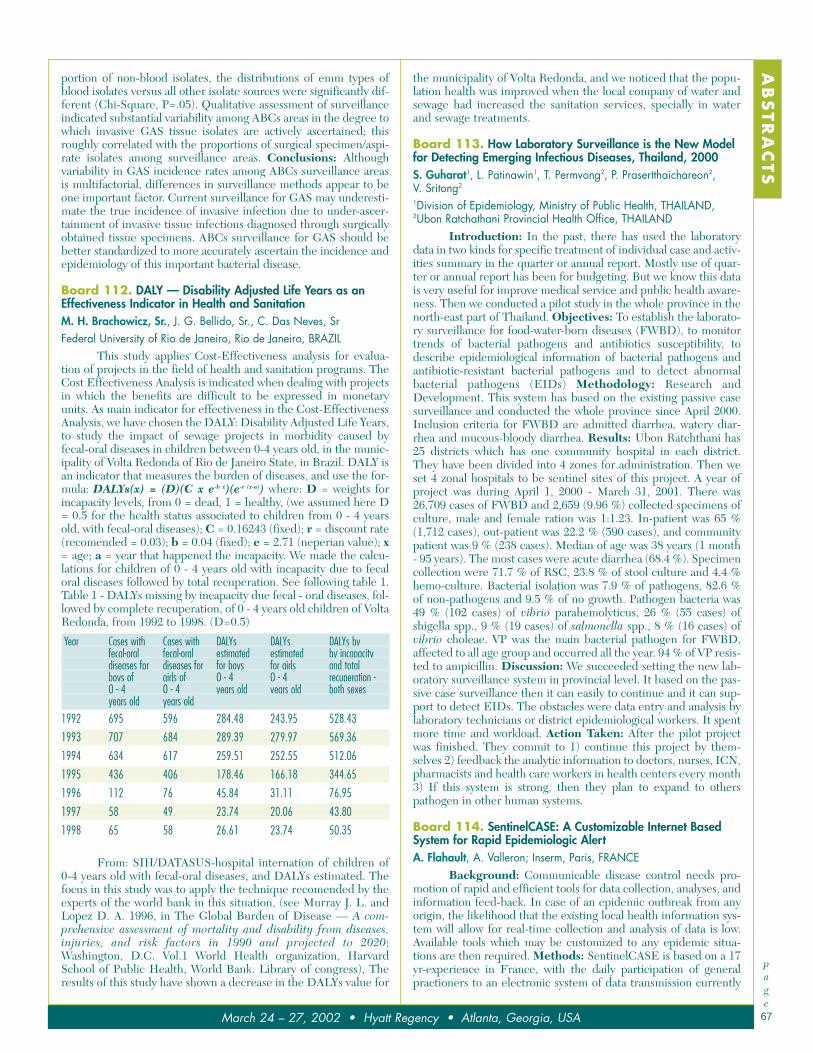
portion of non-blood isolates, the distributions of emm types ofblood isolates versus all other isolate sources were significantly dif-ferent (Chi-Square, P=.05). Qualitative assessment of surveillanceindicated substantial variability among ABCs areas in the degree towhich invasive GAS tissue isolates are actively ascertained; thisroughly correlated with the proportions of surgical specimen/aspi-rate isolates among surveillance areas. Conclusions: Althoughvariability in GAS incidence rates among ABCs surveillance areasis multifactorial, differences in surveillance methods appear to beone important factor. Current surveillance for GAS may underesti-mate the true incidence of invasive infection due to under-ascer-tainment of invasive tissue infections diagnosed through surgicallyobtained tissue specimens. ABCs surveillance for GAS should bebetter standardized to more accurately ascertain the incidence andepidemiology of this important bacterial disease.
Board 112. DALY — Disability Adjusted Life Years as anEffectiveness Indicator in Health and Sanitation M. H. Brachowicz, Sr., J. G. Bellido, Sr., C. Das Neves, SrFederal University of Rio de Janeiro, Rio de Janeiro, BRAZIL
This study applies Cost-Effectiveness analysis for evalua-tion of projects in the field of health and sanitation programs. TheCost Effectiveness Analysis is indicated when dealing with projectsin which the benefits are difficult to be expressed in monetaryunits. As main indicator for effectiveness in the Cost-EffectivenessAnalysis, we have chosen the DALY: Disability Adjusted Life Years,to study the impact of sewage projects in morbidity caused byfecal-oral diseases in children between 0-4 years old, in the munic-ipality of Volta Redonda of Rio de Janeiro State, in Brazil. DALY isan indicator that measures the burden of diseases, and use the for-mula: DALYs(x) = (D)(C x e-b x)(e-r (x-a)) where: D = weights forincapacity levels, from 0 = dead, 1 = healthy, (we assumed here D= 0.5 for the health status associated to children from 0 - 4 yearsold, with fecal-oral diseases); C = 0.16243 (fixed); r = discount rate(recomended = 0.03); b = 0.04 (fixed); e = 2.71 (neperian value); x= age; a = year that happened the incapacity. We made the calcu-lations for children of 0 - 4 years old with incapacity due to fecaloral diseases followed by total recuperation. See following table 1.Table 1 - DALYs missing by incapacity due fecal - oral diseases, fol-lowed by complete recuperation, of 0 - 4 years old children of VoltaRedonda, from 1992 to 1998. (D=0.5)
Year Cases with Cases with DALYs DALYs DALYs byfecal-oral fecal-oral estimated estimated by incapacitydiseases for diseases for for boys for girls and totalboys of girls of 0 - 4 0 - 4 recuperation -0 - 4 0 - 4 years old years old both sexesyears old years old
1992 695 596 284.48 243.95 528.43
1993 707 684 289.39 279.97 569.36
1994 634 617 259.51 252.55 512.06
1995 436 406 178.46 166.18 344.65
1996 112 76 45.84 31.11 76.95
1997 58 49 23.74 20.06 43.80
1998 65 58 26.61 23.74 50.35
From: SIH/DATASUS-hospital internation of children of0-4 years old with fecal-oral diseases, and DALYs estimated. Thefocus in this study was to apply the technique recomended by theexperts of the world bank in this situation, (see Murray J. L. andLopez D. A. 1996, in The Global Burden of Disease — A com-prehensive assessment of mortality and disability from diseases,injuries, and risk factors in 1990 and projected to 2020;Washington, D.C. Vol.1 World Health organization, HarvardSchool of Public Health, World Bank. Library of congress), Theresults of this study have shown a decrease in the DALYs value for
the municipality of Volta Redonda, and we noticed that the popu-lation health was improved when the local company of water andsewage had increased the sanitation services, specially in waterand sewage treatments.
Board 113. How Laboratory Surveillance is the New Modelfor Detecting Emerging Infectious Diseases, Thailand, 2000S. Guharat1, L. Patinawin1, T. Permvong2, P. Prasertthaichareon2, V. Sritong2
1Division of Epidemiology, Ministry of Public Health, THAILAND,2Ubon Ratchathani Provincial Health Office, THAILAND
Introduction: In the past, there has used the laboratorydata in two kinds for specific treatment of individual case and activ-ities summary in the quarter or annual report. Mostly use of quar-ter or annual report has been for budgeting. But we know this datais very useful for improve medical service and public health aware-ness. Then we conducted a pilot study in the whole province in thenorth-east part of Thailand. Objectives: To establish the laborato-ry surveillance for food-water-born diseases (FWBD), to monitortrends of bacterial pathogens and antibiotics susceptibility, todescribe epidemiological information of bacterial pathogens andantibiotic-resistant bacterial pathogens and to detect abnormalbacterial pathogens (EIDs) Methodology: Research andDevelopment. This system has based on the existing passive casesurveillance and conducted the whole province since April 2000.Inclusion criteria for FWBD are admitted diarrhea, watery diar-rhea and mucous-bloody diarrhea. Results: Ubon Ratchthani has25 districts which has one community hospital in each district.They have been divided into 4 zones for administration. Then weset 4 zonal hospitals to be sentinel sites of this project. A year ofproject was during April 1, 2000 - March 31, 2001. There was26,709 cases of FWBD and 2,659 (9.96 %) collected specimens ofculture, male and female ration was 1:1.23. In-patient was 65 %(1,712 cases), out-patient was 22.2 % (590 cases), and communitypatient was 9 % (238 cases). Median of age was 38 years (1 month- 95 years). The most cases were acute diarrhea (68.4 %). Specimencollection were 71.7 % of RSC, 23.8 % of stool culture and 4.4 %hemo-culture. Bacterial isolation was 7.9 % of pathogens, 82.6 %of non-pathogens and 9.5 % of no growth. Pathogen bacteria was49 % (102 cases) of vibrio parahemolyticus, 26 % (55 cases) ofshigella spp., 9 % (19 cases) of salmonella spp., 8 % (16 cases) ofvibrio choleae. VP was the main bacterial pathogen for FWBD,affected to all age group and occurred all the year. 94 % of VP resis-ted to ampicillin. Discussion: We succeeded setting the new lab-oratory surveillance system in provincial level. It based on the pas-sive case surveillance then it can easily to continue and it can sup-port to detect EIDs. The obstacles were data entry and analysis bylaboratory technicians or district epidemiological workers. It spentmore time and workload. Action Taken: After the pilot projectwas finished. They commit to 1) continue this project by them-selves 2) feedback the analytic information to doctors, nurses, ICN,pharmacists and health care workers in health centers every month3) If this system is strong, then they plan to expand to otherspathogen in other human systems.
Board 114. SentinelCASE: A Customizable Internet BasedSystem for Rapid Epidemiologic Alert A. Flahault, A. Valleron; Inserm, Paris, FRANCE
Background: Communicable disease control needs pro-motion of rapid and efficient tools for data collection, analyses, andinformation feed-back. In case of an epidemic outbreak from anyorigin, the likelihood that the existing local health information sys-tem will allow for real-time collection and analysis of data is low.Available tools which may be customized to any epidemic situa-tions are then required. Methods: SentinelCASE is based on a 17yr-experience in France, with the daily participation of generalpractioners to an electronic system of data transmission currently
AB
STR
AC
TS
March 24 – 27, 2002 • Hyatt Regency • Atlanta, Georgia, USA
page
67
available on the Internet 24-hrs a day (http://www.u444.jussieu.fr/sentiweb). The server has been developed on a PC witha Linux OS, an Apache HTML server, Java code, a MySQL rela-tional database. Clients are microcomputers with Internet connec-tion. Results: Health professionals are collecting data onSentinelCASE through a securized web interface. After havingidentified the notifier, the system allows data to be stored into aMySQL relational database. Any authorized persons (or the worldwide public if no access restrictions is considered) can consult ona real-time basis all entered data on maps, graphs, and tables. Rowdata may be downloaded on a spreadsheet for further analyses.The engine of SentinelCASE is currently in use at an internation-al level for the ongoing global surveillance of Influenza (FluNet)or dengue (DengueNet, see on http://oms.u444. jussieu.fr/). It hasbeen chosen by WHO-HQ because of its easyness, flexibility,rapid implementation, and low cost. SentinelCASE may equipwithin a few weeks any networks of health professionals. Althougha good knowledge in Linux and Apache is required for the localimplementation and management of the server, no specific com-puter skills are required for the database management, or for cus-tomization of the surveillance system. Through an Internet inter-face, the system is easily and rapidly customizable. The autorizeduser defines items to be monitored. SentinelCASE may be usedfor epidemiologic purpose, but also for entomologic surveillanceor for monitoring any environmental indicator. The use of the sys-tem is self-understandable. None of the 1270 French sentinelGPs, nor the 110 national influenza centres have been specificallytrained for its use. Conclusion: Inserm (French Institute ofHealth and Medical Research) provides a user-friendly tool whichallows for a rapid impementation of epidemiologic surveillance inany part of the world.
Board 116. Use of the Web To Enhance Disease Surveillanceand Bioterrorism Preparedness N. M. M'ikanatha1, D. Welliver2, D. Rohn3, J. Rankin1
1Pennsylvania Department of Health, Harrisburg, PA, 2HersheyMedical Center, Hershey, PA, 3Maryland Department of Health andMental Hygiene, Balitmore, MD
Background: Timely reporting of notifiable diseases iscritical to early detection and response to outbreaks or bioterror-ism (BT) attacks. Previous studies indicate some clinicians areunaware of what, when, how or where to report diseases. The Weboffers an unprecedented opportunity to provide high quality,accessible information and also a means to report disease and BTrelated syndromes. Objectives: To evaluate accessibility and use-fulness of Web-based information on infectious diseases reporting(DR) and BT preparedness, and to suggest areas for improvement.Methods: (1) In November 2001, we surveyed 57 jurisdictionsthat report notifiable diseases to the Centers for Disease Controland Prevention (CDC) through the National ElectronicTelecommunication System for Surveillance (NETSS). An E-mailquestionnaire asked epidemiologists about availability ofreportable diseases lists (RDL) on their Web sites, and plans to usethe Web for DR and syndromic surveillance. (2) In December2001, we used the Google search engine to locate online DR infor-mation. We used a standardized instrument to collect data on avail-ability of reportable diseases lists and reporting instructions, capa-bility for online disease reporting, and information on six CDC BTCategory A agents. Results: (1) Fifty-six of the 57 jurisdictionsresponded by email or subsequent phone interview. Forty-nine(88%) indicated that they have online RDL, and 42 (75%) of thoseare current. Sixteen (29%) provide means for electronic reportingof notifiable diseases; four provide Web-based reporting. Threehave Web-based syndromic surveillance in use; nine plan to start inthe next six months. (2) We located 45 (90%) of the 49 Web siteswith DR information. Eleven (24%) of the jurisdictions requirereporting of all Category A agents while at other sites BT agents are
reportable only as “unusual conditions.” Thirty-three 33 (73%) pro-vide a telephone number for DR, 18 (40%) provide a fax number,and 14 (31%) had an online DR card. Forty-five (90%) indicate therequired time frame for reporting different diseases, and 36 (80%)explained the basis for reporting. Conclusions: The Web is beingused to promote DR but its potential for facilitating timely report-ing of diseases and syndromes has not been fully realized. Up-to-date DRL, telephone and fax numbers for reporting, on online DRcards and instructions could readily be provided on jurisdictions’Web sites. Some states already have recognized the benefits ofWeb-based systems. The Web offers a cost-efficient way toenhance surveillance and BT preparedness.
Board 117. Time Series Modeling and Space-Time Clusteringof DoD-GEIS Data For Early Outbreak Detection H. S. Burkom1, J. Lombardo1, F. Mostashari2, Y. Elbert3, J. Pavlin3, P.Kelley3
1Johns Hopkins Applied Physics Laboratory, Laurel, MD, 2New York CityDepartment of Health, New York, NY, 3DoD/GEIS, Washington, DC
The objective of this effort is the capability for early indica-tion of the spatial location and extent of an outbreak of disease. Theexigency of responding to recent terrorist activity and advances indatabase technologies are increasing the timely availability of sur-veillance data while presenting the problem of how to analyze thegrowing volume of data for prompt outbreak recognition.
The U.S. Department of Defense Global EmergingInfections System (DoD-GEIS) has developed the ElectronicSurveillance System for Early Notification of Community-basedEpidemics (ESSENCE) to enable outbreak alerting using syn-dromic surveillance. This system reduces the surveillance of out-patient visits to the inspection of counts in a manageable numberof syndromes, defined as logical groupings of ICD9 codes.Researchers at DoD-GEIS and the Johns Hopkins Applied PhysicsLaboratory have calculated time series predictions for backgroundlevels of daily visits expected in each monitored region for the var-ious syndromes. Autoregressive modeling and other time seriesmethods have yielded predictions that account for temporal pat-terns of consumer behavior. Small spatial resolution is needed forearly alerting so that increases in visits at the neighborhood levelare not masked by normal variability at the state or county level.We have used the patient-residence zipcodes provided by theESSENCE system to perform spatial-temporal cluster analysis ofthe variations of daily data from the levels predicted by the mod-els. The resulting clusters indicate approximate regions of outbreakfor further investigation by human expertise or by other automat-ed means. To seek potential clusters, we employ Kulldorff’s space-time scan statistic because it gives a p-value for each cluster alongwith the component zipcodes and avoids selection bias as well asthe zipcode resolution allows. Applications have located unlikelyclusters of syndrome visits using model-based expectations. Wehave also clustered visits for single ICD9 codes based on the rep-resentation of each zipcode in the ESSENCE system. The result-ing clusters may be inspected using GIS plots. Simulations havebeen used to assess the utility of this approach for early detectionof a point-source outbreak. We used the Sartwell lognormal modelto estimate the epidemic curve for diseases regarded as biowarfarethreats, and we estimated the spatial case dispersion to inject casesinto authentic background data. Simulations are parametrized bythe total infected to determine how severe an outbreak may bedetected according to time after exposure.
I C E I D 2 0 0 2 A B S T R A C T Spage 68
14 ICEID 2002 KeynoteSessionSunday, March 24, 5:00 p.m.Centennial Ballroom
15 Meet-the-Experts IMonday, March 25, 7:30 a.m.Regency Ballroom VI/VII
16 Plenary Session I Monday, March 25, 8:30 a.m.Centennial Ballroom I/II
17 Plenary Session II Monday, March 25, 8:30 a.m.Centennial Ballroom III/IV
20 Antimicrobial Resistance IMonday, March 25, 10:00 a.m.Grand Hall East
Board 1. Mutational Analysis of Position Arg164 in TEM-1and SHV-1: Two Highly Similar ß-Lactamases F. K. Majiduddin, T. G. PalzkillBaylor College of Medicine, Houston, TX
Resistance to ß-lactam antibiotics is a serious threat to theeffective treatment of infectious disease. Several mechanisms areinvolved in resistance, the most prevalent being the production ofß-lactamases. The TEM-1 and SHV-1 ß-lactamases are importantcontributors to resistance to ß-lactam antibiotics in Gram-nega-tive bacteria. These enzymes share 68 % amino acid sequenceidentity and their atomic structures are nearly superimposable.Extended spectrum cephalosporins were introduced, in part, toavoid the action of TEM -1 and SHV-1 ß-lactamase. However, thewidespread use of these antibiotics has lead to the evolution ofvariant TEM and SHV enzymes that can hydrolyze extendedspectrum antibiotics. Interestingly, despite being highly similar instructure, the TEM and SHV enzymes have evolved somewhatdifferently in response to the selective pressure. Examples of thisare at residues Arg164 and Asp179, two positions that form a saltbridge at the base of an omega-loop structure. Among TEM vari-ants substitutions are only found at position 164 while substitu-tions are only found at position 179 among SHV variants. Toexplain this observation, we examined the effects of mutations atposition 164 in both TEM-1 and SHV-1 on antibiotic resistanceand catalytic efficiency. In addition, competition experiments
were carried out between mutants to understand why certainsubstitutions preferentially evolve due to the selective pressure ofantibiotic therapy. Based on these competition assays, the SHVArg164Ser variant is more fit than wild-type SHV-1, but is unableto compete effectively versus the SHV Asp179Gly variant. Amongthe TEM variants, the Asp179Gly variant is more fit than strainsexpressing the parental TEM-1 enzyme, but this mutant is unableto compete effectively against its Arg164Ser counterpart. Thisexplains why only mutants at position Asp179 exist in SHVenzymes and Arg164 mutants in TEM enzymes in the clinical set-ting. Subtle unseen differences in the structures of TEM-1 andSHV-1 may account for the divergent evolutionary paths thesetwo enzymes are undertaking.
Board 2. Synthetic Peptide Antibiotics: Solutions for theNew MilleniumD. WadeHelsinki University, Helsinki, FINLAND
The current public health problems of antibiotic resistantmicroorganisms, and bioterrorism, necessitate the development ofnew antibiotic strategies and therapeutics. During the past twodecades, a new category of antibiotics has been discovered, thegene-encoded peptide antibiotics of the animal, plant, and bacter-ial kingdoms. Research interest in these peptide antibiotics hasgrown exponentially, and there is a hope that they may provide apartial solution to the problems mentioned above. Several hundrednew, naturally occurring, peptide antibiotics have been isolated,and one of the most prolific sources of these new antibiotics hasbeen the skin of frogs and toads (anurans). One group of theseantibiotics, the temporins and temporin-like peptides, are com-posed of 31 different peptides from 8 species of frogs. Surprisingly,there are an additional 8 peptides with very similar structures tothe frog peptides that have been isolated from the venoms ofwasps. One of the frog skin peptides, temporin A (TA), has beenshown to have antibacterial activities against antibiotic resistantbacteria. Synthetic manipulations of the structure of TA has shownthat it is possible to improve its antibiotic properties and reduce itstoxicity, and they have provided information about the structuralfeatures of TA that endow it with antibiotic properties. It is expect-ed that further work with the temporins, and the other members ofthe new class of gene encoded peptide antibiotics, will eventuallyprovide at least a partial solution to the problem of antibiotic resist-ant microorganisms.
Board 3. Growth Inhibition by Cisplatin in Group-I IntronContaining Pathogens: A Study in Candida albicans K. Chandrasekhar, R. MalathiDr.ALM Post Graduate Institute of basic Medical Sciences, Universityof Madras, Taramani Campus, Chennai, INDIA
We previously reported that antitumor agent cisplatin,which has been clinically used in treatment of wide range of can-cer has been found to inhibit group-I intron splicing invitro fromTetrahymena thermophila1. Interestingly this has been found toarrest the growth of C.albicans, an opportunistic human fungalpathogen that has become a major cause of morbidity and mortal-ity in immunocompromised as well as cancer and HIV patients2,3.This gains lot of significance from clinical point of view sinceC.albicans is known to possess this group-I intron in 25s rRNA andtheir absence in human will pave way for an alternative target site,in addition to the well known cell-membrane therapeutic agentswhich are gaining resistance in present conditions. Recently wehave carried out an invitro-screening assay using ATCC and 4 clin-ical strains and the parameters include disk diffusion method,growth curve analysis, RNA isolation and a fluorescence micro-scopic study using a new staining method “Ethidium Bromide” inorder to analyse the morphological features. The above experi-ments were carried out with normal as well as with drug treated
AB
STR
AC
TS
March 24 – 27, 2002 • Hyatt Regency • Atlanta, Georgia, USA
page
69
C.albicans strain. The results have indicated that, in growth curve,cis-DDP at 60µg concentration shows a remarkable inhibition ofmultiplication of C.albicans cells with respect to control after 10hour incubation period while analyzing O.D values at 600nmrange. On the other hand low molecular weight RNA [25s, 18srRNA] from the culture was isolated, analysed using agarose gelelectrophoresis and the results suggest that cis-DDP might havecompletely abolished the 25s rRNA with high efficiency. While themicroscopic study has clearly shown that the cis-DDP treated cellsat 60µg concentration shows inhibition of hyphae growth as well asenlargement of ovoid cell irrespective of the control whose ovoidcells are smaller and the hyphae with many branches/budding visu-alized under 40X and 100X of Nikon Fluorescence Microscope andthe same experiment was also carried out to compare with EtBrstain using normal C.albicans stain, the results were similar. Thesefindings suggest that cis-DDP if properly oriented /targeted withepitope/liposome delivery, could possess antifungal propertybesides their antitumor activity especially in cancer patients.Moreover as pathogens continue to evade therapeutic targetssearch of novel agents with new target site is becoming a seriousproblem in persistence of human life, not only for fungal pathogensbut also for other pathogenic organism. Reference: 1.Malathi R.and Chandrasekar K. Journal of Biosciences. 1999 suppl.11, 96.2.Chandrasekar.K, Shyla J., and Malathi,R. Journal of ClinicalMicrobiology. 2000 vol.10 (38). 3.Leibowitz M.J., AntimicrobialAgents and Chemotherapy. 2000, Vol.44, No.4, 958-966.
Board 4. Hemolytic and Non-Hemolytic Vancomycin-Resistant Enterococcus faecalis from Imported Beef in Malaysia N. H. Fifadara1, G. R. Rusul2, S. Radu3, Z. Hassan2, L. R. Beuchat11University of Georgia, Griffin, GA, 2University Putra Malaysia,Serdang, MALAYSIA, 3Department of Biotechnology, UPM, Serdang,MALAYSIA
Twenty-two strains of vancomycin resistant Enterococcusfaecalis were isolated from 9 (6%) of 150 imported beef samples.12 strains (54.5%) were b-hemolytic and all strains harbored vanAgene. Hundred percent of the strains tested were resistant to 10 ofthe antibiotics including vancomycin and teicoplanin. Strains were95.4% resistant to trimethoprim-sulfamethoxazole, 68.8% resistantto chloramphenicol and 41% resistant to ampicillin and penicillin.Small sized plasmids ranging from 1.5 to 5.8 Kb were detected in8 (36.4%) strains. The twenty-two isolates were differentiated intotwenty RAPD-types. It was concluded that vancomycin-resistantEnterococcus faecalis with different clones is widespread amongisolates from imported beef in Malaysia.
Board 5. Phenotypic Typing and Molecular Fingerprinting ofFluoroquinolone-Resistant Campylobacter Species Isolatedfrom a Treated Broiler Flock H. A. Wadda1, G. Domingue2, D. J. Griggs3, M. Siccardi2, M.Johnson3, J. A. Frost1, T. Humphrey4, L. J. Piddock3
1Campylobacter Reference Unit, Central Public Health Laboratory,London, UNITED KINGDOM, 2Public Health laboratory Service,FMRU, Exeter, UNITED KINGDOM, 3Antimicrobial Agents ResearchGroup, University of Birmingham Medical School, Birmingham, UNIT-ED KINGDOM, 4Department of Clinical Veterinary Science, Universityof Bristol, Bristol, UNITED KINGDOM
Aim: To investigate the incidence, types and mechanisms ofresistance in Campylobacter spp. during therapeutic treatment ofa poultry flock. Background: Antimicrobial resistance in campy-lobacter isolated from human infections has increased over theyears. Monitoring this increase is important for those severe infec-tions where antimicrobial therapy is essential. It is known thatresistance can be spread from one ecosystem to another ie. trans-fer of resistant strains from farm animals to humans, humans toenvironment, environment to animal and vice versa. Poultry meatconsumption is thought to be one of the main routes of transmis-
sion to humans. Methods: Samples were taken from one poultryflock before, during and at 1 and 2 weeks after fluoroquinolonetreatment. A total of 66 chickens tested positive for Campylobacterspp. and 3 picks were isolated from each bird. These isolates werespeciated and characterised using serotyping, phagetyping and bio-typing methods at the Campylobacter Reference Unit. Drug resist-ance screening using 12 antibiotics was performed using themethod developed as part of the NCCLS working party onCampylobacter susceptibility testing. Pulsed Field GelElectrophoresis (PFGE) and Amplified Fragment LengthPolymorphisms (AFLP) were used for more detailed strain charac-terisation. Mutations in gyrA and gyrB was determined withDHPLC. Results: Both Campylobacter jejuni and Campylobactercoli were isolated in all three of the treatment phases. ResistantC.jejuni were isolated mostly pre-treatment and during treatment,whilst resistant C.coli were seen in the post-treatment phase (noresistant C.coli were isolated in pre-treatment). Results indicatedthat 17.4% of the positive chickens at the pre-treatment phasewere fluoroquinolone-resistant, 100% during the treatment phaseand 82.8% at the post-treatment phase. Phenotypic analysis of theresistant strains showed C. jejuni HS27/PT67 to be the predomi-nate strain at the pre-treatment stage, but this was not seen in theother two phases. C.jejuni HS31/PT1 was the predominant resist-ant strain during treatment but less commonly detected at thepost-treatment phase. More C.coli strains were seen in the post-treatment phase where C.coli HS56/PT44 was the predominantstrain. Isolates examined to date all had a substitution of Thr86 ingyrA. Conclusion: An increased proportion of resistant strainswere seen during treatment, these persisted post treatment.Changes in species and subtype prevalence also occurred duringthe course of this study. The resistant mechanism observed so faris the substitution of Thr86 in gyrA.
Board 6. Enhanced Surveillance for Antimicrobial ResistanceAmong Enteric Bacteria: NARMS Retail Food Study J. E. Stevenson1, D. G. White2, D. J. Torpey, III3, A. S. Craig4, K. E.Smith5, M. M. Park6, M. A. Pascucilla7, A. D. Anderson1, . and theNARMS Working Group1
1Centers for Disease Control and Prevention, Atlanta, GA, 2U.S. Foodand Drug Administration, Laurel, MD, 3University of Maryland,Baltimore, MD, 4Tennessee Department of Health, Nashville, TN,5Minnesota Department of Health, Minneapolis, MN, 6Georgia PublicHealth Laboratory, Atlanta, GA, 7Connecticut Emerging InfectionsProgram, New Haven, CT
Background: The food supply, including meat andpoultry, is an important source of enteric bacteria, includingCampylobacter, E. coli, Salmonella and possibly enterococci.Antimicrobial resistance among these foodborne bacteria is notuncommon and often is associated with the use of antimicrobialagents in food animals. Retail food represents the point of exposurethat is closest to the consumer and, when combined with data fromslaughter plants and on-farm studies, provides a more representa-tive picture of the prevalence of resistance in foodborne pathogens.To focus efforts to mitigate antimicrobial resistance and to betterunderstand the contribution of the food supply to antimicrobialresistance among enteric bacteria, the National AntimicrobialResistance Monitoring System (NARMS) for Enteric Bacteria isextending surveillance of antimicrobial resistance to bacteria iso-lated from food. Program Description: The NARMS RetailFood Study is a collaborative effort between the Centers forDisease Control and Prevention (CDC), five FoodNet sites(Connecticut, Georgia, Maryland, Minnesota and Tennessee) andthe U.S. Food and Drug Administration (FDA). The NARMSRetail Food Study has adopted a standard method to monitor theprevalence of antimicrobial resistance among Campylobacter, E.coli, Salmonella and enterococci isolated from a convenience sam-ple of meat and poultry from selected grocery stores in the UnitedStates. Data collection will begin January 1, 2002. Each site will
I C E I D 2 0 0 2 A B S T R A C T Spage 70

visit at least one grocery store per month, not returning to the samestore for at least two months, and purchase packages of meat orpoultry including 10 packages of chicken breasts, 10 packages ofpork chops, 10 packages of ground turkey and 10 packages ofground beef. Isolation procedures have been adapted from theFDA’s Bacteriological Analytical Manual. Each site will culture therinse from each sample for the presence of Salmonella andCampylobacter. In addition, Georgia, Maryland and Tennessee willculture the rinse for E. coli and enterococci. Isolates will be for-warded to FDA for antimicrobial susceptibility testing.Conclusion: This collaborative surveillance project will providedata and isolates useful for focusing efforts to mitigate antimicro-bial resistance in enteric bacteria. Enhanced efforts are needed tomitigate the increasing prevalence of antimicrobial resistanceamong foodborne bacteria. By examining the prevalence of antimi-crobial-resistant enteric bacteria we will better understand theextent of antimicrobial resistance in the food supply and will bebetter equipped to implement mitigation strategies.
Board 7. Multidrug Resistance Among Human Non-TyphoidalSalmonella Isolates in the United States: NARMS 1999-2000 S. Rossiter, K. Joyce, J. E. Stevenson, T. Barrett, A. D. Anderson, andthe NARMS Working Group;Centers for Disease Control and Prevention, Atlanta, GA
Background: An estimated 1.4 million Salmonella infec-tions occur in the United States annually. Emerging antimicrobialresistance, including multidrug resistance, contributes to thehuman health burden of Salmonella and threatens the utility ofantimicrobial agents, including third generation cephalosporins(e.g., ceftriaxone). The National Antimicrobial ResistanceMonitoring System (NARMS) for Enteric Bacteria was establishedin 1996 as a collaborative effort among the Centers for DiseaseControl and Prevention (CDC), participating state and local healthdepartments, the U.S. Food and Drug Administration (FDA), andthe U.S. Department of Agriculture (USDA). NARMS data arehelpful in directing applied research and mitigation efforts.
Methods: In 1999 and 2000, 17 participating public healthlaboratories forwarded every tenth non-typhoidal Salmonella iso-late to CDC. Salmonella isolates received at CDC were tested bybroth microdilution (Sensititre®) for minimum inhibitory concen-trations (MIC) to 15 antimicrobial agents including ampicillin (A),chloramphenicol (C), kanamycin (K), streptomycin (S), sulfon-amides (Su), and tetracycline (T), using NCCLS interpretive stan-dards. Isolates with decreased susceptibility to ceftriaxone (MIC ≥16 µg/ml) were tested by E-test (AB Biodisk).
Results: Of the 2876 human Salmonella isolates tested in1999-2000, 26% (743) were resistant to ≥ 1 agent; 21% (601) weremultidrug-resistant (resistant to ≥ 2 agents). Three multidrug-resistant strains, S. Typhimurium R-type ACSSuT, S. TyphimuriumR-type AKSSuT, and S. Newport R-type ACSSuT accounted for10% (278/2876) of non-typhoidal Salmonella isolates, 37%(278/743) of the resistant isolates, and 46% (278/601) of multidrug-resistant isolates. S. Typhimurium R-type ACSSuT was present inall sites, ranging from 12% of Salmonella from Washington to 5%of Salmonella from California. S. Typhimurium R-type AKSSuTwas present in 16 of the 17 sites; it was most prevalent inWashington where it was 9% of Salmonella. S. Newport R-typeACSSuT was present in all sites, ranging from 8% of Salmonella inKansas to <1% of Salmonella in New York State. Among humanSalmonella isolates, ceftriaxone resistance was present in 2%(3/170) of S. Typhimurium R-type ACSSuT isolates, 3% (2/63) ofS. Typhimurium R-type AKSSuT isolates, 71% (32/45) of S.Newport R-type ACSSuT isolates, and <1% (16/2764) of otherSalmonella isolates.
Conclusion: Three multidrug-resistant strains account fornearly half of multidrug resistance among human Salmonella iso-lates; ceftriaxone resistance also is common among multidrug-resistant Salmonella Newport and present in some multidrug-
resistant Salmonella Typhimurium. Further studies are needed todetermine sources for these multidrug-resistant strains and sup-port prevention efforts.
Board 8. Expanded-Spectrum Beta-Lactam Resistance amongHuman Clinical Enterobacteriaceae in the United States: Resultsand Characterization of 2000 NARMS Surveillance J. M. Whichard1, K. Joyce1, P. D. Fey2, J. McClellan1, F. J. Angulo1, T.Barrett1, and the NARMS Working Group1
1Centers for Disease Control and Prevention, Atlanta, GA, 2Universityof Nebraska Medical Center, Omaha, NE
Background: Third-generation cephalosporins are usefulfor the treatment of invasive salmonellosis and other severe pedi-atric infections. The National Antimicrobial Resistance MonitoringSystem (NARMS) for Enteric Bacteria has identified increasedcephalosporin resistance in human bacterial enteric pathogens; in1999 1.5% of isolates tested exhibited intermediate or resistantminimum inhibitory concentrations to expanded-spectrumcephalosporins. Preliminary molecular characterization of resist-ance determinants in the 2000 NARMS collection is presented.Methods: As NARMS participants, the 17 state and local publichealth laboratories submitted every 10th non-typhoidalSalmonella, every 10th Shigella and every 5th E. coli O157:H7received in 2000 to the Centers for Disease Control andPrevention for antimicrobial susceptibility testing. Minimuminhibitory concentrations (MIC) were determined for 17 antimi-crobials using broth microdilution (Sensititre®). Isolates were cho-sen for further study based on an intermediate or resistant MIC forthe expanded-spectrum cephalosporins: cefoxitin (≥16 µg/ml), cef-tiofur (≥4 µg/ml) or ceftriaxone (≥16 µg/ml). ß-lactamases werecharacterized using isoelectric focusing and pcr for blaCMY-2.Results: Of the 2236 isolates tested in 2000, 57 (2%) met the MICcriteria and were selected for further study. These 57 isolates com-prised 3% (46/1378) of the non-typhoidal Salmonella isolates test-ed, 2% (7/451) of Shigella, and 1% (4/407) of E. coli O157:H7.Overall, 52 (91%) of the 57 isolates produced a ß-lactamase with apI≥8.4, consistent with an ampC-type ß-lactamase. Forty-four(77%) were pcr-positive for a blaCMY gene. All 4 E. coli O157:H7isolates tested produced an enzyme with a pI≥8.4 and were alsopcr-positive for a blaCMY gene. All 7 Shigella isolates tested wereS. sonnei and produced an enzyme with a pI≥8.4, but were nega-tive by pcr for blaCMY. Forty of the 46 Salmonella isolates testedproduced an enzyme with a pI≥8.4 and were pcr-positive for ablaCMY gene. Notably, 24 of the 40 blaCMY positive Salmonellawere serotype Newport. Seven of the 57 isolates (12%) producedadditional ß-lactamases, including 4 Salmonella and 3 Shigella son-nei. One isolate (Salmonella serotype Nienstedten) producedenzymes of pI 5.4 and 8.0. Conclusion: The presence of isolatesexhibiting intermediate or resistant MIC to expanded-spectrumcephalosporins increased in 2000. The major determinant in the2000 NARMS collection appears to be a blaCMY-type ß-lacta-mase. S. Newport is the predominant organism among the 2000NARMS isolates exhibiting this resistance. Additional ß-lactamasesare expressed in over 10% of the isolates as well. Because of thepervasive ß-lactam resistance phenotype conferred by the ampC-type enzymes, these additional enzymes would not have been iden-tified by susceptibility testing alone.
Board 9. Irrigation Water and Associated Sediments as aSource of Integron Bearing Enteric Bacteria E. Vega, M. T. Roe, S. D. PillaiTexas A&M University, College Station, TX
Use of antibiotics in humans and animal feed is thought tobe responsible for the increased prevalence of antibiotic resistantbacteria in the environment. The Texas-Mexico border region issignificantly impacted by anthropogenic activities. The Rio GrandeRiver serves as the primary source of irrigation water in this region.
AB
STR
AC
TS
March 24 – 27, 2002 • Hyatt Regency • Atlanta, Georgia, USA
page
71

Considering the numbers and types of possible sources of fecalcontamination of irrigation water, we hypothesized that irrigationwater and agricultural fields harbor a large number of integron-bearing enteric bacteria. Antibiotic resistance in Gram-negativebacteria is primarily thought to be mediated by gene cassetteswithin the integron gene sequence. E. coli was isolated from sedi-ment and water at 11 sampling sites along the Texas-Mexico bor-der. Antibiotic resistant profiles were determined using the agardilution method for E. coli. Out of 326 E.coli isolates, only 31 iso-lates (9.5%) were determined to be resistant to more than oneantibiotic. The isolates were characterized using PCR primers spe-cific for the variable region of the class I integron gene sequences.Of the 31 E. coli multiple drug resistant isolates, only 5 isolates(16.1%) indicated the presence of the class I integron sequence.Sequencing E.coli cassettes verified the presence of the aadA genein three isolates and dhfr XII gene and aadA genes in one isolate.The results indicate that agricultural fields harbor low numbers ofintegron-bearing E.coli. It is, however, critical to understandwhether integron elements are involved in horizontal gene transferof antibiotic resistant genes on fresh produce especially those thatare consumed raw or minimally processed.
Board 10. Infections Caused by Rapidly GrowingMycobacteria: Experience of Infectious Diseases Consultants M. A. De Groote1, L. J. Strausbaugh2,3, D. B. Jernigan4, L. A.Liedtke2,3
1National Jewish Hospital, Denver, CO, 2VA Medical Center,Portland, OR, 3IDSA Emerging Infections Network, Portland, OR,4CDC, Atlanta, GA
Background: Because non-tuberculous mycobacterialinfections are seldom reportable, their epidemiology remains poor-ly understood. Although the National Jewish Medical andResearch Center has appreciated an increase in the number ofinfections due to rapidly growing mycobacteria (RGM); it is notknown whether this experience reflects that of the nation. Toexamine this question, we surveyed members of the InfectiousDiseases Society of America Emerging Infections Network (EIN)about their recent experiences with RGM. Methods: In October2001, a questionnaire was distributed to all 797 infectious diseasesconsultants participating in the EIN. Results: Overall, 374 (47%)of the 797 members responded. Of the respondents, 183 (49%)had encountered 559 cases of RGM during 2000-2001. Moreover,17% had noted more cases in recent years. EIN members from allnine regions of the US (especially the South Atlantic, Pacific andMid-Atlantic) reported seeing increased numbers of cases.Respondents noted a wide spectrum of RGM disease, includingpulmonary, cutaneous, and, to a lesser extent, disseminated andlymphatic. By region, 23 to 54% of EIN members reported seeingM. chelonae cases, 14 to 44% M. abscessus cases, and 23 to 54%M. fortutium cases. Fifty-six (31%) of respondents described envi-ronmental exposures associated with infection: medical procedures(45%), soil or water (21%) and trauma (29%). Predisposing hostconditions included immunocompromise (steroids, HIV, andmalignancy) and chronic lung diseases (emphysema and cysticfibrosis). Some EIN members expressed concern about distin-guishing colonization from infection and establishing the need fortreatment. Most respondents (79%) use in vitro susceptibility test-ing to guide therapy. EIN members indicated that a diverse arrayof facilities, including several national reference laboratories, per-form RGM identification and susceptibility testing. Conclusions:RGM infections are frequently seen by infectious diseases consult-ants in all regions of the country. They appear to be increasing insome areas, especially in coastal states. Clinical features of membercases suggest that RGM are opportunistic pathogens, frequentlyacquired from environmental sources. Since numerous laborato-ries isolate RGM, consistent methods for identification and sus-ceptibility testing will help define the epidemiology and propertherapy of these emerging pathogens.
Board 11. Outbreak of Community-Acquired Methicillin-Resistant Staphylococcus aureus (MRSA) Skin Infections AmongAlaska Natives, 2000 H. C. Baggett1, T. Hennessy1, D. Bruden1, C. Hamlin1, R. Leman2, A.Reasonover1, R. Sparks1, R. Donlan3, S. McAllister3, P. Martinez4, J.Butler1
1CDC, Anchorage, AK, 2Indian Health Service, Albuquerque, NM,3CDC, Atlanta, GA, 4Yukon Kuskokwim Health Corporation, Bethel, AK
Background: Reports of MRSA-infected patients withouthospital exposure indicate that MRSA infection is now acquired inboth healthcare and community settings. In August 2000, anMRSA skin infection outbreak among patients without hospitalexposure was reported in southwestern Alaska. We investigatedthese community-acquired MRSA infections and potential risk fac-tors for disease. Methods: Using ICD9 codes, we determined thepercentage of clinic visits for skin infection to the regional hospitalfrom January 1998-July 2000. We reviewed charts of all patientswith a positive S. aureus culture from May-July 2000. Community-acquired MRSA was defined as culture-confirmed illness in some-one without hospitalization, surgery, dialysis, admission to a long-term care facility, or presence of an indwelling catheter in the prioryear. In one village, we conducted a case-control study using 34case-patients with MRSA skin infections and 94 controls with nohistory of skin infection. MRSA nasal carriage was assessed forcase-patients, controls and their household members. We conduct-ed an environmental assessment of traditional saunas, which havebeen linked to skin infections in prior outbreaks. Pulsed-field gelelectrophoresis (PFGE) was performed on available clinical, car-riage, and environmental MRSA isolates. Results: During March1999 through July 2000, the number of MRSA skin infections atthe regional hospital increased from 5 to 56 per month (p<0.01),and visits for skin infections increased from 1% to 3% of outpatientvisits (p<0.01). In the peak three months of the outbreak (May-July2000), 85% (142/168) of S. aureus skin infections were MRSA and75% (107/142) of these were community-acquired. In the case-control study village, 5.1% (39/760) of residents had confirmed S.aureus skin infections during a 5-month period, and all S. aureusisolates from that village were MRSA. Case-patients used moreantibiotics than controls in the year before the outbreak (median4.0 vs. 2.0 courses, p=0.01). Sauna use was reported by 85% ofcase-patients and was not more common than among controls(89%). However, MRSA was cultured from 8 (17%) of 47 saunas,and 44% of case-patients and 13% of controls used MRSA-positivesaunas (OR=5.6, p<0.01). MRSA in biofilms were identified insauna wood. A single PFGE pattern was seen in 71 (89%) of 80MRSA isolates from clinical, carriage, and environmental sources;an additional 7 (9%) isolates differed by only 2 bands.Conclusions: A large outbreak of community-acquired MRSAskin infections occurred among Alaska Natives in southwesternAlaska. Illness was associated with antibiotic and sauna use.Response has included judicious antibiotic use education and asauna disinfection efficacy study.
Board 12. National and Regional Susceptibility Patterns of Staphylococcus aureus and Methicillin-Sensitive andMethicillin-Resistant Staphylococci: Results of the AntimicrobialResistance Management (ARM) Program J. G. GumsUniv. of Florida, Gainesville, FL
Background: The ongoing ARM program was developedto document susceptibility patterns, including for S aureus, methi-cillin-sensitive S aureus (MSSA) and methicillin-resistant S aureus(MRSA) in inpatient and outpatient isolates. Since 1987, more than10 million isolates have been collected on 19 organisms and 46antibiotics from 105 US hospitals in 5 regions (Northeast, NorthCentral, Southeast, South Central, Southwest). Methods:Antibiograms and sensitivity reports of S aureus, MSSA, and
I C E I D 2 0 0 2 A B S T R A C T Spage 72
MRSA isolates were reviewed for susceptibility to fluoroquinolone(ciprofloxacin, levofloxacin, ofloxacin, trovafloxacin) and otherantibiotics (vancomycin, clindamycin, erythromycin, and naf-cillin/oxacillin). Results: Total number of isolates and percentageof isolates susceptible to each antibiotic were determined bothnationally and regionally. Nationally, S aureus isolates were as sus-ceptible to ciprofloxacin (62.3%, n=194,937) as to levofloxacin(62.2%, n=78925), with a greater sensitivity to ciprofloxacin seen inNorth Central (60.9% vs 58.7%), Southeast (62.7% vs 62.2%), andNortheast (60.7% vs 52.8%). These data suggest cross-resistancebetween ciprofloxacin and levofloxacin. Susceptibility to lev-ofloxacin was greater than to ciprofloxacin in South Central (82.9%vs 72.4%) and Southwest (80.4% vs 54.3%). S aureus isolate sus-ceptibility to erythromycin nationally was 32.5% (n=272,184),accounted for primarily in Southeast (22.9%); it was higher in allother regions: North Central (53.4%), Northeast (54.3%), SouthCentral (61.1%), Southwest (67.1%). Compared with erythromy-cin, S aureus isolates had a much greater susceptibility to clin-damycin (75.9%, n=33180). This difference was seen in everyregion, with the smallest comparative difference noted in SouthCentral (61.5% vs 70.3%). Susceptibility to nafcillin/oxacillinnationally was 64.9% (n=256,121); this ranged from 63.2% inNorth Central to 78.0% in South Central. The majority of changein susceptibility to nafcillin/oxacillin and ciprofloxacin nationallyover the past decade has occurred in the last 3 years (1998 to 2000),with ciprofloxacin sensitivity declining with increasing levels ofMRSA; however, there is a lack of correlation regionally. For exam-ple, in Southeast, S aureus susceptibility to ciprofloxacin declined15.8%, while MSSA declined 0.5%. Nationally and regionally, per-centages of S aureus and MRSA isolates susceptible to vancomycinwere similar (S aureus, range 99.6% to 99.9%; MRSA, range 99.4%to 100%). Conclusions: Nationally and regionally, the majority ofS aureus and MRSA isolates remain sensitive to vancomycin. For Saureus, nafcillin/oxacillin, ciprofloxacin, and levofloxacin show sim-ilar rates of susceptibility; however, as sensitivity to ciprofloxacinhas decreased, MRSA levels have increased nationally.
Board 13. The Association Between Individual Treatmentwith Oxytetracycline and Antibiotic Resistance and VirulenceFactors in Commensal E. coli in Cattle A. O'Connor1, K. Ziebell2, C. Poppe3, S. McEwen4
1Iowa State University, Ames, IA, 2Laboratory for FoodborneZoonoses, Health Canada , Guelph, ON, CANADA, 3Laboratory forFoodborne Zoonoses, Health Canada, Guelph, ON, CANADA,4Department of Population Medicine, University of Guelph, Guelph,ON, CANADA
Commensal enteric bacteria may undergo selection pres-sure and represent a reservoir of resistance or virulence genes forpotentially pathogenic bacteria. We examined the effect of individ-ual antibiotic treatment on the prevalence of resistance to antibi-otics in commensal fecal E. coli isolates from cattle. The E. coliwere also tested for the presence of verotoxin (VT) genes and twoaccessory virulence factors: intimin (eae) and enterohemolysin(hlyA). Fecal samples were collected between October andDecember 1998 from bulls treated with injectable oxytetracyclinefor respiratory disease. For each treated bull, samples were col-lected from 2 untreated pen mates. From 26th December 1998,for 16 days, all bulls received in-feed chlortetracycline. Final post-treatment samples were collected in February 1999. Five E. coliwere isolated from each sample and tested for resistance to 19antibiotics. Fecal samples were tested by a generic polymerasechain reaction (PCR) capable of detecting all vt genes; when posi-tive the 5 E. coli were further subtyped for the presence of vt1, vt2,eae and hlyA genes. The unit of observation and analysis were theisolate and the bull respectively. Wilcoxon‘s rank test compared thedistribution of the ranked prevalence of resistance between indi-vidually treated and untreated bulls. To determine the effect oftime of sampling on the prevalence of VT an exact test for the bino-
mial distribution of the discordant pairs was used, the null hypoth-esis being p = 0.5. Discordant pairs refer to bulls initially positive(Oct to Dec) then negative at the second test (Feb) or visa versa.This tests the concept that if time of sampling was unrelated tobecoming positive, then an equal number of bulls should changefrom positive to negative for VT as change from negative to posi-tive. Fishers exact test for comparison of multiple proportionsexamined the effect of individual treatment on the distribution ofthe counts of the paired outcomes for each virulence gene. From83-paired samples 39 were individually treated and 44 wereuntreated. Individual treatment was associated (p<0.05) withincreased prevalence of resistance to chloramphenicol and sul-fisoxazole. 23 bulls were positive for VT at both samplings. Thepoint estimate and 95% confidence interval for the binomial distri-bution of the discordant pairs tested for VT was 0.64, 0.46-0.80(p=0.12). Among these 23 bulls, individual treatment was not asso-ciated with increased prevalence of virulence genes. Therefore,individual animal treatment appears to be associated with a changein the prevalence of select antibiotics but unassociated with theprevalence of genes. We also noted an effect of timing of samplingon the prevalence of VT and resistance. Two factors that variedbetween sampling times were mixing of the cattle and the additionof chlortetracycline. The individual effects of these cannot be dif-ferentiated given the study design.
Board 14. Analysis of the Aminolgycoside 6'-N-Acetyltransferase Type Ib [AAC(6')-Ib] AcetylCoA PutativeBinding Site R. Sarno, G. Wisedchaisri, M. Petri, S. Delira, K. Kantardjieff, M.TolmaskyCalifornia State University Fullerton, Fullerton, CA
Aminoglycoside antibiotic use is threatened by inactivatingenzymes that eliminate the antibiotic‘s biological activity. ThepJHCMW1 plasmid, isolated from a clinical Klebsiella pneumoniaestrain, carries the transposon Tn1331, which confers resistance toa wide variety of aminoglycosides as well as beta-lactam antibiotics.The resistance to several aminoglycosides such as amikacin,kanamycin, tobramycin, sisomicin, and netilmicin is due to the 6‘-N-acetyltransferase type Ib [AAC(6‘)-Ib] which belongs to theGCN5-related acetyltransferase superfamily. These proteinsinclude 4 conserved regions (motifs A - D). We performed a studyon amino acids present in motif A, a region that has been implicat-ed in binding the donor acetylCoA molecule. Site-directed muta-genesis was used to substitute the highly conserved Gly129, andGly131 amino acids of this motif. Substitutions of Gly131 for Tyr,Ile, Lys, Val, Thr, Ala, or Gln and substitutions of Gly129 for Ile,Leu, Pro, Phe, or Asn resulted in proteins with dramatically dimin-ished ability to confer resistance to kanamycin or amikacin. Onlythe Asn129 derivative was able to confer reduced but substantialresistance to both antibiotics. These results suggest that the Glyresidues at this position play important roles in the enzymatic activ-ity of AAC(6‘)-Ib. Hybrid fold recognition methods have been usedto generate a 3-D model of the AAC(6')-Ib which includes motifsC, D, and A. This model is considered to have the correct fold atthe 99% confidence level. Our mutations occur in a proposed helixloop region flanking the acetylCoA binding site. Altered activityprofiles may be explained by putative conformational changes thatwould result from modified acetylCoA binding to accomodatemutated side chains.
Board 15. Integron Gene Sequences on Retail Chicken Products M. T. Roe, C. M. Haas, S. D. PillaiTexas A&M University, College Station, TX
Integron gene sequences have been shown to provide amechanism for the lateral gene transfer of antibiotic resistancegenes between bacteria. Previous work done in our lab has shownthat integron gene sequences were present on chicken products
AB
STR
AC
TS
March 24 – 27, 2002 • Hyatt Regency • Atlanta, Georgia, USA
page
73
throughout all stages of poultry production. From this we hypothe-sized that the integron gene sequence would be further present onretail chicken products. In this study we examined the prevalence oftwo classes of the integron gene sequence within the microflora ofretail chicken. A total of 15 chicken products (legs, breasts, andthighs) were purchased from 5 retail markets and examined for totalheterotrophic bacterial loads, and the presence and characteristicsof integron sequences. Total microbial community DNA wasextracted and tested for the presence of the class 1 and class 2 inte-gron gene sequence by PCR. All samples showed as expected highlevels of total heterotrophic bacteria (> 1x 10⁵ CFU/ml). Further,all samples 15 out of 15 (100%) showed the presence of the class 1integron gene sequence and 8 out of 15 (53.3%) samples showedthe presence of the class 2 integron gene sequence. A number ofthe class 1 integron variable regions were amplified and furthercharacterized by sequencing. Results indicate that the integrongene sequence is prevalent within on-shelf poultry and serve as areservoir for antibiotic resistance gene cassettes. Studies to eluci-date the potential for these gene cassettes to participate in genetransfer events on chicken products are needed.
Board 16. Community-Onset and Healthcare-AssociatedMethicillin Resistant Staphylococcus aureus in Minnesota, 2000 K. LeDell1, T. Naimi1,2, S. Borchardt1, D. Boxrud1, J. Adams1, A.Glennen1, K. Como-Sabetti1, S. Fridkin2, R. Danila1, R. Lynfield1
1Minnesota Department of Health, Minneapolis, MN, 2Centers forDisease Control and Prevention, Atlanta, GA
Background: Community-onset (CO) methicillin resistantStaphylococcus aureus (MRSA) is increasingly recognized amongpatients outside the hospital setting who lack traditional MRSA riskfactors. To assess and characterize MRSA in MN, we initiatedprospective surveillance in 2000. The object was to compare COand healthcare-associated (HA) MRSA patient and isolate charac-teristics. Methods: 12 sentinel MN hospital laboratories wereselected to represent different geographic regions. Sentinel sitesreported all cases of MRSA. Case information was collected;MRSA isolates were obtained. Antimicrobial susceptibility testingand pulsed-field gel electrophoresis (PFGE) subtyping was doneon all CO-MRSA isolates and 25% of HA-MRSA isolates. CO-MRSA cases were patients with a positive MRSA culture obtainedwithin 48 hours of admission and no permanent indwellingcatheters or percutaneous devices, no history of hospitalization,surgery, long-term care residence or dialysis in the prior year, andno history of MRSA infection or colonization. Patients that met theCO-MRSA case definition by chart review were interviewed to ver-ify their medical history. Results: 4612 patients with positive S.aureus cultures were identified; 25% (1164/4612) were MRSA(range 9-50%). Of all MRSA cases, 85% (991/1164) were HA-MRSA, 11% (133/1164) were CO-MRSA, and 3% (37/1164) couldnot be classified. After interview, 13% of presumed CO-MRSAcases were reclassified as HA-MRSA. CO-MRSA patients wereyounger than HA-MRSA patients (median age 23 yrs vs. 68 yrs).CO-MRSA patients were more likely to have MRSA isolated fromskin compared to HA-MRSA patients (74% vs. 40%; p <0.001).Among all MRSA isolates, 119 distinct PFGE patterns and 5 clon-al groups containing 3 or more isolates were identified. CO-MRSAisolates (71%) were more likely than HA-MRSA isolates (18%) tobe clonal group A (OR 10.9; CI 6.3-18.9). CO-MRSA isolates weremore likely than HA-MRSA isolates to be susceptible to all of thefollowing: ciprofloxacin, clindamycin, TMP-SMX, and gentamicin(OR=17.6; 95% CI 9.4, 33.0). Of CO-MRSA patients for whominformation was available, 63% (67/107) received a beta-lactamantibiotic as initial therapy. Conclusions: Differences in age, site ofinfection, antimicrobial susceptibilities, and PFGE patterns wereobserved between CO-MRSA and HA-MRSA cases. CO-MRSAstrains have distinct features that may have led to their emergencein the general population and may require new approaches for pre-vention and control. CO-MRSA has important clinical conse-
quences, since isolates are resistant to beta-lactam agents, the mostcommon empiric therapy for outpatient infections. Studies areneeded to determine risk factors for CO-MRSA infection.Continued surveillance will further define the epidemiology of CO-MRSA and shape guidelines for prevention and control.
Board 17. Antimicrobial Resistance Pattern of IsolatedBacterial Pathogens Obtained from Diarrheic Patients inIndonesiaB. A. Oyofo1, P. J. Tjaniadi1, D. S. Subekti1, C. H. Simanjuntak2, W.K. Alexander1, A. L. Corwin1, M. Lesmana1,3
1U.S. Naval Medical Research Unit No.2, Jakarta, INDONESIA,2National Institute of Health Research and Development, Jakarta,INDONESIA, 3Medical Faculty Trisakti University, Jakarta, INDONESIA
Antimicrobial resistance of emerging and reemergingpathogens is of increasing concern to health care physicians. Theuse of antibiotics has a well documented risk factor for infection orcolonization with resistant bacterial pathogens. The antimicrobialsusceptibility pattern for 1892 bacterial pathogens isolated fromdiarrheal patients admitted to hospitals and community health cen-ters located in the cities of Jakarta, Padang, Medan, Denpasar,Pontianak, Makassar and Batam, Indonesia, were analyzed by thedisk diffusion method from 1995 to 1999, to determine theirchanging trends in response to 9 antibiotics. Using routine culturemethods, Vibrio cholerae O1 (46.7%) was the pathogen most fre-quently detected, followed by Shigella spp. (17.8%), Salmonellaspp. (14.5%), Vibrio parahaemolyticus (8.2%), Salmonella typhi(4.9%), Campylobacter jejuni (3.4%), Vibrio non O1 (2.8%), andSalmonella paratyphi A (1.0%). Of the 339 Shigella spp. isolated,found were: Shigella flexneri (84%), Shigella sonnei (12%) andShigella dysenteriae (4%). The reemergence of Shigella dysenteri-ae was noted in 1998 after an absence of 15 years. All V. choleraeO1 were resistant to colistin. Shigella spp. were resistant to ampi-cillin, trimethoprim/sulfamethoxazole, chloramphenicol and tetra-cycline. Salmonella typhi was susceptible to all antibiotics tested,while Salmonella spp. showed various resistance patterns accordingto the species grouping. Campylobacter jejuni showed increasingmultiple resistance to ceftriaxon, norfloxacin and ciprofloxacin. Allbacterial isolates were susceptible to ciprofloxacin and norfloxacin,with the exception of Campylobacter spp. and Salmonella group C.Campylobacter jejuni was susceptible to erythromycin. This studyshows the emergence of resistant bacterial pathogens toquinolones in Indonesia.
Board 18. Prevalence of Aminoglycoside Resistance AmongEnterococci Isolated from Poultry and SwineC. R. Hudson, P. J. Fedorka-Cray, J. B. Barrett, S. R. LadelyUSDA-ARS-ARRU, Athens, GA
In this study, the prevalence of resistance to three amino-glycosides (gentamicin, kanamycin, and streptomycin) in entero-cocci isolated from poultry and swine was examined. One hundredand sixty-two enterococci from poultry and 444 enterococci fromswine were isolated and speciated. The predominant speciesidentified were Enterococcus faecium (n=105), followed byEnterococcus faecalis (n=40), and Enterococcus durans (n=8)from poultry and Enterococcus durans (n=175), followed byEnterococcus faecalis (n=120), and Enterococcus faecium(n=106) from swine. Approximately 74% of the poultry enterococ-ci and 60% of the swine enterococci were resistant to at least oneaminoglycoside. Using PCR, the isolates were also examined forthe presence of 12 aminoglycoside resistance genes (ant(6)-Ia,ant(6)-Ib, ant(9)-Ia, ant(9)-Ib, ant(4‘)-Ia, ant(3’)-Ia, aph(3‘)-IIIa,aph(2’)-Ib, aph(2’)-Ic, aph(2’)-Id, aac(6‘)-Ie-aph(2’)-Ia, andaac(6‘)-Ii). Eight of these resistance genes were identified in theisolates, most frequently aac(6‘)-Ii and ant(6)-Ia from E. faeciumfrom both poultry and swine samples. E. faecium also containedthe highest combination of resistance genes for both poultry and
I C E I D 2 0 0 2 A B S T R A C T Spage 74

swine. Thirty-seven percent of E. faecium from poultry containedaac(6‘)-Ii-ant(6)-Ia-aph(2’)-Ic while 21% from swine containedaac(6‘)-Ii-ant(6)-Ia-aph(3’)-IIIa. Seven isolates exhibiting high-level resistance to all three antimicrobials (MIC ≥ 1024 µg/ml)were negative for all genes tested. These data suggest that entero-cocci from animal sources contain diverse and potentially uniden-tified aminoglycoside resistance genes.
Board 19. The Gene bolA Ensures Survival by Linking Cell Wall Metabolism with Cell Division: Implications orß-Lactam ResistanceJ. M. Santos1,2, M. B. Lobo1, A. P. Alves de Matos3,4, C. M. Arraiano1
ITQB/ Universidade Nova de Lisboa, Oeiras, PORTUGAL,2Universidade de Aveiro, Aveiro, PORTUGAL, 3Hospital CurryCabral, Lisboa, PORTUGAL, 4Faculdade de Medicina Dentária/Universidade de Lisboa, Lisboa, PORTUGAL
The gene bolA has been shown to trigger the formation ofosmotically-stable round cells when overexpressed in stationaryphase. Our results show that in poor growth conditions bolA isessential for normal cell morphology in stationary-phase and inresponse to sudden carbon starvation. We also show an unprece-dented bolA-related phenotype in exponentially-growing cellsbased on transmission electron microscopy results. In exponentialgrowth bolA promotes round morphology through a mechanismwhich is exclusively dependent on the two main E. coli D,D-car-boxypeptidases PBP 5 and PBP 6. We show that somehow bolAcontrols the levels of transcription of dacA (PBP 5), dacC (PBP6), and ampC (AmpC), a class C ß -lactamase, thus connecting forthe first time PBPs and ß -lactamases at the level of gene regula-tion. Furthermore, PBP 5 and PBP 6 are shown to be regulatedand to have distinct effects on the the peptidoglycan layer.Presented evidence demonstrates that bolA can confer resistanceto ß -lactams by two different pathways: a stress-mediated mech-anism and a stress-independent route, which involves aquiredtolerance to these antibiotics. bolA seems to be a regulator of cell-wall biosynthetic enzymes with different roles in cell morphologyand cell division.
Board 20. Detection of Antibiotic-Resistant Enteric Bacteria in Groundwater in the Vicinity of Swine Farms in EasternNorth Carolina M. E. Anderson, M. D. SobseyUniversity of North Carolina at Chapel Hill, Chapel Hill, NC
About half of the antibiotics produced globally flow into theagricultural industry. Antibiotics are used heavily by the UnitedStates commercial swine industry for growth promotion and dis-ease treatment. This use of antibiotics has led to high proportionsof multiply antibiotic-resistant enteric bacteria being fecally shedby swine and other agricultural animals, and growing concernsabout the spread of these bacteria into environmental media. Astudy was conducted to determine enteric bacterial occurrence andextent of release of antibiotic-resistant bacteria from swine farmsinto groundwater. Four study sites with known groundwater flowpaths were screened for enteric bacteria and coliphages. Thesesites include two swine farms with lagoons and land application ofthe swine waste, one farm with land application of swine wastes butno swine present, and a farm with only crops and no land applica-tion of swine wastes or swine present. Of a total of 48 study wells,25% were positive for enterococci, 17% were positive forEscherichia coli and only one well (~2%) was positive for bothsomatic and male-specific coliphages. Of the total of 115 entero-cocci isolates, 34% were Enterococcus faecium, and 5% were E.faecalis, both of which are species associated with fecal contamina-tion, and can be pathogenic. Of 45 presumptive E. coli isolates,89% were confirmed by biochemical testing. The bacterial isolateswere tested for antibiotic resistance using a panel of 17 drugs thatare typical of human and veterinary use. The enterococci isolates
were predominantly resistant to four or fewer antimicrobials,including streptomycin, gentamicin, neomycin and clindamycin; afew were pan-resistant and the predominant resistance patterns ofthe isolates differ among study sites. For example, one swine farmisolate was resistant to ten antimicrobials: vancomycin, chlortetra-cycline, tetracycline, trimethoprim, chloramphenicol, erythromy-cin, ampicillin, florfenicol, tylosin base, and clindamycin. Themajority of the E. coli isolates were resistant to three or moreantimicrobials (e.g., tetracycline, tylosin base and clindamycin),with a few also showing pan-resistance to eight or nine drugs anddifferences in antimicrobial resistance among the study sites. Forexample, one swine farm isolate was resistant to nine antimicro-bials: chlortetracycline, tetracycline, trimethoprim, chlorampheni-col, gentamicin, ampicillin, florfenicol, tylosin base, and clin-damycin. The results of this study demonstrate that antibiotic-resistant enteric bacteria are present in groundwaters of swinefarms that have the lagoon and land application system for wastemanagement. The extent to which such contamination of ground-water with multiple antibiotic-resistant enteric bacteria poses risksto human health is uncertain and deserves further investigation.
Board 21. Risk Factors for Emerging AntimicrobialResistance in Helicobacter pylori Infection in the United States W. M. Duck, B. D. Gold, J. M. Pruckler, Q. Song, A. Sulka, B.Swaminathan, J. Sobel and the HARP Working GroupCenters for Disease Control and Prevention, Atlanta, GA
Introduction: Helicobacter pylori (HP) infection is theprincipal cause of primary gastritis, peptic ulcer disease, and isstrongly associated with gastric adenocarcinoma. Therapy entailscomplicated multi-antimicrobial dosing regimens administered forat least two weeks. Widespread use of antimicrobials for various ill-nesses may induce antimicrobial resistance among HP isolates. TheHelicobacter pylori Antimicrobial Resistance Monitoring Project(HARP) is the only prospective, multi-center U.S. network trackingregional and national prevalence rates of HP resistance. Methods:Beginning in 1998, 11 U.S. medical centers submitted HP isolatesobtained at diagnostic upper endoscopy to CDC for antimicrobialresistance testing. Each isolate is linked to a case report form withclinical and epidemiologic patient data. Susceptibility testing wasperformed using agar dilution methodology, with validated qualitycontrols strains and standardized break points according to NCCLSguidelines. Univariate analysis was performed with SAS version 8.2.Results: From December 1998 until May 2001, 352 HP isolateswere submitted by participating sites. Susceptibility testing was per-formed on 325 isolates; 99 isolates (30.5%) were resistant to 1antimicrobial, and 15 (4.6%) were resistant to 2 antimicrobials.Among 325 isolates, 73 (22.5%) were resistant to metronidazole(MET), 24 (7.4%) to clarithromycin (CLA), and 2 (0.6%) to amoxi-cillin (AMOX). Of 15 isolates resistant to 2 antimicrobials, 1 (6.6%)was resistant to CLA and AMOX and 14 (93.3%) were resistant toCLA and MET. One-hundred ninety-four patients (63.0%) weremale and 114 (37.0%) were female, the median age was 58 range, (3-94), 122 (40.3%) were white, 165 (54.5%) were black, and 10 (3.3%)were Asian. In univariate analysis, factors associated with infectionby resistant HP were: antacid use in 12 months preceding endoscopy(OR=1.8, 95% CI, 1.1-3.1); use of ZantacR or TumsR in 30 days pre-ceding endoscopy (OR=2.7, 95% CI, 1.2-6.1 and OR=3.3, 95% CI,1.3-8.8, respectively); non-white race (OR=1.7, 95% CI, >1.0-2.7);earnings > $30,000/yr; (OR=1.8, 95% CI, 1.1-3.2); and residence inrural or farm area (OR=3.5, 95% CI, 1.2-10.8). Conclusions: HPantimicrobial resistance to CLA, AMOX, and MET is prevalentamong endoscopy patients at HARP project sites and is associatedwith preceding antacid use. Association of demographic and soci-coecomonic factors with resistance requires further evaluation, andhas potential future implications for screening and treatment strate-gies. Expanding HARP to increase project population representativ-ness will enhance monitoring HP resistance on a regional andnational level, and help clarify its risk factors in the U.S.
AB
STR
AC
TS
March 24 – 27, 2002 • Hyatt Regency • Atlanta, Georgia, USA
page
75
Board 22. The Epidemiology of Community-OnsetMethicillin-Resistant Staphylococcus aureus (MRSA) Infection/Colonization in the Atlanta Metropolitan Area, 2001V. H. Rego1, J. C. Hageman2, J. Sitz1, W. Baughman1, M. M.Farley3, J. A. Jernigan4, S. K. Fridkin2
1Atlanta VA Med Ctr and Georgia Emerging Infections Program,Decatur, GA, 2Centers for Disease Control and Prevention, Atlanta,GA, 3Emory Univ Sch of Med and Atlanta VA Med Ctr, Atlanta, GA,4Centers for Disease Control and Prevention and Emory Univ Sch ofMed, Atlanta, GA
Background: Methicillin resistance in Staphylococcusaureus is becoming a public health concern because of its emer-gence in the community setting. Most reports have been hospital-based and limited to medical record review. Objectives: To deter-mine the epidemiology of community-onset (CO) MRSA infec-tions/colonizations in the 8 county Atlanta metropolitan area, pop-ulation approximately 3.1 million. Methods: Prospective review oflaboratory reports of MRSA isolates from hospital laboratories andreference laboratories serving 32 hospitals and many outpatientsettings. A case of MRSA is considered healthcare (HC)-associated(HCA) if ANY of the following criteria are present: hospitalized >48 hrs prior to MRSA culture; previous MRSA isolation; dialysis,surgery, or hospitalization in the past year; or a percutaneous orindwelling device in place at the time of culture. Cases are initial-ly screened by consulting hospital personnel and reviewing medicalrecords. If no HCA criteria are documented, the case is inter-viewed to identify previously unrecognized HC associations. If noHCA criteria are present, it is considered a CO case. Results: Datafor January 1 through June 30, 2001 identified 2041 incident casesof MRSA. On initial screen, 141 (6.9%) of the cases met no docu-mented HCA criteria and required a telephone interview. 42 of the141 have been interviewed; 29/42 (69%) were confirmed as CO. 21of the 29 (72.4%) had clinically relevant infections: skin infection(10), sinusitis (3), soft tissue infection (2); and one each ofosteomyelitis with secondary bacteremia, endocarditis, primarybacteremia, conjunctivitis, diverticulitis, and bronchitis. 1 (3.5%)was considered a contaminant; 7 (24.1%) were of indeterminaterelevance. The median age for CO cases was 39.5 yrs comparedwith 69 yrs for HCA cases (p<0.01). 69% of the CO cases weremale compared with 48% for HCA cases (p=0.02). At the time ofinterview, HC exposures were identified that fell outside of theHCA criteria in 18 (62%) of the 29 CO cases: 15 cases receivedantibiotics and 3 were employed in the HC sector in the previousyear; 9 had been hospitalized in the prior 2-5 years. Conclusion:Preliminary results indicate that CO MRSA represents < 7% of allMRSA in Atlanta. CO cases were more common in younger malesand involved skin and soft tissue infections in more than half of theclinically relevant cases. Healthcare exposures were identified dur-ing the interview that may provide insight into the acquisition ofMRSA in the community.
Board 23. Transmission of the blaCMY-2 gene fromSalmonella enterica Serotype Newport in the Dairy FarmEnvironmentS. C. Rankin, H. Aceto, R. Munson, J. Cassidy, J. P. Holt, D. S.Munro, C. E. BensonUniversity of Pennsylvania, Kennett Square, PA
Background: The incidence of multiple drug resistantstrains of Salmonella enterica serotype Newport isolated from farmanimals in Pennsylvania has increased in the past 12 months.Isolates are most frequently bovine in origin and primarily comefrom dairy facilities. The organism is resistant to cephalosporinantibiotics due to the presence of the blaCMY-2 gene on a largeplasmid. The predominant 140kb resistance plasmid has beenshown to be non-conjugative, but it can be mobilized. This studywas designed to determine whether transfer of the resistance plas-
mid, and thus the blaCMY-2 gene, occurred readily betweenorganisms from a dairy farm environment. Methods: InSeptember 2001 the Field Investigation Group were invited to visita 350 cow dairy farm that had recently suffered calf and cow mor-talities due to Salmonella Newport infection. Prior to serotypeidentification, animals on this farm were being treated for diarrhealdisease with ceftiofur. Thirty-five samples were obtained from avariety of sources that included individual cows and calves, feed,drinking water, pooled fecal samples from the calf and cow pens,bird feces, cat feces and a variety of additional environmental sam-ples. All samples were held at 4ºC overnight and delivered to theSalmonella Reference Center for processing. Following pre-enrichment in buffered peptone water, samples were sub-culturedto MacConkey agar (MAC), deoxycholate citrate agar, sodiumselenite broth and Rappaport Vassiliadis broth. Four colonies thatwere classified as non-salmonella by visual identification were sub-cultured from the initial MAC plates to a fresh MAC plate. A cef-tiofur disk (30µg) was added to identify resistant organisms. Allresistant organisms were identified using Sensititre AP80 Gramnegative identification plates and tested for the presence of theblaCMY-2 gene by PCR. Results: Twelve of the 35 samples (34%)were positive for Salmonella Newport. Of 140 non-salmonella-likecolonies tested 29 (21%) were resistant to ceftiofur. One isolate waspositive by PCR for the presence of the blaCMY-2 gene and thisisolate was subsequently found to be Salmonella Newport. Othergenera identified included Enterobacter, Citrobacter, Providencia,Proteus, and Pseudomonas. The alternate mechanism(s) ofcephalosporin resitance are under further investigation.Conclusions: Multiple drug resistant Salmonella Newport was iso-lated from sick cows and environmental samples from aPennsylvania dairy farm. Cephalosporin resistance was observed inother genera but, in this instance, there was no evidence to supporttransfer of the blaCMY-2 gene from Salmonella Newport to otherorganisms in the environment.
Board 24. Susceptibility Testing Practices for Streptococcuspneumoniae, 2000:Are Laboratories Prepared To MonitorAntibiotic Resistance? I. Chuang1, N. L. Barrett2, D. R. Feikin3, A. Reingold4, K. Gershman5,K. McCombs6, L. H. Harrison7, S. Johnson8, J. R. Hibbs9, P. R.Cieslak10, A. Craig11, J. Jorgensen12, C. G. Whitney13
1Centers For Disease Control & Prevention, Atlanta, GA, 2ConnecticutDepartment of Public Health, Hartford, CT, 3Centers for DiseaseControl and Prevention, Atlanta, GA, 4California Emerging InfectionsProgram, Berkeley, CA, 5Colorado Emerging Infections Program,Denver, CO, 6Georgia Department of Human Resources, Atlanta,GA, 7Epidemiology and Medicine, Pittsburgh, PA, 8MinnesotaEmerging Infections Program, Minneapolis, MN, 9New YorkEmerging Infections Program, Albany, NY, 10Oregon EmergingInfections Program, Portland, OR, 11Tennessee Emerging InfectionsProgram, Nashville, TN, 12University of Texas Health Science Centerat San Antonio, San Antonio, TX, 13Centers for Disease Control &Prevention, Atlanta, GA
Background: Pneumococcus causes 63,000 invasive infec-tions and 6,100 deaths per year in the US. A high proportion ofpneumococci have become resistant to antibiotics. Because antibi-otic susceptibility results are increasingly important for guidingtherapy decisions and for monitoring emerging resistance patterns,we conducted a survey to assess laboratory practices for suscepti-bility testing in 2000. Methods: The survey was sent to clinical lab-oratories located in the nine Active Bacterial Core surveillance(ABCs) areas in 2000. Survey questions addressed the susceptibil-ity testing methods used for invasive isolates, compliance with cur-rent National Committee for Clinical Laboratory Standards(NCCLS) guidelines, selection of antibiotics for routine testing,and methods for reporting susceptibility results. Results: Of 659laboratories surveyed, 547 (83%) responded. Three hundred andfifty-three (65%) laboratories reported doing at least some suscep-
I C E I D 2 0 0 2 A B S T R A C T Spage 76

tibility testing of pneumococcal isolates in-house. More than half(n = 188, 53%) of the laboratories performed oxacillin disk screen-ing before doing confirmatory minimum inhibitory concentrations(MICs) testing for invasive isolates, a practice discouraged byNCCLS because it delays reporting of the MIC results. Nearly all(n = 351, 99%) of the laboratories performed MIC either in houseor at a reference laboratory. Of the 250 laboratories performing in-house MIC testing, the majority (n = 222, 88%) of laboratoriesused appropriate MIC methods for penicillin susceptibility testing.Most laboratories (n = 190, 76%) performed susceptibility testingfor penicillin, cefotaxime or ceftriaxone, and vancomycin on iso-lates from patients with life-threatening infections as recommend-ed by NCCLS. Only 39% (n = 98) of laboratories performed sus-ceptibility testing against fluoroquinolones, a first-line agent forcommunity-acquired pneumonia. Some laboratories reportedeither the exact MIC values (n = 35, 14%) or the interpretations(i.e., susceptible [S], intermediate [I] or resistant [R]; n=66, 26%),rather than reporting both (n = 137, 55%) as recommended.Conclusions: Although the majority of clinical laboratories wereusing appropriate methods for pneumococcal susceptibility testing,there were some inconsistencies with NCCLS guidelines. Becauseof the recent increase in fluoroquinolone use, both to treat pneu-monia and in response to the recent anthrax attack, more laborato-ries should consider testing isolates for fluoroquinolone resistance.
21 Syndromes and Diagnosis IMonday, March 25, 10:00 a.m.Grand Hall East
Board 25. Brucellosis among Persons at High-RiskOccupation A. M. Abou-EishaSuez Canal University, Ismailia, EGYPT
Brucellosis is considered one of the most important zoonot-ic diseases constituting a public health problem throughout theworld, particularly in the developed countries. This study was todetermine the prevalence of brucella infections among persons athigh- risk occupation. A total of 1316 persons (886 at high-risk and430 of city dwellers), from Ismailia and Port Said provinces, Egypt,was first screened for brucella antibodies by Rose Bengal test(RBT) to measure the exposed rate. Reactive sera were furtheranalyzed by the standard tube agglutination test (STAT). Theprevalence of brucella infections among the examined persons was5.1% (67 out of 1316 human sera). The highest infection rates wererecorded among veterinary assistants (44.4%) followed by veteri-narians (23.5%) then abattoir workers (11.6%), farmers and theirfamilies (3.1%), and lastly the city dwellers (1.6%) that were not incontact with the farm animals. The seroprevalence among farmersand city dwellers was not influenced by sex. All isolates were iden-tified and biotyped as Brucella melitensis biovar3 (4 isolates) thatisolated from human blood of four seropositive persons at high-riskoccupation had STA-antibody titers of ≥1:1280. The current studyfound a very high risk associated with assisting in animal parturitionand contact with the blood of infected animals but no significantrisk associated with other direct animal contact. Such findingscould be used as means to locate cases of human brucellosis anddesign measures to control brucellosis in man and animals.
22 Bioterrorism IIMonday, March 25, 10:00 a.m.Grand Hall East
Board 26. Bioterrorism-Related Anthrax: CDC’s InternationalResponse C. S. Polyak, J. T. Macy, M. Irizarry-De La Cruz, J. E. Lai, J.McAuliffe, T. Popovic, E. D. Mintz, and the EOC International TeamCenters for Disease Control and Prevention, Atlanta, GA
Immediately following reports of the intentional release ofBacillus anthracis in the United States, epidemiologists, microbiol-ogists and clinicians around the world were called upon to respondto widespread political and public concerns. Specific threats, hoax-es and incidents in other countries directly affected citizens andexpatriate U.S. government employees, businessmen, journalistsand travelers. The Centers for Disease Control and Prevention(CDC) established an international team in the EmergencyOperations Center to respond to inquiries for assistance from othercountries regarding anthrax and bioterrorism.
During 12 October - 12 December, CDC’s internationalteam received 128 requests from 68 countries and 2 territories. Anaverage of 3.3 requests per day (peak 9 requests on October 19)were received by email (56.3%) and phone (43.8%). Of the 128requests, 52 (40.6%) were laboratory related; 51 (39.8%) were gen-eral requests for bioterrorism information; 14 (10.9%) were forenvironmental or occupational health guidelines; and 11 (8.6%)were about developing bioterrorism preparedness plans. Ninety-one (71.1%) of the requests were from persons or agencies affiliat-ed with Ministries of Health; 15 (11.7%) were from other publichealth or medical professionals; 13 (10.1%) were from private citi-zens; and 9 (7.0%) were from international organizations. Europeand Central/South America each accounted for 25% of the totalrequests; followed by Asia (17.2%) and Africa (11.7%).
Of the 68 countries and 2 territories, 55 (78.6%) countriesreceived phone and/or email consultation regarding issues con-cerning bioterrorism events and/or preparedness. The remaining15 (21.4%) countries received a high level of support includingCDC testing of specimens (n=10). CDC confirmed three isolatesfrom outside of the U.S. as B. anthracis. Two of these isolates wererecovered from State Department mail pouches sent to the U.S.embassy in Peru. The other isolate was obtained from Chile in aletter sent to a private physician; this isolate was a different subtypefrom the strain isolated in the U.S.
The team disseminated documents on anthrax and bioter-rorism preparedness to 130 CDC employees stationed in 41 coun-tries and to epidemiologists and microbiologists in 111 countriesthrough the Training Programs in Epidemiology and Public HealthInterventions Network (TEPHINET) and World HealthOrganization (WHO) Global Salm-Surv listservs. The team collab-orated with the WHO and the CDC International EmergingInfections Program in Thailand to develop and support a trainingcourse in anthrax management held in Bangkok and attended byrepresentatives from 14 countries.
The information and technical support provided by CDC’sinternational team helped allay fears, prevent unnecessary antibi-otic treatment and enhance laboratory-based surveillance forbioterrorism events worldwide.
AB
STR
AC
TS
March 24 – 27, 2002 • Hyatt Regency • Atlanta, Georgia, USA
page
77
Board 27. Emergency Department and Walk-in CenterSurveillance for Bioterrorism: Utility for Influenza SurveillanceN. M. Bennett, J. KoneckiUniversity of Rochester School of Medicine & Dentistry, Rochester, NY
Emergency departments (ED’s) and ambulatory care set-tings often identify unusual occurrences of communicable disease.Immediately following the identification of the first case of anthrax,the New York State Department of Health asked local health unitsto establish ED-based surveillance. The Monroe CountyDepartment of Health established ED and walk-in center surveil-lance that included tracking of influenza-like illness (ILI), as wellas total numbers of visits and unusual illness or clusters.
Methods: The Health Director convened a meeting of thedirectors of all the local ED’s to discuss the need and rationale forBT surveillance and to request that they institute the system with-in one week. A form was developed, for submission by fax or e-mail, which included number of total and ILI visits per 24-hourperiod, as well as information regarding any unusual illnesses. Theform also included phone and pager numbers for consultation withpublic health physicians. The surveillance covered the five acutecare hospitals that serve a population of approximately 1.25 millionpeople in the Rochester, NY region. In addition, several geograph-ically separated walk-in clinics reported respiratory vs. non-respi-ratory disease visits. Patterns of ED and clinic use are compared tolaboratory reporting of influenza cultures and results of sentinelsite surveillance and absenteeism.
Results: All ED’s and the walk-in clinics instituted surveil-lance on the requested date (10/8/01). Two ED’s reported by e-mail and three by fax or phone by noon each day. Compliance hasrequired occasional reminders, but data are complete. ED’s with-out electronic triage data have developed systems to identify ILI.ED visits to all hospitals averaged 4435 (SD=227) per week with anaverage of 8% (SD=1%) for ILI at baseline. Clinic visits averaged173 (SD=16) for respiratory illness and 282 (SD=32) for non-res-piratory illness. The sensitivity and specificity of the system foridentifying influenza will be studied during the winter of 2002.
Conclusion: Baseline use of ED’s and walk-in clinics isremarkably consistent suggesting the usefulness of these data fortracking unusual communicable disease activity, including influen-za, in the community. In addition to improving ED and LHU com-munication to assure the early identification of a BT event, this sys-tem has been useful to track influenza and thus, to manage care ina community with insufficient ED and hospital capacity.
Board 28. PHLS Response to the Threat of Bioterrorism inEngland & Wales N. Asgari-Jirhandeh, N. Lightfoot, J. Kramer, D. Cooper, S.Gregor, E. Collins, A. Nicoll, On behalf of PHLS DeliberateRelease Response TeamsPublic Health Laboratory Service, Various Locations, UNITED KINGDOM
The Public Health Laboratory Services (PHLS) is a nation-al network organisation set up in 1939 initially to provide defenceagainst biological warfare during World War Two. It delivers gen-eral and specialist microbiological services, surveillance and publichealth support for communicable disease control and health pro-tection. Its two main centres are the Communicable DiseaseSurveillance Centre and the Central Public Health Laboratory arein London but the network extends throughout England & Wales.The PHLS has been preparing for bioterrorism for a number ofyears and accelerated its work since September 11th and the delib-erate releases of anthrax in the USA. The PHLS has been bothproactive and reactive in its management of the situation. Its worksupporting the UK’s national response to bioterrorism have been inthe following categories:
• Assessing, identifying and strengthening appropriate refer-ence labs for specific diseases
• Preparing detailed peer- reviewed guidelines for cliniciansand health professionals
• Rapidly establishing laboratories with additional testingcapacity
• Technical information on deliberate release on a singleweb-site platform atwww.phls.co.uk/facts/deliberate_releases.htm
• Devising alert and surveillance mechanisms for hospitalsto detect potential cases of key infections
• Testing the potential of a pre-existing primary care tele-phone system (‘NHS Direct’) to detect large scale events
• Identifying and tracking any potentially exposed peoplefrom abroad who may come to UK
• Supporting other agencies to provide cohesive plans in theevent a deliberate release of unknown substances
• Providing expert advice to the Department of Health andthe Cabinet Office
• Acting as a point of information for healthcare providers• Auditing current organisational procedures in order to
improve their effectivenessIn the six weeks following the first anthrax case in the USA,
the volume of phone calls by concerned health care professionalsand public to CDSC increased by over 600% with a peak inOctober, while the number of press enquiries to PHLS jumped by400%. In response to the needs of the health professionals, by thethird week of October, the number of original postings at thePHLS website had increased by over 600% compared to beforeOctober 2001.
During this period, PHLS has been working with otherorganisations to arrange appropriate levels of laboratory supportand division of labour for various biological agents. This was usefulparticularly during follow up of exposed people where serologicalconfirmation was essential. Furthermore, during an epidemic of‘white powder incidents’ in UK, PHLS in conjunction with policeand other government organisations was able to increase thenational anthrax-testing laboratory capacity and respond to theneeds of the emergency services.
Board 29. Urgent Prophilaxis in an Epidemic Focus V. P. Batmanov, V. I. IlyukhinVolgograd Plague Control Research Institute, RUSSIAN FEDERATION
Urgent prophylaxis (preventive treatment) constitutes a com-plex of medical measures to prevent the development of the diseasein humans in case of suspicion of contagion with dangerous infectionagents. Urgent prophylaxis should be conducted immediately afterestablishing a fact of bacterial or viral infection, or appearance ofdangerous infectious disease cases among population, and also incase of massive infectious diseases of unidentified etiology, and itassures prompt protection of infected persons.
Urgent prophylaxis is divided into general and special.General urgent prophylaxis should be conducted before identifica-tion of the type of agent. After the final diagnosis, special urgent pro-phylaxis should be conducted (vaccines, sera, bacteriophages,immunoglobulins). We believe that as means of general urgent pro-phylaxis it is reasonable to use easily available antibiotics with diversemechanism of action, active against the majority of known dangerousinfectious diseases. Duration of the course of general urgent pro-phylaxis is governed by probable incubation period and the timenecessary for identification of the etiological agent and determina-tion of its susceptibility to antibiotics, and averages 2 - 5 days.
Efficiency of the measures of urgent prophylaxis largelydepends on the right selection of drug combinations and theirtimely use. Urgent prophylaxis should be conducted virtually for allthe exposed or infected population, taking into account the usualcounterindications. If urgent prophylaxis for the public is requiredin case of unidentified agent, one of the following combinationscan be recommended:
I C E I D 2 0 0 2 A B S T R A C T Spage 78
1. Doxicycline + rifampicin2. Ciprofloxacin (or ofloxacin, or pefloxacin) + rifampicin3. Co-trimoxazole + rifampicin
These preparations should be prescribed in maximal allow-able daily doses recommended by the manufacturers.
These preparations, with diverse mechanisms of antibacter-ial action, inhibit formation of resistance and act on the strainswhich probably have resistance to one of these preparations. Theabove combinations can be efficacious against the following dis-eases: plague, tularemia, anthrax, brucellosis, pseudotuberculosis,legionellosis, cholera, glanders, and melioidosis.
Board 30. Clinical Validation Study of the EMS 911Syndromic Surveillance System for Bioterrorism or PandemicInfluenza in New York City J. A. Greenko, F. Mostashari, A. Fine, M. LaytonNew York City Department of Health, New York, NY
Introduction: Since March 1998, New York City has usedcitywide Emergency Medical Services (EMS) ambulance dispatchdata to monitor for a community-wide rise in influenza-like illness(ILI), which could be an early marker of a large outbreak causedby bioterrorism (e.g. inhalational anthrax) or influenza. This syn-dromic surveillance system analyzes data from seven of 52 call-types, which were selected because they were markers for respira-tory and/or viral symptoms thought to have the highest sensitivityfor detecting increases in ILI. This study sought to clinically vali-date this novel surveillance system.
Methods: A retrospective review of all visits to six selectedhigh-volume emergency departments (EDs) was conducted for a24-hour period on January 19, 1999. The objectives were to vali-date whether or not these call types accurately capture patientswith ILI, and to compare ILI patients arriving by ambulance tothose presenting to the ED by other means. These cross-sectionalsurveys were conducted using standardized data extraction formsand a standard case definition of ILI consisting of fever ANDcough or sore throat.
Results: A total of 2294 ED visits were reviewed, with 522patients (23%) meeting the case definition for ILI, 64 (12%) ofwhom came by ambulance. The combination of selected call typeshad a sensitivity of 72% for clinical ILI, and a Predictive ValuePositive (PVP) of 24%. When compared to individuals not broughtin by ambulance, those who utilized the EMS system were older,more likely to complain of chest pain and shortness of breath, haveabnormal lung sounds, receive a chest X-ray, be diagnosed withpneumonia, and be admitted to the hospital (p<0.05 for all com-parisons). A third of those brought in by ambulance had been sickfor one day or less. However, the median time since onset of symp-toms was similar for the two groups (48 hours).
Conclusion: This ambulance dispatch data-based syn-dromic surveillance system is sensitive for early detection of ILI.Individuals with symptoms that might occur during the prodromalphase of inhalational anthrax (chest pain, shortness of breath) weremore likely to utilize the EMS system, and usually did so early inthe course of illness. This population-based surveillance system hasthe potential to assist in the early detection of a bioterrorist attackor a significant respiratory event and would facilitate prompt pub-lic health intervention.
Board 31. Enhanced Passive Surveillance for Anthrax, NewYork City, 2001 S. Balter1, A. Fine1, D. Weiss1, M. Phillips1,2, K. Bornschlegel1, C.Van Beneden3, M. Fischer2, D. Feikin2, B. Nivin1, P. Thomas1, M.Layton1, The NYCDOH-CDC Anthrax Surveillance Team1,2
1New York City Department of Health, New York, NY, 2Centers forDisease Control and Prevention, Atlanta, GA, 3Centers For DiseaseControl and Prevention, Atlanta, GA
Background: Following the terrorist attacks on the WorldTrade Center on Sept. 11th 2001, and the subsequent announce-ment of a case of inhalational anthrax in Florida on Oct 4th, 2001,the New York City Department of Health (NYCDOH) initiatedenhanced passive surveillance for a biologic agent attack includinganthrax in New York City. Methods: On Sept. 12, the NYCDOHbegan sending broadcast fax alerts promoting bioterrorism aware-ness to all New York City Hospitals. On October 4th, all hospitalEmergency Departments, Infection Control Departments and lab-oratories in New York City were called to alert them to the Floridacase and request immediate reporting of suspect cases.Laboratories were instructed to call and submit any isolate that wasa non-motile, non-hemolytic gram-positive bacilli. A broadcast faxalert on clinical recognition, diagnosis and reporting of anthrax wassent to Emergency Departments, laboratories, Infection ControlDirectors, Infectious Disease Directors, Radiology Departmentsand intensive care units via broadcast fax and e-mail. Broadcastalerts were faxed and e-mailed 1-2 times/week to update providersand posted on the NYCDOH website. An anthrax hotline for med-ical providers was staffed 24 hours a day. Hotline numbers werepublicized in the weekly alerts and on the web. Providers reportingsuspect cases were transferred to a 24-hour on-call communicabledisease physician. Dermatology consultation was arranged at twoacademic dermatology clinics so cutaneous cases could be quicklyassessed. Suspect cases were triaged over the phone as a)highlysuspect b)additional evaluation needed by provider, c)not likely tobe a case. Highly suspect cases were actively managed by NYC-DOH personnel including: physical examination and/or digitalphotographic examination, interview, chart review, and submissionof specimens to the NYC Public Health Laboratory and CDC.Frequent follow-up occurred to monitor clinical status and to relaylaboratory results. Results: Seventeen broadcast alerts were sentin Oct. and Nov.; the communicable disease hot-line received >600 calls reporting suspected cases of anthrax. Ninety-four caseswere highly suspect. Seven cases of cutaneous anthrax and 1 caseof inhalational anthrax were identified: 3 were reported by infec-tious disease physicians (including the inhalational case), 1 by adermatologist, 1 by a public health doctor, 1 by an emergencyphysician and 2 patients self-reported. Conclusion: This outbreakhighlighted the need for public health agencies to develop proto-cols for rapid implementation of enhanced surveillance in responseto terrorism attacks including several modalities--broadcast fax,hotlines, websites, e-mail-- to facilitate communication withhealthcare providers to rapidly identify new cases.
Board 32. Response to Anthrax Terror Attacks in the Winter Season A. Flahault, F. Carrat, C. Viboud, T. Hanslik, A. ValleronInserm, Paris, FRANCE
The fear to suffer from anthrax rather than influenza evenafter having detected influenza outbreak in the area may lead topanic attitudes. We recommend that all potentially exposed per-sons should be preventively vaccinated against influenza, such ashealth professional, municipal employees, postmen and post-women and other people who may be involved at any stage in thecontrol process of bioterrorism.
An appropriate search for risk factors of anthrax exposuremay be practiced in all influenza-immunized persons consultingwith ILI and in all non previously immunized persons with ILI (i.e.having received or handled a suspicious mail, or having been in aplace of known exposure to Anthrax as determined by public healthauthorities). These two simple measures (influenza vaccination inrisk groups and individual search for risk factor to anthrax expo-sure), may avoid antibiotics supplies to shorten, desorganization tooccur, and true victims of anthrax not to receive appropriate care.These attitude may also dissuade the temptation for such a confu-sion by the terrorists. Even if ILI due to other natural viral originsmay occur in a vaccinated person (field efficacy of the vaccine
AB
STR
AC
TS
March 24 – 27, 2002 • Hyatt Regency • Atlanta, Georgia, USA
page
79
against ILI has been estimated to be around 66% IC95% [52%-79%], ranging from 16% and 83% depending on the age, influen-za vaccination may limit extensive usage of antibiotics in viral dis-eases and may also help people to decrease their acute perceptionof risk of a bioterrorist consequence on their health in this highlysensitive period of the year. It may also help preparedness plan forthe next months. As an example, in a large urbanized city withabout 10 million people, it may be expected that between 170,000and 1 million inhabitants will suffer from ILI in the absence ofinfluenza vaccination and consult their GP for that reason duringwinter in Northern hemisphere. If a bioterrorist attack was tooccur at that time, mass influenza vaccination coverage wouldallow to reduce by 60% people consulting with ILI next winter andthen the undue utilization of antibiotics.
Board 33. Syndromic Surveillance for Bioterrorism – NewYork City, Oct-Dec 2001 A. Karpati1,2, F. Mostashari1,3, R. Heffernan1, J. Leng1, P. Thomas1, D.Weiss1, J. Ackelsberg1, S. Balter1, D. Das1, S. Young1, K.Bornschlegel1, B. Cherry1, A. Fine1, M. Layton1
1New York City Department of Health, New York, NY, 2Centers forDisease Control and Prevention, Atlanta, GA, 3OutbreakDetect, Inc.,New York, NY
Background: Syndromic surveillance for bioterrorism isaimed at identification of population-level outbreaks in their earlystages, when patients might present to the health-care system withmild, non-specific illness. Early detection of a bioterrorist eventwould allow public health authorities to expedite public healthinterventions (e.g, mass prophylaxis) to help lessen morbidity andmortality. The recent World Trade Center disaster and anthraxattacks have heightened the need for such systems.
Methods: In October 2001, the NYCDOH instituted asyndromic surveillance system using hospital emergency depart-ment (ED) data. Each morning, 7 days per week, hospitals elec-tronically transfer a line list of all patients seen in the ED in theprevious 24 hours. Each dataset includes the patients' chief com-plaints, home zip codes, and ages. The data are compiled and thechief complaints are computer-coded, using key words, into dis-tinct clinical syndromes (e.g., respiratory illness, non-specificfebrile illness, gastrointestinal illness) corresponding to likely pro-dromal manifestations of illness caused by potential bioterrorismagents. Using a modified, temporal-spatial "scan" statistic, propor-tions of all visits due to the syndromes of interest are compared tothe previous 14-days' values, and a quantitative measure of geo-graphic and/or temporal clustering is obtained. Unusually elevatedrates, or "alarms", defined statistically as clustering in time or spacewith probability due to chance of <0.01-0.05, prompt an immedi-ate epidemiologic and/or clinical investigation by the Departmentof Health. Results: The NYCDOH receives daily reports from 29hospitals in all five NYC boroughs (approximately 5000-6000records per day) via e-mail or file-transfer protocol (FTP). FromNovember 1 - December 4, 2001, statistical "alarms" that prompt-ed further investigation occurred 16 times on 10 separate days(including alarms for respiratory, non-specific febrile, and gastroin-testinal illness at each geographic level: citywide, hospital, and zipcode). Responses to these alarms have included reviews of the cur-rent-day ED logs to verify whether the previous day's increase hadbeen sustained, outreach to hospital emergency departments andinfection-control departments for intensified surveillance and diag-nostic activities, and chart reviews and telephone contact ofpatients in the clusters. One citywide outbreak of viral gastroin-testinal illness has been detected. Conclusions: Electronic report-ing of chief complaint data from emergency departments is aviable, though labor-intensive, method for conducting syndromicsurveillance in a large urban area. Key optimization issues for thefuture include balancing sensitivity with specificity in statisticalthresholds for response, and in developing sustainable protocolsfor rapid investigation of elevated rates.
Board 34. Towards a Theoretical (and Practical) Frameworkfor Prodromic Surveillance F. Mostashari1,2
1New York City Department of Health, New York, NY,2OutbreakDetect, Inc, New York, NY
When syndromic surveillance seeks to detect an increase inmild symptoms that presages an outbreak of potentially fatal ill-ness, this can be termed "prodromic surveillance" and has particu-lar applicability to the timely detection of new and emerging infec-tious diseases, whether naturally occurring or intentional. Based onour experience with developing, implementing, and evaluatingprodromic surveillance for biologic terrorism over the last 3 years,the authors conclude that:
1. Prodromic Surveillance is only one component of bioter-rorism surveillance
• Prodromic surveillance aims to identify population-levelincreases in mild illness, not one or several cases of unusu-al severe illness. It is unknown, and perhaps unknowable,whether the first detectable signal from a large-scalebioterrorist attack will come through prodromic surveil-lance or reporting from the astute clinician.
• If the bioterrorist attack is first detected through diagnosisof a single individual with severe illness, prodromic sur-veillance still can provide critical information on the mag-nitude of the outbreak, location and timing of release, andcase finding.2. Data sources must be carefully considered. Many possi-
bilities have been examined (e.g., pharmaceutical sales, school andwork absenteeism, nurse's hotline calls, ambulance dispatches,emergency department visits). Ideal characteristics include:
• Routinely collected for other purposes, imposing no addi-tional burden on data collectors.
• Computerized.• “Syndromic”— can be categorized into illness categories
(e.g. respiratory, non-specific febrile, gastrointestinal)• Includes geographic information (e.g., zip code).• Secure and timely electronic transmission possible
3. Outbreak detection algorithms are needed that are epi-demiologically informed and statistically sound, such that:
• We move away from "the eyeball".• Algorithms detect clustering in space as well as time.• Different analytic approaches are used, depending on
potential confounders and the availability of baseline data.• Once an algorithm is developed, alarm thresholds are set
according to how many alarms the epidemiologists arewilling and able to investigate.4. Prodromic surveillance must be complemented by a
rapid epidemiologic response capability:• Health departments must be able to support investigations
365 days per year.• An important first step is distinguishing natural variability
from an illness outbreak. Has the increase been sustainedin the last 12 hours, or returned to baseline?
• The key goal of the investigation is to quickly move from thesyndromic "signal" to specific diagnosis. This requiresenhancing surveillance for severe illness as well as enhanceddiagnostics among individuals with mild illness-clinical testsin individuals who would not normally receive them.
Board 35. Public Health, Bioweapons and the Canadian-American Border: Present Problems and Future Policies D. H. Avery, IIUniversity of Western Ontario, London, ON, CANADA
This paper will examine the connection between immigra-tion, infectious diseases and bioweapons within a historical andcontemporary framework. Or, more specifically, it will examine thequestion of why immigrants coming from Canada are now per-
I C E I D 2 0 0 2 A B S T R A C T Spage 80

ceived, by many American government officials and members ofCongress, as representing a serious health and security threat tothe United States. While these concerns have been evident duringthe past five years, the events of September 11 greatly intensifiedfears about the capabilities of terrorist organizations to move acrossthe 49th parallel, possibly armed with bioweapons.
In many ways this is a timely topic because of the on-goingattempts by Washington to convince Canadian authorities to adoptmore stringent health and security measures in the newImmigration Act, and thereby establishing conformity withAmerican screening standards. At the same time that these talksare proceeding, there is increased cooperation between publichealth officials in the two countries. This was evident during theNovember meetings in Ottawa between Tommy Thompson, USHealth and Human Services Minister, and Allan Rock, the Ministerof Health Canada, to discuss the possibilities of collaboration in thelarge scale development of smallpox vaccines. In addition, the longand extensive inter-action between bioweapons specialists of theCanadian Armed Forces, and their counterparts in the UnitedStates Army Medical Research Institute of Infectious Diseases, hasalso been greatly expanded.
The goal of this study is to provide an analytical frameworkfor the on-going discussion of how Canada and the United Statescan deal with the threat of bioterrorism, and the spread of infec-tious diseases while at the same time maintaining the relatively'open' border. Although some reference will be made to earlierborder controversies, including the 1962 smallpox incident involv-ing a Canadian visitor, most of the emphasis will be placed ondevelopments since 1995 when the threat of bioterrorism hasbecome a matter of grave concern for both counties. Indeed, theCanadian government has recently demonstrated that it appreci-ates the grim warnings of the United States Commission onNational Security in the 21st Century that "the most serious threatto our security may consist of unannounced attacks on Americancities by sub-national groups using genetically engineeredpathogens."
In this project I will utilize a wide range of sources within acomparative framework. I have written extensively about contem-porary attempts to reconcile Canadian and American immigrationpolicies, as well as how the two countries have cooperated in thefield of bioweapons defensive research, and civil defense coordina-tion. As well, I am involved in a multi-disciplinary study onCanada's broader international role in dealing with public healthissues associated with bioterrorism.
23 Vectorborne Diseases IMonday, March 25, 10:00 a.m.Grand Hall East
Board 40. The Isolation of Yellow Fever Virus ofHaemagogus leucocelaenus Mosquitoes Lots Obtained Nearthe Argentina Border at Rio Grande do Sul State, Brazil, 2001P. F. Vasconcelos1, A. Sperb2, H. O. Monteiro1, M. N. Torres2, M. R.Souza1, H. B. Vasconcelos1, L. Mardini2, S. G. Rodrigues1
1Instituto Evandro Chagas/FUNASA, Belém, Pará State, BRAZIL,2Secretaria de Saúde do Rio Grande do Sul, Porto Alegre, BRAZIL
Following monkey's (Alouatta fusca) death in the munici-palities of Garruchos and Santo Antonio das Missões, in RioGrande do Sul state, Brazil, near Uruguay river in the Argentinaborder, two strains of yellow fever virus (YFV) were isolated intosuckling mice from lots of Haemagogus leucocelaenus mosquitoes.The minimal infection rate obtained of the infected species was
8.7%. YFV in this species was only reported in two prior occasions:in the 30's years in Brazil in the same region (Shanon et al., 1938),and 40's in the Amazon region of Colombia (Bugher et al., 1941).No human cases were reported, but YFV antigen was detected byimmunohistochemistry in a monkey liver fragment of a death ani-mal. The YFV isolation, and the absence in the area ofHaemagogus janthinomys, the most important YFV vector inBrazil, suggest that Haemagogus leucocelaenus, considered unim-portant secondary YFV vector in Brazil, play an important role inthe disease epidemiology in the Southern Cone, perhaps being themain vector.
This work was supported by IEC/FUNASA, Secretaria deSaúde do Rio Grande do Sul, and CNPq (process 521294/97-5
Board 41. Characterization of the Major Antigenic Protein 2of Ehrlichia canis and E. chaffeensis and Its Application for theSerodiagnosis of Ehrlichiosis T. T. Knowles1, H. L. Sorenson2, D. C. Marciano1, E. Breitschwerdt3,S. Harrus4, A. F. Barbet1, A. R. Alleman2, M. Bélanger1
1University of Florida, College of Veterinary Medicine, Dept. ofPathobiology, Gainesville, FL, 2University of Florida, College ofVeterinary Medicine, Dept. of Physiological Sciences, Gainesville, FL,3North Carolina State University, College of Veterinary Medicine,Dept. of Companion Animal and Special Species Medicine, Raleigh,NC, 4The Hebrew University of Jerusalem, Rehovet, ISRAEL
Ehrlichioses have been identified globally as importantemerging tick-transmitted diseases. Canine ehrlichiosis (CE),caused by Ehrlichia canis, may result in extensive morbidity andmortality. Clinical and hematologic abnormalities are often non-spe-cific during E. canis infections. Co-infections with other tick-trans-mitted agents, such as E. chaffeensis, the causative agent of humanehrlichiosis, may be common. Thus a definitive diagnosis may be dif-ficult to reach. Currently, the indirect fluorescence antibody (IFA)assay is the method most widely used to diagnose E. canis or E. chaf-feensis infections. It is considered the “gold standard”, howeverresults are often subjective and non-specificity is observed due tocross-reacting epitopes between species. Although currently notavailable, a rapid, in-house method for serological diagnosis, usingthe recombinant antigens of E. canis and E. chaffeensis, would be auseful tool for clinical diagnoses and epidemiological studies.Previously, we cloned and expressed the Major Antigenic Protein 2(MAP2) of E. chaffeensis and E. canis. We developed two indirectELISAs using the rMAP2 from both species. No cross-reactivity wasobserved with eight other important canine-infecting microorgan-isms in the E. canis rMAP2 ELISA. However, cross-reactivitybetween E. canis rMAP2 and E. chaffeensis-positive serum has notbeen tested. Results of the present study show that the E. chaffeen-sis rMAP2 ELISA failed to differentiate between E. canis and E.chaffeensis infections. In addition, we further characterized theMAP2 by determining if the map2 gene is conserved among variousgeographic isolates of E. canis and E. chaffeensis, and if a single copygene encodes the MAP2. Sequences of the map2 gene of four geo-graphically different isolates of E. chaffeensis (Arkansas, Wakulla,Osceola, and Liberty) and five isolates of E. canis (Oklahoma,Florida, Israel, DJ, and Jellybean) were compared. The sequences ofthe map2 gene were found to be highly conserved among the vari-ous isolates of each respective bacterial species. Genomic Southernblot analysis with a DIG-labeled species-specific map2 gene probesuggested that the map2 is a single copy gene. Thus, sequence con-servation between isolates, the absence of multiple copies of thegene, as well as previous results obtained with the rMAP2 ELISAsuggests that the MAP2 of E. chaffeensis and E. canis is a potentialantigen for the serodiagnosis of Ehrlichiosis. However, because sig-nificant differences in the MAP2 amino acid composition betweenE. canis and E. chaffeensis were observed, we believe that mono-clonal antibodies against a specific epitope of the rMAP2 of E. canisor E. chaffeensis could be used to develop a competitive ELISA, thusimproving the specificity of the tests.
AB
STR
AC
TS
March 24 – 27, 2002 • Hyatt Regency • Atlanta, Georgia, USA
page
81

Board 43. Fifty-five Years of Tularemia in Alaska, 1946-2001 L. CastrodaleAlaska Division of Public Health, Anchorage, AK
The first documented human case of tularemia in Alaskaoccurred in 1946; eight more cases occurred in the next twodecades. Since 1972, Alaska has recorded approximately one caseeach year. Although the disease occurs infrequently, health careproviders must remain alert for the clinical presentation oftularemia as wildlife and environmental reservoirs for the bacte-ria persist.
Tularemia results following the introduction of Francisellatularensis into the body through the bite of certain arthropods(e.g., ticks or deer flies); exposure of pre-existing or new wounds tocontaminated water or carcasses; inhalation of dust from contami-nated hay or soil; or consumption of inadequately cooked infectedmeat. Three to five days later, six presentations of tularemia -ulceroglandular, glandular, oropharyngeal, typhoidal, pneumonicor oculoglandular - may develop depending upon several factors,including the portal of entry or the virulence of the infecting strain.Most cases respond well to appropriate antibiotic therapy; less than4% of cases are fatal.
From 1946 to 2001, Alaska recorded 33 cases of tularemia.Complete data for all cases were not available. Males accounted for76% (19 of 25) of case-patients. Eighty-six percent (25 of 29) ofcase-patients were white, and 14% (4 of 29) were Alaska Native.The median age was 36 years (range 18-54 years, data available for24 persons). Onset dates for the majority of cases (63% or 15 of 24)were in June-August. Sixty-seven percent (22 of 33) of case-patients resided in centraleastern Alaska; 24% (n=8) in the greaterAnchorage area; 6% (n=2) in north-western and 3% (n=1) in south-eastern Alaska.
No Alaska cases of tularemia were fatal. Ulceroglandularpresentations were most common (84% or 16 of 19) with twotyphoidal cases and a single pneumonic tularemia case also report-ed. Of case-patients with detailed exposure histories (n=19), 17(89%) had direct contact with animals - 16 with a known wildlifereservoir, e.g., snowshoe hare or muskrat, and one received a catbite. Among Alaska wildlife, F. tularensis has been directly culturedfrom ticks; and detected indirectly through serologic testing of var-ious animals, e.g., bears and hares.
Cases of tularemia are reportable in Alaska to public healthauthorities by laboratories and health care providers. Tularemia isenzootic in Alaska so that sporadic human cases are expected,especially among persons who skin hare or muskrat. Imperviousgloves worn while skinning animals are an effective means of dis-ease prevention. Case reports receive immediate attention fromthe Alaska Section of Epidemiology to ensure that appropriateantibiotics are prescribed, laboratory personnel performing testsare kept safe, and information is funneled in a timely manner tobioterrorism surveillance personnel due to the concern that F.tularensis could be weaponized.
Board 44. Arbovirus Activity and Surveillance in Australia –Changes and Challenges R. C. RussellUniversity of Sydney, Westmead, AUSTRALIA
More than 75 arboviruses are recorded for Australia with 13implicated in human disease. The most common (>5000 cases p.a.)is the alphavirus Ross River, widely endemic with frequent out-breaks of polyarthritis. Most severe is the flavivirus Murray Valleyencephalitis with a few cases almost annually in the endemic northand rare epidemics in the south. In recent years other local andexotic viruses have 'emerged': the alphavirus Barmah Forest hasfrequent widespread activity and local epidemics; the flavivirusKunjin (a West Nile subtype) has occasional widespread low levelactivity in northern and southern regions; Dengue viruses are fre-quently introduced and locally transmitted with risks for endemic-
ity and haemorrhagic fever; Japanese encephalitis virus establishedin the northeast may spread southwards through extensive feral pigpopulations and a competent local vector. Also, the establishmentof an exotic vector of Japanese encephalitis across northernAustralia, a Dengue vector being repeatedly imported, and apotential vector of West Nile gradually moving northwards, havecaused further concern.
Arbovirus surveillance is undertaken variously in mostStates. Monitoring includes mosquito populations for abun-dance/virus isolation, sentinel animals for seroconversion, weatherdata, and reports of human cases. Data are reported directly tostate health authorities who contact local operatives for action.Surveillance programs are detecting increased urban activity ofRoss River, increased activity of other flaviviruses such as Edge Hilland Stratford, and changing status among vectors. There is nonational funding and no national uniformity in surveillance, but astate arbovirus website (http://www.arbovirus.health.nsw.gov.au/) isbeing expanded to serve as a national site for data coordination/dis-semination and a national government expert advisory committeewas recently formed.
There are a number of surveillance and control issues beingaddressed, such as problems of remote locations, selection ofappropriate animal sentinels, relative importance of mosquitomonitoring and sentinel chickens, and significance of wetlands andirrigation practices in maintaining reservoir and vector habitat.Predicted climate change is likely to enhance virus transmission insome areas, and there are substantial and poorly understood com-munity costs associated with the infections. The potential forincreased incidence reinforces the need for efficient surveillanceand control but, with no therapeutics or vaccines for the indige-nous viruses, vector management is critical to disease manage-ment. Mosquito control is not well organised in many risk areas andthere are serious concerns for control of widespread epidemics.There is an urgent need for commitments by state and localauthorities for funding and organization of mosquito control toreduce disease risks.
Board 45. Survey for Rickettsia, Ehrlichia, and BorreliaOrganisms in Ticks from Mississippi J. Goddard1, C. Paddock2, W. Nicholson2, J. Sumner2, J. Shen2, J. Piesman3
1Mississippi Department of Health, Jackson, MS, 2Centers for DiseaseControl and Prevention, Atlanta, GA, 3Centers for Disease Controland Prevention, Fort Collins, CO
Objective: Objective of this study was to search for threedisease agents in ticks from Mississippi -spotted fever group rick-ettsiae, Ehrlichia chaffeensis, and Borrelia-like spirochetes.Methods: From Nov 1999 to Oct 2000, ticks were collected foranalysis from vegetation as well as deer, dogs, and humans. Tickswere cut longitudinally to make smears on three microscope slides.Remaining tick pieces were frozen at -65°C for additional testing.Tick smears were stained by direct immunofluorescence assays(FA) for Rickettsia and Borrelia, while indirect FA (IFA) was usedfor Ehrlichia. The tests for each pathogen group are not specificfor a single species. The corresponding tick for each positive smearby FA/IFA was removed from the freezer and sent to the CDC forPCR analysis. Results: A total of 149 adult ticks (four species)were collected from eleven collection sites from southwestern tonorthern Mississippi. Amblyomma americanum was most com-monly collected (n=68), followed by Ixodes scapularis (n=53). Thebird tick, Ixodes brunneus, (usually rare) was the third most com-monly collected tick (n=17). Eleven Dermacentor variabilis werealso collected. FA test results on the 149 ticks were sometimesambiguous due to non-specific staining of tick tissues and otherbacterial contaminants. None of the 149 ticks tested were FA pos-itive for Borrelia-like organisms. However, smears of 30 (20%)and 32 (22%) ticks reacted with anti-E. chaffeensis sera and anti-R. rickettsii sera, respectively. None of the ticks staining with the
I C E I D 2 0 0 2 A B S T R A C T Spage 82
IFA for Ehrlichia revealed DNA of E. chaffeensis by using PCR;however, 23 (72%) of 32 FA-positive ticks for SFG rickettsiaeyielded amplicons of the appropriate size when tested by using aPCR assay for SFG rickettsiae, corresponding to an overall infec-tion rate among the collected ticks of 15%. Incidentally, smears of12 (71%) of 17 I. brunneus revealed abundant bacilliform bacte-ria. PCR amplification of DNA from a single I. brunneus that con-tained bacilliform bacteria was performed using universal primersfor the 16S rRNA gene as well as Borrelia-specific primers. PCRusing the Borrelia-specific primers was negative. The predomi-nant sequence obtained using the universal primers did not matchany known sequence in GenBank, but showed 91% identity withan endosymbiont of Acanthoamoeba. Conclusions: Borrelia-likespirochetes were not detected in Mississippi. SFG rickettsiae arecommonly associated with Mississippi ticks-especially A. ameri-canum. An as-yet-unidentified bacterium apparently infects alarge portion of I. brunneus ticks. Further study will be needed toidentify this species.
Board 46. Detection of Yersinia pestis and a NovelBartonella Genotype in Prairie Dogs and Their Fleas UsingMultiplex PCR H. L. Stevenson1, B. Ying2, M. Y. Kosoy1, J. A. Montenieri1, J. L.Lowell1, M. C. Chu1, K. L. Gage1
1CDC, DVBID, Fort Collins, CO, 2Yunnan Institute of Endemic DiseaseControl and Research (YIEDCR), Dali, Yunnan, CHINA
We developed a multiplex polymerase chain reaction (PCR)assay that simultaneously detects three types of flea-associatedmicroorganisms. The targets for the assay were sequences encod-ing from the gltA, 17 kDa antigen, and pla genes of Bartonella spp.,Rickettsia spp., and Yersinia pestis, respectively. A total of 260 fleasamples containing blood meal remnants were analyzed from fleascollected from abandoned burrows at the site of an active plagueepizootic in Bear Creek Lake Park, Jefferson County, Colorado.Results indicated that 34 (13.6%) fleas were positive for Bartonellaspp., 0 (0%) were positive for Rickettsia spp., and 120 (46.1%) werepositive for Y. pestis. Twenty-three (8.8%) fleas were coinfectedwith Bartonella spp. and Y. pestis. A second group of 295 bloodmeal-containing fleas were analyzed from abandoned burrows atRed Lion Wildlife Area, Logan County, Colorado where an epi-zootic had occurred 2-4 months before the time of sampling. Ofthese 295 fleas, 7 (2.3%) were positive for Bartonella spp., 0 (0%)were positive for Rickettsia spp., and 46 (15.6%) were positive forY. pestis. Coinfections were not observed in fleas from the older(Logan County) epizootic site. No positives were identified amongthe 100 unfed fleas examined from each site. The multiplex PCRalso was used successfully to identify Y. pestis and Bartonella in thetissues of prairie dogs. Although plague is routinely identified inprairie dogs and prairie dog fleas, this report represents the firstidentification of Bartonella from these animals and their fleas. Inorder to further characterize these agents, selected BartonellaPCR amplicons from prairie dogs and prairie dog fleas weresequenced. Phylogenetic analyses indicate that the sequences ofthese prairie dog and prairie dog flea bartonellae cluster tightlywithin a clade that is distinct from those containing other knownBartonella genotypes. The ability of the multiplex PCR to simulta-neously test flea samples and rodent tissues for multiple pathogensshould save time and sample material, serve as an efficient screen-ing technique, and provide more ecological information on thepresence of coinfections or other topics.
Board 47. Molecular Identification of Crimean-CongoHemorrhagic Fever Virus in Human Clinical Cases inSouthern Russia A. E. Platonov1, L. S. Karan1, S. B. Yazyshina1, E. M. Krasnova2, N.V. Rusakova2, V. V. Lazorenko2, V. A. Antonov3, M. M. Shvager4, M.V. Govorukhina4, I. L. Obukhov5, G. A. Shipulin1, V. V. Maleev1
1Central Research Institute of Epidemiology, Moscow, RUSSIAN FED-ERATION, 2State Center for Sanitary and Epidemic Control,Volgograd, RUSSIAN FEDERATION, 3Anti-Plague Institute, Volgograd,RUSSIAN FEDERATION, 4State Center for Sanitary and EpidemicControl, Rostov, RUSSIAN FEDERATION, 5State Institute for Control ofVeterinary Products, Moscow, RUSSIAN FEDERATION
Crimean hemorrhagic fever (CHF) was diagnosed in theRussian Federation since 1944. 334 human cases were registeredin the Rostov region from 1963 to 1971 years, 51 cases (15%) werefatal [1]. CHF outbreaks have been reported also in the Republicsof Kalmykya and Dagestan, the Stavropol, Astrakhan andKrasnodar regions of the Southern Russia. After the period of rel-ative composure the CHF epidemic situation has become worselast years. In 2000-2001 more than 80 CHF cases with 10 deathswere serologically confirmed in Russia [2]. Both old endemicregions and new region (Volgograd) were affected. In the latterregion there were 15 CHF cases in 2000 and 9 cases in 2001; theCrimean-Congo hemorrhagic fever virus (CCHFV) antigen wasfound in the ticks.
The Central Research Institute of Epidemiology has devel-oped the specific RT-PCR-based assay for prompt diagnosis ofCHF. To date 15 CHF clinical cases, occurred in the South Russianregions, were confirmed by this assay. Nine isolates were charac-terized by direct sequencing of the gene fragment (215nucleotides) locating in RNA small (S) segment and coding nucle-oprotein. These data (AF432115-AF432121) were compared withother CCHFV sequences available in GenBank. Pairwisenucleotide p-distance ranged from 0 to 0.038 within recentSouthern Russian isolates, whereas the p-distance betweenRussian isolates and the isolates from Africa, China, andKazakhstan was greater than 0.11. Russian variant of CCHFVappeared to be rather stable, because the difference betweenrecent isolates and strain Drosdov, isolated in 1967 in Astrakhan,varied from 0.024 to 0.033. Interestingly, the strains isolated in2001 in Kosovo belonged also to Russian or, possibly, "European"genovariant. Although the number of observations was low, therewere probably Stavropol and Volgograd topovariants withinRussian CCHFV isolates; in Rostov region, which located inbetween Stavropol and Volgograd, both topovariants were present-ed. Most nucleotide substitutions within Russian CCHFV isolatesdid not result in the amino acid change. One of the isolates fromRostov-2001 had one amino acid substitution and Drosdov strainhad two substitutions.
1. Aristova VA, Kolobukhina LV, Schelkanov MI, Lvov DK.Vopr Virusol 2001, 46(4): 7-15.
2. www.depart.drugreg.ru. Russian Ministry of PublicHealth.
AB
STR
AC
TS
March 24 – 27, 2002 • Hyatt Regency • Atlanta, Georgia, USA
page
83
24 Waterborne Infections IIMonday, March 25, 10:00 a.m.Grand Hall East
Board 48. Waterborne Diseases: A Global View W. PasiniWHO Collaborating Centre, Rimini, ITALY
The WHO estimates that at the beginning of the 21 centu-ry about 1.1 billion people haven’t got access to safe and cleandrinking water (Bartram and Hueb,2000). Waterborne diseasetransmission occurs by drinking contaminated water. This hastaken place in many dramatic outbreaks of faecal-oral diseases suchas cholera and typhoid fever. Outbreaks of waterborne disease con-tinue to occur across the developed and developing world. Water-washed disease occurs when there is a lack of sufficient quantitiesof water for washing and personal hygiene. Diarrhea is the mostimportant public health problem affected by water and sanitationand can be both waterborne and water-washed. Approximately 4billion cases of diarrhea each year cause 2.2 million death, mostlyamong children under the age of five. Intestinal worms infectabout 10% of the population of the developing world. 200 millionpeople in the world are infected with schistosomiasis of whom 20million suffer severe consequences. Diarrhea is by far the com-monest cause of illness in travellers. It affects an estimated 20-50%of all the travelers. It may cause anything from embarrassment andinconvenience to disruption of travel and business plans. For vul-nerable people it may even be fatal, if not promptly and effective-ly treated. Waterborne and foodborne diseases are transmitted byconsumption of contaminated drinks and food. Dehydration is theclinical effect of diarrhea and it can be serious, expecially in chil-dren. Dehydration may also be caused by a inadequate intake ofwater. A book on Hydration and Health has been written in late2001 and will be distributed to the medical community in 2002 inmany countries, including Europe, North and Latin America. Thebooks covers all the benefits of a proper hydration, the risks ofdehydration, the waterborne diseases and chemical pollutants,water and children, water and seniors and many other aspects.Outbreaks of diseases associated with recreational water arereported from all parts of the world. Surface water including rivers,lakes and oceans, swimming pools and hit pools put health risks tohumans through microbial pollution (Hunter 1997). Pesticides andnitrates in drinking water and recreational water constitute a seri-ous problem worldwide. Freshwater can contain hazardous con-centration of lead, arsenic, fluoride and radioactivity (WHO 2001).Adequate quantities of safe water and good sanitation are neces-sary conditions for healthy living, but their impact will dependupon how they are used.
Board 49. Outbreak of Legionellosis in Stavanger, Norway H. H. Blystad1, M. Holm2, O. Natas3, E. Bjorlow4,1, P. Aavitsland1
1Norwegian Institute of Public Health, Oslo, NORWAY, 2MedicalHealth Office, Stavanger Municipality, Stavanger, NORWAY, 3CentralHospital of Rogaland County, Stavanger, NORWAY, 4EuropeanProgramme for Intervention Epidemiology Training, Oslo, NORWAY
Background: Indigenous legionellosis is a rare disease inNorway with only a handful of sporadic cases diagnosed every year.Prior to this outbreak, no clusters or outbreaks have been docu-mented in Norway. Several studies, however, have shown the pres-ence of various Legionella (L) species in cooling towers at hospitalsand hotels. We report here an outbreak of legionellosis in the cityof Stavanger (population 100,000) on the west coast of Norway.Methods: For the descriptive study, a case was defined as a person
staying in the city of Stavanger within two years prior to September2001 with a clinical or radiographic diagnosis of pneumonia and aconfirmed laboratory diagnosis of L pneumophila serogroup 1(LpS1) confirmed by culture, by a fourfold or greater rise in spe-cific serum antibody titre seroconversion by indirect immunofluo-rescent antibody test or by microagglutination or a single high titre,using reagents to LpS1 or other L species and serogroups, or byantigen detection of specific L antigen in urine. Patients, relativesor the patients' doctor were interviewed. Based on these inter-views, 81 water samples were taken from suspected sources.Isolates from patients were compared with environmental isolatesby genotyping using the AFLP method. Results: From 18 July to7 September 2001, 26 cases were reported to NIPH. All cases hadbeen staying in Stavanger within a ten days period prior to theonset of symptoms. The case fatality ratio was 23% (6/26). The agerange was from 16 to 94 years (median 54). The median age of fatalcases was 84 years. The sex ratio (M/F) was 3.3 (20/6). One casewas a British tourist identified through the reporting system of theEuropean Working Group for Legionella Infections (EWGLI). Allcases had been in a limited area in the city center within a 500mradius within ten days prior to the onset of symptoms. Three of thepatients had been staying in a hotel in the same area. Water sam-ples taken from the cooling tower of this hotel subsequentlyshowed presence of LpS1. The air outlet of the cooling tower wassituated five metres above ground level, close to a bus terminal.The onset of symptoms of the last reported case occurred nine daysafter the cooling tower in the hotel was taken out of service and dis-infected on 29 August. Nine isolates from patients and five isolatesfrom the cooling tower were genetically compared and showedsimilarities, and were different from other known Norwegian L iso-lates. Discussion: Close cooperation between local health authori-ties, the local microbiological laboratory, and NIPH was essentialin this investigation of a classic outbreak of Legionnaires' disease.To prevent further outbreaks, an information programme aimed athotels, engineering consultants, and health professionals will beimplemented. Current Norwegian prevention guidelines will beupdated in accordance with the forthcoming European preventionguidelines produced by members of EWGLI.
25 Emerging Opportunistic InfectionsMonday, March 25, 10:00 a.m.Grand Hall East
Board 50. An Outbreak of Blastomycosis in Minnesota J. Bender1, R. Danila2, T. Naimi2, L. Sundberg3, B. Juni2, J. Bartkus2,J. Lindquist31University of Minnesota, Saint Paul, MN, 2Minnesota Department ofHealth, Minneapolis, MN, 3St. Louis County Department of PublicHealth, Duluth, MN
Background: Blastomycosis outbreaks are infrequentlyidentified and thus opportunities for epidemiologic investigationare limited. In August 2000, a cluster of blastomycosis cases wasreported from a small town in northern Minnesota. This reportsummarizes the investigation of a large outbreak, risk factors forcases, and climatic/environmental conditions contributing to theoutbreak. Methods: Active surveillance for human cases was doneby contacting hospitals and laboratories in the area and encourag-ing submission of clinical specimens from suspect cases. Active sur-veillance for cases in dogs was done by contacting veterinarians. Ahuman case was defined as one in which Blastomyces dermatitidiswas cultured or visualized from sputum or bronchial lavage. Serum
I C E I D 2 0 0 2 A B S T R A C T Spage 84

samples were collected from humans and tested for B. dermatitidisA antigen by immunodiffusion and complement fixation. A casecontrol study examining risk factors for disease was conductedcomparing cases to healthy neighborhood controls. A case in a dogwas identified as one in which B. dermatitidis was cultured or visu-alized from sputum, skin lesion, or bronchial lavage; a suspect casehad either a chronic cough or unresolving skin lesion and lived inthe affected neighborhood. Soil samples from the area were testedby mouse assay. Available human and dog isolates were examinedby RAPD PCR and sequencing the ITSI region. Historical precip-itation, humidity, and temperature data were reviewed. Results:Eighteen human cases were identified. All cases lived in a singleneighborhood of approximately 200 households. The median agewas 38 years (range, 7 to 70). Thirteen (72%) cases were female.Ten (56%) were hospitalized 1 to 22 days. Onset of illness rangedfrom 7/11/00 to 9/7/00. Serologic testing did not identify addition-al cases. Only 2 patients developed an antibody response. Caseswere younger (p= 0.08), had a history of a chronic medical condi-tion (OR=2.6, p=0.08), reported other family members ill(OR=6.8, p<0.01), and lived closer to a recent excavation site thancontrols (p<0.05). Nineteen confirmed and 4 suspect canine caseslived in the same neighborhood with onsets ranging from 8/23/00to 10/23/00. All soil testing was negative for B. dermatitidis. All ofthe outbreak isolates (human and dog) were the same genotype.The preceding months had above average precipitation (p<0.05),humidity (p<0.01), and temperature (p<0.01). Conclusions:Weather conditions and recent disruption of vegetation and soilcontributed to this outbreak. Improved diagnostics are needed forblastomycosis as evidenced by the lack of serologic response in cul-ture confirmed cases. RAPD PCR was useful in linking outbreakrelated cases.
Board 51. The Emerging Diarrhoeal Pathogen: Cyclosporacayetanensis and HIV Seropositive Individuals in Lagos,NigeriaE. G. AlakpaNigerian Institute of Medical Research, Lagos, NIGERIA
The advent of the Human Immunodeficiency Virus (HIV)infection and detection has spontaneously changed the spectral ofinfectious disease management and has lead to the reclassificationof some once accepted non infectious agent as pathogenic agentwith the subsequent increase in the number of supposed newerpathogen. One of these ‘newer’ agents is the emerging diarrhoealprotozon, Cyclospora cayetanensis whose involvment as an emerg-ing diarrhoeal pathogen has been globaly reported. We report thefirst documented isolation of this pathogen form HIV individualsfrom the first ever study conducted in Nigeria and the West coastof Africa. Stool and blood samples were obtained from 182 con-firmed HIV seropositive individuals from some governmentaccredited HIV screening centers in Lagos. Stool samples werescreened parasiologically. Seven (3.8%) stools were confirmedpositive for the presence of the pathogens’ oocyst and all positivestools were form diarrhoeal patients. Confirmation of oocyst wasdone at the SMP Reference Laboratory, Glasgow, UK.Bacteriology was negative in these positive stools. The result wassignificant (p < 0.05), and of interest as patients whose stoolswere positive, have been diarrhoeic for over 3 weeks despite hav-ing been on antidiarrhoeal drugs. Also, majority of the healthworkers and physicians in the State and country were completelyignorant of the existence of this pathogen. The need for morestudies and education of health practitioners can not be overemphases as this study has shown the existence of this pathogenin this state and country. Key words: Cyclospora cayetanensisHIV, diarrhoea, Lagos, Nigeria
Board 52. Prevalence of Virulence Factors in Aeromonashydrophila and Aeromonas caviae Isolates from Groundwaterand Human Stool Samples V. C. Ardi1, M. A. Borchardt2, J. H. Standridge3, B. H. Olson1
1UC Irvine, Irvine, CA, 2Marshfield Med. Res., Marshfield, WI, 3U ofWisconsin, Madison, WI
The US Environmental Protection Agency has addedAeromonas hydrophila to the contaminant candidate list for drink-ing water, because of its suspected pathogenic importance andubiquitous occurrence in water. One study estimated aeromonadsto cause 13% of the reported gastroenteritis cases in the US. A.hydrophila was identified as the causative agent in 36% of diar-rheal patients at a long-term care facility with ingestion of contam-inated food or water as the potential source. The pathogenicity ofA. hydrophila and A. caviae is related to production of adherencefactors, enterotoxins, and toxin activators (serine proteases). A pre-vious report compared Aeromonas spp. isolates from diarrhealpatients and their drinking water for relatedness by DNA finger-printing. One could hypothesize that DNA fingerprints would besimilar between the clinical and groundwater isolates if water wasthe source of infection, but no relatedness among these isolateswere found. To determine if the linkage between pathogenicity anddisease potential might be better understood by comparing viru-lence factors in these samples, we screened the 17 human clinical(fecal) and 78 groundwater isolates for the toxin and toxin activat-ing genes. These virulence factors were detected using PCR withconsensus sequence primers derived from multiple DNAsequences for hemolysin and cytolytic enterotoxin genes from A.hydrophila and A. caviae (GenBank), aligned using Clustal W. ThePCR product is a 224 bp amplicon. Since no serine proteasesequence has been published for A. caviae, the three reported acti-vator sequences from A. hydrophila were used to develop primerswith a 175 bp amplicon. Of the 10 fecal and 46 water isolates iden-tified as A. hydrophila, none of the clinical and 29 (63%) of thewater isolates contained enterotoxin genes. Of the remaining iso-lates, 1 clinical and 10 water isolates also had at least one virulencegene. To date, 6/17 A. caviae from water origin are positive. Theprevalence of hemolysin/cytolytic enterotoxin genes was 5.9%(1/17) and 50% (39/78) for fecal and environmental isolates,respectively. Our findings support the previous work suggesting lit-tle relatedness among clinical and water isolates as determined byvirulence factor occurrence. Our data suggest that 94.1% ofAeromonas from these clinical sources do not have thehemolysin/cytolytic enterotoxins reported in the literature. Otherenteropathogenic microbes including Salmonella, Campylobacter,and Cryptosporidium, were found in 5/17 clinical isolates. Ourfindings suggest either Aeromonas was not the caustive agent, theenterotoxin traits were on a plasmid that had been deleted, or aLPS-toxin that dissolves the lipid bilayer of cells, not tested for,may be responsible. The mechanism of pathogenicity is an impor-tant to resolve, since 50% of water isolates possessed enterotoxingenes suggesting potential pathogenicity.
Board 53. Bordetella pertussis Infection in US MilitaryRecruit Populations J. C. Sammons, C. Le, J. M. Fuller, S. Poddar, M. K. Ryan, K. L. RussellNaval Health Research Center, San Diego, CA
Background: Pertussis (whooping cough) is a highly con-tagious respiratory disease caused by infection with Bordetella per-tussis. Some epidemiological evidence suggests that the prevalenceof pertussis infection is on the rise among adolescents and adults,potentially due to waning vaccine-acquired immunity. Confinedpopulations, including those in daycare centers, nursing homes,and military camps, are particularly at risk. The extent to which B.pertussis infection contributes to acute respiratory disease amongmilitary trainees has not been extensively studied. Therefore, sur-veillance was established at 4 basic training sites throughout the US
AB
STR
AC
TS
March 24 – 27, 2002 • Hyatt Regency • Atlanta, Georgia, USA
page
85

in 1999. Objective: Measure the proportion of prolonged-coughillness attributable to B. pertussis infection among US militarytrainees. Methods: Recruits at four military training sites meetingthe case definition of seven or more days of cough were eligible toparticipate. One nasopharyngeal swab (NP), one throat swab, and15 cc each of acute and convalescent sera were collected. The NPspecimens were cultured on Regan-Lowe with cephalexin andBordet-Gengou without cephalexin. The NP and throat specimensunderwent PCR assay for a 153-bp region with primers for a B.pertussis repeated genome sequence. Serologic assays were per-formed using the MarDx PT/FHA EIA kit. This kit tests collec-tively for IgG, IgM and IgA specific antibodies against pertussistoxin (PT) and filamentous hemagglutanin (FHA). An individualwas considered a positive B. pertussis case if they were culture pos-itive, PCR positive, or sero-converted from negative to positive intheir paired sera. Results: Of 130 specimens tested by all threemethods, 10% (13) were identified as B. pertussis positive. One(0.78%) individual was positive by culture and PCR, 9 (7%) werePCR positive from NP and/or throat specimens, and 3 (2%) werepositive by sero-conversion. Conclusions: This study demon-strates that pertussis infection is potentially an important pathogenamong military recruits, accounting for 10% of prolonged-coughillness. If acellar pertussis vaccine becomes available to youngadults in the US, immunization with this product may be warrant-ed in this population. Additional surveillance may address the valueof such a practice in the future.
26 Emerging Aspects of HIV and STDsMonday, March 25, 10:00 a.m.Grand Hall East
Board 54. Etiology of Leucorrhoea in HIV Seropositiveand HIV Seronegative Women with Special Reference toBacterial VaginosisV. A. Palan, IIIB Y L Nair hospital & T N Medical college, Mumbai, INDIA
200 HIV seropositive female patients with multiple sexpartners and 100 seronegative female patients with single sex part-ners presenting with chief complaint of leucorrhoea were studiedover a period of 2 years.Multiple STD'S were seen more common-ly in former group with significant P=0.0176 when comparedbetween two groups.Common multiple STD was Candida albicanswith Trichomoniasis. Bacterial vaginosis was also found more com-monly in HIV seropositive patients.Prevotella - Porphyromonasgroup was associated in 93.2% of patients with bacterial vagi-nosis.Among the Amsel's and Nugent's criteria used for diagnosingbacterial vaginosis, χ2=0.0043; Df=1; P=0.9476{NS} Amsel’sCriteria v/s Nugent’s Criteria: Kappa Agreement Test=0.0618(kappa < 0.60 indicates moderate agreement). There was increasein incidence of non albicans Candida particularly C.glabrata amongHIV seropositive patients.
Board 55. HIV Infection in Northwestern Russia: AnEpidemic Unfolding E. Bjørløw1, A. Chernev2, A. Gavrilov3, A. Tulisov4, P. Aavitsland5
1European Programme for Intervention Epidemiology Training, Oslo,NORWAY, 2Regional State Sanitary and Epidemiological ControlCenter, Murmansk, RUSSIAN FEDERATION, 3Republican AIDSCentre, Petrozavodsk, Republic of Karelia, RUSSIAN FEDERATION,4State Sanitary and Epidemiological Control Center, Archangel,RUSSIAN FEDERATION, 5Norwegian Institute of Public Health, Oslo,NORWAY
Background: In the mid nineties an explosive spread ofHIV infection was noted in the Russian county of Kaliningrad onthe Baltic coast. From 1999, a similar explosive increase has beenobserved in the city of St. Petersburg and surrounding LeningradCounty and in other parts of the central Russian Federation. Theepidemic has been driven by unsafe injecting practices amonginjecting drug users (IDU). Norway borders to Murmansk county(1.0 million inhabitants) and has also close links with the two otherRussian counties in the Barents region, Archangel (1.5 million) andthe republic of Karelia (0.8 million). Our objective was to assess thespread of HIV in these parts of the Russian Federation. Methods:In the Russian Federation, newly diagnosed cases of HIV infectionis reportable by name on a specific form to the State Sanitary andEpidemiological Control Centre in the county. Cases diagnosedafter anonymous testing are not reported. We reviewed casesreported up to October 31 2001. Results: In Murmansk, a cumu-lative total of 173 cases had been reported up to 2001. During thefirst ten months of 2001, 387 cases (37 per 100,000) were reportedcompared to only 70 cases for the whole of year 2000. 92% of thecases in 2001 were reported to have been infected by injectingdrug use. In Archangel, 37 cases (2.5 per 100,000) inhabitants werereported in by October 31 2001 compared to 11 for the whole ofyear 2000. 17 of the cases were infected by injecting drug use. Inthe Republic of Karelia, 58 cases (7.5 per 100,000) were reportedby October 31 2001 compared to 51 for the whole year of 2000.Discussion: The Russian HIV epidemic is driven by injecting druguse and has unfolded during 2001 in the far northern county ofMurmansk and may be starting in neighbouring Archangel andKarelia in 2002. The rate of spread in Murmansk is comparable tothe rate metropolitan St. Petersburg (50 per 100,000). In neigh-bouring Norway, the annual rate of newly diagnosed HIV infectionis 3 per 100,000, possibly making the Norwegian-Russian borderthe international border with the highest HIV rate gradient in theworld. The epidemic may currently constitute the biggest threat topublic health in Murmansk. In the future, the epidemic mayspread by sex to the larger heterosexual population and vertically tonewborns. Preventive measures are urgently needed to save youngRussians from this grave infection.
Board 56. Genetic Analysis of Perinatal Transmission of HIV-1 Subtype C Isolates from India S. R. Das1, S. Sehgal2, S. Gopalan2, N. Ahmad3, S. Jameel11ICGEB, New Delhi, INDIA, 2PGIMER, Chandigarh, INDIA,3University of Arizona College of Medicine, Tucson, AZ
The HIV epidemic in India is at critical levels with an esti-mated 3.5 million Indians presently infected. It is known that HIV-1 subtype C is responsible for majority of infection in India. Thedynamics of perinatal transmission and the viral factors responsiblefor it have not been worked out in this situation. The actual mech-anisms of perinatal transmission are unknown but theoretically itoccurs at three stages: prepartum (transplacental passage), intra-partum (exposure of infants skin and mucus membrane to mater-nal blood and vaginal secretion) and postpartum (breast feeding).To date, there are no clearly defined factors, viral or host, associat-ed with maternal transmission of HIV-1. Host factors like lowCD4+ lymphocyte counts, maternal immune response, high viralload or disease progression may contribute to transmission. Somestudies on HIV-1 subtype B have shown that only minor variantsfound in the genetically heterogeneous population of viruses in themother are transmitted to the infants. Whether it is cell free orcell-associated HIV-1 that is transmitted, the envelope glycopro-tein(s) will play an important role on account of its interaction withCD4 receptor and one of the co-receptors. Mutations in the enve-lope V3 region could potentially affect mother-infant transmissionof HIV-1, since this region is an important determinant for cellulartropism and virus neutralization. To study the relationship betweentransmission and genetic diversity, we are analyzing the HIV-1envelope sequences in perinatally infected mother-child pairs by
I C E I D 2 0 0 2 A B S T R A C T Spage 86

examining the proviral sequences. After PCR amplification the C2through V5 region of ~650 bp, it was cloned in pGemT Easy vec-tor. For each pair, 20 clones from the mother and 20 clones fromthe child were sequenced. After alignment of the sequences andphylogenetic analysis we have observed that the predominant vari-ant from the mother is not the dominant one in the child. Rather,a minor variant from the mother was dominant in the child. Thus,perinatal transmission for subtype C Indian isolates appears to fol-low a pattern similar to HIV-1 subtype B, where the minor variantsare transmitted to the child. The data from multiple mother-childpairs, its analysis and implications will be presented.
Board 57. Addressing Emerging Sexually TransmittedDiseases (STDs) in the US MilitaryV. H. MacIntosh1, S. L. Ludwig2, K. T. McKee, Jr.3, P. O. Renzullo4,W. B. Calvert, III5, C. E. Bader6, M. Malakooti7, J. C. Gaydos8,4
1Air Force Medical Operations Agency, Washington, DC, 2Office ofthe US Coast Guard Commandant, Washington, DC, 3US ArmyMedical Research Institute of Infectious Diseases, Ft. Detrick, MD,4Henry M. Jackson Foundation, Rockville, MD, 5Naval EnvironmentalHealth Center, Norfolk, VA, 6J4 Medical Readiness Division, JCS,Pentagon, Washington, DC, 7Navy Environmental Health Center,Norfolk, VA, 8Department of Defense Global Emerging InfectionsSurveillance and Response System, Silver Spring, MD
Background: Sexually transmitted diseases (STDs) remainsignificant problems for the military services. Department ofDefense (DoD) study panels and civilian consultants have identi-fied inadequate reporting, complacency and inconsistent clinicalpractices as possible contributing factors to persistence of theseproblems. Additionally little attention has been given to the viralSTDs, human papillomavirus (HPV) and herpes simplex virus(HSV) infections. Methods: Surveillance data from service (Army,Navy, Marine Corps, Coast Guard and Air Force) reportableevents systems, as well as data from available peer-reviewed publi-cations were reviewed. Surveillance systems were assessed usingpublished guidance for evaluating surveillance systems. Results:Service surveillance systems were uniformly passive. Althoughbased upon identical reportable disease lists, data reported by thedifferent service systems were not standardized. Laboratory testresults and tests used were often unknown. Herpes simplex virus(HSV) and human papillomavirus (HPV) were not reported. Nostudies of reporting completeness and validity of STD diagnoseswere available. Disease rates/100,000 persons (or person-years)ranged from 73-340 for N. gonorrhea (GC), 54-1127 for C. tra-chomatis (CT), and 2-9 for syphilis. Only 1 installation reportedantibiotic resistance data for GC. Peer reviewed studies, conduct-ed on selected populations, tended to yield more uniform data.Limited prevalence data were available for GC (0-3%) and syphilis(0.02%). Among basic trainees, CT prevalences of 9-11% in Armyand Marine females, and 5% in Army males, were found. Amongmilitary members who had completed basic training, CT preva-lences ranged from 3-7% in Navy women, 7-12% in Army women,and 3-5% in male Marines. Trichomonas vaginalis prevalences hadbeen reported only for female Marine recruits (2%) and an Armyfemale clinic population (6%). HPV had been studied only amongArmy women seeking care (45-51% prevalence). No reports onHSV were found. Large population-based studies of incidencewere lacking. Conclusions: Surveillance data on STDs in the mil-itary are inconsistent and inadequate. Wide variations in STD ratesexist among services and within individual services over time.Reasons for variations could not be determined, but lack of stan-dardization and reporting deficiencies probably contributed.Attention must be given to validating completeness and accuracy ofdata submitted to the surveillance systems. All recruits should haveSTD screening and periodic exams mandated for high-risk servicemembers. Steps must be taken to capture data on antibiotic resist-ance and viral STDs.
Board 58. Prevalence of the Ist/Aids in the Laboratory of theHealth Center King Baudouin of Guediawaye in Senegal I. NidrHealth Center King Baudouin Of Guediawaye, Dakar, SENEGAL
Objectives: The prevalence of the IST/AIDS at patientsthat present themselves to the laboratory for the tracking of anist/aids.
Methodology: A retrospective survey based on the exam ofpatient files that is presented themselves to the laboratory duringthe year 1999 for the tracking of an IST/AIDS.
Results: * Vaginal prelevement: the IST find out on 857taking is : - Vaginose bacterienne : 383 (44,7%); - Vaginal candi-dose: 316 (36,8%); - Vaginal trichomonose : 86 (10%); - Vonococcie: 02 (0,2%).
*Verologie chlamydienne : on 388 patients,175 patient havea serology positive (45%) . The indications most frequent clinicsare - The pelvic pains (28,5%); - The secondary barrenness(27,4%); - The primary barrenness (17,7%). *Recherche ofmycoplasmes patient : surs 263 ,198 have a positive research(75%). Indications the most frequent clinics are : - The secondarybarrenness (26,3%); - The primary barrenness (22%); - The pelvicpains (21,7%). * Syphilitic serology : on 2164 patient ,47 had a RPRpositif and confirmed by a spécific serology (TPHA) either 2% .Most of patients came for a prenatal balance. * Retroviral serology:on 215 patient presenting themselves to the laboratory for a retro-viral serology, 56 had a positive serology : HIV 1 (44 patients),HIV2 (4 patients), HIV1+2 (8 patients). The mean of age is 26years. It is about of patient symtomatiquess or having greatly jour-ney either of partners of people living with the VIH.
Conclusion: Seen the prevalencess of the IST/AIDS as inthe Center of Health King Baudouin of Guediawaye, we put inœuvre of struggle modes to master the fast development of theIST/AIDS as to know : IEC, the adapted management of the casesdiagnostiques, the systematic depistage of the syphilis at the preg-nant woman and proposition of the test VIH at the pregnant woman.
Board 59. A Large Military Chlamydia Testing ProgramDemonstrates the Need for Increased Screening L. C. Canas, A. T. McComb, C. Otremba, V. R. Carpenter, L. Bock,B. Delgado, Jr.Brooks Air Force Base, San Antonio, TX
Chlamydia trachomatis continues to be the most commonbacterial sexually transmitted disease (STD) in the United States.The burden of this infectious disease to the US military has greatimpact both in terms of health care dollars and lost training/worktime. Following the recommendations of the Centers for DiseaseControl and Prevention (CDC) concerning guidelines for costeffective screening programs, the Armed Forces EpidemiologicalBoard (AFEB) has recommended routine chlamydia screening fornew female recruits within the first year of service, all female mil-itary service members under the age of 25 during each recom-mended routine Papanicolaou Smear, and male military personnelwith identifiable risk factors or symptoms. Screening is also con-sidered to be the standard of care for obstetrics patients. Manychlamydia infections are asymptomatic and are particularly prob-lematic in females often leading to pelvic inflammatory disease,infertility, ectopic pregnancy, and neonatal transmission. TheBrooks Air Force Base (BAFB) virology laboratory in San Antonioroutinely conducts diagnostic chlamydia testing for those militarymedical treatment facilities (MTF) that choose to send the test outrather than do it on site. Reasons for this decision are mainly dueto a shortage of trained personnel or too few requests to make onsite testing cost effective. The BAFB laboratory now averages>8,000 clinical specimens/month and performs testing using theligase chain reaction test (Abbott Laboratories, Chicago, IL). Thisnucleic acid amplification test is approved for both female andmale genital and urine specimens. During the month of July 2001,
AB
STR
AC
TS
March 24 – 27, 2002 • Hyatt Regency • Atlanta, Georgia, USA
page
87

8100 patient samples from 69 MTFs around the world wereprocessed and results were reported. Of those, 84.5% (6844/8100)were females and 5.9% (401/6844) of those results were positivefor C. trachomatis. Urine samples accounted for only 2.2%(180/8100) of the female specimens but made up 3.5% (14/401) ofthe female positive results. By contrast, only 15.5% (1219/8100) ofthe specimens were from males, but 22% (273/1219) of those werepositive. Urine samples accounted for 20% (242/1219) of the malesamples of which 8.7% (21/242) of those urines were reported tobe positive. The military has recognized the value of chlamydiascreening programs to detect disease presence and prevent asymp-tomatic transmission.
27 Emerging Zoonoses IIMonday, March 25, 10:00 a.m.Grand Hall East
Board 60. The Selectiv Extraction Method of B. anthracisCapsul Antigen B. M. Suleimenov, Sr.1, I. S. Arakelyan, Sr.2, E. E. Li, Sr.2, E. E. Li, Sr.2
1Kazakh science Center for quarantine and zoonotic diseases,Almaty, KAZAKHSTAN, 2Kazakh Scientific Center of Quarantine andZoonotic Infections, Almaty, KAZAKHSTAN
The B. anthracis capsule antigen detects at 36º C and it’sthe most perspective antigen for creating of diagnostic prepara-tions as compared with O- antigen or lipopolysaccharide of the cellwall. We were detected 4 preparations which are in different inten-sity of minor concentration selectively extracts with B. anthaciscapsule antigen. On the based was devised the technique of cap-sule antigen selective extraction with the most peculiar and sensi-tive production of capsule antigen by Zenkovsky’s vaccine strain.Peculiarity and sensitivity of antigen were tested in indirect hemag-glutination and erythrocytic immunoglobulin diagnosticum in thereaction of immuno-fluorenscenses. The devised technique has nolacks of E. Baker’s (1952) technique with using the high concen-tration of sulfate ammonium and is no complicated on the fulfill-ment and is perspective for production of native B. anthracis cap-sule antigen.
Board 61. Detection and Characterization of Swine HepatitisE Virus from Herds in Costa Rica J. A. Kase1, M. T. Correa2, C. Luna3, M. D. Sobsey1
1University of North Carolina, Chapel Hill, NC, 2North CarolinaState University, Raleigh, NC, 3National University of Costa Rica,Heredia, COSTA RICA
Recent findings, including the discovery of almost geneti-cally indistinguishable swine and human Hepatitis E virus (HEV)strains, have suggested a potential role of swine in the transmissionof HEV. Routes of human exposure to swine HEV are uncertainbut may include direct and indirect animal contact and exposure toswine fecal matter. Serological evidence indicates that HEV isenzoonotic in swine regardless of the status of human HEVendemicity. Although swine HEV isolates from North America,Europe, and Asia have been genetically characterized, little isknown about the strain presumed to be circulating in swine fromSouth and Central America. The virus is known to circulate amongthe population in Costa Rica as shown by a seroprevalence studyconducted in 2000. In addition, the extent to which HEV may bepresent in fecal waste generated from intensive swine operationsfound globally, is largely unknown. In this study, four commercialswine production sites served by the Veterinary School in CostaRica were surveyed for fecal HEV in freshly passed swine fecescollected from individual barns. A questionnaire was presented in
Spanish to all farm managers to elicit information on swine demo-graphics and waste management strategies. Methods for concen-tration and purification of HEV from stool suspensions consisted ofclarification, solvent extraction of sedimented solids, and polyeth-ylene glycol precipitation. Because HEV cannot be cultivated, viralgenomic RNA was extracted and RT-PCR, with primers located inORF2, was utilized for the detection of viral RNA. Of the samplesanalyzed, 7/21 produced a product of the expected size followingtwo rounds of (i.e. semi-nested) amplification. Five of the sevenpositive samples were from swine between the ages of 1-1/2 to 3months of age. Notably, samples from swine aged 1 month and 4months were negative. Our data corroborate the previous findingof seroconversion, based upon the detection of anti-HEV antibod-ies, occurring in piglets approximately three months of age. Theidentity of all RT-PCR products was confirmed by nucleic acidsequencing and all isolates were found to be genetically similar.Based upon GenBank sequence comparisons with characterizedswine and human HEV strains and phylogenetic analysis of nucle-ic acid regions amplified by ORF2 primers, our isolates closelyresembled and clustered with the US swine and human strains(nucleic acid identity >90%). The majority of the nucleotidechanges resulted in no differences at the amino acid level. Our dataprovide further insight into the natural ecology of HEV in swineand raise further public health concerns about occupational as wellas off-farm exposures, such as via fecally contaminated surfacewater or food and xenotransplantation.
Board 62. Laboratory Diagnosis and Antibiotic SensitivityTesting of Brucella Species in Egyptian Patients T. Ismail1, M. Wasfy1, F. G. Youssef1, M. Abdel-Maksoud1, K. C.Earhart1, M. Rakha2, F. Mahoney1,3
1U.S. Naval Medical Research Unit No. 3, Cairo, EGYPT, 2CentralPublic Health Lab, Ministry of Health, Cairo, EGYPT, 3Centers forDisease Control and Prevention, Atlanta, GA
Introduction: Brucellosis is a zoonotic infection that rep-resents an important health problem in Egypt. In a surveillancestudy on acute febrile illness (AFI) in Egypt, a blood culture sys-tem was established in 12 fever hospitals throughout the countryand Brucella isolates were received for quality control purposes.The objective of this study was to test the in vitro antibiotic activi-ty of 80 Brucella strains against 6 antibiotics commonly used totreat brucellosis. Methods: Brucella strains were characterized byPCR using B. melitensis specific primers. Antimicrobial sensitivitywas obtained using Kirby-Bauer disk diffusion and E-test methods.Results: All isolates were confirmed as Brucella melitensis by stan-dard microbiological methods and a Polymerase Chain Reaction(PCR) technique. The average Minimum Inhibitory Concentration(MIC’s) obtained by the E-test [MIC100] were tetracycline (TCN)(0.125 mic/ml), doxycycline (0.19 mic/ml), trimethoprim-sul-famethoxazole (SXT) (0.25 mic/ml), gentamicin (Gn) (1.5 mic/ml),rifampicin (R) (2.0 mic/ml) and streptomycin (SM) (2.0 mic/ml).Zone diameters obtained by Kirby-Bauer techniques showed ahigh reverse correlation with the MIC’s of TCN, R, SXT, Gn andceftriaxone. The ‘r’ values were -0.98, -0.98, -0.93, -0.92 and -0.97,respectively. Conclusion: Brucella melitensis is the major cause ofBrucellosis in Egypt. Our in vitro findings support the currentempirical regimen for the treatment of brucellosis with eitherdoxycycline and streptomycin or septra and tetracycline. Sensitivityresults supports a treatment role for all six antibiotics. The closecorrelation between inhibition zone size with the MIC suggests theKirby-Bauer method could be standardized for Brucella. Furtherstudy and standardization of antibiotic susceptibility of Brucellaspecies may prove particularly important in situations such asrelapse or treatment failure.
I C E I D 2 0 0 2 A B S T R A C T Spage 88

Board 63. The Development of an In vitro Replication Systemfor Nipah Virus K. Halpin, B. Harcourt, W. J. Bellini, P. A. Rota; CDC, Atlanta, GA.
A new member of the family Paramyxoviridae emerged inMalaysia in late 1998. Nipah virus (NV) was responsible for thedeaths of over 100 people, and also caused a respiratory disease inpigs, which devastated the country’s pig farming industry. Initialcharacterization of NV showed that it is very closely relatedHendra virus (HV). NV and HV represent a new genus within theParamyxoviridae, which has some unique genetic properties.However, the biocontainment classification of NV (BSL-4) restrictsthe type and frequency of work conducted on this virus. Therefore,alternatives to the use of live virus for further characterizationexperiments are highly desirable. Here we report the establish-ment of a system to study NV replication that does not require theuse of infectious virus. A minigenome was constructed to encodean NV genome-sense RNA analogue containing the gene for chlo-ramphenicol acyltransferase (CAT) under the control of the puta-tive NV transcription motifs and flanked by faithful copies of theNV genomic termini. Transfection of plasmids encoding the NVminigenome, nucleocapsid protein (N), phosphoprotein (P) andthe polymerase protein (L), each under the control of T7 promot-er, into CV1 cells infected with a vaccinia virus recombinantexpressing the T7 RNA polymerase gave rise to detectable CATactivity. The levels of CAT expressed by the NV mini-genome repli-cation system were equivalent to those expressed by a well-charac-terized, measles mini-genome system. This replication system willnow be used to study transcription and replication of NV and toinvestigate the functional significance of the unique genetic prop-erties of NV.
Board 64. Functional Properties of the Fusion andAttachment Glycoproteins of Hendra and Nipah Viruses A. Tamin, B. Harcourt, P. E. Rollin, T. G. Ksiazek, W. J. Bellini, P. A. RotaCDC, Atlanta, GA
Hendra virus (HV) and Nipah virus (NV) are recentlyemergent, related viruses that are capable of causing severe diseasein humans and animals. The goal of this study was to examine theimmunogenic and functional properties of the fusion (F) andattachment (G) glycoproteins of NV and HV. Vaccination of micewith recombinant vaccinia viruses (rVV) expressing either the F(rVV/NV-F) or the G (rVV/NV-G) proteins of NV induced neutral-izing antibody responses to NV, with higher titers produced aftervaccination with rVV/NV-G. Co-infection of Vero cells withrVV/NV-F and rVV/NV-G produced multi-nucleated giant cells,which were not visible in cells infected with either of the rVVs indi-vidually, and cell fusion was inhibited by antibodies to either NV-Gor NV-F. When the homologous pairs of G and F proteins fromeither HV or NV were co-expressed in a transient expression sys-tem, extensive syncytia formation was observed within 24 hours.An equivalent amount of syncytia formation was observed whenthe heterologous pairs of F and G proteins from HV and NV wereco-expressed. Therefore membrane glycoproteins of HV and NV,like the membrane proteins of other paramyxoviruses, are able toinduce a neutralizing antibody response, and both proteins are nec-essary for cell fusion.
Board 65. The Laboratory Diagnostics of Plague Patientsin Kazakhstan B. M. Suleimenov, Sr.1, B. B. Atshabar, Sr.2
1Kazakh science Center for quarantine and zoonotic diseases,Almaty, KAZAKHSTAN, 2Kazakh Scientific Center of Quarantine andZoonotic Infections, Almaty, KAZAKHSTAN
In the natural plague foci of Kazakhstan mainly are regis-tered bubonic and bubonic-septic forms of plague. In single cases
are dermal, anginouse-bubonic and pneumonic forms. The com-plex of plague laboratory diagnostics consists of three stages: theexpress-method with native material; accelerated methods (thecrop of material on nutritious mediums, the infection of whitemice and research after 10-15-24 hours); developed method (bac-teriological and biological). Mandatory for the research, are blood,material from tonsil, the type of another material depends on dis-ease forms. 10 ml of blood take in two tubes - 3-4 ml for the serumderiving, remaining with 20-30 unite heparin for other methods.From contents vesicle and pustule make washing by a syringe witha physiological solution. There is investigate differentiate the cor-tex from dermal ulcer, separated ulcers, punctate of an edge of anulcer. Separately - right and left the tonsile, patch on tonsil andpurulent tap. At the first 24-72 hours is investigate punctate of thecenter of central inflammation of bubon. In 72 hours after meltingof tissue study punctate of an edge of the center. Differentiallytake contents fluctuating of the center and separated bubon ofavailability of a fistula. From the phlegm - traces of blood, pus. Thefeces of septic and anginouse forms. At meningitis syndrome isinvestigate the liquor. At a corpse - besides from parenchymatousorgans, marrow (if necessary), surface lymphatic gland, is neces-sary mesenteritic lymphatic gland. For increase of possibility ofisolation microbe from material to make crop of 4-6 samples of thesame material. For the serological research to Fга 1to use at once three variants of sample - are native and in titers1:10, 1:100. These methods used in practice during many tensyears. The last cases of people’s plague in Kazakhstan were regis-tered on station Saksaulsk (Kyzylorda oblast, August, 2001). Atfirst in this region two patients at the ages of 13 and 41after greatnumber of fleas bites was registered with bubonic-septic forms ofplague with thigh localization of bubo. One of them is died (latehospitalization). After 24 hours in blood and bubo punctate wasobserved continuous growth. The Fra 1 from bubo punctate is1:81920, from blood is 1: 320. The white mice died from plague in2-3 days. The microbe was isolated from lymphatic gland and lungs(2 colonies). The Fra 1 was founded only in lymphatic gland -1:5120. During medical treatment of second patient were foundedthe antibodies to Fra1 after 8 days - 1:320, 10 days -1:2560. In 1999the rare anginouse-bubonic form of plague was registered. Y.pestiswas isolated only from right tonsil with marked center of inflam-mation and from faces. The titer of antibodies is 1:320 on the 7thand 1:80000 on the 15th days. The devised scheme of plague lab-oratory diagnostic is simple in execution, effective and adapt tolocal conditions.
Board 66. Efficiency Improved Enterotoxic AntigenErithrocyte Immunoreagents in Cholera Diagnostics B. M. Suleimenov, Sr.1, I. S. Arakelyan, Sr.2, E. E. Li, Sr.2, I. B.Utepova, Sr.2, R. S. Mussagalieva, Sr.2, T. Khamzin, Sr.3
1Kazakh science Center for quarantine and zoonotic diseases,Almaty, KAZAKHSTAN, 2Kazakh Scientific Center of Quarantine andZoonotic Infections, Almaty, KAZAKHSTAN, 3Atyrau Antiplague sta-tion, Atyrau, KAZAKHSTAN
The registration of specific antibodies to enterotoxic -choleragen with allowance of regional features of cholera epidemi-ology, represents not only diagnostic, but also preventive value.The regular research of serums of patients blood with acute intes-tinal infections, in regions with often cholera diseases of people, itis rationally for information maintenance of preventive measures.Therefore it is necessary the availability of a highly sensitive andspecific preparation for registration of antibodies to choleragen.The most active is commercial enterotoxic antigen erythrocytepreparation, registers maximum titer of antibodies in patientsblood in cultivation of serum 1:10 000. We advance the method ofproduction choleragen by following parameters: there is construct-ed the new selective medium for production choleragen and theoptimum conditions are developed for production of deriving ery-throcyte preparation. It was produced the choleragen V. cholerae
AB
STR
AC
TS
March 24 – 27, 2002 • Hyatt Regency • Atlanta, Georgia, USA
page
89

569B of type Inaba, V. cholerae 680 of type Ogava, V. cholerae O-139, V. cholerae 505 non 01. All these strains, except V. cholerae569B we were isolated from the patients. All antigen erythrocytepreparation obtained on the bases of choleragen from differentstrains have the high specificity and sensitivity. All of them gavenegative results in Reaction of Indirect Hemagglutination (RIHA)with commercial agglutinative serums О, Ogava and Inaba.The liquid variants antigen preparation till to lyophilization, regis-tered specific antibodies in serum of cholera patients (О-139) for 20 day of disease in the range of 1:500 000-1:1000 000.After the lyophilization were registered the positive results of reac-tions in the range of this serum of 1:125 000-1:500 000. In 2001(September) was investigated about 300 serums of patients infect-ed by cholera Eltor type of Inaba in Aktau, Mangyshlak oblast. Therange of positive reactions of RIHA from the patients in criticalperiod of process changed from 1:20 to 1:160000. In control reac-tions with commercial preparations the titer of positive reactionsdid not exceed 1:10 000 Obtained preparations are perspective incholera diagnostics and in realization of prophylaxis measures.
Board 67. Case of Hantavirus Pulmonary Syndrome in Europe P. Caramello1, F. Lipani1, R. Balbiano1, F. Canta1, L. Bonino2, C.Moiraghi2, F. Navone2, V. Gay2
1Malattie Infettive A, Osp. Amedeo di Savoia, Torino, ITALY,2Medicina d'urgenza, Ospedale Molinette, Torino, ITALY
A young Romanian man was admitted for malaise, fever,headache, blurred vision, conjunctival injection, abdominal andback pain lasting for about two days. He arrived to Italy after a 20days makeshift trip through the Balkans. Laboratory examsshowed:PTL: 9,000/mm3; WBC: 21,000/mm3 with immunoblasts;Htc: 61,9%, Hb:20,8 g/dl, INR:1,81, Creat: 2,2 mg/dl; Urea: 137mg/dl; proteinuria: 3.4 g/24 h; LDH: 770 UI, AST/ALT: 74/131 UI.ARDS developed needing non invasive ventilation support. A com-plete clinical and radiological recovery was obtained on 18th day. Adiagnosis of Hantavirus Pulmonary Syndrome (HPS) due to a virusof the Puumala group was made on the serologic results.Hantaviruses were known as the agents of HFRS and EpidemicNephritis (EN) in the Eastern Hemisphere. Since 1993, newstrains of hantavirus (mostly SNV) have been emerging in the USas the cause of an acute respiratory disease now termed HantavirusPulmonary Syndrome. Eurasiatic hantaviruses are classified in twosubfamilies, depending on the host: murinae (HTN, DOB, SEO)and arvicolinae (PUU), while American hantaviruses belong to sig-modontinae. Phylogenetic trees show two major lineages of han-taviruses, one leading to HTN, SEO, Thailand and DOB, and oneleading to PUU, SN, Prospect Hill, Andes and other New Worldhantaviruses. HTN and SEO are the most genetically homoge-neous and stable in nature, PUU virus isolates vary the most. BothHFRS and HPS can be divided into distinct clinical subtypes, andthe viral strain seems to be a key determinant of the severity andnature of the clinical abnormalities. Pathogenesis of HFRS andHPS is the same, consisting in a functional impairment of vascularendothelium. Clinical differences depend on different comparti-mentalization of the selective immune response: in both forms ofillness, renal and pulmonary involvement may occur, even if thedegree of severity varies. In HFRS, slight pulmonary involvementis sometimes reported, although a severe pulmonary syndrome israrely observed. Because of Puumala virus is the most phylogenet-ically similar to American hantavirus, a predominant pulmonaryinvolvement is not surprising and has to be referred to the geneticpolimorphism and to the intrinsic characteristics of the strain.Hantavirus outbreaks have been associated with changes in thehost rodent population density and contact with humans. Theobservation of a case of HPS substained by Puumala virus, could bethe expression of an accidental contact between rodent population,carrying Hantavirus with prevailing pulmonary trophism (and usu-ally murine restricted), and humans. That could reflect the change-ments in social and economical conditions and massive migrations.
Public health must be prepared to face unusual health problems,due to immigration from endemic countries. Hantavirus have to bekept in mind as a differential diagnosis in case of acute respiratorydistress even in Europe.
Board 68. West Nile Virus Activity in Israel In 2001: Humanand Mosquito Infections H. Bin1, M. Hindiyeh1, U. Shalom2, H. Pener3, L. Orshan2, L.Shulman1, H. Shcnur3, D. Gandacu4, L. Weiss1, S. Schlezinger1 andE. Mendelson1,5
1Central Virology Laboratory, Public Health Services, Ministry ofHealth, Chaim Sheba Medical Center, Tel-Hashomer, ISRAEL, 2Ministryof the Environment, Jerusalem, ISRAEL, 3Laboratory of Entomology,Central Laboratories Jerusalem, Public Health Services, Ministry ofHealth, ISRAEL, 4Department of Epidemiology and Infectious Diseases,Public Health Services, Ministry of Health, ISRAEL, 5Faculty of LifeSciences, Bar-Ilan University, Ramat Gan, ISRAEL
Background and Objectives: Following an outbreak ofWest Nile (WN) fever in 2000 the virus activity was closely moni-tored in humans and mosquitoes in 2001. We present laboratoryresults of the human and mosquito testing. Methods: The casedefinition for laboratory diagnosis in 2001 included only meningi-tis and encephalitis cases. Human infections were monitored bytesting for IgM antibodies primarily in adults age 18 y and above.RT-PCR was attempted on CSF and pre-seroconversion sera.Between April 24 and November 14, mosquitoes were collected inhigh-risk areas. 534 mosquito pools were tested for presence ofWN virus genome by real-time RT-PCR, TaqMan., identifying theENV and NS5 regions. Virus isolates were compared to the year2000 isolates. Results: In 2001, 44 out of 1358 patients were posi-tive (3.2%) and 3 patients died (case fatality rates: 6.8%). The fatalcases were confirmed by positive TaqMan results. In 2000, 439 outof 1600 patients (27%) were positive and 29 died (case fatality rate:6.6%). 20 out of 534 mosquito pools (3.7%), containing 899 out of19499 (4.6%) of Culex pipiens and Culex perexigus species hadWNV sequences. Virus was isolated from 8 pools, and molecularanalysis of the PrM/M/ENV genes revealed 2 lineages. Similar to2000 findings, one most closely related to the 1999 New-York iso-lates, and the other to the 1997 Romanian isolate. These lineageswere reported by others in Israel in birds and horses since 1998 aswell. The geographic distribution of the clinical cases and the pos-itive mosquito pools was partially overlapping. Two main geo-graphic foci of WN fever in 2000 were not affected in 2001.Conclusions: Given the narrow case definition for WN laboratorytesting in 2001, and the high activity of Echo4 causing asepticmeningitis during the same months as the WNV activity, we havenot yet enough evidence to determine if the 2001 activity repre-sents an outbreak or normal endemic situation. However, the pres-ence of the same WN genomic lineages in birds, humans and mos-quitoes for more than 3 years is suggestive of endemic activity.
Board 69. Environmental Risk Factors and Campylobacterinfection – An Ecological Approach Using a GeographicalInformation System K. Nygård1, Y. Andersson2, Å. Svensson2, J. Lindbäck2, T.Kistemann3, J. Giesecke2
1European Programme for Intervention Epidemiology Training,Swedish Institute for Infectious Disease Control, SWEDEN, 2SwedishInstitute for Infectious Disease Control, Stockholm, SWEDEN, 3Instituteof Hygiene and Public Health, University of Bonn, GERMANY
Campylobacter spp is the most common cause of acute bac-terial gastroenteritis in Sweden. Case-control studies to identifyrisk factors have been conducted in several countries during the90s, but still many cases remain unexplained. The geographic dis-tribution of Campylobacter infections varies substantially, andthere are many environmental factors that may influence theobserved pattern, but few studies have investigated the influence
I C E I D 2 0 0 2 A B S T R A C T Spage 90
Abstract
Withdrawn
of these factors. Geographical Information Systems (GIS) offers anopportunity to use routinely available surveillance date in ecologi-cal studies in order to investigate the association betweens vari-ables showing a geographic pattern and disease incidence. Thismethod can complement traditional approaches to the investiga-tion of risk factors for Campylobacter infections, both in outbreaksituations and for sporadic cases. We conducted an ecological studyon the association between environmental variables related towater and livestock and Campylobacter incidence in Sweden 1998- 2000. Information on cases and environmental factors were col-lated and plotted using GIS. Pearson correlation coefficients andmultiple Poisson regression were used to estimate the strength ofthe associations. Positive ecological associations were foundbetween Campylobacter incidence and average water-pipe lengthper person, with ruminant density, and a negative association withpercentage of people having public water supply. This indicatesthat drinking water and contamination from livestock may beimportant factors in explaining part of the sporadic humanCampylobacter cases.
Board 70. Induction of Cellular Angiogenic Factors inMacrophages by Bartonella henselaeS. I. Resto-Ruiz, M. W. Schmiederer, C. Newton, D. Sweger, T. W.Klein, H. Friedman, B. E. AndersonUniversity of South Florida College of Medicine Department ofMedical Microbiology and Immunology, Tampa, FL
Bartonella henselae is a bacterial human pathogen whosenatural reservoir is the domestic cat. The emerging zoonotic syn-dromes associated with B. henselae are cat-scratch disease (CSD)and bacillary angiomatosis (BA). CSD is the primary manifestationof B. henselae infection in the immunocompetent individual andthe most common cause of chronic, benign adenopathy in children.However, in the immunocompromised individual, infection tendsto be systemic and includes fever with bacteremia. In addition, B.henselae-induced angiogenesis or BA is the primary manifestationof infection in HIV-positive, immunosuppressed individuals, andchronic alcoholics. The mechanism by which BA developesremains unclear. We hypothesize that B. henselae-infectedmacrophages may induce the production of cellular angiogenic fac-tors that may in turn cause endothelial cell (EC) proliferation. Theinduction of angiogenic factors in differentiated human promono-cytic THP-1 cells upon infection with live or killed B. henselae wasevaluated. Supernatants analyzed by ELISA demonstrated thatboth, vascular endothelial growth factor (VEGF), a direct positiveregulator of angiogenesis, and interleukin-1 beta (IL-1ß), a poten-tiator of VEGF, were induced in THP-1 upon infection with live orUV-killed bacteria. Similar results were observed whenmacrophages were pre-treated with cytochalasin D, a phagocytosisinhibitor, suggesting that bacteria-cell attachment is sufficient forVEGF and IL-1ß induction. Neither VEGF nor IL-1ß was pro-duced when heat-killed (70º or 100º C) bacteria were used. In addi-tion, conditioned medium from infected macrophages induced theproliferation of human microvascular endothelial cells (HMEC-1)thus demonstrating angiogenic activity. The data suggests thatVEGF and IL-1ß are linked to a paracrine angiogenic loop pro-moting the development of bacillary angiomatosis during B. hense-lae infection.
Board 71. A Case of Fatal Autochthonous AcquiredAngiostrongylus cantonensis Infection in Jamaica G. Codrington1, J. F. Lindo2, C. Escoffery2, C. Cunningham-Myrie3,D. Ashley3, M. L. Eberhard4
1National Public Health Laboratory, Kingston, JAMAICA, 2Universityof the West Indies, Kingston, JAMAICA, 3Ministry of Health, Kingston,JAMAICA, 4CDC, Atlanta, GA
Human infection with Angiostrongylus cantonensis can beserious and life threatening. Once thought to occur only in
Southeast Asia and Pacific islands, infection in both man and rats,the normal definitive host, appears to be spreading. Infected ratshave been reported from Egypt and the Caribbean. Recently,human infection has been recognized in the United States(Louisiana), Australia, Africa and Jamaica. We report the occur-rence of a fatal infection with A. cantonensis in a 14-mo-old maleJamaican child who, until the time of his illness was developingnormally. Initial complaints were itching and restlessness progress-ing to poor appetite, abdominal distention, and fever. The childwas admitted to hospital, where his condition progressively deteri-orated, marked by respiratory stridor, poor head control, weaknessof limbs, and decreasing consciousness. Differential diagnosisincluded encephalitis, Guillian-Barre syndrome, and meningitis.Treatment with antibiotics and dexamethasone was started butdeath occurred on day 35 post-admission. Examination of stainedsections of the brain and lungs revealed intense inflammation,eosinophilic meningo-encephalitis and numerous sections ofworms identified as A. cantonensis. The child had not traveled out-side of Jamaica. This is the first case of fatal A. cantonensis infec-tion recognized in Jamaica. The case in conjunction with a recentoutbreak in a group of travelers to Jamaica and the finding of theparasite in rats and snails on the island indicate that A. cantonensisis well established in Jamaica and poses risk of infection to bothresidents and travelers alike.
Board 72. The Incursion of West Nile Virus into Canada in 2001 M. A. Drebot1, I. Barker2, R. Lindsay1, P. Buck3, H. Weingartl4, F.Hunter5, G. Ebel6, L. Kramer6, R. Lanciotti7, P. Sockett3, H. Artsob1
1Zoonotic Diseases and Special Pathogens, Health Canada,Winnipeg, MB, CANADA, 2University of Guelph, Guelph, ON,CANADA, 3Bureau of Infectious Diseases, Health Canada, Ottawa,ON, CANADA, 4Canadian Food Inspection Agency, Winnipeg, MB,CANADA, 5Brock University, St. Catharines, ON, CANADA,6Arbovirus Laboratories, New York State Department of Health,Albany, NY, 7Division of Vector-Borne Infectious Diseases, Centers forDisease Control and Prevention, Fort Collins, CO
West Nile (WN) virus was first detected in the WesternHemisphere during a 1999 outbreak of viral associated illness anddeath among humans, animals, and birds in New York state. As ofDecember 14th, 2001 the virus had spread to 26 additional states,the District of Columbia, the Cayman Islands and Canada (thisreport). To undertake surveillance for the possible incursion of WNvirus into Canada, an avian surveillance system was put into placethe spring of 2001. Kidney and brain tissues from dead birds(Corvid species) collected at various sites in Saskatchewan,Manitoba, Ontario, Quebec, and the Atlantic Provinces werehomogenized in tissue culture medium and RNA isolations per-formed. Purified RNA was screened for the presence of WN virusgenome by TaqMan and one-step reverse transcriptase (RT)-PCRdiagnostics. Confirmation of WN virus infection within bird tissueswas carried out by viral isolation and immunohistochemical (IHC)procedures using WN virus specific antisera. To date a total of 2828Canadian birds have been analysed for WN virus infection in avariety of tissue types. PCR based diagnostics revealed the pres-ence of viral genome in a crow collected in Windsor, Ontario onAugust 8/ 2001. Virus isolation, IHC analysis, and ampliconsequencing confirmed that this bird was infected with the NorthAmerican strain of WN virus that was responsible for the1999/2000 outbreaks in north eastern United States. From mid-August onward, WN virus has been detected in a total of 127 birds(106 crows, 21 blue jays) from 12 different health units in southernOntario. The latest date documented for finding a WN virus infect-ed bird was October 29. Dead bird submissions from otherCanadian provinces have thus far shown no evidence of infection.In addition to dead bird surveillance a study was undertaken todetermine the occurence of WN virus in various mosquito speciescollected at distinct sites in southern Ontario during the months of
AB
STR
AC
TS
March 24 – 27, 2002 • Hyatt Regency • Atlanta, Georgia, USA
page
91

August and September. A total of 1092 pools of mosquitoes (5852individuals) were screened for the presence of WN virus genomeby RT-PCR. Three Culex pipiens pools were found to be positivefor viral RNA. Sequencing of capsid and membrane protein encod-ing regions of a number of Canadian WN virus isolates showednucleotide sequence changes when compared to various genotypescurrently circulating in the US. Further genetic characterization isongoing to determine the extent of sequence divergence thatpresently exists among Canadian and American isolates.
Board 73. Intestinal Heterophyidiasis: An Emerging ParasiticZoonosis In Southern Philippines V. Y. Belizario, Jr.University of the Philippines College of Public Health, Manila, PHILIPPINES
Heterophyidiasis is an infection of the small bowel causedby heterophyid flukes. In the Philippines, the prevalence of het-erophyidiasis has been noted to be low. In in connnection with anoutbreak of intestinal capillariasis in Southern Philippines, com-munity surveys showed heterophyid infection rates of 16.7% and15.7% in 1998 and 1999, respectively. An active case detection wasconducted in San Isidro, Monkayo, Compostela Valley in the earlypart of May 2000 by a team from College of Public Health,University of the Philippines Manila. An interview of patients witha history of bowel disturbance in the past 4 weeks prior to the sur-vey was conducted, and stool specimens from the same patientswere examined using Kato-Katz method and/or Formalin etherconcentration technique. With Kato-Katz method, heterophyideggs were counted, and a frequency distribution was preparedusing a proposed scheme for classification of intensity. Key inform-ant group interviews were also conducted to help describe prac-tices of food preparation and eating habits that possibly exposelocal residents to infection. Possible intermediate hosts were alsocollected from the field and were forwarded to the Institute ofMarine Biology of the University of the Philippines for specimenidentification and examination of the larval stages of heterophyidfluke and other helminths. A total of 87 patients in Barangay SanIsidro, were diagnosed to have heterophyidiasis. Overall infectionrate was 36.0%. Infection rates were higher in age groups 5 yearsold and over, although infection rate in children less than 5 years ofage may not be considered insignificant. Males 60 years old andyounger had higher infection rates compared with females in thesame age groups. To determine efficacy of treatment, stool speci-mens were collected from patients who were diagnosed to haveheterophyids 7 to 14 days after treatment with praziquantel. Curerate among those with heterophyidiasis was excellent at 97.1%.Collected samples of possible intermediate hosts, which includedfreshwater fishes and other aquatic animals were examined andfound to be infected with various parasites in different life cyclestages. Parasites were found in the scales, gills, muscles, intestines,and caeca. The high incidence of parasites in freshwater fishes mayhave an important impact on the health of the community as awhole considering that fish is a major source of food in the area.Early diagnosis and treatment of heterophyidiasis are important toensure prompt resolution of infection, hence, decreased morbidityand decreased chances for complications like heart or braininvolvement. This study illustrated the importance of recognitionof the possible problem of heterophyidiasis and other food-borneparasitoses, accurate laboratory diagnosis, and recognition of possi-ble intermediate hosts that will be crucial for control and preven-tion of this health problem.
Board 74. Expansion of WNV Activity in MassachusettsDuring 2001 B. G. Werner, R. Konomi, Z. Berrada, M. Cumming, D. Hoffman, P.Zarcone, A. Clain, S. Hennigan, W. N. V. Response TeamMassachusetts State Laboratory Institute, Boston, MA
Following the emergence of West Nile Virus (WNV) inNew York City in 1999, the Massachusetts Arbovirus SurveillanceProgram was expanded to track not only Eastern EquineEncephalitis Virus but also WNV. In 2000, the virus was found pri-marily in birds, especially crows. During the 2001 season, however,virus was associated with three human cases, (ages 70, 72 and 89),including one fatality, with onsets from mid-September throughearly November. The record breaking warm weather during the fallis thought to have played a role in the extension of this arboviralseason. Another striking feature of the reappearance of WNV wasan outbreak in horses; 37 clinical cases were laboratory confirmedwith onsets between August 28 and October 15. Only one clinicalcase and a second infection were documented previously in thestate. Once again large numbers of dead birds were reported, and1104 tested positive for WNV by RT-PCR. The total increased overthe 849 found positive in 2000, even though the routine collectionswere suspended in many towns during September and Octoberand not all submissions were tested. Crows and blue jays account-ed for 93% of the positives, but 18 additional avian species werealso virus positive. Geographically, the virus spread throughouteastern Massachusetts. A total of 25 mosquito pools were WNVpositive in cell culture and/or RT-PCR compared to 4 isolates theprevious summer. Mosquito trapping will resume in the spring, buthuman and veterinary surveillance testing continues.
Board 75. Epidemiological Survey of Bacterial Colonizationin Poultry Production Workers and a Human ReferentPopulation J. P. FurunoDepartment of Epidemiology and Preventive Medicine, University ofMaryland School of Medicine, Baltimore, MD
Background: Cases of human disease have been attrib-uted to bacterial infections associated with consumption of poultryproducts. Few studies have addressed the possible transfer ofmicroorganisms from agricultural production into the human com-munity through other (non-food) pathways. We conducted an epi-demiological survey of bacterial colonization in poultry productionworkers and a human referent population. Methods: This was apilot, cross-sectional study in the Delaware/Maryland/Virginia(Delmarva) region; an area with high-density chicken growing andprocessing. Poultry industry workers and community residents >18yrs were enrolled; persons working in health care and those withrecent foreign travel were excluded. Exposure assessment was per-formed using questionnaires administered by trained interviewersin English or Spanish. Additional exposure assessments were con-ducted by visits to chicken houses in Delmarva. Each participantprovided a fresh stool sample. Aliquots of specimens were incubat-ed at 42o C in enrichment broth and transferred to blood agarplates under microaerophilic conditions. Campylobacter jejuniwere identified biochemically as oxidase and catalase positivecolonies that hydrolyzed both hippurate and indoxyl acetate.Results: Thirty-four subjects were enrolled in the study, includingwhites, African Americans, and Hispanics. A high prevalence ofcampylobacter colonization was found: among community resi-dents 100% (9/9) were positive; among workers in contact with livechickens 41% (7/17) were positive; and among workers in poultryprocessing plants 63% (5/8) were positive for campylobacter.Conclusions: These results suggest a high prevalence of campy-lobacter exposure among residents in Delmarva living near poultrygrowing and processing facilities. Furthermore, they suggest thatthere are likely other pathways of exposure in rural communities in
I C E I D 2 0 0 2 A B S T R A C T Spage 92

addition to occupational exposures. These data highlight theimportance of studying the prevalence of colonization in commu-nities with a high likelihood of exposure. Research supported by acontract with US FDA, CVM.
Board 76. Reverse Genetics for Bunyaviridae: Rescue ofCrimean-Congo Hemorrhagic Fever Virus (CCHF) Minigenomes R. Flick1, H. Feldmann1, F. Elgh2
1Canadian Science Centre for Human and Animal Health, SpecialPathogens Program, Winnipeg, MB, CANADA, 2Swedish Institute forInfectious Disease Control, Centre for Microbiological Preparedness,Stockholm, SWEDEN
In order to manipulate the viral RNA genomes of differentmembers of the family Bunyaviridae, we have used an RNA poly-merase I expression system based on the approach recently pub-lished for Uukuniemi virus (J. Virol. 75: 1643-1655). A cDNA con-sisting of the CAT- or GFP-ORF in the antisense orientationflanked by the 5` and 3` terminal non-coding sequences (S-seg-ment) of a Crimean-Congo Hemorrhagic Fever (CCHF) virus wasinserted between an RNA polymerase I promoter and terminator.Following transfection, the cellular RNA polymerase I generatedan artificial vRNA segment (minigenome) without any end modifi-cation, e.g. CAP structure, poly(A)-tail. The viral proteins neces-sary for transcription and replication of this RNA minigenomewere provided by CCHF-superinfection, which was performedunder biosafety level 4 conditions. Using this approach, CAT activ-ity/GFP fluorescence was observed, demonstrating that the helpervirus was able to recognize the pol I transcript as an artificialgenome segment. Our data further suggests that the RNA poly-merase I expression system can be used for the manipulation ofgenomes of bunyaviruses with a high efficiency, despite the factthat the replication cycle of these viruses takes place exclusively inthe cytoplasm of the cell. Different strategies to optimize the pro-duction of recombinant virus will be discussed. This is the first steptowards the successful rescue of a human-pathogenic member ofthe family Bunyaviridae. In future we will use the RNA pol I sys-tem in combination with expression plasmids encoding the viralproteins which will allow us to rescue infectious CCHF particleswithout the need of a helper virus. We believe that the continuedimprovement of this system will facilitate the development ofattenuated, recombinant CCHF viruses that can be used for study-ing the pathogenesis and the host immune response of this impor-tant viral pathogen.
28 Food Safety IIMonday, March 25, 10:00 a.m.Grand Hall East
Board 77. Surveillance and Genotyping of Norwalk-likeVirus (NLV) in Specimens of Viral Gastroenteritis Outbreaks inNew York State (NYS) Over a Twenty-Month Period M. E. Fuschino1, M. P. Kleabonas1, K. Rush-Wilson1, T. Church1, N.K. Chatterjee1, D. Morse2
1Wadsworth Center, New York State Dept. of Health, Albany, NY,2Office of Science and Public Health, New York State Dept. ofHealth, Albany, NY
NLVs have been recognized as one of the leading causes ofviral gastroenteritis with an estimated 23 million cases nationwideper year. Based on trend analysis of 1979-1995, approximately3,000 adult deaths occurred annually from an estimated 267 mil-lion disease episodes. But an etiologic agent could be detected in<10% of these cases due to unavailability of sensitive and specificdiagnostic tests. Molecular methods now available enable the
detection of NLVs in clinical specimens. Consequently, NLVstrains can be identified and linked to outbreaks in multiple loca-tions, tracing them to their sources in contaminated food andwater. The endemic disease burden caused by NLVs remainsunknown. Furthermore, contributions by other diarrhea-causingviruses like rota, enteric adeno and some enteroviruses have notbeen thoroughly investigated. In a collaborative study with theCDC and other EIP sites, this investigation was designed to assessthe contribution of viruses to gastroenteritis outbreaks in NYS.From January 2000 to August 2001, we tested approximately 180non-bacterial stool specimens received from 43 gastroenteritis out-breaks throughout the state. Methods used were RT-PCR andnucleotide sequencing for detecting NLVs; ELISA for rota-; andtissue cultures (RhMK, A549, RD) with IFA and neutralization forentero- and enteric (types 31, 40, 41) adenoviruses. Two primersets and automated nucleotide sequencing analyzed the strainsfrom both genogroups I and II of NLV in these outbreaks. Thepatients, mostly adults, with the majority female (54% : 43%)exhibited symptoms such as diarrhea, nausea, vomiting, fever,abdominal cramps and headache. NLVs were the sole viral agentdetected in 54% (81/150) of all specimens tested from 30 out of 43outbreaks (70%). Genogroup II NLV strains predominated in>90% of the outbreaks, including Spykenisse and Gwynedd. Theoutbreaks appeared to be more frequent (69% in 2000, 61% in2001) in the 6 warm weather months, perhaps relating to increasedoutdoor activities involving food. Interestingly, two NYS countiesexperienced multiple outbreaks during 2000 and 2001. In onecounty, the same genogroup II virus caused more than one out-break, while in the other, viruses of different genogroups wereresponsible. Thus, it is evident that NLVs are a predominantpathogen of non-bacterial gastroenteritis in NYS. Furthermore,trends of NLV-infections can now be investigated both among andwithin regions across the counties in NYS with a focus on connec-tions between genogroups and geographic location. (Supported inpart by a CDC/EIP Viral Gastroenteritis Project Grant).
Board 78. Acute and Long-Term Mortality Associated withFoodborne Bacterial Gastrointestinal Infections: A RegistryBased Study M. Helms, P. Vastrup, P. Gerner-Smidt, K. MølbakStatens Serum Institut, Copenhagen, DENMARK
Background: Despite marked improvements in the livingconditions and hygienic standards, foodborne bacterial infectionshave a major impact on the public health and economy of theindustrialized countries. We conducted a registry based, matchedcohort study to determine the excess mortality associated withinfections with Salmonella, Campylobacter, Yersinia enterocoliticaand Shigella spp and to address the issue of interaction with pre-existing illness. Method: By using data from the National Registryof Enteric Pathogens and the Danish Civil Registration System, wewere able to estimate the long-term effect on survival. Additionallywe collected data on admission and discharge diagnoses from TheDanish National Patient Registry and the Cancer Registry, therebyallowing us to control for pre-existing illness. By survival analysis(conditional proportional hazards regression) we estimated one-year-mortality among patients compared with a sample of the gen-eral population in Denmark, matched by age, gender and county ofresidence. Results: A total of 48,857 patients, distributed over26,974 Salmonella-cases, 16,180 Campylobacter, Yersinia 4,045and Shigella 1,658 were registered in the National Registry ofEnteric Pathogens. Among patients 1,026 (2.10%) deaths wereregistered up to one year after infection. 3,353 (0.69%) deathswere identified among 487,150 people in the reference group.Patients infected with one of the four enteric pathogenic specieshad a 3.2 times higher mortality than referents (95 % confidenceinterval (CI) 3.0-3.5). After adjusting for co-morbidity this wasreduced to 2.3 (95 % CI 2.2-2.5). The relative mortality within thefirst 30 days of infection (acute mortality) was high in all four bac-
AB
STR
AC
TS
March 24 – 27, 2002 • Hyatt Regency • Atlanta, Georgia, USA
page
93
terial groups (Salmonella: RR 11.8, Campylobacter: RR 5.0,Shigella: RR 31.8 and Yersinia: RR 4.7), compared with the refer-ence group. Furthermore we found an excess long-term mortalityup to six month after infection with a Campylobacter (RR 1.6) orYersinia (RR 2.1) species, and up to 1 year in Salmonella species(RR 1.3). In the group of patients without known pre-existing ill-ness, we found an excess mortality among Salmonella (RR 2.4) andYersinia (RR 2.8) infected patients up to one year after infectioncompared to the reference group. Conclusion: The four foodborne bacterial species were all associated with a significant acutemortality, even when pre-existing conditions were taken intoaccount. As a new finding, Salmonella, Campylobacter and Yersiniawere associated with a significant long-term excess mortality. Onlya part of this excess mortality could be explained by co-morbidity,in particular HIV-infection, metastatic cancer and leukemia. Ourdata suggest that current estimates of the burden of foodborne dis-eases underestimate the number of deaths from bacterial gastro-intestinal infections.
Board 79. Meningococcemia-Liked Syndrome (Streptococcussuis type 2) is New Emerging Zoonotic Disease in Thailand,1999 - 2000S. GuharatMinistry of Public Health, Nonthaburi, THAILAND
Background: On August 27, 2000, local epidemiologist inLumpun Provincial Health Office (PHO) was notified from com-munity hospital in Lumpun province where is in the northernpart of Thailand that there was two suspected meningococcalmeningitis cases. Then the rapid response team investigated thisevent during August 27 - September 30, 2000. Objectives: Toverify diagnosis and the outbreak, to describe the outbreak, todetermine mode of transmission and risk behaviors, to recom-mend prevention, control and further surveillance.Methodology: Reviewed medical record of both cases. Activecases finding by interview physicians, review medical records inprevious year and search for contact case in community. The def-inition of the case was high grade of fever, severe myalgia andpetichiae/echymosis with S. viridans or S. suis finding from bloodor CSF or urine culture. And environmental survey was conduct-ed within that village. Results: Two cases were male, 44 and 40years old. Dates of onset were August 21, 2000 and August 25,2000. The places at illness were different subdistrict but in thesame district. Both of them died from sepsis and shock on August26, 2000 and August 27, 2000, respectively. Clinical signs andsymptoms were fever, severe myalgia and echymosis at face andtrunk. The laboratory finding from hemo-culture of both caseswere Streptococcus suis type 2. Medical record reviewing duringJan 1999 - July 2000, eight cases that met case definition werefound. All of them were male, 44 years old mean of age (40 - 50years old), all died. Date of onset of first case was 26 March,1999. From epidemic curve, it showed 3 times of outbreak andthe last time showed common source outbreak. 7/10 cases hadhistory of few days taking raw pork or raw pork’s blood before illand had history of heavy alcoholic consumption more than 10years. All of them had no history of feeding or contact with livedor dead pigs for 30 days before ill. Discussion: Previously inThailand, there had no document of severe S. suis type 2 infec-tion. From these outbreaks, we suspected meningococcemia andwe could verify diagnosis and outbreak that caused from S. viri-dans or S. suis. The mode of transmission was taking raw pork orraw pork’s blood and the risk was heavy alcoholic consumption.Action Taken: Health education was done in communities. InLumpun province, the new surveillance for seeking meningococ-cemia-like syndrome from S. viridans or S. suis has been con-ducted the whole province and expand to adjacent provinces.Then we can find this syndrome in another two provinces.
Board 80. Investigation of an Escherichia coli O111:NMOutbreak in a Daycare in South Dakota C. D. CarlsonSouth Dakota State Public Health Laboratory, Pierre, SD
Background: Escherichia coli O111:NM is one of manynon-O157 shiga toxin producing E. coli (STEC) known to causediarrheal illness in humans. Very few outbreaks of non-O157 STEChave been reported in this country. Surveillance conducted by theState Public Health Laboratory (SPHL) the past two years hasshown that this is one of the more common non-O157 serotypesisolated in SD. The SD Department of Health initiated the inves-tigation after receiving a report of a shiga toxin positive, stool spec-imen. Methods: The index case was detected through the state’spassive surveillance system on September 28, 2001. The initialinvestigation revealed that the patient attended a home daycare.Further investigation identified two additional symptomatic chil-dren from the same daycare. Stool samples tested at the SPHLwere positive for shiga toxin by EIA on both additional sympto-matic children. Stool samples were plated using standard culturemethods. Individual colonies were tested for shiga toxin produc-tion by EIA. Toxin positive isolates were typed by pulsed-field gelelectrophoresis (PFGE) using the standard PulseNet protocol forE. coli and sent to the Centers for Disease Control and Prevention(CDC) for serotyping. Results: The onset date of the initial casewas Sept. 20, 2001. Possible sources of exposure identified includ-ed pet rabbits and the father’s occupation at a feed mill. Littleinformation was obtained from the food history. No other familymembers were ill. The second case had an onset of Sept. 25, 2001with this child’s brother becoming ill on Oct. 3, 2001. The only riskfactor observed for these cases was contact with the index case atthe daycare. The shiga toxin positive isolates on all three cases wereidentified by the SPHL as Escherichia coli and all had indistin-guishable pulsed-field gel electrophoresis patterns. Laboratoryresults from CDC indicated all were serotype O111:NM. Theywere also positive for several virulence factors by PCR includingshiga toxin 1 and 2, enterohemolysin and the attaching and effac-ing gene. All cases had positive stool samples for at least 3 weeksafter onset of illness. Conclusions: This is the first identified out-break of the O111:NM serotype in South Dakota. The source ofthis outbreak has not been clearly identified. The length of timefrom the onset of illness to diagnosis (8 days) resulted in a poorfood history. The length of time between onset dates and the typi-cal incubation period of STEC would suggest person to persontransmission resulted in the 2 additional cases. Efforts need to bemade to increase awareness of this type of organism and decreasethe time between onset and reporting.
Board 81. Stool Specimen Practices in Clinical Laboratories,FoodNet Sites, 1995-2000 A. C. Voetsch1, T. Rabatsky-Ehr2, S. Shallow3, S. M. Thomas4, P. M.Cassidy5, E. Swanson6, T. Root7, D. E. Gerber8, M. A. Hawkins9, P. J.Shillam1, J. G. Wells1, F. J. Angulo1, P. M. Griffin1, and the EIPFoodNet Working Group1
1Centers for Disease Control and Prevention, Atlanta, GA,2Connecticut Emerging Infections Program, Yale University, NewHaven, CT, 3California Emerging Infections Program, San Francisco,CA, 4Georgia Division of Public Health, Atlanta, GA, 5OregonDepartment of Human Services, Portland, OR, 6MinnesotaDepartment of Health, Minneapolis, MN, 7New York StateDepartment of Health, Albany, NY, 8Tennessee Department of Health,Nashville, TN, 9University of Maryland School of Medicine,Baltimore, MD
Background: Clinical laboratory practice influencespathogen isolation rates and may affect the interpretation of labo-ratory-based surveillance data trends. To determine laboratorypractice in the Centers for Disease Control and Prevention'sFoodborne Diseases Active Surveillance Network (FoodNet) sites,
I C E I D 2 0 0 2 A B S T R A C T Spage 94
microbiologists at clinical laboratories which process stool speci-mens from FoodNet residents were surveyed in 2000 and resultswere compared to previous surveys conducted in 1995 and 1997.In 2000, FoodNet sites included Connecticut, Georgia, Minnesota,and Oregon and select counties in California, Colorado, Maryland,New York, and Tennessee. Methods: We ascertained laboratorymethods for routine testing of stool specimens for Salmonella,Shigella, Campylobacter, Escherichia coli O157:H7, Yersinia, andVibrio species, and estimated the number of stool specimensprocessed per year by clinical laboratories. FoodNet conductedactive surveillance in those laboratories for all culture-confirmedcases of those pathogens. Results: Four hundred fifty-six laborato-ries processed stool specimens from FoodNet residents; the labo-ratories processed an estimated 440,000 stool specimens per year.The number of stools processed per 100,000 persons in all sites was1504(range, per site, 823 to 2675 per 100,000 persons.) These lab-oratories reported routinely testing for Salmonella, Shigella, andCampylobacter; only 63% of laboratories routinely tested for E.coli O157:H7, 50% for Vibrio, and 49% for Yersinia. Among allstools submitted, the mean isolation rate for Campylobacter was1.2% (range: 0.8% to 1.7%), 0.9% for Salmonella, (range: 0.5% to1.1%), 0.4% for Shigella (range: 0.2% to 0.5%), and 0.2% for E. coliO157:H7 (range: 0.1% to 0.4%). Among the 160 laboratories sur-veyed in all three years, the proportion that reported routinely test-ing for E. coli O157:H7 increased from 59% in 1995 to 68% in2000. Discussion: Variation in the identified rate of culture-con-firmed illness caused by these pathogens may be explained, in part,by variation in laboratory practice; other potential factors includevariation in physician practice and rates of illness in the population.Adherence to recently published IDSA/CDC guidelines for diag-nosis and management of diarrheal diseases may help to balanceconcerns for patient management and public health surveillance inthe era of managed health care.
Board 82. Surveillance for Guillain-Barré Syndrome in Oregon B. Shiferaw1, K. Steingraber2, W. Chapin2, C. Finnegan2, M.Kennedy3, P. Cieslak1, and the EIP FoodNet Working Group3
1Oregon Department of Human Services, Portland, OR, 2OregonHealth Sciences University, Portland, OR, 3Centers for DiseaseControl and Prevention, Atlanta, GA
Background: Guillain-Barré syndrome (GBS) is an acutedemyelinating condition that may complicate Campylobacter orupper respiratory infections. GBS is not reportable in Oregon, andits incidence is not known in that state. We sought to determine theburden and the proportion of GBS cases attributable toCampylobacter infection in Oregon. Methods: Campylobacterinfections reported in Oregon during 1997 were reviewed. Oregon1997 hospital discharge data (HDD) were queried for ICD-9 code357.0. Hospitals with cases so identified were asked to review theirown records for additional cases. Duplicates due to repeat admis-sions were removed. Data were abstracted from discharge sum-maries and laboratory records. Cases were classified as definite,probable, possible or non-cases according to published criteria.Results: Eighty-two cases of GBS were identified through the ini-tial HDD query, including 24 cases identified by hospitals. Fourmedical charts were unobtainable. Twenty-two (28%) of theremaining 78 were classified as definite cases, 12 (15%) were prob-able cases, 10 (13%) were possible cases, and 34 (44%) were non-cases. Treatment modalities included plasmapheresis (32%), andIVIG (54%). The majority of the patients were discharged home(64%), 32% were discharged to extended-care facilities, and oneperson died. The mean number of days in the intensive care unitwas 13 days. The statewide incidence of definite, probable, andpossible cases was 1.4/100,000. Persons ≥ 65 years of age older hada higher incidence than younger persons (2.1 vs 1.3/100,000). Menhad higher rates than women (1.6 vs 1.2). Of the 44 definite, prob-able or possible cases, 8 (18%) reported preceding gastrointestinal
illness; 3 (7%) had confirmed Campylobacter infection. Seven-hundred thirty-seven cases of laboratory-confirmed campylobacte-riosis were reported in Oregon during 1997. Conclusion: GBScaused 1 death and substantial morbidity in Oregon in 1997.Diagnosed Campylobacter infection was associated with at least7% of GBS. About 0.4% of laboratory-confirmed Campylobacterinfections were complicated by GBS. Efforts to prevent campy-lobacteriosis are likely to result in a decreased incidence of thisserious complication.
Board 83. Trends in Indigenous Foodborne Disease (IFD) andDeaths, England and Wales - 1992 to 2000 G. K. Adak, S. M. Long, S. J. O'BrienPublic Health Laboratory Service, London, UNITED KINGDOM
Background: Commitment to food safety is evidenced byhigh profile governmental initiatives around the globe. To measureprogress towards targets policy makers need to know the baselinefrom which they started. Aim: To describe the burden (mortality,morbidity, new presentations to general practice, hospital admis-sions and hospital occupancy) and trends of indigenous foodbornedisease (IFD) in England and Wales between 1992 and 2000.Methods: Routinely available surveillance data, special surveydata, hospital episode statistics (HES) and National Statistics mor-tality data were collated and arithmetic employed to estimate theburden and trends of IFD in England and Wales. Adjustmentswere made for underascertainment of disease through national sur-veillance and for foreign travel. The final estimates were comparedwith those from the United States of America. Results: In 1995there were an estimated 2,365,909 cases, 21,138 hospital admis-sions and 717 deaths in England and Wales due to IFD. By 2000this had fallen to 1,338,772 cases, 20,759 hospital admissions and481 deaths. In terms of disease burden the most importantpathogens were campylobacters, salmonellas, Clostridium perfrin-gens, Verocytotoxin-producing Escherichia coli (VTEC) O157 andListeria monocytogenes. The ratio of food-related illness in theUSA to IFD in England and Wales in 2000 was 57:1. Taking intoaccount population rates this ratio fell to 11:1 and converged whenaetiology and disease severity were considered. Conclusion:Reducing IFD in England and Wales means tackling campylobac-ter. Lowering mortality rates, however, also requires better controland prevention of salmonellas, Cl. perfringens, L. monocytogenesand VTEC O157.
Board 84. Outbreak of Multidrug-resistant SalmonellaNewport Associated with Consumption of Italian-style SoftCheese, Connecticut T. McCarthy1, Q. Phan2, P. Mshar2, R. Mshar2, R. Howard2, J. L.Hadler2
1Centers for Disease Control and Prevention, Atlanta, GA,2Connecticut Department of Public Health, Hartford, CT
Background: In the previous few years, multidrug-resist-ant Salmonella Newport (MDR-SN) has emerged in multiplestates as an important cause of human illness. In Massachusettswhere a sustained increase has been seen, infection with MDR-SN was associated with exposure to dairy farms. In April 2001,the Connecticut (CT) Department of Public Health (DPH)Epidemiology Program noticed through routine surveillance anincrease in reports of S. Newport isolates and began an investiga-tion. Initial typing by pulsed-field gel electrophoresis (PFGE)suggested that the increase was caused by a single MDR strain,indistinguishable from the Massachusetts strain. Methods: Todetermine the magnitude and source of the outbreak, wereviewed previous surveillance data and conducted a case-controlstudy and an environmental investigation. A case-patient wasdefined as a person reported to the DPH with illness onset April1-25 and an isolate of the same PFGE type as the initial cases.Two or more controls for each case-patient were obtained
AB
STR
AC
TS
March 24 – 27, 2002 • Hyatt Regency • Atlanta, Georgia, USA
page
95
through progressive digit-dialing matching on local prefix and agegroup. Environmental investigation included collecting raw milkand cheese samples from the subsequently implicated cheesemanufacturer and leftover cheese from ill cheese consumers.Samples were cultured for S. Newport and assayed for alkalinephosphatase. Results: Before April, only sporadic cases of S.Newport with a matching PFGE pattern were reported, averag-ing one every 2-3 months since January 2000. From April 1through May 31, 26 MDR-SN isolates were reported. Case-patients lived in five CT counties; median age was 56 years(range: 15-88); 20 (77%) were female. Twenty-three case-patientsreceived antibiotics for their illness. Eight cases were hospital-ized, none died. The 15 case-patients included in the case-controlstudy were more likely than their 40 controls to have consumedany of three fresh soft Italian style cheeses (100% versus 38%,p<0.0005). Results from consumption of basket cheese revealedthe strongest association (73% versus 0%, p<0.00001). The impli-cated cheese maker obtained raw milk from a large multistateconsortium. The cheese making process involved a heating stepbut not a formal pasteurization step. The outbreak strain was iso-lated from a sample of raw milk, but not cheese. Alkaline phos-phatase was detected in four of eight cheese samples obtainedfrom ill persons during the outbreak period. Conclusions: Thisoutbreak was most likely caused by periodic inadequate heattreatment of contaminated raw milk used to make soft cheese.Beginning the on-site cheese making process with raw milk mightadd an unnecessary element of risk to cheese making, particular-ly for fresh soft cheese. The emergence of an MDR strain of S.Newport as a cause of human illness associated with dairy farmcontact and/or consumption of dairy products appears to be agrowing problem in the U.S.
Board 85. An Outbreak Of Typhoid Fever In FloridaAssociated With An Imported Frozen Fruit D. J. KatzFlorida Department of Health, Miami, FL
An outbreak of typhoid fever involving at least 16 persons inFlorida during the winter of 1998-99 was investigated using casecontrol, environmental and laboratory methods. The genomic pro-files of the 15 confirmed cases were identical. Consumption of fruitshakes made with frozen mamey, a tropical fruit, was significantlyassociated with illness (matched odds ratio=7.6, 95% confidenceinterval, 1.4 to 81.3). Laboratory testing showed the fruit was heav-ily contaminated with fecal coliforms; no Salmonella entericaserovar Typhi (S. Typhi)was isolated. The frozen mamey was pre-pared in plants in Guatemala and Honduras. No further casesoccurred after the frozen product was recalled. As our nation'sfood sources become increasingly globalized, outbreaks of exoticdiseases linked to contaminated imported food will becomeincreasingly common. This outbreak highlights the need for newapproaches to ensuring the safety of our food supply.
Board 86. Higher Incidence of Listeria Infections amongHispanics: FoodNet, 1996-2000 J. C. Lay1, J. K. Varma1, R. Marcus2, T. Jones3, S. F. Tong4, C.Medus5, M. Samuel6, P. M. Cassidy7, F. P. Hardnett1, C. R. Braden1,and the EIP FoodNet Working Group1
1Centers for Disease Control and Prevention, Atlanta, GA,2Connecticut Emerging Infections Program, Yale University, NewHaven, CT, 3Tennessee Department of Health, Nashville, TN,4Maryland Department of Health and Mental Hygiene, Baltimore,MD, 5Minnesota Department of Health, Minneapolis, MN, 6CaliforniaEmerging Infections Program, San Francisco, CA, 7OregonDepartment of Human Services, Portland, OR
Background: Listeriosis, a disease caused by infectionwith Listeria monocytogenes, carries high morbidity and mortality.Infection with L. monocytogenes has been most commonly associ-
ated with the consumption of unpasteurized milk, soft cheeses, hotdogs, and deli meats. Prominent outbreaks among Hispanic com-munities in Los Angeles and North Carolina have been associatedwith the consumption of Mexican-style soft cheese made withunpasteurized milk. We sought to determine if people of Hispanicethnicity have an increased rate of listeriosis in general. Methods:Active surveillance for listeriosis has been conducted in FoodNetsites since 1996. In 2000, listeriosis surveillance was conducted ineight FoodNet sites (California, Connecticut, Georgia, Maryland,Minnesota, New York, Oregon, Tennessee) encompassing approxi-mately 29.5 million persons, or 11% of the US population. We ana-lyzed demographic data on all cases of culture-confirmed listerio-sis identified by FoodNet from 1996 to 2000. Results: From 1996to 2000, a total of 474 culture-confirmed cases of listeriosis werereported in FoodNet sites. The five year average incidence for allsites was 0.4 per 100,000 population, ranging from 0.3 in MN to 0.7in CT. The average incidence was 0.2 per 100,000 among non-Hispanics and 0.7 among Hispanics during the study period.Among non-Hispanics, the incidence was 0.2 in Native Americans,0.2 in blacks, 0.2 in whites, and 0.4 in Asians. Although the inci-dence remained higher in Hispanics across almost all age groups,the disparity between Hispanics and non-Hispanics was greatestamong infants < 1 year of age (11.9 per 100,000 vs. 1.0 per 100,000respectively) and among Hispanic women of childbearing age (15-39 years, 1.1 per 100,000 vs. 0.1 per 100,000 respectively). Thehighest incidence of illness among Hispanic women of childbear-ing age was observed in the 30-34 age group (2.7 per 100,000).When comparing incidence by gender, Hispanic females (0.9 per100,000) had a notably higher incidence than Hispanic males (0.5per 100,000); incidence was similar by gender for non-Hispanics.Conclusion: In FoodNet sites from 1996 to 2000, there was ahigher incidence of listeriosis among Hispanics compared withnon-Hispanics, particularly in infants and women of childbearingage. Hispanic infants had a 12-fold greater incidence of listeriosisthan their non-Hispanic counterparts; for Hispanic women 30-34years of age, the incidence was 13-fold greater than for non-Hispanic women in the same age group. Additional studies of lis-teriosis focusing on these groups are needed to determine specificrisk factors for infection. To reduce the burden of listeriosis, pre-vention strategies and educational campaigns that focus on pro-tecting infants and women of childbearing age should be targetedtowards the Hispanic community.
Board 87. Comparability of FoodNet and United StatesPopulations F. P. Hardnett, R. M. Hoekstra, M. H. Kennedy, F. J. Angulo, and theEIP FoodNet Working GroupCenters for Disease Control and Prevention, Atlanta, GA.
Background: A key objective of the Centers for DiseaseControl and Prevention's Foodborne Disease Active SurveillanceNetwork (FoodNet) is to estimate the burden of foodborne illnessin the United States. FoodNet activities, however, are conductedwithin selected state health departments. The selection of thesesites was not chosen to be representative of the U.S. population.We therefore evaluated the comparability of the FoodNet popula-tion to the U.S. population on the basis of several demographiccharacteristics and health indicators. Methods: Using 1996 U.S.Census data, we performed a demographic comparison of the orig-inal FoodNet population (Minnesota, Oregon and selected coun-ties in California, Connecticut and Georgia) and U.S. populationon the basis of age, gender, race and urban residence (metropoli-tan statistical area (MSA) distribution). Using Community HealthStatus Indicator (CHSI) Project data, we also compared the twopopulations on the basis of population density (persons per squaremile) and percent at or below poverty. For the purpose of thisinvestigation, poverty is defined as having a household income lessthan the poverty thresholds established by the U.S. CensusBureau. These thresholds vary by family size and composition.
I C E I D 2 0 0 2 A B S T R A C T Spage 96
Results: The original FoodNet (count: 14,281,096) and U.S.(count: 265,189,794) populations had similar age and gender dis-tributions, but differed slightly with regard to race. The Asian pop-ulation was overrepresented (FoodNet: 6%, U.S.: 4%). The Blackand Hispanic populations were underrepresented (FoodNet: 11%and 6%, U.S.: 13% and 12%, respectively). The populations alsodiffered in their proportion of urban residents (FoodNet: 99%,U.S.: 80%). County-level comparison indicated a lower populationdensity among the 135 FoodNet counties (FoodNet: median= 31persons per square mile; U.S.: median= 41 persons per squaremile). The FoodNet population also had a smaller percentage ofpersons living at or below poverty (FoodNet: 11%; U.S.: 15%).Conclusions: The generalizability of FoodNet studies is some-what limited due to slight demographic differences (e.g., Hispanicand Asian populations). Despite these differences, however, thedistribution of the FoodNet population across several other demo-graphic factors and health indicators is similar to that of the U.S.population. These data support the generalizability of FoodNetdata to the U.S. population for the purpose of understanding theepidemiology of foodborne illness. Every year since its inception,the FoodNet catchment area has increased from approximately 14million in 1996 to 34 million in 2001. Further analysis is being con-ducted to compare the expanding FoodNet population with theU.S. population.
Board 88. Age, Ethnic and Racial Disparity in Salmonellasertotype Enteritidis (SE): FoodNet, 1998-2000 R. Marcus1, T. Rabatsky-Ehr1, J. C. Lay2, J. Mohle-Boetani3, M. M.Farley4, C. Medus5, B. Shiferaw6, M. A. Hawkins7, S. M. Zansky8, T.F. Jones9, J. L. Hadler1, and the EIP FoodNet Working Group2
1Connecticut Emerging Infections Program, Yale University, NewHaven, CT, 2Centers for Disease Control and Prevention, Atlanta, GA,3California Department of Health Services, San Francisco, CA,4Georgia Emerging Infections Program, Atlanta, GA, 5MinnesotaDepartment of Health, Minneapolis, MN, 6Oregon Department ofHuman Services, Portland, OR, 7University of Maryland School ofMedicine, Baltimore, MD, 8New York State Department of Health,Albany, NY, 9Tennessee Department of Health, Nashville, TN
Background: Salmonella serotype Enteritidis (SE)emerged as the most common Salmonella serotype in the UnitedStates in the mid-1990s, reaching a peak in 1995. SE infections aremost often associated with consumption of raw or undercookedshell eggs. The objective of this analysis is to describe the variationin SE incidence rates among the CDC’s Foodborne Disease ActiveSurveillance Network (FoodNet) sites by state, age group, race,and ethnicity to determine where prevention efforts might be tar-geted. Methods: Since 1996, FoodNet sites have been conductingactive laboratory-based surveillance at clinical laboratories forselected foodborne pathogens including Salmonella. Clinical labo-ratories forward Salmonella isolates to public health laboratoriesfor serotyping. In 2000, FoodNet sites included Connecticut,Georgia, Minnesota, and Oregon, and selected counties inCalifornia, Tennessee, Maryland, and New York (29.5 million);11% of the U.S. population. Results: In 1998-2000, 11,657Salmonella cases were ascertained, of which 12% (1425) were SE.Enteriditis was the 2nd most common serotype. SE incidence rateswere 1.9 per 100,000 in 1998, 1.7 in 1999 and 2.0 in 2000. Averageannual incidence was highest in Maryland (4.8/100,000), followedby Connecticut (3.7) and was lowest in Georgia (0.9). The averageannual age-specific incidence of SE was highest among children <5years of age (4.2/100,000) and 5-9 years of age (2.1/100,000). Therewas no difference in age-specific incidence rates by gender. Of the939 (66%) SE cases over 3 years of age whose race/ethnicity wasknown, average annual incidence was highest among Blacks(2.0/100,000) followed by Hispanics (1.2), and Whites (1.1).Incidence among Blacks was highest in Maryland (3-year average7.4/100,000). Conclusions: Incidence of SE varied by site with thehighest incidence in Maryland and Connecticut. Children under 5
years of age had an incidence of SE twice as high as other agegroups. The incidence of SE in Blacks was higher than in otherracial or ethnic groups. These differences in SE incidence warrantfurther study to identify risk factors for infection that may explainthese variations. Surveillance data are useful for identification ofgroups for targeted educational programs to reduce the incidenceof SE infection.
Board 89. A Pilot Study in FoodNet of the Use of StoolCollection Kits Delivered to the Home to Improve Confirmationof Etiology in Gastroenteritis Outbreak Investigations R. L. Garman1, T. F. Jones1, S. N. Bulens2, S. F. Tong3, R. A. Meyers3,S. S. Gettner4, P. S. Mead2, U. D. Parashar2, and the EIP FoodNetWorking Group2
1Tennessee Department of Health, Nashville, TN, 2Centers for DiseaseControl and Prevention, Atlanta, GA, 3Maryland Department ofHealth and Mental Hygiene, Baltimore, MD, 4California Departmentof Health Services, Berkeley, CA
Background: In 68% of foodborne disease outbreaksreported to the Centers for Disease Control and Prevention(CDC), no etiologic pathogen is identified. In two-thirds of out-breaks of unconfirmed etiology, no stool specimens are submittedfor laboratory testing. We studied the utility of using stool collec-tion kits delivered to the homes of patients to improve rates ofspecimen submission and identification of an etiology in food-borne disease outbreaks. Methods: CDC and FoodborneDiseases Active Surveillance Network (FoodNet) sites inCalifornia, Maryland, and Tennessee initiated a prospective pilotproject to collect stool specimens using kits during gastroenteritisoutbreaks. Each site designed, implemented, and evaluated easy-to-use kits specific to the needs of their populations and healthdepartment laboratories. All kits included instructions, shippinglabels, transport and packaging material, and a stool collection“hat” for the toilet. Two sites used a single specimen collectioncontainer, and one site used separate bacterial and viral collectioncontainers with different media. The sites employed commercialand health department couriers and U.S. mail to deliver andretrieve the kits. Results: From April 1 to October 31, 2001, stoolcollection kits were deployed in 12 outbreaks (7 in Tennessee, 4 inMaryland and 1 in California), involving 248 ill persons. Kits weredistributed to 59 ill persons, and 42 (71%), which included > 1specimen from 11 of the 12 outbreaks were returned to state lab-oratories. Of these, 28 were returned via courier and 14 by U.S.mail. “Inability to produce a specimen” after receiving the kit fromthe health department was the most common reason for non-sub-mission. The mean time from start of the outbreak investigation toreceipt of specimens at the laboratory was 4.9 days. Of the 11 out-breaks for which kits were returned, an etiologic organism wasconfirmed in eight (72.7%); 6 Norwalk-like virus, 1Staphylococcus aureus, and 1 Salmonella serotype Enteritidis.Conclusion: In over two-thirds of gastroenteritis outbreaks inwhich these stool collection kits were successfully deployed, anetiologic organism was identified. Delivery of kits to patientshomes to improve rates of stool collection in outbreaks in whichspecimens might otherwise not be submitted could substantiallyreduce the number of outbreaks with an unknown etiology.However, these preliminary findings are based on a small numberof outbreak investigations. The cost-effectiveness and feasibility ofroutine use of these kits requires further evaluation.
AB
STR
AC
TS
March 24 – 27, 2002 • Hyatt Regency • Atlanta, Georgia, USA
page
97

Board 90. Eating in Restaurants: A Risk Factor for FoodborneIllness? Findings from FoodNet to Be Explored by EHS-Net T. F. Jones1, D. J. Vugia2, C. Selman3, F. J. Angulo3, and the EIPFoodNet Working Group3
1Tennessee Department of Health, Nashville, TN, 2CaliforniaDepartment of Health Services, Berkeley, CA, 3Centers for DiseaseControl and Prevention, Atlanta, GA.
Background: Over 80% of Americans eat out at least onceper week, and 46% of the U.S. food dollar is spent on food awayfrom home. The Centers for Diseases Control and Prevention’sFoodborne Disease Active Surveillance Network (FoodNet) pro-vides a unique opportunity to investigate the potential relationshipbetween foodborne illness and consumption of food outside thehome. Methods: We compiled results of a random digit dialingtelephone survey and several multi-state case-control studies, per-taining to consumption of food outside the home as a risk factor forfoodborne illnesses. Results: Among 12,755 respondents to the1998-1999 population survey, 83% said that they eat out at leastonce per week, and 16% ate out an average of 5 or more times perweek. In a case-control study of Salmonella serotype Enteriditiswith 182 cases enrolled, among persons with no recent interna-tional travel, consumption of chicken outside the home was asso-ciated with a matched odds ratio (mOR) of 2.1 (95% CI 1.2-3.4)and a population attributable risk of 25%. In a case-control studyof E. coli O157:H7 with 200 cases enrolled, among persons con-suming ground beef, eating a hamburger at a restaurant that wasnot part of a major fast-food chain was associated with a mOR of10 (CI 1.3-82). In a case-control study of Salmonella serotypeHeidelberg with 44 patients enrolled, illness was associated witheating eggs prepared outside the home (mOR=6.2, CI 1.2-31.7),particularly runny eggs (mOR=11.1, CI 1.22-63.1), with popula-tion attributable risks of 33% and 56%, respectively. In a case-con-trol study of 64 persons with fluoroquinolone-resistantCampylobacter, among persons without recent international trav-el, illness was associated with eating chicken or turkey at a com-mercial establishment (mOR 4.3, CI 1.2-15). Conclusions:Findings from a number of FoodNet case-control studies suggestthat consumption of food outside the home is associated withincreased risk of specific foodborne illnesses. Refining data on theperiod of exposure for common risk factors will be important inbetter understanding this issue. The CDC’s EnvironmentalHealth Specialist Network (EHS-Net) is a new program devel-oped to improve our understanding of environmental causes of ill-ness. Its initial focus will be evaluating risk factors associated witheating outside the home. Given the numbers of persons eating inrestaurants regularly, further study is warranted to better under-stand the nature of those risks.
Board 91. Drinking Water Exposures and Perceptionsamong 1998-1999 FoodNet Survey Respondents S. H. Lee, D. A. Levy, A. W. Hightower, B. C. Imhoff, and the EIPFoodNet Working GroupCenters for Disease Control and Prevention, Atlanta, GA
Background: In 1996, Congress mandated that EPA andCDC produce a report to define a national estimate of waterborneillness attributable to municipal drinking water. As part of thereport process, it was determined that there was a need to bettercharacterize the drinking water consumption, behaviors, and expo-sure outcomes of the U.S. population. Methods: The EmergingInfections Program's Foodborne Diseases Active SurveillanceNetwork (FoodNet) conducts population surveys that collectdemographic, medical and food consumption information. EPAand CDC integrated water consumption and exposure questionsinto FoodNet surveys administered via random digit dialing tohouseholds within 7 FoodNet sites from February 1998 throughFebruary 1999. Results: Among 12,755 respondents, 63.8% iden-tified municipal water, 17.8% bottled water, and 15.0% private well
water as their primary source of drinking water. Residents of ruralor farm areas were more likely to drink private well water thanmunicipal or bottled water (p=0.001). Reasons for drinking bottledwater included improved taste or odor (49.1%), avoiding chemicals(28.0%), and avoiding germs (16.5%). Bottled-water drinkers withchildren were more likely to express concern about germs in water(p=0.02). Thirty percent of tap water drinkers treated their water.The most cited treatment method, filtration (76.0%), was associat-ed with higher income and higher education (p<0.001). Those withannual incomes less than $15,000 who treated their water favoredpitcher filters or boiling their water. The reasons for choosing totreat water did not vary by income or education. Respondents(65.0%) did not know if their filter removed Cryptosporidium.Respondents did not consistently use the primary source of drink-ing water to prepare beverages. One-third of those who drank bot-tled or treated tap water reported using untreated tap water to pre-pare cold beverages. Diarrheal episodes (3 or more stools lasting 1day or more or impairing daily activity, except episodes linked withchronic illness) did not show any significant associations with waterexposures by univariate analysis. Conclusion: The results from the1998-1999 population survey indicate socioeconomic factors andgeographic location may influence the type of drinking watersource and selection of treatment. Responses generally indicatethat the public chooses water treatment for palatability, rather thanto prevent harm from possible chemical or microbial contaminants.Continued data collection will indicate whether these patterns andbeliefs remain temporally and geographically consistent as moreFoodNet sites are included and will assist in the development of anational estimate of waterborne illness.
Board 92. Population-based Incidence of Infection withSelected Enteric Bacterial Pathogens for Children under 5 Yearsof Age, FoodNet, 1996-1998 K. M. Koehler1, T. Lasky2, S. B. Fein1, S. M. Delong3, M. McGavern4,T. Rabatsky-Ehr5, S. M. Ray6, B. Shiferaw7, E. Swanson8, D. J. Vugia9,and the EIP FoodNet Working Group3
1Center for Food Safety and Applied Nutrition, Food and DrugAdministration, College Park, MD, 2FoodSafety and InspectionService, U.S. Department of Agriculture, Washington, DC, 3Centersfor Disease Control and Prevention, Atlanta, GA, 4MarylandDepartment of Health and Mental Hygiene, Baltimore, MD,5Connecticut Emerging Infections Program, Yale University, NewHaven, CT, 6Emory University School of Medicine, Atlanta, GA,7Oregon Department of Human Services, Portland, OR, 8MinnesotaDepartment of Health, Minneapolis, MN, 9California Department ofHealth Services, Berkeley, CA
Background: Previous studies have shown that the diseaseburden of bacterial enteric infections falls disproportionately onchildren under 5 years old. This study describes population-basedincidence rates of laboratory-confirmed infections withCampylobacter, E. coli O157, Listeria, Salmonella, Shigella, andYersinia in children < 5 years of age in the CDC's FoodborneDiseases Active Surveillance Network (FoodNet), 1996-1998.Methods: Culture-confirmed cases of infection with these 6pathogens were ascertained through active laboratory surveillance.The FoodNet catchment area included Minnesota, Oregon, andselected counties in California, Connecticut, Georgia, Marylandand New York. Incidence rates for each pathogen per person-yearof observation (py) were calculated by year, site, season, sex, andage. Age-specific postcensus estimates for the 3-year period wereused to calculate the FoodNet population. Incidence rate ratiosand 95% confidence intervals (CIs) were used to estimate relativerisk (RR) in those < 5 years. Results: There were 3,488,746 py forchildren < 5 years and 51,115,328 total py for all ages; children <5accounted for 7% of total py. For children < 5 years, there were5,210 cases of infection with any of the 6 pathogens, accounting for21% of cases for all ages. By pathogen, the number of cases andpercent of cases for < 5 years out of all reported FoodNet cases
I C E I D 2 0 0 2 A B S T R A C T Spage 98
was: Campylobacter 1505 (13%); E. coli O157 359 (29%); Listeria25 (10%); Salmonella 1941 (27%); Shigella 1133 (28%); Yersinia247 (53%). Culture-confirmed cases per 100,000 py for those < 5were 43.1 for Campylobacter, 10.3 for E.coli O157, 0.7 forListeria, 55.6 for Salmonella, 32.5 for Shigella, and 7.1 forYersinia. For age < 5, RRs compared with age 5 and older for therespective pathogens were 2.1 for Campylobacter, 5.7 for E. coliO157, 1.4 for Listeria, 5.1 for Salmonella, 5.4 for Shigella, and15.3 for Yersinia. Incidence rates varied widely across the 7FoodNet sites. In general, for the 5 pathogens other than Listeria,sites with higher incidence rates for age < 5 also had high rates forage 5 and older, compared with other sites. However, forCampylobacter, E. coli O157 and Shigella, sites with higher ratesfor age < 5 also had a greater contrast between age < 5 and age 5and over than did other sites. Conclusion: This population-basedstudy confirmed a disproportionate disease burden for theseenteric bacterial infections in children < 5 as both percent of casesand per 100,000 py. The absolute and relative disease burden forage < 5 years differed by pathogen and by FoodNet site. This dis-ease burden suggests that investigation of risk factors specific tothis age group and a review of current prevention and controlstrategies and their enhancement specifically for young childrenmight lead to appreciable reductions in illness.
Board 93. Changing Epidemiological Patterns ofSalmonella serotype Enteritidis in Barbados: Implications for Tourism and Trade L. Indar1, P. N. Levett2, R. Knight3, M. Ingram3, E. Commissiong1, G.Baccus-Taylor1, P. Prabhakar4, J. Hospedales4
1Univ of the West Indies, St.Augustine, TRINIDAD AND TOBAGO,2Univ of the West Indies, Sch Clin Med & Res, Bridgetown, BARBA-DOS, 3Min of Health, Bridgetown, BARBADOS, 4CAREC, Port ofSpain, TRINIDAD AND TOBAGO
Objective: To determine the etiology, sources and risk fac-tors for Salmonella Enteritidis (SE) infections in Barbados, to com-pare its epidemiology with that of SE infections in Trinidad and todevelop preventive measures. Methods: Retrospective (1996-1998) and prospective (August 1998-November 2000) descriptiveepidemiological studies were conducted to determine the occur-rence, distribution and potential risk factors for SE infections inBarbados. To determine the etiology for SE infections, a matchedcase control study of 30 cases and 60 age- and neighborhood-matched controls was conducted between February 1999 toNovember 2000. Standard written questionnaires were used forthe prospective and case control studies, administered throughface to face interviews. SE outbreaks were also investigated.Salmonella isolates were serotyped and phagetyped using standardmethods. Data was analyzed in Epi Info version 6.04c software.Findings: The isolation rate per 100,000 population of SEincreased from 1.1 in 1990 to 10.0 in 1999. Children < 10 yearswere most susceptible to SE infections (44 % cases, 39 infectionsper 100,0000). No distinct seasonal pattern was observed in theoccurrence of SE cases. SE infection was found to be associatedwith the consumption of undercooked eggs (matched odds ratio[mOR]=43.5, 95% confidence interval [CI]=6.79-1737,p<0.00001) and undercooked chickens (mOR 8.23, 95%CI=1.57-73.6, p<0.01). In particular soft boiled eggs (mOR = 29.0,p<0.0001), scrambled eggs with soft yolks (mOR=8.39, p<0.001)and caesar salad (mOR =10, p<0.001) were the main implicatedundercooked egg-containing foods. Three hotel SE outbreaks,involving visitors and 6 family outbreaks were investigated. Caesarsalad, soft boiled and scrambled eggs, and undercooked chickenswere implicated. Of the 60 isolates phagetyped, PT8 was mostprevalent (33 cases, 55%), followed by PT4 (19 cases, 32%), PT2(6 cases, 10%), PT24a (1 case) and PT11 (1 case) Ten cases (17%)were hospitalized , and 2 died (case fatality rate 6.7%).Conclusion:. This is the first reported analytical study of SEinfections in Barbados. It highlights that in Barbados, the con-
sumption of undercooked eggs and undercooked chickens are themajor vehicles for SE infections, unlike our previous findings inTrinidad. SE PT8 and PT4 predominate. This is the first time thatundercooked chicken has been associated with SE infection in theCaribbean, and it is also the first time that PT2 has been identifiedin the Caribbean. The findings of this study have important impli-cations for public health, food safety, trade and tourism inBarbados. It has demonstrated the increasing impact of SE infec-tion in local and visitor population which requires a "Farm toTable" for effective prevention and control.
Board 94. Hospitalizations Among Cases with the MostCommon Serotypes of Salmonella: FoodNet, 1996-2000M. P. Finke1, P. J. Shillam1, T. McGivern2, X. Wang3, D. J. Vugia4, S.D. Segler5, D. E. Gerber6, S. Stenzel7, S. M. Zansky8, M. H.Kennedy9, R. Marcus10, and the EIP FoodNet Working Group9
1Colorado Department of Public Health and Environment, Denver, CO,2Oregon Department of Human Services, Portland, OR, 3MarylandDepartment of Health and Mental Hygiene, Baltimore, MD, 4CaliforniaDepartment of Health Services, Berkeley, CA, 5Georgia EmergingInfections Program, Atlanta, GA, 6Tennessee Department of Health,Nashville, TN, 7Minnesota Department of Health, Minneapolis, MN,8New York State Department of Health, Albany, NY, 9Centers forDisease Control and Prevention, Atlanta, GA, 10Connecticut EmergingInfections Program, Yale University, New Haven, CT
Background: Salmonella is a leading cause of gastroin-testinal illness in the United States. Although there are over 2000known serotypes of Salmonella, four serotypes account for themajority of reported infections. This analysis assessed severity ofillness among the major serotypes of Salmonella cases reported tothe Centers for Disease Control and Prevention’s FoodborneDisease Active Surveillance Network (FoodNet), as measured byhospitalization rates and duration. Methods: A case of salmonel-losis was defined as the isolation of a Salmonella species from anyclinical source in a resident of the FoodNet catchment area duringthe years 1996-2000. In 1996, the catchment area includedMinnesota, Oregon, and selected counties in California,Connecticut, and Georgia; subsequently, selected counties in NewYork, Maryland (1998) and Tennessee (1999) were added. The topfour reported Salmonella serotypes were abstracted from the dataset and analyzed for hospitalization and length of hospital stay. Themedian duration of hospitalization was calculated for each serotypeand the proportion of hospitalized cases was compared, byserotype, using the chi-square test. Results: Between 1996 and2000, 15,931 cases of Salmonella infection were reported toFoodNet. One hundred ninety-two cases with incomplete hospi-talization data were excluded from this analysis. Of the 15,739remaining, 2671 (17%) were hospitalized. Four Salmonellaserotypes (S. Typhimurium, S. Enteriditis, S. Heidelberg, and S.Newport) comprised 53% of the hospitalized cases. Hospitalizedpatients with one of these serotypes had a median hospital stay of3 days (range 1-157). S. Typhimurium accounted for 4015 (26%)cases; 778 (19%) were hospitalized and had a median hospital stayof 3 days. There were 2144 (14%) cases of S. Enteriditis; of which322 (15%) were hospitalized with a median hospital stay of 4 days.S. Heidelberg represents 961 (6%) cases; 207 (22%) were hospital-ized and had a median hospital stay of 4 days. There were 1045(7%) cases of S. Newport, of which 102 (10%) were hospitalizedwith a median hospital stay of 3 days. Conclusion: The propensityof individual Salmonella serotypes to cause hospitalization amongresidents of FoodNet sites varies, ranging from 10% (S. Newport)to 22% (S. Heidelberg). However, the median duration of hospi-talization among cases infected with the most frequent serotypes ofSalmonella is similar, suggesting similar recovery rates independ-ent of the serotype causing infection. Further studies are neededto determine reasons for the elevated hospitalization rates betweenS. Heidelberg and S. Typhimurium.
AB
STR
AC
TS
March 24 – 27, 2002 • Hyatt Regency • Atlanta, Georgia, USA
page
99
Board 95. Restaurant-Associated Behavior from the FoodNetPopulation Survey, 1998-99 R. L. Garman1, D. J. Vugia2, R. Marcus3, S. D. Segler4, M. A.Hawkins5, A. Bogard6, B. J. Anderson7, T. F. Jones1, L. Kirchhoff8, andthe EIP FoodNet Working Group9
1Tennessee Department of Health, Nashville, TN, 2CaliforniaDepartment of Health Services, Berkeley, CA, 3Connecticut EmergingInfections Program, Yale University, New Haven, CT, 4GeorgiaEmerging Infections Program, Atlanta, GA, 5Univeristy of MarylandSchool of Medicine, Baltimore, MD, 6Minnesota Department ofHealth, Minneapolis, MN, 7New York State Department of Health,Albany, NY, 8University of Iowa, Iowa City, IA, 9Centers for DiseaseControl and Prevention, Atlanta, GA
Background: A large proportion of foodborne outbreaksreported in the United States are associated with restaurants, butlittle is known about risk factors for sporadic acute gastrointestinalillness associated with restaurants. It is estimated that 46% of theaverage U.S. household’s food-dollar is spent in restaurants.Methods: From March 1998 through February 1999, the Centersfor Disease Control and Prevention’s Foodborne Diseases ActiveSurveillance Network (FoodNet) conducted a random-digit dialingtelephone survey to better understand factors potentially associat-ed with acute diarrheal illness. The survey was performed inConnecticut, Minnesota, and Oregon and selected counties withinCalifornia, Georgia, Maryland, and New York (population 21 mil-lion persons). We attempted to interview 150 persons each monthin each state. Persons of all ages were eligible. Respondents wereasked about restaurant patronage in the past 7 days and food pref-erences. Results: The questionnaire was administered to 12,755persons. Of these, 463 (4%) reported eating at a fast food or sit-down restaurant frequently (≥7 times in the past 7 days). Amongmales, 7% ate out frequently as compared to 3% of females(p<0.001). Over 10% of persons between the ages of 16 and 25years of age and 6% of young adults (26-45 year-olds) ate out fre-quently. Blacks (7%) were more likely to eat out ≥7 times in theprevious week than whites (4%, p<0.001). Of all respondents, 10%ordered their restaurant hamburgers cooked rare or medium-rare.Of these, 87% considered a hamburger having pink on the insideto be cooked, compared to 20% of those who ordered medium,medium-well, or well-done restaurant hamburgers (p<0.001). Onethird (33%) of rare or medium-rare hamburger eaters cut theirhamburgers to check how they were cooked as compared with two-thirds (66%) of non-rare hamburger customers (p<0.001).Conclusions: A large proportion of this survey’s respondents ate≥7 meals per week at fast food and sit-down restaurants. A sub-stantial number of respondents admit to preferring establishedhigher-risk foods, such as undercooked hamburgers, when they eatout. Further studies are necessary to explore the associationbetween frequent restaurant patronage and acute diarrheal illness.
Board 96. Marked Regional Variation in the Incidence of Laboratory-Confirmed Bacterial Foodborne Illness:FoodNet, 2000 F. J. Angulo1, T. F. Jones2, M. A. Hawkins3, S. Hurd4, P. Smith5, D. J.Vugia6, S. Johnson1, M. H. Kennedy1, P. M. Griffin1, and the EIPFoodNet Working Group1
1Centers for Disease Control and Prevention, Atlanta, GA, 2TennesseeDepartment of Health, Nashville, TN, 3Univeristy of Maryland Schoolof Medicine, Baltimore, MD, 4Connecticut Emerging InfectionsProgram, Yale University, New Haven, CT, 5New York StateDepartment of Health, Albany, NY, 6California Department of HealthServices, Berkeley, CA
Background: Each year in the United States, an estimated5 million persons contract bacterial foodborne illnesses. TheFoodborne Diseases Active Surveillance Network (FoodNet) is theprincipal foodborne disease component of the CDC’s EmergingInfections Program. FoodNet strives to monitor the burden of
foodborne illnesses and interventions designed to reduce them.Methods: In 2000, FoodNet conducted population-based activesurveillance for laboratory confirmed cases of Campylobacter, E.coli O157, Listeria, Salmonella, Shigella, Vibrio, and Yersinia infec-tions in Connecticut, Georgia, Minnesota, and Oregon, and select-ed counties in California, Maryland, New York, and Tennessee(total population 29.5 million). FoodNet contacts approximately450 clinical laboratories at least monthly to ascertain cases.Results: 12,125 cases were identified; 4640 Campylobacter, 4237Salmonella, 2324 Shigella, 631 E. coli O157, 131 Yersinia, 101Listeria, and 61 Vibrio. The incidence per 100,000 population washighest for Campylobacter (15.7), followed by Salmonella (14.4),and Shigella (7.9). Lower incidences were reported for E. coliO157 (2.1), Yersinia (0.4), Listeria (0.3) and Vibrio (0.2).Substantial variation in incidence was reported among sites. Theincidence of Campylobacter infections ranged from 6.6 per100,000 in TN to 38.2 in CA. The incidence of Salmonella infec-tions was less variable ranging from 8.9 in OR to 18.0 in GA. Ratesfor infections with specific Salmonella serotypes also varied; infec-tions with S. Typhimurium ranged from 1.9 in CA to 3.7 in TN, S.Enteritidis from 1.0 in NY and TN to 5.1 in MD, and S. Newportfrom 0.3 in OR to 3.5 in TN. Shigella infections ranged from 1.1 inNY to 18.8 in MN. E. coli O157 infections ranged from 0.5 in MDto 4.6 in MN. Incidence also varied by age, especially forCampylobacter and Salmonella infections; for children <1 year ofage, the incidence was 88.4 and 33.6, respectively, substantiallyhigher than for other age groups. Conclusion: Campylobacter wasthe most frequently diagnosed pathogen; however, substantialregional variation occurred. The incidence of Campylobacter andSalmonella among infants is particularly high. Focused researchinto the reasons for these local differences may provide informa-tion about prevention that is of general use. Further preventionefforts are needed to meet the Healthy People 2010 objectives forCampylobacter (12.3/100,000 population), Salmonella (6.8/100,000population), and E. coli O157 (1.0/100,000).
Board 97. The Burden of Diarrheal Illness in FoodNet, 2000-2001 M. A. Hawkins1, S. M. DeLong2, R. Marcus3, T. F. Jones4, S. Shallow5,D. L. Morse6, K. G. McCombs7, A. K. Courtney8, C. Medus9, B.Shiferaw10, B. C. Imhoff2, and the EIP FoodNet Working Group2
1University of Maryland School of Medicine, Baltimore, MD, 2Centersfor Disease Control and Prevention, Atlanta, GA, 3ConnecticutEmerging Infections Program, Yale University, New Haven, CT,4Tennessee Department of Health, Nashville, TN, 5CaliforniaEmerging Infections Program, San Francisco, CA, 6New York StateDepartment of Health, Albany, NY, 7Georgia Division of PublicHealth, Atlanta, GA, 8United States Department of Agriculture,Washington, DC, 9Minnesota Department of Health, Minneapolis,MN, 10Oregon Department of Human Services, Portland, OR
Background: An estimated 76 million food-related illness-es occur annually in the United States. Since 1996, the Centers forDisease Control and Prevention’s Foodborne Disease ActiveSurveillance Network (FoodNet) has conducted periodic surveysof the population to determine the prevalence of diarrheal illness,and frequency at which persons with diarrhea seek medical careand submit stool specimens. These data are useful in determiningthe burden of foodborne diseases. Methods: The FoodNet popu-lation survey was designed to ascertain demographic, health, andfood preference information among residents of Connecticut,Georgia, Minnesota, Oregon, and select counties in California,New York, Maryland, and Tennessee (population 29.5 million).The 131-item questionnaire was administered by telephone usingstandard Behavioral Risk Factor Surveillance System methodology.One member of each household contacted was randomly selectedto complete the interview. Participants were asked about activitiesin the month prior to interview. We attempted to interview 150persons per month in each of the FoodNet sites between March
I C E I D 2 0 0 2 A B S T R A C T Spage 100

2000 and February 2001. We defined acute diarrheal illness as ≥ 3loose stools in 24 hours with impairment of daily activities or dura-tion of diarrhea >1 day. Results: Of the 14,647 persons inter-viewed, 5761 (39%) were male; the median age was 41 years. Ofthe 14,046 respondents who denied a chronic gastrointestinal dis-ease, 659 (5%) reported an acute diarrheal illness during themonth prior to the interview. The prevalence of illness was high-est among children less than 5 years (9%) and lowest among thoseover 65 years (2%). Rates of illness were similar among education,income, gender, and residence groups but ranged from 2% inAsian/Pacific Islanders to 10% in those of mixed race. Of thosewith an acute diarrheal illness, 18 (3%) reported blood in theirstool, 152 (23%) sought medical care; of those seeking medicalcare 26 (17%) were asked to submit a stool specimen, and 73(48%) were prescribed antibiotics for their illness. Only 7 (27%) ofthe 26 who submitted stool samples were prescribed antibiotics.Discussion: Diarrheal illness continues to impose a large burdenof disease with a marked prevalence and high proportion of ill per-sons seeking medical care. However, few persons seeking medicalcare submitted a stool specimen, illustrating that there are manymore ill persons for each laboratory-confirmed infection.Including a factor of 0.7 to account for test sensitivity, these dataindicate there are roughly 36.5 cases of diarrheal illness for eachdiagnosed infection. This is similar to multipliers used to developrecent estimates, and suggests that changes in reported incidencemay reflect changes in illness rates rather than changes in healthseeking behavior.
Board 98. Isolation and Characterization of Campylobacterjejuni Isolates Obtained from Asymptomatic Human Volunteers D. Torpey, N. Golden, M. A. Hawkins, J. Ruark, D. Acheson, J. G. Morris, Jr.University of Maryland School of Medicine, Baltimore, MD
Background: Campylobacter jejuni is the foremost causeof bacterial gastroenteritis in the United States. It is also demon-strating increased resistance to antibiotics, especially the fluoro-quinolones. In Maryland, the incidence of gastrointestinal diseasedue to C. jejuni (6.8 cases/100,000 population) is significantly lessthan that caused by Salmonella (17.7 cases/100,000 population). Inorder to ascertain if there is an asymptomatic state forCampylobacter that might contribute to the decreased incidenceof symptomatic disease, the Centers for Disease Control andPrevention‘s Foodborne Diseases Active Surveillance Network‘s(FoodNet) University of Maryland site analyzed fecal specimensfrom healthy human volunteers. Methods: Stool specimens werecollected from Maryland residents who had not experienced a sig-nificant episode of diarrhea nor had taken antibiotics in the sixmonths prior to specimen donation. Specimens were processed byadding an aliquot of stool into 5 ml of Campylobacter enrichmentbroth. Following a 48 hr incubation at 420C, an aliquot of theenrichment culture was transferred to a Campylobacter blood agarplate and incubated an additional 48 hr at 420C in amicroaerophilic environment. Colonies were identified as C. jejuniif they were Gram negative rods, grew at 420C on the selectivemedia, were both oxidase and catalase positive, and hydrolyzedboth hippurate and indoxyl acetate. Results: Stools from 27 of the222 asymptomatic volunteers (12.2%) yielded colonies that wereidentified as C. jejuni. Six of these asymptomatic isolates, alongwith 12 clinical isolates obtained from hospitalized patients, wereanalyzed for the presence of two putative virulence genes using thepolymerase chain reaction. None of the asymptomatic isolates werepositive for the presence of cytolethal distending toxin (CDT)genes, while 8/12 clinical isolates contained CDT genes. In vitro,CDT induces cell cycle arrest at the G2 phase and ultimately celldeath. In addition, the CiaB gene (associated with secretion of bac-terial virulence factors) was not amplified in any of the asympto-matic isolates but was amplified in 11/12 clinical isolates.Conclusion: C. jejuni may be found in the gastrointestinal tract of
individuals who do not have overt signs of disease nor a recent his-tory of significant diarrheal illness. Currently the remaining asymp-tomatic isolates are undergoing analysis for the presence of CDT,CiaB and other putative virulence genes. Functional cell invasionassays and antibiotic susceptibility tests are also being performed tobetter characterize the differences between isolates that cause clin-ical disease and those that do not.
Board 99. Comparison of Disease Severity BetweenOutbreaks of Known and Unknown Etiology with ≥ 10 IllPersons, FoodNet Sites, 1999-2000 S. M. DeLong1, T. F. Jones2, M. McGavern3, Q. Phan4, M. Samuel5,W. Keene6, K. M. Lane7, R. F. Woron8, F. J. Angulo1, and the EIPFoodNet Working Group1
1Centers for Disease Control and Prevention, Atlanta, GA, 2TennesseeDepartment of Health, Nashville, TN, 3Maryland Department ofHealth and Mental Hygiene, Baltimore, MD, 4Connecticut Departmentof Public Health, Hartford, CT, 5California Emerging InfectionsProgram, San Francisco, CA, 6Oregon Department of HumanServices, Portland, OR, 7Georgia Division of Public Health, Atlanta,GA, 8New York State Department of Health, Albany, NY
Background: Approximately 550 foodborne disease out-breaks are reported to CDC annually and two-thirds of these havean unknown etiology. Several factors, including disease severityand specimen collection and testing, are thought to contribute to asuccessful investigation. Methods: To compare disease severitybetween outbreaks of known and unknown etiology with ≥10 illpersons, we reviewed outbreak data collected in FoodNet sites in1999 and 2000. FoodNet, the CDC’s Foodborne Diseases ActiveSurveillance Network, includes Connecticut, Georgia, Minnesota,and Oregon and portions of California, Maryland, New York, andTennessee, representing approximately 29 million persons (11 % ofthe United States population). A foodborne outbreak is defined astwo or more cases of a similar illness resulting from ingestion of acommon food. Etiology was determined using the guidelines forconfirming a foodborne disease outbreak [MMWR 2000; 49(No.SS-1):54-61]. Results: From 1999 to 2000, 185 foodborne diseaseoutbreaks with ≥10 ill were reported from FoodNet sites. Six out-breaks of ≥10 ill persons per million population were reported inFoodNet (range 2 per million persons in TN to 11 per million per-sons in MN.) Of these, 96 (52%) were outbreaks with a known eti-ology. Among these, 57 (59%) were due to Norwalk-like virus, 17(18%) to Salmonella spp., 6 (6%) to Escherichia coli O157, and 16(17 %) to other etiologies. Among the 96 outbreaks of known eti-ology, the median number of persons with vomiting was 12 (range0 to 186), with diarrhea was 18 (range 0 to 243), and with fever was9 (range 0 to 148). Among the 89 outbreaks of unknown etiology,the median number of persons with vomiting was 11 (range 0 to72), with diarrhea was 14 (range 1 to 72), and with fever was 5(range 0 to 30). In 18 (19%) outbreaks of known etiology, 50% ormore of those ill sought health care; this compares to 2 (2%) out-breaks of unknown etiology. Outbreaks with a known etiology werealso more likely to be outbreaks where ≥50% of those ill were hos-pitalized [4 (4%) outbreaks vs. 0 (0%) outbreaks] and where≥ 25%of those ill had bloody diarrhea [14 (15%) vs. 1 (1%)].Conclusions: A little more than half of the foodborne disease out-breaks with ≥10 ill persons reported to FoodNet from 1999 to 2000had a known etiology. Outbreaks of known etiology were more like-ly to result in health care visits, hospitalizations, and bloody diar-rhea, possibly prompting a more aggressive investigation includingmicrobial testing. Additional efforts are needed to determine theetiology of outbreaks with less severe symptoms. The use of couri-er services and mail-in kits to increase the number of clinical spec-imens submitted for etiologic testing during outbreaks mayincrease pathogen yield in outbreaks where patients are less likelyto seek medical care.
AB
STR
AC
TS
March 24 – 27, 2002 • Hyatt Regency • Atlanta, Georgia, USA
page
101

Board 100. Outbreak of Salmonella Enteritidis AmongScandinavian Tourists Returning from Greece: The Needfor Communication in the Detection of Outbreaks in Tourist DestinationsP. J. Guerin1,2, K. Nygard3,4, L. Vold1,2, M. Kuusi5, O. Alvseike1, A.Siitonen5, J. Lassen1, Y. Andersson3, P. Aavitsland1
1Norwegian Institute of Public Health, Oslo, NORWAY, 2EPIET(European Programme for Intervention Epidemiology Training), Oslo,NORWAY, 3Swedish Institute for Infectious Disease Control,Stockholm, SWEDEN, 4EPIET (European Programme for InterventionEpidemiology Training), Stockholm, SWEDEN, 5Finnish NationalPublic Health Institute, Helsinki, FINLAND
Background: Greece is a popular holiday resort forScandinavians. By July 2001, the Norwegian Institute of PublicHealth (NIPH) had received notifications of an unusually highnumber of cases with Salmonella Enteritidis among tourists return-ing from Greece. The National Reference Laboratory forEnteropathogens at NIPH noticed a cluster of cases with uncom-mon Salmonella Enteritidis (S.E.) of phage type 14b. An increasein the same phage type was also registered at the Swedish Institutefor Infectious Disease Control (SMI) and the Finnish NationalPublic Health Institute (KTL). In previous years, only sporadiccases infected by S.E. phage type 14b have been reported toScandinavian surveillance systems. Methods: For the descriptivestudy, a case was defined as a person living in Norway, Sweden orFinland with a positive finding of S.E. phage type 14b after travel-ling to Greece within the incubation period for SalmonellaEnteritidis. In addition, in order to identify possible source(s) ofinfection, case-control studies were conducted in Norway andSweden. The Norwegian case control study was restricted totourists who had been in the same area of western Crete between30 July and 2 September. Twenty-five cases were matched with 45controls by time of travel and tour operator. Isolates from patientswere characterised by standard biochemical assays and phage typ-ing methods at the reference laboratory. Results: The first casewas reported in Norway on 28 May 2001. The first peak of caseswas observed in mid-August, corresponding to the summer holi-days in Scandinavia, and a second peak in mid-October correspon-ding to the autumn holiday. By 1 December, 301 cases had beenreported in Scandinavia, 89 in Norway, 149 in Sweden and 63 inFinland. Fifty-one percent of the cases had been infected in Crete.The median age was 35 years and the sex ratio (M/F) was 0.9. Inthe Norwegian case control study, consumption of chicken wasassociated with illness (OR = 3.3, 95% CI: 1.0 - 11.3). Results fromthe Swedish study and the environmental investigation in Greeceare not yet available. Discussion: Because the place of infection isnot included in most salmonella notification systems, it is likely thatthe S.E. phage type 14b outbreak reported in Scandinavia repre-sents only "the ears of the hippopotamus" of a larger outbreak inEurope. Poultry was the probable vehicle of infection.Collaboration among national institutes of public health can be animportant tool in the detection and alert of outbreaks that occur intourist destinations. Infections are often reported only in thetourists’ home country, because they often prefer to seek medicalcare after returning. Therefore public health authorities in thetourist destinations may not be aware of the problem.
Board 101. Foreign-Travel-Associated Salmonellosis andShigellosis: Oregon 2000-2001 T. E. McGivern1, B. Shiferaw2, M. Cassidy1, S. Mauvais1, P. Cieslak2
1Oregon Department of Human Services, Oregon Public HealthLaboratory, Portland, OR, 2Oregon Department of Human Services,Office of Disease Prevention and Epidemiology, Portland, OR
Introduction: Salmonellosis and shigellosis are importantenteric diseases, and both are reportable in Oregon; during 2000the incidences were 8.4 confirmed cases/100,000 population and2.4 per 100,000 (excluding outbreak cases), respectively. Efforts to
reduce their incidence by making food safer are under way.However, the extent to which salmonellosis and shigellosis inOregon are attributable to foreign travel (FT) is unknown.Methods: Oregon's FoodBorne Diseases Active SurveillanceNetwork (FoodNet) program has conducted active, laboratory-based surveillance for Salmonella and Shigella since 1996. Clinicallaboratories report Salmonella and Shigella cases to local publichealth officials and submit all isolates to the Oregon State PublicHealth Laboratory for subtyping. Local health department caseinvestigations are submitted to the Oregon Department of HumanServices Office of Disease Prevention and Epidemiology for analy-sis. The case-investigation forms solicit information on FT duringthe five days before illness onset for non-typhoidal salmonellosis,three days for shigellosis, and 21 days for typhoid fever. We evalu-ated case travel histories by pathogen for the years 2000-2001 andcompared them to travel histories reported during the same yearsin a FoodNet telephone survey of a stratified random sample ofOregonians. Results: Excluding outbreak cases, from January2000 through October 2001, 518 salmonellosis and 168 shigellosiscases were reported from Oregon residents, for a total of 686 spo-radic cases. Case-investigation forms were submitted for 665 (97%)of these. 59 (11%) of the salmonellosis cases and 46 (27%) of theshigellosis cases were associated with FT. FT was associated with 9(82%) of 11 cases with Salmonella Typhi and 21 (32%) of 65 withSalmonella Enteritidis, but only 4 (3%) of 144 with SalmonellaTyphimurium. 22 (31%) of 72 of Shigella sonnei cases and 16(19%) of 86 Shigella flexneri cases were associated with foreigntravel. Of the 105 FT-associated cases, 55 (52%) had traveled toMexico; 24 (23%) to Asia; 9 (9%) to Africa; 8 (8%) to other LatinAmerican countries; 5 (5%) to Europe; and 4 (4%) to other areas.In the 2000-2001 FoodNet population survey of 1,600 Oregon res-idents, only 1% reported FT in the seven days before the interview.Conclusions: 15% of sporadic salmonellosis and shigellosisreported in Oregon are associated with FT. Some of this mayreflect a bias as to who is cultured; nevertheless, the striking dif-ferences in reported FT by Salmonella serotype suggest that muchof the observed association is real. Studies are needed to identifythe sources of these infections abroad.
Board 102. Restaurant-Associated Foodborne Outbreaks inOregon, 1996-2001 L. E. Lee, J. Mack, B. Shiferaw, P. R. CieslakOregon Department of Human Services, Portland, OR
Background: Food preparation errors in restaurantkitchens expose the general population to viruses, bacteria and par-asites. Much is known about the food preparation errors in restau-rant (and other) kitchens that produce foodborne illness. Less isknown about the proportion of restaurant-associated foodborneoutbreaks attributable to these errors. Methods: We reviewed indetail summaries of foodborne outbreaks investigated by Oregonstate and local health department epidemiologists from 1996through 2001. We defined an outbreak as two or more non-house-holders who develop similar gastrointestinal illness at about thesame time and are epidemiologically linked to a common source.Foodborne outbreaks were grouped by where the food was pre-pared (restaurants, private kitchens, institutions, or other settings).We then idenified restaurant-associated outbreaks that could havebeen prevented in the restaurant's kitchen. Results: From 1996-2001, Oregon epidemiologists investigated a total of 135 foodborneoutbreaks. Of these, 64 (47%) occurred among restaurant patrons,29 (22%) among persons who ate food prepared in institutions, 20(15%) among persons who ate food prepared in private kitchens,and 22 (16%) among persons who ate food prepared in other set-tings. Of the 64 restaurant-associated foodborne outbreaks,causative agents were confirmed by laboratory testing in 19 (30%)and a food was convincingly implicated in 19 (30%). Four out-breaks were the result of eating products contaminated before theyreached the restaurant; in 26 outbreaks food preparation errors
I C E I D 2 0 0 2 A B S T R A C T Spage 102
were not documented on the investigation summaries. Thirty-twooutbreaks (50%) could have been prevented in the restaurantkitchen. These outbreaks were attributed to food handlers withNorwalk virus preparing food eaten raw (n=22); food not appro-priately "hot-held" or thoroughly cooked (n=9); and cross-contam-ination of an uncooked food with a raw ingredient of animal origin(n=1). Conclusions: Restaurant-associated foodborne outbreaksaccount for nearly half of all outbreaks investigated. This mayreflect the fact that such outbreaks are more likely to be reportedthan outbreaks originating in other settings. The most commoncause of preventable outbreaks in restaurants was Norwalk virus,which has a human reservoir and which spreads as a result of poorfood handler hygiene. Prevention research should focus on within-restaurant changes that make it difficult for food handlers not towash their hands after using the toilet.
Board 103. Clinical and Molecular Investigation of aSalmonella Typhimurium DT104 Outbreak Due to Consumptionof Raw Beef V. Agmon1, D. Zamir2, J. Kopelowitz1, R. Yishai1, E. Khavalevsky1, S.Scharf2,3, N. Azor3, M. Gdalevich3
1Government Central Laboratories, Public Health Services, Ministry ofHealth, Jerusalem, ISRAEL, 2Barzilai Medical Center, Ashkelon,ISRAEL, 3Ashkelon District Health Office, Public Health Services,Ministry of Health, Ashkelon, ISRAEL
Background: Salmonella enterica serotype Typhimurium,definite phage type (DT)104 is a zoonotic pathogen. Since 1990 ithas become an increasingly common cause of human gastroenteri-tis worldwide. In Israel the first human cases were reported in1994. This strain is frequently associated with multidrug resistanceto antimicrobial agents. The common R-type in Israel is ACST (A.ampicillin; C. chloramphenicol; S. streptomycin; T. Tetracycline).Objectives: To determine the epidemiological, clinical and molec-ular characteristics of an outbreak of severe gastroenteritis in aclose community of East Asian workers. Materials And Methods:A comprehensive epidemiological investigation was carried out bythe Ashkelon District Health Office employing interviews and sur-vey of medical records. Salmonella spp. isolated from clinicalsources and from suspected beef and cow blood samples wereidentified and tested by classical laboratory methods (serotyping,phagetyping and antibiotic resistance typing). The epidemiologicaland serological data were complemented by molecular investiga-tion, using pulse field gel electrophoresis (PFGE). Results: In July2001, fifty nine individuals of an East Asian group of workersdeveloped severe diarrhea. Twenty five were hospitalized. Twopatients developed secondary bacteremia. Salmonella typhimuri-um DT104 was isolated from the stool of all the 25 patients and thetwo blood samples. The epidemiological investigation revealed asuspected common source: Raw beef eaten by all on the day beforeonset of symptoms. The estimated attack rate in the exposed pop-ulation was 100%. The median incubation time was 18h. The sameS. typhimurium DT104 was isolated from samples of the suspect-ed beef and cow blood. Application of PFGE with the restrictionenzymes XbaI and AvrII to all isolates revealed the same PFGEpattern for all human and food isolates. The clonality was con-firmed by a common drug resistance pattern (ACSTN; N. nalidix-ic acid). Conclusions: The main characteristic of the reported out-break is its definite focal pattern. We assume that this food borneoutbreak is correlated with the specific food handling practice.Similar events have been reported worldwide, emphasizing theglobal importance of Salmonella typhimurium DT104.
Board 104. Salmonella Newport in Georgia, 1998-2000 K. G. McCombs1, S. M. Thomas1, S. D. Segler2, M. R. Bates3, K. E.Lee3, W. R. MacKenzie1, S. Lance-Parker1
1GA Division of Public Health, Atlanta, GA, 2GA Emerging InfectionsProgram, Atlanta, GA, 3GA Public Health Laboratory, Atlanta, GA
Background: The rate of reported serotyped Salmonellainfection in GA between 1998 and 2000 was 16.4 *10-5person/years (p/y), which is one of the highest rates in the UnitedStates. S. Typhimurium is the most common serotype in GA and inthe country, but S. Newport, the second most common serotype inGA, is 16% of Salmonella isolates in GA compared to only 8% inthe United States. Understanding the descriptive epidemiology ofS. Newport in GA will guide development of targeted environ-mental studies and hypothesis-testing epidemiologic studies.
Methods: S. Newport data were collected through GAEmerging Infections Program's (EIP) active surveillance systemand through passive reporting to the Georgia Division of PublicHealth (GDPH). Only reports that included Health District (HD),age and Salmonella serotype were used in the analysis. A three-year average rate for each of the nineteen Health Districts (HDs)was calculated. Analysis was done with EpiInfo 6 and Excel.
Results: The three-year overall rate of S. Newport in GAwas 2.66*10-5 p/y. Most HDs have a rate less than 4.5*10-5 p/yexcept for two adjoining south-central GA HDs where the rateswere 16.23*10-5 and 13.66*10-5 p/y. More than 50% of GA S.Newport cases were children <5 years old. The three-year averagerate of S. Newport in children <5 years old was 19.40*10-5 p/ycompared to a rate for those 5 years old and older of 1.33*10-5 p/y.These data have encouraged focus on the south-central GA HDsand on those <5 years old. The rate of S. Newport in these south-central districts in children 5 years old and older is 6.3*10-5 p/ywhich is still high for the state but low compared to the rate in chil-dren <5 years old of 123.45*10-5p/y. Although cases of S. Newportoccur throughout the year, an increase is seen in July with a peakin September or October. This increase is also marked in the twosouth-central GA HDs with high rates of S. Newport infection.
Conclusion: The high rate of reported S. Newport in twosouth-central GA HDs suggests that studies in these areas focusingon activities, habits and food consumption of children less than 5years old will be most useful. Environmental studies should also beconsidered in this geographic region of the state during late sum-mer and early fall. Targeted environmental and epidemiologic stud-ies may identify the route of transmission of S. Newport to youngchildren in south GA and result in effective prevention programs.
29 MalariaMonday, March 25, 10:00 a.m.Grand Hall East
Board 105. The Thrombospondin Related AutonomousProtein (TRAP) from P. falciparum Interacts with the Liver D-glucuronyl C5-epimerase in HumansS. K. Lal, J. Shrivastava, S. Tyagi, V. S. ChauhanInternational Centre for Genetic Engg. & Biotechnology, New Delhi,INDIA
Malaria remains one of the major infectious diseases of theworld, affecting the health of hundreds of millions of people. Aseries of molecular and cellular interactions occur during passageof the parasite through human blood and liver cells. The sporozoitebinds to specific receptors on liver cells that include glycoamino-glycan chains of heparin sulfate proteoglycans. The co-receptor onsporozoites involves, in part, the thrombospondin domains on thecircumsporozoite protein and on TRAP (thrombospondin related
AB
STR
AC
TS
March 24 – 27, 2002 • Hyatt Regency • Atlanta, Georgia, USA
page
103

autonomous protein). Using Plasmodium falciparum pfTRAP as“bait” in the yeast two-hybrid system, we have screened a humanliver cDNA library for interacting proteins. We have found that D-glucuronyl C5-epimerase (gC5E) interacts with pfTRAP. A molec-ular dissection of this interaction shows that a 79 amino acid regionof pfTRAP, containing the unique RGD region interacts withgC5E. We have tested this interaction using the full-length clonedgC5E using various in-vivo and in-vitro methods. Finally, we haveshown that the pfTRAP-gC5E interaction is unique to pfTRAP andnot to another closely related protein Plasmodium cynomolgiTRAP, in which the RGD region is absent. The uniqueness of thisinteraction and its implications in parasite invasion are discussed.
Board 106. Reemergence of Malaria Related to ClimaticFactors in U.S. Forces and Republic of Korea Soldiers andCivilians 1993-2000 B. J. Killenbeck1, J. Gaydos2, J. Pavlin3, A. Anyamba4, T. Klein5, H. C. Lee5
1U.S. Navy, Environmental Health Officer, Bowie, MD, 2U.S. DOD,GEIS, Walter Reed Army Institute of Research, Silver Spring, MD,3U.S., DOD, GEIS, Walter Reed Army Institute of Research, SilverSpring, MD, 4NASA Goddard Space Flight Center, BiosphericSciences Branch, Greenbelt, MD, 5Preventive Services, 18th MedicalCommand, REPUBLIC OF KOREA
In 1948, the World Health Organization (WHO) estimatedthe prevalence of Malaria at about 300 million people infected,possibly causing 3 million deaths per year. The Republic of Korea(ROK) enjoyed several Malaria-free decades and the WHOdeclared the ROK malaria free in 1979. But, in 1993 P. vivaxreemerged near the western edge of the Demilitarized Zone(DMZ) and cases tripled in 1995 and quadrupled in 1997. Thecause of this reemergence is unknown. Many studies have used sin-gle or multiple remote sensing techniques to derive models toevaluate climatic variables that directly/indirectly influence envi-ronmental factors that may independently or collectively influencethe transmission of vector-borne diseases. Factors like vegetationcover, landscape structures, and water bodies that may be potentialbreeding sites can be measured to see if these variables are associ-ated with the rate of human cases. Instruments used for these stud-ies have been Landsat‘s Multispectral Scanner and ThematicMapper, the National Oceanic and Atmospheric Administration‘sAdvanced Very High Resolution Radiometer, and France‘sSysteme Pour l‘Observation de la Terre. Validating such studiespresent many confounding variables that have to be controlled orexplained, for example, the interaction of socio-economic changesand mobility of populations are difficult to quantify. Additionally,documenting changes in public health practices and systematicdata collection are very challenging obstacles. Methods: We ana-lyzed metrological data for Korea provided by the Global HistoricalClimatology Network and the National Climatic Data Center alongwith Outgoing Longwave Radiation (OLR) anomaly time series1981 to 2000. Results. Results: from this analysis indicate severalobservable shifts over time of increasing surface temperature cou-pled with a series of corresponding increased positive OLR anom-alies, which imply decreased cloud coverage indicating decreasedprecipitation. Conclusion: We conclude that the observed trends,particularly increasing temperature may have enhanced the para-site development inside of mosquitoes, therefore changing thefeeding frequency and allowing the vector to transmit the diseaseearlier and longer in its life cycle. This may help to explain theincrease in malaria cases along the DMZ in the ROK.
Board 107. Non Pharmacological Methods to PreventMalaria in an Urban Community in India : Is It Working ? G. SrinivasTn.Dr.MGR Medical University, Chennai 32, INDIA
Background: Chennai city in accounts for over 70% of theurban malaria in this southern state of India. This is inspite of widespread use of anti-mosquito like personal protection measures. Thequestion arises whether these are really effective.
Objective: To measure the protection from malaria of thepersonal protection measures used against mosquito bites
Design: Case-Control Study Setting: The corporationhealth clinics, which serve as primary level malaria clinics forChennai.
Participants: New febrile patients with blood smear posi-tive or negative for malarial parasites were grouped as cases andcontrols respectively. 84 cases and 87 controls were studied.
Main Outcome Measure(s): Proportion exposed to pro-tective measures in cases and controls.
Results: OR of 0.97(0.53,1.78)(p=0.940) was detected, forthe use of self-protective measures against malaria. The usage ofcoil showed 7% protection (0.41,2.09) (p=0.849). Logistic regres-sion analysis showed 19% (0.40,1.65) (p=0.572) protection on theuse of netted windows in houses. The power of the study was 63%.
Conclusions: The protective measures used by the popu-lation do not appear to offer any protection against malaria. Thereasons for the lack of protection need to be studied further withregard to the quality and duration of action of the repellants, thenocturnal biting habits of mosquitos.
Board 109. Target Based In vitro Assays for NewAntimalarial Drug Development B. L. Tekwani1, A. K. Tripathi1, J. A. Ogunbanwo1, S. I. Khan1, L. A.Walker1, M. A. Avery2
1National Center for Natural Products Research, University ofMississippi, University, MS, 2Department of Medicinal Chemistry,School of Pharmacy, University of Mississippi, University, MS
Malaria remains a major global health problem with over 40% world’s population at the risk of the disease. The problem hasbeen further aggravated due to spread of the drug resistance.Identification of novel lead molecules, which are active against theresistant strains of the parasite, is highly necessary for new anti-malarial drug development. Current approaches of combinatorialchemistry generate large number of novel chemical entities.Improved technologies for isolation and identification of novelcompounds from natural resources has provided promising newmolecules. Target based in vitro assays may expedite the process ofnew lead identification and optimization. We have developed andstandardized some in vitro assays which are based on some vitalmolecular functions of malaria parasite.The target functions showsignificant selectivity for the malaria parasite.
During intraerythrocytic proliferation large amount ofhemoglobin is degraded by malaria parasite. This process generatestoxic free heme. Malaria parasite is equipped with distinct metabol-ic process for detoxification of heme through its conversion to acrystalline black brown pigment commonly known as hemozoin.Biosynthesis of hemozoin (b-hematin) is a target for action of sev-eral blood schizontocidal antimalarials. We have developed a filtra-tion based micro plate assay for in vitro b-hematin formation. Theassay is highly robust and cost-effective. Sensitivity of this assay isalmost similar to the tedious and expensive radiometric assay. Theassay has been validated with several standard antimalarials.
The erythrocytes infected with malaria parasites are contin-uously exposed to oxidative stress. Reduced glutathione is a majorantioxidant which plays essential role in defense of malaria parasiteto the oxidative stress. This is kept in the functional state by a FADcontaining enzyme, glutathione reductase (GR). The enzyme fromPlasmodium falciparum shows distinct differences over the mam-
I C E I D 2 0 0 2 A B S T R A C T Spage 104

malian counterparts. The selective inhibitors of GR may be thepromising antimalarials. Recombinant P. falciparum as well humanglutathione reductases have been prepared from the E. coli cul-tures. The enzymes have been purified by affinity chromatography.A spectrophotometric microplate assay for GR has been standard-ized. The assay has been validated with some standard GRinhibitors. The purified malarial and human GR proteins may beused for high throughput screening and identification of selectiveenzyme inhibitors. Recently we also identified a putative gene forglutathione s-transferase (GST) from the P. falciparum genomedata-base. The gene has been cloned by PCR and over expressedin E. coli cultures. GST may have important role in drug resistancein malaria parasite. Assays for several other target functions arebeing developed. Availability of these tools may expedite new drugdevelopment
30 Molecular Epidemiology IMonday, March 25, 10:00 a.m.Grand Hall East
Board 108. Echovirus Type 13 Associated Outbreak of ViralMeningitis in Israel, 2000 R. Handsher1, D. Sofer1, L. Shulman1, B. Abramovitz1, I. Silberstein1,E. Somech2, T. Shohat3, E. Mendelson1,4
1Chaim Sheba Medical Center, Tel-Hashomer, ISRAEL, 2WolfsonMedical Center, Holon, ISRAEL, 3The Israel Center for DiseaseControl (ICDC), Tel-Hashomer, ISRAEL, 4Bar-Ilan University, Ramat-Gan, ISRAEL
In the summer and fall of 2000 an outbreak of viral menin-gitis due to Echovirus type 13 (Echo13) occurred in the central,most populated region of Israel. Similar outbreaks due to Echo 13where reported at the same time in Germany, England and Wales1,2
and a year later in the USA3. Echo 13 did not cause outbreaks inthese countries previously, and had been isolated very rarely. Wereport here the history of Echo 13 activity in Israel for the last 20years and describe characteristics of the cases diagnosed by theCentral Virology Laboratory (CVL). Between 1980 and 1999 Echo13 was isolated only twice: from a healthy contact of an AFP casein 1998 (a 5 years old), and from an AFP case in 1999 (an 11 yearsold). Between July and October, 2000 the laboratory isolated Echo13 from 91 patients with aseptic meningitis hospitalized in 6 med-ical centers in the central region of Israel, from Hadera in theNorth to Rehovot in the South. The Monthly distribution of thelaboratory confirmed enteroviral infections during the outbreakperiod are summarized in the table below.
Month | CSF | Stool
No. No. No. Total No. No. No. Totalsamples Echo other No. of samples Echo other No. of
13 (%) entero isolates 13 (%) entero isolatesviruses (%) viruses (%)
July 113 33(29) 3 36(32) 25 4(16) 4 8(32)
Aug 160 28(17.5) 2 30(18.7) 40 6(15) 3 9(22.5)
Sept 92 11(12) 1 12(13) 26 1(3.8) 0 1(3.8)
Oct 55 7(12.7) 1 8(14.5) 30 1(3.3) 0 1(3.3)
Total 420 79(18.8) 7 86(20.5) 121 12(9.9) 7 19(15.7)
The age distribution of 72 patients was 40 (55%), 28(38.9%), 2 (2.8%) and 2 (2.8%) in age groups 0-4y, 5-14y, 15-19yand 20-41y, respectively. Females were 44% and males were 56%.
The age distribution was similar to those reported in Germany andthe US (app 95% of the cases were below 15y) but different fromthe outbreak reported in England and Wales where 68% of thecases were in patents over age 15y1, 2,3. A preliminary sequenceanalysis of the 5'UTR of the Israeli 1998 and 1999 isolates and 12isolates from July 2000 revealed a 97.4% homology between the1998 and 1999 isolates, and 98.7% homology between the 2000 iso-lates (which were all nearly identical) and each of the 1998 and1999 isolates. Comparatively, the 5'UTR homology between theEcho 13 isolates and an Echo 7 isolate from 2000 was in the rangeof 91.5 and 94 %. A more detailed molecular and antigenic analy-sis of Echo 13 isolates worldwide may lead to the understanding ofthe emergence of this virus as the causative agent of viral meningi-tis outbreaks. 1. Eurosurveillance Weekly 2000;4 at <http:www.eurosurv.org/> 2. Comm. Dis. Rep CDR Wkly 2000:10277,280.MMWR 14, 2001 50:36, 777-780.
Board 110. Molecular Detection of Human Calicivirus in Young Children with Acute Gastroenteritis in Spain, During 1997 N. Negredo1, E. Roman2, R. M. Dalton1, I. Wilhelmi3, A. SánchezFauquier1
1Centro Nacional de Microbiología, Madrid, SPAIN, 2Servicio dePediatría, Hospital Severo Ochoa, Leganés, Madrid, SPAIN,3Servicio de Microbiología, Hospital Severo Ochoa, Leganés,Madrid, SPAIN
Human Calicivirus are increasingly being recognized as oneof the viral aetiological agents that cause acute gastroenteritis.However, to date routinely detection has not been possible. HuCVcan be categorized into two genera named Norwalk-like virus(NLV) and Sapporo like viruses (SLV). NLV is the major cause ofoutbreak and sporadic gastroenteritis in adults. SLV has been asso-ciated mainly to gastroenteritis in non-hospitalisated children. Toestablish the importance of HuCV among Spanish children a largeprospective study has been carried out in children attended in ahospital’s pediatric emergency room. A total of 822 stool specimenswere collected from children under 4 years during the period of ayear. Samples in which no enteric pathogen was detected (neitherbacterial nor viral) by routine diagnosis test were analysed by NLVspecific RT-PCR. This infectious agent was detected in 54 (27%) of201 samples tested by RT-PCR. A selection of this samples arebeing analysed by sequence analysis of a fragment POL codingregion for their phylogenetic grouping (genotype). From this study,we could estimate the annual incidence rate of sporadic acute gas-troenteritis due to HuCV. This rate was calculated in 4,23 annualcases/1000 children under 4 years. In addition, hospitalisation inci-dence rate of HuCVs infected children was 0,52 annualcases/1000children under 4 years. In this study the annual inci-dence rate of sporadic acute gastroenteritis closed to HuCVs was4,23 annual cases/1000 children under the age of 4 years. In addi-tion, hospitalisation incidence rate of HuCVs infection was 0,52annual cases/1000children under the age of 4 years.
Board 111. Seroprevalence of SEN Virus Among Individualsat High or Low Risk for Infection with Blood–Borne Pathogensin ManitobaM. R. Dawood1, G. Smart2, L. A. Forrester3, A. Giulivi3, K. D. Kaita4
1Univeristy of Manitoba, Winnipeg, MB, CANADA, 2CadhamProvincial Laboratory, Winnipeg, MB, CANADA, 3Health Canada,Ottawa, ON, CANADA, 4University of Manitoba, Winnipeg, MB,CANADA
Objectives: 1- To determine the seroprevalence of SEN-Vamong individuals at high or low risk for infection with blood-borne pathogens (BBP). 2- To compare the nucleotide sequencesof different SEN-V isolates in Manitoba. Methods: Twentypatients with no known risk factors for infection with BBP, 10 froma hepatology clinic and 50 patients from HIV clinics were included
AB
STR
AC
TS
March 24 – 27, 2002 • Hyatt Regency • Atlanta, Georgia, USA
page
105
in the study. Participants were assigned study codes and wereasked to complete a questionnaire regarding their history of liverrelated diseases, injection drug use (IDU), blood transfusion (BT)and hospitalization. DNA was extracted from serum samples usingQiagen DNA Mini kit followed by PCR amplification. The oligonu-cleotide primers were able to amplify a 348 bp segment from thedifferent SEN-V genotypes and other related viruses. Ampliconswere analyzed on 2% agarose gel electrophoresis. The 348 bp seg-ments from positive cases were subject to nucleotide sequenceanalyses using ABI Prism 310 genetic analyzer and nucleotidesequences were compared to sequences in the gene bank database.Results: SEN-V was detected in forty cases. The positivity ratesamong individuals from 2 clinics with no known risk factors are10% and 60%. The positive patients from these 2 clinics have onlyhistory of hospitalization. The SEN-V prevalence rates among indi-viduals seen in the hepatology or the HIV clinics are 60% and 54%respectively. Nucleotide sequence analyses was performed on allthe positive specimens. The sequence analyses of the ampliconsindicated close relation to SEN-V but not to TTV, YONBAN orSANBAN viruses. Isolates from individuals at low risk of SEN-Vinfection indicated that one patient has mixed genotype infectionand six of genotypes B (1), C (1), D (1), F (1) and H (2). Thenucleotide sequence analyses of the 33 specimens from high riskindividuals indicated that 22 patients are infected with more thanone genotype. The other 11 individuals were infected with geno-types A (2), B (1), F (1), H (4) and 3 do not match the sequencesof the published SEN-V genotypes. Conclusion: The high preva-lence of SEN-V among individuals with low risk for blood-bornepathogen infection may indicate that the virus can be transmittedby other means other than IDU or BT. The nucleotide sequencesdemonstrate that 86% and 33% of patients with low and high riskrespectively are infected with single genotype. The infection withmore than one genotype may indicate that infection with one geno-type may not protect against infection with other genotypes.
Board 112. The Characteristic of the Francisella tularensisMediaasiatica A. M. Aikimbayev, T. N. Kunitsa, G. A. Temiraliyeva, B. A.Atshabar, L. Y. LukhnovaKazakh Scientific Center of Quarantine and Zoonotic Diseases,Almaty, KAZAKHSTAN
There are 4 types of tularemia natural foci in Kazakhstan:foothills-stream, flood-lands-marsh, steppe, tugai. Into the firstthree foci F.tularensis holarctica circulate. Masgut Aikimbayev[1966, 1967] in 1965 studying the fermentative activity of 56tularemia microbe strains, isolated in the natural tugai foci ofKazakhstan, found out that some of these strains reiterate only anumber of features of two subspecies of tularemia microbe (hol-arctica, nearctica). Possessing ferment -citrulline ureidase andmaking acid from glycerol, its can’t make acid from glucose andmaltose, are little pathogenic for the laboratory rabbits. The authorproposed to isolate the given variety as subspecies F.tularensismediaasiatica. Besides these signs it has been determinated, thatstrains F.tularensis mediaasiatica in comparison with F.tularensisholarctica are susceptible to erythromycin and macrolides. All thestrains of tularemia microbe of the F.tularensis mediaasiatica sub-species are agglutinated by the licensed tularemia serum, receivedfrom immunisated by F.tularensis holarctica animals. The microbeis well coloured with tularemia luminescent serum both in smearsout of the bacterial culture and in smears from the animals’organs. The circulation of the given tularemia microbe subspeciesin general takes place among Meriones tamariscinus and Lepustolai Pall.; the epizootic process often involves Ondatra zibethica,small mice-like rodents and Rhombomis opimus. The main carri-ers are ticks — D.dagestanicus, Rhipicephalus pumilio. F.tularen-sis mediaasiatica does not posses oxidase activity, and are catalasepositive (oxidase-, catalase +). The majority of strains of tularemiamicrobe of the F.tularensis mediaasiatica subspecies produce
tularecins and at the same time are sensible both to their owntularecins and to tularecins produced by other subspecies oftularemia microbe. The literature cites data concerning the use ofolegonucleotide hybridization analysis under the study oftularemia microbe of the F.tularensis mediaasiatica. On the basisof an oligonucleotide hybridization analysis that detects a singlebase substitution in 16S rRNA, Sandstrom et al.[1972] havedetermined that F.tularensis mediaasiatica generally share signifi-cant identity to F.tularensis tularensis. For identification oftularemia microbe subspecies at the taxonomic level the multilo-cus VNTR- analysis is especially important.
Board 113. Molecular Epidemiology of an Outbreak Causedby Salmonella enterica Serovar Newport in Argentina M. G. Pichel1, N. C. Castaneda1, M. I. Caffer1, M. C. Monzón2, L.A. Merino3, N. Binsztein1
1Instituto Nacional de Enfermedades Infecciosas ANLIS "Carlos G.Malbran", Buenos Aires, ARGENTINA, 2Hospital "Juan Pablo II",Corrientes, ARGENTINA, 3Instituto de Medicina Regional,Universidad Nacional del Nordeste, Resistencia, ARGENTINA
Between July 28 and August 25 2001, a total of 70 cases ofgastroenteritis were reported at the “Juan Pablo II” PediatricHospital, in the Province of Corrientes, Argentina. Among thesecases, Salmonella spp. was isolated as the only pathogen from thestools of 52 infants under 8 years old. All these isolates were sus-ceptible to 12 antimicrobial agents assayed. The strains were sub-mitted to the National Reference Laboratory, where the biochem-ical and serological tests were performed and the isolates wereidentified as Salmonella enterica serovar Newport. In our country,S. Newport is not frequently isolated, being Enteritidis andTyphimurium the most prevalent serovars up to now. In order toestablish the genetic relationship among the S. Newport isolates, atotal of 25 isolates were analyzed by pulsed field gel electrophore-sis (PFGE) using the restriction enzyme XbaI. For comparisonpurposes, 16 strains of S. Newport isolated from sporadic cases indifferent time periods and from distinct regions of the countrywere included in the study. The genotypic analysis revealed that allthe isolates from the children in the Province of Corrientes showedan identical PFGE profile, confirming that the outbreak wascaused by a single strain. In contrast, the isolates from sporadicinfections exhibited different DNA band patterns, that sharedbetween 7.4% and 80.4% of the bands with the outbreak profile.
Board 114. Isolation and Molecular Subtyping of Listeriamonocytogenes from Pristine, Urban, and Agricultural Sources B. D. Sauders, K. G. Evans, K. Nightingale, M. WiedmannCornell University, Department of Food Science, Ithaca, NY
Although previous reports describe the ecology and trans-mission of Listeria monocytogenes (Lm) in and among food pro-cessing environments, there are few reports describing the occur-rence of Lm among environments outside those used for food pro-duction. These few reports suggest that Lm is distributed ubiqui-tously and transmission dynamics may be difficult to assess. Toassess the occurrence and molecular diversity of Lm in differentenvironments in New York State (NYS), we cultured pristine,urban, and agricultural environments for Lm. Lm subtypes werecompared with all reported Lm from animal and human listeriosisin the prior five years. Samples were obtained from soil, plantmaterials, and water from all environments. In addition contactand floor sponges were taken from urban areas and stool speci-mens were obtained from all farms. Four separate sites each werechosen for urban and pristine environments. Agricultural siteswere sampled when bovine or goat cases were reported to theCornell University Veterinary Diagnostic Laboratory. One controlfarm (without reported listeriosis) was chosen for each case farm.Samples were enriched in Listeria Enrichment Broth for 24 and 48hours at 30 ºC and then streaked for isolation on Oxford medium.
I C E I D 2 0 0 2 A B S T R A C T Spage 106
Typical colonies were confirmed as Lm biochemically and by poly-ermase chain reaction detection of the hly gene. All Lm isolateswere genetically characterized by automated ribotyping(Qualicon™). There were 7/176 (4%) Lm positive urban speci-mens, 2/163 (1%) Lm positive pristine specimens, and 45/238(19%) Lm positive farm specimens. Urban isolates were repre-sented by five different ribotypes. DUP-1038B accounted for 13%of the human and 2% of the animal listeriosis in NYS in the 5 yearsprior. DUP-1045B accounted for 3% of the human cases and <1%of the animals cases. DUP-1039 was found only in <1% of animallisteriosis cases. Both 116-702-S-3 and 116-704-S-5 were uniqueribotypes that were not previously described. Among pristine iso-lates, there were 2 ribotypes. DUP-1045A represented <1% of allhuman and no animal cases. DUP-1053C was not found in anyclinical cases. From 31 farm isolates, there were 12 ribotypes. Fiveof the 12 were found previously among both human and animalcases, 4 were found only among human cases, 2 were found onlyamong animal cases, and 1 was never found among either animalor human cases. While this study was aimed at determining thegenetic diversity of Lm among different environmental sourcesoutside the normal conceptualized foodborne transmission route,we found that a much higher proportion of Lm was readily cul-tured from farm and urban environments than from pristine envi-ronments. Our results show that a considerable diversity of Lmgenotypes exists in non-food related environments and that highdensity animal populations (such as farms) may provide a favorableenvironment for the survival and transmission of Lm.
Board 115. UPGMA: Are We Correctly Describing theRelationships Between Bacterial Isolates? A. G. Fonseca Pacheco1, L. H. Harrison2
1University of Pittsburgh Graduate School of Public Health, Pittsburgh,PA, 2Infectious Diseases Epidemiology Research Unit, University ofPittsburgh Graduate School of Public Health and School of Medicine,Pittsburgh, PA
Introduction: Genetic relatedness clustering techniquesfor bacterial isolates, such as the unweighted pair group methodusing arithmetic averages (UPGMA), are used for the study of phy-logenetic trees. Even though UPGMA is used to describe the rela-tionships among the isolates and yield an unequivocal dendrogram,distortions of the original data can occur and more than one den-drogram can be obtained form the same dataset — tie dendro-grams. In this study, we present a procedure to address these issues.Methods: Three articles with published distance matrices for clus-ter analysis with bacteria were selected from the literature to serveas an example for the techniques described. For each distancematrix, the order of the isolates was shuffled 1,000 times and all pos-sible dendrograms were constructed. In the case of more than onedendrogram, the ones that presented the least distortion in the orig-inal distance structure, measured by the cophenetic coefficient(CC) were chosen. If more than one tie dendrogram had the small-est CC, an isolate-wise analysis was performed to compare the spe-cific differences between them. The distortion in the original dis-tance in each dendrogram was compared using two parameters tocheck if: 1) the original distance between an isolate in one clusterand all isolates not in that cluster is smaller than the cluster heightand 2) the original distance between the isolates within a cluster isbigger than the height of the next cluster. Results: All threedatasets yielded more than one dendrogram — 2, 3 and 8 respec-tively. In the first, the CC was around 0.97, and the published den-drogram was retrieved in 50% of the replications. In the second, theCC was around 0.70, and the published dendrogram could not bereproduced. In the last one, the CC was around 0.91 and the pub-lished dendrogram appeared in 10.3% of the replications. The iso-late-wise distortion was much greater for the example with thesmallest CC, and so were the differences between specific isolatesin the tie dendrograms. Discussion: Multiple dendrograms can beobtained for the same dataset, depending on the presence of tied
distances. The great majority of statistical packages yield only onedendrogram, which is arbitrarily chosen depending on the order ofthe data. Although it is believed that a CC greater than 0.70 is suf-ficient to guarantee a low rate of distortion, we show that even val-ues as high as 0.90 can present significant isolate-wise distortions.The examples presented illustrate the necessity of a closer analysisof results drawn from dendrograms constructed with UPGMA, tocheck for severe distortions, and to check for tie dendrograms. Wesuggest that whenever UPGMA is employed, the analysis shouldinclude all possible dendrograms obtained from the data, along withtheir CC, and a refined isolate-wise analysis of the dendrogramswith the highest CC should be performed.
Board 116. Development of a Multilocus Sequence Typing(MLST) Scheme for Listeria monocytogenes S. C. Smole1,2, F. Pagotto3, S. Lute1, J. Farber31Boston VA Medical Center, Boston, MA, 2State Laboratory Institute,Boston, MA, 3Health Canada, Ottawa, ON, CANADA
The foodborne pathogen, Listeria monocytogenes, isresponsible for a number of outbreaks of listeriosis worldwide.The high fatality rate associated with this foodborne pathogencombined with its ability to grow at refrigerated temperatureshave prompted the FDA and FSIS (USDA) to establish a zero tol-erance for ready-to-eat foods. These periodic outbreaks result incostly outbreak investigations and large food recalls. To investigatethe population biology of this organism and to aid in the develop-ment of a molecular typing tool based on nucleic acid sequence,multilocus sequence typing (MLST) was set up by choosing eightdifferent metabolic genes of which 450-550 bp of sequence wereobtained. A diverse set of 140 isolates were chosen from a largecollection of clinical, environmental, and food isolates represent-ing sporatic episodes and food outbreaks from 1971-2000. Thisdiverse set of isolates represents serotypes (1,1a, 1b, 1c, 1/2a, 1/2b,1/2c, 3b, 4, 4b, 4d, 4e, 6b, and 7) and, for comparison, these iso-lates were all characterized by pulsed-field electrophoresis usingAsc I and, if necessary, Apa I. Geographically, the isolates wereobtained from North America, South America, and Europe. Thistyping method will provide a ongoing view into the populationstructure of this species as well as a better understanding of itsglobal epidemiology.
Board 117. PFGE Diversity Among Salmonella Heidelberg inthe United States F. W. Virgin, Jr., K. A. Kubota, E. M. Ribot, S. B. HunterCenters for Disease Control and Prevention, Atlanta, GA
PulseNet is the national network of public health laborato-ries that perform molecular subtyping using pulsed-field gel elec-trophoresis (PFGE) on foodborne bacterial pathogens. PFGE pat-terns are shared and compared for the purpose of cluster identifi-cation through a national database located at the at the Centers forDisease Control and Prevention (CDC). PFGE patterns are sub-mitted by U.S. Public Health Labs, food regulatory agencies, aswell as Canadian public health laboratories through PulseNetNorth. The PulseNet national database for Salmonella serotypeHeidelberg was established in 1998. In addition to the S.Heidelberg isolates submitted by PulseNet laboratories, thenational database also includes PFGE patterns from retrospectiveisolates from 1985, 1990, and 1995 that were sent to CDC. All iso-lates were PFGE typed using PulseNet standardized protocol forSalmonella. XbaI is used as the primary restriction endonuclease todigest total DNA. The endonuclease BlnI was also used for someof the isolates. Gel images were analyzed using Molecular Analystv 1.5 (Bio-Rad, Hercules, CA) and dendrograms were calculatedusing the Dice coefficient and UPGMA. Within the S. Heidelbergdatabase there were 85 unique XbaI PFGE patterns. Five hundredtwenty-eight isolates in the database which were restricted withXbaI have been assigned pattern numbers. Two hundred ninety-
AB
STR
AC
TS
March 24 – 27, 2002 • Hyatt Regency • Atlanta, Georgia, USA
page
107

five (56%) of those isolates were designated pattern JF6X01.0022.Of the 295 isolates, 104 were digested with BlnI. Out of the 104isolates, 82% were indistinguishable and were assigned the patternnumber JF6A26.0001. The remaining 19 isolates were assigned 10different pattern numbers represented by 1 to 5 isolates. Isolateswith patterns JF6X01.0022 and JF6A26.0001 were seen in 1985,1990, and 1995. Although there appears to be PFGE pattern diver-sity within S. Heidelberg, a predominant clone has emerged circu-lating among strains in the U.S. This data supports a previous studyshowing the continual presence of this clonal strain since 1985. Itappears that PFGE is useful in two ways. Since there are over 80unique patterns within the Heidelberg database, PFGE can beused to distinguish among isolates that are not patternsJF6X01.0022 and JF6A26.0001. Finally, it can also be useful inidentifying the predominance of the clonal strain.
Board 119. Correlation of Epidemiologic Trends inMeningococcal Infection with the Strains of NeisseriaMeningitidis Causing Invasive Disease M. McEllistrem1, P. Pass2, J. A. Kolano1, L. H. Harrison1,2
1University of Pittsburgh, Pittsburgh, PA, 2Johns Hopkins BloombergSchool of Public Health, Baltimore, MD
Background: The incidence of meningococcal disease inpersons 15-24 years of age increased from 1990-97, before declin-ing in 1998-99. An increase in this age group is characteristic of aclonal outbreak of meningococcal disease. From 1990-99, the inci-dence of meningococcal disease steadily increased among personsat least 25 years old. Methods: Pulsed-field gel electrophoresis(PFGE) was performed on N. meningitides isolates obtained fromactive, laboratory based surveillance from Maryland during 1992-99. PFGE-based clonal groups were defined as strains at least 80%genetically related by dendrogram. The degree of genetic related-ness between strains was calculated using the mean of the Dicecoefficients. Results: Among the 84% (246/293)of the cases withknown serogroups, 46% (29/62) of persons 15-24 years old wereinfected with a serogroup C strain compared to 21.5% (20/93) ofadults at least 25 years old (p < 0.01). Likewise, 32% (19/62) of per-sons 15-24 years old were infected with a serogroup Y strain com-pared to 44% (41/93) of adults at least 25 years old (p = 0.09). From1992-97, the mean of the Dice coefficients was 83.7 for persons 15-24 years old versus 67.1 for children < 15 (p < 0.01) and 66.8 forpersons at least 25 years old (p < 0.01). During 1999, 88% (7/8) ofthe serogroup C infections in persons 15-24 years old were due toan unique PFGE pattern which was not present in previous years.Seventy-six (58/76) percent of the serogroup Y strains could beclassified into 2 clonal groups (1 and 2). The proportion of clonalgroup 2 strains increased from 11% (1/9) in 1992 to 57% (12/21) in1999 (p = 0.01, Chi square for trend); this trend was seen amongadults atleast 25 years old, but not in the < 15 and 15-24 year agegroups. Conclusion: During the rise in meningococcal incidencein persons 15-24 years old, serogroup C strains were more geneti-cally related in this age group than in the other two age groups.Interestingly, during the decline of meningococcal disease, aserogroup C clone emerged in persons 15-24 years of age. Amongadults at least 25 years old, the increase in meningococcal diseasewas partially due to an increase in one serogroup Y clonal group.
31 New or Rapid Diagnostics IMonday, March 25, 10:00 a.m.Grand Hall East
Board 120. A Rapid Modified Antibody-Capture Enzyme-Linked Immunosorbent Assay (ELISA) for Detection HSV-1Primary Infection in Human M. Ravanshad1, M. Roostaee1, A. Mostafaee2, F. Sabahi11Virology Department, Medical Sceinces School,Tarbiart ModarresUniversity, Tehran, IRAN (ISLAMIC REPUBLIC OF), 2Medical Faculty,Kermanshah University, Kermanshah, IRAN (ISLAMIC REPUBLIC OF).
Objective: Development and application of an enzyme-linked immunosorbent assay (ELISA) for detection and measure-ment of IgM antibody to herpes simplex virus type 1 (HSV-1) inhuman sera. Methods: An antibody-capture ELISA based on thereaction between anti-human IgM, prepared in rabbits, and IgM ofserum samples and HSV-1 conjugated with horesradish peroxidasewas developed and applied. Five hundred sera from healthy per-sons and 26 sera from persons with primary HSV-1 infection weretested for the presence of IgM antibody against HSV-1. The speci-ficity and sensitivity of the test were compared with a commercialtest kit, and was acceptable. Results: Based on the results obtainedfrom this method, it was shown that this method is suitable todetect IgM to HSV-1. Seven point three percent of healthy serawere positive for IgM. The sensitivity and specificity of the test wasmeasured and were 94.4% and 93.91% respectively. Conclusion:Detection of HSV-1 IgM provides a sensitive, rapid and economicmethod for the diagnosis of HSV-1 primary infection. The ELISAtest developed in this study had acceptable sensitivity and is a suit-able method for the detection of HSV-1 primary infection.
Board 121. Evaluation of Eight Rapid Screening Tests forAcute Leptospirosis in Hawaii P. V. Effler1, A. K. Bogard1, H. Y. Domen1, A. R. Katz2, H. Y. Higa1,D. M. Sasaki11State of Hawaii Department of Health, Honolulu, HI, 2John A. BurnsSchool of Medicine, University of Hawaii, Honolulu, HI
Leptospirosis is a major public health problem throughoutthe world. Clinical recognition of leptospirosis is challenging andthe definitive serologic diagnostic assay, the microscopic agglutina-tion test (MAT), is time-consuming and difficult to conduct.Various serologic screening tests have been developed but theirperformance among ill persons in the US has not been established.Eight screening tests were compared on 379 sera obtained from aseries of 236 patients (33 with confirmed infection) collected in1998 and 1999. The median number of days between illness onsetand specimen collection was nine. The overall sensitivity, by speci-men, for each test was as follows: IHA (MRL Diagnostics-Cypress,Calif.): 29%; INDX Leptospira Dip-S-Tick™ (PanBio InDx, Inc.,Baltimore, MD): 52%; Biognost™ IgM IFA test (Bios GmbHLabordiagnostik, Gräfelfing, Germany): 40%; Biolisa™ IgMELISA (Bios GmbH, Labordiagnostik, Gräfelfing, Germany):48%; Leptospira IgM ELISA™ (Pan Bio Pty Ltd., Brisbane,Australia): 36%; SERION ELISA classic Leptospira™ (InstitutVirionoSerion GmbH, Würsburg, Germany): 48%; LEPTODipstick™(Organon-Teknika, Ltd, Amsterdam, The Netherlands):34%; Biosave™ latex agglutination test (LATEX) (Bios GmbHLabordiagnostik, Gräfelfing, Germany): 86%. Test specificityranged from 85% to 100% among all tests except LATEX whichwas significantly lower, at 10%. Test sensitivity was particularly low(<25%) for all tests (except LATEX) on specimens collected duringthe first week of illness. This is the most comprehensive field trial
I C E I D 2 0 0 2 A B S T R A C T Spage 108

of leptospirosis screening tests reported to date. The data indicatethat IgM detection tests have limited utility for diagnosing lep-tospirosis during the initial evaluation of patients seen in Hawaii, atime when important therapeutic decisions are made. Improvedleptospirosis screening tests are needed.
Board 122. Enhancing Surveillance for Influenza Virus byIncorporating Newly Available Rapid Diagnostic Tests P. Effler, M. Ieong, M. Nakata, T. TomState of Hawaii Department of Health, Honolulu, HI
Objectives: To determine the effect of incorporatinginfluenza rapid testing into public health virologic influenza sur-veillance activities.
Methods: Beginning with the 1999-2000 influenza season,physicians throughout Hawaii ordering a viral culture for patientswith suspected influenza were also offered influenza rapid testing.We compared the number and results of viral respiratory culturessent to the Hawaii Department of Health (HDOH) and the num-ber of providers who participated in influenza surveillance overconsecutive influenza seasons. Data from 37 virology laboratorieselsewhere in the United States were reviewed for comparison.
Results: HDOH received 306, 396, 1112, and 2169 speci-mens for influenza culture during the 1997-1998, 1998-1999, 1999-2000 and 2000-2001 influenza seasons, respectively, and the num-ber of influenza isolates recovered was 73, 64, 137, and 491 respec-tively. The number of Hawaii providers submitting one or moreinfluenza cultures rose from 34 to 196 between the 1998-1999 and1999-2000 seasons; the number of cultures submitted at compari-son laboratories rose just 5% during this same period.
Conclusions: In our setting, incorporating rapid testsinto public health surveillance greatly increased the number ofspecimens submitted for viral culture and the number ofinfluenza isolates recovered. Coupling rapid tests with culturesappears to be an effective incentive for physicians to participatein virologic surveillance.
Board 123. New Express Method of Virus-Cell InteractionDynamic Monitoring and Diagnostics.A. S. Fedchuk1, O. P. Fedchuk2, A. O. Fedchuk2, P. O. Fedchuk3
1Ukraine Antiplague Research Institute, Odesa, UKRAINE,2I.I.Mechnikov Odesa National University, Odesa, UKRAINE, 3OdesaState Academy of Refrigeration, Odesa, UKRAINE.
We propose new original and objective express-method forvirus-cell interaction dynamic monitoring. Fractal approach waschosen because it is stated experimentally that main systems andorgans of human organism beginning from cellular level have frac-tal structure. Samples of Hep-2 cells cultivated for 24 and 48 hourswere used as the substrate for further infection with Herpes sim-plex virus of US-1 strain. The substrates were pretreated with E-aminocaproic acid (EACA) and tested 8, 16, 24 and 48 hours afterinfection. The non-pretreated cell culture samples were tested aswell. Thickness and homogeneity of samples were checked usinginterferometric techniques. Diffraction patterns (DP) of the spec-imens containing system of virus-cell interacting particles weregenerated due to the specific changes of their optical density andafterwards registered by VZM 1000 Color LabVideo system. Thusobtained DP were transmitted to Intel Pentium III 600 MHz PCand processed using original image processing software elaboratedin our group. As a result of few minutes long PC work, one couldobtain an objective parameter — fractal dimension D, which ismuch more sensitive than those obtained using regular techniquessuch as direct counting of pre-colored infected cells in opticalmicroscope. All types of specimens under consideration in presentwork were tested as well through traditional techniques and theresults were compared with one another. Sufficient differences inD values were registered even at early stages (8 hours and evenless) of viral infection. We could state that proposed method will be
of exceptional need in drug design research and clinical practicebecause of its sensitivity and express nature.
Board 124. New NAT Assays for Quantitative Detection ofNon-Enveloped Model Virus, PPV, in Blood Product SafetyStudiesG. G. Prikhod'ko, H. Reyes, I. Vasilyeva, H. Xue, J. He, A. Ralston,T. F. BusbyAmerican Red Cross Holland Laboratory, Rockville, MD
Improvement in donor selection and plasma screening hasresulted in a tremendous reduction of risk in transfusion- and plas-ma product-transmitted infections over the past two decades. In1999, European Union regulators began to require that all plasmabe tested by nucleic acid amplification technology (NAT) assays forHCV if derivatives made from such plasma were to be sold inEurope. This announcement provoked and accelerated the devel-opment and implementation of NAT assays for blood and plasmascreening in the United States and other developed countries.Virtually all whole blood and plasma donations collected in theUnited States are been screened for both HCV and HIV-1 by NATassays. Moreover, recently evaluated NAT assays for detection ofHBV and B19 in plasma pools are expected to reduce the risk ofviral transmission via transfusion and plasma products. Still theblood supply remains vulnerable to new and reemerging infec-tions, since several hepatitis and other viruses have been discov-ered. For this reason, current US and European guidelines andregulations recommend using at least 2 viral clearance techniquesto create plasma products that more closely approach zero risk forpathogen transmission. Porcine parvovirus (PPV) has been recog-nized as a model virus for non-enveloped human viruses like par-vovirus B19 and HAV in viral inactivation/removal studies. Thestandard, cell-based PPV-specific (TCID50) assay provides a quan-titative analysis of PPV-containing samples within 6 to 7 days witha sensitivity of approximately 2 log10 TCID50. However, neither areferenced method for the quantitative PPV NAT assay nor a kit forquantification of PPV has been published or become available onthe market. We developed two PPV NAT assays (a PPV-specificnested-Q-PCR assay and a PPV-specific LightCycler nested-PCRassay) that are designed to be used with widely distributed PCRsystems from companies such as Perkin Elmer, MJ Research andRoche Molecular Biochemicals. Both assays are simple and repro-ducible, and provide an accurate quantification of PPV (single copyof PPV per PCR run) within several hours. The assays contain aPPV DNA standard, against which all PPV controls can be cali-brated, and the proof test of the PPV-specificity of amplified prod-ucts. PPV NAT assays were evaluated in PPV inactivation studies,quantification of PPV in human plasma and in nanofiltration of α1-proteinase inhibitor (API) spiked PPV samples.
32 Environmental ChangesMonday, March 25, 10:00 a.m.Grand Hall East
Board 125. Behavioral Environmental Influence onPathology and Outcome of Chronic Viral Hepatitis Y. ShcherbaSt.Petersburg State Medical University, Scientific Research Institute ofInfluenza of the RAMS, Saint Petersburg, RUSSIAN FEDERATION
Background/Objectives: Nowadays it is well known thatbehavioral environmental epidemiology can predict acute andchronic communicable diseases of public health importance,
AB
STR
AC
TS
March 24 – 27, 2002 • Hyatt Regency • Atlanta, Georgia, USA
page
109
including the newly emerging or re-emerging infections. In thisinvestigation we have undertaken to assess the influence of under-lying or superimposed drug abuse on the pathomorphogenesis ofchronic viral hepatitis and evaluate its possible prognostic signifi-cance. Methods: Prospective and retrospective epidemiological,clinical and histoenzymological investigation of liver tissueobtained with the help of liver biopsy in 400 patients with chronichepatitis B, C or B and C with and without intravenous opiate andephedrine addiction. Results: In chronic viral hepatitis patientswithout drug abuse an increase in the number of capillarized sinu-soids, marked by enzymes alkaline phosphatase and 5-nucleoti-dase, and decrease in the activity of the second link tissue oxidationenzyme NAD-diaphorase were determined in the blood-hepato-cytic barrier. The changes were much greater in the group of asso-ciate B and C etiology. In chronic viral hepatitis patients with drugabuse the number of capillarized sinusoids was very large and theenzyme activity was persistently very low, regardless of the partic-ular etiology — B, C or B and C respectively. Discussion/Conclusions: The data obtained are in accordance with the fact ofsevere deleterious drug interference with the oxidation-reductionprocess in living tissues and intensive capillarization of the liversinusoids, being especially expressed in opiates. Simultaneous Band C viral infection of the liver in association with drug abuse,both being the very consequences of the behavioral environmentalinfluences, can cause greater alteration and subsequent structuraldamage to the blood-hepatocytic barrier. The study demonstratedthat the quantitative data obtained correlated sufficiently well withthe severity and outcome of chronic hepatitis B, C or B and C (theextent of inflammation, fibrosis and cirrhotic transformation).
Board 126. Bioecological Assessment and EnvironmentalControl by Women's Reproductive Function Indices E. Ailamazyan1, T. Belyaeva2
1St.Petersburg Institute of Obstetrics and Gynecology, SaintPetersburg, RUSSIAN FEDERATION, 2St.Petersburg State MedicalUniversity, St.Petersburg Institute of Obstetrics and Gynecology, SaintPetersburg, RUSSIAN FEDERATION
Objectives: To sustantiate a new method of bioecologicalassessment and environmental control by women’s reproductivefunction indices. Methods: More than 250000 women with andwithout pregnancy and 50000 newborn infants have been exam-ined prospectively and retrospectively. The women’s clinical, bio-chemical, hormonal, immunologic, instrumental data testifying totheir reproductive function in the areas with and without variousunfavourable ecological influences (industrial, agricultural andsanitary) have been compared. Results: The most informativewomen’s reproductive function criteria for the assessment of andmonitoring the ecological situation in the region in this respectwere selected. They included: the rates of the hazard of inter-rupted pregnancy (incompetent pregnancy), gestosis, prematuredelivery, labor activity anomalies, hypogalactia, fetal hypotrophy,chronic intrauterine hypoxia, in-delivery fetal hypoxia and asphyx-ia, perinatal mortality, newborn morbidity, ovarian hormonalinsufficiency, menopausal age, osteopenia degree in early post-menopause, and climacteric syndrome degree. The quantitativevalues of the criteria selected can be used for the assessment ofthe ecological situation of the region in this respect as beingfavourable/unfavourable. Conclusions: Making use of the sug-gested criteria and corresponding indices gives the possibility toensure and carry out ecological regional cartography and individ-ual prophylaxis of ecologically dependent obstetric, gynecologic,perinatal and neonatal pathological conditions.
33 Foodborne Illness andAntibiotic ResistanceMonday, March 25, 10:30 a.m.Centennial Ballroom I
Campylobacter coli – What’s the Big Deal? S. J. O'Brien, G. K. Adak, S. M. Long, I. A. Gillespie, J. A. FrostPublic Health Laboratory Service, London, UNITED KINGDOM
Background: In the 1980s campylobacters emerged as themost common bacterial cause of acute gastroenteritis in NorthAmerica and Western Europe. Routine surveillance and specialstudies have consistently shown that, within the genus,Campylobacter coli lies second to C. jejuni as a cause of human ill-ness. Data on the impact of C. coli infection were scarce becausestrains were infrequently speciated and incidence studies werelacking. However following the completion of the Study ofInfectious Intestinal Diseases in England and the Sensor Study inthe Netherlands, incidence data are now available for two industri-alized countries. The development of a method for estimating theimpact of food-related illness by the United States Centers forDisease Control and Prevention (CDC) has made it possible toexplore the impact of C. coli infection within the context of allfood-related disease. Methods: We refined the CDC method forquantifying the impact of food-related disease to correct forimported infection. We estimated the number of illnesses, presen-tations to primary care physicians (presentations), hospitalizations,days of care in hospital (days) and deaths due to indigenous food-borne C. coli in England and Wales in 2000 and compared thesedata with those for other pathogens. Results: Indigenous food-borne C. coli infection resulted in 24560 illnesses, 11695 presenta-tions, 990 hospitalizations (for 5500 days) and 6 deaths. C. coliinfection led to more hospitalizations than all foodborne pathogensother than C. jejuni and Salmonella enterica serotype Enteritidisand more illness and presentations than Shiga-toxin producingEscherichia coli, Salmonella enterica serotype Typhimurium orListeria monocytogenes. Conclusion: The importance of C. coli asa foodborne pathogen has been underestimated, possibly becauseit lies in the shadow of C. jejuni. This is the first time that theimpact of illness due to this pathogen has been considered againstthe background of all other causes of food-related infectious intes-tinal disease on a national scale. Our analyses show that C. coli isan important pathogen in its own right and that control of thispathogen is a necessary part of any strategy that is developed toreduce the burden of food-related illness. However in order tocontrol C. coli we must learn more about its epidemiology andmicrobiology.
Is Drinking Water a Risk Factor for Endemic Cryptosporidiosisin the Immunocompetent General Population of the SanFrancisco Bay Area? A. Khalakdina1,2,3, D. J. Vugia2,4, J. Nadle2, J. M. Colford, Jr.1,2
1Division of Public Health Biology and Epidemiology, Centers forFamily & Community Health and Occupational & EnvironmentalHealth, School of Public Health, University of California at Berkeley,Berkeley, CA, 2California Emerging Infections Program, Oakland,CA, 3Community Health Epidemiology and Disease Control, SanFrancisco Department of Public Health, San Francisco, CA, 4Divisionof Communicable Disease Control, California Department of HealthServices, Berkeley, CA
Background: Cryptosporidiosis is a protozoan enteric ill-ness that has received much attention in immunocompromisedpersons and in outbreak settings (frequently waterborne). Thereare, however, no population-based studies on the risk factors for
I C E I D 2 0 0 2 A B S T R A C T Spage 110

community-acquired cryptosporidiosis in the immunocompetentgeneral US population. Methods: We undertook a case-controlstudy in 8 counties in the San Francisco Bay Area as part of anational study to ascertain the major routes of transmission forendemic cryptosporidiosis in this population. Our specific goal wasto evaluate any risk associated with drinking water. Cases wererecruited from among those identified by a population-based,active surveillance system that is in place in the region. At least twoage-matched controls per case were recruited using random-digitdialing schemes for three possible types: household, neighborhoodand different water district. Cases (n=26) and controls (n=62) wereinterviewed by telephone using a standardized questionnaire thatincluded extensive questions about the following exposures in the2-week period prior to the onset of illness in the index case: drink-ing water, recreational water, food items, travel, animal contact,and person-to-person fecal contact. For adults, additional ques-tions were asked about sexual practices. Results: In multivariateconditional logistic regression analyses the major risk factor forcryptosporidiosis was travel to another country (matchedOR=24.12; 95% CI: 2.64-220.62) (Table). Conclusions: Our studyfound a high association between foreign travel and cryp-tosporidiosis. No significant association with drinking water wasdetected. These findings can be used both to design larger studiesof endemic cryptosporidiosis and to guide the analyses of nationaldata that have already been collected.
Cryptosporidiosis among the immunocompetent, San FranciscoBay Area: Multivariate OR (95% CI)
Exposure All Controls(n=62) Neighborhood Controls(n=45)
Drink boiled water 1.00 (reference) 1.00 (reference)
Drink filtered or bottled water 2.29 (0.11–46.52) 2.11 (0.08–54.71)
Drink tap water 3.56 (0.11–46.52) 2.91 (0.15–-54.91)
Travel to another country 34.66 (3.58–27.96) 24.12 (2.64–20.62)
Washington Clinical Laboratory Initiative – Assessment ofLaboratory Practice J. M. CountsUniversity of Washington, Seattle, WA
The University of Washington Clinical Laboratory Initiativeis a demonstration project, that is part of the National LaboratorySystem (NLS), funded by CDC. The goal of the NLS is tostrengthen critical laboratory testing for public health surveillanceand investigations through systematic assessments of laboratorypractice, focused training, improvement of diagnostic testingthrough the promotion of consensus testing standards and respon-sive laboratory delivery systems that provide diagnostic testing forthe diagnosis, treatment and surveillance of infectious diseases.The Initiative is conducted in collaboration with the CDC,Foundation of Health Care Quality and numerous other stake-holders in the state of Washington as a quality improvement initia-tive. The current focus of the Initiative is the assessment of labora-tory practice that addresses antimicrobial susceptibility testing inpublic health, hospital, commercial, and physician office laborato-ries. National studies have indicated that antimicrobial susceptibil-ity testing (AST) may not be of the quality necessary to ensureappropriate patient care and effective surveillance. Data will bepresented from studies, conducted by the Clinical LaboratoryInitiative, which identified serious deficiencies in laboratory prac-tice such as utilization of outdated NCCLS tables, failure torespond appropriately to case studies, which address contemporaryAST issues, and failure to utilize appropriate number and type ofantimicrobials in AST which may contribute to erroneous resultsleading to treatment failures and false assumptions about antimi-crobial resistance.
Real-Time International Surveillance of AntimicrobialResistance by the Enter-net Surveillance NetworkI. S. Fisher1, N. Gill1, B. Reilly2, H. Smith3, J. Threlfall3, on behalf ofthe Enter-net participants4
1PHLS Communicable Disease Surveillance Centre, London, UNITEDKINGDOM, 2Scottish Centre for Infection and Environmental Health,Glasgow, UNITED KINGDOM, 3PHLS Central Public HealthLaboratory, London, UNITED KINGDOM, 4Enter-net surveillance hub,London, UNITED KINGDOM
Background: Many fora and reports have highlighted theimportance of surveillance of antimicrobial resistance across arange of organisms over the last few years. Illness from entericpathogens often presents with self-limiting symptoms, but this isnot always the case, particularly for the immunocompromised orthose with severe infection. With no new antimicrobials on theimmediate horizon it is vital to monitor the efficacy of currenttreatments and identify the emergence of resistance to those cur-rently in use. However international surveillance of resistance hasalways been dogged by the question of comparability of results. In1998 Enter-net — the international surveillance network for theenteric pathogens salmonella and shiga-toxin producing E.coli —undertook an international study to compare the qualitative (resist-ant, intermediate resistance or sensitivity) results of antimicrobialsusceptibility testing to ascertain whether international surveil-lance could successfully be achieved. Forty-eight strains were sentto each of the 18 National Reference Laboratories in the networkto be tested using their own methods against the panel of 11antimicrobials recommended by Enter-net so that the results couldbe analysed and the degree of concordance (or non-concordance)between them could be determined. Over 8,500 tests for antimi-crobial susceptibility were undertaken and, with the exception oflow-level resistance to ciprofloxacin, it was clear that there was agreat deal of concordance between the results, over 99% of thetests for resistance yielded the same results. Therefore data onantimicrobial resistance were incorporated into the specificationfor the international databases created by Enter-net. ResultsProvisional data for 2000 (the last full year for which data are avail-able) show that in the salmonella database antimicrobial suscepti-bility testing results are available for almost 18,000 strains. Of these6,098 (47.1%) were fully sensitive, 5,343 (41.2%) showed resist-ance to between one and four antimicrobials, and 1,513 (11.7%)strains were resistant to five or more antimicrobials. Conclusion:International surveillance of antimicrobial susceptibility results hasbeen achieved. Comparable data on these results are being collat-ed in the international Enter-net databases. Although the inclusionof data on testing results has begun relatively recently, its true valuewill be seen in the course of the next few years as the data becomemore complete and significant, and trends and the emergence ofnewly resistant strains are identified. In addition the exchange ofantimicrobial resistance data has proved invaluable in confirmingoutbreak strains of salmonella infections on an international basis.
AB
STR
AC
TS
March 24 – 27, 2002 • Hyatt Regency • Atlanta, Georgia, USA
page
111

34 Vectorborne Diseases IMonday, March 25, 10:30 a.m.Regency Ballroom V
The Emergence of Rift Valley Fever in the Arabian Peninsula, 2000 H. El Sakka1, R. Graham1, E. Mohareb1, D. Salma1, S. Lewis1, A. ElKholy2, M. Ibrahim2, F. Mahoney1,3
1U.S. Naval Medical Research Unit No. 3, Cairo, EGYPT, 2FieldEpidemiology Training Program, Egyptian Ministry of Health andPopulation, Cairo, EGYPT, 3Centers for Disease Control andPrevention, Atlanta, GA
Background: On September 17 2000, the Yemen Ministryof Agriculture and Irrigation (MoAI) and Ministry of Health(MOH) received reports about the occurrence of disease compati-ble with Rift Valley Fever (RVF) in the Tihama region of Yemen.Under terms of a cooperative agreement to rapidly respond to out-breaks, a WHO mission was quickly organized to provide assistancein the outbreak investigation. The Saudi Arabian Ministry ofHealth has described a simultaneous outbreak of RVF in the Jizanregion of KSA.
Methods: Using standardized case investigation forms andmobile surveillance teams, a computerized database was estab-lished to monitor the outbreak. Laboratory capacity was developedto perform serologic assays for IgM-class antibody to Rift ValleyFever virus. A cross-sectional survey was conducted to evaluateattack rates and risk factors for disease.
Results: Case finding - Between August 7 to December 31,2000, 1358 suspect case-patients were identified including 147(11%) persons who died. The mean age of suspect case-patientswas 31.4 years (range 1 month- 95 years). Among 555 case-patientswith serological testing, 142 (26%) had IgM class antibody to RVFvirus, 20 (4%) were weakly reactive. The clinical spectrum of dis-ease was typical of RVF and included patients with benign andcomplicated disease (eg hemorrhagic disease, hepatitis, retinitis,and encephalitis). Overall, laboratory confirmed disease occurredin 14 districts in 5 different governorates. The majority of case-patients (>75%) reported exposure to sick animals, handling anabortus or slaughtering of animals in the week prior to illness.Among the 721 persons tested in the cross-sectional survey, 151(21%) had serologic evidence of past or recent infection with RVFvirus. Risk factors for disease included exposure to animal abortus(OR 3.3; P < 0.05), animal contact, (OR 3.1; P <0.05) and contactwith sick family members (OR 2.2; P <0.05). Thirty six percent ofparticipating households reported a similar outbreak in 1998 withwaves of abortions and sudden death in the farm animals.
Conclusions: The outbreak response was highly effectivein mobilizing resources in a rapid manner. Surveillance effortsdocumented widespread disease among humans and animalsthroughout the northern Tihama. Enhanced surveillance effortsare ongoing to monitor the potential for RVF virus to becomeendemic in the region.
Microheterogenicity of the Volgograd Clone of West Nile Virus A. E. Platonov1, L. S. Karan1, S. B. Yazyshina1, K. O. Mironov1, E.M. Krasnova2, V. V. Lazorenko2, N. V. Rusakova2, A. N. Zhukov2, V.A. Antonov3, I. L. Obukhov4, G. A. Shipulin1
1Central Research Institute of Epidemiology, Moscow, RUSSIAN FED-ERATION, 2State Center for Sanitary and Epidemic Control,Volgograd, RUSSIAN FEDERATION, 3Anti-Plague Institute, Volgograd,RUSSIAN FEDERATION, 4State Institute for Control of VeterinaryProducts, Moscow, RUSSIAN FEDERATION
In August and September 1999, an outbreak of West Nile(WN) encephalitis/fever occurred in the Volgograd region of SouthRussia (> 600 human clinical cases). WN clinical cases were diag-nosed in Volgograd region in 2000 and 2001 also (31 and 15patients). Brain specimens from non-survivors were positive inspecific RT-PCR assays confirming the presence of WN virus. Wehave sequenced the fragment of WN viral envelope (E) geneobtained from the brain tissue samples of 16 Volgograd patientswith clinical and pathomorphological signs of aseptic meningoen-cephalitis. Fourteen patients died from September 3 to September7, 1999. Nucleotide substitutions occurred at 5 of the 512 positionsof the E gene fragment of 14 isolates. So 5 genovariants (VLG-1,AF412270; VLG-2, AF412271; VLG-3, AF412272; VLG-6,AF412273; VLG-11, AF412274) were presented. Variant VLG-1,found in 9 isolates, prevailed significantly (p = 0004, chi-squaretest). Within group mean p-distance was equal 0.0019 ± 0.0009(pairwise p-distance ranged from 0 to 0.0059). All substitutionswere in third codon, only one of them lead to amino acid change(Met-Ile in VLG-3). As far as all patients contracted the disease atthe same time and at the same place, these isolates appearedbelonging to the same clone. Minor differences might arise as aresult of “genetic noise" or adaptation to host or vector. After addi-tional analysis of GenBank WN sequences submitted from theUSA 1999-2000 and Israel 1999-2000, we suggested to define con-ventionally the WN clone as the group of strains having withingroup mean p-distance less than 0.005 and the maximal pairwise p-distance less than 0.015 for nucleotide sequence. The E gene frag-ment of two WN isolates from Volgograd 2000 were identical toVLG-1 genovariant, that argued for the endemicity of this clonefor the region. The nearest relatives of the Volgograd WN cloneare the mosquito isolates from Romania-1996 [AF260969] andKenya-1998. Their mean p-distances from the Volgograd clonewere equal 0.006 ± 0.003, pairwise distance ranges from 0.0020(one substitution without amino acid change in VLG-3 isolates incomparison to Romania-1996M isolate) to 0.0078 (four substitu-tions). According to our definition, Volgograd, Romanian andKenyan strains belong to the same clone. The comparison of thecomplete genome of one Volgograd isolate [AF317203] andRomania-1996M isolate confirmed this hypothesis. The strains dif-fered from each other in 46 nucleotide positions, 43 of them werelocated in coding region. 35 mutations were silent so the strainsdiffered in 8 amino acid only. So the homology of WN-Volgograd-4 and WN-Romania-1996M strains was 99.77% for aa sequenceand 99.58% for nt sequence. Although Volgograd-1999/2000,Romania-1996M, and Kenya-1998-mosquito isolates demonstratean unusual degree of similarity, the exact mode of the clone intro-duction into Russia remains unclear.
Rapid Screening and Identification of West Nile Virus inCaptive and Wild Birds Using Non-Invasive EnvironmentalSamples and a Portable TaqMan RT-PCRJ. D. Callahan1, T. S. McNamara2, A. L. Glaser3, M. Turell4, K.Gaffney1, S. Ellis1, W. M. Nelson1
1Tetracore, Inc, Gaithersburg, MD, 2Wildlife Conservation Society,NY, NY, 3Cornell University, Ithaca, NY, 4US Army Medical ResearchInstitute of Infectious Diseases, Fort Detrick, MD
Zoo animal populations in large cities could serve as excel-lent sentinels for the epidemiological surveillance of West Nile(WN) virus or other diseases if sample collection and screening forpathogens could be made easier and faster. Current rapid testingmethods for WN include RT-PCR amplification of viral RNA fromtissues, which is useful only if infected animals die. We describe thedevelopment of a Taqman RT-PCR assay for the specific real-timedetection of WN and Kunjin (KUN) viruses suitable for environ-mental samples that can be performed in less than two hours on aportable instrument. A set of oligonucleotide primers and a singlefluorogenic probe were designed to target a conserved area withinof the 3’noncoding region common to all known sequences of WN
I C E I D 2 0 0 2 A B S T R A C T Spage 112
and KUN. Preliminary evaluation and optimization of the assaywere performed on cell culture derived stock viruses representingWN, KUN and multiple flaviviral near neighbors. The assay suc-cessfully detected all WN and KUN strains tested but did notamplify closely related flaviviruses. Archived RNA samples from tis-sues, blood, cloacal and orpharyngeal swabs, and guano represent-ing zoo and wild birds populations as well as serially collected serumand guano from birds experimentally infected with WN wereobtained for evaluation of the assay. RNA was extracted from thelater sample set using the Qiagen, QIAamp viral RNA mini kit.Testing of archived RNA samples from captive and wild animal pop-ulations previously identified as infected with WN and control ani-mals indicated that the Taqman assay had 100% correlation withviral isolation and gel based RT-PCR results for all sample types andspecies tested. Testing of serum and guano samples from experi-mentally infected crows revealed a 100% correlation betweenTaqman and viral isolation and plaque assay results for serum andguano for all samples with viral titers of at least 10e+3.0 PFU/ml.These results indicate that the assay may be suitable for testing forWN in guano samples from live birds. Preliminary results appearpromising and highlight the potential use of this assay for screeningfor WN or KUN. The ability to test multiple sample types includingnon-invasive guano samples from live birds facilitate the potentialuse of the assay as a rapid tool for the epidemiological surveillanceand diagnostic investigation of West Nile and Kunjin viruses.
West Nile Virus First Transmission Season in Florida, 2001 –More than 400 Horse Cases and 170 Chicken Seroconversionsbut Only Sporadic Human DiseaseC. Blackmore1, L. M. Stark2, R. L. Oliveri3, L. A. Conti3, S. T.Wiersma3
1Florida Department of Health, Jacksonville, FL, 2Florida Departmentof Health, Tampa, FL, 3Florida Department of Health, Tallahassee, FL
With endemic St. Louis Encephalitis virus and EasternEncephalomyelitis virus transmission in Florida, the state has awell established arbovirus surveillance program mainly by usingsentinel chicken flocks, monitoring equine arboviruses and byenhanced passive human encephalitis case investigation. In antic-ipation of the arrival of West Nile (WN) virus in the state, an inter-agency task force was formed and, based on the experience fromstates in the North East, protocols for additional surveillanceusing dead bird reporting, dead bird testing and horse testingwere established.
After the virus was first detected in Florida in early July2001, intensive surveillance efforts over the following 4 monthsuncovered evidence of WN virus infections in 763 wild birds, 404horses, 175 sentinel chickens and 11 people. West Nile virusinfected wild birds were found in 65 of the 67 Florida counties.However, unlike the predominantly urban 1999 and 2000 epi-zootics in New York and other northeastern states, the epicenter ofthe Florida epizootic was in the mostly rural north and north cen-tral Florida. Only 7% of WN virus positive dead birds, 4% of WNvirus positive horses and 17% of WN virus positive chicken foundstatewide, were from counties in the central and southern parts ofthe state, the focus of SLE virus transmission in Florida.
Eight human cases were diagnosed from 7 north and northcentral Florida counties. Three additional cases were diagnosed inMonroe County (the Florida Keys). Seven case-patients weremales, median age was 50 years and onset dates ranged from July13 to October 28, 2001. Seven were hospitalized with no fatalities.All human cases were preceded by detections of virus infections inanimals in the state. However, only seven of the eleven cases werepreceded by reports of WN virus detection in birds or horses in thelocal county.
The reasons for the limited number of human WN viruscases are unclear.
Surveillance data and disease prevention messages werepublicized on the web, in news letters and in regular state and local
press releases, measures that have been shown to increase the pub-lic awareness of the need to protect themselves against mosquitobites in previous outbreaks. In addition, extensive mosquito controlefforts were undertaken in counties with mammalian virus trans-mission. Ecological and behavioral characteristics of the local mos-quito vector(s) may also be of importance. Mosquito collection datasuggest the human disease vectors may be one or several localspecies in the Culex family. Virus isolations have also been madefrom Anopheles atropos, Culiseta melanura, Ochlerotatus atlanti-cus and Oc. taeniorhynchus.
35 Molecular Diagnosticsand Epidemiology IMonday, March 25, 10:30 a.m.Centennial Ballroom III
A Molecular Approach to the Epidemiology of Giardia guodenalis in a Peruvian Shantytown P. J. Hoover1, C. Reed1, G. D. Sturbaum1, R. H. Gilman2, C. R.Sterling1
1University of Arizona, Tucson, AZ, 2Johns Hopkins University,Baltimore, MD
Giardia duodenalis is a protozoan parasite that causessevere diarrhea, abdominal pain and rapid weight loss. As the mostcommon intestinal parasite of humans in developed countries, G.duodenalis has been named in several U.S. outbreaks occurring indaycare centers and small communities. The situation, however, isfar worse in developing countries where living conditions are oftencrowded and sanitation is poor. In fact, these conditions create anenvironment where people are continually exposed to infection.This setting has proven especially devastating for children, whoseimmune systems are not developed enough to fight off repeated G.duodenalis infections. Children with multiple infections early inlife do not develop equally with their age-matched counterparts.While infectious from person to person, controversy remains as towhether or not the organism is transmissible from animals tohumans. Furthermore, transmission patterns within an endemiccommunity are poorly understood. This study attempts to provideinsight into these issues by examining an endemic locality in Peru.Because the organism is passed in the feces, stool samples werecollected from 12 households and their dogs living in a shantytownoutside of Lima, Peru from May 20 to July 31, 2001. Householdsconsisted of five to nine members, and their ages ranged from 0 to58 years. Fecal samples were collected weekly from 81 humans and4 dogs. Samples were microscopically screened for G. duodenalis,and positive fecal samples were purified using differential cen-trifugation techniques. G. duodenalis DNA was extracted frompurified samples using a QIAamp DNA Stool Mini Kit. To identifydifferent strains of G. duodenalis affecting the shantytown, agenomic region variable among G. duodenalis strains was analyzed.The small subunit ribosomal (SSU) RNA gene was targeted utiliz-ing the polymerase chain reaction and primers RH4 and RH11. A292-bp product was produced from positive samples andsequenced to compare strains. Infections were tracked graphicallytaking into consideration each participant’s age, infection duration,and the infecting G. duodenalis strain. During the study, 37 of 81human participants microscopically screened positive for G. duo-denalis, while one of four dogs screened positive. Comparison ofthe SSU-RNA sequences from human isolates reveals the predom-inance of two distinct genotypes within the endemic locality (thedog isolate could not be sequenced). However, only a single G.duodenalis genotype was found among infected members from the
AB
STR
AC
TS
March 24 – 27, 2002 • Hyatt Regency • Atlanta, Georgia, USA
page
113
same household during the study. These results suggest that infec-tion may occur from a single source, and that transmission is likelyto occur among household members.
Increasing Detection of Malaria in U.S. Hospitals Using theOptiMAL Rapid Diagnostic Test C. J. Palmer1, J. A. Bonilla1, D. A. Bruckner2, E. Barnett3, N. S.Miller4, J. Masci5, M. A. Haseeb6
1University of Florida, Gainesville, FL, 2UCLA Medical Center, LosAngeles, CA, 3Boston Medical Center, Boston, MA, 4WashingtonHospital Center, Washington, DC, 5Elmhurst Hospital Center,Elmhurst, NY, 6Kings County Hospital, Brooklyn, NY
There are greater than 1,000 cases of malaria diagnosedeach year in the United States. While most cases are imported,U.S. malaria cases have also been attributed to blood transfusion orlocal mosquito transmission. Reported numbers, however, may beartificially low because most U.S. hospital laboratory technologistshave limited experience in identifying malaria parasites and areonly infrequently requested to screen for this disease. In this study,a rapid malaria diagnostic test, the OptiMAL (Flow Inc., Portland,OR) was introduced to selected U.S. hospitals in an effort to assistand increase the identification of malaria cases. The OptiMAL testis an immunochromatographic test that both identifies and speci-ates malaria parasites based on detection of parasite lactate dehy-drogenase (pLDH) from a drop of patient blood. Results are visu-ally read as colored bands on a nitrocellulose strip. In this study, atotal of 83 patients with suspect malaria were tested by bothmicroscopy and OptiMAL. Patients reporting to these hospitalshad recently traveled from Nigeria, Kenya, Uganda, Senegal,Gambia, Ghana, Sudan, Iraq, India, Ecuador, Indonesia, orBangladesh. Results indicated that 19 were positive for malaria bybloodfilm (13 falciparum, 6 vivax) while 18 were positive byOptiMAL (13 falciparum, 5 vivax). Initially, there was one dis-crepant sample wherein the bloodfilm was positive and theOptiMAL was negative. However, further evaluation of the patientblood sample by that state’s Dept. of Health revealed that thepatient was infected with Babesia spp. and not malaria. Thus theOptiMAL was correct and the initial blood film reading was incor-rect. To date, we report 100% sensitivity and 100% specificity inthe U.S. hospital study using the OptiMAL with eight additionalmonths left in the study. Participating hospitals have reported thatthe OptiMAL rapid malaria test was well accepted and enthusias-tically welcomed by those working in their diagnostic laboratories.Integration of the OptiMAL rapid malaria diagnostic test into theU.S. healthcare infrastructure can provide an important and easy touse diagnostic tool for the detection of malaria.
Incidence and Type Distribution of Astrovirus Among Spanish Children R. M. Dalton1, E. Roman2, N. Negredo1, I. Wilhelmi3, A. SánchezFauquier1
1Centro Nacional de Microbiología, Madrid, SPAIN, 2Servicio dePediatría, Hospital Severo Ochoa, Leganés, Madrid, SPAIN,3Servicio de Microbiología, Hospital Severo Ochoa, Leganés,Madrid, SPAIN
Our understanding of the epidemiology of astrovirus-asso-ciated acute gastroenteritis is changing markedly with eachimprovement in detection methods. The incidence of astrovirusinfection in children less than 4 years of age diagnosed with acutegastroenteritis in a hospital of Madrid during one year was deter-mined by the combination of two methods (EIA and RT-PCR). Inthis study, faeces from 822 children diagnosed in a Hospital’s pae-diatric emergency room were analysed. In every sample, the pres-ence of astrovirus was analysed by EIA (IDEIA, DAKO). Besides,samples in which no enteric pathogen was detected (neither bacte-rial nor viral) were analysed by astrovirus specific RT-PCR.Astrovirus was detected in 36 (4.38%) of 822 specimens tested by
EIA (IDEIA, Dako) and in 50 more specimens (6.08%) by RT-PCR. This represents 10.5% (86) of all children tested for entericpathogens, including viral and bacterial, over the survey period.Using RT-PCR as a method of detection of astrovirus this viralagent can be considered as the second cause of acute gastroenteri-tis of viral origin. A selection of the 86 astrovirus positive samplesof this study are being analysed by RT-PCR and nucleotidesequencing of 348pb within the capsid protein precursor region ofthe genome for their phylogenetic grouping (genotype).Preliminary results point out that HAst-1 could be the predomi-nating genotype in this study.
Detection and Typing of Enterovirus in Cerebrospinal Fluid B. I. Rosen, B. Slater, M. Dupuis, R. Hull, R. Ferrera, C. HuangNew York State Department of Health, Slingerlands, NY
Enteroviruses are the most common cause of acute asepticmeningitis and meningoencephalitis. Reverse transcription-poly-merase chain reaction (RT-PCR) assays have recently been devel-oped for the detection of enterovirus RNA directly from clinicalsamples. The primers used in most of these assays are targeted toconserved sequences within the 5’ noncoding region (5’ NCR) ofthe enterovirus genome. Primers targeted to the VP1 sequence ofenteroviruses have facilitated the molecular typing of enterovirus-es. We have assessed RT-PCR assays with primers targeted to twogenomic regions (5’ NCR and VP1). Cerebrospinal fluid (n =1545)received at our encephalitis PCR laboratory since July 1997 wereexamined. Each clinical sample was assayed for the presence of 11different viruses, including enterovirus. Primers targeted to theconserved 5’ NCR of the enterovirus genome were used routinelyfor diagnostic detection. PCR products were visualized on agarosegels stained with ethidium bromide. Amplicons of the expectedsize were recovered and sequenced to confirm the identity of theviruses. A total of 207 cases of enterovirus infections were identi-fied. Seventy percent of all cases occurred during the months ofAugust, September, and October. Molecular typing of enteroviruswas performed with degenerate primers targeted to the VP1region. The sequences of the amplicons were compared with adatabase containing the sequences of all 66 human enterovirusserotypes. Of 83 cases examined for the presence of VP1sequences, 49 (59.1%) were echovirus 18, 23 (27.7%) wereechovirus 13, 8 (9.6%) were echovirus 30, 2 (2.4%) were echovirus6, and 1 (1.2%) was coxsackie A9. Our data suggests that (1) RT-PCR with primers targeted to the enterovirus 5’ NCR region pro-vides a rapid and sensitive procedure for routine laboratory diag-nosis of enterovirus infections, and (2) RT-PCR with primers tar-geted to VP1 provides a useful tool for the identification ofenterovirus. The typing of enteroviruses is important for epidemi-ologic studies of enterovirus disease outbreaks.
36 Late Breakers IMonday, March 25, 10:30 a.m.Centennial Ballroom IV
37 Smallpox ResponseMonday, March 25, 10:30 a.m.Centennial Ballroom II
I C E I D 2 0 0 2 A B S T R A C T Spage 114

38 First Encounters with New Diseases: The Clinician’s PerspectiveMonday, March 25, 11:30 a.m.Regency Ballroom VI/VII
39 Zoonotic DiseasesMonday, March 25, 1:00 p.m.Centennial Ballroom I
Shared Animal and Human Influenza Viruses: A Role in the Next Pandemic? K. F. ShortridgeThe University of Hong Kong, Hong Kong, HONG KONG SPECIALADMINISTRATIVE REGION OF CHINA.
The H5N1 virus that infected chicken and humans inHong Kong in 1997 had poor human-to-human transmissibility.Would it have given rise to a full-blooded rather than an incipi-ent pandemic if had it undergone prior reassortment with a pre-vailing human influenza virus in a pig to acquire the gene(s) thatfacilitate this? Does the H5N1 virus still pose a pandemic threatgiven the isolation of different genotypes from fatal infections ofchicken in Hong Kong in 2001? Does the H9N2 virus pose agreater pandemic threat than H5N1 given the isolation in HongKong recently of one lineage from children with influenza-likeillness and another from pigs? The recent isolation of H4N6virus from pigs with pneumonia in Canada extends the range ofknown avian H subtypes isolated from naturally infected pigs.Could the pig play a consolidating role in a chain of events as amixing vessel for the reassortment of avian and human influenzaviruses in the genesis of the next pandemic virus?
The Emergence of Infectious Diseases Among Wildlife and the Origin of Human ZoonosesP. DaszakConsortium for Conservation Medicine, Palisades, NY
Using the criteria that define emerging infectious diseases(EIDs) of humans, we can identify a significant group of EIDsaffecting wildlife, many that have caused mass mortality events andpopulation declines. A key example is chytridiomycosis, a recentlydiscovered fungal disease of amphibians implicated in global pop-ulation declines and species extinctions. Our research strongly sug-gests that the causative agent has been disseminated international-ly via globalized trade in amphibians. House finch mycoplasmalconjunctivitis; canine distemper in lions, African wild dogs andmarine mammals, infectious salmon anemia and the majority ofwildlife EIDs have been driven by anthropogenic introduction ofhosts and/or pathogens to new geographic regions (“pathogen pol-lution”). What can these phenomena teach us about disease emer-gence in human populations? The majority of human EIDs arezoonotic, either classically or in the broadest sense due to recentpathogen evolution following a host jump. The complex multi-hostlife cycles of some pathogens and abundance of unknownpathogens with zoonotic potential presents a serious challenge tosurveillance and prediction of emergence. This requires analysis ofthe factors that alter contact and transmission rates between popu-lations. Our analysis shows that for wildlife EIDs these factors arealmost entirely anthropogenic environmental changes. They
include close parallels of commonly cited drivers of human EIDsand reflect a historically unprecedented level of interactionbetween humans, domestic animals and wildlife. Many are poorlyunderstood, but clearly have the potential to alter transmission andcontact rates and promote zoonotic emergence. These includehabitat fragmentation, urban sprawl, blocking of wildlife migrationroutes, deforestation, encroachment, overfishing and others.Ecologists and conservation biologists have developed a wealth ofknowledge on the direct impacts of these changes on wildlife pop-ulations, but have largely ignored their impact on pathogen trans-mission. We propose that predicting zoonotic emergence will bepossible for some pathogens, but only by a fusion of theseapproaches and closer collaboration between disciplines. This inte-gration requires us to step outside the security of our narrow fieldsand tackle complex issues in related disciplines. For medics andpublic health researchers, this means understanding ecosystemprocesses, conservation biology and wildlife population biology.
40 Innovative Surveillance SystemsMonday, March 25, 1:00 p.m.Centennial Ballroom II
41 Foodborne/Waterborne DiseaseMonday, March 25, 1:00 p.m.Centennial Ballroom III
42 Public Health Policy/LawMonday, March 25, 1:00 p.m.Centennial Ballroom IV
Legal and Policy Issues To Consider Before a Bioterrorist AttackR. E. HoffmanUniversity of Colorado Health Sciences Center, Denver, CO
A bioterrorism attack has the potential to overwhelm pub-lic health agencies, hospitals, and health care providers, especiallyif the bioterrorist agent may be transmitted from person to person.The response will require coordination among these groups as wellas local, state, and federal political leaders, public information spe-cialists, emergency managers, public safety workers, law enforce-ment officials, and mental health workers. The key to an effectiveresponse is assuring that all possible workers and support staff aregiven adequate personal protective equipment and organized intoteams of sufficient numbers with efficient communications. To thisend, it is recommended that existing legal authority be reviewedand revised or drafted as necessary. Consideration should be givento assessment of emergency public health powers as well as reduc-tion or elimination of legal obstacles that prevent private entitiesand public agencies from working together in a public health crisisfor the welfare of the community. Laws, regulations, executiveorders, and emergency plans should be flexible and not undulyrestrictive or burdensome so that those leading the response can
AB
STR
AC
TS
March 24 – 27, 2002 • Hyatt Regency • Atlanta, Georgia, USA
page
115
use the best laboratory methods, forensic science, and epidemiolo-gy to adapt rapidly to highly unusual circumstances facing the com-munity and nation. The response to an attack can be optimizedthrough detailed planning and training. Issues surrounding the useof restrictive measures, such as isolation and quarantine, for largenumbers of persons will be discussed. Examples of Coloradobioterrorism response laws, regulations, and draft executive orderswill be presented.
West Nile Virus in New York City W. LopezNew York City Department of Health, New York, NYIn late August of 1999 a cluster of encephalitis cases was detectedin Queens County, New York City, through the fortuitous combi-nation of early notification to the City Health Department by atreating physician and amazing detective work on the part of cityepidemiologists. Quick consultations ensued between the cityDepartment of Health (CDOH), the state Department of Health(SDOH) and the federal Centers for Disease Control andPrevention (CDC), and human specimens were submitted tothem for laboratory analysis. In the days that followed those dis-cussions were expanded to include the Mayor’s Office ofEmergency Management (OEM), the state Department ofEnvironmental Conservation (DEC), and the federalEnvironmental Protection Agency (EPA). The city respondedimmediately by applying larvicide to standing bodies of water andby spraying the pesticide Malathion in northern Queens by heli-copter to control adult mosquitoes. A few weeks later thecausative agent was determined to be West Nile virus (WNV), atype of encephalitis that had never before been transmitted in thewestern hemisphere.
New York City’s experience as the epicenter of WNV offersmany lessons for the practitioners of public health and of publichealth law. WNV reminds us all of the need to maintain a publichealth infrastructure that does not lose sight of the old threats, andof how they were brought under control, even in the face of newemerging threats such as bio-terrorism. Nuisance control andabatement is one of the oldest public health mandates, and one ofthe most traditional uses of the police power to protect the publicfrom the neglect and/or abuse of the individual. WNV requires there-engineering, re-invigoration, and also the re-learning, of nui-sance control practice and of its underlying laws, all in an age ofneeded environmental conservation. Indeed, WNV has forced thepublic health and environmental governmental establishments towork together in ways that have sensitized both to the importanceof the other’s role. In the end, the mission of both, as with all ofgovernment, is similar and complimentary; i.e., to provide for thehealth, safety and welfare of the public.
Perhaps the most fundamental lesson of the WNV experi-ence is the reaffirmation of the fact that the public health practi-tioner, today, cannot implement public health policy and interven-tions without sound legal advice that is cognizant of not only thenuances of traditional public health law, but also of the law thatgoverns kindred agencies. In effect, public health law is broaderand more complicated than in the past. The WNV experience inNew York City shows how closely law intertwines with policy andprogrammatic initiatives. Hopefully, it also shows how law, insteadof being an obstacle to sound policy, can be a vehicle that facilitatesa better public health outcome.
43 Disease EradicationMonday, March 25, 1:00 p.m.Regency Ballroom V
44 Emerging Zoonoses IMonday, March 25, 3:00 p.m.Regency Ballroom V
Emergence of Canine Visceral Leishmaniasis in Dogs in North AmericaZ. Duprey1, P. M. Schantz2, F. Steurer2, J. Jackson3, J. Rooney4, E.Rowton5, M. Gramiccia6, E. Breitschwerdt71Division of Parasitic Diseases, NCID, CDC, Atlanta,, GA, 2Divisionof Parasitic Diseases, NCID, CDC, Atlanta, GA, 3Walter Reed ArmyInstitute of Research, Silver Spring, MD, 4Virginia State Department ofHealth, Richmond, VA, 5Walter Reed Army Institute of Research,Washington, D.C., DC, 6Institute of Public Health, Rome, ITALY,7College of Veterinary Medicine, North Carolina State University,Raleigh,, NC
Beginning in the late summer of 1999, foxhounds at a huntclub in Dutchess Co., NY, developed illness with manifestationsthat included bleeding, wasting, seizures, hair loss, skin lesions,kidney failure, and swollen limbs and joints; several dogs died.Leishmania spp. was isolated from lymph nodes and other tissuesof 15 seropositive dogs. investigation was expanded and screeningof foxhounds in other states has revealed evidence of widespreadinfection. Through April, 2001, sera from more than 10,000 fox-hounds and other hunting dogs throughout North America havebeen tested and positive titers were detected in 1.8%. At least oneseropositive dog was detected in 60 different kennels of foxhoundsin 21 U.S. states and 2 Canadian provinces. The organisms isolat-ed from 40 hounds in multiple states and provinces were typed atthe Institute of Public Health (Rome) and determined to be L.infantum MON1. The routes of transmission in these dogs remainunclear. Phlebotomine species exist in most of these areas, how-ever, vector transmission has not been demonstrated and epi-demiologic characteristics of the infection do not support vectortransmission. Serotesting of pet dogs (n~600) and wild canids(n=300), many of them from geographic localities close to infect-ed foxhounds, have not revealed evidence of infection. Foxhoundscommonly live in close contact with each other and mixing of dogsfrom hunt clubs in different states is common; therefore, directdog-to-dog transmission may occur. To date there have been nocases of autochthonous human visceral leishmaniasis reported inthe United States. Investigations are continuing to identify infec-tions in dogs and other potential hosts, to determine how theinfection is being transmitted, and to determine the potential pub-lic health significance.
Detection and Genetic Analysis of Swine Hepatitis E Virus inFarm Waste T. L. Cromeans1,2, A. S. Foust1,2, A. E. Fiore1, M. D. Sobsey2, B. P.Bell1, H. S. Margolis1, B. H. Robertson1
1CDC, Atlanta, GA, 2University of North Carolina, Chapel Hill, NCHepatitis E virus (HEV), an unclassified calici-like virus,
was first detected in hepatitis outbreaks caused by gross fecal con-tamination of water supplies in endemic countries. Increasing evi-dence suggests that HEV may be a zoonotic agent whose full rangeof natural hosts has yet to be defined. Serologic evidence indicatesthat antibodies to HEV are present in swine from Australia,Canada, China, Germany, Korea, Nepal, New Zealand, Taiwan,Thailand, the United States (US) and Vietnam. Evidence of HEV-related antibodies have also been reported in rats, rhesus monkeys,chickens, dogs, sheep, goats, and cattle. The sequences of HEV-like viruses from swine and chickens have recently been published;swine virus is closely related to human isolates of HEV, while thechicken related HEV is genetically different. Swine HEV has pre-
I C E I D 2 0 0 2 A B S T R A C T Spage 116

viously been detected in individual or pooled fecal samples by RT-PCR. Barn waste that is composed of fecal matter, urine and othersubstances and is commonly stored in pits or lagoons, also may con-tain HEV shed from animals, but sHEV detection in such materi-al has not been reported. HEV presence in such waste would haveimplications for swine waste management systems. We processed 5liters of liquid barn waste (lagoon-type) collected in the fall of 2000(2 liters) and in the spring of 2001 (3 liters) from a midwesternfacility in the US for the detection of sHEV. Each one-liter samplewas separated into solid and liquids by a low speed centrifugation.The liquid fraction was concentrated, extracted, and reconcentrat-ed to produce ~ 10 ml samples. The solids were re-extracted toproduce two fractions: liquid and solid. Each of these fractions wasprocessed separately to produce 2 more 10 ml samples, for a totalof three ~10 ml concentrates from each liter of liquid manure.RNA extracts of the concentrates were analyzed by RT-PCR forsHEV with primers specific for sHEV from the ORF-2 region ofthe genome. All fifteen samples were RT-PCR positive and serial10-fold dilutions revealed that concentrates averaged ~10 5 per ml.Sequencing of amplified DNA yielded two different sequencescorresponding to the fall and spring collection times, respectively.Comparison with available sequences in this HEV ORF 2 regionrevealed that these sequences were genotype III. This genotypecontains the previously published swine HEV sequence (US- Sw1,also from the Midwestern US) and two sequences from sporadichuman cases recently identified in the US. In summary, we haveisolated from liquid swine manure waste two different sequencesof sHEV that are distinct from the other published midwesternsHEV. The concentration method used will be useful for detectionof sHEV in mixed waste from different barns, farms, or herds.
Enhanced Laboratory-based Surveillance of Shiga Toxin-pro-ducing Escherichia coli O157, the NetherlandsY. T. Van Duynhoven, Sr.1, C. M. De Jager1, A. E. Heuvelink, Sr.2,W. K. Van der Zwaluw1, H. M. Maas1, W. Van Pelt, Sr.1, W. J.Wannet, Sr.1
1National Institute of Public Health and the Environment, Bilthoven,NETHERLANDS, 2Inspectorate for Health Protection and VeterinaryPublic Health, Zutphen, NETHERLANDS
In January 1999 an enhanced surveillance of Shiga toxin-producing Escherichia coli (STEC) O157 was implemented in theNetherlands. All laboratories report positive cases to the publichealth services and submit isolates for typing to the reference lab-oratory. Public health services collect clinical and risk factor infor-mation of patients. In February 2000, a questionnaire was sent toall laboratories to assess the criteria for testing, diagnostic toolsused, and participation in the surveillance. Between January 1999and June 2001, 93 symptomatic cases were reported, 25% aged 0-4 years. Based on O-, H- and stx-typing, two types dominated:O157:H7, stx2 (47%) and O157:H-, stx1, stx2 (24%). Distributionsof types were similar for HUS and non-HUS patients. However,less stx1-positive strains (mainly also stx2-positive) were isolatedfrom hospitalised cases (30% vs 41% for non-hospitalised cases, notsignificant) and urination problems were less often reported bypatients with stx1-positive strains (20% vs 49%, Chi2=4.5, p=0.03).Pulsed-field gel electrophoresis, using Xba I, showed 17 clusters ofisolates with at least 95% fragments in common. Although severalhad a known relationship (mainly household), 13 clusters (also)included isolates with an unknown epidemiological link. Within 6clusters, based on the interval between dates of onset of illness, acommon source for the infections was imaginable. Of all patients,52% reported a known risk factor, such as contact with farm ani-mals or manure, consumption of raw or undercooked beef, rawmilk, raw-milk cheese or contact with a symptomatic individual.Compared to data from control persons from a general practice-based study on gastroenteritis, especially contact with farm animals(manure) was reported clearly more often by STEC patients (21%vs 7%), suggesting that this might be an important risk factor for
STEC O157 in our country. For a 1-year old boy, this could be con-firmed: indistinguishable PFGE patterns were obtained for isolatesfrom the boy and goats at the petting zoo that he visited. Responseto the laboratory survey was high (97%). Ninety-five percent of lab-oratories tested for STEC O157. The majority (88%) used cultureon (CT-)SMAC as detection method. Confirmation was primarilyperformed with commercially available latex agglutination assays(95% of laboratories) and biochemical characterisation with theAPI 20E test (42%). Most laboratories (92%) used selection crite-ria for testing: bloody diarrhoea and other clinical information werereported by 81% of laboratories and young age by 10%. It is con-cluded that STEC O157 seems a limited public health problem inthe Netherlands. However, testing is mainly performed selectivelyusing clinical and age criteria. PFGE results suggest that smallclusters regularly occur, but often go unnoticed. Molecular typingis a useful tool for cluster identification in routine surveillance.
Outbreaks of Multidrug-Resistant Salmonella SerotypeTyphimurium Infections Associated with Small Animal VeterinaryFacilities in Idaho, Minnesota and Washington, 1999 J. G. Wright1, K. E. Smith2, L. Tengelsen3, J. Grendon4, D. Boxrud2,B. Holland1, A. D. Anderson1
1Centers for Disease Control and Prevention, Atlanta, GA, 2MinnesotaDepartment of Health, Minneapolis, MN, 3Idaho Department ofHealth and Welfare, Boise, ID, 4Washington State Department ofHealth, Olympia, WA
Background: An estimated 1.4 million persons are infect-ed with Salmonella annually in the United States. While the major-ity of these infections are acquired from eating contaminatedfoods, Salmonella also is transmitted through contaminated waterand contact with reptiles, farm animals, and pets. Outbreaks ofhuman Salmonella infections associated with veterinary facilitieshave been infrequently reported and most such outbreaksoccurred in large animal (e.g., cattle, horses) facilities. Outbreaksassociated with small animal (e.g., dog, cat) facilities are rare.Methods: Routine laboratory-based Salmonella surveillance wasconducted in Idaho, Minnesota, and Washington; clinical laborato-ries forwarded Salmonella isolates to the state public health labo-ratories for serotyping. Field investigations were conducted by thestate health departments. Selected Salmonella isolates were for-warded to CDC for phage typing and antimicrobial susceptibilityto 17 antimicrobial agents using broth microdilution (Sensititre®).Selected Salmonella isolates from the state veterinary diagnosticlaboratory in Minnesota also were forwarded to the state publichealth laboratory for pulsed-field gel electrophoresis (PFGE).Results: In 1999, outbreaks of Salmonella serotype Typhimuriumassociated with small animal veterinary facilities were reported inIdaho, Minnesota, and Washington. In Idaho, 10 of 20 employeesof a veterinary clinic became ill after employees cared for kittens illwith diarrhea; ill employees reported no other common exposures.Specimens were not collected from the kittens, but stool speci-mens from five ill persons yielded S. Typhimurium resistant toampicillin, chloramphenicol, sulfonamides, streptomycin and tetra-cycline (R-type ACSSuT). In Minnesota, S. Typhimurium DT104R-type ACSSuT was isolated from 9 cats at an animal shelter thatdied from enteritis. These isolates were indistinguishable by PFGEfrom isolates from 7 ill persons, 6 of whom reported a connectionwith the animal shelter. In Washington, S. Typhimurium DT104 R-type ACSSuT was isolated from three ill employees or clients of aveterinary facility. Specimens from 14 cats associated with the facil-ity also yielded S. Typhimurium DT104 R-type ACSSuT.Conclusion: Three states reported outbreaks of multidrug-resist-ant S. Typhimurium infections among employees or clients of smallanimal facilities. In each facility, employee or client illness followedillness in animals. These outbreaks demonstrate the need forincreased hygiene within small animal facilities and at home, espe-cially after handling of animal feces.
AB
STR
AC
TS
March 24 – 27, 2002 • Hyatt Regency • Atlanta, Georgia, USA
page
117

An Outbreak of Salmonella Javiana Associated withAmphibian Contact – Mississippi, 2001 P. Srikantiah1, J. C. Lay1, J. A. Crump1, S. Hand2, J. Campbell2, V.Janakiraman1, H. Fletemier1, R. Middendorf1, S. Van Duyne1, M.Currier2, P. S. Mead1, K. Molbak1
1CDC, Atlanta, GA, 2Mississippi State Department of Health, Jackson, MS
Background: Although Salmonella Javiana is the fifth mostcommonly isolated Salmonella serotype among humans in theUnited States, the risk factors and sources for infection are largelyunknown. Infection with S. Javiana occurs primarily in infants andyoung children and appears to be restricted to specific geographicregions of the country. During the summer of 2001, an outbreak ofS. Javiana infections occurred among young children in greaterJackson, Mississippi, providing an opportunity to examine risk fac-tors for infection. Methods: We conducted a case-control study toidentify risk factors for infection. We defined a case as infectionwith S. Javiana between August and September 2001 in aMississippi resident. Two age-and county- matched controls percase were randomly selected from the Mississippi state birth reg-istry and through sequential digit dialing. Pulsed-field gel elec-trophoresis (PFGE) was performed on all S. Javiana isolates fromstool cultures. Results: We enrolled 55 case patients and 109 con-trols. Among patients, thirty-three (58%) were female and themedian age was 24 months (range 3 months to 70 years).Symptoms included fever (85.5%), abdominal pain (82.6%), andbloody diarrhea (44%). The median duration of illness was 7 days.Nine (16.4%) patients were hospitalized; none died. Molecularsubtyping of 51 isolates yielded 18 distinct PFGE patterns, sug-gesting multiple sources of infection. The most common patternwas found in 20 (39%) isolates. Thirty-two (58%) case-patientsreported exposure to amphibians (frogs, toads and turtles), definedas owning an amphibian, touching an amphibian, or seeing anamphibian on one’s property, compared with 32 (31%) controls(matched odds ratio 3.3, p=0.003). In addition, consumption ofwatermelon was associated with S. Javiana infection, but the fre-quency of this exposure was low and could only account for six(12%) infections. Conclusions: Amphibians may be a reservoir forS. Javiana. In our study, contact with amphibians and and theirenvironments was a risk factor for infection. Such exposures maybe more common among toddlers, who are likely to have increasedhand to floor contact, potentially explaining why S. Javiana dispro-portionately affects young children. Interestingly, the geographicdistribution of certain amphibian species mimics that of S. Javianainfection; both cluster in the Southeastern United States. Publichealth officials should promote proper hand washing after contactwith amphibians to decrease the risk of infection with S. Javiana.
Outbreaks of Salmonellosis at Elementary Schools Associatedwith Dissection of Owl Pellets F. Anderson1, C. Medus2, F. Leano2, J. Adams2, K. Smith2
1Washington County Department of Public Health and Environment,Stillwater, MN, 2Minnesota Department of Health, Minneapolis, MN
Background: On May 22, 2001 the Washington CountyDepartment of Public Health and Environment (WCDPHE)received reports from two physicians of children from the sameelementary school with acute febrile gastroenteritis. Symptomsincluded high fevers and bloody stools, and Salmonella had report-edly been cultured from the stool of one child. The school nursereported high absenteeism rates during the same week. Methods:Staff from the WCDPHE and Minnesota Department of Healthinterviewed students from the elementary school about illness his-tory, meals at school, and school-related activities. Food service atthe school was evaluated. A case was defined as a school attendeewho developed diarrhea (≥3 stools in a 24-hour period) and feveron or after May 14. Animals and animal materials used in schoolactivities were cultured. Salmonella isolates were subtyped by
pulsed-field gel electrophoresis (PFGE). Results: Of 352 stu-dents, 205 (58%) were interviewed. Of students interviewed, 39(19%) met the case definition. Salmonella Typhimurium was cul-tured from the stool of 27 cases; all but one isolate had an indistin-guishable PFGE pattern (designated TM353). Dates of illnessonset ranged from May 17 to May 25. Illness was associated withbeing a member of the science club (odds ratio [OR], 38.0; 95%confidence interval [CI], 5.0-308; p<0.001) and being a member ofthe after-school latchkey program (OR, 7.0; 95% CI, 3.0-17.0;p<0.001). On May 16, science club members dissected owl pellets(i.e., regurgitated indigestible fur, bones, from a recent meal) on atable in the school’s cafeteria. Concurrently, the latchkey programwas being held in the cafeteria. The same table was used for snacksfor the latchkey children following science club and for lunch thenext day before it was sanitized. Subsequent to this outbreak, anoutbreak was identified among science club members at a secondelementary school. In this outbreak, seven of nine students becameill after dissecting owl pellets on May 24; S. Typhimurium TM353was recovered from six of these students. The pellets in both out-breaks originated from a single barred owl that was housed at alocal nature center. Cultures of left over pellets and fresh fecesfrom the owl yielded S. Typhimurium TM353. The owl shed S.Typhimurium in its feces for at least 4 months (when culturing wasdiscontinued). Prior to the outbreaks, the owl’s diet consisted ofmice and chicks from commercial sources; TM353 was recoveredfrom all four chicks left over from the shipment used to feed theowl prior to and during the outbreaks. Conclusions: These are thefirst reported outbreaks of salmonellosis due to contact with owlpellets. As with all animal-derived materials, handling of owl pel-lets should be followed by sanitation of contact surfaces and thor-ough hand washing. Sterilized owl pellets for use in educationalactivities can be obtained from commercial sources.
45 BioterrorismMonday, March 25, 3:00 p.m.Centennial Ballroom II
Public Health Laboratory Response to an Anthrax Incident in Connecticut D. Barden, T. Brennan, G. Budnick, E. Gaynor, A. Kinney, D. R.Mayo, M. Ridley, K. KelleyCT State Health Department, Hartford, CT
In mid-November of 2001, a case of anthrax was confirmedin a 94-year-old woman in Connecticut. The ConnecticutDepartment of Public Health (DPH) laboratory had been process-ing various kinds of specimens up to that point but the number ofspecimens processed, the urgency to produce results and thedemand for data increased dramatically after the incident. TheBio-Response Action Team (BRATs, a volunteer group of microbi-ologists) was fully activated along with considerable administrativesupport. We will present our practical experience as well as data onthe numbers, variety and temporal distribution of the greater than2100 specimens we have processed to date as well as descriptiveinformation on the technical changes that were made in the labo-ratory to accommodate the changing demands. We hope that thisinformation will help other state health laboratories to better pre-pare for a similar incident
I C E I D 2 0 0 2 A B S T R A C T Spage 118

Rapid Molecular Identification of B. anthracis in New YorkState in Response to Recent Bioterrorism Incidents K. Musser, M. Kelly, S. Davis, C. Egan, N. Dumas, A. Waring, T.Halse, P. Maupin, K. Hechemy, J. Hibbs, D. Morse, R. LimbergerNew York State Department of Health, Albany, NY
In the three years prior to September 11th, 2001, the NYS-DOH received and tested an average of six suspect bioterrorismspecimens a year for Bacillus anthracis. From September 11thuntil December 14th, 850 specimens were received for Bacillusanthracis testing. Our response plan to this crisis included modifi-cations to our existing protocols as well as the decision to use ger-mination followed by the polymerase chain reaction (PCR) as theprimary method for detection of B. anthracis and culture as a con-firmatory method. Three separate PCR assays were developedwhich required prior assessment of sensitivity, specificity, as well asovercoming potential PCR inhibitors often found in environmentalsamples. We found that our PCR assays can detect <25 bacteriaand have 100% specificity when tested against Bacillus spp., andother non-Bacillus bacteria. Analysis of past bioterrorism speci-mens revealed that a significant number of samples containedinhibitors to PCR which necessitated methods to inactivateinhibitors in environmental specimens. A total of 26 substanceswere analyzed for PCR inhibition including common householdpowders, laboratory agents, soil, blood samples, several envelopetypes, photographs, and past bioterrorism samples. The majority ofPCR inhibitors were removed by a DNA extraction together witha modification of the components of the PCR reaction mix. Whentested on biothreat samples, only 30 of the 838 specimens contain-ing PCR inhibitors that were not removed. The samples thatdemonstrated persistent inhibition in PCR assays as well as PCR-positive samples were cultured. An integral part of our identifica-tion/confirmation scheme for B. anthracis includes phage sensitiv-ity and direct fluorescent antibody of both the cell wall and cap-sule. Of the 850 specimens received, 838 were processed; 778 werenegative by PCR and 47 were negative by culture for B. anthracis.Additionally, 11 samples were positive by both PCR and culture,one sample was positive by PCR but negative by culture, and onesample was negative by PCR and positive by culture, with only onecolony of B. anthracis isolated. The new streamlined protocol hasimproved our turn-around time for ruling out B. anthracis fromtwo to five days before this crisis to our current 7-18 hours. Thecurrent protocol also can now accommodate close to 200 speci-mens per day if necessary whereas previous methodology limitedour laboratory capacity to testing 5 specimens per day. We are alsoworking on incorporating new CDC-developed ABI Prism 7700and Smart Cycler assays that should further increase capacityand/or decrease turn-around time. This experience demonstratesthe need to build a strong public health infrastructure in order toincrease laboratory surge capacity as well as advanced training todeal with the increasing threat of bioterrorism events.
Laboratory Response in the Commonwealth of Virginia to theIntentional Release of Bacillus anthracisD. A. Pettit, J. V. Carroll, E. M. Basinger, M. R. Ettinger, S. S.Walker, T. L. York, J. L. PearsonDivision of Consolidated Laboratory Services, Richmond, VA
The intentional release of Bacillus anthracis in the UnitedStates has resulted in increased laboratory testing to identify andconfirm the presence or absence of B. anthracis in clinical speci-mens and environmental samples. To address an unprecedenteddemand to process, test and store a large number of potentiallyinfectious legal samples, the Division of Consolidated LaboratoryServices (DCLS) in the Commonwealth of Virginia, rapidlyreviewed and revised protocols for sample collection, transport,submission, chain-of-custody, triage, and evidence storage andretrieval. Pathways to establish communication of test results tolaw enforcement and local public health departments were rede-
fined. The Laboratory Response Network (LRN) protocols,reagents, and control organisms, were used to process over 800samples consisting of 127 clinical samples, 614 suspicious mailitems and/or powders, and 84 other environmental samples. Threeof 47 clinical isolates submitted for confirmation were positive forB. anthracis, as determined by gamma phage lysis, M’Fadyeanstaining, and direct fluorescent antibody tests for B. anthracis cellwall-associated polysaccharide and capsule. In addition, B.anthracis specific DNA was amplified from these isolates usingthree independent real time polymerase chain reactions. No B.anthracis colonies were obtained from environmental samples sub-mitted for testing. However, 101 isolates containing gram positiverods with characteristics that were morphologically indistinguish-able from B. anthracis on sheep blood agar plates were detected.All of these isolates were non-hemolytic and 78% were non-motile.None of the 101 isolates exhibited gamma phage lysis and all iso-lates requiring further characterization were negative by direct flu-orescent antibody tests for B. anthracis cell wall-associated poly-saccharide and/or capsule and real time PCR for B. anthracis.Further characterization of B. anthracis-like colonies is on going.Spores were detected by malachite green staining in six letters thatcontained a visible powder. Cultures of the powder yielded beta-hemolytic, motile, gram positive rods that were further character-ized as being in the B. cereus group. These activities allowedDCLS to process high volumes of samples in an accurate and time-ly manner.
Monitoring of Human Exposure to Bacillus thuringiensis afterAerial Applications for Insect ControlD. B. Levin, G. V. de AmorimUniversity of Victoria, Victoria, BC, CANADA
Aerial applications of Foray 48B, which contains Bacillusthuringiensis (Bt) strain HD1 (Btk HD1), were carried out on May09-10, May 19-21, and June 08-09, 1999 to control European gypsymoth, Lymantria dispar, populations in Victoria, British Columbia,Canada. An assessment of the health impact of Btk was conductedby the Office of the Medical Health Officer of the Capital HealthRegion, during this period. Environmental (air and water) andhuman (nasal swab) samples, collected before and after aerialapplications of Foray 48B, both in the spray zone and outside of thespray zone, were analyzed for the presence of Btk HD1-like bacte-ria. Random Amplified Polymorphic DNA (RAPD) analysis, crygene specific polymerase chain reaction (PCR), and Dot-blot DNAhybridization techniques were used to screen over 11,000 isolatesof bacteria. We identified bacteria with genetic patterns consistentwith those of Btk HD1 in 9,102 of 10,659 (85.4%) isolates obtainedfrom the air samples, 13 of 440 (2.9%) isolates obtained from thewater samples, and 131 of 171 (76.6%) isolates from the nasal swabsamples. These analyses suggest that bacteria applied from air-planes over urban areas result in both extensive environmentalcontamination and human exposure, even if individuals are indoors at the time of the application.
Threat Letter Menace: The Fiji Experience K. Kishore1, E. Buadromo2, S. Singh2
1Fiji School of Medicine, Suva, FIJI, 2Colonial War MemorialHospital, Suva, FIJI
Objective: To process and identify the suspected materialsent in threat letters to embassies and other local offices.
Methods: Pathology laboratories at the CWM hospitalserves as the referral laboratory for the country. Microbiology labprocesses the routine diagnostic specimens it receives from thehospital. It also provides its services to the police for its forensicinvestigations. Threat letters were sent to embassies, governmentoffices, and offices of the prominent citizens. They were reportedto police, who cordoned off the premises, obtained the specimensof suspected materials from those threat letters and sent to micro-
AB
STR
AC
TS
March 24 – 27, 2002 • Hyatt Regency • Atlanta, Georgia, USA
page
119

biology lab for identification, and sealed the office where these let-ters were opened. Microbiology lab processed these specimens,identified them with available lab resources, and advised the policeaccordingly. Offices where these letters were opened remainedsealed during this period.
Results: A total of 11 specimens were sent to lab for pro-cessing. First of these was sent to an embassy, and the letter origi-nated from overseas. Rests all sent to local offices and persons andwere sent locally. All of these contained white powder, whichimmediately drew the suspicion of possible anthrax spores. Theywere processed as per CDC guidelines, and other reference text-books. Microbact TM, API 20 E TM and API 50 CH TM wereused for confirmation for their identity. None of them were foundto be containing spores of B. anthracis.
Conclusions: Following the events of September 11 inUSA and subsequent anthrax scare, similar tactics was used forcreating panic and confusion among the people. Although possiblyof anthrax spores sent to any one remained a remote possibility, issucceeded in creating a sense of apprehension, scare, and height-ened state of alert. It also resulted in enormous waste in terms ofwork hours for the offices where these were sent, and extra bur-den in lab processing, resulting in resource drains on facility oflimited resources.
Enhanced Emergency Department Surveillance SystemFollowing the World Trade Disaster – New York City,September 14 to October 10, 2001 D. Das1, D. Weiss1, S. Balter1, T. Treadwell2, J. McQuiston2, L.Hutwagner2, A. Karpati2,1, F. Mostashari1, K. Bornschlegel1, B.Cherry1, S. Mathew2, A. Fine1, M. Layton1
1New York City Department of Health, New York, NY, 2Centers forDisease Control and Prevention, Atlanta, GA
Background: After the events of September 11, 2001, theNYCDOH was under a heightened alert status for bioterroristattacks in the city. As a result, an Emergency Department (ED)syndromic surveillance system was implemented with the assis-tance of CDC to monitor for an increase in disease syndromes thatmight represent a bioterrorist event. The system included thelargest deployment of Epidemic Intelligence Service Officers inCDC history. Methods: From September 13th - 29th, CDC staffwere stationed at 15 sentinel EDs on a 24-hour basis (“fullstaffing”) and between September 30th and October 10th sta-tioned at 12 EDs on an 18-hour basis due to resource limitations(“part staffing”). A standardized form was used to classify each visitinto one of 12 distinct syndrome categories and to obtain demo-graphics. Six of these syndrome categories represented prodromalmanifestations of illness that may be caused by bioterrorist agents.Visits were classified by the ED physician, nurse, or EIS officersand based on discharge diagnosis and data was entered on-site.Data and ED census numbers were transferred to the NYCDOHevery 24 hours. Data was analyzed daily for individual hospital andgeographically-based statistical increases (“alarms”) by syndrome.Retrospective analysis of the data was completed using SAS 8.0 todetect trends of syndromes and differences in reporting betweencoverage hours. Results: The NYCDOH received daily reportsfrom 12 EDs during the entire surveillance period. Comparison oftotal visits recorded with ED census data indicated 85% of visitswere recorded during “full staffing” while 75% of visits wererecorded during “part staffing” (µ2=997, p<0.0001). Statistical“alarms” occurred 16 times with an “alarm” size of 2 to 105 visits.Each “alarm” was investigated by chart review through on-siteCDC staff and no infectious disease outbreaks were detected. Ofthe total visits classified, 12.8% of visits were trauma related, 5.5%were related to exacerbation of an underlying respiratory condi-tion, and 1.5% were associated with an unexplained death with his-tory of fever. All other syndromes accounted for less than 1% of allED visits. Conclusions: Daily analysis of the ED visits providedthe opportunity for timely detection of increases in clinical syn-
dromes citywide. On-site CDC staff promptly investigated statisti-cal “alarms”. The system was sensitive, detecting small increases insyndromes, but required 53 to 83 full-time staff to function opti-mally. The usefulness of implementing this type of surveillance inresponse to a high profile event should be further evaluated toassess the validity of syndromic coding and the timeliness of“alarm” investigations.
46 Chronic DiseasesMonday, March 25, 3:00 p.m.Centennial Ballroom III
Failure to Detect C. pneumoniae in Atherosclerotic Specimensfrom Major Arteries of 93 PatientsJ. Bishara1, S. Pitlik1, A. Vojdani2, A. Kazakov1, G. Sahar1, M.Haddad1, S. Rosenberg3, Z. Samra3
1Rabin Medical Center; Beilinson Campus, Petach-Tikva, ISRAEL,2Immunosciences Lab, Inc., Drew University School of Medicine andScience, Los Angeles, CA, 3Chlamydia and Mycoplasma NationalCenter, Department of Microbiology. Rabin Medical Center, BeilinsonCampus, Petach-Tikva, ISRAEL
Background: Seroepidemiological studies have indicated apossible association between chronic Chlamydia pneumoniae (Cp)infection and atherosclerosis. Several studies, using different diag-nostic methods have demonstrated Cp or its components in ather-osclerotic lesions. Interestingly, these findings have not been con-firmed by all researchers. Methods and Results: Punch speci-mens of the aortic wall of 61 patients undergoing coronary-aorticbypass graft, and carotid atheromas of 32 patients undergoingcarotid endarterectomy, were examined for Cp by cell culture(HEp-2 cells), and two PCR assays in two different laboratories.Anti Cp IgG antibodies were found by MIF and ELISA in 67.5%and 55.4% of patients’ serum specimens; IgA in 22.7%, and 35%respectively. Anti Cp IgM was found in one patient only. All cul-tures and PCR tests for Cp were negative. Conclusions: In ourstudy population, we found no evidence that Cp exists withinatheromatous arteries. The speculated association between Cpinfection and atherosclerosis remains controversial and warrantsfurther investigation
Prevalence of Hepatitis C and other Chronic Liver DiseaseEtiologies in Primary Care Practices T. St. Louis1, V. Navarro1, A. Sofair1, B. Bell21Connecticut Emerging Infections Program, New Haven, CT, 2Centersfor Disease Control and Prevention, Atlanta, GA
Background: Little is known of the prevalence of chronicliver disease (CLD) in general, and hepatitis C in particular, in pri-mary care patient populations. We conducted a cross-sectionalstudy among primary care practices in Waterbury, CT to estimateCLD prevalence and its attributable causes among patients seek-ing primary care and to explore primary care practitioners’ (PCPs)CLD-diagnosis documentation and referral habits. Methods: All46 PCPs located within the Waterbury city limits were invited toparticipate. Patient charts were selected from each participatingpractice’s active patient roster according to a simple weighted, ran-dom sampling scheme. Demographic and clinical data, substanceuse history, diagnosis of CLD, and referrals to gastroenterologistswere recorded. A diagnosis of CLD was assigned using standarddefinitions for probable and possible CLD. Standard criteria wereused to assign CLD etiologies where appropriate. Results: Of the46 Waterbury PCPs, 31 (67%) participated, comprising 11 prac-tices (7 group, 4 solo). A total of 1,610 charts (range 65-608 perpractice) were screened. The median patient age was 42 years
I C E I D 2 0 0 2 A B S T R A C T Spage 120
(range 18-96); 39% were male. A total of 60 patients met our defi-nition for CLD, making the overall prevalence in our sample 3.7%(95% CI: 2.8-4.7%). The age, gender, and racial distributions ofCLD cases were similar to the remaining 1,550 screened patients.Sufficient clinical data were available to assign an etiology for CLDin 38 (63%) cases. Of these, 18 (30%) had fatty liver and 15 (25%)had hepatitis C. Hepatitis B, alcohol-related liver disease, autoim-mune hepatitis, hemochromatosis, and primary hepatic malignantdisease each accounted for 2%. Of the 28 (47%) patients docu-mented as having CLD by the PCPs, 12 (43%) were referred togastroenterologists in a median period of 14 months after diagno-sis (range 0-64 months). Hepatitis C was the most common etiolo-gy for which CLD patients were referred to gastroenterologists (6,50%). Of the 48 patients who were not referred to gastroenterolo-gists, 17 (35%) had fatty liver and 9 (19%) had hepatitis C.Conclusions: In our study of a primary care patient population,we found a CLD prevalence of 3.7% and 0.9% of patients had rec-ognized hepatitis C. However, these figures most likely reflect min-imum estimates because not all patient charts contained sufficientclinical information to determine if a CLD diagnosis was appropri-ate, including testing for chronic hepatitis C virus infection. Fattyliver and hepatitis C were the most common etiologies for CLD.More than half of CLD cases were not documented as such orreferred by the PCP, indicating the possibility of under-diagnosisand limitations in access to subspecialized care for CLD patients.This is especially important in cases of hepatitis C, where curativetherapy is available.
Intestinal Anaerobic Bacteria in Early Rheumatoid Arthritis R. Manninen1, S. Vartiainen1, J. Jalava1, R. Luukkainen2, T. Möttönen3,E. Eerola1, P. Toivanen1
1Department of Medical Microbiology, Turku University, Turku, FIN-LAND, 2Satalinna Hospital, Harjavalta, FINLAND, 3Division ofRheumatology, Department of Medicine, Turku University CentralHospital, Turku, FINLAND
The etiology of rheumatoid arthritis (RA) has remainedunknown. Recently, increasing attention has been paid to the nor-mal intestinal flora as a potential source of etiological agents.Clinical studies have indicated that changes in the intestinal flora,due to fasting or diet, may accompany reduction of disease activityin RA. Changes in the flora reflect improvement of the patientswhen they are divided into high and low-responders. Based onanalysis of bacterial fatty acids, evidence has been presented thatcomposition of the intestinal flora in the early RA is different fromthat of non-RA controls. The difference was due primarily toanaerobic bacterial species. The present study was designed tocompare the fecal microbiota of the patients with early RA with themicrobiota of the control patients using 16S rRNA oligonucleotideprobes, detecting a variety of anaerobic bacteria in the normalintestinal flora. Fecal samples of 25 early, disease modifyingantirheumatic drugs naive RA patients and 23 control patients suf-fering from non-inflammatory pain were investigated. The contri-bution of five bacterial groups was determined by using whole cellhybridization with seven fluorescently labeled 16S rRNA-targetedoligonucleotide probes. These probes cover one third to a half ofthe total bacteria in the human intestine. Patients with early RAhad significantly less bacteria belonging to the Bacteroides,Prevotella and Porphyromonas genera than the controls (4.7% vs.9.5%, p=0.00005). The finding was confirmed with a probe specif-ic for bacteria of the Bacteroides fragilis group (1.6% vs. 2.6%,p=0.02). The samples of RA patients and the controls did not dif-fer significantly when five other oligonucleotide probes wereapplied. They were detecting bacteria in the genera Atobium,Coriobacterium, Collinsella, Bifidobacterium and Fusobacterium,and in the Eubacterium rectale-Clostridium coccoides group. Weconclude that the content of anaerobic bacteria in the intestinalflora of the patients with early RA is significantly different com-pared with the controls. The number of bacteria belonging to the
Bacteroides-Prevotella-Porphyromonas group was, on the average,in RA patients only half that of the controls. If this finding can beconfirmed, together with a recent suggestion that certainBacteroides species are required for fortification of the barrierfunction in the intestinal epithelium, it adds further evidence tothe hypothesis that intestinal bacterial flora plays a role in theetiopathogenesis of RA.
Postdiarrheal Hemolytic Uremic Syndrome in New York State B. Tserenpuntsag, H. H. Chang, M. Kacica, P. F. Smith, D. L. MorseNew York State Department of Health, Albany, NY
Objectives: To estimate the number of Hemolytic UremicSyndrome (HUS) cases in New York State exclusive New YorkCity; to assess the sensitivity of the Communicable DiseaseSurveillance System in New York State for HUS; and to describethe epidemiologic and clinical features of HUS cases. Methodsand Materials: Medical records for patients with HUS as a pri-mary or any secondary discharge diagnosis (ICD-9-CM code283.11) from the Statewide Planning and Research CooperativeSystem (SPARCS) between January 1998 and December 1999were reviewed and matched with the postdiarrheal HUS cases thatwere reported to the Communicable Disease Surveillance Systemfor the same period. Capture- recapture method was used to esti-mate the total number of postdiarrheal HUS cases and to evaluatethe sensitivity of HUS reporting to the surveillance system. Casesidentified in SPARCS were matched to the surveillance cases bylast name and date of birth. Data abstracted from medical recordswere analyzed to describe the epidemiologic and clinical charac-teristics of HUS patients. Results: From January 1998 toDecember 1999, a total of 36 confirmed postdiarrheal HUS caseswere reported to the surveillance system. From 246 dischargescoded as HUS in SPARCS, 54 cases were identified as confirmedpostdiarrheal HUS after chart review. The estimated total numberof reportable HUS cases in 1998-99 was 65 (95% CI: 59, 70) andthe surveillance sensitivity was 55% (39% in 1998 and 67% in1999). From the medical record review of the 54 confirmed cases,the highest rate of HUS was observed among children in the 3-10year age group with a rate of 1.4 per 100,000. Females were morelikely to develop HUS than males (RR=2.0, 95% CI: 1.0, 4.0). Themedian length of hospital stay was 11 days. Death occurred in 5(8%) cases. 93% of the cases had an acute or bloody diarrhea (75%of them bloody). Stool samples were tested for any pathogen in84% of the patients and E. coli was isolated in 68% of the testedsamples. Renal dysfunction was present with protein- or hematuriain 50 (79%). The mean serum creatinine was 4.2 mg/dl, and meanblood urea nitrogen (BUN) was 74 mg/dl. Thrombocytopenia(platelet <150,000/ml) was present in 90% of the patients, and 76%had hemolytic anemia with microangiopathic changes on peripher-al blood smear. Hemodialysis was necessary for 30% of the casesand anemia was treated with blood transfusion in 60% of the cases.Conclusions: The 55% sensitivity of the communicable diseasesurveillance system for HUS indicates that the incidence of diseaseis higher than suggested by the number of HUS reports received.Since HUS is a leading complication of E. Coli 0157:H7 infection,it is important to check hospital discharge reports to ensure com-plete reporting.
AB
STR
AC
TS
March 24 – 27, 2002 • Hyatt Regency • Atlanta, Georgia, USA
page
121

Outbreak of Cryptococcus neoformans var. gattii in aRestricted Zone in British Columbia M. Starr1, M. Fyfe1, W. Black1, P. Kibsey2, L. MacDougall1, S. Mak1,M. Pearce3, J. Isaac-Renton1, M. Romney1, L. Stein1, C. Stephen4, D.Patrick1
1University of British Columbia Centre for Disease Control, Vancouver,BC, CANADA, 2Victoria General Hospital, Victoria, BC, CANADA,3Capital Health Region, Victoria, BC, CANADA, 4Centre for CoastalHealth, Nanaimo, BC, CANADA
Background: Cryptococcus neoformans var. gattii (CNVG)is a human and animal pathogen which is distinct from the morecommon C. neoformans var. neoformans (CNVN). It more com-monly causes disease in immunocompetent individuals. The preva-lence of CNVG has been shown to be highest in geographicallyrestricted areas in tropical and subtropical regions. The incidence ofCNVG in endemic areas like Australia is approximately 1 case permillion per year. A specific association between CNVG and partic-ular species of eucalyptus trees has been identified. Infection hasnot been known to be endemic in Canada, where there are feweucalypts. Moreover, an outbreak of CNVG infection has not previ-ously been described. Since January 1999, 38 patients with crypto-coccal disease have been identified on the east coast of VancouverIsland, giving an estimated incidence of 27 cases per million peryear. Methods: Clinical histories for the 38 patients with pul-monary disease or meningitis due to CN were reviewed over thepast 36 months. Epidemiological features were collected and ana-lyzed for each case. Microbiological isolates were retrieved and clas-sified as CNVG or CNVN by serological methods. GeographicInformation Systems (GIS) technology was used to map the distri-bution of cases. Environmental samples were collected from thegardens of the cases’ homes as well as the surrounding areas.Results: All of the patients in the outbreak were diagnosed withlaboratory confirmed (culture and/or histopathology) cryptococcallung disease and/or meningitis. Of the 38 cases, 64% had no evi-dence of underlying immunodeficiency per se, although 25% weretaking steroids in doses exceeding 20mg per day. 82% were smok-ers. None of the patients were HIV positive. Of the 15 microbio-logical isolates, 14 were identified as CNVG, and one as CNVN.GIS mapping revealed a distribution of cases confined to a singlebiogeoclimatic zone. Environmental samples have not yielded asource for the outbreak as yet. Conclusions: We report the emer-gence of CNVG disease in a temperate region not previously knownto harbour this fungus. This unique outbreak of cryptococcosis inpredominantly immunocompetent hosts and companion animalsallows for further elucidation of the epidemiology of CN infections.A seroepidemiological case-control study examining possible expo-sures and reservoirs, molecular typing of the clinical isolates, and aninvestigation into animal cases are all ongoing.
Tuberculosis Gene Deletion Typing, Not YATM ("Yet AnotherTyping Method") Y. GoguetStanford University, Stanford, CAYves-Olivier Goguet de la Salmonière, Charlie Kim, Mary SloanSiegrist and Peter Small
Molecular epidemiology is useful in assisting classical epi-demiology. It allows better outbreak tracking, understanding oftransmission dynamics, and helps tuberculosis management suchas cross-laboratory contamination investigation or hospital organi-zation improvements. Many different typing methods for M. tuber-culosis exist, but few are frequently used. RFLP typing with theIS6110 marker remains the gold standard; although it is time con-suming. Spoligotyping is also appreciated for its simplicity, rapidityand discriminatory power for some strains.
Recently, a new approach for genetically analyzing strainshas been developed using a whole genome microarray for detect-
ing genomic deletions (Kato-Maeda et al., 2001, 11:547-554). Thistechnique has now been applied to over 100 strains of MTB, butcannot be applied for large typing studies because of the prohibi-tive cost and the technical complexity of this approach. So far, morethan fifty different DNA deletions have been detected by hybridiz-ing DNA from clinical strains with the “DNA chip,” representingthe genome of H37Rv. More deletions will be identified as morestrains are analyzed. By using the deletions detected as markers ina reverse line blotting method, we created a new typing methodthat is both straightforward and inexpensive, even affordable forsome developping countries. This approach will be easily standard-ized, encouraging international collaborations. We have developedMTB membranes that can interrogate 43 of these regions simulta-neously. Preliminary data suggest that this simple reproduciblemethod promises to be helpful for different typing purposes whilealso providing biological information. Given the nature of themarkers used, it will yield reliable phylogenetic information. Inaddition this technique generates information about genome genescontent that provides a new way of correlating genotype with thebiological properties of individual strains.
47 Foodborne and Waterborne Illness IMonday, March 25, 3:00 p.m.Centennial Ballroom IV
Outbreak of Viral Gastroenteritis and an Ill Baker Who Should Have Known Better: Novel Application of Email forRapid Investigation M. Widdowson1, M. A. de Wit2, H. Vennema2, M. Koopmans2
1Centers for Disease Control, Atlanta, GA, 2National Institute ofPublic Health and the Environment, Bilthoven, NETHERLANDS
Introduction: On 18 January 2001, a Dutch municipalhealth authority received a report that more than 200 of about 850government staff developed vomiting and diarrhea within 24 hoursof a reception in a restaurant. An investigation was conducted todetermine the source of the outbreak and to implement controlmeasures. Methods: The restaurant provided a food list and infor-mation about restaurant staff who were ill. Food-inspection servicestaff visited the kitchens, took food samples, and implemented gen-eral hygiene measures. Fecal samples were requested from bothrestaurant and government staff who became ill. An electronicquestionnaire was emailed to all government employees to ascer-tain the number who became ill and the foods they had consumed.A case was defined as an employee attending the reception whodeveloped vomiting or diarrhea (>2 times/24h) within 72 hours.Data on returned questionnaires were analyzed as a case-controlstudy. Results: Of 550 (response approximately 65%) governmentstaff who returned questionnaires, 231 met the case definition(attack rate [AR] among respondents = 42%). The occupationalphysician estimate of the total number of cases was around 240(estimated overall AR = 28%). Viral etiology was suspected since76% of cases reported vomiting and diarrhea and recovery gener-ally within 2-3 days. An additional 5 of 8 food handlers reportedonset of illness after the reception. Food and fecal samples werenegative for common bacterial pathogens, but 24 (62%) of 39 fecalsamples were positive for Norwalk-like virus (NLV) by RT-PCR; allthe NLV-positive specimens contained the same genotype, whichhad not previously been identified. A rapid assessment of 150 ques-tionnaires within 6 hours demonstrated that all 39 cases had eatenat least one roll, compared with 103 of 111 controls (p=0.11).Further analysis with all 550 questionnaires failed to indicate a sig-nificant association of disease with any particular roll, but did show
I C E I D 2 0 0 2 A B S T R A C T Spage 122
a strong association with increasing number of rolls eaten(p<0.001). Additional investigation found that the baker had haddiarrhea and had vomited in the bakery sink while baking and slic-ing the rolls. A fecal sample from the baker was positive for the out-break strain of NLV. Strict hygiene measures were implemented inthe bakery. Conclusions: This investigation shows that contami-nated bread was the probable cause of this large outbreak of NLVillness and underscores the ease with which food can be contami-nated. Use of email and electronic questionnaires allowed for rapiddata collection, and identification of the vehicle and source of theoutbreak. Awareness of food handlers and food health authoritiesof the transmissibility of NLVs needs to be improved. Of interest,10 outbreaks in the subsequent 6 months in the Netherlands wereattributed to this strain, suggesting that this outbreak may havebeen a seeding event.
Challenges in the Interpretation of Classical and MolecularEpidemiology Results; Two Calicivirus Outbreaks Due toOysters; Denmark at New Year 2000 F. X. Hanon1, S. Corbet1, B. Böttiger1, A. C. Schultz2, P. Saadbye2, A.Perge2, K. Mølbak1
1Statens Serum Institut, Copenhagen, DENMARK, 2National FoodAgency, Søborg, DENMARK
Objective: Oysters are a recognized source of foodborneCalicivirus outbreaks. We evaluated sequencing of the Caliciviruspolymerase gene as a typing method to compare strains betweenpatients and oysters causing outbreaks in Denmark, 2001.Background: We investigated two Calicivirus outbreaks. January3, 2001, two incidents of gastrointestinal illness were reported. Twoand five persons were ill, and the attack rate was 100% among indi-viduals who ate oysters. Oysters from several origins were on theDanish market at that time, but the patients incriminated a singlebatch of imported oysters (recalled January 4). From January 3 to16, 297 cases of oyster-associated illness were reported from allover the country; one patient was hospitalized. In an epidemiolog-ical study, we estimated the attack rate among exposed to be 80%.Late in February, two restaurants reported gastrointestinal illnessamong 6 and 3 patrons, following the consumption of oysters; twopatients were hospitalized. The attack rate was 90%. The oysterswere imported from the same producer, but were not from thesame batch as the one causing the New Year outbreak. Testing:Nested Reverse Transcriptase PCR for the polymerase ofCalicivirus were positive in 6 of 15 oyster samples from the impli-cated batches. None of the employees at the importer’s facilityreported being sick prior to handling the implicated batch. One of18 environmental samples taken on the premises was positive usingRT-PCR. Stools samples from 17 patients were available;Calicivirus was detected in 13 patients using electronic microscopyand RT-PCR with JV12 and JV13 primers. The analysis of 285 bpof the polymerase gene from patients’ samples revealed variousstrains, different from those in oysters. In one incident, twopatients sharing the same meal shed different strains. In onepatient, different strains were found in the same sample. Otherpatients, who independently ate oysters from the same batch, wereinfected with similar strains. Finally, a patient-strain from the NewYear outbreak had 100% similarity with a patient-strain from theFebruary outbreak, although the oysters were from a differentbatch. Discussion: By using classical tools of epidemiology, weconcluded that the disease was associated with consumption ofcontaminated oysters. However, sequencing data demonstrated awide heterogeneity of strains circulating in the outbreaks. This sug-gests that sewage contamination of the sites where the oysters wereharvested or stored was the likely cause. Our observations indicatethat molecular typing is not sufficient to link exposure (food item)to patient material, nor does differences between strains precludesuch a link. To better understand why a given patient harbors onestrain rather than another, and under which conditions Calicivirusmutate and re-combine, we recommend examining different
strains from the same sample. Part of EU project QRLK1 99 594
Coordinating Environmental Public Health Practice withEpidemiology and Laboratory Analysis: A WaterborneOutbreak of "Norwalk-like Virus" in the Big Horn Mountainsof Wyoming S. A. Seys1, H. M. Mainzer2, A. G. Heryford1, A. D. Anderson3,4, S.S. Monroe4, G. S. Fout5, J. P. Sarisky2, K. J. Musgrave1
1Wyoming Department of Health, Cheyenne, WY, 2EnvironmentalHealth Services Branch, Division of Emergency and EnvironmentalHealth Services, National Center for Environmental Health, CDC,Atlanta, GA, 3Epidemic Intelligence Service, Epidemiology ProgramOffice, CDC, Atlanta, GA, 4Respiratory and Enteric Virus Branch,Division of Viral and Rickettsial Diseases, National Center forInfectious Diseases, CDC, Atlanta, GA, 5National Exposure ResearchLaboratory, U.S. Environmental Protection Agency, Cincinnati, OH
Background: In February 2001, the WyomingDepartment of Health received reports of acute gastroenteritisamong persons who had recently been on a snowmobiling vacationin the Big Horn Mountains. Initial interviews and laboratory test-ing suggested that exposure to a "Norwalk-like virus" in drinkingwater from a lodge was responsible for the illness. Methods:Environmental health specialists and epidemiologists from severalstate and federal agencies coordinated an investigation of environ-mental risk factors and system failures. The environmental assess-ment of the three lodges in the area included food service opera-tions, water supply systems, and sewage disposal. A retrospectivecohort study was conducted among 82 persons identified fromguest registers to identify risk factors associated with illness. Stooland water system samples were collected for laboratory analysis.Results: Statistical analysis from the retrospective cohort studysuggested that illness was associated with water consumption atone lodge (RR=3.3, 95% C.I.=(1.4, 7.7)). A chi-square test for lin-ear trend showed that risk of illness increased significantly with thenumber of glasses of water consumed (p=0.0003). The consump-tion of individual food items was not statistically associated with ill-ness. Reverse transcriptase-polymerase chain reaction (RT-PCR)testing on 13 stool samples yielded 8 positives for "Norwalk-likevirus" (NLV) genogroup II, with 3 distinct sequence types detect-ed. Fecal contamination of one of three operating wells was alsofound and one of the samples tested positive for NLV genogroupII. The environmental assessment of the property revealed that aninadequately installed sewage system was delivering effluent intoshallow soil with poor filtering capacity. This effluent likely con-taminated drinking water of an overloaded water system.Conclusion: This event represents the largest waterborne out-break ever reported in Wyoming. It illustrates the potential forwaterborne transmission of viral gastroenteritis and the advantagesof coordinating environmental public health practice with tradi-tional epidemiologic and laboratory investigations. It is essentialthat personnel representing each of these entities participate fullyin outbreak investigations and that environmental health specialistsreceive training in systems analysis and sampling methodologies.Outbreak investigations should address all of the systems of a facil-ity including food service, water supply, and sewage systems.
Sesame-Seed Paste Caused an International Outbreak Due toSalmonella Typhimurium DT104 – The Investigation in Norway O. Alvseike1, P. J. Guerin1,2, T. Stavnes1, P. Aavitsland1
1Norwegian Institute of Public Health, Oslo, NORWAY, 2EPIET(European Programme for Intervention Epidemiology Training), Oslo,NORWAY
Background: The incidence of domestically acquired sal-monellosis is generally low in Norway. The last three years up to 12annual sporadic domestic cases due to S. Typhimurium definitivephage type 104 with R-type ACSSuT (DT104) have been diag-nosed. By April 2001, the Norwegian Institute of Public Health
AB
STR
AC
TS
March 24 – 27, 2002 • Hyatt Regency • Atlanta, Georgia, USA
page
123

(NIPH) had received notifications of an unusual cluster of caseswith DT104. Oriental names were over-represented among cases.Methods: Isolates from patients were characterised by standardantimicrobial drug assays and phage typing methods at theReference Laboratory of Enteropathogenic Bacteria (RLEB). Acase was defined by the isolation of DT104 from a person living inNorway while being ill. Descriptive epidemiological analyses wereundertaken. Meanwhile, the Swedish Food Authority detectedDT104 from helva, a Turkish sesame-seeds paste with differentflavours. This enabled a more directed case-control study to inves-tigate the association between cases and helva. The cases werematched with controls for age, sex, region and “ethnic back-ground.” The questionnaire contained colour pictures of helvaproducts and other food items from a shop to ease the communi-cation. We collected data from 18 cases and 20 controls. The datawere analysed by stratified exact logistic regression. Results: Thefirst case recorded in Norway had onset on 3rd November 2000,months before the positive batch from Sweden was distributed forretail sale. By July 31st, 28 cases had been reported in Norway.Twenty-two cases (79%) belonged to minority ethnic groups fromEastern-Mediterranean countries. On average, 3.1 cases permonth were recorded. The median age was 23 years. In the casecontrol study, consumption of helva the week before onset wasassociated with illness (ORe = 8.8, 95% CI: 1.2-394). DT104 waslater also isolated from helva leftovers from two patients and fromtwo retail sale packages, all from the same factory in Turkey.Discussion: Because the Norwegian notification system recordsplace of acquisition, we were able to discover this outbreak ofdomestic DT104, a rare infection in Norway, and launch an inves-tigation. The pictures of food items in the questionnaire partiallysolved language barriers. The association between disease andhelva consumption was clear. The group of ethnic minorities wasmore likely to be infected because of food habits, but none of thesix cases with Norwegian names had consumed helva. This numberis however within the range of what could be expected from spo-radic cases. Related outbreaks were reported from Sweden,Australia, Germany and the UK.
Changing Epidemiological Patterns of Salmonella serotypeEnteritidis in Barbados: Implications for Tourism and Trade L. Indar1, P. N. Levett2, R. Knight3, M. Ingram3, E. Commissiong1, G.Baccus-Taylor1, P. Prabhakar4, J. Hospedales4
1Univ of the West Indies, St.Augustine, TRINIDAD AND TOBAGO,2Univ of the West Indies, Sch Clin Med & Res, Bridgetown, BARBA-DOS, 3Min of Health, Bridgetown, BARBADOS, 4CAREC, Port ofSpain, TRINIDAD AND TOBAGO
Objective: To determine the etiology, sources and risk fac-tors for Salmonella Enteritidis (SE) infections in Barbados, to com-pare its epidemiology with that of SE infections in Trinidad and todevelop preventive measures. Methods: Retrospective (1996-1998) and prospective (August 1998-November 2000) descriptiveepidemiological studies were conducted to determine the occur-rence, distribution and potential risk factors for SE infections inBarbados. To determine the etiology for SE infections, a matchedcase control study of 30 cases and 60 age- and neighborhood-matched controls was conducted between February 1999 toNovember 2000. Standard written questionnaires were used forthe prospective and case control studies, administered throughface to face interviews. SE outbreaks were also investigated.Salmonella isolates were serotyped and phagetyped using standardmethods. Data was analyzed in Epi Info version 6.04c software.Findings: The isolation rate per 100,000 population of SEincreased from 1.1 in 1990 to 10.0 in 1999. Children < 10 yearswere most susceptible to SE infections (44 % cases, 39 infectionsper 100,0000). No distinct seasonal pattern was observed in theoccurrence of SE cases. SE infection was found to be associatedwith the consumption of undercooked eggs (matched oddsratio [mOR]=43.5, 95% confidence interval [CI]=6.79-
1737,p<0.00001) and undercooked chickens (mOR 8.23, 95%CI=1.57-73.6, p<0.01). In particular soft boiled eggs (mOR = 29.0,p<0.0001), scrambled eggs with soft yolks (mOR=8.39, p<0.001)and caesar salad (mOR =10, p<0.001) were the main implicatedundercooked egg-containing foods. Three hotel SE outbreaks,involving visitors and 6 family outbreaks were investigated. Caesarsalad, soft boiled and scrambled eggs, and undercooked chickenswere implicated. Of the 60 isolates phagetyped, PT8 was mostprevalent (33 cases, 55%), followed by PT4 (19 cases, 32%), PT2(6 cases, 10%), PT24a (1 case) and PT11 (1 case) Ten cases (17%)were hospitalized , and 2 died (case fatality rate 6.7%).Conclusion: This is the first reported analytical study of SE infec-tions in Barbados. It highlights that in Barbados, the consumptionof undercooked eggs and undercooked chickens are the majorvehicles for SE infections, unlike our previous findings inTrinidad. SE PT8 and PT4 predominate. This is the first time thatundercooked chicken has been associated with SE infection in theCaribbean, and it is also the first time that PT2 has been identifiedin the Caribbean. The findings of this study have important impli-cations for public health, food safety, trade and tourism inBarbados. It has demonstrated the increasing impact of SE infec-tion in local and visitor population which requires a “Farm toTable” for effective prevention and control.
Health Impact of the Salmonella enterica Serotype EnteritidisPhage Type 4 Epidemic G. K. Adak, S. M. Long, S. J. O'Brien, L. R. WardPublic Health Laboratory Service, London, UNITED KINGDOM
Background: In the 1980s Salmonella enterica serotypeEnteritidis emerged as a major foodborne pathogen. Rising levelsof human disease were linked to a developing panzootic in poultry.During the 1990s the UK Government and industry introduced arange of measures to reduce contamination in chicken meat andeggs, culminating in a flock vaccination program starting in 1996.We examined the impact of S. Enteritidis phage type (PT) 4 andthe effect of control measures on human health in England andWales. Methods: We refined the United States Centers forDisease Control and Prevention (CDC) method for quantifyingthe impact of food-related disease to correct for imported diseaseand to describe trends. We estimated the number of illnesses, pre-sentations to primary care physicians (presentations), hospitaliza-tions, days of care in hospital (days) and deaths due to indigenousfoodborne S. Enteritidis PT4 (IFSE4) over 20 years. Results:Between 1981 and 2000 IFSE4 infection resulted in an estimated586000 illnesses, 418000 presentations, 18000 hospitalizations(104000 days) and 1300 deaths in England and Wales. In 1981there were 750 illnesses, 540 presentations, 20 hospitalizations(130 days) and 2 deaths. Infection rates rose to reach a plateaubetween 1990-7. In 1993 there were 59000 illnesses, 42000 pre-sentations, 1800 hospitalizations (10400 days) and 130 deaths.Annual rates of IFSE4 infection have fallen by over 70% since1997. In 2000 there were 15000 illnesses, 11000 presentations, 470hospitalizations (2800 days) and 30 deaths. The fall in IFSE4 hasmeant 88000 less illnesses, 63000 less presentations, 2700 less hos-pitalizations (16000 less days) and 190 fewer deaths in three years.Conclusion: In the last 20 years no single strain of foodbornepathogen has impacted on human health in England and Wales inthe way that S. Enteritidis PT4 has done. However our analysessuggest that the introduction of flock vaccination has had a majoreffect in rapidly reducing the burden of disease attributable to thispathogen. The refinements that we have made to the CDC methodfor measuring the impact of food-related disease enable both theimpact of pathogen emergence and the effectiveness of interven-tions to be objectively assessed.
I C E I D 2 0 0 2 A B S T R A C T Spage 124

48 Antimicrobial Resistance IMonday, March 25, 3:00 p.m.Centennial Ballroom I
Antibiotic Susceptibility and the Mechanisms of MacrolideResistance in Invasive Group B Streptococcus, Minnesota,1998 and 2000J. Bartkus, C. Morin, S. Vetter, E. Thompson, A. Glennen, R. LynfieldMinnesota Department of Health, Minneapolis, MN
Background: Group B Streptococcus (GBS) is the leadingcause of invasive neonatal infections in the U.S. Intrapartumantibiotic prophylaxis with penicillin or ampicillin is recommendedto prevent neonatal disease. For women allergic to β-lactam antibi-otics, clindamycin (C) or erythromycin (E) is recommended.Although resistance to penicillin has not been confirmed, resist-ance to macrolides and lincosamides is well described. In GBS,resistance is typically conferred either by methylation of the 23Sribosomal subunit (erm) or by efflux pumps (mefA, mefE).Methylation usually results in cross-resistance to macrolides, lin-cosamides and streptogramin B (MLS phenotype) while the effluxmechanism confers resistance only to macrolides (M phenotype).Few studies have evaluated MLS resistance mechanisms in GBS.Our objectives were to determine the prevalence of E and C-resist-ance and to characterize the resistance mechanisms among inva-sive GBS isolates. Methods: Invasive GBS infection is reportablein Minnesota. Cases are reported to MDH through a statewideactive surveillance system. Surveillance includes collection of epi-demiologic data and submission of isolates to MDH for antimi-crobial susceptibility testing by broth microdilution. Resistantstrains were evaluated by PCR and isolates were tested forinducibility of the MLS phenotype for the mef and erm genes.Results: Among the 101 isolates tested in 1998, 21 (21%) were E-resistant and 8 (8%) were C-resistant. Among the 220 isolates test-ed in 2000, 50 (23%) were E-resistant and 27 (12%) were C-resist-ant. All C-resistant strains were E-resistant. Sixty-four of the 71 E-resistant GBS isolates were evaluated by PCR. Of the 20 1998 iso-lates, 7 with an MLS phenotype contained ermB (3) or ermTR (4).One MLS resistant strain failed to yield products with the primerstested. Twelve 1998 isolates had M phenotype; all containedmefE. Of the 44 year 2000 isolates, 18 had M phenotype and 26had MLS phenotype. All 44 isolates contained the ermB gene. Eminimum inhibitory concentration values were higher in ermB-containing strains (p<0.02). Twelve MLS phenotype isolates test-ed were of the constitutive MLS type, while 15 of the M pheno-type (by broth microdilution) isolates from 2000 were of theinducible MLS type. Three M phenotype isolates, all of whichcontained mef as well as ermB, failed to induce MLS resistance.Conclusion: Although there was no increase in E-resistance ininvasive GBS between 1998 and 2000, there was a trend towardsincreased C-resistance. This was accompanied by an increase inthe prevalence of the ermB gene in both MLS and M phenotypeisolates. The reason for this shift in resistance determinants in notknown, but could be a consequence of selective pressure by use ofthese antibiotics. E and C resistance have important implicationsfor prophylaxis and treatment of GBS disease; these susceptibilitypatterns need to be monitored.
Multi-Drug Resistant Neisseria gonorrhoeae with DecreasedSusceptibility to Cefixime, Hawaii, 2001 S. A. WANG1, M. V. Lee2, C. J. Iverson1, N. O'Connor3, R. G.Ohye2, J. A. Hale4, J. S. Knapp1, P. V. Effler2, H. S. Weinstock1
1CDC, Atlanta, GA, 2Hawaii Dept of Health, Honolulu, HI, 3HawaiiDept of Health State Laboratory, Pearl City, HI, 4Seattle GISPLaboratory, Seattle, WA
Background: Gonorrhea, the second most commonlyreported communicable disease in the United States, increasesHIV transmission and causes pelvic inflammatory disease, whichcan lead to ectopic pregnancy and infertility. Over the years,Neisseria gonorrhoeae (GC) have readily acquired resistance tovarious antibiotics, including sulfanilamide, penicillin (pen), tetra-cycline (tet), spectinomycin (spec), and ciprofloxacin (cipro).Report: We report on 3 patients in Hawaii who were diagnosedwith multi-drug resistant GC that also had markedly decreased sus-ceptibility to cefixime (cfx). Patient A, a 36 year old white male,presented in 2/2001 with urethritis and was treated with cfx 400 mgand azi 1 gm. Subsequently, his culture grew GC which was specsensitive, but resistant to pen (Minimum Inhibitory Concentration[MIC] of 8.0 mg/L), tet (MIC 8.0 mg/L), and cipro (MIC 8.0mg/L), and had decreased susceptibility to azi (MIC 0.25 mg/L)and cfx (MIC >0.125 mg/L). The patient returned in 4/2001 withurethritis and was treated with spec 2 gm and doxycycline 100 mgBID x 7 days. His second culture grew GC with the same antibi-ogram as his first. A test-of-cure performed 3 weeks later was neg-ative. Patient A reported having 2 female sex partners, both ofwhom visited Hawaii from Japan. One sex partner could not belocated; the second, Patient B, presented in 5/2001. Patient B wasa 27 year old Japanese female who reported having yellow vaginaldischarge since 2/2001, having only Patient A as a sex partner, andhaving sex with Patient A once in 2/2001 and once in 4/2001. Shewas treated with ceftriaxone 125 mg and azi 1 gm. Her culturegrew GC which was spec sensitive, but resistant to pen (MIC 4.0mg/L), tet (MIC 2.0 mg/L), cipro (MIC 16.0 mg/L), and haddecreased susceptibility to azi (MIC 0.25 mg/L) and cfx (MIC 0.25mg/L). Patient C was a 30 year old Pacific Islander male who pre-sented in 3/2001 with urethritis and was treated with cfx 400 mgand azi 1 gm. His culture grew GC which was spec sensitive, butresistant to pen (MIC 4.0 mg/L), tet (MIC 8.0 mg/L), and cipro(MIC 8.0 mg/L), and had decreased susceptibility to azi (MIC0.125 mg/L) and cfx (MIC >0.125 mg/L). A test-of-cure performed3 months later was negative for GC. Patient C reported having onefemale sex partner who could not be located. These first reports ofmulti-drug resistant GC with decreased susceptibility to cfx in theUnited States are worrisome because their existence and spreadmay further limit treatment options for GC.
Shigella dysenteriae Serotype 1 in West Africa: InterventionStrategy for an Outbreak in Sierra Leone, 1999-2000 P. J. Guerin1,2,3, C. Brasher4, E. Baron4, D. Mic4, F. Grimont5, M.Ryan6, P. Aavitsland3, D. Legros1
1Epicentre, Paris, FRANCE, 2EPIET (European Programme forIntervention Epidemiology Training), Oslo, NORWAY, 3NorwegianInstitute of Public Health, Oslo, NORWAY, 4Médecins Sans Frontières,Paris, FRANCE, 5Centre National de Référence de TypageMoléculaire Entérique. Unité Biodiversité des Bactéries PathogènesEmergentes. Institut Pasteur, Paris, FRANCE, 6Department ofCommunicable Disease Surveillance and Response, World HealthOrganisation, Geneva, SWITZERLAND
Background: Bacillary dysentery has been a disease affect-ing poor and crowded communities throughout history, and con-tinues to be a major cause of morbidity and mortality in developingcountries. In the past two decades, large outbreaks due to Shigelladysenteriae serotype 1 (Sd1) have been reported in Central andSouthern Africa. In November 1999, a Médecins Sans Frontièresteam based in the south eastern part of Sierra Leone (Kenema dis-
AB
STR
AC
TS
March 24 – 27, 2002 • Hyatt Regency • Atlanta, Georgia, USA
page
125

trict) reported an increased number of cases of bloody diarrhoea.We describe the investigation of the outbreak and report the out-come of cases treated with a 5 day regimen of ciprofloxacin.Methods: A case was defined as a person living in this region, pre-senting blood in the stools, observed by a health worker, fromDecember 1999 to March 2000. Microbiological confirmations atInstitut Pasteur, Paris, were performed for the early cases. Mobileteams visited the affected area and referred patients at high risk ofdeath from dysentery (children less than 5 years old, adults 50years of age or older, malnourished older children and adults) tomedical facilities for isolation and a 5 day treatment ofciprofloxacin. Other patients received Oral Rehydration Salts andwere referred to isolation centres if needed. Hygiene advice wasgiven. Results: A total of 4,218 cases of dysentery was reported inKenema district from December 1999 to March 2000. The overallattack rate was 7.5%. The attack rate was higher among childrenunder 5 years compared to the rest of the population (11.2% vs6.8%). The case fatality ratio was 3.1% (131/4,218), also higher forchildren under 5 years (6.1% vs 2.1%). Most deaths occurred at thebeginning of the outbreak. The strain isolated was resistant tocommon antibiotics (amoxicillin, amoxicillin + clavulanic acid,tetracycline, trimetoprim-sulfamethoxazole and choramphenicol),and was sensitive to nalidixic acid, ciprofloxacin and ceftriaxone.583 patients considered at higher risk of death were treated withciprofloxacin in isolation centres. The treatment compliance was99.7% and the case fatality ratio was 0.9% (5/583). Discussion: Thisis the first time a large outbreak caused by Sd1 has been reportedin West Africa. The disease is not yet well known by local healthcare workers and health authorities of the region should be warnedof the potential epidemic risk. In this setting, with poor medicalfacilities, a 5-day ciprofloxacin regimen was highly effective in themost severe bloody diarrhoea cases and the case fatality ratio wasless than that observed with nalidixic acid treatment during previ-ous outbreaks in Central Africa. Treatment with ciprofloxacinshould be restricted to isolation units and directly observed inorder to delay emergence of resistance.
The Acquisition of Ciprofloxacin Resistance in Travel-Associated and Home-Acquired Campylobacter jejuniInfection: A Case-Case ComparisonI. A. Gillespie1, S. J. O'Brien2, J. A. Frost21Public Health Laboratory Service, London, UNITED KINGDOM,2Public Health Laboratory Service, London, UNITED KINGDOM
Aim: To determine factors independently associated withthe acquisition of a ciprofloxacin resistant Campylobacter jejuniinfection. Background: Campylobacter enteritis is usually anunpleasant but self-limiting disease. Management is typically limit-ed to maintaining fluid balance. Where antimicrobial therapy isindicated the treatments of choice have tended to be erythromycinor ciprofloxacin. However, the emergence of resistance against flu-oroquinolones, and ciprofloxacin in particular, has become a majorpublic health problem worldwide. Methods: Human campylobac-ter isolates referred from laboratories within a sentinel surveillancesystem (population ~12.5 million) were speciated and sub-typed.Antimicrobial resistance was determined by an agar dilutionmethod supported by minimum inhibitory concentrations forciprofloxacin and erythromycin. Epidemiological information, cap-tured by self-completion postal questionnaire, was linked with thetyping data using the patients’ surname and date of birth. Theexposures of cases infected with a ciprofloxacin resistant C. jejuniinfection (‘cases’) were compared with those infected with a sensi-tive strain (‘controls’) using single risk variable analysis and logisticregression. The analysis was restricted by travel status to control forthe confounding effect of foreign travel. Results: Nineteen per-cent of strains were resistant to ciprofloxacin. Over half (55%) ofthe campylobacter infections acquired abroad were resistant tociprofloxacin, compared with 10% of UK strains (relative risk 5.31;95% Confidence Interval (CI) 4.69-6.02; P<0.001). Amongst trav-
el-associated cases of C. jejuni infection, ciprofloxacin resistancewas independently associated with the consumption of chicken(Odds Ratio (OR) 2.45; 95%CI 1.28-4.70; P=0.007) and bottledwater (OR 2.09; 95%CI 1.15-3.81; P=0.016) in the two weeks priorto illness. Indigenous cases who were infected with a ciprofloxacin-resistant strain of C. jejuni were more likely to report the con-sumption of pre-cooked cold meats (OR 2.13; 95%CI 1.44-3.13;P<0.001) than cases infected with a sensitive strain.Conclusions:The risk of acquiring antimicrobial resistant campylobacter infec-tion was strongly associated with foreign travel. Restricting ouranalyses by travel status revealed different sets of risk exposures foracquiring a resistant C. jejuni strain, suggesting that different inter-vention strategies might be required. Case-case comparisonsproved to be a useful tool for the generation of hypotheses forcampylobacter infection from surveillance data.
Prevalence and Consequences of Fluoroquinolone-ResistantCampylobacter Infections: NARMS 1997-2000 J. McClellan, S. Rossiter, K. Joyce, K. Stamey, A. D. Anderson, andthe NARMS Working GroupCenters for Disease Control and Prevention, Atlanta, GA
Background: Campylobacter causes 2.4 million infectionseach year in the United States. Fluoroquinolones (e.g.,ciprofloxacin) commonly are used in adults with Campylobacterand other infections. Fluoroquinolones also are used in livestockand poultry. Human infections with fluoroquinolone-resistantCampylobacter have become increasingly common and are associ-ated with consumption of poultry. These, and other data, prompt-ed FDA to propose withdrawal of fluoroquinolone use in poultry in2000. Methods: In 1997, the National Antimicrobial ResistanceMonitoring System (NARMS) for Enteric Bacteria began monitor-ing antimicrobial resistance among Campylobacter in theFoodborne Disease Active Surveillance Network (FoodNet) sites;by 2000, the number of sites had increased to 9. Additionally, acase-control study of sporadic Campylobacter infections was con-ducted in 7 FoodNet sites between 1998-1999. Isolates collectedfrom individuals with Campylobacter infections were forwarded toCDC for speciation using hippurate test and PCR, and susceptibil-ity testing to ciprofloxacin using the E-test. Results: NARMS test-ed 1202 Campylobacter isolates from 1997-2000; 1145 (95%) wereC. jejuni, 44 (4%) C. coli, 7 (0.6%) C. upsaliensis, and 6 (0.5%)other Campylobacter species. Fourteen percent of isolates(163/1202) were ciprofloxacin-resistant (MIC ≥ 4 µg/ml); 13%(155) of C. jejuni isolates, 16% (7) C. coli, and 0.6% (1) C. upsalien-sis. Among 775 patients in the FoodNet Campylobacter case-con-trol study, 11% (85) had ciprofloxacin-resistant infections. Among421 persons with a Campylobacter infection who did not take astrong antidiarrheal medication (e.g., Imodium, Lomotil, or pre-scription), persons with ciprofloxacin-resistant infections had alonger duration of diarrhea than persons with ciprofloxacin-sus-ceptible infections (8 vs 7 days, p=0.05). Of these 421 persons, 126(30%) took fluoroquinolones and no other antimicrobial agent fortheir illness. Among the 126 persons who took fluoroquinolones,the mean diarrhea duration was longer in patients withciprofloxacin-resistant infections than in patients withciprofloxacin-susceptible infections (8 vs 6 days, p=0.04).Conclusions: NARMS surveillance data illustrates emerging fluo-roquinolone resistance of Campylobacter in humans. Persons withciprofloxacin-resistant Campylobacter infections have a longerduration of diarrhea than persons with ciprofloxacin-susceptibleCampylobacter infections. Fluoroquinolones commonly are usedto treat human infections; additional efforts are needed to protectthe efficacy of fluoroquinolones.
I C E I D 2 0 0 2 A B S T R A C T Spage 126

High Prevalence of Antibiotic Resistance in Enterotoxigenic E.coli (ETEC); Minnesota 2000 - 2001 J. M. Besser, K. Smith, C. Taylor, P. Gahr, C. MedusMinnesota Department of Health, Minneapolis, MN
Background: Enterotoxigenic E. coli (ETEC) is a leadingcause of morbidity and mortality worldwide. It is the primarycause of traveler’s associated diarrhea, and can acquired fromimported produce. Antibiotics, such as ciprofloxicin (CIP),trimethoprim-sulfamethoxazole (SXT), and doxycline (DOX) areoften prescribed to travelers for self-administration in case of ill-ness. Recent studies have shown a high incidence of CIP resist-ance among cases of Campylobacter jejuni, another travel-associ-ated diarrheal agent. Limited information is available aboutantibiotic resistance in ETEC, the more common cause of travel-er’s diarrhea. Methods: The population examined were patientspresenting with diarrhea at a large HMO serving a predominate-ly urban population, and at a hospital serving a predominatelyrural population during 2000 and 2001. Travel, antibiotic use,exposure histories, and demographic data were collected by caseinterviews. Samples were cultured on SMAC agar, and colonysweeps were examined using a CYBR-green PCR assay. Threeprimer sets were used for detection of the LT gene (eltB) and theST alleles STa1 (estA1) and STa2-STa3 (estA2 and estA3), and iso-lates were biochemically identified. Fifty-five isolates of ETECwere tested for susceptibility to a standard antibiotic battery,which includes CIP, SXT, and tetracycline (TE) using NCCLSdisk diffusion protocols. Results: Thirty-two of the 55 patients(58.2%) reported travel outside the U.S. One of 55 (1.8%) isolateswere resistant to CIP, 14/55 (25.5%) to SXT; and 27/55 (49.1%) toTE. The single case with a CIP resistant isolate had traveled toThailand, and had been treated with multiple, unspecified med-ications. Travel and antibiotic use histories were available for13/14 (92.9%) cases with SXT resistance. Reported travel includ-ed South America (2), Mexico (3), India (1), Central America (2),Europe (1), and no travel (4). None of these cases (0/13) report-ed SXT use. Ten patients were <18 years old, and of those 3/10(33%) had isolates resistant to SXT. Two patients treated with TEprior to illness had TE-resistant isolates. Temporal trends werenot observed in any of the 12 antibiotics tested. The sources ofdomestic acquisition were not determined. Conclusions: Thisstudy examined cases of ETEC acquired in MN and in foreigncountries. The prevalence of resistance to CIP continues to bevery low, but should be further monitored due to increasing useof this drug. The prevalence of SXT resistance was high, and like-ly global in nature. The prevalence of DOX resistance, represent-ed by TE, was very high. These data, when taken with C. jejunisusceptibility data, suggest that current recommendations fortreatment of traveller’s diarrhea need to be re-evaluated.
49 International HealthPanelTuesday, March 26, 7:30 a.m.Regency Ballroom VI/VII
50 Antimicrobial Resistance IITuesday, March 26, 8:30 a.m.Grand Hall East
Board 1. High Frequency of Metronidazole andClarithromycin Resistance in Helicobacter pylori isolates fromAlaska Natives M. G. Bruce1, T. W. Hennessy1, B. J. McMahon2, F. D. Sacco2, A. L.Reasonover1, J. M. Morris1, D. J. Parks1, D. A. Hurlburt1, C. E.Hamlin1, A. J. Parkinson1
1CDC/NCID/Arctic Investigations Program, Anchorage, AK, 2AlaskaNative Medical Center, Anchorage, AK
Background: Helicobacter pylori (H. pylori) infection iscommon (75% seroprevalence of IgG antibodies) among AlaskaNatives (AN). In this population, it is a major cause of gastritis andpeptic ulcer disease, and may be associated with rates of gastriccancer that are over twice the national average. Elsewhere in theUnited States, H. pylori cure rates are high (85-95%) and reinfec-tion rates are low (< 5% over a 2-year period); however, prelimi-nary data from an ongoing study in Alaska indicate markedly lowereradication rates (60-70%), and higher reinfection rates (16.3% at2 years) among AN. Methods: We analyzed H. pylori data fromCDC’s sentinel site surveillance in Alaska to determine: 1) the rateof culture positivity of biopsy specimens from AN adults undergo-ing routine upper endoscopy, and 2) the susceptibility of H. pyloriisolates to metronidazole, clarithromycin, tetracycline and amoxi-cillin. We cultured clinical specimens obtained from biopsies of ANpatients undergoing upper endoscopy from 7/99 through 7/01 atmedical facilities located in Anchorage, Bethel, and Dillingham.Susceptibilities of all culture-positive H. pylori isolates were deter-mined by Agar dilution. Results: Of 643 biopsy specimensobtained from 435 AN patients, 302 (47%) were culture positive,representing 222 (51%) patients. Females were more likely to beculture-positive compared to males (173 (49%) of 355 vs. 129(45%) of 288, OR 1.2, p = .35). Mean age of culture-positive andculture-negative patients was 50 years. AN living in Western Alaskawere more likely to test positive by culture when compared tothose who lived in other regions of Alaska (86 (67%) of 129 vs. 216(42%) of 514, OR=2.8, p < .001). 46% of isolates had a minimuminhibitory concentration (MIC) of > 8 µg metronidazole/ml, 33%had an MIC of > 1 µg clarithromycin/ml, 2% of isolates had anMIC of > 1 µg amoxicillin/ml and no isolates were resistant totetracycline (> 2 µg tetracycline/ml). Resistance to metronidazole(MtzR) and clarithromycin (ClaR) varied by region (ranges, 39-46% and 24-46%, respectively). Females were more likely thanmales to show MtzR (56% vs. 33%, OR=2.9, p < .01) and ClaR(36% vs. 29%, OR=1.4, p = .22). No difference in mean age wasobserved for MtzR (mean age resistant 48.5 yrs vs. mean age sen-sitive 51.2 yrs, p = .21) or ClaR (mean age resistant 50.3 yrs vs.mean age sensitive 49.7 yrs, p = .65). Conclusions: Rates of resist-ance to metronidazole and clarithromycin are elevated among ANwhen compared with the rest of the United States. A high preva-lence of infection with H. pylori in AN, combined with a high fre-quency of antibiotic resistance in H. pylori isolates could result inhigh rates of treatment failure among AN.
AB
STR
AC
TS
March 24 – 27, 2002 • Hyatt Regency • Atlanta, Georgia, USA
page
127

Board 2. Trends in the Use of Azithromycin/Clarithromycinand the Quinolones for Selected Infectious Diseases inAmbulatory Care Settings in the US, 1993/94-1999/2000 L. F. McCaig, C. W. BurtNational Center for Health Statistics, CDC, Hyattsville, MD
Background: The increased use of antibiotics has coincid-ed with the emergence of antimicrobial resistance. With thousandsof Americans taking antibiotics because of the recent anthrax out-break, there is a renewed urgency to monitor antibiotic prescrib-ing. We examined data from the National Ambulatory MedicalCare Survey (NAMCS) and National Hospital Ambulatory MedicalCare Survey (NHAMCS), annual national probability surveys ofoffice-based physicians and hospital emergency departments(ED’s) and outpatient departments (OPD’s), respectively, in theUS to describe trends in the prescribing of specific antibiotics forselected infectious diseases seen in ambulatory care settings and toprovide a baseline in the use of ciprofloxacin. Methods: Data wereanalyzed from the 1993-2000 NAMCS and NHAMCS and wereweighted to produce national annual estimates. Two years of datawere combined to provide more reliable estimates. Each year,~2500 physicians and ~500 hospitals were in the NAMCS andNHAMCS samples, respectively, and data were collected on~25000 visits in each setting. A drug prescribing rate was calculat-ed which was defined as the number of antimicrobial drugs pre-scribed divided by the number of visits. Rates for the quinolones(including ciprofloxacin), azithromycin/clarithromycin, and theerythromycins were calculated for all 3 settings combined (i.e.,physician office, OPD, and ED) for sinusitis, bronchitis, pneumo-nia, and urinary tract infection (UTI). Results: From 1993/94-1999/00, the azithromycin/clarithromycin prescribing rates forsinusitis, bronchitis, and pneumonia in ambulatory care settings inthe US increased on average by 128%, while there was a corre-sponding decrease in the erythromycin prescribing rates of about65%. The quinolone prescribing rate for the 3 respiratory diseasescombined (sinusitis, bronchitis, and pneumonia) and for UTIincreased by 123% and 58%, respectively. The overall ciprofloxacinprescribing rate increased by 23%, (from 6.1 prescriptions per1000 visits to 7.5). Conclusions: The shift from the use of nar-rower spectrum antibiotics (erythromycins) to broader spectrumdrugs (azithromycin/clarithromycin) raises concern and the appro-priateness of their use need to be assessed. Establishing a baselineof prescriptions for ciprofloxacin prior to the anthrax outbreak of2001 is important to allow for the detection of unusual patterns ofprescribing subsequent to the bioterrorist attack. It is expected thatciprofloxacin prescriptions will continue to rise in 2001.
Board 3. Ecological and Human Health Impacts ofAgricultural Use of Antimicrobials S. J. DeVincentAlliance for the Prudent Use of Antibiotics (APUA), Boston, MA
In 1999, APUA instituted its Facts about Antimicrobials inAnimals and the Impact on Resistance (FAAIR) project. The spe-cific objectives of FAAIR have been to:
• Review and analyze scientific evidence pertaining toantimicrobial use in agriculture
• Increase public awareness of related human health issues• Foster communication among stakeholders concerning
benefits and risks• Influence policy makers to implement measures for
improved use• Develop an ecological perspective on the issue of antimi-
crobial resistanceTo meet these goals, APUA convened a Scientific Advisory
Panel of nationally-recognized experts to examine evidence rele-vant to policy debates on this issue. The Panel developed a com-prehensive report, The Need to Reduce Antimicrobial Usage inAgriculture: Consequences of Use on Ecological and Human
Health. This report considers concerns expressed by stakeholdergroups as well as comments made by outside reviewers from gov-ernment and industry.
The FAAIR Report begins with analyses of the extent ofantimicrobial use in food animal production and plant agriculture.It then addresses the origin, spread, and persistence of resistancegenes. There is a review of the epidemiological evidence for a linkbetween the use of antimicrobials in food animals and the inci-dence of infection due to resistant foodborne pathogens inhumans. The final sections of the report deal with risk assessmentand evidence that the agricultural use of antibiotics imposes a bur-den on human health.
Some key findings of the FAAIR Report include:• Use of antimicrobials in agriculture creates selective pres-
sure for the emergence of antimicrobial resistance in bac-teria of food animals.
• These resistance determinants contribute to an environ-mental reservoir of resistance. They can be transmitted tohumans through the food supply or by contact with theanimals or their environment.
• This increase in resistance can result in human health con-sequences.The report includes a series of Conclusions and Policy
Recommendations intended to improve public policy on the agri-cultural use of antimicrobials. In this section, the Panel suggestspractical strategies to encourage more appropriate use of antimi-crobials in agriculture and to preserve the effectiveness of thesedrugs for the treatment of human disease.
Board 4. The Economics of Appropriate Antimicrobial Use inTreating Acute Pharyngitis in Adults K. Roy, M. I. Meltzer, R. E. Besser, J. T. WeberCenters for Disease Control and Prevention, Atlanta, GA
Background: Although viruses are the most commoncause of pharyngitis in adults, antimicrobials are prescribed in 76%[Gonzales, et al., JAMA 1997] of all cases. Such inappropriate useof antimicrobials may exacerbate the growing problem of antimi-crobial resistance. Objective: To conduct an economic evaluationof both recommended and practiced treatment strategies. Theevaluation balances the consequences of untreated Group A β-hemolytic streptococcal (GABHS) pharyngitis, which causes 5-15% of all cases, against decreases in inappropriate antimicrobialuse. Methods: A decision tree, with a societal perspective (direct,indirect, and productivity costs included), was used to determinethe most cost-effective (lowest $/case correctly diagnosed andtreated) strategy from the following options: 1) Prescribe antimi-crobials to all patients with sore throat; 2) Current practice of pre-scribing antimicrobials to 70-100% of patients based on physician’sdiscretionary use of clinical criteria and diagnostic tests; 3) UseCentor’s (1980) clinical decision rules [to treat patients if > 3 symp-toms, not treat if < 2 symptoms, and treat after further diagnostictests if 2-3 symptoms] and diagnostic tests that use both rapid anti-gen testing (RAT) and throat culture (TC) for negative RAT [Note:TC involves delays in optimal treatment while waiting for results];4) Use clinical decision rules and RAT only; 5) Use clinical decisionrules and TC only; 6) No prescription and no tests. Values for theinput variables were obtained from published literature, existingdatabases and expert opinion. Reductions in probabilities ofantimicrobial prescription (PRscript) were calculated but no valua-tion was placed on such reductions. Sensitivity analyses were con-ducted to determine when one strategy would be changed foranother. Results: The most cost-effective strategy depended uponthe probability of GABHS (PRGABHS) in the community. If 0% <PRGABHS < 5% - choose no prescription and no tests (PRscript = 0%);if 5% < PRGABHS < 30% - choose clinical decision rule plus RAT(PRscript = 18-32%); if 30% < PRGABHS<50%choose clinical decisionrule plus RAT and TC (PRscript = 34-46%); if PRGABHS > 50% -choose treat all (PRscript = 100%). Conclusions: Among healthy
I C E I D 2 0 0 2 A B S T R A C T Spage 128
adults with acute pharyngitis typically antimicrobials should beonly prescribed after employing decision rules and RAT (strategy4). Compared to current practice (strategy 2), this will reduce theprobability of antimicrobial prescription from 70% to 23%; whilethe proportion of infections not treated remain the same (4-5%) forboth strategies. Only if the probability of GABHS is >50% shouldantimicrobials be prescribed indiscriminately to all without appro-priate laboratory test(s). These results support recently publishedguidelines for treating acute pharyngitis in adults.
Board 5. Comparison of Animal and Human Multidrug-Resistant Isolates of Salmonella Newport in Minnesota K. Smith1, J. Bender2, S. Stenzel1, C. Lindeman2, J. Adams1, F. Leano1
1Minnesota Department of Health, Minneapolis, MN, 2University ofMinnesota College of Veterinary Medicine, Saint Paul, MN
Background: Multidrug resistance, including resistance toceftriaxone, has been emerging among human clinical S. Newportisolates submitted to the Minnesota Department of Health (MDH)since 1999. Of 56 S. Newport isolates submitted during 1999-2000,13 (23%) were resistant to ≥ 5 antimicrobials, and 12 were resist-ant to ceftriaxone. The objective of this study was to evaluateantimicrobial resistance and molecular subtype characteristics ofclinical S. Newport isolates from animals from a veterinary diag-nostic laboratory in Minnesota and to compare them with humanclinical S. Newport isolates in Minnesota. Methods: S. Newportisolates from clinically ill or dead animals submitted to theMinnesota Veterinary Diagnostic Laboratory from January 2000through September 2001 were forwarded to MDH. Animal isolateswere subtyped by pulsed-field gel electrophoresis (PFGE) andtested for antimicrobial resistance at MDH using the same meth-ods used to test human clinical isolates submitted to MDH throughroutine surveillance. Antimicrobial resistance testing was done bydisk diffusion; Etest for ceftriaxone was done on isolates withintermediate susceptibility by disk diffusion. Results: Isolatesfrom 33 animals (all from different farms or homes) were tested,including 22 cattle, 5 swine, 2 horses, 1 deer, 1 dog, 1 owl, and 1iguana. Of the 33 isolates, 25 (76%) were resistant to > 5 antimi-crobials, and 24 had resistance to at least ampicillin, chloram-phenicol, streptomycin, sulfamethoxazole, and tetracycline (R-typeACSSuT); these isolates were recovered from cattle (n=19), swine(n=2), a horse, a dog, and a deer. Of the 24 ACSSuT isolates, allwere also resistant to cephalothin (Cf), 23 to ceftriaxone (Cro), 20to kanamycin (K), and 19 to gentamicin (Gm). Nineteen of 33 ani-mal isolates (58%) were resistant to 9 antimicrobials (R-typeACSSuTCfCroKGm), including 16 isolates from cattle. PFGE sub-typing of the 33 animal isolates yielded 16 subtype patterns. NinePFGE subtypes represented 16 cattle isolates that were at leastpenta resistant; 5 of these 9 subtypes have been associated withclinical illness in humans in Minnesota. Isolates belonging to thisgroup of 5 PFGE subtypes were also recovered from horses (2 iso-lates), a pig, a dog, and a deer. Conclusions: Clinical animal iso-lates of S. Newport submitted to the Minnesota VeterinaryDiagnostic Laboratory during 2000-2001 were highly multidrug-resistant; over one half of isolates were resistant to 9 antimicrobials,including ceftriaxone. Cattle were the primary host from whichmultidrug-resistant strains of S. Newport isolates were recovered.PFGE subtyping revealed indistinguishable subtypes betweenhuman and animal (particularly cattle) isolates. Future studies todetermine specific risk factors for infection with multidrug-resist-ant S. Newport in humans should include investigation of cattle asa potential reservoir.
Board 6. Antimicrobial Susceptibility Among EnterotoxigenicEscherichia coli (Etec) and Shigella sp. Isolated from RuralEgyptian Children with Diarrhea. S. D. Putnam1, T. Wierzba2, R. Frenck2, M. Riddle3, S. Savarino1, Z.Mohran2, A. ElGendy2, M. Rao4, J. Clemens5
1Naval Medical Research Center, Silver Spring, MD, 2Naval MedicalResearch Unit No. 3, Cairo, EGYPT, 3Uniform Services UniversityHealth Sciences, Bethesda, MD, 4National Institute of Child Healthand Human Development, Bethesda, MD, 5International VaccineInstitute, Seoul, REPUBLIC OF KOREA
Antibiotic susceptibility testing (AST) was conducted on1,203 ETEC and 193 Shigella sp. isolates collected from 875 ruralEgyptian children. Children were enrolled in one of three prospec-tive diarrhea studies conducted near Alexandria from 1995 through2000. For each study subject, twice weekly home visits were con-ducted and a stool sample and rectal swab were collected if a‘loose, liquid, or bloody stool’ was reported. Rectal swabs wereinoculated into Cary-Blair transport medium, and along with thestool sample, placed in an ice chest, and transported to a field lab-oratory. Aliquots of stool were inoculated into Buffered-Glycerol-Saline tubes and all clinical specimens were transported thriceweekly to the microbiology labs at NAMRU3 in Cairo. Standardlaboratory procedures were used for the isolation and identificationof ETEC, Shigella sp., Salmonella sp., and Campylobacter sp. ASTwas conducted by the disk diffusion method and interpretedaccording to NCCLS guidelines. Some of the antibiotics testedinclude, ampicillin, amikacin, aztreonam, cefepime, ceftriaxone,ceftazidime, gentamicin, nalidixic acid, ciprofloxacin, tetracycline,and sulfamethoxazole/trimethoprim (SXT). The overall rates ofresistance for ETEC and Shigella in our study population were:ampicillin (62.6% & 55.4%), SXT (51.0% & 44.6%) and tetracy-cline (40.3% & 75.1%), respectively. Over the length of the study,ETEC demonstrated slight statistical increases in resistance toSXT, while demonstrating minor decreases in resistance to ampi-cillin, tetracycline, and multi-drug resistant isolates. AmongShigella, there was a slight statistical increase in resistance to SXT,while all other drugs generally showed a decrease or had no dis-cernible change in resistance over the study. Little resistance(~1%) among ETEC and Shigella were noted for all other antibi-otics. In this region of the Middle East, antibiotic resistance tocommonly available antibiotics was quite high, but overall demon-strated a slight statistical decrease during the six-year period, withthe sole exception of SXT. This study also suggests that in vitroresistance to newer antibiotics is very low, possibly reflecting limit-ed use among the general population in this rural area.
Board 7. Advantages of a Coordinated System ofIndependent Global Surveillance Databases: A Report from theGlobal Advisory on Antibiotic Resistance Data (GAARD) J. Stelling1, K. U. Travers2, R. Jones3,4, L. Miller5, D. Sahm6, B.Souder2, T. O’Brien2,1, S. Levy2,4
1Brigham and Women's Hosp., Boston, MA, 2Alliance for the PrudentUse of Antibiotics, Boston, MA, 3The Jones Group, North Liberty, IA,4Tufts Univ., Boston, MA, 5GlaxoSmithKline, Collegeville, PA, 6FocusTechnologies, Inc., Herndon, VA
Background: Though antimicrobial resistance (AMR) isrecognized as a serious public health threat, a coordinated globalAMR surveillance system is not in place. Despite a demonstrablerole in AMR control, independent surveillance program data havenot been systematically compared, suggesting the utility of a coor-dinated approach.
Methods: Aiming to serve as a comprehensive global infor-mation resource for AMR surveillance, GAARD analyzed data sub-mitted by three major surveillance programs: the AlexanderProject, SENTRY, and The Surveillance Network (TSN). Data,from 1997-1999, were derived from reference-quality minimuminhibitory concentration (MIC) systems. APUA, coordinated the
AB
STR
AC
TS
March 24 – 27, 2002 • Hyatt Regency • Atlanta, Georgia, USA
page
129
analysis as project manager, and identified issues important to fur-ther collaborative projects.
Results: SENTRY, Alexander, and TSN provided MICdata for 4248, 1306, and 12,960 S. pneumoniae isolates, respec-tively. Comparison was complicated by recent changes in interpre-tive criteria. Also, the use of differing dilution ranges, isolatesources, and sampling and testing methods among the programsrequired careful consideration.
Conclusion: The coordination of AMR surveillance pro-grams requires significant expertise and cooperation to handleinevitable protocol discrepancies. Susceptibility testing against suf-ficiently broad ranges of antibiotic dilutions facilitates programintegration. GAARD exemplifies the capabilities inherent in well-integrated longitudinal collaborative surveillance efforts that willsubstantially guide future international AMR interventions.
Board 8. Resistance to Penicillin in U.S. S. pneumoniaeIsolates Collected from 1997-1999: A Report from the GAARD Project J. Stelling1, K. U. Travers2, R. Jones3,4, L. Miller5, D. Sahm6, B.Souder2, T. O'Brien1,2, S. Levy2,4
1Brigham and Women's Hosp., Boston, MA, 2Alliance for the PrudentUse of Antibiotics, Boston, MA, 3The Jones Group, North Liberty, IA,4Tufts Univ., Boston, MA, 5GlaxoSmithKline, Collegeville, PA, 6FocusTechnologies, Inc., Herndon, VA
Background: GAARD was created to supplement the sig-nificant public health benefits of independent antibiotic resistance(AMR) surveillance programs. Coordination and comparison ofmethods and data strengthen problem-solving capacity and conse-quent AMR control efforts. Methods: GAARD member organiza-tions Alexander Project, SENTRY, and The Surveillance Network(TSN), reported penicillin susceptibility data from U.S. isolates ofS. pneumoniae from 1997-1999 to the project manager, APUA.Alexander and SENTRY reported data for consecutively collectedisolates sent to central laboratories for testing, while TSN reporteddata collected directly from hospital laboratory information sys-tems. The MIC and categorized susceptibility data were analyzedlongitudinally for trends associated with time period and surveil-lance system. Results: Over the three-year period, the annual pro-portions of S. pneumoniae isolates not susceptible to penicillinwere 29%, 28%, 32% in SENTRY; 34%, 34%, 41% in Alexander;and 50%, 47%, and 44% in TSN. SENTRY and Alexander datasuggested a trend toward higher non-susceptibility in 1999; thattrend was statistically significant in the Alexander data (p<0.02).Significantly higher non-susceptibility was noted overall amongTSN isolates (p<0.001). Conclusion: GAARD surveillance datadocumented potentially important trends toward greater penicillinresistance in U.S. S. pneumoniae isolates. The program-specificdifferences in susceptibility may be due to different samplingmethods, network sizes, and institution type. These conclusionshave greater public health importance given the collaborativeanalysis of the joint data.
Board 10. Ability of Proteolytic Inhibitor E-aminocaproicAcid to Amplify Antimicrobial Action of Antibiotics AgainstEmerging Infectious Diseases Agents Y. A. Boschenko, I. Y. Dronova, V. P. Lozitsky, V. A. Pushkina, A. N.Yurdanova, A. S. Fedchuk, N. N. Mankovskaya, L. I. ShitikovaUkrainian I.I.Mechnikov Research Institute of Emerging Diseases,Odesa, UKRAINE
We have shown previously that proteolysis inhibitorsdemonstrate antiviral and antimicrobial activity towards dangerousinfectious agents such as influenza virus, herpes simplex virus, ade-noviruses, toxoplasmosis, etc. The scope of the present work is thestudy of the proteolysis inhibitor E-aminocaproic acid
(E-ACA) influence on the sensitivity of emerging diseasesagents (Francisella tularensis and Vibrio cholerae) to various
antibiotics. We have studied the antimicrobial action of 18 repre-sentatives of 10 antibiotics classes combined with addition of 5 %of E-ACA to the nutrition medium. Standard discs containingneeded quantity of antibiotics under testing. Two strains ofFrancisella tularensis: vaccine strain 15 (FT 15) and virulent strain29 (FT 29) and three strains of Vibrio cholerae: strain 569 (VCC569), El-Tor strain 754 (VCEl 754), Non-1 strain 146/11 (VCNon-1 146/11) were used as the infectious agents. We have discoveredthat all studied infectious agents are resistant to E-ACA actiontaken apart from antibiotics. It was discovered that FT 15 strainwas resistant to carbenicillin and ofloxacin. Lysis zones around thediscs containing these antibiotics were totally absent. Addition ofE-ACA leaded to the increase of FT 15 sensitivity towards antibi-otics. Diameter of inhibition zones were equal to 16 mm in the caseof carbenicillin and to 28 mm in the case of ofloxacin. FT 29 wastotally non-sensitive to cefotaxim and ceftriaxon. Addition of E-ACA caused the effect of FT 29 becoming highly sensitive to theseantibiotics. E-ACA addition has transformed VCNon-1 146/11strain resistant to cefatoxim, ceftriaxon, erithromycin, norfloxacinand tetracyclin into highly sensitive one to these antibiotics. Zonesof growth hindering for VCNon-1 146/11 due to the mentionedantibiotics combined with E-ACA were in the range of 11 to 25mm. It was shown that E-ACA addition increased this strain sensi-tivity to canomycin, ofloxacin and lomefloxacin. Addition of E-ACAsolution caused the sensitivity increase of VCC 569 to ampicillin,gentamycin, sizomycin, vancomycin, canomycin. This strain wastotally resistant to erithromycin. After E-ACA addition the growthhindering zone diameter was increased up to 28 mm. Strain VCEl754 became much more sensitive to the antibiotics of penicillinclass, aminoglycosides, norfloxacin and tetracyclin after combinedapplication of E-ACA. Analysis of the experimental results hasshown that various strains of emerging diseases infectious agentsbecome more sensitive to known groups of antibiotics beingapplied in combination with E-ACA. Addition of this preparationbeing tested and classified as non-toxic one in great majority ofcountries leads to great enhance of efficacy of antibiotics antimi-crobial therapy in laboratory testing cycle. The use of E-ACA as thepromotor of antibiotics antimicrobial action could be recommend-ed for clinical trial.
Board 11. Epidemiology of Community-onset Methicillin-resistant Bloodstream Infection with Staphylococcus aureus inConnecticut, 2000-2001 S. Smolenski1, C. Morin2, N. L. Barrett1, J. L. Hadler1
1Connecticut Active Bacterial Core Surveillance Project, EmergingInfections Program, Dept Public Health, Hartford, CT, 2MinnesotaDept Health, Minneapolis, MN
Background: A previous Connecticut (CT) study demon-strated that community-onset (CO) bloodstream infection withmethicillin-resistant Staphylococcus aureus (MRSA) is an impor-tant public health problem. Starting in 2000, laboratories wererequired to report all bloodstream MRSA isolates in CT residents.In 2001, the system was expanded to include all invasive (sterilesite) MRSA isolates. The objectives of the surveillance system areto determine the incidence, epidemiology, trends and risk factorsfor CO-MRSA. Methods: A case of CO-MRSA was defined as aperson with an isolate of MRSA obtained as an outpatient or nolater than 2 days after admission to a hospital and who was not aresident of a long-term care facility (LTCF) or a hemodialysispatient. A standardized medical record review was done for all CO-MRSA. Patients were further classified as either a) healthcare asso-ciated (HA) if they had a hospital admission, outpatient surgery,previous MRSA infection, or resided in a LTCF in the past year, orb) community-acquired (CA). In addition, they were independent-ly classified as either having an underlying illness (UI) predispos-ing to SA infection or not. Individuals identified as CA-MRSA arebeing interviewed using a standard questionnaire to determine ifother risk factors exist. Results: In 2000, 676 reports of MRSA
I C E I D 2 0 0 2 A B S T R A C T Spage 130

bloodstream isolates were identified; 128 (19%) were CO-MRSA.Medical record review for year 2000 is complete on 117 (91%).Overall incidence of CO-MRSA was 3.8 per 100,000 population.The rate of CO-MRSA was highest in individuals over 65 years ofage (14.9), residents of towns >100,000 in size (4.9), blacks (4.7)and males (4.4). Only one case was <18 years old. For 2001 as ofDecember 1, 220 additional persons with CO-MRSA were identi-fied, of whom 164 (75%) had bloodstream isolates. The projectedyear 2001 rates for all invasive CO-MRSA and for bloodstreamcases are 7.0 and 5.3 per 100,000, respectively. Medical recordreviews have been completed for 240 (69%) cases of CO-MRSAfrom years 2000 and 2001 combined. Of these, 205 (85%) wereboth HA and had UI, 5 (2%) were HA only, 29(12%) appeared tobe CA with UI, and 1 was CA with no UI. Of the 210 HA cases, 180(86%) had been hospitalized and 126 (60%) had undergone sur-gery. UIs were present in 233 of the cases; status of one case wasunknown. The most common UIs were heart disease (44%) anddiabetes (39%). HIV was present in 5%; underlying injection druguse was present in 6%. Conclusions: CO-MRSA is common andappears to be increasing in CT. The epidemiology is similar to thatfound in a previous pilot study. Rates are highest among those over65 years and residents of urban areas. Most CO-MRSA is health-care-associated. Invasive community-acquired MRSA in personswithout underlying conditions is rare. Continued surveillance andexamination of risk factors in those with apparent community-acquired MRSA is needed.
Board 12. Trends in Invasive Pneumococcal Disease,Connecticut, 1996-2000N. L. Barrett1, Z. Fraser1, P. Mshar2, J. Jorgensen3, J. L. Hadler1
1Connecticut Active Bacterial Core Surveillance Project, EmergingInfections Program, Dept Public Health, Hartford, CT, 2ConnecticutActive Bacterial Core Surveillance, Emerging Infections Program,Dept Public Health, Hartford, CT, 3University of Texas Health ScienceCenter, San Antonio, TX
Background: Since 1995, the CT ABCs project has con-ducted statewide active population-based surveillance for invasivepneumococcal disease (IPD), monitored antimicrobial resistanceand, since 1998, serotype trends. Our objectives were to assess the5 year trends in rates of IPD, antimicrobial resistance andserotypes covered by vaccine by demographic subgroups.
Methods: Case data was obtained by medical recordreview. IPD isolates were sent to a reference laboratory for antimi-crobial testing and to CDC for serotyping. Rates of IPD weredetermined per 100,000 population-specific groups. Vaccine cov-erage of serotypes was determined based on the 7-valent vaccine(PCV) for 0-4 year olds and the 23-valent vaccine (PPV) for >4 yearolds. Results: Over 5 years, the rate of IPD decreased from 24 to20.3 cases per 100,000. Between 1999 and 2000, when PCV wasintroduced for infants, the largest drop in rates was in the <2 yearage group (23%). Comparing 2000 rates to the 1996-99 average,rates in <2 year olds decreased most in Hispanics (31%) and whites(22%) but less in blacks (7%). Blacks <2 years still have the highestrate of IPD (250 cases/100,000). Penicillin-resistant IPD (PRSP)increased from 11% to 13.6% of isolates over 5 years. Percentage(%) that were resistant increased for all antimicrobials, except forvancomycin (still 0%). The largest increases in % resistant were forerythromycin (5.2 to 15%), amoxicillin (4.3 to 12%), cefotaxime(3.4 to 7.2%), and meropenem (2.1 to 6.4%). Percentage resistantto erythromycin (ERSP) increased in both penicillin susceptible(2.2% to 4.8%) and non-susceptible isolates (19.2% to 52.7%). The% of isolates that are ERSP increased in all age groups. The % thatwere ERSP and that were PRSP were significantly lower in blacksthan whites (5.4% vs. 10.8%; 8.4% vs. 13.5%, p 17 years old. Atleast 86% of serotypes in CT IPD isolates have been covered byPCV (children) or PPV (adults). No distinct age-specific decreasein % of isolates with serotypes covered by vaccines was seen from1998-2000. When resistant were compared to susceptible isolates
for % vaccine coverage, ERSP were less likely than ESSP isolatesto be covered (77% vs 88%, p<.00001), unlike PRSP compared toPSSP isolates (92% vs 88%, p>.05) Conclusions: In CT, overallIPD rates may be decreasing. Distribution of PCV may already behaving an effect on IPD incidence in infants, but use among blacksmay be lagging. Antibiotic-resistant IPD is progressively increas-ing, especially erythromycin resistance. Fluoroquinolone resist-ance may be emerging. The association between erythromycinresistance and having a lower percentage of serotypes contained inthe vaccines needs further exploration.
Board 13. Erythromycin Resistance Among Invasive Group AStreptococcal Infections in the United States, 1999 C. A. Van Beneden1, R. Facklam1, R. Lynfield2, L. Shealey1, B. Juni3,A. Reingold4, N. L. Barrett5, M. M. Farley6, L. Harrison7, N. M.Bennett8, K. Stefonek9, C. Whitney1,10
1Centers for Disease Control and Prevention, Atlanta, GA, 2MinnesotaEmerging Infections Program (EIP), Minneapolis, MN, 3MinnesotaDepartment of Health, Minneapolis, MN, 4California EIP, Oakland,CA, 5Connecticut EIP, Hartford, CT, 6Georgia EIP, Atlanta, GA,7Maryland EIP, Baltimore, MD, 8New York EIP, Albany, NY, 9OregonEIP, Portland, OR, 10for the Active Bacterial Core Surveillance(ABCs)/ Emerging Infections Program (EIP) Network, Atlanta, GA
Background: Group A streptococcus (GAS) is a commoncause of both invasive and noninvasive infections. Although GAShas remained universally sensitive to penicillin, macrolides areoften used as alternate therapy. Resistance to erythromycin andother macrolides has risen to high levels in many countries outsideof North America (20% to 60%). We sought to determine percentof erythromycin resistance among invasive isolates in geographical-ly diverse regions of the United States using CDC’s ActiveBacterial Core Surveillance (ABCs) system. Methods: ABCs con-ducts active laboratory- and population-based surveillance for inva-sive GAS infections. Bacterial isolates obtained from invasive GASinfections identified by participating ABCs sites (CA, CT, GA, MD,MN, NY, OR) in 1999 were tested for resistance to penicillin,ampicillin, erythromycin, clindamycin, vancomycin and cefotaximeby broth microdilution. According to NCCLS guidelines, strainswith mean inhibitory concentrations (MIC) of 0.5 mg/ml and > 1.0mg/ml to erythromycin were defined as intermediate (EryI) andresistant (EryR), respectively. Emm typing was performed on iso-lates using standard methods. Results: In 1999, 772 cases of inva-sive GAS were identified; 547 (71%) isolates were available forantimicrobial susceptibility testing. Of these, 6.4% (n=35) wereEryR and four were EryI. Significant variation in % EryR wasnoted among sites: 0% (GA), 2.8% (CT), 3.9% (MN), 5.5% (MD),7.7% (NY), 13.7% (CA) and 13.8% (OR). Upon comparison ofpatients with EryR to patients with EryS isolates, no significantdifferences were noted for the following variables: sex, race, hos-pitalization, outcome, and type of infection. Age > 18 years wasassociated with having an EryR infection (P=0.028); persons age35-49 years had the highest % EryR (10.3%). Emm typing wascompleted on 521 (95%) of the available GAS isolates. EryR wasidentified among 12 of the 50 different emm types identified butwas most common in types emm114 (13 of 16 strains), emm83 (4of 7 strains), and emm58 (4 of 8 strains). Twelve of 16 emm114isolates were from GAS cases in CA and OR; nine (75%) wereEryR. The remaining four emm114 strains, found elsewhere(MD=1, MN=3), were EryR. Conclusion: Erythromycin resist-ance among invasive GAS isolates shows significant geographicvariation, with the greatest resistance noted in ABCs sites locatedin two states. Resistance is more common in certain emm types;predominance of resistant strains of emm 114 in the western U.S.could be due to expansion of a resistant clone, although localantibiotic use is likely also a factor.
AB
STR
AC
TS
March 24 – 27, 2002 • Hyatt Regency • Atlanta, Georgia, USA
page
131

Board 14. Rapid Detection of Methicillin-ResistantStaphylococcus aureus by Real-Time PCR Using LightSpeed Probes H. Perry-O'Keefe, M. Fiandaca, J. Coull, H. StenderApplied Biosystems, Bedford, MA
The emergence of methicillin-resistant Staphylococcusaureus (MRSA) has become an important cause of nosocomialinfections and is associated with increased morbidity, mortality andhospital costs. Furthermore, the lack of rapid methods for identifi-cation of MRSA often leads to empirical treatment of S. aureusinfections with vancomycin which can lead to the development ofvancomycin-resistant enterococci, another important antimicro-bial-resistant pathogen. Here we describe a simple 40-minute real-time PCR method for detection of the methicillin resistance gene,mecA, in MRSA using LightSpeed probes. LightSpeed probes aredual-labeled peptide nucleic acid probes with a fluorescent dyeand quencher molecule bound to the opposing termini. Uponhybridization, the fluorophore and quencher are spatially separat-ed and the LightSpeed probe becomes fluorescent. The test is per-formed as a homogeneous sealed tube assay, where the formationof amplicons is detected on-line by an increase in fluorescence.The performance of the test was initially established using sevenreference strains representing both MRSA and MSSA and showed100% accurate discrimination. Evaluation using 16 clinical isolatesshowed 81% (13/16) agreement with antimicrobial susceptibilitytesting. Discrepancies are being examined further.
Board 15. Antimicrobial Susceptibility and Serotype Patternsof Invasive Group B Streptococcus Isolates from Georgia,Minnesota, New York and Oregon, 1996- 2000 M. L. Castor1,2, C. Whitney2, R. Facklam2, P. Cieslak3, M. Farley4, N. Bennett5, S. Lewis2, S. Johnson1, P. Ferrieri6, K. Como-Sabetti1, R. Lynfield1
1Minnesota Department of Health, Minneapolis, MN, 2Centers forDisease Control and Prevention, Atlanta, GA, 3Oregon State HealthDepartment, Portland, OR, 4Emory University, Atlanta, GA, 5MonroeCounty Health Department, Rochester, NY, 6University of MinnesotaMedical School, Minneapolis, MN
Background: Maternal carriage of Group B Streptococcus(GBS) poses a risk for vertical transmission to neonates.Intrapartum GBS chemoprophylaxis recommendations imple-mented in the 1990’s reduced invasive early-onset neonatal diseaseby 65% (1.7 to 0.6 per 1000 livebirths during 1993-1998).Nonetheless, GBS remains the leading cause of neonatal invasivedisease and is an important pathogen for peripartum women andnonpregnant adults. Identifying susceptibility and serotype trendsis critical for guiding chemoprophylaxis recommendations and vac-cine formulation. Methods: Active population-based surveillancefor GBS was conducted in four states during 1996-2000. Case-patients were defined by isolation of GBS from a sterile site; analy-ses were performed on perinatal (neonates and pregnant women)and adult case-patients. Isolates were tested for susceptibility bybroth microdilution and serotyped. Preliminary Results: Of 3,399case-patients; 1,043 (31%) had susceptibility data and 1,638 (48%)were serotyped. No isolates were resistant to penicillin, van-comycin, cephalothin, or cefazolin. Clindamycin resistance wasfound in 110 (11%) and erythromycin resistance in 196 (19%) iso-lates. Comparing 1996-1998 with 1999-2000, erythromycin resist-ance increased from 16% to 21% (p=0.025) and clindamycin resist-ance from 8% to 12% (not significant). Perinatal isolates were com-monly serotype III (40%), Ia or Ia/c (27%) and V (17%). Adult iso-lates were most often serotype V (30%). Serotype V was associatedwith clindamycin or erythromycin resistance among perinatal(p<0.001 for both) and adult isolates (clindamycin, p=0.009; eryth-romycin, p=0.02). Conclusions: Clindamycin and erythromycinare alternatives to penicillin for intrapartum prophylaxis in peni-cillin-allergic women; however, resistance to both was found.
Cefazolin or vancomycin may be preferable alternative agents.Clindamycin or erythromycin resistance was associated withserotype V. Vaccine strategies should consider predominantserotypes and/or serotypes associated with resistance.
Board 16. Phenotypic and Genotypic Comparison ofSalmonella Strains Collected from Humans and Swine in NorthCarolina W. Abebe Gebreyes1, L. Wolf21North Carolina State University, Raleigh, NC, 2North Carolina PublicHealth Laboratory, Raleigh, NC
Background: Multiple antimicrobial resistant Salmonellaeare an emerging problem at the global scale. In the ICEID 2000symposium, we reported that multi-drug resistance with distinctpentaresistant pattern was common among Salmonella entericaisolates collected from swine in North Carolina. We found that36.2% of 484 Salmonella serovar Typhimurium (includingCopenhagen) isolates from swine were of phage type DT104,which commonly exhibits pentaresistance (R-type ACSSuT).Provided this phage type has been increasingly reported fromhuman outbreaks in most parts of the world and the state of NorthCarolina is one of the leading in pork production, there has been agrowing concern that clinical Salmonella isolates identified fromhumans in the swine producing counties would more likely be sim-ilar or identical strains as that of the swine. Methods: In order totest this hypothesis, we compared frequency of resistance as well asthe genotypes of isolates collected from swine and humans. Wereceived 179 isolates from the North Carolina Public HealthLaboratory between the year 2000 and 2001. We tested antimicro-bial susceptibility of isolates for 12 antimicrobial agents usingKirby-Bauer disk diffusion method. To discern similarity betweenhuman and swine isolates, we used genotyping by Pulsed Field GelElectrophoresis (PFGE). Results: Antimicrobial susceptibilitytests revealed that 81% of the isolates from human clinical sam-ples did not exhibit resistance to any of the antimicrobial agentstested. On the other hand more than 85% of isolates from swinewere resistant to at least one antimicrobial agent. AmongTyphimurium isolates from human, 6% exhibited consistentpentaresistance pattern as that of DT104. We found higher fre-quency of multi-drug resistance among serovar Heidelberg iso-lates from humans ranging between 4 and 19% per antimicrobialagent. Serovar Heidelberg often exhibited either no resistance oronly tetracycline resistance among isolates collected from swine.None of the human isolates were found to be resistant tociprofloxacin, amikacin and gentamicin. However, 2% of the iso-lates showed reduced susceptibility to a third generationcephalosporin, ceftriaxone. None of the swine isolates exhibitedresistance to third generation cephalosporins or ciprofloxacin.PFGE comparison of isolates with similar resistance patternshowed disparity of fingerprints (with less than 75% pearson pair-wise correlation) between human and swine isolates. Conclusion:Our findings so far, do not warrant that human clinical isolates aresimilar to those commonly found in swine. Rarely, similar finger-prints have been noticed. The common finding of disparity inresistance pattern as well as DNA fingerprints may imply thatthere could be other common reservoirs of human infection moreimportant than the swine-human transmission cycle.
Board 17. Detection of a Class I Integron in Multi-DrugResistant Salmonella niakhar J. D. Tankson1, P. J. Fedorka-Cray1, C. R. Hudson1, J. T. Gray1, J. B.Barrett1, M. Headrick2
1USDA/ARS/ARRU, Athens, GA, 2FDA-CVM, Athens, GAIn the United States, Salmonella enterica serotype niakhar
is a rarely isolated serotype of Salmonella. Between 1997 and 2000,the animal arm of the National Antimicrobial ResistanceMonitoring System — Enteric Bacteria (NARMS) at USDA-ARS
I C E I D 2 0 0 2 A B S T R A C T Spage 132

in Athens, GA assayed for antimicrobial resistance a total of 22,383Salmonella isolates from various animals (swine, cattle, chickens,turkeys, cats, and dogs). Isolates originated from on-farm studies,veterinary diagnostic laboratories, or raw product collected fromfederally inspected slaughter and processing plants. Only 5 (0.02%)of these isolates were identified as niakhar (designated A-E). Ofthe five, B - D isolates were isolated in the Midwest (dairy cattle),A and E were isolated in the Southern United States (dairy cattleand dog). Antimicrobial resistance testing indicated that isolates A,B, and E were susceptible to all antibiotics tested; whereas iso-late C was only resistant to ampicillin. Isolate D was resistant toampicillin, chloramphenicol, ciprofloxacin, kanamycin, nalidixicacid, streptomycin, sulfamethoxazole, tetracycline, andtrimethoprim/sulfamethoxazole. Class I integrons (intI1) inSalmonella are known to harbor multiple resistance genes. UsingPCR primers to the 5’conserved segment of intI1, a 568-bp frag-ment was amplified from isolate D. The integron from this iso-late was localized to a ~ 12 kb XbaI fragment using PFGE.Further characterization of all isolates is ongoing. This is a firstreport of ciprofloxacin resistant Salmonella from NARMS as wellas the first identification of intI1 in Salmonella niakhar.
Board 18. Assessment of the Etest™ System for Determinationof Antibiotic Susceptibilities of Bacillus anthracis StrainsRepresentative of World-Wide DiversityN. Sirianni1, P. Coker2, P. Turnbull3, M. Hugh-Jones2, L. Peruski11Naval Medical Research Center, Silver Spring, MD, 2Louisiana StateUniversity, Baton Rouge, LA, 3Arjemptur Technologies, Salisbury,UNITED KINGDOM
Twenty-five Bacillus anthracis strains representative of thegeographic diversity of this pathogen were evaluated for resistanceto nine clinically relevant antibiotics using the Etest™ system (ABBiodisk, Solna, Sweden) by standard methods. Relative suscepti-bility and minimum inhibitory concentrations (MIC) were deter-mined for azithromycin, cefotaxime, cefotaxime/clavulanic acid,ciprofloxacin, erythromycin, gentamicin, penicillin, tetracycline,and vancomycin. Initial analysis of these 25 isolates revealed thatthe level of resistance to azithromycin was consistently 4-12 timesgreater than that previously noted (Turnbull et al., unpublishedresults) ranging from 1.5 to 12 mg/L with a median range of 4 to 6mg/L. All strains were susceptible to cefotaxime, cefotaxime/clavu-lanic acid, ciprofloxacin, and erythromycin, in good agreementwith prior reports using different methodologies. Of the remain-ing antibiotics, sensitivity to penicillin was greater than previouslyreported with over 90% of the strains having MICs between 0.012to 0.032. One of these strains, ASC 32, previously reported aspenicillin resistant had a markedly higher MIC to this antibiotic(24 mg/L) than did the other strains but this value was less thanthat reported in a study be Lightfoot and co-workers (64 mg/L).The MICs for gentamicin ranged from 0.019 to 0.5 mg/L whilethose for vancomycin ranged from 0.75 to 3.0 mg/L. We proposethat the Etest™ method provides an effective, simple alternativeto the disk-diffusion and broth dilution methods for the directquantification of B. anthracis susceptibility. The relevance of ourfindings to the therapeutic uses of different antibiotic classes inhuman clinical cases will be discussed. Further studies are under-way to assess the susceptibility of these and other selected strainsto additional antibiotics.
Board 19. National Department of Defense Surveillance forStreptococcus pneumoniae: Update on Antibiotic Resistanceand Serotype Distribution C. A. Cameon, A. W. Hawksworth, C. P. Barrozo, J. M. Fuller, B.Smith, J. C. Sammons, M. K. Ryan, K. L. RussellNaval Health Research Center, San Diego, CA
Background: Streptococcus pneumoniae is commonlyassociated with pneumonia, bacteremia, meningitis, and otitis
media. Recent emergence of antibiotic resistant S. pneumoniae isa growing concern among military and civilian health practitioners.Surveillance for antibiotic resistance and serotype distributionamong pneumococcal isolates can provide critical information forthe effective use of antibiotics and vaccines. Methods: S. pneumo-niae clinical isolates were systematically collected from seven largemilitary hospitals between August 1997 and May 2001. Antibioticsusceptibility testing was completed with E-test gradient strips.Isolates were serotyped by latex agglutination with diagnostic anti-sera and confirmed with the Quellung test. Basic demographic andclinical information was available from existing sources in thehealth care centers. Results: Three hundred four isolates wereevaluated for antibiotic resistance. One hundred seven (35.2%)had intermediate or high-level resistance to penicillin. Seventy-three (24.0%) of the isolates had multi-drug resistance to penicillinand any two of the following: erythromycin, sulfamethoxizole/trimethoprim, or ceftriaxone. All isolates were sensitive to lev-ofloxacin and vancomycin. To date, we have successfully serotyped250 of the 304 isolates, and 20 different serotypes were identified.The most common were serotypes 14(27.6%), 9V(13.6%),19F(10.4%), 6B(12.0%), 4(9.2%) and 23F(8.8%). Serotypes 18Cand 4 were significantly more likely to be penicillin sensitive(p=0.017, and p≤0.001, respectively), while 9V was more likely tobe penicillin resistant (p=0.045). Serotype 4 was significantly morelikely to be multidrug sensitive (p=0.003) while serotypes 19F and23F were more likely to be multidrug resistant (p=0.004, andp=0.045, respectively). Conclusions: Antibiotic resistance is pres-ent in a significant proportion of S. pneumoniae isolates obtainedfrom military healthcare beneficiaries. Furthermore, data indicatea significant penicillin resistance in serotype 9V and significantmulti-drug resistance in serotypes 19F and 23F. Those serotypesfound to be resistant are included in the 23-valent pneumococcalvaccine; this may have important public health ramifications.Continued surveillance of these trends is critical to assure successin future treatment of the diseases caused by this pathogen.
Board 20. Penicillin-Resistant Streptococcus Pneumoniae inNew York State: Population Variability S. M. Zansky1, B. J. Anderson1, N. Spina1, G. Smith2, A. Jameson1,N. Dumas3
1New York State Department of Health, Emerging Infections Program,Albany, NY, 2New York State Department of Health, EmergingInfections Program, Rochester, NY, 3New York State Department ofHealth, Wadsworth Center, Albany, NY
Background: Streptococcus pneumoniae (SPN) is theleading bacterial cause of a number of diseases including pneumo-nia, meningitis, otitis media, and bacteremia resulting in high mor-bidity and mortality in the U. S. The prevalence of drug resistantSPN strains has increased since resistance was first reported in theearly 1990s. Drug resistant, invasive SPN disease becamereportable in New York State in July 1995. In 1999, all invasivepneumococcal infections became reportable in the 15 Albany andRochester counties that comprise the Active Bacterial Core (ABC)component of the NYS Emerging Infections Program (EIP). Thepurpose of this report is to characterize populations at risk for inva-sive disease and, more specifically, those at risk for penicillin-resist-ant SPN. Methods: Confidential case reports were completed forall culture-confirmed invasive cases among residents of the NYSEIP during the period 1999-2000. Isolates were tested locally forpenicillin resistance and results were coded according to NCCLSguidelines. In this analysis, intermediate and fully resistant caseswere collapsed into a single ’resistant’ category. Patient informationincluded demographic characteristics, length of hospitalization,outcome, and underlying medical conditions. Results: During thetwo-year period 1999-2000, 831 invasive SPN cases were reportedin the NYS EIP for an annual incidence rate of 19.9/100,000. Eightcases with ’unknown’ susceptibility results were excluded from theanalysis. Of the remaining 823 cases, 658 were classified sensitive
AB
STR
AC
TS
March 24 – 27, 2002 • Hyatt Regency • Atlanta, Georgia, USA
page
133

(15.8/100,000 per year), and 165 resistant (4.0/100,000). Femaleshad slightly higher rates of infection compared to males (21.1 vs.18.7/100,000 per year) and a higher percent resistant (22.3% vs.17.3%), but these differences were not significant. The differencein overall incidence rates between blacks and whites was highlysignificant (44.5 vs. 17.9/100,000; OR=2.48, p<.00001) but therewas no difference in the percent resistant between the two groups(20% resistant within each race). Younger (< 5 years; 69.1/100,000)and older (> 65 years; 58.2/100,000) individuals were at signifi-cantly greater risk for infection (OR=4.25, p<.00001 and OR=4.27,p<.00001 respectively) compared to all other age categories butthere were no significant differences in the percent resistant byage. Conclusions: Although both age and race were shown to beindependent risk factors for invasive pneumococcal disease, thisanalysis did not identify any significant risk factors for infectionwith penicillin-resistant strains. Identification of risk factors associ-ated with drug-resistant SPN infection will continue to gain impor-tance as the prevalence of these strains rises.
Board 21. Prevalence of Antimicrobial Resistance ofSalmonella in Chickens and Pigs in Thailand: Reflection of the0veruse of Antimicrobial Drugs T. Chalermchaikit, M. Lertworapreecha, J. Sasipreeyajan, S.Lueangyoslurchakul, P. Assawacheep, S. Wattanapunsuk, T.Kienkaew, K. PoonsiriChulalongkorn University, Bangkok, THAILAND
Background: The estimated prevalence of humanSalmonellosis in Thailand is 76-1,053 cases per 100,000 population.Likely sources of infection include food of animal origin such aschicken and pork, which are a favorite protein diet of the Thai pop-ulation. Methods: Fecal samples from 563 native-chickens, 1,645farm-broilers, 114 native-pigs, and 722 farm-pigs were tested. AllSalmonella isolates were tested for antimicrobial susceptibilityusing an agar dilution technique. Results: Salmonella was found in8.9 % (50 isolates) of the native-chicken samples, 5.3 % (87 iso-lates) of the farm-broiler samples, 6.1 % (7 isolates) of the native-pig samples, and 3.1 % (24 isolates) of the farm-pig samples. Theresults showed that Salmonella isolates from native-chickens andnative-pigs had a lower percentage of antimicrobial resistance thanSalmonella isolates from farm-broilers and farm-pigs (Table 1).Table 1 Percentage of antimicrobial resistance of Salmonella iso-lates from feces of native-chickens, farm-broilers, native-pigs andfarm-pigs in Thailand.
| % Resistance of Salmonella Isolates from Feces
Antimicrobial Native- Farm- Native- Farm-Testing Chickens Broilers Pigs Pigs
Ampicillan 0.96 24 0 75
Chloramphenicol 0 13.3 14.3 16.7
Kanamycin 0 13.3 14.3 25
Nitrofurantoin 0 11.7 0 4.2
Tetracycline 10.2 29.4 0 95.8
Nalidixic acid 33.3 59.2 28.6 45.8
Ciprofloxacin 0 0 14.3 20.8
Furazolidone 4.8 49.7 28.6 4.2
Sulfamethoxazole 9.3 16.2 0 70.8
Sulfamethoxazole + Trimethoprim 9.3 12.8 0 6.7
Conclusion: Native-chickens and native-pigs are raised inrural areas, consume natural feed, and are rarely given antimicro-bials as growth promoters or for disease prevention; this couldexplain the lower prevalence of antimicrobial resistance amongthese samples. Farm-broilers and farm-pigs, however, are raised
intensively and usually given antimicrobials as growth promoters orfor disease prevention. Since 2001, Thailand has fully adopted“Recommended International Code of Practice for Control of theUse of Veterinary Drugs (CAC/RCP 38-1993)” and implemented“Good Farm Practices” in all industrial food-animal farms.Additionally, the Ministry of Public Health has established food-safety plans, which address all major national food-safety problems.Related governmental sectors understand that food safety requiresthe integration of all stakeholders rather than any individual organ-ization; therefore, co-operation among stakeholders is improving.However, the insurance of “Safe from Farm to Table” is still a chal-lenging task that requires hard work from all related agencies. Tohelp reduce the burden of foodborne diseases and implement pru-dent use of antimicrobials in food-animals, a strong collaboration ofall stakeholders involved in the food chain, including both govern-ment and private agencies (i.e. veterinarians, farmers, food micro-biologists, epidemiologists, etc.), is essential.
Board 22. The Use of Pulsed Field Gel Electrophoresis andAutomated Ribotyping to Monitor the Increased Prevalence ofa Multidrug Resistant Salmonella serotype Newport inMassachusetts Associated with Cows J. L. Fontana1, A. Stout1, M. A. Tyndall1, B. L. Bolstorff2, R. Timperi11Mass. State Laboratory Institute, Boston, MA, 2MassachusettsDepartment of Public Health, Boston, MA
A strain of multidrug resistant Salmonella serotypeNewport (MDR S. Newport) has recently emerged in the UnitedStates, first appearing in Massachusetts in April 1999. This strain ischaracterized by resistance to at least ampicillin, cephalothin, chlo-ramphenicol, clavulanic acid, streptomycin, sulfamethoxazole,tetracycline and ceftriaxone. Between January 1999 and December2001, 167 human and 17 bovine isolates were identified as S.Newport at the Massachusetts State Laboratory Institute (SLI).The rate of MDR among S. Newport isolates received at SLI rosefrom 23% in 1999, to 49% in 2000 and to 54% in 2001. All 17bovine isolates were MDR, whereas 66 of the human isolates wereMDR suggesting a possible bovine reservoir for this particularstrain of S. Newport. Automated ribotyping discriminated S.Newport from other serotypes and correctly identified MDR S.Newport as one of two dominant ribotypes. There were 71 pulsedfield gel electrophoresis (PFGE) patterns of XbaI digests of S.Newport in both MDR S. Newport (n=8) and other S. Newport(n=63). The dominant PFGE pattern in the MDR S. Newport iso-lates was MA-JJP0034. Thirteen of 17 the bovine and 43 of the 66human isolates displayed this pattern. Thus, both typing methodswere helpful in identifying the MDR strain of S. Newport; howev-er, automated ribotyping was particularly useful in rapidly identify-ing the serotype and PFGE in detecting associations that aided epi-demiologic investigations.
Board 23. Comparison of Two Campylobacter IsolationMethods and Their Effect on Antimicrobial Resistance Patterns P. J. Fedorka-Cray1, M. L. Headrick2, B. Rose3, M. D. Englen1, J.Plumblee1, E. Adams1
1USDA-ARS, Athens, GA, 2FDA-CVM, Rockville, MD, 3USDA-FSIS,Washington, DC
Introduction: Campylobacter spp. are fairly ubiquitous inthe environment but their fastidious nature make detection andisolation in the laboratory difficult and time consuming. As part ofthe National Antimicrobial Resistance Monitoring System(NARMS), we routinely isolate Campylobacter from chicken car-cass rinses. We developed a new Campylobacter isolation methodand compared it to our conventional isolation method routinelyused in our ARS laboratory. Methods: In the conventional method(CM), Bolton enrichment broth (BB) was inoculated with a cotton-tipped applicator soaked in the carcass rinse while in the new, orspin, method (SM), 10 mL of the carcass rinse was pelleted, the
I C E I D 2 0 0 2 A B S T R A C T Spage 134

supernatant was decanted and the pellet was resuspended in BB.For both methods, broths were incubated at 42°C for 48 h underreduced atmosphere, streaked to Cefex agar, and incubated at42°C for 48 h. Typical Campylobacter isolates were picked andconfirmed by PCR. Results: Using the SM, 92/331 (27.8%) rinseswere positive for Campylobacter while only 356/2564 (13.9%) rins-es were positive using the CM. Isolates were tested for resistanceto azithromycin (Az), ciprofloxacin (Ci), chloramphenicol (Cl),clindamycin (Cm), erythromycin (Em), gentamicin (Gm), nalidixicacid (Na), and tetracycline (Tc). No isolates were resistant to eitherCl or Gm. The total percent C. jejuni isolated by the SM (n=48)that were resistant to Az (4.2%), Ci (22.9%), and Na (22.9%) washigher than total percent C. jejuni isolated by the CM (n=217):2.8%, 17.5%, and 19.4%, respectively. Conversely, resistance to Tcwas 33.3% from the SM versus 49.3% from the CM. For C. coli,the total percent resistant to Az (13.6%), Ci (18.2%), Em (13.6%),and Na (18.2%) from the SM (n=44) was lower than those from theCM (n=139): 22.3%, 25.2%, 22.3%, and 29.5%, respectively.Resistance to tetracycline was also reversed with more (61.4%) iso-lates resistant from the SM versus the CM (54.0%). Preliminaryanalysis of the isolates recovered from the same sample by bothmethods (21/331; 6.3%) does not indicate any resistance differ-ences between methods. Conclusions: The increase in recoveryfrom the SM versus the CM suggests that total numbers ofCampylobacter are low within the sample and/or there may be atendency for more fit bacteria to survive and propagate in onemethod over the other, most likely by concentration of numbers.Additionally, we have observed more particulate material in thepellet which may have a higher association with Campylobacter.The difference between methods for both recovery and resistancedata when isolates are not recovered from the same sample clearlydemonstrates that outcome is affected by methodology and mustbe considered when analyzing data from different studies.
Board 24. Comparison of Antibiotic Susceptibility Patterns ofEnteric Pathogens Between Local Populations and U.S.Travelers to Thailand from 1995 through 2000 M. D. Lewis1, D. Tribble2, S. Baqar2, J. Sanders2, A. Srijan1, C.Pitarangsi1, L. Bodhidatta1
1Armed Forces Research Institute of Medical Sciences, Bangkok,THAILAND, 2Naval Medical Research Center, Silver Spring, MD
Antibiotic resistance among enteric pathogens is of a criti-cal concern and influences the effectiveness of treatment forpatients suffering from traveler’s diarrhea (TD). The ArmedForced Research Institute of Medical Sciences (AFRIMS) rou-tinely tests enteric pathogen isolates from patients for antibioticssusceptibility (AS) patterns during the course of conductingresearch on TD in South East Asia. We reviewed our databases forthe years 1995 through 2000 to compare the AS patterns of isolatesfrom U.S. adults traveling to Thailand to isolates collected fromstudies done by AFRIMS in Thailand with local populations. Wecompared the differences of resistance in bacterial isolates toampicillin (AMP), tetracycline (TCN), trimethoprim-sul-phamethoxazole (SXT), ciprofloxacin (CIP), and azithromycin(AZM). Percentage of antibiotic resistance among entericpathogens isolated from U.S. adults traveling to Thailandcompared to isolates from Thai local populations.
| Bacterial Pathogen [% Resistance (n=)]
Antibiotic Isolate Campylo- E.coli ETEC Salmonella Shigella Vibriosource bacter (non-ETEC) non-typhi
AMP Thai 24(25) 47(900) 57(345) 27(996) 39(379)US 40(210) 22(82) 51(73) 20(184) 13(16) 58(12)*
TCN Thai 41(27) 54(899) 41(345) 54(995) 91(379)US 92(266) 85(82) 67(73) 80(184) 100(16) 0(12)
SXT Thai 85(26) 49(900) 48(345) 37(996) 92(379)US 99(264) 38(82) 47(73) 23(184) 88(16) 17(12)
CIP Thai 78(876) 1(900) 1(344) 0(996) 0(379)US 91(329) 4(82) 0(73) 0(184) 0(16) 0(12)
AZM Thai 6(839) 17(101) 14(58) 2(190) 0(76)US 2(328) 4(82) 3(71) 8(143) 33(3)* 0(12)
Bolded numbers denote a statistical difference ofp<0.05 between the isolate sources. * While a statistical dif-ference exists between sources, would disregard due tosmall “n” for U.S. isolates. Overall, widespread resistance isnoted for enteric isolates to AMP, TCN, and SXT as well as forCampylobacter to CIP. With the exception of SXT, Vibrio appearsto continue to be susceptible to these antibiotics. AZM has the leastpercentage of resistance of these antibiotics. Differences in the ASpatterns between the two sources of isolates are noted especially toTCN. Higher percentages of resistance from U.S. isolates may bea result of the widespread prophylactic use of doxycycline by theU.S. military against malaria when deployed to Thailand.Interestingly, Thai isolates appear to have higher percentages ofresistance to AZM, though still a relatively small percentage. Aconsistent difference is noted among pathogens only forCampylobacter where U.S. isolates had a higher percentage ofresistance than Thai isolates. More research on Campylobacter isneeded to explore this phenomenon. Reporting of results fromresearch studies and surveillance systems throughout Asia will beincreasingly important for tracking changes in AS patterns andmaking recommendations for the clinical treatment of TD.
Board 26. Comparison of Antimicrobial Resistance Patternsin Campylobacter Species Isolated from Broiler Chickens in1999 and 2000 P. J. Cray1, M. L. Headrick2, N. Anandaraman3, M. D. Englen1, J.Plumblee1, E. Adams1
1USDA-ARS, Athens, GA, 2FDA-CVM, Rockville, MD, 3USDA-FSIS,Washington, DC
Introduction: Antimicrobial resistance has been increas-ing in many bacteria. The National Antimicrobial ResistanceMonitoring System — Enteric Bacteria (NARMS) was establishedto monitor for emerging resistance in food borne and commensalbacteria of human and animal origin. Methods: The animal arm ofNARMS conducted antimicrobial susceptibility testing onCampylobacter species isolated from broiler chicken carcass rinsescollected at federally inspected slaughter and processing plants in1999 (C. jejuni n=77; C. coli n=19) and 2000 (C. jejuni n=90; C.coli n=16). All isolates were tested for susceptibility to eight antimi-crobials: azithromycin, ciprofloxacin, chloramphenicol, clin-damycin, erythromycin, gentamicin, nalidixic acid, and tetracyclineusing the E-test (AB Biodisk) as per manufacturer and NARMSprotocol. Data were analyzed by dividing the US into regions rep-resenting the Northeast (Region 1; R1), Southeast (Region 2; R2),Central North (Region 3; R3), Central South (Region 4; R4), andWest (Region 5; R5). More isolates of C. jejuni were available thanC. coli in R2 and R4 (representing regions with the highest broilerproduction) for both years. For C. jejuni, total numbers by regionfor 1999 and 2000, respectively, were R1 - 14 and 9, R2 - 32 and39, R3 - 4 and 6, R4 - 25 and 29, and R5 - 2 and 7. For C. coli, totalnumbers by region for 1999 and 2000, respectively were R1 - 2 and
AB
STR
AC
TS
March 24 – 27, 2002 • Hyatt Regency • Atlanta, Georgia, USA
page
135
1, R2 - 9 and 10, R3 -3 and 0, R4 - 3 and 5, and R5 - 2 and 0,respectively. Results: In both years, all isolates were susceptibleto chloramphenicol and gentamicin. Resistance to other antimi-crobials varied by year and district; however, isolates were moreresistant to tetracycline than any other antimicrobial. For C. jeju-ni, total numbers of isolates resistant to ciprofloxacin were 9 for1999 and 10 for 2000. Multiple resistance (defined as resistanceto 2 or more antimicrobials) among C. jejuni varied betweenregions for both years. Total number of C. coli resistant tociprofloxacin decreased from 1999 (5) to 2000 (1). For both years,multiple resistance was highest in R2 for C. coli isolates.Conclusions: Regional distribution indicates that a majority ofthe isolates originated from the Southeast (R2) and Central South(R4) which correlates with the major poultry production areas.The relatively low numbers of isolates from R1, R3 and R5 con-found analysis of the data. Collectively, there appears to be littlevariation among resistance patterns throughout the regions withthe exception of multiple resistance in C. coli as multiple resist-ance was observed in all 5 regions for 1999 and while only in R2and R4 in 2000. Continued monitoring and increasing total num-bers of isolates tested is warranted.
Board 27. Multiple Resistance Among Salmonella Isolates ofAnimal Origin from The National Antimicrobial ResistanceMonitoring System -Enteric Bacteria 1997-2000 P. J. Fedorka-Cray1, M. L. Headrick2, N. Anandaraman3, J. T. Gray1,D. A. Dargatz4, J. Hermosillo1, T. Ball11USDA-ARS, Athens, GA, 2FDA-CVM, Rockville, MD, 3USDA-FSIS,Washington, DC, 4USDA-APHIS, Fort Collins, CO
Introduction: Developing antimicrobial resistance to newand older drugs is of global concern. However, the growing trendof multiple resistance involving both new and older drugs will exac-erbate the problem. The National Antimicrobial ResistanceMonitoring System — Enteric Bacteria (NARMS-EB) was estab-lished to monitor emerging resistance in food borne and commen-sal bacteria of human and animal origin. Methods: As part of theanimal arm of NARMS, Salmonella isolates were submitted fromraw product collected at slaughter and processing plants (designat-ed SI) or from feces or tissue samples collected from diagnosticsubmissions (designated DI) from chickens (CH), cattle (CL),swine (SW) and turkeys (TK) for 1997 through 2000. Antimicrobialsusceptibility testing was conducted using the SensititreTMSystem (Trek Diagnostics, Inc.) as per manufacturer’s directions.Antimicrobials were selected and configured in a 96 well custommade panel. Results were analyzed by year, type (SI or DI), source(CH, CL, SW, TK), and serotype. Total percent pan-susceptibleand resistance to only 1 antimicrobial were combined for thisanalysis. Multiple resistance was defined as resistance to ≥ 2antimicrobials. Results: Overall, SI from CL, SW, and TK weremore susceptible than DI regardless of year. For CH isolates, how-ever, multiple resistance was 34% in 1997 and 1998 and 33% in1999 and 2000 among SI while multiple resistance among DI was23%, 40%, 26%, and 26% for 1997-2000, respectively. Regardlessof type, turkey isolates had more multiple resistance than CH, CL,or SW. The least multiple resistance was observed among CL sam-ples collected from SI. The most common resistance pattern fromall sources was to Streptomycin, Sulfamethoxazole andTetracycline. Resistance to more antimicrobials was observedamong DI. Analysis within source by year was variable and notrend among sources was detected. The top 5 serotypes also variedwidely between sources by year. Conclusions: These data indicatethat multiple resistance is less in CL, SW and TK SI when com-pared to DI. This is not unexpected as diagnostic submissions areoften made after an animal is ill or dead, and depending uponsource, may have been treated for prior illness. The data observedfor CH isolates was not expected. It is possible that this reflectssubmission of samples from earlier periods in production prior toextended exposure or use of antimicrobials or variations in
serotype. The most common multiple resistance pattern for all iso-lates is to older drugs. However, when multiple resistance includ-ed newer drugs, resistance was also observed to the older drugs.Molecular analysis of multiple resistant cassettes will assist in fur-ther analysis of the data. While multiple resistance does not appearto be significantly changing among SI, analysis by serotype andregion will provide the additional detail which is necessary todetermine if trends are emerging.
Board 29. Alternative Antibiotics with Specific Anti-Staphylococcal Activity A. PauliReviewScience, Zirndorf, GERMANY
The development of multiple resistance of Staphylococcusaureus towards clinically relevant antibiotics was the reason to ana-lyze the effectiveness of patented antibiotics towards S. aureusback to the year 1948. The number of patents covered by thisanalysis exceeds 600 and the number of antibiotics 1100 (1).
The effectiveness of the respective antibiotics is expressedas ‘Therapeutic Index’ (TI) according to the formula TI = LD50/MIC, which calculates the toxicity of antibiotics towards microor-ganisms in vitro (MIC = minimal inhibitory concentration in ppm)and the toxicity towards animals in vivo (LD50 = that dose causingdeath of 50% of test animals in mg/kg/body weight). The toxicitydata of antibiotics towards animals have been determined on sev-eral administration routes, and therefore, it was distinguishedbetween oral, intraperitoneal (ip) and intravenous (iv) ‘TIs’. TheTIs (iv, ip) of vancomycin were used as reference and only suchantibiotics are listed among results which show higher TIs than thereference compound itself.
To select antibiotics with anti-staphylococcal activity havingdifferent chemical basic structures than antibiotics used in thetherapy of S. aureus infections, antibiotics belonging to the follow-ing groups were excluded from this analysis: tetracyclines, beta-lac-tams, macrolides, aminoglycosides, ansa-type antibiotics andquinolones. In a second attempt the selected antibiotics werechecked for occurrence of microbial resistances and positive com-pounds were also excluded.
The selected antibiotics on the basis of iv toxicity data are nosiheptide, platomycin B, diumycin A, moenomycin,pholipomycin, thiostrepton, siomycin, platomycin A, and FR-900451. On the basis of ip toxicity data danomycin, nosiheptide,thermorubin, thiostrepton, siomycin, platomycin B, A-82846-A,diumycin A, enduradicin, A-47934, and thiopeptin A1 are selected.On the basis of oral toxicity data chlorobiocin, pactamycin, resisto-mycin Bayer, actinomycin C complex, carriomycin, cervinomycinA2 monoacetate, cervinomycin A1 triacetate, AM-5344-A2, macro-momycin, aflastatin A, T-2636-C and amicetin showed promisingresults. The data on the selected antibiotics indicate that a greaternumber of compounds exist being more effective against S. aureusthan vancomycin. Their usefulness as clinical therapeutics dependson further factors, such as the failure of cross-resistance to thera-peutically relevant antibiotics, satisfactory bioavailability, andabsence of side effects.
References1) AMICBASE-EssOil: Database on Natural
Antimicrobials, ReviewScience, Germany (1999-2002)
I C E I D 2 0 0 2 A B S T R A C T Spage 136

51 Syndromes and Diagnosis IITuesday, March 26, 8:30 a.m.Grand Hall East
Board 30. Surveillance for Patients with Acute Meningitis in Egypt M. A. Azab1, H. El Sakka1, F. G. Youssef1, T. Ismail1, S. El Oun2, H.Mansour1, F. Mahoney1,3
1U.S. Naval Medical Research Unit No. 3, Cairo, EGYPT, 2Ministryof Health and Population, Cairo, EGYPT, 3Centers for Disease Controland Prevention, Atlanta, GA.
Background: Acute meningitis is an important cause ofmorbidity and mortality in Egypt. Surveillance for patients withmeningitis is a high priority to determine the etiology and designprevention strategies. In June 1998, a project to develop enhancedsurveillance for patients with meningitis and encephalitis wasimplemented in diverse areas of Egypt. This report describes clin-ical and epidemiological features of bacterial meningitis from thisnetwork during the first 3 years. Methods: Clinical and laboratorytraining was conducted to optimize recovery of bacterial pathogensand standardize collection of clinical information. Results: Of the6868 patients evaluated between June 1998 and August 2001, 691(10.1%) had culture-confirmed bacterial meningitis. The etiologyof disease included 244 patients (35%) with S. pneumonia (SP),129 (19%) with H. influenzae (HI), 125 (18%) with N. meningitides(NM), 106 (15%) with Mycobacterium tuberculosis (MTB), and 87(13%) with other bacterial causes of disease. The mean age of cul-ture-positive patients was 13 years for patients with SP, 1.0 forpatients with HI, 15 for patients with meningococcal disease, and24 years for patients with MTB. The overall case fatality ratio was25.5% and was highest among patients with MTB (46.2%), fol-lowed by HI (25.6%), SP (23%) and NM (16%). The mean inter-val from the onset of illness to date of hospital admission was high-est among patients with TB meningitis (13 days) and was higheramong patients who died (7 days) compared to patients who sur-vived (4.6 days). Sensitivity to penicillin and ceftriaxone was56%/94% (SP), 71%/90% (HI) and 14%/81% (NM-ampicillinused). Conclusion: SP is the leading cause of bacterial meningitisin Egypt followed by HI, NM and TB. The case fatality ratio forpatients with all forms of meningitis is higher than what is report-ed in United States and European countries and could be due todelayed presentation and the emergence of antibiotic resistance.
52 Travelers’ HealthTuesday, March 26, 8:30 a.m.Grand Hall East
Board 31. The Association of Enterotoxigenic Escherichia coliwith Hospitalized Diarrheic Patients in Denpasar, IndonesiaD. S. Subekti1, M. Lesmana1,2, P. J. Tjaniadi1, N. N. Safari1, C. H.Simanjuntak1, S. Sukarma3, N. Sriwati4, W. K. Alexander1, A. L.Corwin1, B. A. Oyofo1
1U.S. Naval Medical Research Unit No.2, Jakarta, INDONESIA,2Medical Faculty Trisakti University, Jakarta, INDONESIA, 3SanglahHospital, Denpasar, Bali, INDONESIA, 4Wangaya Hospital,Denpasar, Bali, INDONESIA
Infection caused by enterotoxigenic Escherichia coli(ETEC) poses a serious health problem among children and adults
in developing countries. Colonization of the small intestinalmucosa by ETEC strains is mediated by antigenically specific fim-brae. The relationship between ETEC and hospitalized patientswith acute diarrhea was examined in two hospitals in Denpasar,Indonesia. A total of 489 hospitalized patients with acute diarrheawere enrolled in this study and their stools were screened forenteric bacterial pathogens. Toxins, colonization factor antigens(CFAs), in vitro antimicrobial susceptibility and seasonal distribu-tion patterns associated with ETEC were ascertained. The diagno-sis of ETEC infection and CFA association were performed withGM-1 ELISA and Dot blot immunoassays. ETEC was isolatedfrom the stools of 14.9% of the patients. The distribution of toxinsamong the ETEC strains was ST- 51 at 69.9% and LT and ST/LTwith 28.8% and 1.3%, respectively. A high isolation rate of ETECwas found among children between 1 and 5 years. CFAs were iden-tified in 28.8 % of the ETEC strains. A high prevalence of CFAswas found among the stools of patients with ST isolates. A highresistance to ampicillin, trimethoprim/sulfamethoxazole, chloram-phenicol, tetracycline and cephalothin was observed among ETECstrains. All ETEC strains were susceptible to norfloxacin,ciprofloxacin and nalidixic acid. The results of this study documentthe association of ETEC bacteria among hospitalized patients withacute diarrhea in Denpasar, Indonesia. These data may help cur-rent research efforts on the development of CFA-based vaccinesfor humans against ETEC and provide additional information forfuture ETEC vaccine trials in Indonesia.
53 Blood SafetyTuesday, March 26, 8:30 a.m.Grand Hall East
Board 32. Hepatitis C Virus: Tainted Blood in Latin America G. A. Schmunis1, J. R. Cruz1, F. Zicker2
1Pan American Health Organization, Washington, DC, 2World HealthOrganization, Geneva, SWITZERLAND
The number of cases of transfusion-transmitted infectiousdiseases can be estimated from the number of blood donors, per-centage of donors screened, and the prevalence rate of serologicmarkers in the donor population(1,2).We report here such infor-mation for 9 Latin American countries in 1993 (Chile, Colombia,Ecuador, El Salvador, Guatemala, Honduras, Nicaragua, Peru, andVenezuela, with 1,125,579 screened donors for all these countries);for 9 countries in 1994 (Colombia, Ecuador, El Salvador,Honduras, Nicaragua, Panama, Peru, Uruguay and Venezuela,with 976,670 screened donors); for 12 in 1995 (the same countriesas in 1994 plus Argentina, Costa Rica, and Paraguay, with1,929,124 screened donors); and for 13 in 1997 (the same countriesas in 1995 plus Chile, with 2,326,685 screened donors for all thesecountries). Estimates were based on official national reports of thenumber of blood donors, percentage of donors screened, andprevalence of HCV by EIA in the donor population (1-3). Theprevalence of HCV in the different countries varied from 0.5 to9.4/1,000 donors during the study period. To calculate the poten-tial number of tainted units of blood the following assumptionswere made (1,2): 1) Reagents and blood bank procedures were ofgood quality and appropriate, 2) fractionation indices used werethose published elsewhere (3); when data were not available, it wasassumed that each blood donation was used for a single transfusionto one recipient; 3) The prevalence of HCV for the non-screenedpopulation was considered to be equivalent to that reported amongthe screened donors. We estimated that there were 6,675 taintedunits of blood in 1993; 2,231 in 1994; 3,540 in 1995, and 797 in1997. No country screened 100% of donors, and the percentage ofscreening varied from 25% to 57% in the 9 countries for which data
AB
STR
AC
TS
March 24 – 27, 2002 • Hyatt Regency • Atlanta, Georgia, USA
page
137
were available in 1993. Donors were not screened in Bolivia andCosta Rica in 1993, or in Paraguay in 1994. On the other hand, 4and 6 countries, respectively, screened 100% of donors in 1995 and1997. In those that did not, screening coverage varied from 25 to99%. The number of tainted units per 10,000 blood donorsdecreased from 59 in 1993, to 23 in 1994, 18 in 1995 and 3 in 1997.Continuous collection of this type of information is essential forobtaining the support needed to expand blood donor screening.References: Schmunis GA, Zicker F, Pinheiro F, Brandling-Bennett D, 1998. Risk of transfusion transmitted infectious dis-eases in Central and South America. Emerg Infect Dis 4: 5-11.Schmunis G, Zicker F, del Pozo A, Segura E, 2000. Blood trans-mitted infectious diseases: Argentina 1995 through 1997.Transfusion 40: 1048-1053. 3. Pan American Health Organization.Third Meeting of the Task Force on Surveillance for Emerging andRe-Emerging Infectious Diseases, 1998. November 16-17. MexicoCity, Mexico, PAHO/HCP/HCT/141/99.
Board 33. Detection of Viable Chlamydia pneumoniae inPeripheral Blood Mononuclear Cells Obtained from HealthyBlood Donors Y. Yamamoto1, H. Ikejima1, H. Yamaguchi1, H. Friedman1, G. F.Leparc2
1University of South Florida College of Medicine, Tampa, FL, 2FloridaBlood Services, St. Petersburg, FL
C. pneumoniae (Cpn) is an obligate intracellular bacteriumassociated with respiratory tract infections. Current studies suggesta possible involvement of Cpn in some chronic diseases, such asasthma, arthritis, and atherosclerosis. In particular, atherosclerosisis recognized as a chronic inflammatory disease and the involve-ment of Cpn in this disease has been strongly indicated. In thisregard, current studies showed that Cpn antigens can be detectedby PCR not only in atherosclerotic plaques but also in peripheralblood mononuclear cells (PBMCs) obtained from patients withcoronary artery disease. However, the viability of these bacteria inthe specimens is not known due to the difficulty of Cpn culture.Therefore, it is not clear whether the bacteria detected in speci-mens, particularly in PBMCs, can be a potential risk factor for thediseases associated with Cpn infection. In the present study, weexamined the prevalence of Cpn in PBMCs from healthy blooddonors by PCR assay specific for Cpn as well as by Cpn antigendetection with FITC-conjugated anti-Cpn antibody. Furthermore,the cultivation of PBMCs for detection of viable bacteria using RT-PCR and determining increased numbers of Cpn was also per-formed. The PBMCs of healthy blood donors were found positivefor Cpn in a significant number (8.9 %; 21 positive/237 tested) asdetermined by both PCR and staining for Cpn. When a highly sen-sitive PCR protocol, established in our laboratory, was utilized fordetection of Cpn in blood specimens, the prevalence of Cpn in theblood obtained from healthy donors increased significantly.Culture in vitro of PBMCs obtained from health donors resulted inan increase of Cpn numbers as well as message levels of Cpn genesdetermined by staining with specific antibody and RT-PCR,respectively. These results clearly indicate that Cpn detected in thePBMCs are viable and can proliferate. The demonstration of thepresence of viable Cpn with growth potential in blood of healthydonors reveals a potential risk factor carried by a significant num-ber of healthy people, as well as the possibility of transmission ofthis pathogen through blood transfusions.
Board 34. Comparaison by PFGE of S. marcescens StrainsIsolated from Post Transfusional InfectionsH. Tali-Maamar1, P. Pina2, F. Sahli3, P. Allouch2, K. Rahal11Pasteur Institute, Algiers, ALGERIA, 2Centre hospitalier AndréMignot, Versailles, FRANCE, 3Centre Hospitalo-Universitaire de Sétif,Sétif, ALGERIA
We are reporting the analysis by PFGE of 32 strains ofS.marcescens isolated to investigate of nosocomial infections (gyne-cology and surgery wards) . The strains prove from blood culture ofpatients have presented a post transfusion hypothermia, from sur-face groups and pockets of blood, some of which were adminis-tered to some of these patients. All these cases happened in thesame period (on one month). A study of antibiotic sensitivity showsa same profile of all, and identical at there of the wild strain(cephalosporinase low level). The pulsed field gel electrophoresis,after restriction with XbaI, reveal 3 different groups according (A,B and C) to migration profil. Nevertheless the pulsotype A repre-sent the majority (30/32=93.73%). For one patient we had: thesame strains isolated from blood culture and pocket blood that wastransfused for this patient. This presume that this patient was con-taminated by the transfusion, and for all specimens the samesource of contamination was determined. If infection is a rare com-plication of blood transfusion, this is often fatal. Bacterial control atdifferent steps of blood construction would have to be made.
54 Detection of Novel AgentsTuesday, March 26, 8:30 a.m.Grand Hall East
Board 35. Group I Intron as Therapeutic Target : Screeningof Chemotherapeutic Agents Lodging Amino Groups AgainstCandida albicans R. MalathiDepartment of Genetics, Dr.ALM PG IBMS, University of Madras,Taramani Campus, Chennai, INDIA.
Earlier in vitro studies have shown that certain antimicro-bial antibiotics act as potential inhibitors of group I intron selfsplicing mechanism. Some of these agents act as competitiveinhibitors, while some lodging amino groups bind strongly tounique regions of the RNA. Moreover the presence of the catalyt-ic RNA in the essential genes of the pathogens, and their absencein humans suggest that group I introns can be a potential target sitefor such agents, especially if they are properly oriented and deliv-ered to their target site.
The above reports were an initiative to take up the in vitroscreening of certain aminoglycoside antibiotics such as strepto-mycin, amikacin, tobramycin, gentamycin, kanamycin; antimicro-bial antibiotic - tetracycline; antitumour agents like mitoxantroneand mitomycin C, against ATCC 10261 and three clinical strains ofCandida albicans. This pathogenic fungus is a normal commensalorganism causing mild, superficial infections in an immuno com-petent host but capable of causing life threatening systemic, dis-seminated disease in a compromised patient, for which self splic-ing group I introns have been identified in the 25S rRNA gene,denoted as Ca.LSU. Disc diffusion, growth curve analysis, brothmacro dilution, RNA isolation and fluorescent microscopic studieswere carried, and the results indicated that tetracycline, amikacin,streptomycin, mitoxantrone and mitomycin C had inhibitedremarkably the multiplication of Candida albicans. Mobility shiftswere observed in the band corresponding to 25S rRNA isolatedfrom aminoglycosides and tetracycline treated candida and degra-
I C E I D 2 0 0 2 A B S T R A C T Spage 138

dation in antitumor agents treated ones, suggesting the interactiosof drugs with RNA. Marked changes in the morphology of theovoid cells and hyphae were observed in the drug treated cellsstained with EtBr viewed under Nikon fluorescent microscope at100X magnification.
These interpretations may suggest that detailed molecularanalysis as well as drug delivery system of the above drugs throughliposomes/epitopes would further aid in reacting with respectivetarget sites of RNA of Candida albicans.
REFERENCE:1. Von Ahsen, U. and Schroeder, R. (1991). Streptomycin
inhibits splicing of group I introns by competetion with guanosinesubstrate. Nucl. Acids. Res. 19 : 2261-2265.
2. Milletti, K.E., Leibowitz, M.J.,(2000). Pentamidine inhi-bition of group I intron splicing in Candida albicans correlates withGrowth inhibition. Antimicrobial Agents and Chemotherapy. 2000,44, No.4: 958-966.
Board 36. Natural Infection by SIV in Indian PrimatesJ. S. NandiNational Institute of Virology, Pune, INDIA
It is generally believed that the Asian primates, unlike theAfrican primates are not naturally infected by SIVs. However sero-logical and PCR based experimental evidence suggets that the wildIndian rhesus monkeys (Macaca mulatta) are naturally infected byputative SIV. Phylogenetically it is likely to be related to but dis-tinct from the known African SIVs. The relevant data are present-ed and discussed.
Board 37. Stealth-Adapted Viruses and Viteria: Insights intoVirus Construction, Replication and Potential Therapies Basedon DNA Sequence Analysis of an African Green MonkeySimian Cytomegalovirus-Derived Stealth VirusJ. MartinCenter for Complex Infectious Diseases, Rosemead, CA
Stealth-adaptation is a mechanism that allows cytopathicviruses to evade immune elimination through the deletion of genescoding the major antigens targeted by the cellular immune system.A prototype stealth-adapted virus, repeatedly cultured from apatient with chronic fatigue syndrome (CFS), was cloned and par-tially sequenced. The virus has a fragmented, genetically unstable,genome. It has retained numerous viral sequences that can bealigned to various regions of the genome of humancytomegalovirus (HCMV). Where the comparison can be made,the sequences match much more closely to those of African greenmonkey simian cytomegalovirus (SCMV), indicating an unequivo-cal origin from SCMV. Kidney cells from cytomegalovirus seropos-itive African green monkeys were, until recently, routinely used toproduce live poliovirus vaccine. The SCMV-derived stealth-adapt-ed virus has five adjacent, but divergent, open reading frames thatpotentially code for molecules related to the US28 CC chemokinereceptor protein of HCMV. In addition, the virus has acquired cel-lular sequences from infected cells, including a set of three diver-gent genes that potentially code for proteins related to the putative
oncogenic CXC chemokine known as melanoma growth stimulato-ry activity (MGSA/Gro-alpha). The amplification of chemokine andchemokine receptor related genes in the prototype SCMV-derivedstealth-adapted virus, supports current experimental therapeuticapproaches based on chemokine suppression. Interestingly, theMGSA-related genes generally lack introns and were, therefore,presumably assimilated into viral DNA from cellular RNA throughreverse transcription. The virus has also acquired geneticsequences from various bacteria. This finding has led to the sec-ondary designation of this type of novel microorganism as viteria.Molecularly heterogeneous viruses, inducing similar cytopathiceffects in culture (and when examined; non-inflammatory vacuo-lating cellular damage in brain and tissue biopsies), have been cul-tured from numerous patients with severe neurological, psychi-atric, immunological and neoplastic diseases. In controlled, blind-ed, studies, cytopathic effects were recorded in 9% of healthy indi-viduals donating blood for transfusion; in contrast to the positiveresults recorded in virtually all blood samples from patients withvarious illnesses. The differing clinical manifestations in infectedpatients may reflect the assimilation of different cellular and othersequences in various stealth-adapted viruses. Stealth-adaptedviruses (and viteria) pose a major threat to Public Health. Furtherinformation is available on the internet at www.ccid.org
Board 38. New Rickettsiae and Ehrlichiae Detected in Ticksfrom Thailand and Vietnam P. Parola1,2,3, J. P. Cornet4, J. P. Gonzalez4, H. V. Thien5, D. Raoult3,R. S. Miller2, S. R. Telford, III1, C. Wongsrichanalai21Harvard School of Public Health, Boston, MA, 2Armed ForcesInstitute of Medical Sciences (AFRIMS), Bangkok, THAILAND, 3Unitedes Rickettsies, Marseille, FRANCE, 4Institut de Recherche pour leDeveloppement, Mahidol University, Bangkok, THAILAND, 5Bao LocGeneral Hospital, Bao Loc, Lam Dong Province, VIETNAM
Background: To date, 2 spotted fever group (SFG) rick-ettsia have been identified in ticks in Thailand including (i) theThai tick typhus Rickettsia (TT-118, suggested to be a strain ofRickettsia honei, which is an emerging pathogen prevalent onFlinders Island, Australia) and (ii) a new rickettsia of unknownpathogenicity (“R. thailandii sp. nov.’ Kollars et al. 2001). Theirrole as agents of human disease is not known. Ehrlichioses of vet-erinary importance are well-known to occur in Thailand, mainlycanine ehrlichiosis due to E. canis and transmitted by the browndog tick Rhipicephalus sanguineus. Although human ehrlichioseshave been suspected to occur in Thailand based upon on sero-logical data, ehrlichiae that are recognised as human pathogenshave not yet been identified from patients or in human-bitingticks. Accordingly, we analysed ticks collected from peridomesticor other common animals for evidence of rickettsial andehrlichial infection.
Methods: Between September and December 2001,around 500 ticks were collected in the central part of the Thai-Myanmar border (Sangkhlaburi District, Kanchanaburi Province,Thailand) by flagging vegetation, and from animals or people. Ticksincluded 8 species from 5 different genera. Thirty specimens col-lected in Vietnam were also included in this study. The DNA ofeach tick was extracted and rickettsial DNA was detected by PCRusing RpCS.877p-RpCS.1273r which amplify an informative frag-ment of the citrate synthase gene. Ehrlichial DNA was detectedwith EHR16SR-EHR16SD primers which amplify a fragment ofthe 16S rRNA gene of ehrlichiae; larger portions of the 16S rRNAgene were subsequently amplified from tick DNA samples thatwere found to be positive, using the universal primers fD1 and rp2.All amplicons were sequenced and analysed by routine phyloge-netic procedures.
Results: Five new ehrlichial genotypes were identifiedfrom ticks infesting peridomestic animals, including (i) Bm52 andBm218 from Boophilus microplus collected on cattle in Thailand,(ii) Hh317 and Hh324 from Haemaphysalis hystricis collected on
AB
STR
AC
TS
March 24 – 27, 2002 • Hyatt Regency • Atlanta, Georgia, USA
page
139

wild pigs in Vietnam, and (iii) Aj360 from Amblyomma javanensecollected on pangolins in Thailand. Bm52, Bm218, Hh317 andHh324 were assigned to the Ehrlichia canis genogroup and Aj360to the E. phagocytophila genogroup. Two new SFG rickettsialgenotypes were also detected from A. testudinarium and from apool of Dermacentor larvae; other genes are now being studied tobetter characterize these new rickettsiae. We conclude that rick-ettsiae and ehrlichiae are common infections of peridomestic ani-mals in Thailand and Vietnam; their role as agents of human infec-tion, however, remains to be described.
55 VaccinesTuesday, March 26, 8:30 a.m.Grand Hall East
Board 39. Spectrum of Gut Immunologic Reactions to NewVibrio cholerae and its Toxin K. Walia1, H. Vohra2, N. K. Ganguly1
1Indian Council of Medical Research, New Delhi, INDIA,2Postgraduate Institute of Medical Education and Research,Chandigarh, INDIA
Vibrio cholerae is a normal inhabitant of aquatic environ-ments. However, contaminated supplies in some areas of the worldhave enabled some clones of the species to become pathogenic forhumans. We chose to work on an epidemic isolate of V.choleraeWO7 which was devoid of ctx, zot and ace genes, but elaborated anextracellular factor that evoked fluid accumulation in rabbit ilealloop and elongated Chinese Hamster Ovary cells in culture. Thecharacterization and sequencing of purified toxin revealed that thistoxin was distinct from the known cholera toxins. The analysis ofthe nature of immune responses evoked by this V.cholerae WO7 inthe gut of mice revealed striking differences as compared to thoseelicited by V. cholerae569B. Oral infection with both V.choleraeWO7 and 569B resulted in an increased number of CD4+ cells inspleen, peyer’s patches and mesentric lymphnode compartments.Significant differences were seen in the expression of CD45RO oncells in different compartments with V.cholerae WO7 as comparedto V cholerae 569B.Infection with V.cholerae WO7 resulted inenhanced expression of IL-2R on the peyer’s patches and spleniclymphoid cells. Oral infection with V cholerae 569B yielded aTh2type of response at peyer’s patch as well as splenic level withtraces of Il-12 and IFN-γ. However V.cholerae WO7 infectionresulted in release of Th1 type of cytokines at gut as well as at sys-temic level during early stages which subsequently transformed toTh2 type during later stages. Infection with both V.cholerae WO7and 569B induced toxin specific IgA secreting cells at gut as well assplenic level which was potentiated by the release of cytokines likeIL6 and IL-10 at the gut as well as splenic level. The abundant IgMresponses seen during initial stages decline gradually followed byIgG1 during later stages. Although there was appearance of otherIgG isotypes but the net outcome in both the groups aws persist-ing IgG1 at gut as well as splenic level along with IgA. This under-standing of toxin specific T cells and their activation will providefurther basis for development of strategies for prevention and con-trol of cholera infections.
Board 40. Haemophilus Influenzae Type B (Hib): Alternativeand Fractional-Dose Regimens of Hib Vaccines for the Preventionof Hib Meningitis and Pneumonia in Resource-Poor CountriesD. Seyoum1, K. W. Johnson2, N. L. Blum3
1The United States Pharmacopeial Convention, Inc., Rockville, MD,2Management Sciences for Health (MSH) [formerly with the USP],Arlington, VA, 3The United States Pharmacopeia (USP), Rockville, MD
Background: Invasive disease caused by Haemophilusinfluenzae type b (Hib), including meningitis, pneumonia, sepsisand epiglottitis, is associated with high mortality and serious neu-rological sequelae in children under 5 years of age. Hib conjugatevaccines provide substantial protection against nonbacteremicpneumonia, particularly those cases with alveolar consolidation,pleural effusion or other signs of likely bacterial infection. Hibmeningitis is near elimination from many developed countries fol-lowing the introduction of Hib conjugate vaccines into childhoodimmunization schedules; however, Hib remains a major cause ofmeningitis and pneumonia in many developing countries. AlthoughHib conjugate vaccines have been successful, nearly eradicatingHib disease in countries where used routinely, they are relativelyexpensive, compared with the vaccines routinely used in theExpanded Program for Immunization (EPI).
Methods: Although Hib vaccination would be considered acost-effective public health intervention, it may be cost-prohibitiveto implement in resource-poor countries. Members of the USPDrug Information Expert Committees reviewed the publishedclinical trials on alternative and fractional-dose regimens for vacci-nation against Hib presented to them in the form of evidencetables. The evidence tables were based on evidence ratings. Themain outcome of the study was safety, effectiveness, and cost ofHib vaccines can be within reach of developing countries that cur-rently cannot afford these vaccines.
Results: Published clinical trials of alternative and frac-tional-dose regimens of Hib conjugate vaccines were analyzedbased on their evidence ratings. The immunogenicity of fractional-dose and two-dose regimens of Hib conjugate vaccines was com-pared to the standard three-dose series to identify more economi-cal vaccination schedules. This analysis included the following reg-imens: three full doses, three fractional doses consisting of one halfor one third of the full dose, and a regimen of two full doses (at age4 and 6 months).
Conclusions: Alternative and fractional-dose regimens ofHib vaccines are safe and effective and these alternative and frac-tional-dose regimens could bring the cost of Hib vaccines withinthe reach of countries that currently cannot afford them.Therefore, the members of USP Drug Information ExpertCommittees recommend that fractional doses of Hib conjugatevaccines may be used in countries that currently cannot afford thestandard 3 dose-series.
Board 41. Human T Cell Proliferative Responses Inducedby ChimeriVaxTM-JE, an Experimental Live-AttenuatedVaccine, and the Licensed Inactivated Japanese EncephalitisVirus Vaccine S. Green1, A. L. Rothman1, J. Cruz1, F. Guirakhoo2, T. P. Monath2, F.A. Ennis1
1University of Massachusetts Medical School, Worcester, MA,2Acambis Inc., Cambridge, MA
Japanese encephalitis (JE) virus is a significant cause ofneurologic morbidity and mortality in Asia. Although an effectivevaccine is available, it is relatively expensive, requires multipledoses and is reactogenic. ChimeriVaxTM-JE is a candidate second-generation live-attenuated vaccine based on the safe and effica-cious Yellow Fever (YF) 17D vaccine in which the prM and Egenes have been replaced with those of JE virus. We examined Tcell proliferative responses in 36 subjects enrolled in a Phase I clin-ical trial of ChimeriVaxTM-JE. Subjects were stratified by immune
I C E I D 2 0 0 2 A B S T R A C T Spage 140
status to YF and each stratum divided into three groups:ChimeriVaxTM-JE 5 logs, ChimeriVaxTM-JE 4 logs, or YF-Vax® ,the currently licensed YF vaccine. Blood specimens were collectedpre-vaccination and on day 31 post-vaccination, and peripheralmononuclear cells were separated and cryopreserved. T cell prolif-eration assays were performed using inactivated antigens preparedas crude lysates of Vero cells infected with YF, JE Nakayama strain,JE SA14-14-2 or the ChimeriVaxTM-JE virus.
Neutralizing antibody responses to JE viruses were detect-ed in all subjects who received either dose of ChimeriVaxTM-JE.Proliferation responses to inactivated JE antigens were detectablein a minority of subjects, all of whom were in the YF-immunegroup. In addition, less than half of the YF-naïve subjectswho received either ChimeriVaxTM-JE or YF-Vax® developed Tcell proliferative responses directed against YF or ChimeriVaxTM-JE inactivated antigens by day 31. In contrast, T cell proliferativeresponses directed against YF or ChimeriVaxTM-JE were detectedin the majority of YF-immune subjects on day 0; responses to YFantigen declined in some of these subjects by day 31.
We also studied two subjects who received 3 doses of thelicensed inactivated JE vaccine as a control. PBMC from thesedonors responded to JE antigens 3 and 30 days following the thirdimmunization. Serial samples from one donor demonstrated per-sistent T cell proliferative responses as long as 235 days followingthe third immunization.
We conclude that T cell proliferative responses to JE virus-es can be induced by immunization with ChimeriVaxTMJE or bythe licensed inactivated JE vaccines. Studies of CD4+ and CD8+T cell responses to JE following ChimeriVaxTM-JE are ongoing.
Board 42. The Use of Surface Antigens from Gram-negativeBacteria for VaccinesV. N. Parkhomenko1, E. D. Moroz1, M. T. Parkhomenko2
1Institute of Epidemiology and Infectious Diseases, Kiev, UKRAINE,2Institute of Biochemistry, Kiev, UKRAINE
The aim of investigations was to develop the vaccines andimmune serum that are effective under hospital infections causedby bacteria Escherichia, Proteus, Salmonella and Pseudomonas,especially in consequence of immunodeficiency. Antigens wereobtained by J.E.Scott, W.Nimmich, investigated biochemical, oncell cultures Vero, HeLa and neurons, on white mousses using pro-bit analysis of lethality curves, in IHA. Samples were grouped infour complex surface antigens (CSA) according to genera. Contentsof CSA: polysaccharide from 6-9 kDa to 300 kDa with small impu-rities of nucleic acid. LD50 of CSA were from 2.21 to 3.26 mg. Wetested by factor analysis the method of serum obtaining with titersof antibodies: Salmonella 1:2560, Escherichia 1:2560,Pseudomonas1:1280, Proteus 1:2560. Rise of im-munity were after vaccination ofrabbit: increase of IgM, IgG and of bacteri-cidal, opsonic and cyto-toxic neutralizing activity of serum, higher phagocytosis of bacteriaby polymorphonuclear and macrophage cells, enlargement of T-helper and B lymphocytes. Protective action of serum was investi-gated on nor-mal and compromised white mice. For reduction ofimmunity the mice were X-rayed. LD50 for normal and LD*50 forX-rayed animals were determined for all strains. Immune serum(1:960) against Escherichia defended the normal mice from deathunder injection 4 LD50 and compromised mice under injection of3 LD*50 E.coli K1. Immune serum (1:960) against spp. Proteusdefended the normal mice from death under injection of 4 LD50and X-rayed mice under in-jection 3 LD*50 P.mirabilis. Immuneserum (1:1920) against P.aeruginosa de-fended the mice from deathunder injection 4 LD50 and x-rayed mice under in-jection of 4LD*50 P.aerugiosa O2. Immune serum (1:1260) against Salmonelladefended the normal mice from death under injection 5 LD50 andx-rayed mice under injection of 3 LD*50 S.typhimurium. LD50 andLD*50 differed in 3,7 - 5,2 times. Thus antibodies against surfaceantigens of Escherichia, Proteus, Salmo-nella, Pseudomonas cansignificantly increase efficiency of macroorganisms re-sistance.
Board 43. Randomized, Placebo-Controlled Field Trial of anAutoclaved Leishmania amazonensis Vaccine Plus BCGAdjuvant Against New World Cutaneous Leishmaniasis R. X. Armijos1,2, M. Weigel3,2, A. Hidalgo1, W. Cevallos1, J. Correa1
1Universidad Central del Ecuador, Quito, ECUADOR, 2FundacionInternacional "Biociencias", Quito, ECUADOR, 3Virginia Tech,Blacksburg, VA
The randomized, placebo-controlled study evaluated thesafety, immunogenecity, and efficacy of an autoclaved-killed, wholecell L. amazonensis vaccine (Leishvaccin) against cutaneous leish-maniasis (CL) in an endemic tropical Ecuadorian population.Vaccine group subjects (n=1009) received two intradermal doses ofvaccine plus a BCG adjuvant administered two months apart.Control group subjects (n=986) received two doses of an inertplacebo administered in the identical manner. No serious adverseeffects were recorded in either vaccination group. Significantlymore vaccine than control group subjects converted to a positiveleishmanin skin test (LST) two months after the second vaccina-tion dose (74.4% vs. 14.7%; X2= 438; P= 0.000001). The inci-dence of parasitologically confirmed CL at follow-up months 0-26 was not significantly different. Likewise, the cumulative inci-dence of CL was similar in the vaccine and control groups (2.0%vs. 1.3%; X2= 1.1; p > 0.05). PCR analysis identified L. Vianniaas the subgenus responsible for infecting 91% of the vaccine and100% of control group subjects. It also confirmed the presenceof L. Viannia and L. Leishmania DNA within the same lesion ofthe remaining vaccine group subject. The lack of vaccine effica-cy observed for the vaccine differs from that reported for anoth-er candidate based on L. (V) brazilensis, L. (V) guayanensis, andL. (L) amazonensis and tested in an adjacent population (J InfectDisease 177:1352-7). It is possible that a L. amazonensis-basedvaccine may not adequately protect against infection caused byspecies belonging to the L. Vivannia subgenus. Supported bySpecial Programme for Research and Training in TropicalDiseases (UNDP/WB/WHO/TDR) #TDR 970191.
Board 44. Field Trial of a Vaccine Against New WorldCutaneous Leishmaniasis in an At-Risk Ecuadorian Child Population: Protection During Months 13-60 ofFollow-UpR. X. Armijos1,2, M. Weigel3,2, L. Romero1, M. de la Cruz1
1Universidad Central del Ecuador, Quito, ECUADOR, 2FundacionInternacional "Biociencias", Quito, ECUADOR, 3Virginia Tech,Blacksburg, VA
In a previous work (J Infect Disease 177:1352-7), wereported on the results of a randomized, double-blinded, con-trolled Phase III study which evaluated the effectiveness of aphenol-killed whole parasite vaccine based on three Leishmaniaspecies plus BCG adjuvant in an at-risk child population livingin an area of Ecuador endemic for cutaneous leishmaniasis(CL). Briefly, the results of the first study indicated that leish-manin skin test (LST) conversion was greater in vaccine com-pared to control group subjects (who received BCG only) onemonth after the second vaccination dose (85.8% vs.19.9 %; X2=282; p= 0.000001). CL incidence was significantly reduced inthe vaccine compared to control group during the first 12-months of follow-up (2.1% vs. 7.6%; X2= 8.95, P=0.0028). At theend of 12 months, the study was re-blinded to permit the addi-tional 48-months of follow-up. The results revealed that CLincidence was significantly reduced in the vaccine compared tocontrol group between months 13-18 (5.9% vs. 13.8%; X2=8.8,p=0.003). In addition, at follow-up month 18, 67% of vaccinegroup subjects had a positive LST compared to only 36% in thecontrol group (X2=34.8, P= 0.001). However, by follow-upmonths 24, 30, 36, 42, 48, 54 and 60, the incidence of CL in thetwo vaccination groups was not significantly different (P > 0.05)
AB
STR
AC
TS
March 24 – 27, 2002 • Hyatt Regency • Atlanta, Georgia, USA
page
141
Likewise, at month 60 of the follow-up, the proportion of sub-jects in the vaccine versus control groups with a positive LSTwas not significantly different (40.8% vs. 32.5%, X2=1.9, P >0.05). The results suggest that the periodic administration ofboosters may be necessary to maintain protection against NewWorld CL in killed, whole-parasite vaccines. Supported byUSAID-Ecuador #518-0058EXT.
Board 45. Adverse Event Reports Associated with a MassMeningococcal Vaccine Campaign A. Huang, J. Iskander, R. K. PlessCDC, Atlanta, GA
Background: Mass vaccination campaigns are one strate-gy to control invasive meningococcal disease. Decisions to con-duct a campaign are taken by public health authorities based onthe epidemiology of disease. During early 1998 approximately152,000 residents of Rhode Island were immunized withquadravalent meningococcal polysaccharide vaccine. Publishedreviews of the safety of this vaccine have found that seriousadverse events were rare.
Methods: Reports to the Vaccine Adverse Event ReportingSystem (VAERS) temporally and geographically associated withthe campaign were identified and analyzed in comparison tomeningococcal vaccine associated VAERS reports overall. BiologicSurveillance data was used to calculate event reporting rates.
Results: A total of 47 reports were received. Only 5(10.6%) were considered serious and there were no deaths. Thereporting rate of 30.9/100,000 (3.3/100,000 serious reports) con-trasts with the 7.0/100,000 overall reporting rate for meningococ-cal vaccine (rate ratio 4.4) in the VAERS database. Further, it wassimilar to the reporting rate seen in other mass campaigns withmeningococcal vaccines.
Conclusions: Meningococcal and other mass vaccine cam-paigns may stimulate adverse event reporting above baseline levels.Planning for expected increases in adverse event reports duringmass campaigns conducted over short periods of time will helpalleviate undue concerns for the vaccine while highlighting theneed for swift investigation.
Board 46. Implementation of a Large, Double-Blind,Placebo-Controlled Trial of the 23-Valent PneumococcalVaccine in Healthy Young Adults K. L. Russell, C. Baker, M. K. Ryan, P. Vaccine Study TeamNaval Health Research Center, San Diego, CA
Background: Streptococcus pneumoniae infections area significant cause of morbidity among U.S. military popula-tions, especially trainees. With increasing antibiotic resistance,efforts to prevent pneumococcal infection must be pursued. A23-valent pneumococcal vaccine has been available for nearly20 years, but its value has not been well studied in healthyyoung adults. The US military was charged with performing arigorous vaccine trial, although implementation of such a studywas anticipated to be complex.
Methods: A sample size of 191,808 subjects was calculat-ed using estimates of vaccine efficacy, percent coverage, andattrition. Collaboration with public health, academic, and othermilitary institutions, as well as a vaccine manufacturer, Wyeth-Ayerst Pharmaceuticals, was essential for the design and initiationof the study.
Military site investigators were identified to help securesupport from four U.S. Army, Navy, and Marine basic training sites.Research assistants (RAs) dedicated to the study were hired fordaily maintenance; local investigators provide direct supervision.Trainees are enrolled during medical in-processing in basic train-ing. The RAs brief the trainees as a group. Those choosing to par-ticipate complete consent forms and a brief information card.Vaccine or placebo injections are then administered.
RAs identify participants with radiographically confirmedpneumonia during basic training and collect laboratory specimens.Diagnostic tests include evaluation for S. pneumoniae, M. pneu-moniae, C. pneumoniae and viral studies. All participants also pro-vide survey information of respiratory symptoms at the completionof basic training. Cases of pneumonia and respiratory illness afterbasic training are captured with comprehensive medical informa-tion systems in the military.
Advanced software is used to design, scan, store, and inter-pret all written data.
Results: As of December 2001, the study is in its 14thmonth of enrollment and over 70,000 subjects have enrolled.Average participation rate is 70%. Radiographically confirmedpneumonia has been identified in 391 participants to date.Laboratory testing is ongoing.
The study has been well-received at all participatingsites, and no adverse events related to study participation havebeen reported.
Conclusion: The US military provides a unique setting forthe successful completion of a study of this magnitude. The largenumber of healthy young adults living and working in controlledenvironments, and the comprehensive medical databases availableto the military, are attributes unique to the Department ofDefense. Careful planning and coordination have resulted in suc-cessful implementation of this complex study. As one of the largestvaccine trials ever undertaken in military history, this work will bet-ter define the value of vaccinating healthy young adults againstpneumococcal disease.
Board 47. Long Lasting Protection Against Canine Kala-azarUsing The Fml-quila Saponin Vaccine In An Endemic Area OfBrazil(são Gonçalo Do Amarante, Rn) C. B. Palatnik de Sousa, Sr., G. P. Borja Cabrera, N. N. CorreiaPontes, V. O. da Silva, E. Palatnik de Sousa, W. R. Santos, E. M.Gomes, K. G. Luz, M. Palatnik, C. P. Palatnik de SousaFederal Univ. Rio de Janeiro, Rio de Janeiro, RJ, CA
Human kala-azar is an emergent zoonotic canid disease.The development of vaccines that could protect dogs form infec-tion would reflect in the reduction of both canine and human casesof the disease. Naturally exposed dogs of an endemic area werevaccinated with the fucose mannose ligand (FML) antigen ofLeishmania donovani in formulation with QuilA saponin. 100% ofvaccinees were seropositive to FML and showed intradermal reac-tion to L. donovani lysate, 2 months after vaccination. Theabsorbency values and size of intradermal reaction were both sig-nificantly higher in vaccinees than in controls during a 3.5-yearperiod (ANOVA, p<0.0001). Twenty five percent of the control ani-mals (2 dogs on the first year and 6 dogs on the fourth year, respec-tively) and 5% of the vaccinees (1 dog during the fourth year)developed clinical and fatal disease by the end of experiment. Thisdifference was significant (c2= 3.93, p<0.05). This means that 95%protection against kala-azar was achieved in vaccinees, after FML-QuilA vaccination (vaccine efficacy 80%). Leishmania infectionwas also confirmed, 3.5 years after vaccination, in saline controlsthat showed positive PCR for Leishmania DNA and FML-serolo-gy with no intradermal reaction. Higher seropositivities and intra-dermal reactions with no Leishmanial DNA were detected in vac-cinees. The FML-QuilA vaccine induced a significant, long-lastingand strong protective effect against canine kala-azar in the field.
I C E I D 2 0 0 2 A B S T R A C T Spage 142
56 Vectorborne Diseases IITuesday, March 26, 8:30 a.m.Grand Hall East
Board 48. Severe, Concurrent Lyme Disease, Babesiosis andEhrlichiosis in a Healthy 23 Year-Old Connecticut Resident. M. Villanueva1, B. Behjatnia1, J. I. Meek2
1Waterbury Hospital, Waterbury, CT, 2Connecticut EmergingInfections Program, New Haven, CT
Background: Lyme disease (LD), babesiosis, and humangranulocytic ehrlichiosis (HGE) are emergent zoonotic diseasesaffecting residents in the Northeastern and Midwestern UnitedStates. The etiologic agents of each disease are perpetuated in anatural cycle between vertebrate reservoir hosts and the tickvector, Ixodes scapularis. The geographical and ecological over-lap between HGE, LD and babesiosis suggests the potential forco-transmission, co-infection and co-morbidity. We describe thecase of a Connecticut resident simultaneously infected with allthree agents.
Case Report: A previously healthy, 23 year-old male pre-sented to an emergency department (ED) after five days ofheadache, fever, sweats, myalgia, and fatigue. He recalled a tickbite about one week earlier. He was given the diagnosis of LD andtreated with clarithromycin. Approximately two weeks later, thesymptoms returned and he presented to our ED. Laboratory eval-uation revealed leukopenia, thrombocytopenia, and a hematocrit of41%. Liver function tests, and erythrocyte sedimentation rate werenormal. LD serologies were sent. On evaluation, he was found tohave tonsillar, cervical, and inguinal lymphadenopathy andsplenomegaly that was confirmed by abdominal CT scan.
Five days later, the patient returned to the ED with acuteleft upper quadrant pain, and persistent fevers. Hematocrit was29%. Repeat abdominal CT scan demonstrated increasedsplenomegaly and new left flank and pelvic fluid. The patient wasadmitted to the surgical intensive care unit with the presumeddiagnosis of splenic rupture. Laboratory data were significant forleukopenia, thrombocytopenia, elevated LDH and AST, anddecreased haptoglobin. The hospital course was remarkable forpersistent high-grade fevers to 104.6° F. Lymph node biopsyshowed follicular lymphoid hyperplasia and no evidence for malig-nancy. A peripheral smear showed intraerythrocytic ring formsconsistent with Babesia. The patient was treated with clindamycinand quinine with rapid defervescence.
LD serologies from the previous ED visit were positive onboth ELISA and Western Blot. After completing a course of clin-damycin and quinine, the patient was started on a course of doxy-cycline. Serum samples were sent for HGE and Babesia microtiserology. Indirect fluorescent antibody staining methods revealedan HGE titer of 1:4096 and a B. microti titer of 1:640. HGE-spe-cific ELISA was confirmatory. Nine months later, repeat testingdemonstrated a ≥ 4-fold decrease in antibody titer to both HGEand B. microti.
Conclusion: This patient had concurrent infection withthree I. scapularis-borne diseases. The unexpected severity of ill-ness in this otherwise healthy host can in part be attributed to thedelay in diagnosis resulting from an atypical presentation and thepossible immunosuppressive effects resulting from the interplay ofthe three diseases.
Board 49. Assessment of the Competence Of Field-CollectedCulex tarsalis Mosquitoes to Transmit West Nile Virus UnderLaboratory Conditions R. Lindsay1, M. Drebot1, A. Dibernardo1, H. Artsob1, R. Gadawski2,H. Weingartl3, J. Copps3
1Health Canada, Winnipeg, MB, CANADA, 2City of Winnipeg,Insect Control Branch, Winnipeg, MB, CANADA, 3Canadian FoodInspection Agency, Winnipeg, MB, CANADA
Based on testing of field-collected mosquitoes in theUnited States, ornithophilic mosquito species such as Culex pipi-ens are the primary enzootic vectors of West Nile virus (WNV). InCanada, Cx. pipiens has a discontinuous distribution. It is abundantin parts of British Columbia and from Ontario eastward but isabsent from the prairie provinces (i.e., Manitoba, Saskatchewanand Alberta). The niche typically occupied by Cx. pipiens is filledon the prairies by the closely related species, Cx. tarsalis. Currentlyin Canada, the ranges of WNV and Cx. tarsalis do not overlap.However, since 1999 the range of WNV in North America hasexpanded dramatically. If WNV is introduced into the prairieprovinces or western states it is anticipated that Cx. tarsalis, aproven vector of other related arboviruses (e.g., St. Louisencephalitis virus), might be an important vector. To assess the vec-tor competence of Cx. tarsalis, females were collected at a site nearWinnipeg, Manitoba, using CDC light traps, and allowed to feedupon groups of 3 or 4 white leghorn chicks (4-8 day old) that weresyringe-inoculated subcutaneously 2, 4 and 6 days earlier with 0.1ml of NY99 strain of WNV (titre-105 PFU/ml). To confirm chickswere infected, blood samples were collected immediately aftermosquito feeding and tested for viral genome using standard RT-PCR. Female mosquitoes that fed on syringe-infected chickenswere held for up to 22 days at 23.5 C (16 h Light: 8 h Dark), thenre-fed on uninfected chicks. Sera were collected from mosquito-infected chickens 3 weeks post-feeding and tested for IgG and IgMantibodies against WNV using an in-house ELISA. Viral RNA wasextracted from the bodies (non-disseminated infection) and legs(disseminated infection) of selected females that fed on syringe- ormosquito-infected chickens. WNV genome was then amplifiedusing a real time RT-PCR assay. WNV RNA was detected in all butone syringe-inoculated chick and the amount of viral genomedetected decreased with days p.i. In total, 38.9% (28 of 72) of Cx.tarsalis that fed showed evidence of infection (non- or disseminat-ed infection) and WNV RNA was detected only in mosquitoes fedon the 2 day p.i. group of birds. In these mosquitoes, the infection(bodies only) and dissemination rates (bodies and legs) were 90.3(range, 81.2-100%) and 77.4% (range, 68.8-87.5%), respectively.However, only 1 of 3 uninfected birds exposed to these mosquitoesdeveloped WNV antibodies (titre, 1:32,000). The failure of all birdsin this group to acquire WNV may be due to differences in virustitre in feeding Cx. tarsalis or because most mosquitoes did notfeed to repletion when exposed to the uninfected chicks. Overallthese results provide evidence that Cx. tarsalis is a competent lab-oratory vector of WNV and it may be capable of contributing to theenzootic amplification of virus should WNV be introduced intoareas where this mosquito species is abundant.
Board 50. Comparison of the Effectiveness of Two TopicalParomomycin Treatments with Meglumine Antimoniate forNew World Cutaneous Leishmaniasis M. M. Weigel1,2, R. X. Armijos3,2, M. Calvopina3, M. Mancheno4
1Virginia Tech, Blacksburg, VA, 2Fundacion Internacional"Biociencias", Quito, ECUADOR, 3Universidad Central del Ecuador,Quito, ECUADOR, 4Fundacion, Quito, ECUADOR
The randomized, controlled study compared the therapeuticefficacy and safety of two topical paromomycin preparations withGlucantime (meglumine antimoniate) and with each other in 120Ecuadorian patients. The two paromomycin treatment comparisonswere double-blinded. Group 1 (n=40) received 15% paromomycin
AB
STR
AC
TS
March 24 – 27, 2002 • Hyatt Regency • Atlanta, Georgia, USA
page
143
plus 12% methylbenzonium chloride (PMBC) dissolved in a softwhite paraffin base, applied twice daily for 30 days. Group 2 (n=40)was also treated for 30 days with 15% paromomycin plus 10% urea(PRU) dissolved in the same paraffin base. Group 3 (n=40) received20mg/kg/day of IM Glucantime (GLU) for 10 days as per MPH con-vention. Post-treatment lesion redness, inflammation, and sorenesswere significantly more frequent in the two paromomycin groupscompared to GLU group (P < 0.05); lesion burning was more fre-quent in the PRU than GLU group (P < 0.05). However, the fre-quency of treatment-associated side effects was not significantly dif-ferent between the paromomycin groups. The 10-day treatment wascompleted by 90% of the GLU group while 72.5% and 75% of thePMBC and PRU groups completed their 20-day treatment regimes(P > 0.05). Six weeks after beginning treatment, 80.6% of the GLU(80.6%) were clinically cured compared to 48.3% and 40% of thePMBC and PRU groups (X2=12.6, P=0.002). At 12 weeks, the pro-portion of clinically cured subjects in the GLU (91.7%), PMBC(79.3%) and PRU (70%) groups was similar (X2=5.1, P >0.05). GLU-treated subjects clinically cured by 12 weeks had a faster mean heal-ing time than the PMBC- and PRU-treated groups (29.5 ± 12.2 daysvs. 43.1 ± 14.4 days vs. 43.5 ± 17 days; F=8.9; P= 0.0001). During the12-month post-treatment follow-up period, infection reactivationwas observed in 15.2% (GLU), 17.4% (PMBC), and 10.5% (PRU) ofthose subjects diagnosed as clinically cured by 12 weeks (X2= 0.6, P>0.05). The results suggest that although healing time is longer, top-ical paromomycin may be an acceptable therapeutic alternative incases where Glucantime is not available or is medically contraindi-cated. Supported by: Pan American Health Organization-Washington (#HDP/HDR/RG/ECU/1218).
Board 51. RNA isolated from Mosquito Pools Inhibits West Nile Virus Real Time RT-PCR: A Case Study Using the Smart Cycler S. Sakallah, D. Bolton, S. MacRaeNH Department of Health & Human Services, Concord, NH
In the period between August and November 2000, wetested 440 birds and 200 mosquito pools for West Nile Virus. Thevirus was detected in 7 birds, but none of the mosquito pools test-ed positive for the virus. Since these pools were collected in theareas where positive birds were identified, the possibility exists thatsome positive mosquito pools may have been missed due to inhibi-tion of the RT-PCR reaction. Preliminary experiments werelaunched in the spring of 2001 to answer this question. Mosquitopools were spiked with a known amount of RNA that tested posi-tive for WNV. The Ct values reported for these reactions werecompared with that from the positive RNA. Real Time RT-PCRreactions (25 ul) were carried out using the TaqMan One Step RT-PCR kit (Roche) and a FAM-tagged probe. Thermal cycling wasperformed in the Smart Cycler (Cepheid) starting with 30 minutesat 50°C followed by 45 cycles of 15 seconds at 95°C and 1 minuteat 60°C with fluorescence detection.
Our preliminary data show that the majority of mosquitopools tested (57 out of 66) exhibited some level of inhibition.Inhibition patterns could be grouped into 3 categories: minor(where Ct values increased by up to 3 cycles), significant (Ctincreased by 3-5 cycles), and total (Ct = 0, i.e. signal disappearedcompletely). Thirty-seven pools exhibited minor inhibition, 18showed significant inhibition and 2 pools were totally inhibited.The majority of these pools (42 out of 57) contained 1-4 mosqui-toes each, 12 pools had 6-25 mosquitoes each, while 3 pools had26-50 mosquitoes each. There was no apparent correlationbetween the number of mosquitoes in the pools tested and thelevel of inhibition observed. Furthermore, we found no clear linkbetween mosquito genus or species and inhibition.
The cause of RT-PCR inhibition was not the focus of thisstudy. One possible explanation however could be the presence ofinhibitors in the blood mosquitoes feed on. Our results demon-strate the importance of internal controls in these tests to ensure
the validity of the negative results. We are currently in the processof developing controls for WNV and related viruses.
Board 52. RNA Isolated from Mosquito Pools Inhibits West NileVirus Real Time RT-PCR: A Case Study Using the Smart Cycler S. Sakallah, D. Bolton, S. MacRaeNH Department of Health & Human Services, Concord, NH
In the period between August and November 2000, wetested 440 birds and 200 mosquito pools for West Nile Virus. Thevirus was detected in 7 birds, but none of the mosquito pools test-ed positive for the virus. Since these pools were collected in theareas where positive birds were identified, the possibility exists thatsome positive mosquito pools may have been missed due to inhibi-tion of the RT-PCR reaction. Preliminary experiments werelaunched in the spring of 2001 to answer this question. Mosquitopools were spiked with a known amount of RNA that tested posi-tive for WNV. The Ct values reported for these reactions werecompared with that from the positive RNA. Real Time RT-PCRreactions (25 ul) were carried out using the TaqMan One Step RT-PCR kit (Roche) and a FAM-tagged probe. Thermal cycling wasperformed in the Smart Cycler (Cepheid) starting with 30 minutesat 50 oC followed by 45 cycles of 15 seconds at 95 oC and 1 minuteat 60 oC with fluorescence detection.
Our preliminary data show that the majority of mosquitopools tested (57 out of 66) exhibited some level of inhibition.Inhibition patterns could be grouped into 3 categories: minor(where Ct values increased by up to 3 cycles), significant (Ctincreased by 3-5 cycles), and total (Ct = 0, i.e. signal disappearedcompletely). Thirty-seven pools exhibited minor inhibition, 18showed significant inhibition and 2 pools were totally inhibited.The majority of these pools (42 out of 57) contained 1-4 mosqui-toes each, 12 pools had 6-25 mosquitoes each, while 3 pools had26-50 mosquitoes each. There was no apparent correlationbetween the number of mosquitoes in the pools tested and thelevel of inhibition observed. Furthermore, we found no clear linkbetween mosquito genus or species and inhibition.
The cause of RT-PCR inhibition was not the focus of thisstudy. One possible explanation however could be the presence ofinhibitors in the blood mosquitoes feed on. Our results demon-strate the importance of internal controls in these tests to ensurethe validity of the negative results. We are currently in the processof developing controls for WNV and related viruses.
Board 53. Selection and Characterization of AntigenicVariants of West Nile Virus Using Monoclonal Antibodies andMembrane Receptor Preparations Which Recognize thePutative Envelope Protein Receptor-Binding Domain III D. W. Beasley, A. D. BarrettDept. of Pathology, Sealy Center for Vaccine Development, andWHO Collaborating Center for Tropical Diseases, The University ofTexas Medical Branch, Galveston, TX
The flavivirus envelope (E) protein is the major outer mem-brane protein and plays important roles in attachment to andfusion with target cell membranes. In particular, domain III of thisprotein has been proposed to function as the likely receptor-bind-ing domain and is an important target for virus neutralizing anti-bodies. Five monoclonal antibodies were prepared against a NorthAmerican isolate of West Nile (WN) virus. Four of these antibod-ies were found to specifically bind to purified recombinant WNvirus E protein domain III (derived from strain 385-99, an isolatefrom the 1999 outbreak in New York) in ELISA and Western Blotassays. The same four antibodies were also able to neutralize WNviruses representing both genetic lineages I (which includes thevirus currently circulating in North America) and II, confirmingthe importance of E domain III in WN virus infectivity. However,neutralization of lineage II WN strains was significantly less effec-tive than for lineage I viruses, suggesting differences in the pres-
I C E I D 2 0 0 2 A B S T R A C T Spage 144

entation of domain III epitopes between WN lineages. The fifthantibody did not neutralize any WN virus tested. In addition, apolyclonal rabbit serum prepared against the purified recombinantdomain III was also able to neutralize WN strains of both lineages,albeit with better efficiency against lineage I strains. We are cur-rently evaluating potential neutralization escape variants that wereplaque purified in the presence of neutralizing antibodies to iden-tify residues in domain III critical to neutralization of WN virus.We have also investigated binding of WN virus strains to mousebrain tissue membrane receptor preparations (MRPs). Severalstrains in both lineage I and II were identified that bound stronglyto mouse brain MRPs and binding escape variants have beenselected. One variant, derived from lineage II strain H-442, con-tained a single nucleotide substitution in the E gene encoding aLys-Thr mutation at E332, a residue which lies on the upper, out-side face of domain III in structural models of the WN virus E pro-tein. Additional MRP binding variants are being characterized.The virulence of these antigenic variants for mice is being studiedto assess the role of E protein domain III in the virulence pheno-type of WN virus.
Board 54. Epidemic Chikungunya Virus Transmission inWestern Indonesia Occurs Over a Foci of EndemicTransmission J. L. McArdle1, W. Suharyono2, M. Juffrie3, R. Tan1, C. Beckett1, A.Corwin1, K. Porter4
1NAMRU-2, Jakarta, INDONESIA, 2Ministry of Health, Jakarta,INDONESIA, 3Gadjah Mada University, Yogyakarta, INDONESIA,4NMRC, Forest Glen, MD
We present data confirming six recent episodes of epidem-ic Chikungunya virus (CHIK) transmission in Indonesia and sug-gesting one foci of endemic CHIK transmission. In April 2000, acountrywide CHIK surveillance program was initiated with theassistance of the Indonesian Ministry of Health to investigate anec-dotal reports of CHIK transmission in Java and Sumatra. Forty-eight representative acute samples from five geographic regionswere analyzed, 61% were CHIK IgM positive (+) and 42% wereCHIK RT-PCR (+). Two isolates were recovered and a portion ofthe E2 gene was sequenced to confirm CHIK etiology. Ten percentof the samples were dengue IgM (+) and 14% were dengue RT-PCR (+). CHIK transmission has been confirmed by PCR andserology in a 6th region of Indonesia in December 2001, suggest-ing that CHIK transmission is far more widespread acrossIndonesia than previously thought. Evidence also suggests thatYogyakarta represents the first recognized foci of endemic CHIKtransmission. CHIK transmission was documented in Yogyakartaduring 1983, and again during 1998/1999. However, physiciansthere report that they regularly treat patients with CHIK-like ill-ness. A case-control study was initiated in Yogyakarta in December2000, to verify ongoing CHIK transmission. Of cases examined,34% (85/248) were CHIK RT-PCR (+) and 30% were CHIK IgM(+). Three percent were RT-PCR (+) for dengue. Three cases weredually infected with CHIK and dengue viruses (two with dengue 2and one with dengue 4). Of controls, 7% (5/75) were CHIK IgM(+). Dengue serology is pending. A longitudinal study was then ini-tiated in Yogyakarta in June 2001 to estimate yearly CHIK attackrates. Preliminary serological testing established that 19%(156/800) of the volunteers were IgG seropositive for CHIK atenrollment. Fifty-two of the 156 seropositive volunteers were bornafter the 1983 CHIK outbreak. These 52 individuals did not reportCHIK-like illness in the past five years and were not living in thearea where CHIK transmission occurred in 1998/1999. For thepopulation born after 1983, (age 17 and under) this equates to0.8% of the cohort seroconverting on average per year. Based onthese data, we hypothesize that endemic CHIK transmissionoccurs in Yogyakarta and elsewhere in Indonesia.
Board 55. Sentinel Chickens Appear to be Useful Monitors ofWest Nile Virus in Florida L. M. Stark1, C. Blackmore2, R. L. Oliveri3, L. A. Conti3, S. T. Wiersma3
1Florida Department of Health, Tampa, FL, 2Florida Department ofHealth, Jacksonville, FL, 3Florida Department of Health, Tallahassee, FL
Flocks of sentinel chickens have been used in Floridasince 1978 for monitoring activity of St. Louis Encephalitis(SLE) virus and Eastern Equine Encephalomyelitis (EEE) virus.Historically, seroconversions to SLE virus in sentinel chickenspredates human cases in Florida. During 2001, flocks of 6 chick-ens each were placed in 31 counties; the number of flocks percounty varied from 2 to 21.
Blood was collected weekly or biweekly from the chickensand sent to the Department of Health Laboratory in Tampa wheresera were assayed by the Hemagglutination Inhibition test for totalantibody to flavivirus and to alphavirus. SLE virus strain TBH-28sucrose acetone processed suckling mouse brain was used for fla-vivirus antigen. We have previously determined this antigen cross-reacts with antibody to other flaviviruses (Dengue 1-4) in the HAItest. Sera positive for flavivirus antibody in the HAI assay wereassayed for IgM antibody to West Nile (WN) virus. Sera with lowP/N ratios in the Elisa and a sample of HAI negative sera wereassayed by serum neutralization plaque reduction assay (SNPR)against WN and SLE viruses to confirm the etiology of infection.
Mosquitoes are active throughout the year in Florida, andtransmission of SLE and EEE viruses has been detected duringwinter months. Testing is performed every week of the year,although many counties only maintain their flocks from Maythrough December. This year approximately 23,000 sentinel serawere submitted for testing. The first detections of WN virus inbirds, horses and chickens occurred within 2 weeks of one another,in late June early July, 2001. Over the following 5 months, 177 sen-tinels from 20 counties demonstrated antibody development toWN virus. Seroconversions to SLE virus were minimal. Sentinelseroconversions predated detection of virus in dead birds and/ orhorses in 7 counties. In counties where chickens were not the firstindicator of WN virus activity, seroconversions started on average7.1 weeks after the first WN positive bird or horse was detected.Human cases were reported from 2 counties with sentinel chickensurveillance programs. In both counties, chicken seroconversionsto WN virus predated the human cases.
Board 56. Northern Expansion of Human BabesiosisInfection in New York State G. S. Johnson1, R. Westphal1, J. Keithly1, J. Ennis1, A. Teal1, S.Wong1, A. Huang2, A. S. Evans, Jr.3, M. C. Caldwell3, M. Fabiano4,D. J. White1, B. J. Wallace1
1New York State Department of Health, Albany, NY, 2WestchesterCounty Department of Health, New Rochelle, NY, 3Dutchess CountyHealth Department, Poughkeepsie, NY, 4Columbia County HealthDepartment, Hudson, NY
Babesiosis is a protozoan tick-borne infection endemic infocal geographic locations throughout the United States. It is pri-marily transmitted by Ixodes scapularis, but occasional infectionsoccur through blood transfusion or organ transplantation. In north-eastern United States, human Babesia microti infections areendemic in areas of Massachusetts, Connecticut, Rhode Island andNew York State (NYS). In NYS, locally acquired infections havebeen limited to Long Island, primarily the eastern portion. In 2001,four cases of babesiosis were reported in NYS residents who hadnot lived in, nor traveled to previously recognized babesia-endem-ic areas. None had a history of blood transfusion or organ trans-plantation. One patient resided in Westchester County, two residedin Dutchess County, and one in Columbia County. Three (75%)patients had significant exposure to tick habitat in their county ofresidence and had evidence of previous or concurrent Lyme dis-
AB
STR
AC
TS
March 24 – 27, 2002 • Hyatt Regency • Atlanta, Georgia, USA
page
145
ease. The fourth patient had a travel history outside of NYS, but itwas to an area where locally acquired babesiosis has never beendocumented and she had reported a recent tick bite acquired inher county of residence. The mean age of the cases was 52 yearswith a range of 42-77. Three (75%) patients reported an acutefebrile illness with onset during the summer months. One (25%)patient, an afebrile 77 year-old female presenting with increasingdyspnea, was hospitalized. Based upon the laboratory finding ofhemolytic anemia in this patient, peripheral blood smears werereviewed and intraerythrocytic ring-forms/parasites were noted.Three weeks prior to her admission, the patient had been placedon a tapering dose of steroids, an antihistamine and asthma inhalerfor coughing, wheezing and shortness of breath. In addition, severeaortic stenosis was identified. No other immunosuppressive condi-tions were noted. None of the other patients had any history ofimmunosuppressive conditions. All patients had intact spleens.Each patient had at least one laboratory specimen confirmed byNYS Department of Health Wadsworth Center. Peripheral bloodsmears were available on 3 patients; B. microti was identified on allsmears. Indirect fluorescent assay (IFA) was conducted on speci-mens from 3 patients; all had titers ≥ 128. Two patient specimenswere tested by PCR; one was positive. This is the first documenta-tion of human babesiosis acquired in NYS outside of Long Island.However, all 3 counties where the patients reside have high inci-dence rates for Lyme disease and human granulocytic ehrlichiosis,both of which are also transmitted by Ixodes scapularis. The pres-ence of I. scapularis and the occurrence of non-travel associatedbabesiosis in 3 counties north of New York City and east of theHudson River suggests a northern expansion of B. microti and acorresponding risk of human babesiosis infection.
Board 57. Chagas Disease in a Domestic Transmission Cyclein Southern Texas, USA C. B. Beard1, G. Pye2, F. J. Steurer1, Y. Salinas3, R. Campman2, A. T.Peterson4, J. M. Ramsey5, R. A. Wirtz1, L. E. Robinson2
1Centers for Disease Control and Prevention, Chamblee, GA, 2TexasDepartment of Health Zoonosis Control Division, Harlingen, TX,3Cameron County Health Department, Harlingen, TX, 4Natural HistoryMuseum, The University of Kansas, Lawrence, KS, 5Instituto Nacionalde Salud Pública, Cuernavaca, Morelos, MEXICO
Chagas disease is caused by the protozoan Trypanosomacruzi and is transmitted by insects in the Reduviidae subfamilyTriatominae. In the United States, the disease exists almost exclu-sively as a zoonosis, with only five cases reported in humans. OnJanuary 6, 1998, the Texas Department of Health Zoonosis ControlDivision office in Harlingen, TX was contacted following the diag-nosis of Chagas disease in a canine pet that died of acute car-diomyopathy, following the death of a previous pet canine, inSeptember of 1997, with a similar diagnosis. Both animals werepurchased as puppies and owned for four or five months beforebecoming ill. On April 8, 1999, following the death of a third dogat the same location, personnel from CDC, TDH and the CameronCounty Health Department conducted an investigation of thepremises that included inspection of the home, garage, andgrounds. Blood was drawn from three additional canine pets andtested for antibodies to T. cruzi. Because the homeowner recalledfinding bugs inside the house on at least one occasion, two adultsliving in the home were also tested. The same veterinarian whodiagnosed the initial cases reported an additional dog, from a ken-nel approximately two miles from the first site, to have Chagas dis-ease-like symptoms. This second site was also inspected and blooddrawn from the symptomatic dog and three additional dogs fromthe same kennel. A total of three dogs from the first site and onedog from the second site tested positive. No humans tested posi-tive. A follow-up serological study was completed through theCameron County Animal Control. A total of 28 of 347 stray dogs(7.5%) tested positive for antibodies against T. cruzi, with titersranging from 1:32 to 1:512. During inspection of the first site, a
total of 31 adult and immature Triatoma gerstaeckeri were collect-ed underneath concrete slabs of a patio and inside an adjacentgarage. Upon dissection, 24 or 31 were positive for hindgut try-panosomes. Cultures were established and confirmed as T. cruzi.No bugs could be found at the second site. Triatoma gerstaeckeriis considered a sylvatic species, most frequently associated withpack rat burrows. While it occasionally invades domestic dwellings,it is not known to colonize these habitats. In this instance, colo-nization appears to have occurred, the evidence being the observa-tion of large numbers of bugs, including immature stages. At thefirst site, six dogs either died or tested positive for T. cruzi, and 24of 31 bugs contained hindgut trypanosomes. These observationsdemonstrate the existence of a domestic transmission cycle. Toevaluate the potential for the occurrence of this problem in otherlocations besides Cameron County, we used a niche analysis modelto predict the areas where T. gerstaeckeri is likely to be found. Abroad distribution was predicted, extending from central Mexiconorth along the Texas-Mexico border, into the Texas panhandle andNew Mexico.
Board 58. Diagnosis and Treatment of Southern TickAssociated Rash Illness: An Emerging Infections NetworkSurvey A. V. Bode, E. B. HayesCenters for Disease Control and Prevention, Ft Collins, CO
Lyme Disease (Ld) is endemic in areas of the Northeastand upper Midwest. For more than a decade, the Centers forDisease Control and Prevention (CDC) has received reports ofpatients with an erythema migrans (EM)-like rash, often followingtick bites, from non-Lyme disease endemic areas in the South andsouthern Midwest. Studies in these areas have found no evidenceofBorrelia burgdorferi in the majority of these patients; rather,there is evidence that a newly described Borrelia species might beassociated with this condition . The Emerging Infections Network(EIN) recently surveyed their members about the diagnosis andtreatment of this “Southern Tick-Associated Rash Illness (STARI).”Of the 278 physicians queried in the South and Missouri, 188responded. Of these physicians, thirty nine (21%) had seen 96patients for an EM-like rash during the summer of 2000. Thirtytwo (82%) of these 39 physicians said they usually ordered Ldserology on such patients, and 12 (31%) said they always (1) orsometimes (11) obtained skin biopsies from these patients. Twenty-seven (69%) of the 39 said they always treated the patients withantibiotics; ten (26%) of the 39 said they sometimes treated withantibiotics. Several antibiotics were used to treat these patients.Doxycycline was prescribed by 33 physicians, amoxicillin by 15physicians, ceftriaxone by 3 physicians and cefuroxime by 2 physi-cians. Optimal management of STARI will depend on further def-inition of its etiology and natural history.
Board 59. The Relevance of Geometric Morphometry inDistinguishing Cryptic Species of the "Prolixus Complex"J. P. Dujardin, Sr.1, A. Matias2, J. Villegas3, D. Feliciangeli4, F.Monteiro5, C. B. Beard5
1IRD, Chamblee, GA, 2Ministerio de Salud, La Paz, BOLIVIA, 3EMSO,Maracay, VENEZUELA, 4Universidad de Carabobo, Maracay,VENEZUELA, 5CDC NCID, Chamblee, GA
Rhodnius prolixus is a vector of Chagas disease to humans,R. robustus is not, however they have long been two problematictaxa as to their species identities and distribution. Using both tra-ditional and geometric morphometrics, we compared wing size andshape in both sexes of 64 R. robustus and 38 R. prolixus reared inthe same laboratory during one generation. R. robustus specimenswere collected from palm trees in the state of Mérida (Venezuela),and R. prolixus were collected from houses in the state of Cojedes(Venezuela). Even after one generation of living in the same labo-ratory conditions, the two lines showed distinct sizes, divergent
I C E I D 2 0 0 2 A B S T R A C T Spage 146

allometric trends, and significant allometry-free differences inshape. Due to the epidemiological importance of this question, afurther comparison was performed using other samples from othergeographic areas of Venezuela, on specimens already verified asdistinct species after mtDNA sequencing. Thus, 15 wings of R.robustus collected in Venezuela were compared to 45 wings of R.prolixus also from Venezuela. The wings were described by 9 land-marks: the 6 landmarks already used by us before, and 3 landmarkslocated at the junction between corium and membrane. The samesignificant differences were observed, again poorly related to sizevariation, while other differences were revealed at the corium-membrane limit. Since the species compared were genetically themost related pair within the so-called "prolixus complex", one couldexpect that morphometrics would be an efficient tool for accuratespecies diagnostic. As an example, using a discriminant analysis onwing shape variation, we were able to assign to R. robustus one sin-gle, unknown specimen of Rhodnius which was collected inBolivia. In that study, 7 landmarks were used for comparing thequestioned specimen to 64 confirmed R. robustus, 20 R. prolixus,52 R. stali and 36 R. pictipes. Thus, it appears that geometric mor-phometry provides a highly discriminating tool for morphologicaldiagnosis of cryptic species in Triatominae.
Board 60. The Genetic Characterization of Snowshoe HareVirus Strains from Across Canada M. A. Drebot, Jr., E. Burbidge, H. ArtsobHealth Canada, Winnipeg, MB, CANADA
Snowshoe Hare (SSH) virus is a mosquito-transmitted bun-yavirus belonging to the California serogroup. SSH virus has beendocumented across Canada and has been associated with cases ofmeningitis and encephalitis in Quebec, Ontario, New Brunswick,and Nova Scotia. As part of a study to determine the genetic diver-sity of SSH virus in Canada, 69 different viral isolates collectedfrom five provinces and two territories during the 1960s, 70s, and80s were characterized by reverse transcriptase (RT)- PCR ampli-fication and amplicon sequencing. Sequencing of amplified por-tions of the M and S genomic segments indicated that the isolatescould be grouped into six distinct genogroups. G2 glycoproteinencoding regions exhibited nucleotide sequence divergence ashigh as 23 %; however, all but one isolate displayed 100 % aminoacid sequence similarity within the G2 portion analysed.Nucleotide sequencing of amplicons generated from the nucleo-capsid encoding region revealed less sequence variation comparedto the M segment genomic portions analysed in this study. All iso-lates from Ontario displayed high RNA sequence homology (> 98%) despite being collected over a 15 year time period. In contrast,phylogenetic characterization of SSH strains from the Yukon andQuebec revealed the existence of at least two distinct co-circulat-ing genotypes within each province. A SSH “variant” isolated froma larval mosquito in Saskatchewan displayed significant divergencewith respect to both nucleotide and amino acid sequence composi-tion when compared to Canadian and American isolates. Despitethe M and S segment amino acid sequence changes displayed bythis isolate this virus did not appear to display any features consis-tent with being a serotypic variant. Phenotypic and genetic prop-erties that may distinguish this strain from other SSH isolates willbe the subject of future studies. This genetic variant provides fur-ther evidence for the extensive evolution that these RNA virusesmay undergo during their transmission and amplification cyclesand the high degree of genetic diversity that may exist amongstrains within the same serotype.
Board 61. Mosquito, Rodent and Tick Surveillance andVector-Borne Disease Threats at Selected US Forces KoreaInstallations and Training Sites, Republic of Korea, 2000-2001 T. A. Klein1, H. C. Lee1, J. W. Song2, J. S. Chae3
1Preventive Services Directorate, 18th Medical Command, Unit15281, Seoul, REPUBLIC OF KOREA, 2Department of Microbiology,College of Medicine, Korea University, Seoul, REPUBLIC OF KOREA,3College of Veterinary Medicine, Chonbuk National University,Chonju, REPUBLIC OF KOREA
Introduction: Vector-borne diseases are potential publichealth threat to US Forces Korea (USFK). Malaria, transmitted byAnopheles mosquitoes, is only one of several diseases that mayimpact on military readiness and operations. Other vector-bornediseases of primary interest include Japanese encephalitis, han-taviruses, scrub typhus, murine typhus, leptospirosis, ehrlichiosisand other potential tick-borne parasites. Surveillance systems pro-vide USFK the capability to collect and assess data for of potentialexposures and to rapidly deploy and implement appropriate coun-termeasures. While data collection in a combat situation may bedifficult and hazardous, during armistice there are many opportu-nities for data collection analysis and prediction that are requiredto obtain health threat information critical to the protection of thefighting strength. Methods: 1. Mosquito surveillance is currentlyconducted at selected US military installations. Because the taxon-omy of anophelines is not well understood, mosquitoes werereared for morphological and DNA studies for species determina-tion. 2. Rodents were collected at selected installations and train-ing sites and assayed for hantaviruses, scrub typhus, murine typhusand leptospirosis. 3. Ticks were collected by cloth tick drags atselected US installations and training sites and assayed by PCR orEhrlichia/Anaplasma, Ehrlichia chaffeensis, and human granulo-cytic ehrlichiosis parasites. Results: 1. During 1999 and 2000,>26,000 mosquitoes were assayed for malaria sporozoite antigen.Positive mosquitoes were identified from CP Gary Owen and CPBonifas, near the DMZ. Of ?4,300 Cx. tritaeniorhynchus, 12 isola-tions of Japanese encephalitis were made. Examination of progenybroods of anophelines showed that An. sinensis could not be dif-ferentiated from An. lesteri, based on current keys. It is also sus-pect that there are at least two types of An. yatsushiroensis. 2.Rodent surveillance indicated a very high prevalence of han-taviruses at selected training sites near the DMZ (range 0-61% fordifferent trapping periods. Scrub typhus was also prevalentthroughout the same area, extending south. Murine typhus wasidentified from house mice at Yongsan Garrison in Seoul.Leptospirosis was infrequently isolated. 3. More than 95% of allticks collected were Haemaphysalis longicornis, the bush tick. PCRresults showed a high incidence of Ehrlichia/Anaplasma positiveticks. In addition, tissues from rodents showed that >75% of allrodents examined were positive for Ehrlichia/Anaplasma by PCR.Conclusion: Surveillance of arthropods and rodents providesinformation that can be used for threat analysis and provide datafor developing disease reduction strategies.
Board 62. Inferential Modeling of Factors DeterminingAnopheles Distribution and Predictions of Potential Ranges R. S. Levine1, T. A. Peterson2, M. Q. Benedict31CDC/APHL, Chamblee, GA, 2Natural History Museum, Univ. ofKansas, Lawrence, KS, 3CDC/NCID/Entomology, Chamblee, GA
The distribution of malaria vectors has been reported basedon mosquito collections, and factors influencing their distributionhave been identified by various methods. These methods oftenemploy statistical analyses to correlate the collections with a single,or combination of, environmental variable(s) such as temperatureand rainfall. We have applied a powerful new technique known asecosystem niche analysis to model the distribution of the six mem-ber species of the Anopheles gambiae complex throughout theAfrican continent. In addition, as a species complex for which both
AB
STR
AC
TS
March 24 – 27, 2002 • Hyatt Regency • Atlanta, Georgia, USA
page
147

high resolution base data and distribution information is available,we analyzed the North American A. quadrimaculatus complex.Niche analysis utilizes a genetic algorithm to produce a set ofexpert rules describing the entire ecological nice occupied by anorganism. Since our maps elucidate the presence of areas wheresuitable niches for A. gambiae (s.l.) exist, we describe regionswhere collections could be made to validate these predictions. Inaddition, because our predictions are based on the occurrence ofknown ecological niches, we have created distribution maps thatpredict the species’ prospective worldwide ranges based on theoccurrence of the explicit niches. Using these worldwide maps, wedepict the effect of various scenarios of global climate change onthe distribution of these human malaria vectors and its implicationsfor broadening of the vector habitat and subsequent control of thedisease. Finally, we transform the world distribution maps intoworld risk-maps, which illustrate the areas of potential vectorestablishment either through accidental or hostile release of aspecies. We draw upon past incidents — the establishment of A.gambiae s.l. in Brazil and Egypt — to show that establishment ofmalaria-infected A. gambiae (s.l.) among non-immune human pop-ulations has dire public health consequences, due to its superiorityas a vector of human malaria, and should not be ignored as anemerging threat to global health.
Board 63. Dead Bird Clustering: An Early Warning Systemfor West Nile Virus Activity F. Mostashari1,2, M. Kulldorff3, J. Miller1, and the West Nile VirusSurveillance Working Group1,4
1New York City Department of Health, New York, NY,2OutbreakDetect, Inc, New York, NY, 3Division of Biostatistics,Department of Community Medicine and Health Care University ofConnecticut School of Medicine, Farmington, CT, 4New York StateDepartment of Health, Albany, NY
Introduction: Since the 1999 West Nile (WN) virus out-break in New York City (NYC), health officials have been search-ing for an early warning system that could signal increased risk ofhuman WN infection, and provide a basis for targeted public edu-cation and increased larval and/or adult mosquito control. Birdsand mosquitoes with laboratory evidence of WN virus precededmost human infections in 2000, but sample collection and labora-tory testing are time-consuming and costly. We describe a method-ology for detecting small area clustering of dead bird reports, pres-ent the results of its application to data from 2000, and review itsutility in providing an early warning of West Nile virus activity inNYC in 2001.
Methods: All unique non-pigeon dead bird reports weregeocoded, and categorized as “cases” if occurring in the prior 7days, “controls” if occurring during a historic baseline, or censored.The most likely cluster area was determined using the spatial scanstatistic, and evaluated using Monte Carlo hypothesis testing.Analyses were performed in a prospective simulation for 2000 andin real time during 2001.
Results: For the 2000 data, dead bird clustering was foundin Staten Island on June 14, 2000, over 4 weeks prior to laboratoryevidence of WN virus in birds and mosquitoes from this area.Clustering also occurred in other areas of the City prior to thedetection of bird, mosquito, and human infections. The imple-mentation of this system in 2001 led to intensified larval control onJune 27, 2001 in eastern Queens, over three weeks prior to labora-tory confirmation of bird, mosquito, and human infections fromthis cluster area. Dead bird clusters were identified around the res-idence of five of seven human infections, from 0-40 days (median12) prior to the onset of illness, and 12-45 days (median 17) priorto human diagnosis.
Conclusions: Prospective geographical cluster analysisof dead bird reports may provide early warning of increasingviral activity among birds and mosquitoes, and of subsequenthuman infections.
Board 64. Risk Factors for Dengue Infection, East MauiDengue-1 Outbreak – Hawaii, 2001 J. M. Hayes1, J. G. Rigau-Pérez2, P. Reiter2, P. Effler3, L. Pang4, V.Vorndam2, S. Hinten5, K. Mark6, M. Myers6, K. Street7, L. Bergau8, C.Meyer9, M. Amador2, M. Napier10, G. G. Clark2, D. J. Gubler11
1Dengue Branch (DB), Division of Vector-Borne Infectious Diseases(DVBID), National Center for Infectious Diseases (NCID), Centers forDisease Control and Prevention (CDC), San Juan, PR, 2DB, DVBID,NCID, CDC, San Juan, PR, 3State of Hawaii Department of Health,Honolulu, HI, 4Maui District Health Office (MDHO), Wailuku, HI,5Epidemiology Program Office (EPO), CDC, Atlanta, GA, 6EPO,CDC, Atlanta, GA, 7Hana Public Health Nurse Station (HDHS),MDHO, Hana, HI, 8HPHNS, MDHO, Hana, HI, 9HPNS, MDHO,Hana, HI, 10Pacific Disaster Center, Kihei, HI, 11DVBID, NCID, CDC,Fort Collins, CO
Background: In September 2001, the Hawaii Departmentof Health reported a dengue outbreak in East Maui, the firstrecorded transmission of dengue in Hawaii since 1945.Entomologic surveys revealed Aedes albopictus as the only poten-tial vector. A survey was undertaken to identify risk factors for ill-ness at the individual, household, and community level.
Methods: We surveyed communities A and B in the affect-ed area, with high (19.3%) and low (0.4%) incidence respectively,as determined by surveillance. A seroepidemiologic survey wasconducted, inquiring about travel history, symptoms, and potentialexposures to areas with observed high populations of mosquitoes.Recent infection was defined as the detection of anti-dengue IgMantibodies in persons without recent foreign travel, or IgG anti-bodies in persons under age 56 without history of foreign travel orimmunization against Japanese encephalitis or yellow fever.
Results: Community A had 60 households and 135 resi-dents, of which 39 (65%) and 90 (67%), respectively, participated,compared to 44 of 49 (90%) households and 207 of 229 (90%) res-idents in B. In A, 72 (80%) participants contributed serum sam-ples, compared to 131 (63%) in B. Median household and lot sizein A and B were 2 persons and 2.8 acres, and 4 and 0.8, respec-tively. There were 27 (38%) recent infections detected in commu-nity A and 5 (4%) in B. Univariate analysis indicated a significantassociation between recent dengue infection and living in a prop-erty large enough that the neighboring house is not visible (19[79%] of 24 cases vs. 27 [16%] of 164 non-cases, Odds Ratio(OR)=19.28, 95% Confidence Interval [CI]=6.14-70.25), living in ahouse without window screens (10 [34%] of 29 cases vs. 6 [4%] of160 non-cases, OR=13.51, 95% CI=3.93-48.05), living in a proper-ty with taller bushes (mean estimated height for case properties, 11ft. vs. 6 ft. for non-cases, p < 0.0001), greater vegetation cover nearthe house (67% vs. 33%, p < 0.0001), living in a property with mos-quito positive containers (23 [79%] of 29 cases vs. 73 [43%] of 171non-cases, OR=5.11, 95% CI=2.04-14.37), and the use of payphones (6 [19%] of 31 cases vs. 8 [5%] of 165 non-cases, OR=4.71,95% CI=1.30-16.88). These conditions are also associated withinfection within each community (except the payphone, only forA), but significance is lost with the reduction in sample size.Multivariate analysis is currently underway.
Conclusions: Our survey indicates a 2 to 10-fold greaterincidence of dengue infection than detected by surveillance, andrisk factors consistent with a mosquito vector (Ae. albopictus) thatbreeds in natural and artificial containers. Our findings suggest thatcommunity A’s pay phone area was a focus for dengue transmission,and that interventions such as using repellent when outdoors,installing window screens, trimming vegetation, and eliminatingpotential breeding sites may reduce the risk of dengue infection.
I C E I D 2 0 0 2 A B S T R A C T Spage 148

Board 65. Detection of West Nile Virus Activity in theCommonwealth of Virginia’s Avian Populations S. S. Walker, J. M. Hall, W. A. Holzrefe, S. T. Kelly, D. A. PettitDivision of Consolidated Laboratory Services, Richmond, VA
To identify the presence of West Nile virus (WNV) in theCommonwealth of Virginia, the Division of ConsolidatedLaboratory Services has implemented a real-time reverse tran-scription polymerase chain reaction (RT-PCR) assay to detectWNV in tissues of recently dead birds. Over 1,253 dead wild birds,were submitted by the Virginia Department of Health in 2001.These birds were primarily crows (75%), blue jays (16%), and rap-tors (6%) and were necropsied to remove kidney, brain, liver, andheart tissues. Qiagen QIAamp Viral RNA kits were utilized toextract RNA from kidney tissues. RNA extracts were analyzed witha one-step RT-PCR assay using the LightCycler System with FAM-and TAMRA-labeled probes and primers that have been previous-ly reported (Lanciotti et al. J Clin Micro 38:11 and Briese et al.Lancet 355:9215). The LightCycler assay amplified WNV c-DNAand identified 212 positive tissue samples obtained from birds col-lected from 19 counties. Of those tissue samples that yielded posi-tive results, approximately 98% were derived from crows and 2%were derived from blue jays. WNV positive dead birds were thefirst indicator of WNV activity in 19 of the 20 counties in Virginiawith WNV activity. In one county a WNV positive horse was thefirst indicator of WNV activity and in that instance, only onerecently dead bird had been submitted for testing prior to the sero-conversion of the horse. These data suggest that real time RT-PCRusing the LightCycler System is a sensitive, specific, and cost effec-tive method for the detection of WNV activity in the environment.
Board 67. Surveillance for West Nile Virus Infection inMosquitoes, Birds and Mammals in New York City, 1999-2001 V. Kulasekera1, C. Glaser1, A. Fine1, B. Edwin1, L. Kramer2, I.Poshni1, J. Gibson1, J. Miller1
1New York City Dept. of Health, New York, NY, 2Wadsworth Center,Albany, NY
Local transmission of West Nile virus (WNV) was firstdetected in New York City (NYC) in 1999. Infected mosquitoes,birds, and mammals were detected in Queens, Brooklyn and theBronx, with the largest number of human infections in Queens. Inorder to prevent a recurrence of WNV in 2000, a comprehensivesurveillance and control plan was implemented by the New YorkCity Department of Health. A network of adult mosquito traps anddead bird sightings were monitored citywide to detect the pres-ence of the virus. In 2000, West Nile virus transmission was mostintense on Staten Island, with positive mosquitoes and birds firstdetected during the first week in July. Of the 4800 mosquito poolstested 205 were positive. The virus was detected in 8 species, Culexpipiens, Cx. salinarius, Cx. restuans, Aedes vexans, Ochlerotatuscantator, Oc. triseriatus, Psorophora ferox and Anopheles punc-tipennis. Overall, 14,849 dead bird reports were received from thepublic of which 1083 birds were submitted for laboratory testing.Corvus brachyrhynchos was the most prevalent species, accountingfor 133 out of 185 WNV positive dead birds. The 2000 outbreakresulted in 14 laboratory-confirmed human cases. Ten cases werefrom Staten Island, 2 from Brooklyn, 1 from Queens and 1 fromManhattan. The number of fatalities associated with NYC wasreduced from 5 in 1999 to 1 in 2000. Other mammals infected withthe virus were 3 horses, and 1 squirrel. In 2001, the preliminaryresults of surveillance activities resulted in 208 WNV positive mos-quito pools out of 6226 submitted for testing. The species that test-ed positive were Cx. pipiens, Cx. restuans, Cx. salinarius and Ae.vexans. The public reported 5212 dead bird sightings, with 667birds submitted for laboratory testing. Out of the 165 that testedpositive for WNV, 121 were Corvus brachyrhynchos. Active andenhanced passive human surveillance resulted in 7 laboratory con-firmed cases and no fatalities. Staten Island, Brooklyn and Queens
had two cases each, and 1 case was from Manhattan. There was noevidence of other mammals being infected with WNV. The utilityof mosquito and bird surveillance data from 2000-2001 as earlypredictors of human illness will be discussed.
57 Global Climate ChangeTuesday, March 26, 8:30 a.m.Grand Hall East
Board 68. Climate, Weather, and the Outbreak and Spreadof Infectious Diseases: A Strategy L. J. Pietrafesa1, E. Buckley2
1North Carolina State University, Raleigh, NC, 2NOAA CoastalServices Center, Charleston, SC
Complex interactions and feedbacks across biological andphysical systems and/or simple structural threshold like responsesthat cascade throughout are characteristic of the manner in whichecological systems function. Yet, historically most efforts have beenfocused on attempting to describe such complex nonlinearresponses as more readily tractable linear and even stationaryprocesses. Albeit, to anticipate and avoid surprises, particularly inthe area of human health, a more comprehensive understanding ofthe inner relationships between human health, the outbreak andspread of infectious diseases and their relationships to the weatherand climate of the coupled land, atmosphere, oceanic and hydro-logic systems must be established. This paper describes a method-ology which will result in a predictive capability for the outbreakand spread of infectious diseases which are related to environmen-tal factors and pose the greatest risk to US coastal populations,including the Great Lakes. The intrinsic and ultimate objectives ofthe proposed approach will be to: identify processes and biophysi-cal gradients likely to generate non-linear responses and thereforeunexpected behavior under future environmental conditions; com-prehend when nonlinear responses are important to scaling issuestemporally and spatially; quantify transform functions betweenforcings and responses for verifying and parameterizing predictiveprocess models; develop new tools to study nonlinear responsesand thresholds; formulate new hypotheses derived from mathe-matical treatment of model systems, such as spatial scale depend-ence of perturbations that can be tested using data dependentreconstructive methods; and employ this knowledge of the nonlin-ear system responses to better guide human health policy develop-ment for adaptive and mitigation strategies to future environmen-tal weather events and climate variability. Data compiled frommeteorological, oceanographic, hydrological, epidemiological,environmental, and ecological monitoring would be utilized by amultidisciplinary team in the analysis of the complex connections,likely non-linear and non-stationary, between weather events andclimate conditions and the outbreak and/or spread of infectiousdiseases affecting human health.
AB
STR
AC
TS
March 24 – 27, 2002 • Hyatt Regency • Atlanta, Georgia, USA
page
149

58 Health CommunicationTuesday, March 26, 8:30 a.m.Grand Hall East
Board 70. Disparities in Foodborne Disease EducationAmong Physician Specialties that Treat At-Risk Populations S. M. Thomas, K. G. McCombs, S. Lance-ParkerGeorgia Division of Public Health, Atlanta, GA
Background: Foodborne disease is a significant problemin the United States with an estimated 70 million cases each year.Immunocompromised and pregnant individuals are at higher riskof acquiring a foodborne disease and of having a more severe ill-ness if infected. Research has shown that consumers are not welleducated about food safety, but are concerned about infectious dis-ease. Because of the higher risk of illness in immunocompromisedand pregnant individuals and the inherent lack of food safety edu-cation, physicians whose specialties are intimately involved in thetreatment or management of at-risk individuals are essential in theeducation of these populations about foodborne disease.
Methods: In 2000, Georgia, as a member of the EmergingInfections Program, conducted a survey targeting four physicianspecialties that treat immunocompromised or pregnant individuals.The specialties included obstetricians, oncologists and hematolo-gists, infectious disease (ID) physicians, and nephrologists. Thesurvey focused on physician’s beliefs, knowledge, and practicesconcerning education of their patients about foodborne diseaserisk and prevention.
Results: Three hundred nine surveys were distributed tothese four specialties; 87 of the 134 questionnaires returned wereused in the analysis. No difference was found in the proportion ofphysicians surveyed that treat at-risk individuals among the fourspecialties, with 98% of physicians reporting that they treated atleast some at-risk patients. The proportion of patients that request-ed information regarding foodborne disease also did not differamong the four specialties, with 80% of physicians reporting thattheir patients request information on food safety and foodborne dis-ease prevention. Thirty-eight percent of physicians reported thattheir practice provides information about foodborne disease topatients. ID physicians’ practices were more likely to provide infor-mation than both nephrology (p=0.001) and obstetric (p=0.004)practices, while oncology/hematology practices were more likely toprovide information than nephrology practices (p=0.043).
Conclusions: There is a disparity in foodborne diseaseeducation of patients among the four different physician special-ties. ID physicians and oncologists/hematologists are more likely toensure that their patients receive education on foodborne diseaserisk and prevention. Physician specialty appears to have the great-est impact on whether a physician’s practice educates patientsabout foodborne disease. This study highlights two physician spe-cialties in Georgia that should be targeted for increased educationabout foodborne disease and its impact on at-risk individuals.
Board 71. Multi-agency Coordination: The Key for West NileVirus Surveillance and Health Communication in Florida,January- December 2001 R. L. Oliveri, L. Conti, C. Blackmore, S. WiersmaFlorida Department of Health, Tallahassee, FL
A history of endemic eastern equine encephalitis and peri-odic outbreaks of St. Louis encephalitis in Florida resulted in inter-agency cooperation among local, state and federal governmentagencies, state university research centers and local mosquito con-trol agencies. The identification of West Nile (WN) virus in the
United States presented an opportunity to expand arbovirus sur-veillance and expand partnerships in Florida leading to the earlydetection of WN virus and subsequent public health response. Thecollaboration and commitment of the interagency partnershipbrought together a network of arbovirus expertise and experienceresulting in an inclusive and comprehensive response.
A Florida West Nile Virus Interagency Workgroup devel-oped and provided consultation on a state West Nile virusSurveillance and Response plan. This provided guidance to theFlorida Department of Health in its efforts to detect WN virus andcoordinate response. This group met weekly to discuss programdata and initiatives and provide input into public health communi-cations, which included 1) press releases with health alerts, weeklystatewide public conference calls to disseminate and clarifyarbovirus surveillance information; and 2) establishment of astatewide hotline and internet-based reporting site for dead-birdreporting and regular internet summaries and GIS maps of sur-veillance data on each agency website with links to other membersites, as well as national arbovirus information sites.
Novel approaches to data access included development ofan internet-based web-board by the Department of Health to pro-vide automatic and immediate notification of dead bird reports andtest results and the use of the Department’s weekly Epi Update.Health alerts and data summaries were published through theBureau of Epidemiology’s Epi Update, a weekly electronic newslet-ter targeting a broad public health audience. Through direct partic-ipation and communication, complex surveillance strategies weresuccessfully implemented across several agencies to meet publichealth objectives. Direct accomplishments achieved through multi-agency coordination included early virus detection, continuing datacollaboration, consistent interagency public health messages,decreased interagency program competition, shared resources, andincreased public participation in dead bird reporting and apparentbehavior modification to reduce risk of mosquito bites.
Board 72. Marketing Strategy in the Development of aSentinel Surveillance System. J. C. Hagen, B. ParotaDuPage County Health Department, Wheaton, IL
The critical need for, and role of, local public health sur-veillance has become even more apparent in recent months. Thissurveillance must reside at the local level, where both obstaclesand opportunities are clearly understood. This is true not only withcurrently publicized bioagents, but to emerging diseases, and tothe health effects of environmental agents. Carefully chosen sen-tinel events or conditions become foundation stones to this system.The weakest link is the relationship between the heath care com-munity and population-based public health. Soliciting and solidify-ing a mutually beneficial interaction is a complex mix of education,health promotion, and the innovative use of marketing. DuPageCounty Health Department has undertaken a process to designand implement an innovative marketing concept to enhance col-lection of necessary surveillance data from several sectors.Extensive input has been gained from staff and targeted profes-sionals concerning methods for achieving marketing outcomes.Five specific marketing elements are: clear, precise sentinel condi-tions or events to be reported, provision of emerging disease work-shops and training, rapid email/messaging or broadcast fax mecha-nisms, dedicated interactive website for surveillance system part-ners, and constant reporting back to those involved. Early resultsindicate an increasing interest and level of support for this sentinelsurveillance system. The key has been to attract and involve thehealth care professionals so critical to success of the system. Recentevents have identified gaps that exist in our system We must takeadvantage of this opportunity to better protect the health and livesof our residents.
I C E I D 2 0 0 2 A B S T R A C T Spage 150
Board 73. The Pedagogy of Emerging Disease: TeachingMedical Microbiology Using a Multifaceted Approach R. D. SiegelStanford, Stanford, CA
The control of emerging infectious disease demands a cadreof physicians and allied health professionals well versed in the con-cepts of microbiology and infectious disease. Medical students andothers often have difficulty in mastering this information. Severalfactors contribute to this difficulty: 1) the large number of organ-isms, 2) the variable clinical manifestations of infection even for asingle organism, 3) the complexity of interactions betweenpathogen and host, 4) the rapid rate at which new genomic and vir-ulence information is being acquired, and 5) the complexities andprotean nature of epidemiology and treatment. In addition, stu-dents vary widely in the ways in which they learn most effectively.
In light of these considerations, we have developed a mul-tifaceted approach to the instruction of microbiology and infec-tious disease. This course is aimed at clinical medical students butis also taken by graduate students and could be adapted to otherpedagogic settings.
Students can choose from a wide variety of instructionalmodalities that best suit their learning needs. These modalitiesinclude: 1) live daily lectures, 2) clinicopathological case discus-sions, 3) demonstration laboratories, 4) video streamed web basedlectures, 5) teaching assistant led discussions and reviews, 6)detailed course reader, 7) printouts and online versions ofPowerPoint presentations, 8) recommended textbooks, 9) optionalweekly problem sets, 10) online mastery quizzes of microbial tax-onomy, 11) numerous practice tests in both final exam format andarranged by organ system, 12) Microbe — a multimedia microbi-ology teaching tool available on the web or on CD, 13) theInteractive Simulated Patient, 14) an email distribution list, 15) acourse specific web site containing many of the above resourcesand well as links to numerous other microbiology related websites,16) PDA resources in conjunction with the Stanford Palm Project,17) a resource bank of slides and video tapes, and 18) numerousopportunities to interact with the infectious disease consult serviceon working rounds and grand rounds.
Overall, the course is organized into nine sections: an initialsection on host-pathogen interactions and an intro tostructure/function relationships of the major pathogenic groups.This is followed by seven sections organized by organ systems andtaught using a syndromic approach. The sections are: respiratory,GI, skin, genitourinary, liver, CNS, and systemic. The final sectionof the course deals with special topics including: use of antibioticsand antibiotic resistance, nosocomial infection and hospital epi-demiology, surgical infections, zoonoses, travel medicine, emerginginfections, and biological weapons.
This presentation will elaborate on the more innovative andless obvious aspects of these teaching modalities. We will present adetailed description of the website and how it is utilized.
59 InfluenzaTuesday, March 26, 8:30 a.m.Grand Hall East
Board 74. Scenario Analysis of Expected Number ofHospitalizations and Deaths Due to Pandemic Influenza in TheNetherlands M. L. Genugten, van, M. L. Heijnen, J. C. JagerNational Institute of Public Health and the Environment (RIVM),Bilthoven, NETHERLANDS
Introduction: Another influenza pandemic, following the1918, 1957 and the 1968 pandemics, is likely if not inevitable. In a‘regular’ influenza epidemic 5%-20% of the population is tobecome clinically ill; during a pandemic this percentage can mountto 30% or even 50%. A pandemic could cause substantial social dis-ruption, insofar it would involve a large proportion of the popula-tion contracting a serious or less serious form of the illness. Inorder to minimize the effects of such a potential pandemic theDutch Ministry of Health has drawn up an influenza pandemicpreparedness plan. Within the scope of this plan, the Ministryasked us to calculate the expected number of hospitalizations andmortality during an influenza pandemic.
Method: As many uncertainties are involved in this type ofstudies, we have developed alternative scenarios and consultedexperts to give their opinion on these scenarios and on the under-lying model and assumptions. Various interventions are comparedwith the ‘do nothing’ scenario on their effect in terms of avoidedhospitalization and mortality. Possible interventions are influenzavaccination, pneumococcal vaccination or therapeutic use of neu-raminidase inhibitors.
Results: Age-specific attack rates and complication ratesappeared to be crucial to the outcomes in terms of number of hos-pitalizations and deaths. In case of a pandemic with an overallattack rate of 30%, age-specific attack and complication rates, andhealth care as in a ‘regular’ epidemic, we estimated that influenzavaccination of risk groups and health care workers could preventaround 60% of hospitalizations and deaths, therapeutic use of neu-raminidase inhibitors for each person with an influenza-like illnessabout 50%, and pneumococcal vaccination of risk groups forinfluenza about 26% of hospitalizations and 3% of deaths.However, it is unlikely that an influenza vaccine will be available intime. Treatment with neuraminidase inhibitors should start within48 hours after first symptoms, which would be a logistic challenge,whereas pneumococcal vaccination can be given before a pandem-ic threat.
Conclusions: Our analysis makes assumptions explicit andshows gaps in knowledge and data. Describing and comparing thealternatives gives insight into the impact of the pandemic in termsof number of hospitalizations and deaths, the impact of the variouspossible interventions in terms of avoided influenza-related hospi-talizations and deaths and in the crucial model parameters.Scenario-analysis will be helpful in policy making and planning ofcontrol measures on national and regional level. When there is anacute pandemic threat, the availability of the underlying decisionsupport model provides the opportunity to update estimates ofhospitalizations and mortality based on surveillance data from thepandemic source region. The model can also be used for othercountries, using country-specific data.
AB
STR
AC
TS
March 24 – 27, 2002 • Hyatt Regency • Atlanta, Georgia, USA
page
151

Board 75. Assessment of Antiviral Drugs for Influenza APrevention in Critical Occupational Groups J. L. Kingsbury1, J. C. Gaydos2
1Walter Reed Army Institute of Research, Silver Spring, MD, 2DoD-Global Emerging Infections Surveillance & Response System, SilverSpring, MD
Statement of Purpose: This review identified and evaluatedimportant variables related to the use of antiviral drugs to protectlarge critical occupational groups when vaccination cannot be accom-plished and influenza A outbreaks occur.
Statement of Methods Used: Four FDA approved drugsfor influenza A (amantadine, rimantadine, oseltamivir, and zanamivir)were reviewed. Each drug’s mechanism of action, prophylaxis effica-cy, dosing, side effects, effect on humoral response to infection andvaccination, frequency of drug resistance, cost, shelf life, storagerequirements, production capacity, and availability were reviewed.Information was obtained through literature search, search of select-ed websites, and interviews. Efficacy data were obtained from stud-ies on healthy adults with laboratory proven influenza illness.
Summary of Results: All 4 drugs have 70-90% preventionefficacy. Major distinguishing features are: side effects, cost, and fre-quency of drug resistance. The lowest cost drug is amantadine($1.00/5 days, Federal price). Its limitations are 30% drug resistanceemergence and 10% occurrence of central nervous system (CNS)symptoms. Jobs that require high levels of CNS functioning maymake this drug undesirable. Rimantadine is similar to amantadine butonly 2% experience CNS symptoms. The cost is higher ($6.80/5 days)and drug resistance emergence is 30%. The neuraminidase inhibitoroseltamivir costs $32.30/5 days; 10% of patients experience nauseaand vomiting but only 1.5% show drug resistance emergence.Zanamivir is not approved for prophylaxis but studies show it is effi-cacious and may have a low rate of drug resistance. It has cost limita-tions ($26.76/5 days) and occasionally has caused bronchospasm andairflow reduction in chronic respiratory disease patients.
Statement of Conclusions Reached: All 4 antiviral drugshave similar prevention efficacies. Only 3 are approved for pro-phylaxis. Shelf life, storage requirements, production capacity andavailability are under study. Decisions for use will have to balancecost with risks associated with side effects and consideration ofresistance emergence.
Board 76.V. P. LozitskyUkrainian Anti-Plague Institute, Odessa, UKRAINE
In this research we investigated the influence of molecularstructure of macrocyclic pyridinophanes and their analogs on theiranti-influenza and antiherpetic activity. This task was solved bymeans of elaborated 4D-QSAR approaches on basis of simplex rep-resentation of molecular structure. Such representation for biolog-ically active substances allows to unify the description of spatialstructure of compounds with saving of the complete stereochemi-cal information. It enables easily to determine common fragmentsof spatial structure either promoting or interfering the concretebiological activity for researched molecules. On base of suchapproaches it is easily to realize molecular design of compoundswith the given level of activity with the help of generation of theallowed combinations of such types simplexes, which determineresearched property. Statistic characteristics for QSAR of PLS(Partial Least Squares) models are satisfactory (R=0,92-0,97;CVR=0,76-0,86). The molecular fragments that increase anddecrease antiviral activity were defined and will be demonstrated.This information is useful for design and directed synthesis ofnovel antiviral agents. Several compounds with predicted high anti-influenza and antiherpetic activities were already synthesized. Andtheir activities were confirmed experimentally.
This work was partially supported by INTAS foundation(grant INTAS 97-31528).
Board 77. Early Detection of Pneumonia and InfluenzaExcess Mortality in 13 Big Cities in Japan Y. Ohkusa1, N. Shindo2, N. Okabe2
1Institute of Social and Economic Research, Osaka University, Osaka,JAPAN, 2National Institute of Infectious Diseases, Tokyo, JAPAN
Background: There is a well-developed infectious diseasesurveillance system in Japan that enables timely information aboutthe size of influenza epidemics. To know the real impact and sever-ity of the circulating virus, mortality related to influenza isrequired. In this project, we tried to find early warning signs ininfluenza mortality. Formerly, influenza related mortality (if‘influenza’ was listed in any row of the death certificate) reportingwas requested from all Public Health Centers in the country.However, the number reported was about half of the primary causeinfluenza deaths reported to the vital statistics office. In this proj-ect, we chose 13 big cities covering one eighth of the total popula-tion throughout Japan.
Methods: We produced mortality curves for each cityusing pneumonia and influenza (P&I) mortality data (1987-1999).Predicted mortality curves were produced by stochastic frontierestimation. P&I mortality was reported from Public HealthCenters weekly by electronic files. The result was compared withmorbidity and viral surveillance data. Death cases are reported bythe date of death, sex, age, cause of death (pneumonia or influen-za) and place of death.
Results: Within the cities with completed mortality reports(Sapporo, Chiba, Kobe, Hiroshima and Kita-kyushu) no significantexcess deaths were found during the 2000/2001 season. Reportingdelays were different in each city. Sendai, Tokyo and three othercities seemed to have some missing data, and the rest had difficul-ty in complete reporting.
Conclusion: Low mortality observed during the seasonwas explained by the low morbidity especially among elderly andbecause influenza B and A/H1N1 mainly circulated in Japan. InJapan, large excess mortality is found when drifted A/H3N2appears and co-circulates with influenza B.
In Japan, death certificates are brought to ward offices, andthen the records are transferred to the Public Health Center,which is responsible for electronic data entry. This process of trans-fer is not electronic; the main cause of delay exists here and someinfrastructure changes should be made. Preliminary reports ofdeath used in United States’ 122 cities are not available in ourcountry for legal and other reasons. Prompt reporting is essential ifthis system is desired for early warning purposes.
Board 78. An Empirical Research for Demand of InfluenzaVaccination in the Elderly in Japan Y. OhkusaOsaka University, Ibaragi, JAPAN
Purpose: This paper examine to analyze what determines thedemand for vaccination in elderly as high risk group. Then, by using theestimation results, this paper evaluate how the law recommendationand/or subsidy affect their demand. Methods: Original data wereobtained from two survey to the elders living with decendants and theelders living without decendants , conducted by the author. The surveycontain the information about the elders, the household, experience ofinfluenza during the last season, immunization during and hypotheticalquestionare about immunization for Conjoint Analysis. The three esti-mations are performed for actual behavior, Conjoint Analysis and JointEstimation which are combine the first two estimation. Results:Among estimation results, cost, number of immunization, immuniza-tion in night or weekend, and law recommendation heavily affect theirdemand. Experience of influenza and immuzation in the last season,are one of the most important determinant. Moreover, the superior ofthe Joint Estimation is confirmed. Conclusions: The estimation resultsimplies that about 8.9 million elderlys will demand for vaccination if nocost and with law recommendation. Conversely, it will reduce to be 3.2
I C E I D 2 0 0 2 A B S T R A C T Spage 152

million if cost is 6000 yen (about 50 dollars) and without law recom-mendation. The change from no cost to just 500 yen (about 4 dollars)depress the demand by 1.6 million elders. Law recommendation alonecan push up 2.0 million elderlys.
Board 79. Forecasting the Geographical Spread of InfluenzaEpidemics by the Method of AnalogiesC. Viboud1, P. Boelle1,2, F. Carrat1,2, A. Valleron1,2, A. Flahault1,2
1INSERM U444, Paris cedex 12, FRANCE, 2Centre Hospitalo-Universitaire Saint-Antoine, Paris cedex 12, FRANCE
This work presents a method for prediction of the time andgeographical spread of influenza epidemics, using a non parametricforecasting technique, the “method of analogies,” previously pro-posed for forecasting weather by Lorenz (1). No statistical or model-ling approach is currently considered a benchmark for forecasting thespread of an epidemic disease. Geographical mechanistic modelshave been proposed, but require extensive data on transportationflows (2). Statistical models for time series analysis demand assump-tions that are rarely met, because the time series is neither stationarynor exactly periodic. The method of analogies does not require exoge-nous information for prediction, but uses retrospective data and asuitably chosen measure of proximity between contemporary obser-vations and historical observations to approximate the dynamics ofthe epidemic. Incidence forecasts are based on the average evolutionof past patterns, so-called ‘neighbours’, that most closely resemblecontemporary observations.
We applied the method of analogies to forecast influenza-likeillness (ILI) incidences in France and in the 21 administrative regionsof France from a series of 807 consecutive weeks of ILI surveillance(16 epidemics) spanning 1984-2000 and recorded by the FrenchSentinel Network (3). Forecasts were made up to 10 weeks inadvance. Parameters estimation was done by minimization of a cross-validation criterion based on the root mean square error of the pre-dictions. The accuracy of the prediction was assessed by direct com-parison of the distribution of observed and predicted incidences, andby estimating the Pearson correlation coefficient between observedand predicted incidences. The accuracy of the method of analogieswas in turn compared with that of a linear auto-regressive models fit-ted to each series.
For the method of analogies, the correlation betweenobserved and predicted regional incidences (see Figure) was above0.62 (10-week ahead forecasts) and up to 0.81 (1-week ahead fore-casts). Similar results were obtained for national incidences. Thismethod yielded forecasts more accurate than those of linear AR mod-els (P<0.001). However, the predicted incidences were slightly closerto the mean of the series than observed incidences.
The method of analogies is probably suitable for prediction ofother communicable diseases, e.g. acute diarrhoea, as long as the dis-ease displays recurring cycles. As any other prediction method, it alsorequires real-time collection of data to make prediction worthwhile.Finally, the method of analogies is a self-learning process, allowing itsaccuracy to improve with the length of the time series.
Board 80. Is Flu-Like Syndrome or Flu the Main Event of theSeasonal Illnesses in Patients Suffering From C Virus-RelatedChronic Hepatitis (CCH), During IFN Treatment? G. Tarantino1, P. Conca1, A. Perrella1, P. Sorrentino1, P. Ragucci1, E.D' Avanzo1, O. Perrella2
1Dpt of Clinical and Experimental Medicine Federico II UniversityMedical School, Naples, ITALY, 2VII Division Infectious Disease,Hospital D. Cotugno, Naples, ITALY
Among side-effects of IFN therapy the so-called flu-likesyndrome (FLS) is one of the most frequent and fearful experiencethat limits, in some ways, the patients (pts) quality of life (QOL)and the high dose-regimens, during their six- or twelve-monthtreatment period. On the other hand, influenza and other viral res-piratory diseases (VRD) with their impact on general well-beingsense casts shadows whether it should previously be managed withprophylaxis or eventually treated. Both circumstances overlap andsometimes it is difficult to distinguish between them, mainly facingillness mild expressions. Trying to clarify the VRD incidence in oursetting we studied 246 (130 females) pts affected from chronic Chepatitis of which 124 (68 females) had undergone IFN therapyadministered obviously in any year span (but at least started from4 weeks). Were excluded pts with cardiovascular or pulmonary dis-eases, decompensated diabetics, sustained smokers, those olderthan 65 years and the vaccinated ones. On the base of clinical evi-dence coupled with specific serology there were discovered 48cases of VRD including influenza, para-influenza and adenovirusinfections in no treated pts, mostly evidenced during seasonal out-breaks. Four pts suffered from complications (1 secondary bacter-ial pneumonia, 1 marked muscle tenderness, 2 prolonged acutediarrheal illness); in the opposite cohort, only 16 pts showed fever,chills, headache, myalgias and sore throat associated or not withrhinitis whose serology proved 5 viral episodes (chi-square = 13.2;p < .0003), without serious sequelae; the remainder episodes werejudged to be ascribed very likely to FLS, at times tardily emerging.The different hosts were characterized by a major frequency ofsore throat and rhinitis, typical of true VRDs (34/48 cases com-pared with 2/5 ones; 71% versus 40%). Clinical manifestationswere strictly treated by the means of analgesic-antipyretics andanti-inflammatory (overall paracetamol). In the following year thesame pts were monitored to detect the need of administering fluvaccines and surprisingly the IFN-treated cohort had a significant-ly minor incidence of VRD episodes also after having stoppedantiviral treatment (2 versus 39). Which conclusions could bedrawn from these epidemiological observations? The main featureof IFN treatment-period seems to be the well-known FLS. QOL inpts with CCH is sometimes impaired but, globally considering theentire phase of IFN therapy, the decreased frequency of seasonalviral respiratory illnesses improves such feature, cuts-back thetherapeutic costs and, what’s more, lessens hospitalisation periods.
Finally, in our opinion, the IFN immune-modulating andantiviral actions play a pivotal role in inducing a marked reductionof C virus activity and probably other intercurrent viral affections.
Board 81. The Method of Extrapolation of Influenza VirusID50 from One Mammals Species to Another. Perspectives ofIts Application when Controlling and Preventing InfectiousDiseases V. Zhukov1, L. Shishkina1, A. Sergeev1, A. Safatov1, O. Piankova1, V.Petrishenko1, O. Piankov1, E. Ryabchikova2, E. Malkova2, S. Morse3
1Research Institute of Aerobiology, SRC VB "Vector", Koltsovo, RUSS-IAN FEDERATION, 2Institute of Molecular Biology, SRC VB "Vector",Koltsovo, RUSSIAN FEDERATION, 3Columbia University, New York, NY
The scheme of extrapolation is based on a mathematicalmodel of the formation of the infectious process in an organism.The model connects a parameter of an organism susceptibility to avirus, the virus ID50, to parameters of interaction of the virus with
AB
STR
AC
TS
March 24 – 27, 2002 • Hyatt Regency • Atlanta, Georgia, USA
page
153

susceptible cells of the host, the probability of virus productiveadsorption on a susceptible cell and the average virus yield in a cell.The method was tested when extrapolating the value of influenzavirus A/Aichi/2/68 ID50 from outbred mice to outbred rats. Theprimary cells of lungs of both animals were used to measureparameters of virus-cell interaction. The ratio of the numbers ofthe main types of epithelial lung cells susceptible to influenza suchas ciliated cells and pneumo-cytes of the 1st and 2nd types was sim-ilar to the ratio of the cells in lungs. The extrapolated and experi-mentally measured values of the virus ID50 were statistically equal.It indicates the validity of the method of extrapolation. Someapproaches how to use the method in the system of diseases con-trol will be discussed. In particular, the method can be applied tothe solution of the following problems:
• assessment of an infectivity for human of emerging virusesof non human origin;
• epidemiological investigation of human sub-populationwith respect to their susceptibility to viruses.[Supported by ISTC/DARPA Agreement #450p.]
60 International CooperationTuesday, March 26, 8:30 a.m.Grand Hall East
Board 82. Prevalence of Sexually Transmitted DiseasesAmong Injection Drug Users in St Petersburg, RussianFederationN. Abdala1, J. M. Carney2, A. J. Durante1, A. M. Somlai3, A.Shaboltas4, D. Ostrovski5, R. Heimer1
1Yale University School of Public Health, New Haven, CT, 2UrbanHealth Study, UCSF, San Francisco, CA, 3Department of Psychiatry andBehavioral Medicine of the Medical College of Wisconsin, Milwaukee,WI, 4Department of Psychology, St. Petersburg State University, St.Petersburg, RUSSIAN FEDERATION, 5St. Petersburg "Vozvrastcheniye"Return Foundation, St. Petersburg, RUSSIAN FEDERATION
Background: Epidemics of HIV and syphilis haveappeared within the last half dozen years. Injecting drug users(IDUs) are the nexus for these emerging epidemics. Infectious dis-ease prevalence among IDUs can be estimated by testing the smallamounts of blood left inside syringes in a simple, non-invasive way.We have collected used syringes to conduct a point prevalencestudy and have investigated the associated risk behaviors amongthe IDUs visiting a needle exchange program in St. Petersburg,Russia. Method: IDUs returning syringes to the St. Petersburg“Vozvrastcheniye"” syringe exchange program (SEP) from June toJuly 2000 were invited to anonymously complete a 64 item ques-tionnaire requesting demographic, knowledge, and behavioralinformation, and to provide their used syringes to be tested forantibodies indicating previous infection to HIV, Hepatitis C(HCV), Hepatitis B (HBV) and syphilis. Results: A sample of 101subjects was interviewed, and 136 syringes were collected for test-ing. The median age of IDUs was 23 years (IQR: 19-26) and medi-an number of drug injections in the previous month was 40 (IQR:18-70). Syringe prevalences were 10.9% for HIV, 78.2% for HCV,15.8% for HBV, and 6.9% for syphilis. All respondents recognizedthat the sharing of syringes or the use of unsterile syringes is a sig-nificant risk factor for getting AIDS and all were aware that clean-ing used syringes with water alone is insufficient to kill residualHIV. Only 65.3% of subjects recognized condoms as an effectivemeans of STD prevention. 49% thought oil based products weregood lubricants for condoms or were not sure. Although severalitems on the questionnaire were significant predictors for return-
ing contaminated syringes to the SEP, none was an item whichidentified an important behavioral risk or a gap in risk knowledge.Conclusion: The IDU population studied was young and althoughit possessed a high level of knowledge about injection drug use riskbehavior it requires additional interventions to encourage safersexual behaviors. HCV prevalence is approaching saturation andsyphilis infections appear to be much higher among IDUs than inthe general population. The SEP might serve as a useful vehicle toinstitute a STD prevention intervention. Such an interventionmight have the synergistic effect of reducing syphilis infectionswhile keeping the sexual transmission of HIV and HBV low.
Board 83. Surveillance of Viral Gastroenteritis Outbreaks inEurope: 1995 to 2000 B. A. Lopman1, M. Reacher1, D. Brown1, G. Hernández2, M. Koopmans3
1Public Health Laboratory Service, London, UNITED KINGDOM,2Instituto de Salud Carlos III, Madrid, SPAIN, 3Research Laboratoryfor Infectious Diseases, National Institute for Public Health and theEnvironment (RIVM), Bilthoven, NETHERLANDS
Viral agents are the most common cause of acute gastroen-teritis, but estimates of the importance of foodborne transmissionvary widely among different countries. To gain understanding ofsurveillance and epidemiology of viral gastroenteritis outbreaks,data were compiled from ten surveillance systems associated withthe “Foodborne Viruses in Europe” network. Most surveillancesystems found Norwalk-like viruses to be responsible for at least85% of all viral outbreaks. However, the absolute number and pop-ulation-based rates of viral gastroenteritis outbreaks differedmarkedly among European surveillance systems. A wide range ofestimates of the importance of foodborne transmission was alsofound: from nearly 100% in Denmark and France to 7% inEngland and Wales. These differences must be seen within thecontext of the sources of information, clinical definitions and struc-tures of the outbreak surveillance systems.
Board 84. International Trade and Emerging Infections:What Can Trade Data Tell Us? A. Kimball, T. A. Harrison, N. PautlerUniversity of Washington, Seattle, WA
Background: Emergent infections (Nipah virus, AvianInfluenza, Bovine Spongiform Encephalopathy, etc.) have cost bil-lions of dollars in lost trade and travel worldwide over the pastdecade. Control of emerging infections is a new priority within theAsia Pacific Economic Cooperation (APEC), according to theLeaders Declaration (Shanghai, 2001). Beyond humanitarian con-cern, the cost to global trade of emergent diseases is unacceptablebecause: 1) it is not predictable or manageable, 2) it can profound-ly affect corporate and national trade revenues through the disrup-tion of trade, and 3) it can lead to long-term unpredictable changesin global market position for trading nations. The action of APECfollows similar actions by the European Union and ASEAN tradingcommunities. The World Trade Organization (WTO) collects anddisseminates data on trade. However, WTO information has notpreviously been analyzed for the impact of infections on globaltrade. Objectives: 1) To create an analyzable data set of independ-ent variables from the collected urgent measures notifications ofthe World Trade Organization, 2) Use this dataset to describe theexperience with emergent infections and trade restriction asreported to the WTO. Methods: 394 urgent measures reportswere received by WTO, Geneva between April 1996 and August2001. A database to accommodate these forms was designed con-sisting of 16 independent variables. Data was entered usingSTATA, and simple frequency analysis has been carried out. Datalinkage and comparison using other trade data sources is underwayand should be completed by early spring 2002. Results: Of the 394forms analyzed to date, the distribution of reporting economies
I C E I D 2 0 0 2 A B S T R A C T Spage 154

among regions was uneven, with reporting frequency higher forcountries in Europe and Australasia. The two most common caus-es of urgent import restriction were concern for animal health(54%) and food safety (24%). Two infections, bovine spongiformencephalopathy and foot and mouth disease, were the major caus-es of trade disruption reported in this system. Discussion: If vol-ume of trade quantifies exposure to risk for urgent disruption oftrade, we would have expected that high volume countries prefer-entially reported through the system. Our data do not support this.Concern for animal health is the leading cause of trade disruption,however, categories used for reporting are not mutually exclusive.For this reason, data may suffer from misclassification. There istremendous potential value in using this information system todescribe the impact of emerging infections on trade. This value willrequire additional data gathering and refinement to be realized,given current information collection.
Board 85. International Circumpolar Surveillance: InvasiveBacterial Disease Surveillance in the Arctic. A. J. Parkinson1, A. Bell1, J. Spika2, F. Steinz3
1CDC, Arctic Investigations Program, Anchorage, AK, 2LCDC, Bureauof Infectious Diseases, Ottawa, ON, CANADA, 3Institute of the ChiefMedical Officer, Nuuk, GREENLAND
Background: The International Circumpolar Surveillance(ICS) project was established in 1999 and aims to create an infec-tious disease surveillance network of hospital and public health lab-oratories and authorities throughout the Arctic States: USA(Alaska) northern Canada, Greenland, Iceland, Norway, Finland,Sweden and northern regions and Oblasts of the RussianFederation. ICS will allow for the collection, comparison and shar-ing of uniform laboratory and epidemiological data on infectiousdiseases of concern, and assists in the formulation of preventionand control strategies. Methods: In 2000 isolates of Streptococcuspneumoniae (Sp), Haemophilus influenzae (Hi), Neisseia meningi-tidis (Nm), Groups A (GAS) and B (GBS) streptococcus recoveredfrom normally sterile sites at 23 and 14 clinical laboratories inAlaska (AK), and northern Canada (NC), respectively, were for-warded to one of three reference laboratories for characterization.Greenland (GL) submitted only Sp isolates from 17 clinical labora-tories to a reference laboratory in Denmark. Identified cases werereported to local public health personnel who review and provideclinical, demographic, and immunization history. Case and cultureinformation was forwarded to the ICS coordinator at the ArcticInvestigations Program, for analysis, report generation and dissem-ination. Results: A total of 159 Sp isolates were submitted fromAK (111), NC (45) and GL (3) in 2000. Estimated annual rates ofinvasive Sp disease per 100,000 population were 18, 34, and 5 forAK, NC, and GL respectively. Rates among children < 2 were sim-ilar in NC and AK (189 vs 193/100,000), with 82% of Sp isolatesfrom NC and 74% from AK being serotypes contained in the 7-valent vaccine. Among isolates from those > 2 years of age 100%,89% and 100% from NC, AK and GL respectively were serotypescontained in the 23 valent polysaccharide vaccine. The predomi-nant Sp serotype in NC was serotype 1 (31%), and in AK wasserotype 14 (17%). In AK 23% of Sp were non susceptible to peni-cillin. In NC 14% were fully resistant and all were serotype 9V. Theestimated rates for Hi and GAS were 2 times higher in NC thanAK. While no cases of Hi serotype b were detected in Canada in2000, 9 occurred in AK. Rates of Nm and GBS were similar in NCand AK. Conclusions: Collaborative standardized internationalsurveillance for invasive bacterial diseases is feasible and allows fordirect comparisons of disease rates, pathogen characteristics, andintervention effectiveness between participating countries.
Board 86. Shortage of Vaccines During a Yellow FeverOutbreak in Guinea N. NathanEPICENTRE, Paris, FRANCE
Yellow fever (YF) is a viral hemorrhagic fever transmittedby mosquito bites that can be prevented by the 17D YF vaccinewhich protects for at least 10 years. YF is endemic in rural areas inthe vicinity of tropical forests. Large epidemics usually occur whena specific vector (Aedes aegypti) transmits the virus in urban set-tings. The largest outbreaks were reported in the sixties in Ethiopiaand in Nigeria (100,000 cases each). Re-emergence of the diseaseis observed in West Africa. A YF epidemic erupted in Guinea inSeptember 2000. From September 4th 2000 to January 7th 2001,688 clinical cases (of which 88% from rural districts) and 225deaths were reported. The peak of the epidemic occurred betweenNovember 20th and November 26th. The outbreak spread to 16 ofthe 33 districts of the country. Entomological investigations identi-fied Aedes aegypti in the investigated towns (Labé, Coya,Conakry), but at low density. A mass vaccination campaign wasorganised by the Guinean Ministry of Health with the support ofinternational NGOs. Initial target population was all individualsaged more than 9 months living in the affected areas (352,278inhabitants). An appeal for vaccines was made on November 1st.First vaccines (630,000 doses) were available 10 days later.
Because of the rapid extension of the outbreak, the targetpopulation was re-estimated at 1,679,648 persons and a secondappeal for vaccines was made on November 13th. However,300,000 doses only were brought 5 weeks after. Vaccination strate-gies had to be revised and target populations were restricted toaffected urban areas and to the most affected rural areas. The ini-tiation of the mass vaccination campaign was delayed until 4 weeksafter the peak of the epidemic in the region of Labé. At the end ofthe intervention, 856,031 persons had been vaccinated in 2 regionswhere the vaccine coverage was estimated at 56%. The Guineanepisode demonstrated that the international stocks of YF vaccineswere not sufficient to provide an adequate and quick response tolarge outbreaks, in spite of a WHO recommendation made in 1998that an emergency stockpile of 1 million doses be held in Africaand America for outbreak response. Since the outbreak in Guinea,the International Coordinating Group on Vaccine Provision forEpidemic Meningitis Control decided that 2 million doses beingstored as part of a UNICEF stockpile to be used for response tooutbreaks only. This stock shall limit shortages of vaccines duringfuture epidemics. However, the problem of the prevention of theoccurrence of YF epidemics will remain. This can only beaddressed by the organisation of pre-emptive mass vaccinationcampaigns or by the successful introduction of YF vaccination inthe EPI of countries at risk.
Board 87. External Quality Assurance System (EQAS)Demonstrates Continued Need for Improvement in SalmonellaSerotyping and Susceptibility Testing: WHO Global Salm-Surv,2000 and 2001 A. Petersen1, F. M. Aarestrup1, A. B. Jensen1, D. Lo Fo Wong1, M. C.Evans1, F. J. Angulo2, B. C. Imhoff2, H. C. Wegener1, WHO GlobalSalm-Surv3
1Danish Veterinary Institute, Copenhagen, DENMARK, 2Centers forDisease Control and Prevention, Atlanta, GA, 3World HealthOrganization, Geneva, SWITZERLAND
Background: WHO Global Salm-Surv is an internationalnetwork of individuals and institutions involved in Salmonella sur-veillance. WHO Global Salm-Surv strives to enhance Salmonellaserotyping and antimicrobial susceptibility capacity among mem-ber institutions, and thereby improve surveillance data, throughtraining courses and an External Quality Assurance System(EQAS). Materials and Methods: Laboratories participating inEQAS (44 laboratories in 2000 and 103 in 2001) received eight
AB
STR
AC
TS
March 24 – 27, 2002 • Hyatt Regency • Atlanta, Georgia, USA
page
155
blinded Salmonella strains and one E. coli reference strain.Laboratories serotyped and susceptibility tested the isolates fol-lowing their routine laboratory protocols and reported the numberof specimens and Salmonella isolates tested yearly. Further analy-sis is underway to compare performance before and after trainingcourses. Results: Between 2000 and 2001, the proportion of cor-rect serotyping results increased from 73% to 78% and correctantimicrobial susceptibility results of the reference strain increasedfrom 78% to 83%. Correct serotyping results from national refer-ence laboratories were 14% higher than other laboratories, andantimicrobial susceptibility test results of the Salmonella strainsand the E. coli reference strain were 5% and 11% higher, respec-tively. Thirty-nine of the participants in EQAS 2001 had attendeda WHO Global Salm-Surv training course. Trained laboratorieshad a higher percentage of correct results (84% versus 79%) thanuntrained laboratories for susceptibility testing of the referencestrain, but a lower percentage of correct results in serotyping (70%versus 81%). Conclusion: Although progress has been made inimproving the quality of serotyping and susceptibility results, fur-ther improvement is needed. Such performances can be enhancedby WHO Global Salm-Surv activities such as training courses andEQAS. It is anticipated that these activities will improve surveil-lance data worldwide.
Board 88. WHO Global Salm-Surv: StrengtheningLaboratory-Based Surveillance to Reduce the Global Burden ofFoodborne Diseases P. Braam1, B. C. Imhoff2, H. C. Wegener3, M. C. Evans3, A.Petersen3, M. Jouan4, F. J. Angulo2, WHO Global Salm-Surv5
1World Health Organization, Geneva, SWITZERLAND, 2Centers forDisease Control and Prevention, Atlanta, GA, 3Danish VeterinaryInstitute, Copenhagen, DENMARK, 4Institut Pasteur, Paris, FRANCE,5World Heatlh Organization, Geneva, SWITZERLAND
Background: World Health Organization (WHO) GlobalSalm-Surv is a global network of national and regional publichealth, veterinary, and food reference laboratories and individualsinvolved in Salmonella surveillance. WHO Global Salm-Survstrives to enhance the capacity and quality of Salmonella isolation,identification, serotyping, and antimicrobial resistance testingthroughout the world and support local interventions that reducethe human health burden of Salmonella and other foodborne dis-eases. Initiated in January 2000, WHO Global Salm-Surv is coordi-nated by a Steering Committee including WHO Headquarters(Geneva, Switzerland), the Centers for Disease Control andPrevention (Atlanta, Georgia, U.S.), Institut Pasteur (Paris,France), and the Danish Veterinary Institute (Copenhagen,Denmark). Methods: WHO Global Salm-Surv program elementsinclude: regional training courses, a moderated electronic discus-sion group, an external quality assurance system (EQAS), a coun-try databank of annual Salmonella surveillance summaries, a website, and reference testing services. Results: WHO Global Salm-Surv currently has 404 members and 110 participating memberinstitutions from 106 countries. Of the member institutions, 66%test non-human specimens and 58% test human specimens (somelaboratories test both human and non-human specimens). TheWHO Global Salm-Surv members live in the following regions:Africa (5%), the Eastern Mediterranean (9%), Europe (32%), theAmericas (31%), South-East Asia (10%), the Western Pacific(13%). WHO Global Salm-Surv Regional Centers have been estab-lished in Asia (Thailand) and South America (Argentina). In 1999-2001, we conducted 8 regional training courses (one in China,Greece, and Mexico, two in Argentina, and three in Thailand),resulting in training more than 160 microbiologists on Salmonellaisolation, identification, serotyping and antimicrobial susceptibilitytesting methods. WHO Global Salm-Surv sent 80 electronic dis-cussion group messages in 2000 and 2001. Thus far, we have con-ducted 2 cycles of EQAS (2000 and 2001) with 113 laboratoriesfrom 67 countries. A web-based country databank for Salmonella
surveillance summaries has been created; this and other informa-tion, including the WHO Global Salm-Surv Strategic Plan, is avail-able on the WHO Global Salm-Surv website at www.who.int/salm-surv. Conclusion: Future WHO Global Salm-Surv activitiesinclude regional training courses in Poland, Russia, the Caribbean,and Africa, and continuing training courses in Thailand, Argentina,Mexico, and the Eastern Mediterranean. Through these and otheractivities, WHO Global Salm-Surv continues to strengthen thecapacity of local, national and regional laboratories, with the ulti-mate goal of reducing the global burden of Salmonella and otherfoodborne diseases.
Board 89. WHO Global Salm-Surv: Asian Regional Center T. Chalermchaikit1, A. Chongthaleoung1, S. Kanarat1, WHO GlobalSalm-Surv2
1Center for Antimicrobial Resistance Monitoring of FoodbornePathogens, Bangkok, THAILAND, 2World Health Organization,Geneva, SWITZERLAND
Background: Foodborne diseases and antimicrobial resist-ance of foodborne pathogens are major global public health con-cerns. In addition to contributing to a public health burden, food-borne diseases may also result in economic loss. The impact ofthese problems may be more serious in developing countries andfood exporting countries; however, laboratory-based surveillance offoodborne diseases, including antimicrobial resistance of food-borne pathogens, is limited in many of these countries. To addressthese needs, the World Health Organization (WHO) establishedThe Center for Antimicrobial Resistance Monitoring in FoodbornePathogens in Thailand in 1999. The Center’s goals were to estab-lish a laboratory for antimicrobial resistance monitoring of food-borne pathogens, support standardization of antimicrobial suscep-tibility testing in important foodborne pathogens isolated fromfood-animals and food of animal origin in Asian countries, andassist in the development of sustainable national policy for prudentuse of antimicrobials in food-animals. Methods: In 2000, WHOGlobal Salm-Surv, a project created to improve laboratory capacityand decrease the burden of foodborne illness worldwide, estab-lished the WHO Global Salm-Surv Asian Regional Center inBangkok, Thailand. Results: Regional Center activities thus farinclude hosting 3 regional training courses with participants fromCambodia, China, India, Indonesia, Korea, Laos, Myanmar,Malaysia, Nepal, Papua New Guinea, Philippines, Sri Lanka,Vietnam, and Thailand and developing a plan to provide antisera toWHO Global Salm-Surv member institutions. Additionally, duringthe regional training course in 2001, participants noted thatSalmonella serotype Weltevreden was the second most commonserotype in Asia; this serotype is apparently rare elsewhere in theworld. In partnership with the Danish Veterinary Institute, theWHO Global Salm-Surv Asian Regional Center launched a specialstudy to understand the epidemiology of Salmonella Weltevredenin Southeast Asia. The Regional Center is also conducting aNational Program for Antimicrobial Resistance Monitoring inFoodborne Pathogens and a National Program for PromotingPrudent use of Antimicrobial Drugs in Food-Animals, which aresupported by governmental agencies’ research funds. Conclusion:Future goals for the WHO Global Salm-Surv Regional Centerinclude hosting a regional training course for microbiologists andepidemiologists in 2002, providing antisera to regional WHOGlobal Salm-Surv members, and providing leadership in theSoutheast Asian Region.
I C E I D 2 0 0 2 A B S T R A C T Spage 156
61 Molecular Epidemiology IITuesday, March 26, 8:30 a.m.Grand Hall East
Board 90. An Outbreak of Swine Streptococcus suis IllnessesOccurred in China F. C. Zhu, H. Wang, X. S. Hu, H. F. YangJiangsu Province Center for Disease Prevention and Control, Nanjing,CHINA
Streptococcus suis (S. suis) is responsible for a wide varietyof infections in pigs. Illness will happen if human is involved, char-acteristic of meningitis, arthritis, septicaemia, endocarditis, poly-serositis, bronchopneumonia and abortions. Outbreak of illnessescaused by S. suis through contacting infected swines or its carcass-es occurred in Jiangsu , eastern China in 1998. The objectives ofthis study were to elucidate the epidemiological characterizationand scales of the disease through case-control study, and genotypethe isolates of S. suis isolated from cases and infected swines byRAPD with 10 primers. The underlying hypothesis is that if theepidemiological characterization of the diseases caused by S. suis isclear, there will be preventive methods made to control it. All 12isolates isolated from the cases and infected swine were confirmedusing a battery of methods including API biochemical test, serumcoagulate test and thalli fatty acid profile analysis. In 1998, 25 caseswere found in 4 counties of Jiangsu province in the consecutivemonth of July and August, among whom 16 cases progressed intoStreptococcal toxic syndrome, and 9 meningitis. Fourteen of 25cases died of Streptococcal toxic shock syndrome or meningitis. Allthe cases had histories of contacting infected swine or its carcasses4 days before they got illnesses. The isolates were S. suis serotype2. The results of the homologous regions (products of OPB3,OPB8, OPB10, OPB10, OPB12, OPB13, OPB14, REP1, REP2,ERIC1 and ERIC2 as primers ) found in the 12 isolates isolatedfrom the cases and the swine indicated that the outbreak wascaused by contacting swine infected by S. suis. The predominateclinic symptoms of the outbreak are Streptococcal toxic shock syn-drome and meningitis. This is the first report that an emerginginfectious of S. suis occurred in China. The results suggest thathuman can get illness through intimately contacting with livestocksinfected by S. suis. Health promotion that help people know theepidemiological knowledges on the disease , and surveillance for S.suis in livestock need be carried out.
Board 91. A Study of the Emerging Sequence Types ofMultiple Drug Resistant Streptococcus Pneumoniae inQueensland S. Byrne, D. Davis, D. Murphy, J. Bates, J. SavillQueensland Health Scientific Services, Brisbane, AUSTRALIA
A molecular-based surveillance study was carried out inQueensland for the presence of the sixteen recently describedinternational multi-drug resistant Streptococcus pneumoniaeclones. A representative set of twenty-three invasive cases of mul-tiple drug-resistant pneumococcal infection in Queensland chil-dren and adults were selected from all 1600 isolates referred in1998 and 1999.A study of global genetic relatedness was undertak-en. The isolates were examined by BOX -PCR fingerprinting andDNA multi-locus sequencing to determine clonality and sequencetype (ST). The sequence type was determined by sequence com-parison with an international database. BOX-PCR fingerprintingdiscriminated between serotypes and identified clones within sev-eral serotypes. Multi locus sequence typing identified an isolate ofserotype 6A to be a new clone. Isolates representing serotype 6B
were found to be the multi-drug resistant Spanish serotype 6B ST90. Several 19F isolates were found to be a new single locus vari-ant of the Danish ST 77 clone and another 19F to be a new singlelocus variant of the Taiwanese ST 236 clone. Four isolates werefound to be that of the Spanish serotype 23F clone sequence type81. New variants and four recently described multi drug resistantStreptococcus pneumoniae clones were identified for the first timein Queensland isolates. BOX-PCR fingerprinting was demonstrat-ed to be an excellent screening tool for determining clonality. Multilocus sequence typing was found useful to systematically providean internationally derived nomenclature to these clones. Thissequence type data on clones will serve a useful baseline for mon-itoring emerging multi-drug resistance Streptococcus pneumoniaeinfections in the future.
Board 92. Meningococcal Disease in a Christian Pilgrim –Use of Molecular Approaches to Differentiate Sporadic fromOutbreak-Associated W135 Neisseria meningitidis Strains A. Boras1, B. Kaic2, S. Schmink1, C. Sacchi1, I. Ivic3, T. Popovic1
1Centers for Disease Control and Prevention, Atlanta, GA, 2CroatianNational Institute of Public Health, Zagreb, CROATIA, 3UniversityHospital, Split, CROATIA
Background: Over 400 laboratory confirmed cases ofmeningococcal disease caused by N. meningitidis serogroup W135(NMSW135) have been reported in Saudi Arabia and subsequent-ly worldwide among Hajj 2000 and 2001 pilgrims to Saudi Arabiaand their close contacts. Recently, extensive molecular characteri-zation of these isolates demonstrated that they were of porA typeP1.5,2, had identical 16S rRNA gene sequences, indistinguishablePFGE (NheI) profiles, and by multilocus-enzyme electrophoresis(MEE) and multilocus sequence typing (MLST) belonged to theET-37 complex. Here we present the utility of molecular subtypingof NMSW135 strains in an epidemiological investigation of a caseof meningococcal disease in a Croatian Christian pilgrim followinga religious event in a neighboring, predominantly Muslim country.Results: On April 26, 2000, a previously healthy forty-one year oldhousewife presented to the emergency room with signs of sep-ticemia and meningitis. Two days earlier she returned from aCatholic pilgrimage in Bosnia and Herzegovina where hundreds ofpeople from throughout the world were in attendance. No othercases were reported associated with this event. From the patient’sblood, cerebrospinal fluid and a throat swab NMSW135 was isolat-ed. Molecular characterization of the isolate was carried out byPFGE (NheI) and sequencing of the 16S ribosomal RNA gene.Both PFGE pattern and 16S sequence type were clearly differentfrom those identified in all NMSW135 strains associated with theHajj pilgrimage to S. Arabia in 2000 and 2001. Conclusion: Theutility of molecular subtyping for rapid and precise identification ofN. meningitidis isolates associated with epidemic situations wasclearly demonstrated by the ability of molecular subtyping to rap-idly differentiate NMSW135 isolate from a Christian pilgrim inCroatia from those NMSW135 strains associated with the 2000 and2001 outbreaks of meningococcal disease following the Hajj in S.Arabia. Recognition of this case as a sporadic one, rather thanbeing the first sign of potential transmission of the outbreak-asso-ciated NMSW135 strains to a not yet affected country, was helpfulto the public health officials.
Board 93. Prevalence and Virulence Profiles of Escherichia coliO157:H7 in Livestocks in Jiangsu, China in 1999 and 2000 Z. Y. Shi, H. Wang, L. Gu, X. L. Guo, L. ZhuangJiangsu Province Center for Disease Prevention and Control, Nanjing,CHINA
Verotoxigenic Escherichia coli O157:H7 has emerged as animportant foodborne pathogen, causing bloody diarrhea andhemolytic-uremic syndrome (HUS) in most of countries of all theworld. Cattle, sheep and deer are considered as reservoirs of E.coli
AB
STR
AC
TS
March 24 – 27, 2002 • Hyatt Regency • Atlanta, Georgia, USA
page
157
O157. In China, there are some reports about sporadic isolates incattles and pigs, and cases caused by E.coli O157. Jiangsu provinceis an important region in agriculture products in eastern China.The objective of this study is to elucidate the prevalence ofEscherichia coli O157:H7 in livestocks in Jiangsu province throughepidemiological investigation. The underlying hypothesis is that ifthe prevalence of E.coli O157 in livestocks in Jiangsu province,preventive strategies can be made to control probably its outbreak.In the summer of 1999 and 2000, 1733 (in 1999) and 1427 fecalsamples ( in 2000) were detected for E.coli O157:H7 using a bat-tery of methods including Immunomagnetic separation (IMS), CT-SMAC agar, Chromagar®O157, and biochemical test, anti-serumagglutination test. One hundred and sixty seven (167) of total 252isolates were detected for virulence genes ( SLTs, eaeA, hlyA) bymultiplex PCR. In 1999, the prevalence of E.coli O157 was 19.05%in cattles, 12.01% in sheep, 7.75% in chicken and 7.54% in pigs,compared to 9.34% in cattles, 7.47% in sheep, 3.65% in chickenand 4.04% in pigs in 2000. Almost half of detected isolates (82/167) carried at least one virulence gene. The results of virulencegene pattern suggest that SLT2+eaeA+hylA pattern (52/82) is pre-dominate, next is SLT1+SLT2+eaeA+hlyA pattern (15/82). Theseresults indicate that the prevalence of E.coli O157:H7 is signifi-cantly higher in livestock in Jiangsu province than that in otherreports , and livestock are potential infectious source for E.coliO157:H7 outbreak in population. Effective strategies and manage-ment practices for control E.coli O157:H7 transmission need becarried out in Jiangsu province to prevent outbreaks.
Board 94. Use of an AFLP-PCR Technique To Fingerprint andDifferentiate Isolates of Salmonella enterica SerotypeTyphimurium DT104S. C. Rankin, J. P. Holt, S. R. Young, J. Cassidy, D. S. Munro, C. E.BensonUniversity of Pennsylvania, New Bolton Center, Kennett Square, PA
Background: A wide range of methods exist for typingfoodborne pathogens, though none appear to be universally appli-cable or acceptable for Salmonella. This is largely due to the high-ly clonal nature of this pathogen. Sporadic isolates of Salmonellaenterica serotype Typhimurium phage type 104 (DT104) havebeen shown to be virtually indistinguishable and patterns or pro-files derived by a variety of molecular techniques indicate thespread of an epidemic clone, rather than a diverse group of organ-isms. The potential value of highly discriminating and reproducibletyping data cannot be underestimated when supplying informationon outbreak sources and routes of infection. Methods: AllTyphimurium isolates obtained at the University of PennsylvaniaSalmonella Reference Center from 1997 to 1999 were phagetyped. We used a single enzyme AFLP assay and evaluated tworestriction endonucleases (HindIII and EcoRI) and two selectiveprimers for each enzyme to determine the usefulness of this tech-nique for discrimination of Typhimurium DT104. SRC also has anextensive collection of isolates of DT104 from outwith the UnitedStates and 36 of these were included for comparison with DT104strains that originated in the United States. Antibiotic susceptibili-ty testing was performed on all isolates. Results: AFLP-PCR withthe EcoRI-A, EcoRI-C, HindIII-A and HindIII-C primers suggest-ed that discrimination within Salmonella Typhimurium DT104 ispossible with this technique. When results from all four primerswere combined nineteen different profiles could be discerned.Eighty-seven isolates (62%) were classified as the predominanttype and a further 26% of the isolates comprised only four differ-ent combined types. This combined typing is highly discriminatinggiven that DT104 has been shown to be highly clonal. However,the application of four primers would be a costly and time-con-suming process. Therefore, we examined the level of discrimina-tion achieved using a combination of two primers. The most suc-cessful of all the available combinations in this case, EcoRI-C andHindIII-A showed a total of 16 combined profiles rather than the
nineteen observed with all four primers. The two-primer combina-tion had little effect on the distribution of the strains. Eighty-nineisolates (63%) were now classified in the predominant type. Asbefore a further 26% of the isolates comprise only four additionalcombined types. Conclusions: This level of discrimination amongsuch a highly clonal group of organisms clearly indicates the advan-tage of using a sequenced-based method. Minor mutations to thegenome sequence are more readily observed and this leads togreater discrimination between unrelated strains. The ability todiscriminate different “types” within Typhimurium DT104 repre-sents a major asset to the surveillance and subsequent control ofthis pathogen in livestock.
Board 95. Molecular Characterisation of an Outbreak Strainof Multiresistant Salmonella enterica Serotype TyphimuriumDT104 in the United Kingdom A. J. Lawson1, M. Desai1, R. H. Davies2, S. J. O' Brien3, E. J.Threlfall11Central Public Health Laboratory, London, UNITED KINGDOM,2Veterinary Laboratories Agency, New Haw, UNITED KINGDOM,3Communicable Disease Surveillance Centre, London, UNITED KINGDOM
In summer 2000 a significant increase in confirmed cases ofmultiresistant Salmonella enterica serotype Typhimurium, defini-tive phage type 104 (MR DT104) was observed in England andWales — from 150 cases in August 1999 to 372 cases in August2000. The majority of these isolates were from cases in the WestMidlands of England and included one fatality. Outbreak associat-ed isolates were Pulsed-Field Gel Electrophoresis (PFGE) profilextm 1 and were resistant to ampicillin, chloramphenicol, strepto-mycin, sulphonamides and tetracyclines (R-type ACSSuT). Thesecommon profiles occur in 90% and 70% (respectively) of MRDT104. However, Plasmid Profiling (PP) demonstrated that inaddition to the 60 MDa serotype-specific plasmid (found alone in79% of MR DT104), a 2 MDa plasmid was present in isolates fromthe outbreak cases. This profile (PP 60, 2) usually occurs in only5% of MR DT104. In 2000 the frequency of PP 60, 2 amonghuman isolates of MR DT104 rose from 6% in June/July, to 64% inAugust/September, and fell to 9% in October/November. Statisticalanalysis of case-control studies from the outbreak implicated let-tuce and other salad items used as garnish in fast-food establish-ments as the most likely vehicle for infection. MR DT104 is azoonotic pathogen principally associated with infection in cattle.Further investigation of animal isolates collected in 1999/2000detected the ’R-type ACSSuT, PFGE xtm 1, PP 60, 2’ profileamong 13% of MR DT 104 isolates from pigs, 12% from poultryand 3% from cattle. Strains from animals and humans were furthercharacterised by Fluorescent Amplified Fragment LengthPolymorphism (FAFLP). Using the restriction enzymes HindI andHhaI FAFLP analysis demonstrated that the MR DT104 isolatesfrom the summer 2000 outbreak were identical to the predominantFAFLP genotype found in the UK (shared by the first UK humanisolates of MR DT104 from 1984). Non-outbreak human and ani-mal isolates were a mixture of FAFLP genotypes - despite all beingof the same, PP, R-type and PFGE profile. This investigationdemonstrates the importance of a hierarchical molecular approachfor the investigation of outbreaks of S. enterica.
Board 96. Pulsed-Field Gel Electrophoresis (PFGE)Comparison of Pharyngeal and Sterile Site Group AStreptococcal Isolates J. Rainbow, D. Boxrud, S. Johnson, T. Naimi, R. Danila, R. LynfieldMinnesota Department of Health, Minneapolis, MN
Background: The Minnesota Department of Health(MDH) does PFGE subtyping of all group A streptococcal (GAS)isolates from invasive sites in MN. PFGE subtypes from invasiveGAS isolates were compared with throat culture isolates collectedduring a 5-month period. An earlier MN study found that during a
I C E I D 2 0 0 2 A B S T R A C T Spage 158
community-wide outbreak of invasive GAS disease, the PFGE sub-type of the outbreak strain matched the predominant strain foundin pharyngeal isolates. Methods: Cases of invasive GAS diseaseare reported to MDH through active surveillance and isolates aresubmitted to the MDH laboratory. Five clinics representing dif-ferent regions of the state sent up to 30 consecutive pharyngealisolates collected each month for the first 5 months of 1999.PFGE on all invasive isolates obtained during the study periodwere compared to a representative sample of throat isolates.Results: Isolates were available for 96 of 108 (89%) of invasiveGAS cases during the study period. The most common PFGEsubtypes were: GA3 (12, 13%); GA131, GA34 and GA86 (7, 7%each); GA1 and GA5 (6, 6% each); and GA2 (5, 5%). Thirty-fiveother PFGE subtypes were identified, including 8 that wereunique. Two hundred forty-three (45%) pharyngeal isolates weresubtyped. The most common were: GA131 (71, 29%); GA3 (25,10%); GA5 (6, 2%); GA1 (5, 2%); and GA2 and GA9 (4, 1% each).Sixty-two other subtypes were identified, including 11 that wereunique. Geographic variation was noted for the distribution of thepharyngeal GA131 subtype, which accounted for 0-52% of isolatesat each clinic. The GA3 subtype was the most evenly distributedwith a range of 7-18%. Of 19 invasive cases from facilities in thesame regions, 13 (68%) had a PFGE subtype that was found inthroat cultures in that region. Conclusions: The most commonPFGE subtypes for invasive GAS disease were also the most com-mon for GAS pharyngitis. Although regional variability of certainPFGE subtypes was noted for both pharyngeal and invasive iso-lates, a similar distribution of PFGE subtypes was found amongpharyngeal and invasive isolates from the same region. This con-cordance between invasive and pharyngeal GAS PFGE subtypesat a statewide level in a non-outbreak setting is consistent withfindings from a previous regional study during an outbreak ofinvasive GAS disease. Monitoring pharyngeal isolates may be use-ful for detecting the presence of strains that have potential tocause an increased incidence or severity of invasive disease, andmay be useful in guiding development of a GAS vaccine.
Board 97. Evaluation of Potential Behavioral and HouseholdRisk Factors for Pneumocystis carinii Pneumonia K. A. McCullough1,2, M. R. Fox1, G. Groner1, T. R. Unnasch3, H. K.Dillon4, C. B. Beard1
1Centers for Disease Control and Prevention, Chamblee, GA,2Association of Public Health Laboratories, Washington, DC,3University of Alabama School of Medicine, Birmingham, AL,4University of Alabama School of Public Health, Birmingham, AL
Pneumocystis carinii pneumonia (PCP) is the leading life-threatening opportunistic infection in patients with AIDS in theUnited States, where it is responsible for an estimated 3,000 deathsper year. Despite recognition of the disease as an important causeof pneumonia in immunocompromised individuals, its exact modeof transmission remains unclear. This study investigates the poten-tial behavioral and household risk factors for P. carinii exposure inimmunocompromised and healthy individuals. Non-hospitalizedpatients with HIV/AIDS in the greater Atlanta area were recruitedthrough local AIDS relief organizations. Non-immunocompro-mised individuals were recruited at the Centers for DiseaseControl and Prevention — Chamblee campus. Volunteers wereinterviewed using standardized questionnaires that collect dataabout demographics, medical history, potential exposures, andhousehold information. Oral washes and environmental dust anddirt samples were collected and analyzed for the presence of P.carinii. DNA extracted from the samples was amplified by PCR toexamine regions of the mitochondrial large subunit ribosomal RNA(mtlsurRNA) gene and the dihydropteroate synthase (DHPS)gene. Genotypes from environmental samples were compared withthose from oral washes. To date, 65% of the 23 oral washes ana-lyzed from AIDS patients were positive by PCR at one or bothgene loci, while there were no positive samples found in 20 oral
washes from the non-immunocompromised group (p<0.001). Ofthe environmental samples taken from the homes of AIDS patientsand non-immunocompromised participants, 36% and 4% werepositive, respectively N=36, N=27 (OR=14.70, p=0.0024). Thegenotypes observed were qualitatively and quantitatively similar tothose observed in clinical studies. No statistically significant associ-ation (p > 0.05) was observed between a positive P. carinii oralwash and a variety of clinical and environmental or behavioral fac-tors, including CD4 lymphocyte count, PCP prophylaxis, the use ofcentral air, presence of house plants, outdoor activity, and exposureto cigarette smoke. The significant difference in the number of P.carinii positives in the HIV/AIDS versus non-immunocompro-mised groups suggests the possibility of human colonization andcarriage of P. carinii.
Board 98. Heterogeneity in the Genetic Structure ofToxoplasma gondii D. H. Graham1, C. R. Blackston2, J. P. Dubey3, S. M. Gennari4, D. S.Azeredo5, P. Thulliez6, T. Lehmann1
1Centers for Disease Control and Prevention, DPD, EntomologyBranch, Chamblee, GA, 2Fred Hutchinson Cancer Research Center,Seattle, WA, 3United States Department of Agriculture, AgriculturalResearch Service, Animal and Natural Resources Institute, ParasiteBiology, Beltsville, MD, 4Departamento de Medicina VeterinariaPreventiva e Saude Aminal, Faculdade de Medicina Veterinaria eZootecnia, Universidade de Sao Paulo, Sao Paulo, BRAZIL, 5UENF,Rio de Janeiro, BRAZIL, 6Labortoire de la Toxoplasmose, Institute dePuericulture, Paris, FRANCE
Previous studies on the genetic structure of Toxoplasmagondii found little difference in strain composition between animalhosts or geographic locations. We genotyped 120 T. gondii isolatesfrom various hosts including humans, bears, cats, chickens and pigsat 6 microsatellite loci. Homogeneity of allele distribution betweenisolates from different hosts was rejected (P<0.05, Fisher ExactTest) in 5 of the loci, indicating different strain compositionbetween hosts. The most extreme differences in allele compositionwere associated with isolates from chickens. However, nearly allour chicken isolates originated from Brazil whereas most of theother isolates were from North America and Europe. Thus, eitherhost or geographic effects may explain these results. Differenceswere also found in genetic diversity between host groups.Interestingly, diversity measured by the number of alleles (adjust-ed to sample size) was highest in human isolates (P<0.05). Thesame trend was observed based on expected heterozygosity, but thedifference was not significant. Our results suggest a substantiallygreater genetic structure for T. gondii than previous studies haveshown. A comprehensive analysis of these data is under way andwill be presented.
Board 99. Genotyping and Characterization ofMycobacterium avium paratuberculosis Using AmplifiedFragment Length PolymorphismA. R. Ficht1, B. O'Shea2, S. Khare2, L. G. Adams2, T. A. Ficht21Texas A&M Health Science Center, College Station, TX, 2Texas A&MUniversity, College Station, TX
AFLP (amplified fragment polymorphism) has beenemployed to compare Mycobacterium avium subsp. avium (M.avium) and Mycobacterium avium subsp paratuberculosis(MparaTb) for genetic differences that may be used as diagnosticprobes that vary between or within subspecies. The AFLP tech-nology is not simply a fingerprinting technology, but an enablingtechnology for genome research. It detects not only the primersequence, but allows the detection of restriction fragment poly-morphism (RFLP) and can be used to bridge genetic and physicalmaps. AFLP markers can be used to construct high-density genet-ic maps and is an effective method to construct DNA marker maps.We have employed this technology to map deletions in M. avium
AB
STR
AC
TS
March 24 – 27, 2002 • Hyatt Regency • Atlanta, Georgia, USA
page
159
relative to MparaTb and exploit these in the identification ofknockout mutants and the study of host response. Ninety-sixprimer sets have been employed to characterize 23 MparaTb nat-urally occurring isolates and two M. avium isolates. Specifically, 16MseI primers bearing selective dinucleotides at the 3’end wereused in combination with PstI primers also bearing 3’ selectivenucleotides. Using specific primer sets, diagnostic patterns havebeen established for the identification of M. paratuberculosis iso-lates when compared with the ATCC strain of M. avium or with afield isolate of M. avium. One AFLP fragment unique to theMparaTb genome has been cloned, sequenced and identified.Primers generated within this region produce an appropriate PCRproduct in 12 of 19 clinical MparaTb isolates as well as the ATCCMparaTb isolate. The M. avium isolates and 7 of 20 MparaTb iso-lates fail to act as template for this primer set. This work reveals thepresence of at least one polymorphism in the genome of MparaTb.The nature and extent of this polymorphism is under investigation.Additional MparaTb-specific bands, have been cloned and arebeing tested for their diagnostic and epidemiological value.
Board 100. Changing Patterns of Infection with VeroCytotoxin-Producing Escherichia coli in Britain and ContinentalEurope H. R. Smith1, A. Caprioli2, T. Cheasty1, J. E. Coia3, I. S. T. Fisher4, S.J. O'Brien4, W. J. Reilly5, H. Tschäpe6, G. A. Willshaw1
1PHLS Laboratory of Enteric Pathogens Central Public HealthLaboratory, London, UNITED KINGDOM, 2Istituto Superiore di Sanità,Rome, ITALY, 3Scottish E.coli O157 Reference Laboratory, Edinburgh,UNITED KINGDOM, 4PHLS Communicable Diseases SurveillanceCentre, London, UNITED KINGDOM, 5Scottish Centre for Infectionand Environmental Health, Glasgow, UNITED KINGDOM, 6Robert-Koch Institut, Wernigerode, GERMANY
During the 1980s Vero cytotoxin-producing Escherichiacoli (VTEC), and particularly those belonging to serogroup O157,emerged as a cause of severe human disease including haemolyticuraemic syndrome. Increased incidence of infection is due partlyto improved detection and reporting and also the ability to identi-fy VTEC of all serogroups. Surveillance of VTEC infections hasbeen increased both nationally and internationally throughEnternet. Enternet is the surveillance network for VTEC and sal-monella infections, funded by the European Commission and theparticipants include all 15 European Union members plusAustralia, Canada, Japan Norway, South Africa and Switzerland.Laboratory based methods for strain characterisation are appliedin combination with epidemiology and use serotyping, phage typ-ing, VT gene typing as well as DNA-based methods such as pulsedfield gel electrophoresis (PFGE). The rates of infection from 1995to 2001 with VTEC O157 varied considerably from less than 0.2/100,000 in several continental European countries to 1 to 2/100,000 in England and Wales. Rates in Scotland are consistentlythe highest in Britain and peaked at 9.25 / 100,000 in 1996, theyear of the very large central Scotland outbreak. Phage typing ofVTEC O157 isolates from up to 12 countries has allowed the typesto be followed over time. These data show a significant change inBritain with the emergence of PT21/28 as the currently predomi-nant type and the decline of certain other types such as PT49.PFGE has been the key method for examination of heterogeneitywithin the common PTs. Surveillance of non O157 VTEC is limit-ed but outbreaks caused by VTEC O26 have occurred in Europerecently. Detailed strain typing combined with increased surveil-lance and case-control studies have provided the evidence fordemonstrating the importance of transmission of infection direct-ly or indirectly from animals and their faeces in addition to food-borne and person to person routes. Rapid information exchangehas been applied to both national and international outbreaks ofVTEC O157 infection in Europe.
62 New or Rapid Diagnostics IITuesday, March 26, 8:30 a.m.Grand Hall East
Board 101. The First one-block Revese TranscriptasePolymerase Chain Reaction (one-block-RT-PCR) forDiagnostic of Lassa-, Marburg-, Ebola- (Zaire, Sudan &Reston), Rift-Valley- and crimean-congo hemorrhagic FeverVirus in Clinical Specimen A. VahhabzadehVirology, Marburg, GERMANY
The term viral hemorrhagic fever (VHF) refers to a groupof illnesses, which belong to four viral families (Arenaviridae,Bunyaviridae, Filoviridae and Flaviviridae). Most of these areendemic, but the deadly viral hemorrhagic fever could be spread toareas which are previously free from outbreaks. The diseases arecharacterized by an acute onset and, in some cases, a high mortal-ity rate. Typical for these severe diseases is bleeding as a complica-tion. We present some aspects of VHF: epidemiology, anomaly ofblood values, pathogenic mechanism and membrane proteins, risksand prevention of nosocomial transmission, treatment, and futurestudies. The molecular biology of these agents has providednucleotide sequence data that are the basis for the development ofVHF-specific reverse transcriptase-polymerase chain reaction (RT-PCR) assay. Therefore, we developed the first one-block-RT-PCR,that allows simultaneous amplification for the rapid detection,diagnosis, surveillance and identification of viral RNA in specimen.
Board 102. Expression of Hepatitis E Virus Capsid Protein S. Lin1, J. Drobeniuc1, E. Barsov2, L. Lu1, M. Favorov1, C. Humphrey1,B. Robertson1
1CDC, Atlanta, GA, 2National Cancer Institute, Frederick, MDHepatitis E virus (HEV) is a single-stranded positive-sense
RNA virus and the etiological agent of acute hepatitis transmittedby the fecal-oral route. The capsid protein of HEV is encoded byopen reading frame 2 (ORF2), containing 1,980 nucleotides, locat-ed at the 3’ end of the HEV genome. By using recombinant DNAtechnologies, we expressed the full-length ORF2 in two insect cell-based expression systems: recombinant baculovirus (Bac-to-Bac,Gibco-BRL) and InsectSelect (Invitrogen). We took advantage ofthe ability of insect cells to posttranslationally process recombinantproteins in a way that closely resembles posttranslational modifica-tion of proteins in mammalian cells. The HEV capsid protein wasexpressed in Hi-5 cells and purified by sucrose density gradientcentrifugation. Fractions were tested by immunoblotting withpooled sera obtained from HEV-convalescent patients. Weobserved three protein bands, which reacted with antibodies frompatients’ serum: a 75 kDa protein, which corresponded to the pre-dicted molecular weight of the ORF2 gene product, a 150 kDaprotein, which could represent a multimer of the ORF2 protein,and a 30 kDa product that may have been generated by proteolyt-ic processing of the full-length ORF2 polypeptide. Analysis of theprotein preparations by electron microscopy revealed the presenceof self assembled virus-like particles (VLPs). The diameter of theVLPs was approximately 27-30 nm, which corresponds to thediameter of the native HEV virions. Both expression systemsallowed the expression of full-length capsid protein, which assem-bled into VLPs. The time required to produce the recombinantprotein in both systems was similar. The InsectSelect cloning pro-cedure offered the convenience of being able to modify the lengthof the cloned fragment without the need for construction andamplification of recombinant baculovirus. In addition, InsectSelect
I C E I D 2 0 0 2 A B S T R A C T Spage 160
provided the continuous production of the recombinant protein bycontinuous culture.
Board 103. Improvement of Methods of Getting ErythrocytesPreparations for Plague Diagnostic O. Y. Aimanova, T. I. Tugambaev, T. N. Kunitsa, G. I. Denisov, E. A.Ryabushko, A. G. ZhansarinaKazakh Scientific Center for Quarantine and Zoonotic Diseases,Almaty, KAZAKHSTAN
Serological reactions using for plague diagnostic are baseson reveal of capsular antigen or antibodies to them. Conventionalmethod of F1 getting (Baker at al., 1947) has defects, the veryimportance is decay of capsular antigens molecular on fragments,during acetone action. That is why we used for cells inactivationthe warmed microbe mass 30 minutes on 70 C degrees with fol-lowing bacteriological control. Immunology activity of gettingplague erythrocytes antigen diagnostics, with using our capsularantigen was 2-4 higher in compare with commercial preparations,investigated different series of plague agglutinating serums. Wehave got experimental series of erythrocytes preparations, based onplague F2 and LPS. Diagnosticums prepared by using poly-ethilenglycol (PEG). Principle of this polymers action is displace-ment of sensitine to sorbent from solution, in water medium con-taining protein or LPS molecular, that promoting to raped sensibi-lization of formalized erythrocytes and getting possibility of prepa-rations prepare in one stage. Sensitivity of antitoxic plague antigendiagnosticum in different series plague agglutinating serums were1:32950, in serums of immunized laboratory animals - 1:2500.Diagnosticum didn’t give positive result in RDGA with tularensis,brusselosis, salmonellesis ABCDE and enteroyerseniosis aggluti-nating serums, but revealed antibodies with pseudotuberculosisagglutinating serum in titer 1:1280. Specific activity of plague ery-throcytes diagnosticum based on LPS with different series ofplague agglutinating serums were 1:2560, with serums of immu-nized animals - 1:8240. Diagnosticum didn’t reveal antibodies inRDGA with different agglutinating serums, and pseudotuberculo-sis too. Our results permission us to recommend these preparationsfor serological plague diagnostic.
Board 104. Evaluation of Two Biphasic Media for thePrimary Culturing and Transport of Pathogenic AgentsAssociated with Meningitis and Acute Febrile Illness M. Wasfy1, T. Ismail1, M. A. Maksoud1, H. Mansour1, F. Mahoney1,2
1U.S. Naval Medical Research Unit No. 3, Cairo, EGYPT, 2Centersfor Disease Control and Prevention, Atlanta, GA
Background: Bacterial meningitis and undifferentiatedfever (or acute febrile illness) are serious priority diseases in Egypt.As part of a collaborative surveillance activity between theEgyptian Ministry of Health and Population and NAMRU-3, clin-ical samples (CSF and blood, respectively) from both syndromeswere collected from suspected patients presenting to 12 fever hos-pitals in diverse regions of the country. In this setting, blood is inoc-ulated into Biphasic Blood Culture Bottles (BBC, PMLMicrobiologicals, Wilsonville, Oregon) and CSF is centrifuged andthe sediment is Gram-stained, plated on suitable agars (if available)and Biphasic Trans-isolate medium (TI, Ajello et al., 1984), whichhas been designed as a growth and transport medium. Blood cul-tures are incubated for up to 2-3 weeks, while CSF cultures areincubated on TI for 1-2 days. Cultures are then kept at room tem-perature and transported every 3-4 weeks to NAMRU-3 forpathogen identification and QC of hospital results. Objectives: Toevaluate the use of BBC and TI, as growth and transport media fora number of specific blood and CSF pathogens. Methods: Puretriplicate cultures of S. typhi, Brucella melitensis, S. aureus,Neisseria meningitidis, H. influenzae, and S. pneumoniae (clinicaland ATCC strains) were suspended in sterile saline to 0.5MacFarland, serially diluted ten-fold and inoculated into BBC and
TI bottles (1ml inoculum /70 ml BBC and 0.5 ml/15 ml TI). Sevenml of fresh human blood were inoculated into each BBC. 100 ul ofeach bacterial dilution was also spread on blood and chocolateplates for CFU determination. All biphasic bottles were incubatedat 37°C without venting. Identity and viability of growth waschecked on a weekly basis. When no growth was observed within 2weeks, the experiment was repeated. Results: Both types of cul-ture media supported the viability of H. influenzae for 3-4 weeksand 9 weeks for S. aureus and S. typhi when 10-8 - 10-10 bacte-ria/ml (>5 CFU/ml) were inoculated. TI was superior for B.melitensis (>47 vs. 29 days in BBC). BBC seemed better than TIfor N. meningitides and S. pneumoniae (32 and 30 days vs. 24 and25 days, respectively), presumably because of the presence ofblood. Conclusion: TI and BBC can support growth and transportof blood and CSF pathogens for 3-9 weeks depending on thespecies and inoculum concentration. Venting was not performed inthis study to minimize the possible contamination of cultures, amodification which can be of value under field conditions.
Board 105. Use of PCR to Evaluate Patients with Meningitisin Egypt T. Graham1, M. Wasfy1, F. Mahoney1,2
1U.S. Naval Medical Research Unit No. 3, Cairo, EGYPT, 2Centersfor Disease Control and Prevention, Atlanta, GA
Objective: Bacterial meningitis is an important publichealth problem in Egypt and its epidemiology is complex due tothe wide range of pathogens. The objective of this study was toevaluate polymerase chain reaction (PCR) as a method to enhancethe diagnosis of the most important causes of bacterial meningitisin Egypt and evaluate its role as part of a meningitis surveillanceproject. Methods: Bacterial cultures and latex agglutination (atsome institutions) were used to evaluate patients with a clinicaldiagnosis of meningitis admitted to infectious disease hospitals indiverse areas of Egypt. Excess cerebrospinal fluid (CSF) sampleswere stored in liquid nitrogen and sent to NAMRU-3 for furtherevaluation. When possible, bacterial isolates were collected andcultures were confirmed at NAMRU-3. PCR assays were carriedout on specimens classified by laboratory personnel as “purulent”using primers specific for the ply gene (pneumolysin) of S. pneu-moniae (SP), the bexA gene (capsule synthesis) of H. influenzae(HI) and the ctrA gene (capsular transport) of N. meningitides(NM). Results: Of the 105 CSF samples from patients with cul-ture confirmed disease, 101 (96%) were PCR positive for theappropriate bacterial target sequence. The sensitivity of the assayfor CSF from patients with culture confirmed SP was 97%(56/58),HI was 96%(25/26), and from patients with NM 95%(20/21). Ofthe 73 samples that were not culture confirmed at NAMRU-3, 40(55%) were PCR positive for one of the three major agents. Mostdiscordant results occurred in institutions with limited experiencein microbiology or institutions that used latex to diagnose patients.Among the 1,135 culture-negative samples tested by PCR, S. pneu-moniae was detected in 74 samples (6.5%), H. influenzae in 31samples (2.8%) and N. meningitidis in 34 samples (3.2%).Conclusions: PCR is highly sensitive in the evaluation of patientswith the most common causes of bacterial meningitis and can beused to confirm a bacterial etiology in culture-negative patients.PCR can also be used as a quality control measure to evaluate theperformance of clinical laboratories developing microbiologiccapability. The finding that < 15% of patients with unexplainedpurulent meningitis have evidence of infection with SP, HI, andNM by PCR warrants further investigation.
AB
STR
AC
TS
March 24 – 27, 2002 • Hyatt Regency • Atlanta, Georgia, USA
page
161

Board 106. Function of Enterohaemolysin Reaction in MarksShiga-like Toxin (Verotoxin) Production Escherichia coli W. J. YangHenan Provincial Health & Anti-Epidemic, Zhengzhou, CHINA
Function of enterohaemolysin reaction in marks Shiga-liketoxin (Verotoxin) production Escherichia coli Vero (Shiga-like)toxin-producing E.coli(VTEC) infection is an emerging infection inChina. In the last two years, the morbidity of the infection has beenincreasing. Antibiotics are widely used to treat diarrheic patients atall levels and that leads to acute renal failure and HUS in somepatients. In order to control VETC infection, we carried out aninvestigation in 2000. We studied the patients, the surroundingenvironment and the cattle in the near farms. During the normalpathogens culturing and identification, we employed washed sheepblood agar plates which could easily screen out positive-entero-haemolysin-reaction strains. 65 strains 0157:H7 E.coli were isolat-ed from the environment and the health sheep. 89.29% strainsshowed positive enterohaemolysin reaction and harbored one ormore SLT genes by multiplex PCR assay. In some serious bloodydiarrhea fecal samples, we didn’t find 0157:H7 E.coli. It may implythat sporadic cases infection are mainly caused by no-0157:H7 inthe region. For the 98 strains E.coli isolated from diarrheic chil-dren, 5 strains showed clear enterohemilysin reaction. Only onestrain occured agglutination with 0157:H7 serotype. In addition,lysis zones are seen in 11 strains which showed week positiveenterohemilysin reaction, but not as clear as standard 0157 lysiszone(not determined Stx gene at present). There are 9 strains0157:H7 E.coli from diarrhea patients that did not carry SLT genesalso exhibited haemolysis reaction. Although we found that 69 no-E.coli showed enterohemilysin phenomenon in washed sheepblood agar plates, this enterohemilysin reaction still has importantsignificance. At present, many clinical laboratories report the isola-tion of E.coli from 40-60% samples that are not further character-ized. Washed sheep blood agar plate is a simple tool for rapid iso-lation and identification of major VETC. It is especially fit fordeveloping countries, for they lack funding, techniques and equip-ment. As the method induces some question, further studies arestill needed.
Board 107. A Statistical Method to Improve Limit-Of-Detection for Microbead-Based Immunoassays F. Y. Chuang, D. O. NelsonLawrence Livermore National Laboratory, Livermore, CA
The use of microbead-based immunoassays to perform flowcytometric, multi-analyte determinations from single biologicalsamples has grown increasingly popular for a number of clinicalapplications. We propose that this method may be well-suited forrapid screening of respiratory pathogens (such as influenza orinhalational anthrax) in nasopharyngeal swab samples. In order tobe a useful screening tool for early or presymptomatic stages ofinfection, the proposed assay method may be required to detectmicrobial levels that are comparable to (or possibly less than) theconcentration of microbeads used to capture the pathogens.Conventional flow cytometric measures of mean or medianreporter fluorescence inherently require larger numbers of cap-tured particles to meet the criteria for a “positive” test. We per-formed a series of titrations using fluorescent microbeads to cap-ture either influenza Type A virus, or Bacillus globigii (a laborato-ry surrogate for B. anthracis) to characterize the progression of dis-crete changes that occur in the distribution of individualmicrobead reporter fluorescence intensities as the microbial con-centration rises from normal baseline (i.e., zero antigen) to levelsthat approach the conventional limit-of-detection by FACS analy-sis. These observed concentration-dependent changes in intensitydistribution were converted into a mathematical model describingthe distribution of fluorescence intensity as a function of microbialconcentration. We then evaluated a statistical detection approach
based on the likelihood ratio test. In this approach, the observeddistribution of fluorescence intensities in a new sample is used toestimate the underlying microbial concentration and test whetheror not sufficient evidence exists to conclude that the pathogen ispresent. The test is considered positive when a quantity called thelikelihood ratio exceeds a threshold parameter, K (which can becorrelated to a false-positive rate that is deemed acceptable for thegiven screening application). Receiver-Operating Characteristic(ROC) curves were then used to estimate test sensitivity and speci-ficity as a function of K. After comparing the limits-of-detectionusing this method versus conventional FACS analysis, we will dis-cuss strategies to better utilize microbead-based immunoassays fordirect microbial analysis of clinical samples.
Board 108. Rapid One Step RT-PCR for Diagnosis ofOropouche Virus Infection in Human Serum Samples M. R. Nunes1, A. T. Rosa2, S. G. Rodrigues1, G. B. Araújo1, R. B.Tesh2, R. E. Shope2, P. F. Vasconcelos1
1Instituto Evandro Chagas, Belém, BRAZIL, 2University of TexasMedical Branch, Galveston, TX.
Objective: The present study reports the development of arapid one step RT-PCR protocol for diagnosis of Oropouche virus(OROV) infection in human samples. Materials and Methods:RNA extraction of 34 human serum samples (stored at -20°C) withconfirmed OROV isolations was performed using the TRIZOL LStechnique. Subsequently, a rapid RT-PCR protocol using specificprimers for the OROV SRNA segment was used for viral RNAdetection and amplification. The RT-PCR products were thenviewed in a 1.2% agarose gel. Later, the results obtained were com-pared with previous results of serology, standard RT-PCR and virusisolation for determination of the sensitivity and specificity of theone step RT-PCR. Results: From a total of 34 samples tested, 11(32,35%) were positive for OROV genome detection by this modi-fied technique. Electrophoretic analysis in agarose gel revealedproducts with approximately 600 bp according with the molecularweight marker position. This procedure showed more sensitivityand less time consuming in comparison with standard RT-PCR.Conclusion: The development of this technique will allow earlydetection of OROV in future outbreaks, facilitating a rapid diagno-sis and the improvement of control measures during outbreaks.
Board 109. Kinetic Analysis of a Peptide Nucleic AcidSynthesized to Bind the Crystal Protein Gene of Bacillusthuringiensis: Applications as a Simulant for Bacillus anthracisin Diagnostic Assays C. Kolanko1, S. C. Francesconi2, A. Churilla1
1Naval Research Laboratory, Washington, DC, 2Nova Research, Inc.,Alexandria, VA
PNA is a synthetic molecule in which the standard nucle-obases are attached to a polyamide backbone as opposed to thephosphodiester backbone of DNA. It is capable of forming highlystable duplexes with complimentary sequences, therefore makingthem more conducive as a probe used in diagnostic assays for envi-ronmental sampling. A custom peptide nucleic acid (PNA) com-prised of the sequence AAG-TAA-CCC-TGA-AGT-Lys was syn-thesized matching a highly conserved region of the crystal proteingene (CryIA(c) gene) from the organism Bacillus thuringiensissubsp Kurstaki. This organism is being used as a simulant forBacillus anthracis to develop a capture/detection diagnostic assaysystem. B. thuringiensis’s genome is nearly identical to that of B.anthracis except for the presence of extrachromosomal crysequences in B. thuringiensis and in the case of B. anthracis,sequences encoding lethal/edema factors. Employing surface plas-mon resonance (SPR), using a BIAcore 3000 biosensor, the PNAprobe was immobilized on a carboxyl-methyl dextran (CM5) sur-face via amine-coupling through the lysine residue located on theC-terminus of the probe. Direct detection of a 24-bp sequence tar-
I C E I D 2 0 0 2 A B S T R A C T Spage 162

get DNA was achieved over a detection range of 10-100 nM. Aftereach detection of the target DNA, regeneration of the immobilizedprobe was carried out by exposing the surface to a wash of 50 mMNaOH. Additional binding studies were conducted determiningbinding kinetics, varying target DNA size and binding under dif-ferent sampling conditions (salt, pH).
Board 110. Detection of Bacillus anthracis in VeterinaryClinical Samples Using Field Deployable Real-Time TaqManPCR Technology L. A. Cooper1, T. L. Hadfield2, W. Barnes1, K. L. Lohman1
1Molecular Epidemiology Branch, Brooks Air Force Base, SanAntonio, TX, 2Armed Forces Institute of Pathology, Division ofMicrobiology, Washington, DC
Anthrax is a disease of both animals and humans. Itscausative agent, the spore forming bacterium Bacillus anthracis, isendemic to the state of Texas in which periodic animal outbreaksoccur. In early July of 2001, the Texas Animal Health Commissionreported that suspected anthrax cases from the counties ofEdwards, Uvalde, and Val Verde had been confirmed by the TexasVeterinary Medical Diagnostic Laboratory in College Station,Texas. Subsequently, numerous reports of downed animals withantemortem and/or postmortem pathology consistent with anthraxinfection were reported from ranches throughout this region. Fieldobservations also indicated that an unusually large anthrax out-break was underway. In late July 2001, we initiated studies in whichfluorescent real-time PCR was used to detect anthrax DNA in vet-erinary clinical samples. Specifically, we used five sets of B.anthracis-specific PCR primers and TaqMan probes to detectanthrax in cultured material, blood, tissue, nasal swabs, and envi-ronmental samples collected from suspect cases. The PCR targetswere the genes encoding for B. anthracis capsular antigen (CAP),lethal factor (LF), edema factor (EF), and protective antigen (PA).The genetic regions encoding LF, EF, and PA are located on thePX01 plasmid and the CAP gene resides on the PX02 plasmid. Thefifth primer set amplified a B. anthracis genomic region encodingsmall acid soluble proteins (SASP). Each reagent set was user-ready and supplied as pre-packaged ampules containing a drieddown formulation of the primers, probe, and all required PCRreagents. Control organisms included Bacillus cereus, as well as thePasteur and Sterne strains of B. anthracis. Thermocycling was per-formed with a mobile, capillary-based real-time PCR instrumentknown as the Ruggedized Advanced Pathogen IdentificationDevice (RAPID; Idaho Technologies). Our main objective was totest our B. anthracis molecular reagents and protocols using real-world clinical material. Secondarily, the work establishes a proof ofprinciple for the veterinary applications of rapid, field ready molec-ular tests and demonstrates the future applications of this technol-ogy during anthrax outbreaks.
Board 111. A Real-Time PCR Assay for the UniversalDetection of Influenza A Viruses L. T. Daum, L. A. Cooper, L. C. Canas, K. L. LohmanMolecular Epidemiology Branch, AFIERA/SDE, Brooks Air ForceBase, San Antonio, TX
Influenza A viruses infect numerous avian and mammalianspecies and can undergo genetic reassortment in a process knownas antigenic shift. Aquatic birds are the reservoir for fifteen hemag-glutinin subtypes (H1-H15), several of which have crossed thespecies barrier to infect humans and other animals. PCR methodsfor the rapid identification and subtyping of influenza A viruses inhumans have been described but are limited to the detection ofcurrently circulating H1N1 and H3N2 viruses. These assays specif-ically target the hemagglutinin (HA) and neuraminidase genes(NA) of human influenza viruses but would not be able to distin-guish novel influenza A reassortants. To universally detect influen-za A viruses from humans and other animals, a 5’-nuclease TaqMan
PCR assay targeting the highly conserved matrix (M) gene wasdeveloped. This assay was shown to be extremely sensitive, capableof detecting all 15 influenza subtypes (H1-H15) within three hours,and showed no cross reactivity to a reference panel of respiratoryviruses and bacteria. From a public health perspective, this assaycould be valuable for detecting antigenic shift variants that wouldotherwise go undetected using conventional PCR methods whichare based on amplification of currently circulating HA and NAgenes. Furthermore, state and federal laboratories could employthis rapid PCR assay to differentially diagnose influenza A fromother upper respiratory infections and bioterrorism agents thatexhibit flu-like symptoms.
Board 112. Development of a High Throughput FlowAnalysis for the Detection of Bacillus globigii Spores P. Estacio, N. Cahanding, S. Hadley, F. Milanovich, K.VenkateswaranLawrence Livermore National Laboratory, Livermore, CA
Unlike a single, short-lived high-explosion event, the use ofbiological warfare agents has the potential to produce a devastat-ing number of casualty days and possibly months beyond the ini-tial exposure incident. While there is no evidence of infectiontransmission between individuals by direct contact, Bacillusanthracis is capable of maintaining infection capability in a dor-mant spore form, allowing a sustained presence that provides thepotential for delayed exposures. Recent acts of bioterrorisminvolving Bacillus anthracis-containing mail sent through the USPostal Service have left many trace elements of pathogenic sporeson letters and packages, sorting machinery, and workspaces,endangering postal employees and the general public. Inherent inthe timely response to an imminent threat to public health andsafety comes the need to quickly identify anthrax infection-pro-ducing spores contained in a large number of individual sampleswith sensitivity and specificity comparable to current standardprotocols. We demonstrate the development and usefulness of amultiplex immuno assay suited for the detection of bacterialspores with Bacillus globigii as a model system. This system israpid and amenable for high-throughput analysis for differentiat-ing the presence or absence of the bacterial antigen in the sample.Fluorescent-encoded microspheres are used in a liquid suspen-sion array format in this multiplex assay. Measurement of intensi-ty of the fluorescent-reporter antibody bound to each set of dis-tinct microsphere population enables the detection of the bacter-ial antigen in the sample. In our initial investigations, up to onehundred samples were rapidly analyzed simultaneously for thepresence of antigen. Twenty-five random samples contained theanthrax surrogate Bacillus globigii at 1x10⁷cfu/ml. The twenty-fivesamples containing Bacillus globigii were correctly identified. Theresults display promise in a laboratory method that detects thepresence of bacterial agents without sacrificing the integrity of anindividual sample when analyzed in a multiplex format. This rapidand cost effective detection method will be used to evaluate envi-ronmental samples for the Bacillus anthracis presence in parallelwith standard culture methods for comparison analysis.
Board 113. Signature Screening for Assay Development E. A. Vitalis, J. R. Avila, L. E. Danganan, N. K. Montgomery, L.Radnedge, C. L. Strout, L. L. Ott, T. A. Kuczmarski, T. R. Slezak, P. M.McCreadyLawrence Livermore National Laboratory, Livermore, CA
Early detection and accurate identification of biologicalagents are critical for successful handling of bioterrorist attacks oroutbreaks of infectious diseases. The speed and reliability provid-ed by DNA-based diagnostics allow for rapid, unambiguous detec-tion of pathogens. We are developing signature-based real-timePCR assays to be provided to a community of users includingdomestic emergency response, (eg CDC and CBNP Domestic
AB
STR
AC
TS
March 24 – 27, 2002 • Hyatt Regency • Atlanta, Georgia, USA
page
163

Demonstration and Application Programs), USDA, public healthagencies, and others. Accurate, reliable DNA-based pathogendetection requires the identification of genetic signatures that areregions of DNA both unique to that pathogen and conservedamongst its strains. We have developed a computational processthat uses available DNA sequence information to generate a largenumber of potential DNA signatures. To ensure extremely highselectivity and sensitivity, we have established a screening protocolthat rigorously screens DNA signatures. First, the candidate signa-tures are in silicoscreened against an immense DNA sequencedatabase. This database contains sequence from over 600 microbesand viruses and has been compiled from multiple research centersaround the world. Surviving candidate signature primer pairs thenproceed to wet chemistry screening utilizing standard PCR withagarose gel electrophoresis. This step ensures that the primers willreact across strains representative of the diversity of the pathogen(avoid false negative), but will not react with the DNAs of otherorganisms that could be present in a sample (avoid false positives).The collection of over 100 purified DNA templates used in thisscreening process include: 1) Strain panels—representative of thediversity of the target organism in nature; 2) Near neighbors—organisms that are closely related at the genetic level and thereforehave the greatest likelihood of cross-reaction; 3) Soil samples fromaround the country—complex mixtures of organisms from diverseenvironmental collections; 4) Organisms causing similar clinicalsymptoms—organisms causing nonspecific clinical symptoms com-mon to many infections agents; 5) Microbial diversity based on 16Sanalysis—organisms representative of microbial diversity; and 6)Eukaryotic DNA—DNA that may carry over from sample collec-tion procedures. As a result of this screening process, more than300 DNA signatures have been found to be highly specific for sixtop-priority threat bacteria and several high-impact viruses.Signatures are further tested for suitability for real-time TaqMan®fluorogenic PCR detection protocols. Assay performance is vali-dated for a broad range of users in accordance with the FDAapproval process. This work was performed under the auspices ofthe U. S. Department of Energy by the University of California,Lawrence Livermore National Laboratory under Contract No. W-7405-Eng-48.
Board 114. A Study of Virus-Cell Interaction by the Methodof Dielectrophoresis V. M. Generalov, T. S. Bakirov, A. G. Durymanov, A. A. Medvedev,V. D. Poryvaev, V. S. Toporkov, G. I. Tunnikov, A. N. Sergeev, V. A.Petrischenko, L. N. Shishkina, O. V. FefelovResearch Institute of Aerobiology at State Research Center of Virology& Biotechnology "Vector", Koltsovo, RUSSIAN FEDERATION
An attempt to study theoretically and experimentally thechanges in the cell membrane characteristics initiated by a viruseven at the adsorption stage was undertaken in this work. The celldielectrophoresis in the inhomogeneous alternate electric field(IAEF) was used as a basic method for determination of electro-physical characteristics of cells. The experimental data obtainedby us show that adsorption of the viruses of enteritis and grippe inthe cell membrane of erythrocytes of monkey and mouse leads toconsiderable decrease of the membrane capacity and increase ofthe cell equilibrium frequency as early as at the first minutes ofinteraction. The cell equilibrium frequency measurements can beused as a basis of the method for identification of cells infected bya virus and the method for measurement of the virus concentra-tion in cell suspension.
Board 115. A Study of Cell Membrane Properties DuringVirus-Cell Interaction by the Method of Dielectrophoresis A. G. Durymanov, V. M. Generalov, L. F. Bakulina, L. N. Shishkina,B. N. Zaitsev, T. S. Bakirov, O. V. Fefelov, E. P. Goncharova, L. E.Bulychev, V. S. Toporkov, A. A. ChepurnovState Research Center of Virology & Biotechnology "Vector",Koltsovo, RUSSIAN FEDERATION
With the help of the of dielectrophoresis method it wasshown that at the first stage of adsorption of virus by cell, forexample: monkey erythrocyte — parvovirus, chicken erythrocyte— grippe virus, goose erythrocyte — rubella virus, occurs impor-tant changing of the cell membrane electrical capacity. Electronmicroscopy also confirmed that one minute after mixing of celland virus suspensions on the membrane take place considerablemorphological alternations, probably connected with large redis-tribution of the electrical charge on the cell surface. All study ofvirus-cell interactions were made in the special isotonic solutionwith low ionic force. Experimentally chosen components of thesolution provide the possibility of dielectrophoresis (due to smallconductance) and simultaneously very effective virus-cell interac-tion. It was offered a new approach for study the degree of exter-nal physical influences and chemical factors acting on biologicalactivity of the cell.
63 Prions and Public HealthTuesday, March 26, 8:30 a.m.Grand Hall East
Board 117. Modeling the Age Distribution of vCJD andPredictive Epidemiology of the DiseaseA. J. Valleron1, P. Y. Boelle2, R. Will3, J. Y. Cesbron4
1Inserm U444, Paris, FRANCE, 2INSERM U444, University HospitalSaint-Antoine, FRANCE, 3National Creutzfeldt Jakob SurveillanceUnit, Western General Hospital (Edinburgh), UNITED KINGDOM,4Immunité anti infectieuse (JE2236), Université Joseph Fourier(Grenoble), FRANCE
The age distribution of the vCJD cases presently notified inthe UK (as of May 2001) is strikingly young, with a mean age of 28yrs, and only 7% of the subjects are more than 50 yrs. This youngage distribution is not yet explained, as there are no published epi-demiological, nor experimental work which explain this fact.
We have therefore devised a new epidemiological model ofthe vCJD, which focuses on this age characteristic of the vCJD: wehave hypothesized that the risk of infection during the 1980 - 1989period —where roughly 500,000 BSE infected cows are supposedto have entered the chain food- — was strongly dependent on theage: this risk was asumed to follow a plateau function of age beforeage 15, then to decrease exponentially after (Valleron et al.,Science, 2001; 294: 1726-8). From this asumption, analysing theavailable information, we have derived an estimation of the incu-bation time (mean: 16.7 yrs) and of the future number of cases (lessthan 403 cases).
We shall present the results obtained with alternative mod-els of the age-risk function. We shall present the sensitivity analy-sis of the results when possible contaminations that may haveoccured after 1989 are considered (1989 is the date of the UKbovine specified risk materials ban).
The results will be updated using the latest surveillancedata available at the time of the conference.
I C E I D 2 0 0 2 A B S T R A C T Spage 164
64 Plenary Session IIITuesday, March 26, 10:00 a.m.Centennial Ballroom I/II
65 Plenary Session IVTuesday, March 26, 10:00 a.m.Centennial Ballroom III/IV
68 First Encounters with New Diseases: The Clinician’s PerspectiveTuesday, March 26, 11:30 a.m.Regency Ballroom VI/VII
69 Pathogen DiscoveryTuesday, March 26, 1:00 p.m.Centennial Ballroom I
70 Emerging Issues in Healthcare SettingsTuesday, March 26, 1:00 p.m.Centennial Ballroom II
71 Anthrax 2001: Lessons That Stunned UsTuesday, March 26, 1:00 p.m.Centennial Ballroom III
72 Preventing Infectious Disease Through Behavior ChangeTuesday, March 26, 1:00 p.m.Centennial Ballroom IV
73 The World and Its Moving Parts: Implications for Emerging Infectious DiseasesTuesday, March 26, 1:00 p.m.Regency Ballroom V
74 Emerging Zoonoses IITuesday, March 26, 2:45 p.m.Centennial Ballroom I
Emerging Rickettsioses of the Thai-Myanmar Border P. Parola1,2,3, R. S. Miller2, P. McDaniel4, P. E. Fournier3, D. Raoult3, S.R. Telford, III1, C. Wongsrichanalai21Harvard School of Public Health, Boston, MA, 2Armed ForcesInstitute of Medical Sciences (AFRIMS), Bangkok, THAILAND, 3Unitedes Rickettsies, Marseille, FRANCE, 4Kwai River Christian Hospital,Sangkhlaburi, THAILAND
Background: Human rickettsioses known to occur inThailand include mainly murine typhus (caused by Rickettsiatyphi) and scrub typhus (caused by Orientia tsutsugamushi).Spotted fever group (SFG) rickettsioses have been occasionallynoted but to date cases there have only been confirmed by gener-al SFG serology. The etiologic agent(s) of SFG rickettiosis patientsin Thailand have never been specifically identified by isolation ormolecular characterisation, or by the use of specific serologicalassays using panels of SFG antigens. Methods: Fourteen patientsclinically suspected (fever and/or rash and/or eschar and/or arthro-pod bites) or with sera positive for rickettsioses using a dot-ELISAtest (PanBio-InDx multi-test Dip-S-Ticks) or microimmunofluo-rescence were selected from volunteers enrolled in a fever surveil-lance study conducted at the AFRIMS/Kwai River ChristianHospital Clinical Center in 2000-2001. This study site is located onthe central part of the Thai-Myanmar border (SangkhlaburiDistrict, Kanchanaburi Province, Thailand), where rickettsioseshave never been described. There, local Karen, Mon, Burmese andThai populations are commonly exposed to flea- and tick- bites.Diagnostic tools for rickettsioses included serology by immunoflu-orescence using a panel of 13 rickettsial antigens of acute and con-valescent (Day 21) samples. Results: Rickettsioses were serologi-cally documented in all of the 14 patients by evidence of serocon-version and/or IgM. Three were murine typhus cases, three caseswere scrub typhus and eight were SFG rickettsioses. All of theseeight SFG rickettsioses cases demonstrated the highest titres to (i)R. conorii strain Indian, known as an agent of SFG rickettsiosis inIndia, and (ii) R. helvetica, an emerging pathogen known to beprevalent in Europe and in Japan. However, an unknown Rickettsiasp. cross-reactive with R. conorii and R. helvetica could also beimplicated in our cases. These results provide for the first time amore precise documentation of SFG rickettsioses as well as murinetyphus and scrub typhus in Kanchanaburi Province. Attempts atmolecular detection or isolation are being undertaken to specifi-cally identify the SFG agent(s) infecting residents of this area, withthe ultimate objective of estimating its public health burden.
AB
STR
AC
TS
March 24 – 27, 2002 • Hyatt Regency • Atlanta, Georgia, USA
page
165
Clinical Management and Outcomes of Lyme Disease inWisconsin D. R. O'Leary1, E. Belongia2, K. Orloski1, P. Chyou P2, C. Gale2, M.Finkel3, E. B. Hayes1
1United States Centers for Disease Control and Prevention, Division ofVector-Borne Infectious Diseases, Fort Collins, CO, 2MarshfieldMedical Research Foundation, Marshfield, WI, 3Mayo-MidelfortClinic, Eau Claire, WI
Background: Lyme disease (LD) is a rapidly emergingpublic health problem, but little is known regarding diagnostic andtreatment practices. We reviewed medical records of patients diag-nosed with LD between 1992 and 1998 at two integrated healthcare networks in Wisconsin to assess clinical management and out-comes. Methods: We searched a health care database for encoun-ters with ICD-9 codes consistent with LD (088.81, 695.9, 066.9)and reviewed the corresponding medical charts. We classifiedpatients as having erythema migrans (EM), late (disseminated)LD, or not LD. We classified LD as ‘probable’ if the manifestationsmet the national surveillance case definition for LD, and ‘possible’if there was 1) a clinically diagnosed EM with diameter < 5cm orunspecified, or 2) a positive serologic test with recurrent arthralgiasor neurologic manifestations in a patient who did not otherwisemeet the national case definition criteria. We classified illnessepisodes not meeting the criteria for probable or possible LD as‘not LD’. We assessed patient outcomes by reviewing all clinic vis-its up to three years after the initial visit. We classified patients ashaving ’complete resolution’ if there was no mention of LD mani-festations at the last clinic visit, as ‘persistent LD’ if manifestationspersisted at the last visit, and ’insufficient information’ if there wasno follow-up visit documented in their medical record. Results:We identified 1,287 patients who were suspected to have LD bytheir healthcare providers. Six hundred forty-five (50%) of thesepatients had probable or possible LD: 304 (47%) had probableEM; 107 (17%) had possible EM; 72 (11%) had probable late LD;and 162 (25%) had possible late LD. The median age of all LDpatients was 37 years, and 56% were male. Serologic LD testingwas performed for 240 (79%) of probable EM patients, 71 (99%)of probable late LD patients, and 559 (87%) of 642 patients deter-mined not to have LD. Of the 559 patients without LD who hadserologic testing performed, 192 (34%) had three or more tests.Antimicrobials were given to 97% of patients with probable lateLD and 92% of those without LD. Two or more courses of antimi-crobials were given to 16% of patients with probable EM, 57% ofpatients with probable late LD, and 29% of patients without LD.Sixty-nine percent of patients with probable late LD had completeresolution of symptoms based on the medical record; 20% had per-sistent LD, and 11% had insufficient information. Conclusions:In this LD-endemic area, many symptomatic patients without LDhave repeated serologic tests or multiple courses of antimicrobials.The majority of patients with disseminated LD appear to havecomplete resolution of symptoms.
A Neighborhood Outbreak of Q Fever Linked to a Goat Ranchin California M. T. Jay1, J. Douglas2, K. Carter1, J. D. Miller3, J. McQuiston3, G.Rishwain4, E. Schneider4, H. Thompson3, D. Kelaita2
1California Department of Health Services, Sacramento, CA,2Calaveras Public Health Department, San Andreas, CA, 3Centers forDisease Control and Prevention, Atlanta, GA, 4Stockton MedicalGroups, Stockton, CA
Q fever, a rickettsial zoonoses caused by Coxiella burnetii,may manifest in humans as an acute nonspecific febrile illness oran insidious chronic disease often characterized by endocarditis. Inthe past decade, 1-8 sporadic cases of Q fever have been reportedannually in California; two notable outbreaks in urban areasoccurred during that time period and were associated with goats ata humane society and sheep at a petting farm, respectively. We
describe the investigation of a recent goat-associated outbreak of Qfever among neighbors in a rural area of the Sierra Nevada foothillsin California. The index case was a 56-year-old woman who washospitalized in May 2001 with a febrile illness, adenopathy, inter-stitial lung infiltrates, and progressive weakness. A presumptivediagnosis of gall bladder disease led to surgery, but her signs andsymptoms did not resolve. The patient developed hepatitis and wasdiagnosed with Q fever by serology approximately 6 weeks after ill-ness onset. Interviews with the patient revealed that her husbandhad concurrently experienced a nonspecific febrile illness and sub-sequent serologic evaluation indicated a recent Q fever infection.The local public health nurse canvassed their neighborhood anddiscovered a 76-year-old man who had complained of intermittentfever and headache since April. Serologic testing of this patient wasalso consistent with a recent Q fever infection. All three patientswere treated with doxycycline and recovered; however, we contin-ue to monitor them for chronic manifestations. The patients livedat the end of a dusty dirt road approximately one-quarter mile pasta goat herd pastured adjacent to the road. The goats were movedto this neighborhood during the previous year and had kidded inMarch-April. The ranch owner reported no history of reproductiveproblems in the herd, but a serosurvey revealed that 38/40 females(95%) and 4/6 (67%) males demonstrated antibodies to C. burnetii.The patients denied direct contact with the goats, therefore inhala-tion of aerosolized contaminated dust particles spread downwindor while driving past the goats was the suspected source of trans-mission. Recommendations were given to the ranch owner to min-imize environmental contamination during the birthing season. InCalifornia, Q fever is emerging in rural and urban settings wheresusceptible individuals may have direct or indirect contact withinfected small ruminants. This outbreak illustrates the potential forpatients infected with Q fever to present without a history of directcontact with animal reservoirs and it underscores the importanceof a thorough public health investigation to identify additionalcases and implement control measures.
Q fever in the United States: Experience of Infectious Diseaes Consultants and Comparison to National ReportingDuring 2000 J. H. McQuiston1, L. J. Strausbaugh2, D. B. Jernigan1, L. A. Liedtke2,J. E. Childs1, H. A. Thompson1
1CDC, Atlanta, GA, 2VA Medical Center, Portland, ORQ fever, caused by Coxiella burnetii, is a zoonotic disease
that is considered a potential agent of bioterrorism. Adequate sur-veillance and reporting of Q fever is important to understand dis-ease patterns, and to help distinguish natural outbreaks from inten-tional release. Although Q fever was made nationally notifiable in1999, little is known about the current prevalence or geographicendemnicity of this disease in the United States. In order to assesscurrent diagnostic and reporting practices for Q fever, we sent aquestionnaire to 784 infectious disease consultants that were mem-bers of the IDSA Emerging Infections Network (EIN). Wequeried EIN members regarding diagnoses of Q fever made dur-ing 2000, and whether they sought a diagnosis of Q fever for caseswith compatible clinical presentations, including pneumonia orhepatitis of unknown etiology, and cases of culture-negative endo-carditis. We also assessed members’ knowledge about the types oflaboratory tests that were available to them at different diagnosticlaboratories. Responses were received from 419 (53%) EIN mem-bers. Twenty-four cases of Q fever were reported by 16 membersfrom 12 states during 2000, including 16 cases of acute Q fever and8 cases of chronic Q fever. Members also reported encounteringover 3000 cases of pneumonia and over 1000 cases of hepatitis ofunknown etiology; most members (75%) tested less than 5% ofthese cases for Q fever. EIN members also encountered 437 casesof culture-negative endocarditis, of which 64% were tested for Qfever. Serology was the most common laboratory test available tomembers, and most (>50%) used commercial laboratories rather
I C E I D 2 0 0 2 A B S T R A C T Spage 166
than state health department or hospital laboratories. Only 21 casesof Q fever were reported to CDC through the National ElectronicTelecommunications System for Surveillance (NETSS) in 2000. Atleast 18 of the cases identified through the EIN survey were notreported to NETSS based on differences in reporting states. Thissurvey indicates that Q fever is under-reported in the UnitedStates. Most EIN members surveyed did not routinely test cases ofpneumonia or hepatitis of unknown etiology for Q fever, eventhough C. burnetii is sometimes associated with these syndromes.Although C. burnetii is a proven cause of culture-negative endo-carditis, one third of cases encountered during were not tested forthis agent. Furthermore, this survey indicates that at least 75% ofdiagnosed cases go unreported to national authorities. Improvedrecognition and reporting of Q fever by physicians will be impor-tant to assist with national bioterrorism preparedness goals.
Emerging Zoonoses: A Novel Epizootic of Skunks Infected witha Bat Variant of Rabies M. J. Leslie1, C. Hanlon2, J. Smith2, R. Rohde3, R. Cheshier1, C.Rupprecht21Arizona Department of Health Services, Phoenix, AZ, 2Centers forDisease Control and Prevention, Atlanta, GA, 3Texas Department ofHealth, Austin, TX
In January 2001, a striped skunk (Mephitis mephitis), sub-mitted from a residential area in Flagstaff, Arizona was confirmedto be rabid. Historically, Flagstaff and the surrounding areas wereconsidered to have enzootic rabies virus activity in bats, but not ter-restrial mammals. Due to the unusual location of this rabid animal,tissues were sent to a regional reference laboratory at the TexasDepartment of Health for antigenic variant identification.Monoclonal antibody typing and genetic sequence analysis demon-strated that the skunk was infected with a variant of rabies virusassociated with insectivorous bats. In the following six months, 18additional skunks from Flagstaff were confirmed as infected withthe same rabies virus variant. Further analysis at the Centers forDisease Control showed that rabies virus associated with bats waspresent in the salivary glands of some of these skunks. A multi-phased campaign was initiated in response to this novel outbreakincluding active case surveillance, public awareness and education,low cost pet vaccination clinics, and a trap, vaccinate and release(TVR) program to attempt to control the epizootic. As ofDecember 1, 2001, more than 210 skunks trapped in Flagstaffwere parenterally vaccinated, eartagged and released. Surveillanceefforts continue to assess the extent of the epizootic as well as theeffectiveness of the TVR program. This epizootic represents thefirst documented sustained transmission of a rabies virus variantassociated with insectivorous bats among terrestrial mammals. Theimportance of exploring unusual disease events and the need todevelop new techniques to control rabies in wildlife are highlight-ed along with the critical roles of public health laboratories, com-munity rabies prevention programs, and collaboration among local,state, federal and private entities.
Epidemiology of Raccoon and Skunk Rabies in the EasternUnited States M. A. Guerra, A. T. Curns, W. Ivy, III, C. A. Hanlon, C. E.Rupprecht, J. W. Krebs, J. E. ChildsViral and Rickettsial Zoonoses Branch, Division of Viral andRickettsial Diseases, National Center for Infectious Diseases, Centersfor Disease Control and Prevention, Atlanta, GA
Since 1981, an epizootic of raccoon rabies that began nearthe Virginia/West Virginia border has spread through the easternUS. Because of the close association of this species with the humanpopulation, there is the possibility of an increased risk of rabies. Inseveral eastern states cases of rabies among skunks now outnumberrabies cases in raccoons, which has raised concerns about the
emergence of an independent maintenance cycle of rabies virus inskunks which would affect intervention strategies and control pro-grams. The objectives of this study were to determine the tempo-ral and spatial characteristics of rabies epizootics in raccoons andskunks. These data were then used to investigate the evidence forindependent rabies virus cycling in skunk populations versus thesporadic spillover of rabies infection from raccoons. Surveillancedata from 1981 through 2000 was obtained from the databasecompiled yearly by CDC from health departments of Connecticut,Delaware, Massachusetts, Maryland, North Carolina, New Jersey,New York, Pennsylvania, Rhode Island, Virginia, and WestVirginia. The number of rabid raccoons and skunks submitted tocounty health departments per month was used to construct coun-ty-specific epizootic curves for time series analysis and to defineintervals of epizootic versus enzootic rabies in both species. Thefirst raccoon epizootic was significantly larger (median=40; range4-785, p<0.05) than subsequent epizootics in raccoons, and alsosignificantly greater than the first skunk epizootic (median=10;range 4-85, p<0.05). The size of subsequent epizootics among rac-coons showed damped oscillations consistent with theoreticalmodels, while skunk epizootics appeared uniform. There was ahigh degree of spatial correlation between epizootics in raccoonsand skunks in regions where the raccoon-associated variant ofrabies virus was enzootic. In several counties in Massachusetts andRhode Island, cases of skunk rabies outnumbered cases in raccoonsfollowing the initial raccoon epizootic. However, in these countiesthere was a significant cross-correlation between the numbers ofrabid raccoons and skunks. From our analysis we conclude thatskunk rabies is temporally and spatially correlated with raccoonrabies in the eastern United States. The temporal patterning ofskunk rabies is different from raccoons, and also from the patternsexpected from theoretical models of rabies dynamics in raccoonand fox populations. There is currently insufficient epidemiologicand virologic data to conclude independent maintenance of rabiesvirus among skunks in areas where the raccoon-associated variantof rabies virus is enzootic. Future investigations need to assessenvironmental factors, such as differences in habitat suitability andpopulation densities, and evidence of genetic changes in regionalrabies virus variants associated with adaptation to a new host.
75 Foodborne and Waterborne Illness IITuesday, March 26, 2:45 p.m.Centennial Ballroom II
Three Outbreaks of E. coli O157 Infections Due to RetailGround Beef in Minnesota, 2000: Detection, Investigation, andCharacteristics K. Smith, E. Swanson, E. Wagstrom, F. Leano, D. Boxrud, J. Adams,J. Besser, R. Danila, H. F. HullMinnesota Department of Health, Minneapolis, MN
Background: E. coli O157 (O157) is the primary cause ofhemolytic uremic syndrome (HUS) in the United States. Groundbeef is a major source of O157. This report summarizes three O157outbreaks due to retail ground beef that occurred in Minnesotaduring 2000. Methods: In Minnesota, clinical O157 isolates mustbe submitted to the Minnesota Department of Health (MDH).Isolates are subtyped by pulsed-field gel electrophoresis (PFGE)in real time, and cases are interviewed about potential exposures,including ground beef sources. If a temporal cluster of ≥5 caseswith an indistinguishable PFGE subtype is observed, a communitycase-control study may be initiated if a common source is notapparent. Two controls obtained by sequential digit dialing using
AB
STR
AC
TS
March 24 – 27, 2002 • Hyatt Regency • Atlanta, Georgia, USA
page
167
the case’s telephone number are matched to each case by age.Results: Outbreak 1 - Ten O157 isolates with the same PFGE sub-type were submitted to MDH from 5 counties from December1999 to February 2000. The median age of cases was 18 years(range, 4 to 62 years). A case-control study implicated consumptionof ground beef from one chain of grocery stores (matched oddsratio [MOR], 11.8; 95% confidence interval [CI], 1.5-93).Outbreak 2 - In September 2000, MDH identified 3 O157 isolateswith the same PFGE subtype; all 3 cases had consumed the samebrand of ground beef patties; cultures of leftover patties from onecase household yielded the outbreak PFGE subtype of O157.Outbreak 3 - During November 27-29, 2000 MDH identified 10isolates of O157 with the same PFGE subtype. A case-controlstudy implicated consumption of ground beef from one chain ofgrocery stores (MOR, 10.0; 95% CI, 1.0-434). Forty-two laborato-ry-confirmed outbreak cases were ultimately identified from 14counties. The median age of cases was 20 years (range, 1 to 87years); 24 cases were hospitalized, 3 had HUS, and 2 had colec-tomies. Twenty grocery stores were identified as sources of groundbeef for cases. Ground beef exposures for most cases were com-prised of non-hamburger items (e.g., casseroles, spaghetti sauce).O157 was isolated from ground beef from 3 case households, andfrom 18 of 43 intact packages from 3 grocery stores. Positiveground beef packages varied widely in weight and fat content.Store grinding records implicated a single supplier, resulting in anationwide recall of 1.1 million pounds of ground beef.Conclusions: Retail ground beef outbreaks are difficult to identi-fy because cases may not be tightly clustered in time and space.Strategies to enhance outbreak detection include real-time PFGEsubtyping of all O157 isolates, rapid interviewing of cases withdetailed questions about ground beef sources, community case-control studies, and aggressive culturing of ground beef from casehouseholds and retail sources. Contaminated ground beef from acommon source may be sold in numerous grocery stores, in pack-ages of variable weight and fat content.
Epidemiology of Shiga Toxin-Producing Escherichia coli(STEC) Infections in Connecticut, February 1, 2000 - January 31, 2001 Q. Phan1, T. McCarthy2, P. Mshar1, C. Welles3, R. Howard3, T.Rabatsky-Ehr4, J. L. Hadler1
1Connecticut Department of Public Health, Epidemiology Section,Hartford, CT, 2Centers for Disease Control and Prevention, Atlanta,GA, 3Connecticut Department of Public Health, Laboratory Division,Hartford, CT, 4Connecticut Emerging Infections Program, YaleUniversity, New Haven, CT
Background: Infections with Shiga toxin-producingEscherichia coli (STEC) are an important public health problem.E. coli O157 is the most common STEC in the United States (US).However, standard culture methods for E. coli O157 do not detectnon-O157 STEC. Studies in other countries suggest that diseasecaused by non-O157 STEC is as prevalent as O157 STEC.Recognizing that standard cultures do not detect non-O157 STEC,some clinical laboratories in Connecticut have begun using tests todetect Shiga-toxin directly rather than culture for E. coli O157. Wetook advantage of the change in laboratory methods to character-ize the prevalence and epidemiology of non-O157 STEC infection.Methods: To determine the relative frequency of non-O157STEC, we conducted statewide laboratory-based surveillance forSTEC at each of the 40 clinical microbiology laboratories in CT. Aspart of reporting requirements, clinical laboratories submit O157isolates or shiga-toxin positive broths depending on which test theyuse to the State Laboratory for confirmation and further testing.Laboratory audits were performed to ensure that all cases of STECwere reported. To determine the spectrum of illness and risk fac-tors for O157 and non-O157 STEC infections in Connecticut, weinterviewed patients with STEC from February 1, 2000 throughJanuary 31, 2001. Differences between case-patients with non-
O157 and patients with O157 STEC were assessed. Results: FromFebruary 1, 2000 through January 31, 2001, a total of 90 STECinfections were reported: 61 were detected by laboratories thatculture for O157 and 29 were detected by laboratories that testdirectly for Shiga toxin. Among STEC infections identified byShiga toxin testing only, 17 (59%) were found on subsequent test-ing by the state laboratory to be O157 and 12 (41%) were non-O157 STEC, comprising nine different serotypes. Overall, 78O157 STEC and 12 non-O157 STEC were identified. Comparedwith patients who had O157 infection, patients with non-O157were less likely to have diarrhea (p=0.017) or bloody stool(p=0.001), and were less likely to be hospitalized (p=0.005). Nodifferences in demographics, food, or other exposures were identi-fied between patients with non-O157 and O157 STEC infection.Conclusions: Based on results of Shiga toxin testing inConnecticut, non-O157 STEC was detected nearly as often asO157 STEC. Severity of illness caused by non-O157 STEC infec-tion appears to be milder. Differences in risk factors between non-O157 STEC and O157 were not identified. Clinicians evaluatingpatients with diarrhea should consider infection with non-O157STEC. Ongoing surveillance for both O157 and non-O157 STECis needed to better define the incidence and epidemiology ofSTEC infections in Connecticut.
Microbiologic Testing to Identify Shiga Toxin-producing E. coliin HUS Patients: FoodNet 1997-2001 C. R. Braden1, J. C. Lay1, E. Boothe2, D. J. Vugia3, N. Dumas4, B.Shiferaw5, E. Wagstrom6, S. Tong7, S. Burnite8, S. M. Thomas9, S.Hurd10, and the EIP FoodNet Working Group1
1Centers for Disease Control and Prevention, Atlanta, GA, 2TennesseeDepartment of Health, Nashville, TN, 3California Department ofHealth Services, Berkeley, CA, 4New York State Department ofHealth, Albany, NY, 5Oregon Department of Human Services,Portland, OR, 6Minnesota Department of Health, Minneapolis, MN,7Maryland Department of Health and Mental Hygiene, Baltimore,MD, 8Colorado Department of Public Health and Environment,Denver, CO, 9Georgia Division of Public Health, Baltimore, MD,10Connecticut Emerging Infections Program, Yale University, NewHaven, CT
Background: Hemolytic uremic syndrome (HUS) is a lifethreatening illness characterized by hemolytic anemia, thrombocy-topenia, and acute renal failure. In developed countries, nearly allcases of HUS in children are caused by infection with Shiga toxin-producing E. coli (STEC), of which the most well known serotypeis O157:H7. E. coli O157:H7 may be identified by the characteris-tic color of its colonies on Sorbitol-McConkey agar (SMAC). Otherserotypes may be responsible for a portion of HUS cases, but theirisolation from stool specimens is difficult since they do not sharethis distinguishing characteristic. With the advent EIA and PCRtests for Shiga toxin, and the potential for human serology to iden-tify antibodies to STEC in HUS cases, the etiology of STEC inHUS may be better understood. Methods: Since 1997, HUS sur-veillance has been part of CDC’s Foodborne Diseases ActiveSurveillance Network (FoodNet) at all sites (California, Colorado,Connecticut, Georgia, Maryland, Minnesota, New York,Tennessee, Oregon). Pediatric nephrologists in catchment areas forsites were contacted at least monthly for surveillance. Adult casesare reported in a passive system, as are cases outside of catchmentareas. Case information is collected using standard medical andmicrobiologic record abstraction forms. Results: From 1997through October, 2001, 322 cases of HUS were reported. The aver-age incidence among sites was 0.2 per 100,000 population, with arange from 0.04 to 0.5. There were a total of 25 deaths (8%). Of288 cases for which information was available, microbiologic test-ing identified STEC for 169 cases (59%). The proportion of caseswith an STEC isolate identified increased from 38% in 1997 to64% in 2000. All but 2 STEC identified were serotype O157:H7.Among all cases, 97% had stool cultured on SMAC and 38% had
I C E I D 2 0 0 2 A B S T R A C T Spage 168
stool tested for Shiga-toxin. Among cases without STEC identified,only 24% had Shiga toxin testing done. This proportion increasedfrom 5% in 1997 to 47% in 2000. A total of 21 cases had STECserology done to identify anti- O157, O111 or O126 antibody; 10cases (48%) had detectable antibody to O157. No antibody againstnon-O157 STEC serotypes was detected among these cases.Conclusion: E. coli O157 causes the majority of pediatric HUScases: the proportion caused by other STEC is uncertain. Todetermine the etiology of HUS in the United States, a completemicrobiologic assessment should be conducted. Although SMACculture is done for high proportion of cases, serotype indiscrimi-nate tests for Shiga-toxin was conducted for only a minority ofcases. Increased efforts by clinicians and clinical laboratories toconduct complete STEC testing will aid the specific diagnosis.Serologic testing for antibodies against the major STEC serotypesmay be helpful if microbiologic tests are not done or negative.
Risk Factors for Sporadic Escherichia coli O157 Infections inthe United States: a Case-control Study in FoodNet Sites,1999-2000 M. H. Kennedy1, T. Rabatsky-Ehr2, S. M. Thomas3, S. Lance-Parker3,J. Mohle-Boetani4, K. Smith5, W. Keene6, P. Sparling7, F. P. Hardnett1,P. S. Mead1, and the EIP FoodNet Working Group1
1Centers for Disease Control and Prevention, Atlanta, GA,2Connecticut Emerging Infections Program, Yale University, NewHaven, CT, 3Georgia Division of Public Health, Atlanta, GA,4California Department of Health Services, San Francisco, CA,5Minnesota Department of Health, Minneapolis, MN, 6OregonDepartment of Human Services, Portland, OR, 7USDA, Food Safetyand Inspection Service, Washington, DC
Background: Escherichia coli O157 (E. coli O157) infec-tions can cause severe gastrointestinal illness, characterized byabdominal cramps and profuse, often bloody, diarrhea. In theUnited States, E. coli O157 infections cause an estimated 62,000foodborne illnesses, 1,800 hospitalizations and 50 deaths each year.To identify new risk factors for illness and collect more informationon previously identified risk factors, we conducted a matched case-control study of sporadic E. coli O157 infections in 1999-2000.
Methods: Culture-confirmed E. coli O157 cases wereidentified through active laboratory surveillance in 7 sites(California, Connecticut, Georgia, Minnesota, Maryland, NewYork, Oregon) as part of the CDC’s Foodborne Diseases ActiveSurveillance Network (FoodNet). Age-matched controls wereinterviewed for each case within 7 days of the matched-case inter-view. Interviews were conducted by telephone using sequentialdigit dialing and a standardized questionnaire. Information wascollected on demographics, clinical illness, and exposures (e.g.,food, water, animal contact) in the 7 days before the case’s onset.Results: Between February 1999 and April 2000, 326 cases and591 matched controls were enrolled. In preliminary univariateanalysis, infection was associated with eating pink hamburgers inthe home (mOR=2.2, 95% CI= 1.2-4.3), thawing ground beef inmicrowave (mOR=1.5, 95% CI= 1.0-2.2), swimming in a pond,lake, river, or stream with cattle nearby (mOR=15.8, 95% CI=1.9-127.7), drinking pond, lake, river or stream water (mOR=3.5, 95%CI= 1.6-7.6), drinking from water fountains or pool water(mOR=3.5, 95% CI= 1.5-8.2), living on a farm (mOR=1.9, 95%CI= 1.1-3.4), and visiting a farm <12 times a year (mOR=3.0, 95%CI= 1.1-8.5). Consumption of ground turkey, pork chops or roastpork, organic produce, bottled water, or any of 12 produce items(romaine lettuce, red leaf lettuce, raw cabbage, onions, broccoli,carrots, cantaloupe, honeydew, strawberries, watermelon, apples,parsley, cilantro) had odds ratios of less than one. Conclusions:Preliminary analysis indicates undercooked ground beef, surfacewaters, and farms continue to be sources of sporadic E. coli O157infections in the United States. However, unlike previous case-con-trol studies, infections were not associated with restaurant con-sumption of undercooked ground beef, possibly reflecting
improvements in restaurant handling of ground beef or changes ineating habits. Consumption of several produce items was negative-ly associated with E. coli O157 infections. Final interpretationawaits multivariate analysis.
Re-Estimating the Global Burden of Typhoid Fever J. A. Crump, S. P. Luby, E. D. MintzCenters for Disease Control and Prevention, Atlanta, GA
Background: Reliable disease burden information is nec-essary to guide health policy decisions. Typhoid fever is common inthe developing world, where surveillance data are extremely limit-ed because the nonspecific clinical presentation of typhoid feverlimits the value of syndrome-based surveillance and modern labo-ratory methods including blood culture are frequently not availableto confirm infection. In 1986, a global estimate was 16 million ill-nesses and 600,000 deaths annually. We developed a method forrefining this estimate. Methods: We reviewed the literature forpopulation-based studies of typhoid fever incidence, complica-tions, and mortality. Because studies of typhoid fever incidencefrequently used age cohorts, we developed and used age-specificincidence curves for typhoid fever at various levels of endemicityto extrapolate incidence rates from age cohorts to other agegroups within populations. Where countries lacked reliabletyphoid fever incidence data, we extrapolated from neighboringcountries within the regions that experience similar socioeconom-ic conditions. The regional, age-stratified, year 2000 populationestimates of the United Nations Sex and Age Distribution of theWorld Populations were used to derive total annual cases fromincidence data. Morbidity and mortality estimates were madefrom published series. Results: We found 22 population-basedtyphoid incidence studies from which extrapolations withinregions could be made to develop a new estimate of the globaltyphoid fever burden. These were predominantly vaccine studies,or population-based studies designed specifically to measuretyphoid incidence. We found that high typhoid fever incidence(>100/100,000/year) regions include south-central Asia and south-east Asia. Medium incidence (10-100/100,000/year) regionsinclude the rest of Asia, Africa, Latin America and the Caribbean,and Oceania except for Australia and New Zealand. Europe,North America, and the rest of the developed world enjoy lowtyphoid fever incidence (<10/100,000/year). Overall, our modelyields an estimate of the global typhoid fever disease burden of 11million illnesses and 110,000 deaths. Conclusions: New data andimproved understanding of typhoid fever epidemiology allowed usto further refine the global typhoid burden estimate. Using these,we find that typhoid fever remains a major global health burden.More detailed incidence studies in selected countries and regionsare needed to further improve the estimate.
Yersinia enterocolitica Surveillance in Minnesota J. Scheftel1, J. Bender2, F. Leano1, D. Boxrud1, K. Smith1
1Minnesota Department of Health, Minneapolis, MN, 2University ofMinnesota College of Veterinary Medicine, Saint Paul, MN
Background: Yersinia enterocolitica is an important causeof acute febrile enteritis, accounting for an estimated 96,000 casesannually in the U.S. Y. enterocolitica is ubiquitous; human patho-genic strains have been associated with defined bioserogroups.Methods: To improve our understanding of yersiniosis inMinnesota, we reviewed the clinical histories of Y. enterocoliticacases reported to the Minnesota Department of Health (MDH)from 1995 to 2000. We characterized Y. enterocolitica isolates bybiotype and pulsed-field gel electrophoresis (PFGE). From theFoodNet Survey of Clinical Laboratory Practices 2000, we evaluat-ed laboratory practices for enteric culture that might affect sur-veillance for Yersinia in Minnesota. Results: There were 151 casesof Y. enterocolitica reported to MDH from 1995 to 2000. Themedian age of cases was 35 years (range, 2 months to 93 years), and
AB
STR
AC
TS
March 24 – 27, 2002 • Hyatt Regency • Atlanta, Georgia, USA
page
169
59% of cases resided outside the Minneapolis-St. Paul (TwinCities) metropolitan area. Thirty (22%) of 136 patients with knownstatus were hospitalized for a mean of 8.2 days (range, 1-33 days).Of 126 Y. enterocolitica isolates submitted to MDH, 52 (41%) wereclassified as pathogenic biotypes, and 74 (59%) were classified asnon-pathogenic biotypes based on pre-existing literature. Reportedsymptoms and length of hospitalization were similar for caseswhether isolates were characterized as pathogenic or non-patho-genic. Thirty-eight (75%) of 51 pathogenic isolates subtyped wererepresented by one of five closely related PFGE patterns. All 74 ofthe non-pathogenic isolates were biotype 1A, and all 74 had differ-ent PFGE patterns. Of 61 Minnesota laboratories responding tothe Yersinia section of the laboratory survey, 33 (54%) routinelytest for Yersinia as part of their enteric screen. Only two of 28 TwinCities metropolitan area laboratories that handle enteric samplestest for Yersinia; 31 of 33 laboratories that test for Yersinia arelocated outside the Twin Cities metropolitan area in small city orcounty hospitals. Overall, the number of stool samples tested forYersinia, both as part of enteric screens and specific physicianrequests, was 15,229 (21%) of 71,735 stool samples submitted forculture. Conclusion: Case-patients with yersiniosis in Minnesotahad similar clinical illness regardless of whether their Y. enterocol-itica isolates were classified as pathogenic or non-pathogenic basedon biotype; isolates classified as non-pathogenic accounted for 59%of all isolates submitted during 1995-2000. Therefore, the clinicalsignificance of putative non-pathogenic strains of Y. enterocoliticawarrants further investigation. Survey of laboratory testing prac-tices in Minnesota indicated that a minority of stool samples sub-mitted for enteric culture were tested for Yersinia; thus, yersiniosismay be substantially under-diagnosed in Minnesota.
76 Antimicrobial Resistance IITuesday, March 26, 2:45 p.m.Centennial Ballroom III
Randomized Trial of Day Care Staff Education to ImproveParent Knowledge and Attitudes Regarding AppropriateAntibiotic Use. D. R. Croft1, E. Belongia2, M. Knobloch2, P. Chyou2, R. Besser3, J. P.Davis4
1Wisconsin Division of Public Health and the Centers for DiseaseControl and Prevention, Madison, WI, 2Marshfield Medical ResearchFoundation, Marshfield, WI, 3Centers for Disease Control andPrevention, Atlanta, GA, 4Wisconsin Division of Public Health,Madison, WI
Background: Antimicrobial resistance is an emerging pub-lic health problem and has focused attention on the importance ofappropriate antibiotic use. Approximately 45% of children with acommon cold who present to healthcare providers receive antibi-otics inappropriately. Children in day care have frequent viral res-piratory illnesses and parent education might reduce demand forantibiotics. We conducted a group-randomized trial to determine ifpresentations and materials for day care staff promote improvedknowledge and attitudes among parents. Methods: Day care cen-ters with capacity of 50 to 70 children were randomly selected froma database of all licensed facilities in Wisconsin. They wereassigned to the intervention or control group and recruited sequen-tially until 300 households were represented per group based oninformation from the day care centers. Workers in interventioncenters received a presentation about appropriate antibiotic usealong with parent education materials. All centers subsequentlydistributed a self-administered survey to parents of children <5
years old. The survey asked questions about knowledge and atti-tudes of antibiotic use, resistance, and demand. One parent orguardian per household was requested to complete the survey. Thedata were analyzed using univariate, stratified, and multivariateanalyses to control for confounding. Results: Six intervention cen-ters and nine control centers participated and distributed the sur-vey. Surveys were returned by 150 (50%) of 298 of parents at inter-vention centers and 150 (42%) of 361 parents at control centers.The mean age was 32 years in each group; intervention center par-ents were significantly more likely to be non-Hispanic white, col-lege graduates, and insured. Analysis stratified by education levelrevealed that the college-educated intervention group parents cor-rectly answered the nine knowledge questions more often than col-lege-educated control group parents (> median) was significantlyassociated with white race (p=0.02) and being a college graduate(p=0.02) after adjusting for the design effect of day care clusters.There was a borderline association between a high knowledgescore and the intervention group after adjusting for the designeffect (p=0.06). There were no significant differences in attitudesof appropriate antibiotic use between the intervention and controlgroups. Conclusions: Presentations and distribution of education-al materials to day care staff promote improved knowledge amongparents of children <5 years old, particularly among more highlyeducated parents. Changing attitudes and beliefs might requiremore sustained or multifaceted interventions.
Quinupristin/Dalfopristin-Resistant Enterococcus faeciumIsolated from Human Stools, Retail Chicken, and Retail Pork:EIP Enterococci Project K. Gay, K. Joyce, J. E. Stevenson, F. J. Angulo, T. Barrett, and theNARMS Working GroupCenters for Disease Control and Prevention, Atlanta, GA
Background: With the emergence of vancomycin-resistantEnterococcus faecium, quinupristin/dalfopristin (Q/D) has becomean important therapeutic for life-threatening enterococcal infec-tions. Q/D was approved for human use in 1999. However, virgini-amycin, a Q/D analogue, has been used in food animals since 1974.Methods: Between July 1998 and June 1999, laboratories inGeorgia, Maryland, Minnesota, and Oregon used gram-positiveselective media (CNA agar) to culture human stools from outpa-tients submitted to public health laboratories for other diagnosticreasons (n=334) and from chickens purchased from grocery stores(n=407). From July 1999 until June 2000 Michigan was added tothe study and culture of pork samples (n=585) replaced chicken.Enterococcus isolates were forwarded to CDC for antimicrobialsusceptibility testing by broth microdilution and Q/D-resistant iso-lates (MIC ≥ 4 µg/ml) were speciated by biochemical testing.Results: We examined enterococci isolates isolated from outpa-tients (n=286), retail chicken (n=984) and retail pork (n=897). Of119 Q/D-resistant enterococci isolates isolated from humans, 3(2%) were determined to be E. faecium. All 3 of the Q/D-resistanthuman E. faecium isolates had MICs for gentamicin < 250 µg/ml.Of 740 Q/D-resistant enterococci isolates isolated from retailchicken, 299 (40%) were E. faecium. Among the 299 Q/D-resistantE. faecium isolates from chicken, 80 (27%) had MICs for gentam-icin ≥ 1000 µg/ml. Q/D resistance was found in 348 of the entero-cocci isolates isolated from pork. Of the 348 Q/D-resistant porkisolates, 7 (2%) were E. faecium. One of the Q/D-resistant pork E.faecium isolates had a MIC for gentamicin ≥ 1000 µg/ml; the other6 had MICs < 64 µg/ml. Conclusion: Q/D-resistant E. faecium aremore common in retail chicken than pork and human populations.Isolates from retail chickens are more likely than Q/D-resistant E.faecium from pork or human stools to also express high-level gen-tamicin resistance. Q/D-resistant E. faecium from retail chickencould potentially colonize humans, posing a serious threat to pub-lic health. The possibility that genetic determinants for Q/D resist-ance could be transferred from retail chicken and pork to humanenterococcal isolates will be explored further.
I C E I D 2 0 0 2 A B S T R A C T Spage 170

Antimicrobial Resistance in Salmonella Serotype Typhimurium,R-Type ACSSuT, is Associated with Bacteremia: NARMS1996-2000 K. Molbak1, J. K. Varma2, S. Rossiter2, J. C. Lay2, K. Joyce2, K.Stamey2, F. J. Angulo2, and the NARMS Working Group2
1Statens Serum Institut, Copenhagen, DENMARK, 2Centers forDisease Control and Prevention, Atlanta, GA
Background: Antimicrobial resistance in Salmonellaincreased during the 1990s, particularly amongst the most commonserotype, S. Typhimurium. Multidrug-resistant S. Typhimuriumphage type DT104, which usually is resistant to five drugs: ampi-cillin, chloramphenicol, streptomycin, sulfonamides, and tetracy-cline (R-type ACSSuT) is the most prevalent strain among S.Typhimurium. Methods: After serotyping, public health laborato-ries at 17 sites forward every 10th non-typhoidal Salmonella isolateto the National Antimicrobial Resistance Monitoring System(NARMS) for Enteric Bacteria. NARMS performs susceptibilitytesting for 14 antimicrobials, using broth microdilution accordingto NCCLS standards. Results: In 1996-2000, 1,513 (23%) of 6,670isolates were serotype Typhimurium; of which 95% were fromstool and 5% from blood. The median age of patients with S.Typhimurium from stool was 9 years (interquartile range 3-33years) and from blood 42 years (interquartile range 32-63 years).Of the 1,513 isolates, 818 (54%) were resistant to ≥ 1 agent; 6%(53) of resistant isolates were from blood compared with 3% (21)of pan-susceptible isolates (OR 2.2; 95% CI 1.3 to 3.7). 462 (31%)isolates were R-type ACSSuT; 7% (33) were from blood comparedwith 3% of pan-susceptible (OR 2.5; 95% CI 1.5 to 4.5). Adjustingfor age in a multivariable logistic regression model, R-typeACSSuT was 2.5 times (95% CI 1.3 to 4.6) more likely to be fromblood, and resistant isolates other than R-type ACSSuT isolateswere 1.7 times (95% CI 0.9 to 3.5) more likely to be from bloodthan pan-susceptible strains. Conclusions: Antimicrobial resist-ance in S. Typhimurium, particularly R-type ACSSuT, is associatedwith an increased risk of bacteremia. Further studies are needed toconfirm this increased invasiveness and determine the potentialbiological mechanisms.
Antimicrobial Resistance in Salmonella is Associated withIncreased Hospitalization:NARMS 1996-2000 J. K. Varma1, K. Mølbak2, S. Rossiter1, M. A. Hawkins3, T. F. Jones4,S. H. Mauvais5, T. Rabatsky-Ehr6, S. Stenzel7, D. J. Vugia8, M. Park9,K. Joyce1, K. Stamey1, H. Chang10, F. J. Angulo1, and the EIPFoodNet Working Group1
1Centers for Disease Control and Prevention, Atlanta, GA, 2StatenSerum Institut, Copenhagen, DENMARK, 3University of MarylandSchool of Medicine, Baltimore, MD, 4Tennessee Department ofHealth, Nashville, TN, 5Oregon Department of Human Services,Portland, OR, 6Connecticut Emerging Infections Program, YaleUniversity, New Haven, CT, 7Minnesota Department of Health,Minneapolis, MN, 8California Department of Health Services,Berkeley, CA, 9Georgia Division of Public Health, Atlanta, GA,10New York State Department of Health, Albany, NY
Background: Non-Typhoidal Salmonella is a leading causeof foodborne illness, and the prevalence of antimicrobial resistancehas increased. Few studies have explored the human health conse-quences, other than treatment failures, associated with increasingresistance among Salmonella. Methods: The Foodborne DiseasesActive Surveillance Network (FoodNet) has conducted laboratory-based surveillance for Salmonella since 1996. In 2000, nine sites,representing 11% of the U.S. population, completed case reportson all Salmonella infections confirmed at one of the >400 labora-tories in surveillance. Case reports included hospitalization statusat the time of culture collection or up to seven days later. Clinicallaboratories send isolates for serotyping to public health laborato-ries, which forward every 10th non-Typhoidal Salmonella to theNational Antimicrobial Resistance Monitoring System (NARMS).
NARMS performs susceptibility testing via broth microdilutionusing NCCLS standards for 14 antimicrobials. We linked suscepti-bility results from NARMS to FoodNet case reports. Results:From 1996-2000, 15,653 cases of non-Typhoidal Salmonella werereported in FoodNet sites, and 1020 (7%) of these reports hadboth data on hospitalization and NARMS susceptibility results. Ofthese, 557 (55%) patients were female, and 163 (16%) were non-white. The median age was 25 years (inter-quartile range 5 to 42).The most common serotypes were Typhimurium (29%) andEnteritidis (19%). Isolates came from blood in 68 (7%), and hospi-talization occurred in 238 (23%). Resistance to antimicrobials com-monly used to treat Salmonella (cephalosporins, quinolones, oraminoglycosides) was found in 63 patients, 22 (35%) of whom werehospitalized. Patients with isolates resistant to one of these agentshad a higher risk of hospitalization compared to patients with iso-lates susceptible to these agents (OR 1.8, 95% CI 1.1-3.2). Otherrisk factors for hospitalization included age, race, surveillance site,serotype, and bloodstream infection. After controlling for thesefactors in multivariate analysis, the association between resistanceto one of these agents and hospitalization persisted (OR 2.0, 95%CI 1.1-3.7). Hospitalization also occurred more frequently inpatients with isolates resistant to any antimicrobial, compared tothose with pan-susceptible isolates (OR 1.5, 95% CI 1.0-2.2).Conclusions: Antimicrobial-resistant Salmonella infections wereassociated with an increased risk of hospitalization, particularlywhen isolates were resistant to commonly used agents. Given thelimited number of patients studied, further research shouldexplore factors that may have contributed to increased hospitaliza-tion, including failure of empiric antimicrobial therapy, increasedco-morbidity among patients infected with resistant bacteria, andincreased virulence of resistant Salmonella.
Emerging Fluoroquinolone Resistance among Non-TyphoidalSalmonella in the United States: NARMS 1996-2000 S. Rossiter, J. McClellan, T. Barrett, K. Joyce, A. D. Anderson, andthe NARMS Working GroupCenters for Disease Control and Prevention, Atlanta, GA
Background: Fluoroquinolones (e.g., ciprofloxacin) com-monly are used for treating Salmonella infections in adults; fluoro-quinolones (e.g. enrofloxacin) also are used in cattle, chickens, andturkeys in the United States. Among Salmonella, cross-resistanceoccurs for all fluoroquinolones and usually arises from accumula-tion of two mutations in the gyrA gene. A single mutation of thegyrA gene confers decreased susceptibility to fluoroquinolones andhas been associated with treatment failures. Methods: TheNational Antimicrobial Resistance Monitoring System (NARMS)for Enteric Bacteria was established in 1996 to monitor antimicro-bial resistance in Salmonella and other enteric bacteria. Afterserotyping, public health laboratories in the 17 NARMS participat-ing sites forwarded every tenth non-typhoidal Salmonella isolate toCDC for susceptibility testing to ciprofloxacin and 16 other antimi-crobial agents using broth microdilution (Sensititre®) according toNCCLS standards. Patients with isolates exhibiting decreased sus-ceptibility (MIC ≥ 0.25 µ?g/ml) to ciprofloxacin, includingciprofloxacin resistance (MIC ≥ 4 µg/ml), were interviewed.Results: From 1996-2000, 57 (0.8%) of 6970 non-typhoidalSalmonella isolates tested demonstrated decreased susceptibility tociprofloxacin. The percent of isolates that demonstrate decreasedsusceptibility to fluoroquinolones was 0.4% (5/1326) in 1996 and1.4% (20/1378) in 2000. Seven (0.1%) of these isolates wereciprofloxacin-resistant (MIC ≥ 4 µg/ml) and included serotypeSenftenberg (n = 3), Schwarzengrund (n = 3), and Indiana (n = 1).All 7 infections were associated with international travel. Of the 50isolates with ciprofloxacin MICs ≥ 0.25 µg/ml and < 4 µg/ml, themost common serotypes were Enteritidis (n = 14), Berta (n = 7),Typhimurium (n = 6), and Virchow (n = 5). Twenty-eight (56%) ofthese 50 patients were interviewed; 20 (71%) of the 28 patientsinterviewed did not travel internationally in the week before illness
AB
STR
AC
TS
March 24 – 27, 2002 • Hyatt Regency • Atlanta, Georgia, USA
page
171
onset. Conclusion: Emerging fluoroquinolone resistance in non-typhoidal Salmonella is evident. Resistant isolates were associatedwith international travel, whereas other isolates with decreasedsusceptibility were from infections acquired domestically. Thesources of infection were not investigated, but many presumablywere acquired through eating contaminated food. Mitigationefforts in partnership with the agricultural and veterinary commu-nities are needed to limit use of fluoroquinolones and to preservethe efficacy of this commonly used antimicrobial agent.
Prevalence of Salmonella spp. and Campylobacter spp.Following the Discontinued Use of Antimicrobial GrowthPromoters in Broilers and Swine in Denmark M. C. Evans, H. C. WegenerDanish Zoonosis Centre, Danish Veterinary Institute, Copenhagen V,DENMARK
Introduction: Previous studies have suggested that the use ofantibiotics in food-producing animals may affect pathogen shed-ding, causing either an increase or decrease in pathogen load. In1998, the food animal industries in Denmark decided to stop theuse of antibiotics for growth promotion in food animals by the endof 1999. We examined the effect of discontinued use on pathogenload by comparing the mean prevalence of Salmonella andCampylobacter in broiler flocks and swine herds before and afterthe withdrawal of antimicrobial growth promoters in Danish foodanimal production.
Methods: Broiler flocks were examined for Salmonella 3weeks prior to slaughter by sock samples and after slaughter byneck-skin samples. Campylobacter in broilers was measured bycloacal swab samples of ten birds per flock at slaughter. Salmonellain swineherds was monitored by serological testing of meat juicesamples. Herds were then classified into levels 1, 2 or 3 with level1 herds having no or few sero-reactors and level 3 herds having ahigh level of sero-reactors. Salmonella in pork was measured bymonthly slaughterhouse samples. A t-test for comparisons ofmeans was performed using SAS Version 8.0. Equal time periodswere used for comparisons, however periods varied betweenpathogen and animal species.
Results: Salmonella in broilers: The mean percent of flockstesting positive during period 1 (before withdrawal) was 14.4(range: 3.7-33.6) for ante-mortem examination and 17 (range: 8.1-38.8) for post-mortem. Period 2 samples averaged 2.4 (range: 0.2-5.8) and 4.9 (range: 0.7-24.9) respectively. A comparisons of meansshowed that period 2 (after withdrawal) was significantly lower forboth ante-mortem (p<0.0001) and post-mortem samples(p<0.0001). Campylobacter in broilers: The percentage of posi-tive flocks during period 1 was 35.3 (11.4-64.8) and period 2 was40.8 (18-77). There was no difference in the means (p=0.2470).Salmonella in pigs: The percentage of herds classified as level 2 or3 during period 1 was 5 (4.2-6.2) and 3.3 (2.5-4.4) for period 2. Acomparisons of means showed that period 2 was significantly lower(p<0.0001). Salmonella in pork: The percentage of positive sam-ples at slaughter was 1.1 (0.5-1.8) for period 1 compared with 0.8(0.4-1.5) for period 2. A comparisons of means showed that period2 was significantly lower (p=0.0290).
Conclusion: Contrary to producer concerns that the discon-tinued use of antibiotics would lead to an increase in pathogenshedding, our study showed that there was a significant decrease inthe levels of Salmonella in broilers and swine, and no change in theprevalence of Campylobacter in broilers or Salmonella in freshpork. Although it is likely that the decreases are mainly due to suc-cessful control programs and better sampling methods, we cannotdiscount the effect that the removal of growth promoters may play.Additional research and more in-depth analysis are needed to fullyexplain the observed effects.
77 Vectorborne Diseases IITuesday, March 26, 2:45 p.m.Centennial Ballroom IV
A Greenhouse Study to Model Potential Field Use ofGenetically Modified Bacterial Symbionts for Chagas DiseaseControl P. Sen1,2, E. M. Dotson1, A. Betz1, J. Anderson1, G. Groner1, O.Kruglov3, R. V. Durvasula3, C. B. Beard1
1Centers for Disease Control and Prevention, Atlanta, GA,2Association of Public Health Laboratories, Washington, DC, 3YaleUniversity School of Medicine, New Haven, CT
Chagas disease, a parasitic disease caused by the protozoanTrypanosoma cruzi and transmitted by triatomine bugs, affects 16-18 million people annually in Central and South America. WhileChagas disease control strategies have primarily focused on theelimination of vector populations through insecticide spraying,drawbacks to this control method include the development ofinsecticide resistance, environmental hazards, as well as generalissues concerning the sustainability and practicality of continualspraying. In exploring alternate possibilities for control measures,we have looked to genetic modification of the bacterial symbiontsfound in the gut of the triatomine bugs. These symbionts, whichare transferred between bugs by coprophagy, are essential forgrowth and reproduction of the insects. In the vector-symbiontintervention (VSI) project, we genetically modified Rhodococcusrhodnii, a bacterial symbiont of the Chagas disease vectorRhodnius prolixus, to express genes whose products render the tri-atomine bug incapable of transmitting the disease. We also havedeveloped a formulation of these genetically modified (GM) bac-teria (CRUZIGUARD) that would enable introduction and spreadof the GM bacteria into natural insect populations, thereby servingas a novel and potentially effective way to prevent disease trans-mission. In previous laboratory studies, we have shown that thebugs can easily acquire the GM symbionts and that these sym-bionts are stable within the bug, allow for normal development ofthe bug, and can reduce or eliminate the T. cruzi from the gut ofthe bug. In the current study, we have designed and executed asimulated field study inside a reed hut (dimensions 6 feet x 6 feetx 6 feet), enclosed within a double-containment tent system insidea greenhouse. Thirty-six R. prolixus females containing native bac-teria were released in the hut and allowed to lay eggs. After theeggs hatched, CRUZIGUARD prepared with Rhodococcus rhodniigenetically modified to contain only the marker gene LacZ, wasapplied to the hut. Bugs were removed from the hut on four dif-ferent occasions, at approximately one-month intervals, and themidguts were assayed at various instars for bacteria. Of the 67 bugsassayed to date, 38 (56.7%) contained transformed R. rhodnii, and24 (35.8%) contained the native form of these symbiont bacteria.Three bugs harbored Gordona, another native bacterial symbiontof R. prolixus, and no bacterial isolates were obtained from twoearly instars. In addition to the midgut assays, we concurrentlyassayed the CRUZIGUARD each week in order to determine itsviability as a function of time. Over a 5-month period, approxi-mately one log reduction of the bacteria was noted. Observationswere made on various ways of improving efficiency of preferentialuptake of the GM symbionts. These initial results, however, areconsidered to be very positive.
I C E I D 2 0 0 2 A B S T R A C T Spage 172

Ecological Niche Modeling and Differentiation of Populationsof Triatoma brasiliensis Neiva, 1911, the Most ImportantChagas Disease Vector in Northeastern Brazil (Hemiptera,Reduviidae, Triatominae) J. Costa1, A. Townsend Peterson2, C. Ben Beard1
1Centers for Disease Control and Prevention, Atlanta, GA, 2TheUniversity of Kansas, Lawrence, KS
Ecological niche modeling has allowed numerous advancesin understanding the geographic ecology of species, including dis-tributional predictions, distributional change and invasion, andassessment of ecological differences. We used this tool to charac-terize ecological differentiation of Triatoma brasiliensis popula-tions, the most important Chagas disease vector in northeasternBrazil. The species’ ecological niche was modeled based on datafrom the Fundação Nacional de Saúde of Brazil (1997-1999) withthe Genetic Algorithm for Rule Set Prediction (GARP). Thismethod involves a machine-learning approach to detecting associ-ations between occurrence points and ecological characteristics ofregions. Four independent ’ecological niche models’ were devel-oped and used to test for ecological differences among T. brasilien-sis populations (brasiliensis, macromelasoma, juazeiro and melani-ca). These four populations can be distinguished on the basis ofpatterns of coloration. These differences are also reflected in mor-phological, ecological, and genetic variation. These models con-firmed four ecologically distinct and differentiated populations,and allowed characterization of dimensions of niche differentia-tion. Patterns of ecological similarity matched patterns of molecu-lar genetic distances, suggesting that “T. brasiliensis” is a complexof distinct populations at various points in the process of speciation.The brasiliensis population was best able to predict the distribu-tions of the other populations. Interestingly, ecological similaritywas not symmetric — brasiliensis predicts macromelasoma andjuazeiro very well (0.87), yet the converse was not true (0.44 and0.51). The distribution of melanica was poorly predicted by otherpopulations (0.38, 0,10 and 0.21), and melanica was little able topredict other populations. Because of the remarkably widest nichemodel observed for brasiliensis population it is possible that thispopulation also presents the highest potential to invade new areas.Application of GARP modeling technology in understandingspecies’ distributions in geographic and ecological space is rela-tively new, and yet increasingly supported by rigorous empiricaltests. This paper represents a first application in the field of epi-demiology and disease transmission. Future analyses will approachthe challenges of predicting dispersal potential for this species,changes to be expected as a part of climate change, and transitionsbetween sylvatic and domestic distributional situations for Chagasdisease vectors.
In vivo Sensitivity of Plasmodium falciparum to Chloroquineand Sulfadoxine/Pyrimethamine During an Outbreak ofMalaria in Burundi, 2001 F. DantoineEPICENTRE, Paris, FRANCE
Introduction: A major outbreak of Plasmodium falci-parum (Pf) malaria erupted in Burundi in September 2000.Immediately, suspicions were raised about the clinical efficacy ofthe antimalarial drugs of reference used in the country, namelychloroquine (CQ, first-line drug) and sulfadoxine/pyrimethamine(SP, second-line drug). This malaria epidemic led to thousands ofdeaths between September 2000 and May 2001. In this context,the objective of the in vivo sensitivity studies was to estimate theefficacy of CQ and SP and of the combination CQ+SP in the treat-ment of uncomplicated Pf malaria in the provinces of Kayanza andKaruzi.
Methods: The protocol was adapted from the WorldHealth Organization protocol for areas of intense transmission.Children 6 to 59 months old were recruited in health centres, and
classified as therapeutic failures or adequate responders accordingto both clinical and parasitological criteria. Follow-up was of 14days in Kayanza and of 28 days in Karuzi.
Results: The studies were conducted between January andApril 2001. Overall, 465 children were included. Age, mean para-sitaemia and temperature on inclusion were similar among thegroups. The CQ parasitological failure rates (PFR) at day 14 were100% and 97·8% [86·8 - 99·9] in Kayanza and Karuzi respectively.With SP, PFR at day 14 were 73·7% [64·7 - 81·2] and 89·7% [80·8- 94·9] in Kayanza and Karuzi respectively. The PFR of patientstreated with SP rose to 96·1% [90·4 - 98·9] when followed until day28. Finally, among 119 patients treated with CQ+SP, PFR at day 14was 54·8 % [44·8 - 64·5].
Discussion: The high levels of resistance to CQ and SPobserved in Burundi raise questions about the use of these drugsduring the malaria epidemic in this country. Efforts were madeduring and after the outbreak to resort to more effective com-pounds, like combinations of antimalarials including an artemisininderivatives. Future policies for treatment of malaria in Africa willnecessarily rely on more expensive therapies. Their deploymentshould be foreseen along with improvements in the diagnosticcapacities throughout health systems.
Risk Factors for Lyme Borreliosis: A German Case-Control Study J. Fitzner1, A. Ammon1, I. Baumann2, T. Talaska3, A. Schönberg4, K.Stoebel5, V. Fingerle6, B. Wilske6, L. R. Petersen7
1Robert Koch Institute, Berlin, GERMANY, 2Gesundheitsamt Oder-Spree, Beeskow, GERMANY, 3Beratergruppe Lyme Borreliose Oder-Spree, Lindow, GERMANY, 4Federal Institute for Health Protection ofConsumers and Veterinary Medicine, Berlin, GERMANY, 5FederalInstitute for Health Protection of Consumers and Veterinary Medicine,Berlin, GERMANY, 6Max v. Pettenkofer Institute, Ludwigs-Maximilians-Universität, Munich, GERMANY, 7CDC, Fort Collins, CO
Introduction: Although Lyme borreliosis is a public healthproblem throughout much of the world, most risk factor studieshave been carried out in the USA, where only one of the 3 definedhuman pathogenic Borrelia burgdorferi s.l. species is found andwhose tick vector species differ from those in Europe. We studiedthe risk factors for erythema migrans (EM), a characteristic earlysign, in a German district, Landkreis Oder-Spree (LOS), where thereported disease incidence (48 cases/100,000 inhabitants) is highcompared to other districts.
Methods: A case-control study was conducted. Cases werepatients with EM 35 cm, living in the district, reported between 1.May and 31. December 1999. Controls were selected using ran-dom digit dialling, and included if they lived in LOS and had noEM-like lesion during 1999. Standard telephone interviews wereconducted. A 3:1 case-control ratio was attempted. Questions onexposures related to the 4-week interval before the lesion (cases)occurred, or before the interview (controls). In four areas, whichwere selected after mapping the residence and exposure locationof cases and controls, ticks (Ixodes ricinus) were collected and test-ed for B. burgdorferi s.l. by immunofluorescence assay (IFA) andpolymerase chain reaction (PCR).
Results: Analysis included 48 cases and 118 controls.Cases were on average 12 years older than controls (p=0.02).Cases spent twice as much time in gardens (p=0.04) and were 2.2times more likely to have been in a garden within 200 metres ofwoods (CI 95% 1.1 - 4.6). Cases were more likely to have had skincontact with garden bushes (p=0.02) and countryside bushes(p=0.04). Cases were no more likely than controls to work or pur-sue recreational activities in the countryside. There was no differ-ence in frequency of pet ownership, but pets of cases were morelikely to have ticks (OR: 2.6; CI 95% 1.1 - 5.7). In a logistic regres-sion model, age, having pets with ticks, visiting a garden within200m of woods, and skin contact with bushes in the countrysidewere independent risk factors for EM. Sixty-five percent of casesand controls never used preventive measures, such as tick removal
AB
STR
AC
TS
March 24 – 27, 2002 • Hyatt Regency • Atlanta, Georgia, USA
page
173
after potential exposures. Twenty-two percent (100/455) of ticksfrom 3 areas were positive for B. burgdorferi s.l by IFA, with nodifference in prevalence among areas. In contrast 12% (62/514) ofticks were positive by PCR, with a prevalence varying from 2-23%among the four areas.
Conclusion: Peridomestic exposures, particularly in gar-dens, were important risk factors for EM. Having pets with ticksmay signal higher risk. Similar to US findings, these results indicatethat tick exposure prevention in the peridomestic setting will like-ly have the greatest impact on disease incidence. The varyingresults of Borrelia testing of ticks using IFA and PCR indicate aneed to further standardize test methods.
Dengue Fever Outbreak in Hawaii – 2001 P. Effler1, L. Pang2, P. Kitsutani3, M. Nakata1, K. Mills2, V. Vorndam4,K. Street2, J. Elm1, T. Tom1, P. Reiter4, J. Rigau4, J. M. Hayes4, D.Sasaki1, M. Napier5, G. Clark4, D. Gubler6
1State of Hawaii Department of Health, Honolulu, HI, 2Maui DistrictHealth Office, Wailuku, HI, 3Centers for Disease Control andPrevention, Atlanta, GA, 4Centers for Disease Control and Prevention,San Juan, PR, 5Pacific Disaster Center, Kihei, HI, 6Centers for DiseaseControl and Prevention, Fort Collins, CO
Background: Autochthonous dengue infections have notbeen reported in Hawaii since 1945. On September 12, 2001 aphysician in Hana, Maui called the Hawaii Department of Health(HDOH) to report an unusual febrile illness in a local resident withno travel history. Dengue was confirmed at the Centers for DiseaseControl and Prevention and an investigation ensued to determinethe extent of the outbreak and assess factors that contributed to theemergence of dengue in Hawaii.
Methods: Active surveillance for febrile illness was initiat-ed at 50 clinics and hospitals statewide. HDOH staff interviewedsuspected case-patients, queried location-proximal contacts, andobtained blood samples for dengue diagnostic testing at CDC andHDOH. Ad-hoc mosquito surveys were performed on 4 islands.
Results: As of December 1, 2001, 1380 persons have beenevaluated for dengue and 89 locally-acquired infections have beenconfirmed; dengue virus, serotype 1, was isolated from 13 of thosepatients. Laboratory positive autochthonous dengue cases wereidentified with onset in June (4 cases), July (7 cases), August (9cases), September (49 cases), October (17 cases) and November (3cases), 2001. The island of residence was Maui, Oahu, and Kauaifor 65, 20, and 4 of the cases, respectively. Fifty-five (85%) of theMaui cases had exposure in the Hana area; only 2 of 24 Oahu andKauai cases had exposure in Maui. Patient ages ranged from 1 to77 years (mean - 39 years); 62% were male. The most frequentlyreported symptoms for 86 patients with complete histories werefever (92%), body pain (88%), headache (84%), chills (80%), jointpain (69%), rash (62%), and eye pain (52%). Hemorrhagic mani-festations included petechiae (17%), bleeding gums (9%), epistaxis(7%), melena (5%) and menorrhagia (5%). Although 3 patientswere hospitalized, most illnesses were mild; there was no denguehemorrhagic fever. To date, there have been no confirmed dengueinfections among visitors staying in guest accommodations. Allmosquitoes captured in the Hana area in September 2001 wereidentified as Aedes albopictus; Ae. aegypti was not found on anyisland. Preceding this outbreak 24 cases of dengue infection wereimported to the islands of Oahu (15 cases), Hawaii (4 cases), Maui(3 cases) and Kauai (2 cases) in 2001. This compares to a mean of2 imported cases per year for the period 1992-2000 (range 0-8). In3 instances (1 each on the affected islands) one or more autochtho-nous cases were linked to persons with illness after recent travel toother Pacific Islands with epidemic dengue.
Conclusions: This is the first known transmission ofdengue fever in Hawaii in 56 years. Available data implicate Ae.albopictus as the mosquito vector. In response to the outbreak,extensive mosquito control measures were initiated on allislands, including spraying with residual adulticides, house-to-
house source reduction campaigns, and public education to res-idents and visitors.
Re-Introduction of Dengue 3 in Aragua, Venezuela: Clinical,Epidemiological and Laboratory Features of a New Outbreak G. Comach1, M. Jimenez1,2, M. De Quintana1,2, D. Camacho1,2, M.Salcedo1, A. Chiarello1,2, M. Bracho1, M. Alvarez1, M. Soler1, G.Sierra1, E. Sanchez1, I. Villalobos3, J. Duarte3, E. Rojas2, F. Herrera1,L. Urdaneta1, R. Barrios1, N. Zoghbi1, Y. Rubio4, N. Uzcategui5, R.Cuello De Uzcategui6, E. C. Holmes7, E. A. Gould5, F. Liprandi8, A. P.Goncalvez8
1LARDIDEV-BIOMED, Universidad de Carabobo, Maracay,VENEZUELA, 2Corposalud Aragua, Maracay, VENEZUELA, 3HospitalCentral de Maracay, Maracay, VENEZUELA, 4IAESP-MSDS,Maracay, VENEZUELA, 5Centre of Ecology and Hydrology (CEH),Oxford, UNITED KINGDOM, 6Centro Microbiologia y BiologiaCelular, IVIC, Caracas, VENEZUELA, 7Department of Zoology,University of Oxford, Oxford, UNITED KINGDOM, 8Laboratorio deBiologia de Virus, IVIC, Caracas, VENEZUELA
A new outbreak of dengue occurred in Aragua, Venezuela,caused by a Den-3 virus reintroduced after 36 years of its lastdetection. In this study we analyzed clinical, epidemiological andlaboratory-based surveillance data from 2,604 suspected denguepatients reported by sentinel health centers between September2000 and November 2001. Clinical diagnosis was made followingWHO guidelines for dengue case definition. Laboratory diagnosiswere made by virus isolation in C6/36 cells followed by typing withmonoclonal antibodies (VI), reverse transcriptase-polymerasechain reaction (RT-PCR), IgM- and IgG-capture ELISAs. We alsoperformed phylogenetic analysis of 10 dengue 3 isolates fromdengue fever (DF) patients, as well as RT-PCR dengue virus detec-tion and typing on Aedes aegypti captured in some houses withconfirmed dengue patients. Of 1,227 confirmed dengue cases, 979(79.8%) were infected by Den-3, 126 by Den 2 (10.3%), 104 byDen 4 (8,5%) and 18 by Den 1 (1.5%). Also, Den-3 and Den-1infected A. aegypti were detected in neighboring dwellings hous-ing DF patients. Phylogenetic analysis revealed that Den-3 isolatesbelonged to subtype III, which includes viruses from Sri Lanka,India, Samoa, and probably the one introduced in Nicaragua in1994. Den-3 virus transmission occurred uninterrupted through-out the studied period (61weeks), and circulated together with atleast another serotype almost continuously during the 53 weeksand alone the remaining weeks. Den-3 virus was detected in all 17municipalities of Aragua State and hyperendemicity of two or moreserotypes was the rule in 16 (94,1%) of them. As expected, 95.1%of the Den-3 infected cases were <40 years old people. Infectionswere prevalent in males of all age groups but 5 - 9 and ≥ 40 yearsold ones. Undifferentiated febrile and DF were the predominantclinical forms in Den-3 infected patients (99.5%), and the very fewcases that met all four WHO clinical criteria for DHF were gradesI (0.1%), II (0,2%) and IV (0,2%). GAC-ELISA analysis of 46paired sera of Den-3 infected patients revealed that 73.9% weresecondary infections. This new Den-3 outbreak in Aragua,Venezuela, is the worst ever recorded in terms of morbidity andgeographic extension; yet, severe clinical forms (DHF) werealmost absent in spite of the intense hyperendemicity and the highrates of secondary infections.
I C E I D 2 0 0 2 A B S T R A C T Spage 174
78 Prevention and Control StrategiesTuesday, March 26, 2:45 p.m.Regency Ballroom V
An Evaluation of an Educational Videotape to Prevent BotulismAmong Alaska Natives C. Dentinger1, A. Horn2, L. Chiou3, D. Dahlberg2, T. Hennessy4, J.Butler4
1CDC, Anchorage, AK, 2Bristol Bay Area Health Corporation,Dillingham, AK, 3University of Washington, School of Medicine,Seattle, WA, 4CDC/NCID/AIP, Anchorage, AK
Background: Rates of foodborne botulism in Alaska arethe highest in the U.S. Since 1980, the incidence of foodborne bot-ulism has doubled in Alaska, and all cases have been associatedwith eating traditional Alaska Native foods. We conducted a surveyamong southwestern Alaska Natives regarding traditional foodpreparation and consumption practices and used the results toguide prevention messages that were incorporated into an educa-tional video. In April 2000, the video was distributed to all ruralschools and medical facilities in Alaska. We repeated the survey toevaluate the specific impact of the video on food preparation andconsumption practices.
Methods: In-person interviews of a population-based, 16%random sample of persons > 18 years old in 9 villages of south-western Alaska were conducted during August 2001. Data collect-ed included information about video viewing; frequency of con-sumption of traditional Native foods; knowledge about the cause,symptoms, and treatment of botulism; and methods of fermentedfood preparation.
Results: We interviewed 254 adults (40% male, 92% AlaskaNative) and 97 (38%) reported they saw the video. Of these, 78%learned about the video on public televison; and most saw it eitherat home or in a medical facility (78% and 18%, respectively). Intotal, 220 (93%) individuals knew that botulism was a foodborne ill-ness, 169 (77%) of whom knew it was associated with eating fer-mented foods. Compared with the pre-video survey, overall con-sumption (pre-video survey: 77%, post-video survey: 81%) andpreparation (pre: 27%, post: 20%) of traditional Native fermentedfoods did not change. Among those who prepare fermented foods,the proportion who use plastic or metal buckets rather than tradi-tional methods as encouraged in the video, did not differ signifi-cantly between pre and post video surveys (34% versus 33%,respectively); or between those who saw the video and those whodid not (28% versus 37%, respectively). Overall, willingness todecrease the risk of botulism from fermented foods did not changefor behaviors such as boiling foods to destroy toxin (pre: 45%, ver-sus post: 43%), or avoiding foods fermented in plastic containers(pre: 65%, versus post: 68%). The prevalence of misconceptionsabout botulism did not appear to change, including the belief thatsome people were protected from becoming ill from botulism (pre:23%, versus post: 17%), or that antibiotics could cure botulism didnot change (pre: 3%, versus post: 7%).
Conclusion: While the botulism prevention video has beenwell received in Alaska and in the health care community, a yearafter distributing it to rural Alaskan schools and clinics, fewchanges that might reduce the risk of botulism from fermentedfoods were noted. This finding might be reflective of the limitednumber of adults saw the video. Methods to market the use of thevideo, reinforce prevention messages, and support behaviorchange are being explored.
Active Laboratory Surveillance in Massachusetts B. Bolstorff, G. Conidi, J. Daniel, L. Glenn, G. Hamilton, D. Heisey,T. LaPorte, J. Nsubuga, B. MatyasMassachusetts Dept. of Public Health, Jamaica Plain, MA
Introduction: Timely identification and reporting ofinfectious diseases to health departments is necessary to effec-tively implement appropriate control measures and prevent ill-ness. In September 2001, the Massachusetts Department ofPublic Health (MDPH) began a statewide, laboratory-based,active surveillance project to monitor selected organisms andantimicrobial susceptibility patterns (if applicable). Organisms ofinterest include Bacillus anthracis, Brucella species,Cryptosporidium species, E. coli O157:H7, Francisella tularen-sis, Giardia lamblia, Streptococcus pyogenes (groups A and B),Haemophilus influenzae, Listeria monocytogenes, methicillin-resistant Staphylococcus aureus (MRSA), Neisseria meningi-tidis, Salmonella species, Shigella species, Streptococcus pneu-moniae, vancomycin-resistant enterococci and Yersinia pestis.The goals of this surveillance project are to enhance timely iden-tification of foodborne and waterborne outbreaks and unusualevents, such as a bioterrorism incident, and to monitor antimi-crobial resistance across the state.
Methods: Laboratories have been asked to submit retro-spective data from January 2000 forward. Prospective data are sub-mitted either electronically or in paper format, weekly or monthly,depending on laboratory size and capability. Data are entered orimported into an ACCESS database using National ElectronicData Surveillance System (NEDSS) guidelines.
Results: As of 10 December 2001, MDPH epidemiologistsvisited 40 of 85 hospital laboratories and received electronic datafrom 8 laboratories and data in paper format from 7 laboratories.In comparison to active surveillance reports received from the first4 reporting hospitals, the existing passive reporting system hadreceived 100% of the Shigella and Listeria cases, 93% of shigatoxin-positive E. coli cases, 89% of Cryptosporidium cases and 67%of Giardia cases from the same laboratories. Two invasive caseseach of Neisseria meningitidis and Haemophilus influenzaeoccurred, none of which were reported.
Discussion: Through this active surveillance initiative,MDPH expects to increase notification of reportable conditions toallow for appropriate public health control measures, as well as ini-tiate reporting and awareness around emerging pathogens.Additionally, MDPH will obtain antimicrobial resistance data tomonitor resistance trends and facilitate appropriate public healthaction if an unusual or significant resistance pattern is identified.
The Emergence of Serogroup Y Disease and the Epidemiologyof Invasive Meningococcal Disease in Colorado, 1997 - 2001 L. M. Hammond, K. A. GershmanColorado Department of Public Health and Environment, Denver, CO
Background: National surveillance data suggest that therecent increase in serogroup Y disease (SYD) is associated with adifferent clinical and epidemiologic picture of invasive meningo-coccal disease (IMD). We sought to describe the change inserogroup distribution in Colorado over the past decade and tocharacterize the subsequent effect of SYD on the clinical epidemi-ology of IMD.
Methods: A case of IMD was defined as the isolation of N.meningitidis from a sterile source. Eligible cases were identifiedthrough both active (Jul 2000-Jun 2001, ABCs project in theDenver Metro area) and passive (Jan 1997-Jun 2001, statewide)surveillance from 1997 through June of 2001 (N=171, serogroupavailable 77%). Information on type of infection was only availablefrom January 1999 through June of 2001 (N=94, serogroup avail-able 86%). For the purpose of assessing changes in serogroup dis-tribution over time, serogroup data for IMD cases from 1991through 1996 were also used (N= 225, serogroup available 64%).
AB
STR
AC
TS
March 24 – 27, 2002 • Hyatt Regency • Atlanta, Georgia, USA
page
175
Results: The overall serogroup distribution in Colorado hasnotably shifted in the past ten years with Y replacing C as the mostprevalent serogroup. Serogroup Y comprised 7% of IMD casesfrom 1991-1994 and increased to 40% of IMD cases from 1998through mid 2001. Persons with SYD were more likely than per-sons with IMD due to other serogroups to be ≥ 35 years (RR=2.5;95%CI 1.5, 4.2). When stratified by gender, this association waslimited to females (RR=4.3; 95%CI 2.0, 9.4). Female cases of SYDwere significantly older than male cases (median age, 51 vs. 16years; Wilcoxon Rank Sum, P=0.03). All cases of IMD with pneu-monia for which serogroup information was available occurred inpersons ≥ 35 years. Persons with SYD were much more likely tohave pneumonia than were persons with IMD due to all otherserogroups among all ages (RR=18.9; 95%CI 2.6, 137.7) andamong persons ≥ 35 years (RR=5.5; 95%CI 0.9, 35.1). In personswith available outcome data (98%), serogroup C had the highestoverall case-fatality ratio (22.7%). SYD had a higher case-fatality inpersons ≥ 35 years (16.7%) compared to persons < 35 years (4.3%);whereas serogroup C had similarly high case fatality among bothyounger (23.5%) and older (20%) ages. When compared to caseswith other serogroups and non-groupable disease, persons withSYD had similar distributions of race and ethnicity, metro vs. non-metro residence, and gender.
Conclusions: The emergence of SYD in Colorado hasresulted in a changed epidemiology of invasive meningococcal dis-ease. Compared with persons with IMD due to other serogroups,persons with SYD are more likely to have pneumonia and be ≥ 35years. Among those with SYD, females are significantly older thanmales and persons ≥ 35 years have a much higher case fatality ratiothan those < 35. Ongoing enhanced surveillance will continue tomonitor the shift in serogroup distribution and characterize theresulting epidemiology of IMD in Colorado.
Transmission of the Main Viral Pathogens CausingGastroenteritis, NLV, SLV and Rotavirus M. A. de Wit, M. P. Koopmans, Y. T. van DuynhovenNational Institute of Public Health and the Environment, Bilthoven,NETHERLANDS
Viral pathogens are the most common causes of gastroen-teritis in the community. To identify modes of transmission andthereby opportunities for prevention, a case-control study wasconducted in the Netherlands in 1999, nested in a cohort study.Risk factors for Nowalk-like virus gastroenteritis, Sapporo-likevirus gastroenteritis and rotavirus-gastroenteritis were studied,by comparing cases with gastroenteritis that tested positive forthe virus and their matched controls without gastroenteritis, byusing conditional logistic regression analysis. A food handlinghygiene score was calculated based on several variables address-ing hygiene at different steps in foodhandling. For NLV-gas-troenteritis, having a household member with gastroenteritis,contact with a person with gastroenteritis outside the household,and poor food handling hygiene were associated with illness, withpopulation attributable risk fractions (PARs) of respectively 17%,56% and 47%. In a sub-analysis, excluding all cases with contactwith symptomatic persons (with the aim to exclude cases due tocontamination of food in the home), we found a PAR for food-handling hygiene of 12-16%. For SLV-gastroenteritis, only con-tact with a person with gastroenteritis outside the household wasassociated with a higher risk and with a population attributablerisk fraction of 60%. For rotavirus-gastroenteritis, contact with aperson with gastroenteritis outside the household and food han-dling hygiene were associated with a higher risk, with PARs of86% and 46%. Transmission of these viral pathogens occurs pri-marily from person-to-person. However, for NLV-gastroenteritisfoodborne transmission seems to play an important role, with anestimated 12-16% of infections being attributable to contaminat-ed food entering the household. Strict hygiene measures whenbeing in contact with a person with gastroenteritis, and good food
handling hygiene may prevent an estimated 80% of all NLV-gas-troenteritis cases, 60% of SLV-gastroenteritis cases, and 92% ofrotavirus-gastroenteritis cases.
Improving Influenza and Pneumococcal Vaccination Rates forPeople 65+ in Rhode Island through Coalition Building Efforts T. E. BertrandRhode Island Department of Health, Providence, RI
Background: Vaccines are among the greatest publichealth achievements of the 20th Century. In recognition of thepublic health benefits of vaccines, the Ocean State (Rhode Island)Adult Immunization Coalition (OSAIC) was formed in 1997 inorder to reduce the mortality and morbidity associated withinfluenza and pneumococcal disease among Rhode Island’s elderlypopulation (people aged 65+). In its first two years, the Coalitionwitnessed dramatic increases in both influenza and pneumococcaldisease vaccination rates.
Coalition Activities: The OSAIC is composed of thirty-five agencies that are committed to increaseing influenza andpneumococcal immunization rates for people 65+. The membersinclude hospitals, long-term care facilities, vaccine manufacturers,medical societies, managed care, Visiting Nurse Associates, RIDepartment of Health, RI Quality Partners, and community-basedorganizations. Strategies to improve immunization rates include:
1. Providing resources, materials, and training to medicalproviders;
2. Developng and conducting public clinics and educationcampaigns; parrticularly for under-served populations;
3. Facilitating partnerships among communicty groups,health care providers, and private organizations to make vaccinesreadily accessible and free to all seniors;
4. Improving adult immunization tracking and reportingsystems; and
5. Monitoring national trends in adult immunization activi-ties and promoting national adult immunization recommendations.
Trends In Immunization Rates: The immunization ratesfor Rhode Island’s seniors jumped significantly after the OSAICwas formed in 1997. According to the CDC Behavioral Risk FactorSurveillance System, from 1997 to 1999, Rhode Island’s influenzarates increased from 67% to 76%, and for pneumococcal from 43%to 57%. On a national level, these increases propelled RhodeIsland from a ranking of #20 to #1 for influenza, and from #36 to#14 for pneumococcal disease.
Conclusion: Through the collaboration of organizations,significant progress can be made in improving immunization ratesin just two years. Importantly, despite the substantial progressmade by the OSAIC, the Healthy Poeple 2010 Objective is to reachof level of 90% coverage for influenza and pneumococcal.Reaching this objective will require broadening and intensifyingOSAIC’s efforts in coming years.
Evidence of Effectiveness of Egg Quality Assurance Programs,Mandatory Refrigeration, and Traceback Investigations ToMitigate Egg-Associated Salmonella enteritidis Infections in theUnited States G. A. Mumma, P. M. Griffin, M. I. Meltzer, C. R. Braden, R. V. TauxeCenters for Disease Control and Prevention, Atlanta, GA
Background: In the past 30 years, the reported rate ofSalmonella Enteritidis (SE) infections in the United States hasincreased from a low of 0.55/100,000 in 1976 to a high of3.88/100,000 in 1995. Shell eggs have been identified to be a majorcause of SE, and so there is a need for interventions that reduceexposure to SE-tainted eggs. We examined correlations betweenSE incidence and introduction of programs to reduce SE contam-ination of eggs. The programs examined included egg quality assur-ance programs (EQAPs), state refrigeration requirements for eggs,and egg traceback investigations by USDA and FDA.
I C E I D 2 0 0 2 A B S T R A C T Spage 176
Methods: Regression models were used to determine pro-grams associated with changes in national and state SE incidence.SE isolations from humans were reported to CDC by state publichealth department laboratories. Information concerning imple-mentation of EQAPs and refrigeration requirements was obtainedthrough interviews with state agricultural and public health offi-cials, U.S. Census Bureau, USDA, and FDA records.
Results: The average 0 to 7-year post-EQAP SE incidenceincreased by 122% for the 5 states that had industry-sponsoredprograms but decreased by 22% for the 10 states with state-spon-sored EQAPs. The average 7-year post-refrigeration requirementSE incidence for the 17 states requiring egg refrigeration fromfarm to table decreased by 13% and that for the 13 states requiringrefrigeration at retail only decreased by 16%. The average 3-yearpost-USDA traceback SE incidence for 7 states decreased by 14%,and 3-year post-FDA traceback SE incidence for 3 public healthregions decreased by 30%. SE outbreaks and SE outbreaks tracedto eggs decreased by 18 percent and 37 percent, respectively.
The state-level regression model of the impact of EQAPfound the following: A 1% increase in the population at high riskfor SE (defined here as children 65 years) was associated with a10% increase in SE incidence (p<0.01); a 1% increase in eggs pro-duced under EQAP was associated with a 0.47% decrease in SEincidence (p<0.04); and having a state-sponsored instead of indus-try-sponsored EQAP was associated with a 72% reduction in SEincidence (p<0.002). The model for national-level data found thata 1% increase in the population at high risk for SE was associatedwith a 51% increase in SE incidence (p<0.000), a 1% increase ineggs produced under an EQAP was associated with a 2.2%decrease in SE incidence [p<0.05], and an additional FDA trace-back investigation was associated with a 1.2% decrease in the fol-lowing year’s SE incidence (p<0.06).
Conclusions: These data indicate that state-sponsoredEQAPs, egg refrigeration from farm to table, and egg trace backinvestigations probably played a major role in reducing SE illnessin the U.S. and should be continued and expanded. Incidence ofegg-related SE may be reduced through risk-reduction programsfocused on populations at high risk for SE infections.
79 Molecular Diagnostics and Epidemiology IITuesday, March 26, 4:30 p.m.Centennial Ballroom I
Detection of La Crosse Virus in Cerebrospinal Fluid and Tissuesby Reverse Transcription-Polymerase Chain Reaction B. Slater1, K. Bloch2, T. F. Jones3, G. Woodlief4, T. McPherson4, C.Huang1
1New York State Department of Health, Slingerlands, NY, 2VanderbiltUniversity Medical Center, Nashville, TN, 3Tennessee Department ofHealth, Nashville, TN, 4North Carolina State Lab of Public Health,Raleigh, NC
La Crosse encephalitis is one of the most common causes ofreported arboviral illness in the United States. The causative agentis La Crosse virus, which has been historically distributed in theupper Midwestern States — Illinois, Indiana, Iowa, Minnesota,Ohio, and Wisconsin. In recent years, human cases of La Crosseencephalitis have increased in Southeastern States — WestVirginia, Tennessee, and North Carolina. Our interest continues tobe the determination of the appropriate role of nucleic acid ampli-fication methods, particularly RT-PCR, in the diagnosis of suchinfections. Toward this end, a reverse transcription-polymerasechain reaction (RT-PCR) method was used to retrospectively
detect La Crosse virus genome in patients with central nervous sys-tem infections. The RT-PCR method was evaluated directly oncerebrospinal fluid (CSF) and autopsied tissues of patients with LaCrosse encephalitis. Since La Crosse virus is a member ofCalifornia (CAL) serogroup viruses, universal primers for CALserogroup viruses and specific primers for La Crosse virus wereevaluated to determine their effectiveness with clinical specimens.Ten CSF samples from 8 patients were examined with group-spe-cific primers and PCR bands with the expected size were obtainedin 3 samples. In all 9 samples with enough CSF available for addi-tional testing, La Crosse was detected by RT-PCR with virus-spe-cific primers. La Crosse virus RNA was also detected in bothfrontal lobe and spinal cord of autopsied tissues. The results indi-cate that PCR may be a useful technique for identification of LaCrosse sequence, particularly when La Crosse-specific primers areused. PCR offers the advantage of providing a rapid diagnosis atthe time of clinical presentation. Based on the results, we suggestthat PCR be considered as a complementary test for laboratorydiagnosis of suspected cases of La Crosse encephalitis.
Real-time Fluorescence PCR Assays for the Detection andCharacterization of Heat-labile and Heat-stable EnterotoxinGenes from Enterotoxigenic Escherichia coli M. YOUSSEF1, N. STROCKBINE1, N. LEHN2, U. REISCHL2
1Centers for Disease Control and Prevention, Atlanta, GA, 2Institute ofMedical Microbiology and Hygiene, University of Regensburg,Regensburg, GERMANY
Enterotoxigenic Escherichia coli (ETEC) is a major causeof watery diarrhea among children in developing countries andtravelers from the industrialized world to endemic areas. In addi-tion to sporadic infections caused by these organisms, ETECstrains also cause waterborne and foodborne outbreaks.Convenient methods for the detection ETEC are needed for out-break investigations and for diagnostic efforts to estimate the bur-den of diarrheal disease attributable to these organisms. ETECstrains cause disease through the elaboration of one or moreenterotoxins, designated heat-labile toxin (LT) and heat-stabletoxin (ST), and are most reliably detected by assays that target thetoxins they produce or the genes encoding these toxins.Conventional block cycler PCR assays for ETEC have proved use-ful for detecting ETEC; however, recent advances in PCR tech-nology have facilitated the development of real-time fluorescencePCR assays with greatly reduced amplification times and more reli-able methods for the detection of amplified target sequences. Toimprove our ability to diagnose ETEC infections, we developedand evaluated real-time fluorescence PCR assays for theLightCycler? against the enterotoxin genes present in these organ-isms. Separate LC-PCR assays for LT and ST were designed usingthe hybridization probe format to have identical cycling conditionsso they could be run together on the same thermal cycler. Findingsfrom the testing of 160 E. coli isolates of human origin (138 ETECand 22 non-ETEC) with the LightCycler? PCR (LC-PCR) assayswere compared with those obtained by block cycler PCR analysis.The sensitivities and specificities of the LC-PCR assays were each100 % for the LT and ST genes. Findings from the melting curveanalyses of the amplified LT and ST genes revealed sequence vari-ation within each gene that was confirmed by DNA sequenceanalysis of the amplicons. In the case of the ST gene, variationobserved in the melting point temperatures correlated with thepresence of toxin variants ST Ia (STp) and ST Ib (STh). The rapid-ity and specificity of the LC-PCR assays make them attractivealternatives to block cycler PCR assays or other genetic and phe-notypic methods used for the detection and characterization ofETEC.
AB
STR
AC
TS
March 24 – 27, 2002 • Hyatt Regency • Atlanta, Georgia, USA
page
177

Development and Evaluation of PCR-based Diagnostics forIdentification of Salmonella O antigens based on the rfb Locus R. Sherwood, C. Fitzgerald, L. Gheesling, P. I. FieldsCDC, Atlanta, GA
The Kauffman-White serotyping scheme includes about 50different Salmonella O antigen groups, which together with thedetection of H antigens, forms the basis for serotype identification.We have been characterizing the genes required for O and H anti-gen biosynthesis with the goal of developing a DNA-based systemfor serotype determination in Salmonella. We report PCR-baseddiagnostics for the identification of 15 Salmonella O serogroups;eight developed from our de novo sequence data, three developedfrom sequence data reported from other laboratories, and fourevaluated from a previously described assay. We recently complet-ed sequencing the rfb gene clusters from S. Sundsvall (O:6,7), S.Gaminara (O:16), S. Jangwani (O:17), S. Cerro (O:18), S. Marina(O:48), S. Ealing (0:35), S. Roan (0:38), and S. Ipswich (0:41) andused this information to design serogroup-specific primers to tar-get defined regions of the putative wzy and wzx genes for these Oserogroups. Preliminary evaluation indicated that each primer pairis specific for the appropriate O serogroup. We also developedPCR assays for identification of serogroups C1, E, and O:54 basedon wzy (polymerase), wzx (flippase) and wbbF (transferase) O-antigen synthesis genes, respectively, using previously describedsequence data. All 59 strains belonging to serogroups C1, E andO:54 were accurately identified to yield amplicons of 628 bp, 920bp, and 538 bp, respectively. The 289 strains from the 46 otherserogroups were negative in the PCR assays. In addition, a PCR-based assay for identification of Salmonella serogroups A, B, C2and D had been reported previously, and is based on the selectiveamplification of abequose and paratose synthase genes. We arecurrently evaluating the sensitivity and specificity of these assayswith the aim of including them in our panel of serogroup-specificassays. These PCR-based assays will be a rapid and sensitive tech-nique amenable to high throughput O serogroup identification ofSalmonella in the routine public health laboratory environment.
PulseNet Experience: Software Changes and Improvements toOnline E. coli National Database S. B. Hunter, K. A. Kubota, E. M. Ribot, B. SwaminathanCenters for Disease Control and Prevention, Atlanta, GA
PulseNet is the national molecular subtyping network ofpublic health laboratories that performs pulsed-field gel elec-trophoresis (PFGE) on foodborne pathogens. E. coli O157:H7PFGE patterns are electronically shared via the Internet amonglaboratories and the national database maintained at the Centersfor Disease Control and Prevention (CDC). Use of the currentPulseNet online database allows for rapid communication ofPFGE subtyping data needed for the identification of clusters andoutbreaks. The online E. coli database was begun in 1998 usingMolecular Analyst Fingerprint Plus with Data Sharing (Bio-Rad,Hercules, CA) Tools as the analysis software, and an SQL serversoftware was used to store isolate-related information. The localinterface program facilitated uploading of data from a localMolecular Analyst Fingerprinting Plus database and isolate datafrom a local Microsoft Access database. Molecular Analyst itselfcontained minimal isolate information fields, making it necessaryto maintain these data in a separate Access database. Also, securityconcerns also made it difficult to add participants to this systemand hard for participating public health laboratories to connect tothis system. In April 2001, we began migrating to a new systemusing a PulseNet customized version of BioNumerics (AppliedMaths, Sint-Martens-Latem, Belgium). A major advantage of thenew on-line BioNumerics database is the customized entryscreens, which allow for the input of isolate-associated informationsuch as date of isolation, serotype, geographic information, toxintype, phage type, and food vehicle-associated information. Security
is maintained using a SecurID (Security Dynamics Bedford, MA)to authenticate to the CDC firewall and passwords to gain accessto the specific server and database. Currently 28 U.S. publichealth laboratories have direct access to the database.Laboratories are given access after successfully PFGE typing a setof certification strains, obtaining the expected PFGE patterns,and correctly analyzing the resulting tiff image files. Currently,over 8,600 entries have been uploaded to the online server. Thereare 7875 XbaI patterns and 1383 BlnI patterns associated withthese entries. Improvements in the setup of the PulseNetNational online database for E. coli have allowed for more rapidlyadding additional online participants, increased security of data,improved viewing and searches of isolate related data, inclusion ofantimicrobial susceptibility data, and increased possibilities foranalyzing data online.
Molecular Characterisation of a Multiresistant Strain ofSalmonella enterica Serotype Typhimurium DT204bResponsible for an International Outbreak of Salmonellosis E. J. Threlfall1, E. A. Lindsay1, A. J. Lawson1, R. A. Walker1, L. R.Ward1, H. R. Smith1, F. W. Scott1, S. J. O'Brien2, I. S. T. Fisher2, P. D.Crook2, D. Wilson3, D. J. Brown4, H. Hardardottir5, W. J. B.Wannet6, H. Tschäpe7
1Central Public Health Laboratory, London, UNITED KINGDOM,2PHLS Communicable Diseases Surveillance Centre, London, UNITEDKINGDOM, 3County Durham and Darlington Health Authority,Durham, UNITED KINGDOM, 4North Glasgow Univeristy HospitalsNHS Trust, Glasgow, UNITED KINGDOM, 5Landspitali UniversityHosptial, ReyKjavik, ICELAND, 6National Institute of Public Healthand the Environment, Bilthoven, NETHERLANDS, 7Robert-Koch Institut,Werningerode/Harz, GERMANY
From July - October 2000 patients in five European coun-tries (England, Scotland, Germany, The Netherlands, Iceland)were infected with a strain of Salmonella enterica serotypeTyphimurium definitive phage type (DT) 204b (= DT204b) resist-ant to ampicillin, chloramphenicol, gentamicin, kanamycin, strep-tomycin, sulphonamides, tetracyclines, trimethoprim and nalidixicacid, and with decreased susceptibility to ciprofloxacin (R-typeACGKSSuTTmNxCpL). Over 350 laboratory-confirmed caseswere recognised. As a result of epidemiological investigationsshredded lettuce was implicated as the vehicle of infection. Isolatesfrom patients in Iceland, The Netherlands and Scotland were char-acterised in the PHLS Laboratory of Enteric Pathogens and com-pared with those from patients infected in England and frompatients returning to England and Wales after visiting other coun-tries in Europe. Methods included phage typing, antibiogramanalysis, plasmid profile typing, pulsed-field gel electrophoresis(PFGE), fluorescent amplified fragment length polymorphism fin-gerprinting (FAFLP) and integron typing; specific resistance geneswere characterised by PCR; the mutation conferring decreasedsusceptibility to ciprofloxacin was identified by a LightCycler gyrAmutation assay (GAMA). Isolates from Germany and Scotlandwere typed independently using the same phenotypic methods andalso by plasmid profile and PFGE. All isolates possessed five plas-mids ranging from 120 MDa to 2.0 MDa. With the exception ofresistance to nalidixic acid/decreased susceptibility tociprofloxacin, the complete resistance spectrum was transferable toE. coli K12 as an intact linkage group. When PCR amplificationwas performed on DNA extracted from transconjugants, positiveresults for aadA2, blaTEM, sul1 and tetA (class A) were obtainedwhilst blacarb-2, tetA (class G) and tetA (class B) were negative.When studied by integron PCR, one discrete band of 1.6 kilobases(kb) was generated and one faint amplicon of about 4 kb was alsoconsistently produced. When studied by GAMA the gyrA mutationwas that of aspartate to glycine (GAC-GGC) at codon 87. PFGEprofiles of all isolates were indistinguishable. To facilitate epidemi-ological investigations PFGE TIFs of these isolates and those fromScotland and Germany were exchanged electronically. In all cases
I C E I D 2 0 0 2 A B S T R A C T Spage 178

the resultant PFGE profiles were indistinguishable. Finally, TheFAFLP profiles of all isolates were identical. These results con-firmed that the strains of DT204b of R-type ACGKSSuTTmNxCpLresponsible for outbreaks in five European countries in the sum-mer of 2000 represented a single clone. A key aspect of this inves-tigation was the use of harmonised techniques of phage typing andantibiogram analysis coupled with the rapid exchange of molecularfingerprints between laboratories. It would be of major benefit forthe global control of salmonellosis if compatible networks werenow developed for the international exchange of real-time molec-ular data for S. enterica.
Determination of Allelic Diversity in the mec Operon ofMethicillin-Resistant Staphylococcus aureus in Wisconsin S. K. Shukla1, M. E. Stemper1, J. M. Conradt1, R. A. Reich2, S. V.Ramaswamy2, E. A. Graviss2, K. D. Reed1
1Marshfield Medical Research Foundation, Marshfield, WI, 2BaylorCollege of Medicine, Houston, TX
Background: Methicillin resistance in staphylococci isdictated by the presence of the mecA encoded penicillin bindingprotein 2a (PBP2a) which has low affinity for beta-lactam antibi-otics. PBP2a is made in the absence of transcriptional repressiondue to mutated mecI, the repressor gene. It has been proposedthat mecI receives a transducing signal from the membrane boundsensory protein, MecR1 after interacting with beta-lactam drugs.The extent of allelic diversity present in these genes affectingmethicillin resistance is not well studied. We investigated thediversity of sequence variations in the mec operon and its correla-tion with oxacillin minimum inhibitory concentration (MIC),antibiogram, and pulsed-field gel electrophoresis (PFGE) profilesfrom a select group of MRSA strains isolated in Wisconsin.Methods: The isolates in the study were collected from 1989-1999 at Marshfield Laboratories in Wisconsin. We typed 316MRSA strains by PFGE after SmaI restriction digestion.Antibiograms for seven drugs (ciprofloxacin, clindamycin, eryth-romycin, tetracycline, trimethoprim-sulfamethaxazole, gentam-icin, and rifampin) were obtained by using the Vitek system. Inaddition, oxacillin MICs for 216 were determined by the E-test.We sequenced mecA, mecI and mecR genes from 316 MRSA.Colony PCR was used to amplify the mecA complex followed bydirect sequencing of the PCR products. The generated sequenceswere assembled using the DNASTAR program and comparedwith S. aureus N315, a pre-MRSA strain that has an intact mecAcomplex. Results: Of the 316 MRSA isolates studied, 77 PFGEprofiles were identified, of which 55 isolates (17.4%) had uniquefingerprints. Sixteen different susceptibility patterns were identi-fied; the largest group with 124 isolates had resistance to threedrugs in addition to beta-lactam antibiotics. Mutations weredetected in all three genes of the mec operon. Thirty-one percentof the isolates had a nucleotide substitution, G->T at position 7upstream of the start site of mecA; and 64% of these isolates(n=88) had an oxacillin MIC ≥ 96 µg/ml. Interestingly, 70% ofthese strains were also resistant to erythromycin. Ninety-six per-cent of the isolates had a Glu->Gly change at codon 246 of mecA.About thirty isolates (9.5%) had a Asn->Lys change at codon 121of mecI. In addition, mutations resulting in truncation of mecIwere also identified in four isolates. About 8% percent of the iso-lates had synonymous changes in mecR at codon 583 (GAA->GAG). Polymorphisms identified in mecR correlated well withPFGE profiles indicating clustering. Conclusion: Multiple muta-tions were identified in various combinations in the mec operonsuggesting differential regulation of methicillin resistance. Basedon the MICs and sequencing analysis, it seems very likely thatadditional genes may be involved in the mecA regulation.
80 Surveillance and Information SystemsTuesday, March 26, 4:30 p.m.Centennial Ballroom II
Analysis of a Health Indicator Surveillance System: Its Abilityto Detect Annual Influenza Activity for the 1999-2000 and2000-2001 Seasons Compared to Traditional SurveillanceSystems J. Lee, J. Pavlin, Y. Elbert, P. KelleyDepartment of Defense Global Emerging Infections System, SilverSpring, MD
Introduction: We analyzed the ability of an automateddata collection system using outpatient visits in a military benefici-ary population to detect annual influenza epidemics in theWashington D.C. metropolitan area. The system is called theElectronic Surveillance System for the Early Notification ofCommunity-based Epidemics (ESSENCE) developed by theDepartment of Defense Global Emerging Infections System. Weanalyzed illnesses that are coded at clinic visits by ICD9 codes dur-ing annual winter respiratory outbreaks for the 1999-2000 and2000-2001 seasons and compared these to codes used at othernonepidemic times. Currently, ESSENCE has complete surveil-lance data for the Washington, DC area within three days ofpatient visit. Although we realize the need for improved timelinessin this system, we propose that its ability to rapidly detect an out-break exceeds that of most traditional surveillance systems.However, its sensitivity may not be equal to traditional, active sur-veillance systems. Methods: To test this hypothesis, we comparedthe percentage of visits for combinations of specific respiratory andfebrile conditions at primary care clinics and emergency roomsduring the influenza seasons in ESSENCE with what is reportedby the Centers for Disease Control and Prevention’s (CDC) sen-tinel physician surveillance system. We determined the best com-bination of ICD9 codes that most closely approximated the epi-demic curve observed by the CDC system during these twoinfluenza epidemic periods. Results and Discussion: ESSENCEinfluenza data are as accurate and valid as CDC sentinel physiciandata in detecting an influenza outbreak by showing similar out-break curves and peaks. Specifically, ESSENCE’s measurements ofthe start date and the end date of the influenza outbreak season didnot exceed seven days from similar dates reported by CDC sentinelphysicians. Among the types of illnesses that are coded during win-ter respiratory outbreaks, particular ICD9 codes such as fever,upper respiratory infection, viral syndrome and cough, are the bestindicators of respiratory outbreaks.
Lessons Learned from Implementing Electronic LaboratoryReporting, New York StateP. F. Smith, M. Davisson, J. M. Fuhrman, H. Chang, I. J. Gotham, G.Birkhead, D. L. MorseNew York State Department of Health, Albany, NY
Background: One of the main goals of developing theNational Electronic Disease Surveillance System (NEDSS) is topromote the transmission of public health data by establishing uni-form national standards for information exchange. New York State(NYS) has developed the Electronic Clinical Laboratory ReportingSystem (ECLRS) as one application of this approach.Implementation of ECLRS required developing solutions to sev-eral unanticipated challenges. Methods: ECLRS is an automated,internet-based, secure system that permits laboratories to transmittest results electronically to NYS counties to meet public healthreporting requirements. The goals of the system were to replace
AB
STR
AC
TS
March 24 – 27, 2002 • Hyatt Regency • Atlanta, Georgia, USA
page
179
the past paper-based system with electronic reports, to increase thespeed of reporting, to simplify the work of laboratories and to pro-vide an automated alert system for diseases of urgent importance.The system was designed and implemented via a collaborativeeffort among epidemiologists, information technologists, lab spe-cialists and administrators. Results: The initial expectation of inte-grated, standardized reporting capabilities of laboratories wasunrealistic. The system ultimately had to accommodate the widerange of laboratory reporting capabilities. Because most laborato-ries do not use either the Logical Observation Identifier Namesand Codes (LOINC) or the Systemized Nomenclature forMedicine (SNOMED) coding, or the Health Level 7 (HL7) fileformat, ELCRS allows laboratories to transmit results via any oneof three specified file formats, and does not requireLOINC/SNOMED codes. There are 27 laboratories currentlyusing and 22 preparing to use ECLRS. Of the 27 using ECLRS, 3report by ASCII file format, 22 by web page data entry, and 2 byHL7. Four laboratories report with LOINC/SNOMED codes.Quality, completeness and timing of reporting were also chal-lenges. Not all test reports have complete information for patientaddress, which is critical to the correct routing of reports to coun-ties. ECLRS has a hierarchical method of assigning each report toa county; nevertheless, there are still many reports that requiremanual assignment to counties. Many laboratories find it chal-lenging to simply provide electronically what has been sent bypaper in the past, and providing automatic alerts was more com-plicated than expected because of the problem of false alerts andmessages being sent off-hours. Conclusions: The successes ofECLRS have shown the critical importance of involving all actualand potential system users throughout the planning and executionprocess. The challenges encountered indicate the need for evolu-tionary changes in existing commercial laboratory information sys-tems. The successful outcome of this process can be effected onlythrough close partnerships among commercial laboratories, CDCand state and local health.
Outbreak Surveillance: An Important Tool for ControllingCommunicable Diseases M. G. Baker, C. N. ThornleyESR, Wellington, NEW ZEALAND
Background: The identification, control and prevention ofcommunicable disease outbreaks are important and enduringobjective of public health services. Despite the importance of theseobjectives, many outbreaks are not identified or investigated andmuch of the information that could be used for preventing futureoutbreaks is lost because findings are often not reported in a sys-tematic manner. Method: New Zealand introduced a national out-break surveillance system in July 1996. This system provides astandardised way of reporting outbreaks, including details of thecausal agent, characteristics of cases, mode of transmission, con-tributing factors, and outbreak management. The system is fullyelectronic and integrated with other components of the nationalinfectious disease surveillance system. Results: The number ofrecognised outbreaks has increased from 45 in 1996 (6 months) to289 in 2000. In 2000, outbreaks involved a total of 2296 cases, ofwhich 150 required hospitalisation and 5 died. There were 200common source outbreaks reported, of which 164 arose from com-mon events. The most common pathogens or toxins implicatedwere Norwalk like virus (34 outbreaks), Campylobacter (37) andSalmonella (30). The most common non-enteric pathogen or toxinwas Bordetella pertussis (11 outbreaks). Commercial food opera-tions (mainly restaurants or cafés) were implicated in 147 out-breaks. Foodborne transmission accounted for 190 outbreaks in2000. Person to person and waterborne transmission accounted for114 and 23 outbreaks respectively. Abuse of temperature was themost common factor contributing to foodborne outbreaks. Themost commonly implicated food type was poultry (43 outbreaks)though the quality of evidence establishing the link was relatively
weak in most instances. Conclusions: These surveillance data arebeginning to define the burden of disease caused by outbreaks inNew Zealand. More importantly, they provide information thatshould be useful for policy makers, particularly those concernedwith improving food safety. The outbreak surveillance system wassignificantly upgraded in 2000 with an ‘active’ process of integrat-ing epidemiological data with that from communicable disease andfood laboratories to improve the sensitivity and timeliness of out-break reporting.
Lyme Disease Incidence in Wisconsin: A Comparison of StateReported Rates with Rates from a Population-Based Cohort A. Naleway1, E. Belongia1, J. Kazmierczak2, R. Greenlee1, J. Davis2
1Marshfield Medical Research Foundation, Marshfield, WI,2Wisconsin Division of Health, Madison, WI
Background: In endemic areas, Lyme disease (LD) is nowfrequently regarded by physicians as a routine medical conditionand is often managed in urgent care centers or other high-volumeoutpatient settings. As a result, many cases of Lyme disease maynot be reported to state health departments. Few studies in thepast decade have assessed the accuracy and completeness of state-based passive LD surveillance systems, nor have any recent studiesexamined LD incidence using alternative data sources. Methods:Potential LD cases (1992-1998) were identified from chart reviewand laboratory results at two large multispecialty clinic networks inWisconsin. Cases were confirmed if they met the CDC case defi-nition, and they were then compared with cases reported to theWisconsin Division of Public Health (DPH) that also met the CDCcase definition. Using clinic data, incidence rates were calculatedfor residents of the Marshfield Epidemiologic Study Area (MESA),a dynamic population-based cohort of nearly 90,000 residents innorth-central Wisconsin. The Marshfield Clinic is the mainprovider of health care in the 24 zip codes that comprise MESA,and capture of health events for MESA residents is nearly com-plete. Incidence rates were also calculated using DPH data for aneight county region that surrounds and includes MESA, and for theentire state of Wisconsin. Results: Four hundred definite cases ofLyme disease were identified through review of clinic records. Theaverage incidence of definite Lyme disease (1992-1998) was 17.6per 100,000 in MESA. Based on DPH case reports, the LD inci-dence was 17.7 per 100,000 in the eight county region surroundingMESA, and 9.5 statewide. Temporal trends in statewide incidencegenerally were comparable to those observed in MESA. LD inci-dence rose sharply from 1996 to 1998 in both MESA and the sur-rounding eight county region. Sixty-eight percent of definite Lymedisease cases meeting the CDC case definition were not reportedto the Wisconsin DPH. Conclusion: These findings suggest thatthe state passive surveillance system accurately monitors trends inLD incidence, but is less useful for estimating the true burden ofLyme disease due to substantial underreporting of definite cases.
Surveillance for Patients with Acute Febrile Illness in Egypt S. Afifi1, M. A. Azab1, F. G. Youssef1, H. El Sakka1, K. Earhart1, S. ElOun2, F. Mahoney1,3
1U.S. Naval Medical Research Unit No. 3, Cairo, EGYPT, 2Ministryof Health and Population, Cairo, EGYPT, 3Centers for Disease Controland Prevention, Atlanta, GA
Introduction: Acute fever of undetermined etiology (AFI)is a common clinical syndrome that is not well characterized inEgypt. In 1999, training on a standard clinical and laboratory eval-uation of patients with AFI was provided to a network of providersin 13 infectious disease hospitals throughout Egypt. The results forthe first 29 months are reported, demonstrating typhoid and bru-cellosis as the primary etiologies. Methods: Standard case defini-tions were used to identify patients. Case investigations formsincluded clinical, demographic, and risk factor information.Patients were classified as having confirmed S. typhi infection or
I C E I D 2 0 0 2 A B S T R A C T Spage 180

brucellosis based on positive blood cultures. Patients with Brucellatube agglutination titers > 1:160 were classified as having con-firmed brucellosis. Patients with Widal titers > = 1:160 were clas-sified as having probable typhoid fever. Nested analysis was per-formed to calculate age-adjusted prevalence ratios (PR) to identifyrisk factors for patients with brucellosis. Results: Of the 4906patients evaluated between March 1999 and August 2001, 278patients (6%) had positive blood cultures for S. typhi and 516(10.5%) patients were classified with probable disease. Patientswith typhoid fever were identified in all participating institutionswith a peak in the number of cases during the fall (Sept-Nov). Themean age of patients with S. typhi infection was 19 (range 3 - 79);50% were male. Brucellosis was identified in 533 patients (10.9%),including 114 persons with culture confirmed disease. Patientswith brucellosis were diagnosed in all participating hospitals, bothrural and urban, and in all months of the year, with a peak duringthe months of September to October. The mean age of patientswith brucellosis was 32 yrs +/- 14.8; 64.8% of patients were male.Exposures significantly associated with brucellosis, as compared toother patients with AFI, included increasing age (p<0.001), contactwith an animal abortus (PR 3.1), handling raw meat (OR 2.3),slaughtering animals (PR 2.2), daily contact with domestic animals(PRs of sheep 2.4, cattle 2.0, camel 4.9, buffalo 2.1), consumptionof non-pasteurized milk (PR 1.6) and soft cheese (PR 1.2).Conclusion: The surveillance network has identified typhoid feverand brucellosis as the most commonly recognized causes of com-munity-acquired bloodstream infections in Egypt. Improving labo-ratory capacity can lead to greater success in diagnosing causes ofAFI to help tailor therapy and define burden of disease.
Foodborne Viruses in Europe: Web-Based Technologies forInvestigation of Transnational Outbreaks of ViralGastroenteritis M. Koopmans1, B. A. Lopman2, H. Vennema1, M. Reacher2, J.Carrique-Mas3, Y. van Duynhoven4, F. Hanon5, D. W. Brown2
1Research Laboratory for Infectious Diseases, National Institute forPublic Health and the Environment (RIVM), Bilthoven, NETHERLANDS,2Public Health Laboratory Service, London, UNITED KINGDOM,3Swedish Institute for Infectious Disease Control, Solna, SWEDEN,42Research Laboratory for Infectious Diseases, National Institute forPublic Health and the Environment (RIVM), Bilthoven, NETHERLANDS,5Statens Serum Institute, Copenhagen, DENMARK
Viral agents have been estimated to cause more human dis-ease than any other foodborne pathogen. Transnational outbreaksof Norwalk-like virus (NLV) due to international food distributionhave been documented recently as advances in molecular charac-terization have allowed for an improved capability to trace virusthrough the population. A network of surveillance systems of nineEuropean countries has been initiated in order to allow more rapidand internationally standardized assessment of foodborne viruses.We have harmonized the collection of clinical information fromoutbreaks and virological characterization data. A standard ques-tionnaire and minimum dataset for use in outbreak investigationshas been agreed as well as molecular techniques and sequencingprotocols for the characterization of NLV. The web-based datacommunication instruments (Active Server Pages (ASP) for clinicaldata and Bionumerics software for characterization data) will bedescribed. The two databases are relational, accessible to all par-ticipants and data can be viewed over the Internet immediately fol-lowing an outbreak report. Thus, the system functions both as anearly alert tool for investigators and a comprehensive database ofpreviously investigated outbreaks. Results from the winter2001/2002 will be presented.
81 InfluenzaTuesday, March 26, 4:30 p.m.Regency Ballroom V
PER.C6: A Human Designer Cell Line Providing a PandemicProof Platform for the Manufacturing of Safe InfluenzaVaccines M. Pau, C. Ophorst, M. Oerlemans, A. Vooys, J. Pasma, R. Lonsdale,G. Marzio, C. Tuijn, F. G. UytdeHaagCrucell Holland BV, Leiden, NETHERLANDS
Influenza virus is killing thousands of people per year andcauses widespread morbidity with an enormous social and eco-nomic impact. Influenza virus continuously evolves by antigenicdrift causing yearly epidemics. In addition, new strains againstwhich no immunity exists in the population can suddenly appear,giving rise to pandemics with catastrophic results as demonstratedby the Spanish Flu pandemic killing between 20 to 40 million peo-ple. The recent emergence of new strains — H5N1 in 1997 inHong Kong that killed 6 out of 18 infected humans and H9N2 in1999 isolated from children in Hong Kong and China — indeedshow that influenza is ‘standing at the gate’. Are we prepared tocombat a new pandemic?
Vaccination is the cornerstone of influenza managementand its benefits are firmly established.Influenza vaccines arepresently produced on embryonated hen’s eggs.
This production system is not flexible and an undercapacityto produce vaccine for future inter-pandemic periods is anticipat-ed. In case of a pandemic threat it is unlikely that such a manufac-turing system will be able to meet the global demand for a pan-demic vaccine.
The human designer suspension cell line PER.C6TM,derived from human primary retina cells by transformation with anE1 minigene of adenovirus type 5, provides a safe cell substrate forthe manufacturing of influenza vaccines. All strains tested replicatewith fast kinetics (3-4-days) to high titres (1E+07-1E+09 PFU/ml)resulting in excellent yields of hemagglutinin in crude bulk as meas-ured in the SRID (up to>65ug/ml). Furthermore, PER.C6 TM canbe grown in large capacity bioreactors (>1000L) in high cell densi-ties (>6E+06 vc/ml) in an animal component free medium.
Altogether, these properties make PER.C6 TM a powerfulpandemic proof vaccine manufacturing platform .
Acute Respiratory Virus Surveillance in Cairo and Alexandria,Egypt, July 2000 to June 2001 D. E. Salman1, S. Lewis1, E. Aoun2, C. Fayez1, H. M. Esmat2, B.Botros1, A. K. Soliman1, G. D. Chapman1, R. R. Graham1
1U. S. Naval Medical Research Unit No. 3, Cairo, EGYPT, 2Ministryof Health and Population, Cairo, EGYPT
In collaboration with the Egyptian Ministry of Health andPopulation, the U.S. Naval Medical Research Unit No.3(NAMRU-3) conducts influenza and respiratory virus surveillanceas part of a region-wide surveillance program to characterizeinfluenza viruses circulating within the WHO EasternMediterranean Region (EMR). In this report, we describe theresults of this surveillance activity from July 2000 to June 2001 intwo major cities in Egypt, Alexandria and Cairo. A total of 1923 and375 throat swab samples were collected from Alexandria FeverHospital (AFH) and Shoubra General Hospital (SGH) Cairo,respectively. These samples were processed for virus isolation(s) infour different cell cultures i.e. H292, LLCMK2, MRC5 andMDCK. Viral isolates were identified using immunofluorescenttechniques (IFA) with commercial respiratory and enterovirusesvirus screening panels (Chemicon International, Temecula, CA).
AB
STR
AC
TS
March 24 – 27, 2002 • Hyatt Regency • Atlanta, Georgia, USA
page
181
Subtyping of influenza isolates was done by heamagglutinationinhibition (HI) using WHO influenza subtyping kits provided bythe CDC Influenza Branch, Atlanta, GA. A total of 251 influenzaviruses were isolated during the surveillance period with a 12%(229/1923) isolation rate from AFH and 6% (22/375) from SGH.Influenza viruses isolated included 241(96%) type B and 10 (4%)type A. Only influenza A (H1N1) and influenza B(B/Sichuan/379/99-like) were isolated. In addition, 2 parainfluenza,27 adeno and 27 entero viruses were isolated. Representative sam-ples from the influenza A and B isolates were submitted to theWHO Collaborating Center on Influenza at CDC for strain analy-sis. The H1N1 isolates were identified as A/New Caledonia/20/99-like and B isolates as B/Sichuan/379/99-like. The previous year sur-veillance showed a predominance of influenza A (H3N2) similar tothat observed worldwide.
Influenza Surveillance in New York State, 1998-2001M. Kacica, V. RandleNew York State Department of Health, Albany, NY
Background: New York State Department of Healthinfluenza surveillance consists of laboratory surveillance, nosoco-mial outbreak reporting, sentinel physician surveillance, and theState Epidemiologist’s weekly assessment of influenza activity. Thisreport examines the corroboration of data and possible redundan-cy of collected information over a three-year period.
Methods: Ten laboratories in New York State participate inWorld Health Organization and Centers for Disease Control col-laborating laboratory surveillance systems. Throughout influenzaseason (October-May), these laboratories report the number ofrespiratory specimens tested and the number positive for influen-za each week. The state’s hospitals and long-term care facilitiesreport outbreaks of nosocomial influenza, as required by state pub-lic health law. Sentinel physicians report on the number of patientsseen weekly, and the number with Influenza-like Illness (ILI).Based on population, laboratory and outbreak reporting, the StateEpidemiologist categorizes the level of influenza activity (as none,sporadic, regional or widespread) each week. Results: Surveillancedata were reviewed for the period 1998-2001. Peak periods ofinfluenza activity each season were characterized by ≥ 10% of res-piratory specimens testing positive for influenza. Periods ofincreased virus isolation ranged from 11 to 14 weeks. Peak periodswere also characterized by receipt of ≥ 5 outbreak reports/week thefirst two seasons and ≥ 2 reports/week in the third. Periods of ele-vated outbreak activity ranged from 8 to 10 weeks. When ≥ 15% ofspecimens tested positive for influenza, nosocomial outbreaksincreased. During all three seasons, outbreaks at least doubledcoincident with high circulating virus. Increases in ILI appearedearlier, and showed more fluctuation, than increases in viral iso-lates or outbreaks in all seasons. Conclusion: Nosocomial out-breaks increased sharply at the same time laboratories reported ≥15% of specimens testing positive for influenza. Peak periods ofoutbreak activity in healthcare facilities were of shorter durationthan peak periods of virus isolation from the community, whichmay be attributed to aggressive infection control and public healthoversight. Sentinel surveillance is a sensitive system for early sea-son ILI detection; early fluctuations may be due to reporting arti-fact and other circulating viruses. Three of the surveillance systemswere complementary in defining influenza activity. The StateEpidemiologist’s Report was a summary of these surveillance sys-tems for national comparison.
Geographical Coherence of Influenza Epidemics in the US,France and Australia: 1978-98C. Viboud1, A. Flahault1,2
1INSERM U444, Paris cedex 12, FRANCE, 2Centre-Hospitalier Saint-Antoine, Paris cedex 12, FRANCE
There is a substantial inter-annual variability in the circu-lating strains, impact, and time of onset of influenza epidemicsoccurring in temperate areas of the world. It is assumed that thedisease results from human to human transmission of influenzaviruses, and that the global circulation of these viruses stems fromrapid transportation fluxes (1,2). To what extent influenza epi-demics display similarities, or coherence, in geographically dis-tant areas has not been determined. Here we examine the coher-ence of influenza epidemics in the US, France and Australia over20 epidemic seasons.
Weekly number of deaths from pneumonia and influenza(P&I) and demographic data were provided by national agenciesfor vital statistics from Jan 1st, 1978, to Dec 31st, 1998, in the US,France and Australia. We used codes 480-487 from theInternational Classification of Diseases, 9th revision. Mortalitytime series dating back to 1968-77 are currently analysed.
From the national weekly P&I mortality time series, wedefined epidemic weeks and baseline mortality by a linear season-al regression model (3). For each winter season, we measured theoverall and age-specific excess mortality (< or ≥ 65 years old), i.e.the difference of the reported mortality to the expected baselinemortality (4).
We estimated an average of 6,000 P&I excess deaths in theUS over the study period (range 0-14,700), 2,100 in France (range0-8,600) and 300 in Australia (range 0-1,000). Two of 20 influenzaseasons did not result in substantial P&I excess mortality in theUS, 3 in France and 1 in Australia. The duration of the epidemicperiods was similar in the 3 countries : on average 12.3 weeks inthe US (range [4-18]),12.3 in France (range [8-18]), and 10.2 inAustralia (range [2-19]) (P=0.80). The maximum incidence of P&Iexcess mortality in elderly (persons over 65 years old) was40/100,000 in the US, 60/100,000 in France and 42/100,000 inAustralia. The proportion of P&I excess mortality in elderlyranged from 46% to 96% in the US, 87% to 100% in France and40% to 100% in Australia. The correlation coefficient between thenumbers of P&I excess deaths in the US and France in contem-poraneous winter seasons was 0.70 (P=0.001). The number of P&Iexcess deaths in Australia was not correlated with that in Franceor the US in the preceding or following influenza season. In theinfluenza seasons resulting in substantial P&I excess mortalityboth in France and the US, the median time lag between the epi-demic onsets was 1 week (range 0 - 11 weeks).
There is a fairly good coherence in the overall impact ofinfluenza epidemics on P&I mortality in France and the US, con-trary to Australia. Whether the lack of coherence is associated withdifferences in the circulating strains, vaccination coverage, ordemographic factors, will be explored.
A DoD Global Influenza Surveillance Program L. C. Canas, J. S. Neville, A. R. Krull, M. J. Rodriguez, L. T. DaumBrooks Air Force Base, San Antonio, TX
Military global influenza surveillance began in 1976 as aUnited States Air Force public health program. In 1997 theDepartment of Defense (DoD) Global Emerging InfectionsSurveillance and Response System (GEIS) expanded the programto include all US military services, as well as local residents in areaswhere DoD overseas research activities operated. This worldwideDoD surveillance infrastructure provides valuable informationdesigned to provide rapid responses to outbreaks that are poten-tially a significant threat to military readiness. Although this wasfirst recognized during the 1918 influenza pandemic that took thelives of 43,000 US military personnel, it has been underscored by
I C E I D 2 0 0 2 A B S T R A C T Spage 182

more recent outbreaks that have impacted training and operationalmissions. The influenza surveillance initiative combines viral isola-tion, antigenic characterization, and molecular sequencing withclinical and public health information. This information, as well asselected isolates sent for further antigenic characterization, isshared with the Centers for Disease Control and Prevention(CDC) and the World Health Organization (WHO) and has con-tributed to important decisions in influenza vaccine composition.From October 1999-October 2001, nearly 12,000 specimens weresubmitted to the Brooks Air Force Base virology laboratory in SanAntonio, TX. These samples came primarily from 24 sentinel sitesaround the world and were processed according to standard virol-ogy protocols for the presence of any respiratory virus. A retro-spective look at this data demonstrates the unpredictability fromseason to season of the type of virus that will circulate or the impactthe virus will exert and the importance of continuing and evenexpanding the surveillance network. Since there are several respi-ratory viruses that cause “flu-like symptoms,” any isolate is tracked,but influenza is the focus of the program. During the 1999-2000season, 89% of the influenza isolates (337/377) were influenza A.Of those that were antigentically subtyped by hemagglutination-inhibition, 80% were H3N2. In contrast, during the 2000-2001 sea-son, 43% of the influenza isolates (222/521) were influenza A andonly 42% of those subtyped were H3N2. While the prevailing per-ception is that change originates in the Far East, the H3N2(A/Panama) and the H1N1 (New Caledonia) components of the2000-2001 and 2001-2002 seasons have emerged from CentralAmerica and the Southern Hemisphere respectively. Before that,Influenza A/Sydney/05/97(H3N2), also from the SouthernHemisphere, had predominated for three years (1997-2000). TheDoD sponsors a unified program that is global in scope and is actu-ally a microcosm of the larger international programs that pull frommany geographically distinct surveillance programs.
Using CUSUM Techniques To Identify Influenza OutbreaksV. Foster1, J. Matheson1, M. Stoto2
1George Washington University School of Public Health & HealthServices, Washington, DC, 2The RAND Corporation, Arlington, VA
The objective of this research is to develop statistical meth-ods for determining the beginning of an influenza epidemic. Eachyear influenza infections are responsible for large numbers ofexcess deaths, hospitalizations and physician visits in the UnitedStates, all of which are intensified during epidemic years. Influenzais a largely preventable illness because effective vaccines can beprepared and administered in advance of the typical November toMarch season. However, the timing of annual outbreaks can varywithin this broad range, therefore timely identification of changesin the epidemiology of influenza can improve implementation ofcontrol and prevention programs. Additionally, influenza outbreaksoccurring outside the typical season may elude detection untillarge numbers of cases have accumulated.
In the United States the Centers for Disease Control andPrevention’s Influenza Branch coordinates surveillance for influen-za using weekly mortality reports, laboratory data, disease reports,and sentinel physician reports. Because influenza is not areportable disease, however, most cases are not reported to healthofficials. Surveillance is further complicated because many illness-es have “flu-like” symptoms, therefore laboratory confirmation isnecessary for an accurate diagnosis. Reliance on laboratory confir-mation or diagnoses that include influenza or influenza-like illnessmay miss the earliest cases in an epidemic because they appear asnon-specific respiratory illnesses.
Emergency room visits at the George WashingtonUniversity Hospital from Jan. 1, 1998 through May 15, 2001 werecategorized according to five ICD-9 code groups: Flu (influenza),ILI (influenza-like illness), URI (upper respiratory infection), ViralNOS (not otherwise specified), and pneumonia. Exponential
smoothing produced the equivalent of a non-symmetric runningaverage. CUSUM methods with constant background and withexponential smoothing background level were compared to stan-dard outbreak detection models (Z-scores) to detect the start ofepidemics in 1999 and 2000.
For ILI and Viral NOS, CUSUM methods are preferable tostandard Z-score methods because they have fewer false positivesand no delay in recognizing outbreaks. Neither exponentialsmoothing nor constant (Poisson) CUSUM methods are clearlypreferable to the other for identification of influenza epidemics.Because most agents of bioterrorism first appear with flu-likesymptoms, the same data and methods described here can be usedfor early detection of bioterrorism attacks.
82 Global Health and GISTuesday, March 26, 4:30 p.m.Centennial Ballroom IV
Tuberculosis Status Among Iranian and Afghan patientsAdmitted to the National Research Institute of Tuberculosis andLung Disease (1998-2000) M. Yazdanpanah, M. R. Masjedi, H. Masjedi, M. Hosseini, A.VelayatiNational Research Institute of Tuberculosis and Lung Disease, Tehran,IRAN (ISLAMIC REPUBLIC OF)
Objectives: Surveys carried out world wide revealed thatover 50% of refugees develop TB. Tuberculosis is of high preva-lence in Afghanistan. No systematic study has been conducted onthe evaluation of TB and the related treatment status amongAfghan refugees in Iran yet. On the other hand, National ResearchInstitute of Tuberculosis and Lung Disease (NRITLD), as a refer-ral center, receives a high number of Afghans suffering TB fromdifferent parts of the country. Accordingly, during the past twoyears, a study was performed on the TB status among Afghanrefugees in order to access preventive measures and anti-TB pro-grams. Method: A cohort study was conducted on all adult TBcases (>15 years) referring to NRITLD from the beginning of 1998to the end of 2000. A questionnaire containing personal data (age,gender, nationality and occupation) and TB status was distributedamong the cases. The result of sputum smear before the treatmentinitiation and at the end of attack phase of treatment was recordedin the questionnaire as well. Result:A total of 1028 TB cases, 68.3%Iranian and 31.7% Afghan (326 cases) were studied. The mean agewas 32.76 and 48.98 among Afghans and Iranians respectively(P<0.0001). Out of 712 patients with positive smear, grade of spu-tum smear of 686 cases were recorded. The minimum and themaximum of smear grade were +1(43.5%) and +3(37.4%) amongIranians, whereas, they were 32.4% and 44.1% among Afghansrespectively (Table 2) (P<0.0001). Sputum smear was negativeamong 77.4% of Iranians (260 cases) and 63.3% (93 cases) ofAfghans at the end of the second month, (X2=10.36 and df=1 and P<0.001).Sputum smear of 70.6% (89 cases) of Iranians with smear grade of3+ turned to negative at the end of the second month, however,this was 55.6%(32 cases) amongst Afghan cases (P=0.04, df=1 andX2=4.23). Treatment after default was seen among 49.2% ofAfghan with smear positive pulmonary TB and 32.4% of Iranians(X2=40.2, df=1 and P<0.001). Discussion: The recent study sug-gested that Afghan TB patients were younger than Iranians andAfghans developed more severe forms of the disease compared toIranians.Afghan refugees play an important role in transmission ofTuberculosis in Iran; therefore, controlling Afghan refugees forthe presence of TB and the need to follow up the patients, while
AB
STR
AC
TS
March 24 – 27, 2002 • Hyatt Regency • Atlanta, Georgia, USA
page
183

being treated, should be taken into special consideration. As aconsequence we will be able to prevent the dissemination of thedisease, and also take an essential step in control of TB through-out the country.
Modeling Tuberculosis Dissemination in Harris County, Texas,1995-1998, with Spatial Analysis and Geographic InformationSystems (GIS) M. L. Stone1, E. A. Graviss2, L. Teeter2
1University of Texas-Houston, School of Public Health, Houston, TX,2Baylor College of Medicine, Houston, TX
Modeling Tuberculosis Dissemination in Harris County,Texas, 1995-1998, with Spatial Analysis and GeographicInformation Systems (GIS) The main focus of this study was toexamine the spatial distribution of tuberculosis (TB) cases by areain Harris County, Texas over a three-year period, October, 1995 toSeptember, 1998, using geographical information systems (GIS)software and spatial analytical techniques. A major objective of thisstudy was to show how epidemiologic data could be merged withGIS in order to formulate study questions, generate and testhypotheses and critically evaluate topographic density maps thatwere prepared by merging systematic spatial analytical techniqueswith the database management and imaging qualities of a GIS. In-depth spatial analysis was conducted on a subset of 1480 incidentTB cases gathered by the Houston Tuberculosis Initiative (HTI)during a 36-month period. Analytical methods included describingspatial point pattern distributions through the use of exploratoryand analytical techniques in order to observe whether overall TBcases or subsets of TB cases exhibited any systematic patterns asopposed to being randomly distributed (according to theoreticalComplete Spatial Randomness) throughout Harris County duringthe three year period. These methods included spatial point pat-tern techniques such as kernelling estimation, nearest neighboranalysis, (h) function methods, spatial filtering methods and a spa-tial scan statistical method. Analysis, after adjusting for underlyingU.S. Census 2000 population, found that much of the intensity ofTB cases in Harris County centered on a 5 mile radius from down-town Houston, inside the Highway 610 Loop. There was evidenceof clustering of overall TB cases with larger clusters over smallergeographical areas (6-10 miles) and the 3-year incidence estimatesclimbed toward more than 300 cases/100,000 population. Black TBcases were found to be more concentrated in larger clusters over asmaller geographic area than other ethnic groups. Results of thespatial filtering process calculated the mean incidence for BlackTB cases equal to 182 cases/100,000 population over 3 years. A spa-tial scan statistical method found significant (P < 0.05) clusters offour different molecularly characterized, clonally related groups.Further analysis of one specific cluster (Print 4) showed the indi-vidual risk of having TB for this Print type was 9 times higher thanexpected (P < 0.01). In addition, out of 7 individuals who rode pub-lic transportation in this cluster, 6 had a shared bus route.
Mapping of West Nile Virus Risk in the Northeast UnitedStates Using Multi-Temporal Meteorological Satellite Data P. Backenson1, D. J. White1, M. Eidson1, P. F. Smith1, L. D. Kramer1,D. L. Morse1, C. J. Tucker2, M. F. Myers3, S. I. Hay4, S. E. Randolph4,D. J. Rogers4
1New York State Department of Health, Albany, NY, 2NASA, Greenbelt,MD, 3International Research Partnership for Infectious Diseases,Greenbelt, MD, 4Oxford University, Oxford, UNITED KINGDOM
West Nile Virus (WNV) was first discovered in the UnitedStates in September of 1999, after a cluster of cases of human neu-rological illness in the borough of Queens in New York City.Eventually, that outbreak led to 62 human cases of WNV, including7 deaths. Multiple researchers identified and isolated the virus inseveral bird and mosquito species in New York. In 2000, an elabo-rate surveillance system was developed to detect the presence of
WNV before human cases occur. This system was largely success-ful, as the number of WNV detections in birds and mosquitoesincreased tremendously, while the number of human casesdropped to 14. In 2001, this surveillance system, and those like itin other states, detected the spread of WNV to over 20 states, withover 40 human cases. Detecting WNV in both birds and mosqui-toes, however, is a time and labor intensive task, requiring dedicat-ed staff and resources. In New York it has required hundreds ofstaff, and millions of dollars. It often takes at least 10 days from thetime of specimen collection to the time when results are available.To improve efficiency and cost-effectiveness, proxies are soughtto estimate the risk of WNV infection in a given area, preferrablyon a real time basis. The project discussed here utilizes remotelysensed meteorological data to accomplish that goal. Data fromthe Advanced Very High Resolution Radiometer (AVHRR) onthe NOAA series of meterological satellites provided theNormalized Difference Vegetation Index (NDVI) and land sur-face temperature proxies, as well as elevation, and were temporalFourier processed. Bird and mosquito data (both infected anduninfected) were added to these images to suggest conditionsfavoring disease transmission. AVHRR data were also used toanalyze changes over time that might be associated with thearrival of WNV in the United States, and with its potential spreadover time. Maximum likelihood methods applied to these satellitedata allowed production of a series of risk maps that measuredthe similarity of satellite conditions in a given area to the bird andmosquito data collected on the ground. Both bird and mosquitorisk maps showed high kappa indices of agreement. As surveil-lance teams collect more field data on the ground, these riskmaps should become more accurate. These risk maps can then beused by state and local authorities to better direct public healthstaff and resources, and hopefully prevent large-scale outbreaksof West Nile Virus in the future.
Investigation of Q Fever in Bosnia-Herzegovina, 2000: AnExample of International Cooperation J. H. McQuiston1, W. L. Nicholson1, R. Velic2, R. V. Gibbons1, L.Castrodale1, S. H. Wainwright3, T. J. Vannieuwenhoven3, E. W.Morgan4, L. Arapovic2, A. Delilic2, P. Puvacic5, T. Bajrovic2
1CDC, Atlanta, GA, 2Veterinary Faculty, Sarajevo, BOSNIA ANDHERZEGOVINA, 3USDA-APHIS, Riverdale, MD, 4US Army - SFOR,Sarajevo, BOSNIA AND HERZEGOVINA, 5Ministry of Health,Sarajevo, BOSNIA AND HERZEGOVINA
In July 2000, CDC and USDA investigators traveled toBosnia-Herzegovina (BiH) to assist authorities with the investiga-tion of reported outbreaks of Q fever in humans and animals. Localinvestigations of the disease had been hampered by limited diag-nostic testing capability for Coxiella burnetii, the causative agent ofQ fever. The CDC-USDA investigators worked with local healthofficials to establish a laboratory team to test human and animalspecimens by immunofluorescence antibody assays (IFA). Theteam tested specimens from 415 persons, and detected antibodiesin 177 (42.7%) from all over the country. The team also tested over2000 animal specimens, and detected antibodies in 10% of cattleand 4% of sheep. Case control studies were conducted in twotowns (Kakanj and Mostar) to further define risk factors for infec-tion. Cases were defined as a patient with a clinically compatibleillness that was also seropositive. Twenty-five cases and 23 controlswere examined from Kakanj; most cases had illness onset in earlyMarch and early June consistent with two point-source outbreaks.Seventeen of 25 (68%) cases were male; mean age = 37 years,median age = 36 years. Unexpectedly, cases were less likely to havehandled sheep (p=0.07) or cattle (p=0.02) than controls.Consuming milk purchased from a neighbor appeared to be a riskfactor (OR 4.1 p=0.04), while consuming milk collected at homeappeared protective (OR 0.23 p=0.05). Although not significant,cases engaged in general outdoor activities more often than con-trols (OR 2.8, p=0.12). In Mostar, 13 cases were examined with ill-
I C E I D 2 0 0 2 A B S T R A C T Spage 184
ness onset throughout January-June, consistent with sporadicexposures. Seven (54%) were male; mean age = 46, median age =43. Eight cases were compared to matched controls in Mostar;cases appeared similar to controls with respect to livestock contact,but numbers examined were too small to determine significance.The serologic findings suggest widespread exposure in bothhumans and animals in BiH, which contradicts the perception thatQ fever was a recent introduction to the country through animalimportation. Q fever is probably endemic to the region, and theemergence of clinical disease and outbreaks may be associated withundefined changes in farming practices. The outbreak in Kakanjoccurred in mainly persons with limited animal contact, indicatingpossible exposure by wind or dairy products. During their visit,CDC and USDA personnel worked to foster cooperation betweenthe veterinary and medical communities in the Federation of BiH,and facilitated a meeting between veterinary officials from theFederation of BiH and the Republic of Srpska for the first timesince the civil war. The team worked closely with these nationalrepresentatives to make recommendations regarding the diagnosis,control, and prevention of zoonotic diseases such as Q fever.
A WHO Global Salm-Surv Retrospective Study ExaminingSalmonella Serotypes in South America, 2000: Dominance ofSalmonella Serotype Enteritidis WHO Global Salm-Surv South America Working Group1,WHOGlobal Salm-Surv2
1Instituto INEI-ANLIS "Carlos G Malbran", Buenos Aires, ARGENTI-NA, 2World Health Organization, Geneva, SWITZERLAND
Background: Salmonella is a leading cause of foodborneillness around the world. Laboratory-based surveillance data areneeded to understand the epidemiology of Salmonella and otherfoodborne pathogens, influence public health action, and ultimate-ly facilitate a decrease in the global burden of foodborne disease.In response to a need to strengthen national and regional laborato-ry capacity and foster collaboration between microbiologists andepidemiologists involved in human and animal health and food-related disciplines, World Health Organization (WHO) GlobalSalm-Surv was created. This is a collaborative project of WHO, theDanish Veterinary Institute, the Centers for Disease Control andPrevention, and Institut Pasteur. Key components of the programinclude an external quality assurance system and regional trainingcourses. The first South American (SA) regional training coursewas conducted in Argentina in July 2000. During the secondregional training course in September 2001, the WHO GlobalSalm-Surv SA Working Group, which involves microbiologistsfrom national public health, veterinary, and food reference labora-tories from SA countries, was established to exchange informationand communicate Salmonella surveillance data. Methods: As aninitial step in the surveillance of Salmonella in the SA Region, weconducted a retrospective study examining the distribution andrank of Salmonella serotypes during 2000. The SA Working Groupmember laboratories received Salmonella isolates for confirmationand serotyping using the Kaufman-White scheme with the O andH antisera available in each country. The SA WHO Global Salm-Surv members reported Salmonella surveillance data using countryreport forms. Results: In 2000, the SA Working Group memberlaboratories received 9,468 specimens, of which 9402 (99.1%) wereserotyped. The specimen sources were human (n=2476, 26.3%),animal (n=2297,24.4%), food (n=1577, 16.7%), feed(n=2007,21.3%), and environmental (n=1045, 11.1%). Looking atall sources, the most frequent serotype in SA in 2000 was S.Enteritidis (n=4197, 44.6%). In humans, 1148 (46.3%) of theserotyped strains were S. Enteritidis, followed by S. typhimurium(12.7%) and S. typhi (9.8%). In food, S. Enteritidis accounted for60% of the serotyped strains, S. typhimurium was found in only5.2% and Agona in 3.4%. Salmonella Enteritidis was identified in49.5% of the animal isolates, S. Heidelberg in 12.3%, and S.typhimurium in 5.4%. Conclusion: Salmonella Enteritidis was the
most frequently isolated serotype from human and non-humansources in the SA Region in 2000. These data provide baselineinformation for a surveillance program that will monitor, over time,the prevalence of Salmonella serotypes in SA. Regional differencesin isolation rates have been observed and reasons for these differ-ences will be explored by the SA Working Group.
Recurrent Histoplasmosis Outbreaks in Acapulco, MexicoY. A. Roldán, L. E. Soto-Ramírez, G. Rosales, G. R. Pena, J. Sifuentes-Osornio, L. Taylor, G. M. Ruiz-PalaciosNatl Inst Med Sc and Nutrition, Mexico City, MEXICO
Although Histoplasmosis has a worldwide distribution it isoften overlooked in the differential diagnosis of acute respiratoryillness. Recently, 2 outbreaks of acute pulmonary histoplasmosisoccurred in tourists in Acapulco. In a cohort study by CDC of theoutbreak in American students, the only significant risk factor wasstaying at a beach hotel. In September a new outbreak occurred inconvention attendees in the same hotel. We describe clinical andepidemiological characteristics of this outbreak and identificationof the source. Methods: 291 persons, all guests in a hotel inAcapulco in September 13-21, 2001 were studied. Diagnosis wasmade by clinical and X-ray findings and serology and urine antigen.Controls were matched by date of stay; cases and controlsanswered a questionnaire on risk factors. Environmental sourceswere searched for by direct culture, and by peritoneal inoculationof samples and direct exposure of sentinel BALB/C mice. Results:Of 291, 227(78%) were ill (cases). Of these, 152 (67%) were male;median age was 38 yr, range 20-64. The most frequent symptomswere headache (89%), fatigue (82%), fever (81.5%; median39.2°C), night sweats (78%), chills (71%), cough (63%), chest pain(55%), anorexia (52%), weight loss (43%; mean 3.7 Kg) and diar-rhea (14%). Histoplasma Ag was found in 7/27 (26%) urine sam-ples taken after 10 days of illness. Specific Abs were found in 41%and 89% of acute and convalescent sera, respectively, from 104 illpts. Of the environmental samples, H.capsulatum was isolated onlyfrom soil of plants from the beach restaurant. The hotel was closedfor study and remodeled following our recommendations. Therehave been no further cases after the last outbreak. Results of thecase-control study of risk factors will be presented. Conclusions:A third outbreak of acute pulmonary histoplasmosis occurred inAcapulco, affecting 227 convention attendees (attack rate of illness78%). Recurrence of outbreaks stresses the importance of sourcedetection and elimination. Contaminated plant soil prepared fromcompost was the source of the outbreaks. Interestingly, plants andsoil were changed by the same nursery before each outbreak.Histoplasmosis should be considered in the differential diagnosisin travelers with respiratory symptoms who visit endemic areas.Detection of H. capsulatum antigen in urine is low in acute pul-monary histoplasmosis. Acute/convalescent serology is recom-mended, since over 50% of acute sera were negative.
83 Late Breakers IITuesday, March 26, 4:30 p.m.Centennial Ballroom III
84 Meet-the-Experts IIWednesday, March 27, 7:30 a.m.Regency Ballroom VI/VII
AB
STR
AC
TS
March 24 – 27, 2002 • Hyatt Regency • Atlanta, Georgia, USA
page
185

85 Plenary Session VWednesday, March 27, 8:30 a.m.Centennial Ballroom I/II
86 Plenary Session VIWednesday, March 27, 8:30 a.m.Centennial Ballroom III/IV
89 Emerging Vaccines for Emerging DiseasesWednesday, March 27, 10:30 a.m.Centennial Ballroom I
An Effective Proteoliposome-Based Vaccine Against B&CMeningococcal Disease G. SierraFinlay Institute, Habana, CUBA
A purifed vaccine VAMENGOC-BC consisting of purifiedOMP(s) from N. meningitidis B and purified C polysaccharidefrom N. meningitidis C presented as a protein-polysaccharide com-plex based on a Proteoliposome model has been developed andcarefully tested.
In a well controlled, randomized, stratified, prospective,double blinded placebo-vaccine trial this vaccine has shown an effi-cacy of 83%;95% Cl (42%-95%). After ten years (1987-97) of mas-sive application in Cuba and many other countries of more than 40millions of VA-MENGOC-BC doses under epidemic and endemicsituations an effectivity ranging from 75 to 98% have been provedunder different trial conditions (cohort studies, case-control stud-ies, etc.)
A satisfactory low reactogenic and safe profile has beendemostrated for this vaccine in vaccinees ranging from 3 monthesto adult age.
This Finlay’s vaccine is the first in the world with provenclinical efficacy against group B meningococcus coused disease.The protective capacity of this vaccine has been tested againstmany different patogenic Men B-Sero-Subtypes.
Laboratory as well as clinical data based on more than 18years of experience will be discussed during the presentation.
90 Anatomy of EmergingInfectious DiseasesWednesday, March 27, 10:30 a.m.Centennial Ballroom II
91 Antimicrobial ResistanceWednesday, March 27, 10:30 a.m.Centennial Ballroom III
Promoting Appropriate Antimicrobial Drug Use in Developing Countries S. SirinavinMahidol University, Bangkok, THAILAND
Appropriate antimicrobial drug use is defined as use thatmaximizes therapeutic benefit while minimizing development ofresistance, adverse reaction, and cost. Antimicrobial drug use is amajor cause of antimicrobial-resistance problems. Althoughmethodology to maintain the useful life of antimicrobial drugs isavailable, but all are still far from adequacy. “Drugs” and “users”are the two key components.
Antimicrobial drugs are not adequately available in thepoorest countries, while overloading with too many items andpreparations of drugs is the problem in the others. Competitivebusiness promotion of many new drugs usually results in overuseand rapid resistance. Too many drugs also give confusion to usersand appropriate use is more difficult. Locally manufactured drugsare sometime of substandard which can cause inadequate therapyand also contribute to antimicrobial-resistance problems.
Antimicrobial drugs in developing countries are usuallyavailable “over-the-counter”. Effects of antimicrobials on creatingresistant microbes are difficult to be perceived while the drugsseems to be harmless and useful. Antimicrobials are therefore fre-quently used for several illnesses, especially for those with anyinflammatory reaction. Rational antimicrobial prescription needsgood diagnostic approach, including microbiologic diagnosis.Quality of curriculum for professional trainees, etiologic epidemi-ology information and laboratory supports are necessary. These arefrequently insufficient in developing countries. Infectious diseasespecialists who are competent in guiding rational antimicrobial useare also scanty. In addition, antimicrobial drugs are widely used inanimal husbandry without adequate guide or controlled.
In Thailand, inappropriate use of antimicrobial drugs andantimicrobial resistance problems have been concerned. TheNational Committee on Revising Essential Drug List tried to min-imize the antimicrobial items in a National Drug List 2000.National guideline for antimicrobial therapy has been developedsince 1994 and revised in 1996. National Antimicrobial ResistantSurveillance Center for monitoring resistance problems in humanbeings and Center for Antimicrobial Resistance Monitoring inFood-Producing Animals were developed recently. Activities to getpeople who concern about antimicrobial resistance problems inanimals and in human to work and to think together have beenorganized through the National Committee on Controlling Non-Typhoidal Salmonellosis. Several strategies to improve preventionand treatment of infectious diseases, have been implemented inhospitals and community. Drug utilization reviews in hospital set-ting are performed for all restricted antibiotics. So far, inappropri-ate use of antimicrobial drugs and resistance problems in Thailandare still increasing. A strong national policy commitment and sup-port on solving this problem is necessary.
I C E I D 2 0 0 2 A B S T R A C T Spage 186

92 Infectious Diseases in Aging PopulationsWednesday, March 27, 10:30 a.m.Centennial Ballroom IV
93 Foreign Policy and Infectious DiseasesWednesday, March 27, 10:30 a.m.Regency Ballroom V
AB
STR
AC
TS
March 24 – 27, 2002 • Hyatt Regency • Atlanta, Georgia, USA
page
187

I C E I D 2 0 0 2 P R O G R A M a n d A B S T R A C T Spage 188

AARESTRUP, FRANK M............Poster ..............60...............87AAVITSLAND, PREBEN ...........Poster ..............24...............49AAVITSLAND, PREBEN ...........Poster ..............26...............55AAVITSLAND, PREBEN ...........Poster ..............28...............100AAVITSLAND, PREBEN ...........Slide.................47AAVITSLAND, PREBEN ...........Slide.................48ABBOTT, SHARON ....................Poster ..............8.................75ABCS TEAM .................................Poster ..............13...............111ABDALA, NADIA .......................Poster ..............60...............82ABDEL-MAKSOUD, MOHAMED .................................Poster ..............27...............62ABEBE GEBREYES, W .............Poster ..............50...............16ABOU-EISHA, ABDELKARIM M........................Poster ..............21...............25ABRAMOVITZ, B ........................Poster ..............30...............108ACETO, HELEN ........................Poster ..............20...............23ACHARYA, SUBRAT K................Poster ..............5.................32ACHESON, D ..............................Poster ..............28...............98ACKELSBERG, JOEL ................Poster ..............22...............33ADAK, GOUTAM K.....................Poster ..............4.................20ADAK, GOUTAM K.....................Poster ..............8.................73ADAK, GOUTAM K.....................Poster ..............28...............83ADAK, GOUTAM K.....................Slide.................33ADAK, GOUTAM K.....................Slide.................47ADAMS, ERIC .............................Poster ..............50...............26ADAMS, ERIC .............................Poster ..............50...............23ADAMS, JENNIFER ..................Poster ..............20...............16ADAMS, JENNIFER ..................Slide.................44ADAMS, JENNIFER ..................Poster ..............50...............5ADAMS, JENNIFER ..................Slide.................75ADAMS, L G.................................Poster ..............61...............99AFIFI, SALMA ............................Poster ..............2.................15AFIFI, SALMA ............................Slide.................80AGMON, VERED .......................Poster ..............6.................52AGMON, VERED .......................Poster ..............28...............103AHMAD, NAFEES .....................Poster ..............26...............56AIDARA-KANE, AWA ................Poster ..............8.................58AIKIMBAYEV, ALIM M..............Poster ..............6.................44AIKIMBAYEV, ALIM M..............Poster ..............6.................43AIKIMBAYEV, ALIM M..............Poster ..............30...............112AILAMAZYAN, EDUARD .........Poster ..............32...............126AIMANOVA, OLGA Y..................Poster ..............6.................44AIMANOVA, OLGA Y..................Poster ..............62...............103ALAKPA, EDAFESE G ...............Poster ..............25...............51ALEXANDER, WILLIAM K......Poster ..............13...............107ALEXANDER, WILLIAM K......Poster ..............20...............17ALEXANDER, WILLIAM K......Poster ..............52...............31ALEXEENKO, VLADIMIR V ....Poster ..............5.................31ALI, CAMEILE Z.........................Poster ..............13...............101ALLAN, C .....................................Poster ..............1.................9ALLEMAN, ARTHUR R.............Poster ..............23...............41ALLOUCH, PIERRE ..................Poster ..............53...............34
ALVAREZ, MARITZA .................Slide.................77ALVES DE MATOS, ANTÓNIO P ......................Poster ........2019ALVSEIKE, OLE .........................Poster ..............28...............100ALVSEIKE, OLE .........................Slide.................47AMADOR, MANUEL .................Poster ..............56...............64AMMON, ANDREA ...................Slide.................77AMUNDSON, GERALD ...........Poster ..............1.................8ANANDARAMAN, NEENA ......Poster ..............50...............26ANANDARAMAN, NEENA ......Poster ..............50...............27ANDERSON, ALICIA D.............Poster ..............20...............6ANDERSON, ALICIA D.............Poster ..............20...............7ANDERSON, ALICIA D.............Slide.................44ANDERSON, ALICIA D.............Slide.................47ANDERSON, ALICIA D.............Slide.................48ANDERSON, ALICIA D.............Slide.................76ANDERSON, B J..........................Poster ..............28...............95ANDERSON, B J..........................Poster ..............50...............20ANDERSON, BURT E................Poster ..............27...............70ANDERSON, FRED ..................Slide.................44ANDERSON, JENNIFER .........Slide.................77ANDERSON, MAREN E............Poster ..............20...............20ANDERSSON, YVONNE ...........Poster ..............27...............69ANDERSSON, YVONNE ...........Poster ..............28...............100ANGULO, F J ...............................Poster ..............8.................78ANGULO, F J ...............................Poster ..............28...............90ANGULO, F J ...............................Poster ..............28...............81ANGULO, F J ...............................Poster ..............28...............87ANGULO, F J ...............................Poster ..............28...............96ANGULO, F J ...............................Poster ..............28...............99ANGULO, F J ...............................Slide.................76ANGULO, F J ...............................Poster ..............1.................12ANGULO, F J ...............................Poster ..............20...............8ANGULO, F J ...............................Poster ..............60...............87ANGULO, F J ...............................Poster ..............60...............88ANGULO, F J ...............................Slide.................76ANGULO, F J ...............................Slide.................76ANTONOV, VALERII A ..............Poster ..............23...............47ANTONOV, VALERY A ...............Slide.................34ANYAMBA, ASSAF .....................Poster ..............29...............106AOUN, EL SAID .........................Slide.................81ARAKELYAN, IRINA S ...............Poster ..............27...............60ARAKELYAN, IRINA S ...............Poster ..............27...............66ARAPOVIC, LEJLA ....................Slide.................82ARAÚJO, GISELE B....................Poster ..............62...............108ARCARI, CHRISTINE M ...........Poster ..............11...............93ARDI, VERONICA C ..................Poster ..............25...............52ARMIJOS, RODRIGO X .............Poster ..............55...............43ARMIJOS, RODRIGO X .............Poster ..............55...............44ARMIJOS, RODRIGO X .............Poster ..............56...............50ARNOLD, CLARKE ...................Poster ..............13...............110ARRAIANO, CECILIA M...........Poster ..............20...............19
March 24 – 27, 2002 • Hyatt Regency • Atlanta, Georgia, USA
page
189
Author Name Session Type Session # Poster # Author Name Session Type Session # Poster #
AUTHOR INDEX

ARTSOB, HARVEY .....................Poster ..............27...............72ARTSOB, HARVEY .....................Poster ..............56...............49ARTSOB, HARVEY .....................Poster ..............56...............60ASGARI-JIRHANDEH, NIMA .Poster ..............22...............28ASHLEY, D ..................................Poster ..............27...............71ASSAWACHEEP, P ......................Poster ..............50...............21ATSHABAR, BAKHYT A.............Poster ..............30...............112ATSHABAR, BAKYT B ................Poster ..............27...............65AVERY, DONALD H ...................Poster ..............22...............35AVERY, MITCHELL A................Poster ..............29...............109AVILA, JULIE R ...........................Poster ..............62...............113AZAB, MOHAMED A .................Poster ..............2.................15AZAB, MOHAMED A .................Poster ..............51...............30AZAB, MOHAMED A .................Slide.................80AZEREDO, D S............................Poster ..............61...............98AZOR, NEHAMA ........................Poster ..............6.................52AZOR, NEHAMA ........................Poster ..............28...............103BACCUS-TAYLOR, G .................Poster ..............28...............93BACCUS-TAYLOR, G .................Slide.................47BACKENSON, P. BRYON ..........Slide.................82BADER, CHRISTINE E.............Poster ..............26...............57BAGGETT, HENRY C.................Poster ..............20...............11BAJAJ, RAHUL ............................Poster ..............6.................46BAJROVIC, TARIK .....................Slide.................82BAKER, CAROLYN ....................Poster ..............55...............46BAKER, MICHAEL G.................Slide.................80BAKIROV, T S...............................Poster ..............62...............114BAKIROV, T S...............................Poster ..............62...............115BAKULINA, L F...........................Poster ..............62...............115BALBIANO, ROSANNA ............Poster ..............27...............67BALL, TAKIYAH .........................Poster ..............50...............27BALTER, SHARON ....................Poster ..............22...............33BALTER, SHARON ....................Poster ..............22...............31BALTER, SHARON ....................Slide.................45BAQAR, S .....................................Poster ..............50...............24BAQAR, S .....................................Poster ..............50...............9BARBET, ANTHONY F ..............Poster ..............23...............41BARDEN, DIANE ......................Slide.................45BARKER, IAN .............................Poster ..............27...............72BARNES, WILLIAM ..................Poster ..............62...............110BARNETT, ELIZABETH ...........Slide.................35BARON, EMMANUEL ..............Slide.................48BARRETT, ALAN D ....................Poster ..............56...............53BARRETT, JOHN B.....................Poster ..............20...............18BARRETT, JOHN B.....................Poster ..............50...............17BARRETT, NANCY L..................Poster ..............20...............24BARRETT, NANCY L..................Poster ..............50...............11BARRETT, NANCY L..................Poster ..............50...............12BARRETT, NANCY L..................Poster ..............50...............13BARRETT, TIMOTHY ...............Poster ..............20...............7BARRETT, TIMOTHY ...............Poster ..............20...............8BARRETT, TIMOTHY ...............Slide.................76BARRETT, TIMOTHY ...............Slide.................76BARRIOS, ROYBEL ...................Slide.................77BARROZO, CHRIS P...................Poster ..............50...............19BARRY, M A..................................Poster ..............1.................1
BARSOV, EUGENE ....................Poster ..............62...............102BARTKUS, JOANNE ..................Poster ..............25...............50BARTKUS, JOANNE ..................Slide.................48BASINGER, ELLEN M ..............Slide.................45BASSETT, EVERETT J ...............Poster ..............6.................42BATES, JOHN ..............................Poster ..............61...............91BATES, MARTHA R ....................Poster ..............28...............104BATMANOV, VIKTOR P.............Poster ..............22...............29BAUGHMAN, WENDY .............Poster ..............20...............22BAUMANN, INGRID ................Slide.................77BEARD, CHARLES B.................Poster ..............56...............57BEARD, CHARLES B.................Poster ..............56...............59BEARD, CHARLES B.................Poster ..............61...............97BEARD, CHARLES B.................Slide.................77BEASLEY, DAVID W ..................Poster ..............56...............53BECKETT, C ................................Poster ..............56...............54BEECHAM, HARRY J.................Poster ..............13...............107BEHJATNIA, BITA .....................Poster ..............56...............48BÉLANGER, MYRIAM .............Poster ..............23...............41BELIZARIO, VICENTE Y ..........Poster ..............27...............73BELL, ALISON ...........................Poster ..............60...............85BELL, BETH P.............................Slide.................44BELL, BETH ...............................Slide.................46BELLIDO, JAIME G...................Poster ..............13...............112BELLINI, WILLIAM J................Poster ..............27...............63BELLINI, WILLIAM J................Poster ..............27...............64BELONGIA, EDWARD .............Slide.................74BELONGIA, EDWARD .............Slide.................76BELONGIA, EDWARD .............Slide.................80BELYAEVA, TAMARA ................Poster ..............32...............126BEN BEARD, C ..........................Slide.................77...............BENDER, JEFF ..........................Poster ..............25...............50BENDER, JEFF ..........................Poster ..............50...............5BENDER, JEFF ..........................Slide.................75BENEDICT, MARK Q ................Poster ..............56...............62BENNETT, NANCY M................Poster ..............22...............27BENNETT, NANCY M................Poster ..............50...............13BENNETT, NANCY ...................Poster ..............50...............15BENSON, CHARLES E..............Poster ..............20...............23BENSON, CHARLES E..............Poster ..............61...............94BERGAU, LOLY ..........................Poster ..............56...............64BERRADA, ZENDA ...................Poster ..............27...............74BERTRAND, THOMAS E..........Slide.................78BESSER, JOHN M.......................Slide.................48BESSER, JOHN ...........................Slide.................75BESSER, RICH ...........................Slide.................76BESSER, RICHARD E ...............Poster ..............50...............4BETZ, AMY ..................................Slide.................77BEUCHAT, L. R ...........................Poster ..............20...............4BEUCHAT, LARRY R..................Poster ..............8.................55BEUCHAT, LARRY R..................Poster ..............8.................56BHULLAR, VINOD B.................Poster ..............6.................53BIN, HANNA ...............................Poster ..............27...............68BINSZTEIN, NORMA ...............Poster ..............30...............113BIRKHEAD, GUTHRIE ............Slide.................80BISHARA, JIHAD .......................Slide.................46
I C E I D 2 0 0 2 A B S T R A C T S a n d P R O G R A Mpage 190
Author Name Session Type Session # Poster # Author Name Session Type Session # Poster #

BJORLOW, EGIL ........................Poster ..............24...............49BJØRLØW, EGIL ........................Poster ..............26...............55BLACK, WILLIAM .....................Slide.................46BLACKMORE, CARINA ...........Poster ..............10...............85BLACKMORE, CARINA ...........Slide.................34BLACKMORE, CARINA ...........Poster ..............56...............55BLACKMORE, CARINA ...........Poster ..............58...............71BLACKSTON, C R.......................Poster ..............61...............98BLAKE, PAUL .............................Poster ..............8.................79BLASIOLE, DANIEL A..............Poster ..............11...............92BLOCH, KAREN ........................Slide.................79BLUM, NANCY L ........................Poster ..............55...............40BLUMBERG, HENRY M ...........Poster ..............6.................46BLYSTAD, HANS H.....................Poster ..............24...............49BOCK, LISA .................................Poster ..............26...............59BODE, AMY V..............................Poster ..............56...............58BODHIDATT, L ..........................Poster ..............50...............9BODHIDATTA, L .......................Poster ..............50...............24BOELLE, PIERRE Y...................Poster ..............63...............117BOELLE, PIERRE-YVES ..........Poster ..............59...............79BOGARD, A .................................Poster ..............28...............95BOGARD, APRIL K.....................Poster ..............31...............121BOISSON, ELDONNA V............Poster ..............13...............101BOLSTORFF, BARBARA L........Poster ..............50...............22BOLSTORFF, BARBARA ..........Slide.................78BOLTON, DENISE ....................Poster ..............56...............51BOLTON, DENISE ....................Poster ..............56...............52BONEVA, ROUMIANA S ...........Poster ..............6.................53BONILLA, JORGE A...................Slide.................35BONINO, LODOVICA ...............Poster ..............27...............67BOOTHE, E .................................Slide.................75BORAS, ARIJANA .......................Poster ..............61...............92BORCHARDT, MARK A.............Poster ..............25...............52BORCHARDT, STEPHANIE ....Poster ..............20...............16BORJA CABRERA, G P...............Poster ..............55...............47BORNSCHLEGEL, KATHERINE ...............................Poster ..............22...............31BORNSCHLEGEL, KATHERINE ...............................Poster ..............22...............33BORNSCHLEGEL, KATHERINE ...............................Slide.................45BOROVICK, ROMAN V..............Poster ..............6.................45BOSCHENKO, YURIY A ............Poster ..............50...............10BOSCO-NIZEYI, JOHN .............Poster ..............6.................49BOTROS, BOULOS ....................Slide.................81BÖTTIGER, BLENDA ...............Slide.................47BOXRUD, DAVID .......................Poster ..............20...............16BOXRUD, DAVID .......................Slide.................44BOXRUD, DAVID .......................Poster ..............61...............96BOXRUD, DAVID .......................Slide.................75BOXRUD, DAVID .......................Slide.................75BRAAM, PETER .........................Poster ..............1.................12BRAAM, PETER .........................Poster ..............60...............88BRACHO, MERGIORY .............Slide.................77BRACHOWICZ, MARIA H ........Poster ..............13...............112BRACKETT, ROBERT E............Poster ..............8.................56
BRADEN, C R ..............................Poster ..............28...............86BRADEN, C R ..............................Slide.................75BRADEN, CHRIS R ....................Poster ..............8.................80BRADEN, CHRIS R ....................Slide.................78BRANDT, MARY E ......................Poster ..............4.................24BRASHER, CHRISTOPHER ....Slide.................48BREITSCHWERDT,EDWARD .....................................Poster ..............23...............41BREITSCHWERDT, EDWARD .....................................Slide.................44...............BRENNAN, TIMOTHY .............Slide.................45BRINSFIELD, KATHRYN H.....Poster ..............1.................1BROWN, D J.................................Slide.................79BROWN, D W ..............................Slide.................80BROWN, DAVID .........................Poster ..............60...............83BRUCE, MICHAEL G ................Poster ..............50...............1BRUCKNER, DAVID A ..............Slide.................35BRUDEN, DANA ........................Poster ..............20...............11BUADROMO, EKA ....................Slide.................45BUCK, PETER ............................Poster ..............27...............72BUCKINGHAM, STEVE C ........Poster ..............7.................54BUCKLEY, EARLE ....................Poster ..............57...............68BUDNICK, GARY .......................Slide.................45BULENS, S N ...............................Poster ..............28...............89BULKOW, LISA ..........................Poster ..............11...............91BULYCHEV, L E ..........................Poster ..............62...............115BURBIDGE, ERIN .....................Poster ..............56...............60BURDELOV, LEONID A ...........Poster ..............13...............106BURGOS, MARCOS ...................Poster ..............4.................25BURKOM, HOWARD S..............Poster ..............13...............117BURNITE, S ................................Slide.................75BURNITE, STEVEN ..................Poster ..............13...............111BURT, CATHARINE W ..............Poster ..............50...............2BUSBY, T F ...................................Poster ..............31...............124BUTLER, JAY ..............................Poster ..............11...............91BUTLER, JAY ..............................Poster ..............20...............11BUTLER, JAY ..............................Slide.................78BYRNE, SHANE .........................Poster ..............61...............91CAFFER, MARIA I......................Poster ..............30...............113CAHANDING, NICOLAS .........Poster ..............62...............112CALDWELL, MICHEAL C .......Poster ..............56...............56CALLAHAN, JOHNNY D...........Slide.................34CALVERT, WILLIAM B..............Poster ..............26...............57CALVOPINA, MANUEL ............Poster ..............56...............50CAMACHO, DARIA ...................Slide.................77CAMEON, CRAIG A ...................Poster ..............50...............19CAMPBELL, GRANT ................Poster ..............13...............102CAMPBELL, JAMES R...............Poster ..............13...............107CAMPBELL, JANE .....................Slide.................44CAMPBELL-LENDRUM, D H....Poster ..............9.................84CAMPMAN, RICHARD .............Poster ..............56...............57CAMPUS, CAROL ......................Poster ..............2.................16CANAS, LINDA C........................Poster ..............26...............59CANAS, LINDA C........................Poster ..............62...............111CANAS, LINDA C........................Slide.................81CANTA, FRANCESCA ...............Poster ..............27...............67
AU
TH
OR
IND
EX
March 24 – 27, 2002 • Hyatt Regency • Atlanta, Georgia, USA
page
191
Author Name Session Type Session # Poster # Author Name Session Type Session # Poster #

CAPRIOLI, A ...............................Poster ..............61...............100CARAMELLO, PIETRO ............Poster ..............27...............67CARLSON, CHRISTOPHER D.Poster ..............28...............80CARNEY, JOHN M......................Poster ..............60...............82CARPENTER, VERONICA R....Poster ..............26...............59CARRAT, FABRICE ....................Poster ..............22...............32CARRAT, FABRICE ....................Poster ..............59...............79CARRIQUE-MAS, J ....................Slide.................80CARROLL, JUDITH V................Slide.................45CARTER, KRIS K.........................Poster ..............8.................75CARTER, KRIS ...........................Slide.................74CASSIDY, JENNIFER ................Poster ..............20...............23CASSIDY, JENNIFER ................Poster ..............61...............94CASSIDY, MAUREEN ...............Poster ..............28...............101CASSIDY, P M ..............................Poster ..............28...............81CASSIDY, P M ..............................Poster ..............28...............86CASTAÑEDA, NANCY C ...........Poster ..............30...............113CASTOR, MEI L ..........................Poster ..............50...............15CASTRODALE, LOUISA ..........Poster ..............23...............43CASTRODALE, LOUISA ..........Slide.................82CESBRON, JEAN Y .....................Poster ..............63...............117CEVALLOS, WILLIAM .............Poster ..............55...............43CHAE, JOON S.............................Poster ..............56...............61CHALERMCHAIKIT, THONGCHAI ..............................Poster ..............50...............21CHALERMCHAIKIT, THONGCHAI ..............................Poster ..............60...............89CHAN, A .......................................Poster ..............8.................74CHAN, M ......................................Poster ..............8.................74CHANDRASEKHAR, KESAVAN Poster ..............20...............3CHANG, H ...................................Slide.................76CHANG, HWA-GAN H...............Poster ..............13...............105CHANG, HWA-GAN H...............Slide.................46CHANG, HWA-GAN ..................Slide.................80CHAPIN, W .................................Poster ..............28...............82CHAPMAN, GAIL D ...................Slide.................81CHAPMAN, LOUISA E ..............Poster ..............6.................53CHATTERJEE, N K.....................Poster ..............28...............77CHAUHAN, VIRANDER S ........Poster ..............29...............105CHEASTY, T ................................Poster ..............61...............100CHEMERYNSKI, S .....................Poster ..............8.................74CHEN, JINRU .............................Poster ..............8.................56CHEPURNOV, A A ......................Poster ..............62...............115CHERNEV, ANDREI .................Poster ..............26...............55CHERRY, BRYAN .......................Poster ..............22...............33CHERRY, BRYAN .......................Slide.................45CHESHIER, RON ......................Slide.................74CHEUNG, IVAN .........................Poster ..............9.................82CHIARELLO, ANNA .................Slide.................77CHILDS, JAMES E......................Slide.................74CHILDS, JAMES E......................Slide.................74CHIOU, LISA ..............................Slide.................78CHONGTHALEOUNG, A ........Poster ..............60...............89CHRISTIANSEN, LASSE E.......Poster ..............8.................65CHU, MAY C.................................Poster ..............23...............46CHU, PATRICIA .........................Poster ..............4.................22
CHUANG, FRANK Y...................Poster ..............62...............107CHUANG, ILIN ..........................Poster ..............20...............24CHURCH, T ................................Poster ..............28...............77CHURILLA, ALBERT ...............Poster ..............62...............109CHYOU, PO .................................Slide.................76CHYOU P, PO ..............................Slide.................74CICCARESE, PAOLO ................Poster ..............1.................5CIESLAK, P .................................Poster ..............28...............82CIESLAK, PAUL R ......................Poster ..............20...............24CIESLAK, PAUL .........................Poster ..............28...............101CIESLAK, PAUL R ......................Poster ..............28...............102CIESLAK, PAUL .........................Poster ..............50...............15CLAIN, ALISABET .....................Poster ..............27...............74CLARK, GARY G..........................Poster ..............56...............64CLARK, GARY .............................Slide.................77CLEMENS, JOHN ......................Poster ..............50...............6CODRINGTON, G .....................Poster ..............27...............71COIA, J E.......................................Poster ..............61...............100COKER, PAMALA ......................Poster ..............50...............18COLFORD, JOHN M..................Poster ..............5.................29COLFORD, JOHN M..................Poster ..............9.................83COLFORD, JOHN M..................Slide.................33COLLINS, EMILY ......................Poster ..............22...............28COMACH, GUILLERMO .........Slide.................77COMMISSIONG, E ....................Poster ..............28...............93COMMISSIONG, E ....................Slide.................47COMO-SABETTI, KATHRYN ...Poster ..............20...............16COMO-SABETTI, KATHRYN ...Poster ..............50...............15CONCA, PAOLO .........................Poster ..............3.................19CONCA, PAOLO .........................Poster ..............59...............80CONIDI, GIUSEPPE .................Slide.................78CONRADT, JENNIFER M.........Slide.................79CONTI, LISA ...............................Poster ..............10...............85CONTI, LISA A ............................Slide.................34CONTI, LISA A ............................Poster ..............56...............55CONTI, LISA ...............................Poster ..............58...............71COOPER, DUNCAN ..................Poster ..............22...............28COOPER, LYNN A ......................Poster ..............62...............110COOPER, LYNN A ......................Poster ..............62...............111COPPS, JOHN .............................Poster ..............56...............49CORBET, SYLVIE .......................Slide.................47CORNET, J P ................................Poster ..............54...............38CORREA, JOSUE .......................Poster ..............55...............43CORREA, MARIA T ....................Poster ..............27...............61CORREIA PONTES, N N...........Poster ..............55...............47CORWIN, A .................................Poster ..............56...............54CORWIN, ANDREW L...............Poster ..............13...............107CORWIN, ANDREW L...............Poster ..............20...............17CORWIN, ANDREW L...............Poster ..............52...............31COSTA, JANE ..............................Slide.................77COULL, JAMES ..........................Poster ..............1.................11COULL, JAMES ..........................Poster ..............50...............14COUNTS, JON M.........................Slide.................33COURTNEY, A K .........................Poster ..............28...............97CRAIG, ALLEN S ........................Poster ..............4.................22CRAIG, ALLEN S ........................Poster ..............6.................35
I C E I D 2 0 0 2 A B S T R A C T S a n d P R O G R A Mpage 192
Author Name Session Type Session # Poster # Author Name Session Type Session # Poster #
CRAIG, ALLEN S ........................Poster ..............20...............6CRAIG, ALLEN S .......................Poster ..............20...............24CRANFIELD, MICHAEL R ......Poster ..............6.................49CRAY, PAULA J.............................Poster ..............50...............26CRAY, WILLIAM C......................Poster ..............8.................68CROFT, DONITA R.....................Slide.................76CROMEANS, THERESA L........Slide.................44CROOK, P D.................................Slide.................79CROWE, COLLEEN A...............Poster ..............8.................70CRUCETTI, JAMES ...................Poster ..............1.................7CRUMP, JOHN A .........................Slide.................44CRUMP, JOHN A .........................Slide.................75CRUZ, JOHN ...............................Poster ..............55...............41CRUZ, JOSE R..............................Poster ..............53...............32CUELLO DE UZCATEGUI, R ....Slide.................77CUMMING, MELISSA ..............Poster ..............27...............74CUMMINGS, KATE ...................Poster ..............8.................75CUMMINS, JAMES E.................Poster ..............6.................53CUNNINGHAM-MYRIE, C ......Poster ..............27...............71CURNS, AARON T ......................Slide.................74CURRIER, MARY .......................Slide.................44D’ AVANZO, ERNESTO ............Poster ..............3.................19D’ AVANZO, ERNESTO ............Poster ..............59...............80DA SILVA, V O..............................Poster ..............55...............47DAHLBERG, DEBORAH .........Slide.................78DALTON, R M..............................Poster ..............30...............110DALTON, R M..............................Slide.................35DAMBAUGH, TIMOTHY .........Poster ..............2.................16DANGANAN, LINDA E .............Poster ..............62...............113DANIEL, JAMES ........................Slide.................78DANILA, RICHARD ..................Poster ..............1.................8DANILA, RICHARD ..................Poster ..............20...............16DANILA, RICHARD ..................Poster ..............25...............50DANILA, RICHARD ..................Poster ..............61...............96DANILA, RICHARD ..................Slide.................75DANKO, LINDA H .....................Poster ..............13...............104DANTOINE, FRANÇOIS ..........Slide.................77DARGATZ, DAVID A ..................Poster ..............50...............27DAS, DEBJANI ...........................Poster ..............22...............33DAS, DEBJANI ...........................Slide.................45DAS, SUMAN R ...........................Poster ..............26...............56DAS NEVES, CESAR .................Poster ..............13...............112DASILVA, ALEXANDRE J .........Poster ..............6.................49DASZAK, PETER ........................Invited Panel...39DAUM, LUKE T ..........................Poster ..............62...............111DAUM, LUKE T ..........................Slide.................81DAVIES, CLIVE R .......................Poster ..............9.................84DAVIES, R H ................................Poster ..............61...............95DAVIS, DEIRDRE ......................Poster ..............61...............91DAVIS, JEFFREY P .....................Slide.................76DAVIS, JEFFREY ........................Slide.................80DAVIS, STEPHEN ......................Slide.................45DAVISSON, MICHAEL .............Poster ..............13...............105DAVISSON, MICHAEL .............Slide.................80DAWOOD, MAGDY R ................Poster ..............30...............111DE AMORIM, GIOVANA V .......Slide.................45
DE GROOTE, MARY A ..............Poster ..............20...............10DE JAGER, CAROLIEN M........Slide.................44DE LA CRUZ, MARCO .............Poster ..............55...............44DE QUINTANA, MARITZA ......Slide.................77DE WIT, MATTY A ......................Slide.................47DE WIT, MATTY A ......................Slide.................78DELGADO, BERNARDO .........Poster ..............26...............59DELILIC, AZRA .........................Slide.................82DELIRA, STEVE ........................Poster ..............20...............14DELONG, S M .............................Poster ..............28...............92DELONG, S M .............................Poster ..............28...............97DELONG, S M .............................Poster ..............28...............99DENISOV, GEORGY I ................Poster ..............62...............103DENNIS, DAVID T .....................Poster ..............1.................6DENTINGER, CATHERINE ...Slide.................78DERESINSKI, STANLEY ..........Poster ..............5.................27DESAI, MEETA ..........................Poster ..............61...............95DEVINCENT, STEPHEN J........Poster ..............50...............3DIBERNARDO, ANTONIA ......Poster ..............56...............49DICK, J .........................................Poster ..............1.................9DILLON, H K...............................Poster ..............61...............97DOMEN, HARRY Y.....................Poster ..............31...............121DOMINGUE, GIL ......................Poster ..............20...............5DONLAN, RODNEY ..................Poster ..............20...............11DOTSON, ELLEN M..................Slide.................77DOUGLAS, JEANIE ...................Slide.................74DRAYTON, JOYCE R ..................Poster ..............3.................17DREBOT, MICHAEL A..............Poster ..............27...............72DREBOT, MICHAEL .................Poster ..............56...............49DREBOT, MICHAEL A..............Poster ..............56...............60DROBENIUC, JAN ....................Poster ..............62...............102DRONOVA, IRINA Y...................Poster ..............50...............10DUARTE, JOHNNY ...................Slide.................77DUBEY, J P ...................................Poster ..............61...............98DUCK, WILLIAM M...................Poster ..............20...............21DUJARDIN, JEAN-PIERRE P ..Poster ..............56...............59DUMAS, N ...................................Slide.................75DUMAS, NELLIE B....................Poster ..............4.................23DUMAS, NELLIE ......................Slide.................45DUMAS, NELLIE ......................Poster ..............50...............20DUPNIK, KATHRYN .................Poster ..............2.................16DUPREY, ZANDRA ....................Slide.................44DUPUIS, MICHELLE ...............Poster ..............6.................51DUPUIS, MICHELLE ...............Slide.................35DURANTE, AMANDA J .............Poster ..............60...............82DURVASULA, RAVI V.................Slide.................77DURYMANOV, A G .....................Poster ..............62...............114DURYMANOV, ALEXANDER G...........................Poster ..............62...............115DYER, K S .....................................Poster ..............1.................1EARHART, KENNETH .............Poster ..............2.................15EARHART, KENNETH .............Poster ..............4.................21EARHART, KENNETH C ..........Poster ..............27...............62EARHART, KENNETH .............Slide.................80EBEL, ERIC D.............................Poster ..............8.................68EBEL, GREGORY ......................Poster ..............27...............72
AU
TH
OR
IND
EX
March 24 – 27, 2002 • Hyatt Regency • Atlanta, Georgia, USA
page
193
Author Name Session Type Session # Poster # Author Name Session Type Session # Poster #
EBERHARD, MARK L ...............Poster ..............27...............71EDWIN, BARBARA ....................Poster ..............56...............67EEROLA, ERKKI .......................Slide.................46EFFLER, PAUL V........................Poster ..............31...............121EFFLER, PAUL ..........................Poster ..............31...............122EFFLER, PAUL V........................Slide.................48EFFLER, PAUL ..........................Poster ..............56...............64EFFLER, PAUL ..........................Slide.................77EGAN, CHRISTINA ...................Slide.................45EIDSON, MILLICENT .............Slide.................82EIP FOODNET WORKING GRP ..........................Poster ..............8.................78EIP FOODNET WORKING GRP...........................Poster ..............28...............97EIP FOODNET WORKING GRP...........................Poster ..............28...............82EIP FOODNET WORKING GRP ..........................Poster ..............28...............81EIP FOODNET WORKING GRP...........................Poster ..............28...............87EIP FOODNET WORKING GRP...........................Poster ..............28...............86EIP FOODNET WORKING GRP...........................Poster ..............28...............89EIP FOODNET WORKING GRP...........................Poster ..............28...............88EIP FOODNET WORKING GRP...........................Poster ..............28...............91EIP FOODNET WORKING GRP...........................Poster ..............28...............92EIP FOODNET WORKING GRP...........................Poster ..............28...............95EIP FOODNET WORKING GRP...........................Poster ..............28...............94EIP FOODNET WORKING GRP...........................Poster ..............28...............96EIP FOODNET WORKING GRP...........................Poster ..............28...............90EIP FOODNET WORKING GRP...........................Poster ..............28...............99EIP FOODNET WORKING GRP...........................Slide.................75EIP FOODNET WORKING GRP...........................Slide.................75EIP FOODNET WORKING GRP...........................Slide.................76EL KHOLY, A ..............................Slide.................34EL OUN, SAID ............................Poster ..............2.................15EL OUN, SAID ............................Poster ..............51...............30EL OUN, SAID ............................Slide.................80EL SAKKA, HAMMAM .............Poster ..............2.................15EL SAKKA, HAMMAM .............Slide.................34EL SAKKA, HAMMAM .............Poster ..............51...............30EL SAKKA, HAMMAM .............Slide.................80ELBERT, YEVGENIY ................Poster ..............1.................2ELBERT, YEVGENIY ................Poster ..............13...............117ELBERT, YEVGENIY ................Slide.................80ELGENDY, ATEF .......................Poster ..............50...............6ELGH, FREDRIK ......................Poster ..............27...............76
ELLIS, JOSEPH H.......................Poster ..............1.................3ELLIS, STEPHANIE ..................Slide.................34ELM, JOE .....................................Slide.................77ENDLEY, SEEMA ......................Poster ..............8.................64ENGEL, GREGORY A................Poster ..............6.................34ENGLEN, MARK D....................Poster ..............50...............26ENGLEN, MARK D....................Poster ..............50...............23ENNIS, FRANCIS A....................Poster ..............55...............41ENNIS, JILL ................................Poster ..............56...............56EOC INTERNATIONAL TEAM ...........................................Poster ..............22...............26ERDMANN, CHRISTINE ........Poster ..............9.................83ESCOFFERY, C ...........................Poster ..............27...............71ESMAT, HALA M.........................Slide.................81ESTACIO, PETE .........................Poster ..............62...............112ESTRADA, LIBRADA ................Poster ..............9.................82ETTINGER, MATTHEW R........Slide.................45EVANS, ANDREW S ...................Poster ..............56...............56EVANS, KATHRYN G..................Poster ..............30...............114EVANS, MARY C..........................Poster ..............8.................65EVANS, MARY C..........................Poster ..............60...............87EVANS, MARY C..........................Poster ..............60...............88EVANS, MARY C..........................Slide.................76FABIANO, MARCIA ...................Poster ..............56...............56FACKLAM, RICHARD ..............Poster ..............50...............13FACKLAM, RICHARD ..............Poster ..............50...............15FARBER, JEFFREY ...................Poster ..............30...............116FARLEY, M M ..............................Poster ..............28...............88FARLEY, M M ..............................Poster ..............20...............22FARLEY, M M ..............................Poster ..............50...............13FARLEY, M M ..............................Poster ..............50...............15FAVOROV, MICHAEL ...............Poster ..............62...............102FAYEZ, CAROLINE ...................Slide.................81FEDCHUK, ALLA S....................Poster ..............31...............123FEDCHUK, ALLA S....................Poster ..............50...............10FEDCHUK, ANDREW O ..........Poster ..............31...............123FEDCHUK, OLEKSANDR P ....Poster ..............31...............123FEDCHUK, PAVLO O ................Poster ..............31...............123FEDORKA-CRAY, PAULA J.......Poster ..............20...............18FEDORKA-CRAY, PAULA J.......Poster ..............50...............27FEDORKA-CRAY, PAULA J.......Poster ..............50...............17FEDORKA-CRAY, PAULA J.......Poster ..............50...............23FEFELOV, O V.............................Poster ..............62...............114FEFELOV, O V.............................Poster ..............62...............115FEIG, JILL C................................Poster ..............12...............96FEIKIN, DANIEL R....................Poster ..............20...............24FEIKIN, DANNY ........................Poster ..............22...............31FEIN, S B ......................................Poster ..............28...............92FELDMAN, KATHERINE A.....Poster ..............1.................6FELDMANN, HEINZ ................Poster ..............27...............76FELICIANGELI, DORA ...........Poster ..............56...............59FERNÁNDEZ, JORGE ..............Poster ..............6.................48FERRERA, ROCCO ...................Poster ..............6.................51FERRERA, ROCCO ...................Slide.................35FERRIERI, PATRICIA ...............Poster ..............50...............15FEY, PAUL D................................Poster ..............20...............8
I C E I D 2 0 0 2 A B S T R A C T S a n d P R O G R A Mpage 194
Author Name Session Type Session # Poster # Author Name Session Type Session # Poster #

FIANDACA, MARK ....................Poster ..............50...............14FICHT, ALLISON R....................Poster ..............61...............99FICHT, THOMAS A ....................Poster ..............61...............99FIELDS, PATRICIA I..................Slide.................79FIFADARA, NIMITA H ..............Poster ..............20...............4FILATOV, FELIX P......................Poster ..............6.................45FINE, ANNIE ..............................Poster ..............22...............30FINE, ANNIE ..............................Poster ..............22...............31FINE, ANNIE ..............................Poster ..............22...............33FINE, ANNIE ..............................Slide.................45FINE, ANNIE ..............................Poster ..............56...............67FINGERLE, VOLKER ...............Slide.................77FINKE, M P ..................................Poster ..............28...............94FINKEL, MICHAEL ..................Slide.................74FINNEGAN, C ............................Poster ..............28...............82FIORE, ANTHONY E .................Slide.................44FISCHER, MARC .......................Poster ..............22...............31FISHER, IAN S ............................Slide.................33FITZGERALD, COLLETTE ....Slide.................79FITZNER, JULIA ........................Slide.................77FLAHAULT, ANTOINE .............Poster ..............13...............114FLAHAULT, ANTOINE .............Poster ..............22...............32FLAHAULT, ANTOINE .............Poster ..............59...............79FLAHAULT, ANTOINE .............Slide.................81FLETEMIER, HEIDI ................Slide.................44FLICK, RAMON .........................Poster ..............27...............76FOLKS, THOMAS M ..................Poster ..............6.................53FONSECA PACHECO, ANTONIO G.................................Poster ..............30...............115FONTANA, JOHN L....................Poster ..............50...............22FORDE, KENT J..........................Poster ..............12...............96FORRESTER, LESLIE A ...........Poster ..............30...............111FOSTER, VIRGINIA ..................Poster ..............9.................82FOSTER, VIRGINIA ..................Slide.................81FOURNIER, P E..........................Slide.................74FOUST, ANGELA S.....................Slide.................44FOUT, G S .....................................Slide.................47FOX, MELISSA R ........................Poster ..............61...............97FRANCESCONI, STEPHEN C...Poster ..............62...............109FRASER, ZACK ...........................Poster ..............50...............12FREEMAN, REBECCA .............Poster ..............1.................3FRENCK, ROBERT ...................Poster ..............50...............6FRIDKIN, SCOTT ......................Poster ..............20...............16FRIDKIN, SCOTT K ...................Poster ..............20...............22FRIEDMAN, HERMAN ............Poster ..............27...............70FRIEDMAN, HERMAN ............Poster ..............53...............33FROST, JENNIFER A .................Poster ..............8.................66FROST, JENNIFER A .................Poster ..............20...............5FROST, JENNIFER A .................Slide.................33FROST, JENNIFER A .................Slide.................48FUENTES, AGUSTIN ...............Poster ..............6.................34FUHRMAN, JOHN M.................Slide.................80FULLER, JULIE M .....................Poster ..............25...............53FULLER, JULIE M .....................Poster ..............50...............19FURUNO, JON P .........................Poster ..............27...............75FUSCHINO, MEGHAN E .........Poster ..............28...............77
FYFE, MURRAY ..........................Slide.................46GADAWSKI, RANDY .................Poster ..............56...............49GAFFNEY, KRISTIN .................Slide.................34GAGE, KENNETH L..................Poster ..............23...............46GAHR, PAMELA .........................Slide.................48GALE, CRAIG .............................Slide.................74GALENO, HECTOR ..................Poster ..............6.................48GANDACU, D .............................Poster ..............27...............68GANGULY, NIRMAL K ..............Poster ..............55...............39GARCIA, MARIANNE ...............Poster ..............8.................64GARMAN, R L..............................Poster ..............28...............89GARMAN, R L..............................Poster ..............28...............95GAVRILOV, ALEKSANDR ........Poster ..............26...............55GAY, KATHRYN ..........................Slide.................76GAY, VALERIO ............................Poster ..............27...............67GAYDOS, CHARLOTTE A.........Poster ..............11...............93GAYDOS, JOEL C........................Poster ..............1.................4GAYDOS, JOEL C........................Poster ..............12...............97GAYDOS, JOEL C........................Poster ..............12...............98GAYDOS, JOEL C........................Poster ..............13...............100GAYDOS, JOEL C........................Poster ..............26...............57GAYDOS, JOEL C........................Poster ..............29...............106GAYDOS, JOEL C........................Poster ..............59...............75GAYNOR, EUGENE ..................Slide.................45GDALEVICH, MICHAEL .........Poster ..............6.................52GDALEVICH, MICHAEL .........Poster ..............28...............103GENERALOV, V M......................Poster ..............62...............115GENERALOV, V M......................Poster ..............62...............114GENNARI, S M............................Poster ..............61...............98GENUGTEN, MARIANNE L....Poster ..............59...............74GERBER, D E..............................Poster ..............28...............81GERBER, D E..............................Poster ..............28...............94GERNER-SMIDT, PETER ........Poster ..............8.................65GERNER-SMIDT, PETER ........Poster ..............28...............78GERSHMAN, KEN ....................Poster ..............13...............111GERSHMAN, KEN ....................Poster ..............20...............24GERSHMAN, KEN A..................Slide.................78GETTNER, S S .............................Poster ..............28...............89GHEESLING, LINDA ...............Slide.................79GIBBONS, ROBERT V ...............Slide.................82GIBBS, DONNA ..........................Poster ..............6.................35GIBSON, JAMES ........................Poster ..............56...............67GIESECKE, JOHAN ..................Poster ..............27...............69GILL, NOEL ................................Slide.................33GILLESPIE, IAIN A....................Poster ..............8.................60GILLESPIE, IAIN A....................Poster ..............8.................73GILLESPIE, IAIN A....................Slide.................33GILLESPIE, IAIN A....................Slide.................48GILMAN, ROBERT H ................Slide.................35GIULIVI, ANTONIO ..................Poster ..............30...............111GLASER, AMY ............................Poster ..............13...............102GLASER, AMY L..........................Slide.................34GLASER, CARLA .......................Poster ..............56...............67GLASS, GREGORY E..................Poster ..............1.................3GLENN, LYNDA .........................Slide.................78GLENNEN, ANITA ....................Poster ..............20...............16
AU
TH
OR
IND
EX
March 24 – 27, 2002 • Hyatt Regency • Atlanta, Georgia, USA
page
195
Author Name Session Type Session # Poster # Author Name Session Type Session # Poster #

GLENNEN, ANITA ....................Slide.................48GODDARD, JEROME ...............Poster ..............23...............45GOGUET, YVES ..........................Slide.................46GOLD, BEN D.............................Poster ..............20...............21GOLDEN, N ................................Poster ..............28...............98GOLDENBAUM, MARY B.........Poster ..............1.................4GOMES, E M................................Poster ..............55...............47GONCALVEZ, ANA P .................Slide.................77GONCHAROVA, E P...................Poster ..............62...............115GONZALEZ, J P ...........................Poster ..............54...............38GOPALAN, SARLA .....................Poster ..............26...............56GOTHAM, IVAN .........................Poster ..............1.................7GOTHAM, IVAN J........................Slide.................80GOULD, ERNEST A...................Slide.................77GOVE, JAMES .............................Poster ..............11...............91GOVORUKHINA, MARINA V...Poster ..............23...............47GRACZYK, THADDEUS K........Poster ..............6.................49GRAHAM, DOUGLAS H ...........Poster ..............61...............98GRAHAM, R R .............................Slide.................81GRAHAM, ROBERT ..................Slide.................34GRAHAM, TAE WHA ................Poster ..............62...............105GRAMICCIA, MARINA .............Slide.................44GRAVISS, EDWARD A ...............Slide.................79GRAVISS, EDWARD A ...............Slide.................82GRAY, JEFF T...............................Poster ..............50...............27GRAY, JEFF T...............................Poster ..............50...............17GREEN, SHARONE ..................Poster ..............55...............41GREENKO, JANE A....................Poster ..............22...............30GREENLEE, ROBERT .............Slide.................80GREGOR, SIMON ......................Poster ..............22...............28GRENDON, JOHN .....................Slide.................44GRIFFIN, P M..............................Poster ..............28...............81GRIFFIN, P M..............................Poster ..............28...............96GRIFFIN, PATRICIA M .............Poster ..............8.................80GRIFFIN, PATRICIA M .............Slide.................78GRIFFITH, CHRIS J...................Poster ..............8.................72GRIGGS, DEBORAH J ...............Poster ..............20...............5GRIMONT, FRANCINE ............Poster ..............8.................58GRIMONT, FRANCINE ............Poster ..............8.................71GRIMONT, FRANCINE ............Slide.................48GRONER, GENA ........................Poster ..............61...............97GRONER, GENA ........................Slide.................77GU, LING .....................................Poster ..............61...............93GUBLER, DUANE .....................Poster ..............13...............102GUBLER, DUANE J ...................Poster ..............56...............64GUBLER, DUANE .....................Slide.................77GUERIN, PHILIPPE J................Poster ..............28...............100GUERIN, PHILIPPE J................Slide.................47GUERIN, PHILIPPE J................Slide.................48GUERRA, MARTA A ...................Slide.................74GUHARAT, SURIYA ...................Poster ..............13...............113GUHARAT, SURIYA ...................Poster ..............28...............79GUIRAKHOO, FARSHAD ........Poster ..............55...............41GUMS, JOHN G...........................Poster ..............20...............12GUNN, JULIA E ..........................Poster ..............1.................1GUO, XI L .....................................Poster ..............61...............93
HAAS, CRYSTAL M.....................Poster ..............20...............15HAASE, GERHARD ...................Poster ..............1.................11HACKBARTH, ALLISON ..........Poster ..............10...............87HADDAD, MENASHE ..............Slide.................46HADFIELD, TED L ...................Poster ..............62...............110HADIDI, MANSOUR .................Poster ..............1.................8HADLER, J L................................Poster ..............8.................74HADLER, J L................................Poster ..............28...............84HADLER, J L................................Poster ..............28...............88HADLER, J L................................Slide.................75HADLER, J L................................Poster ..............50...............11HADLER, J L................................Poster ..............50...............12HADLEY, SCOTT .......................Poster ..............62...............112HAGEMAN, JEFFREY C ...........Poster ..............20...............22HAGEN, JAMES C ......................Poster ..............58...............72HAHN, J .......................................Poster ..............8.................74HAJJEH, RANA A ........................Poster ..............4.................24HALE, JUDITH A........................Slide.................48HALL, JEFFERY M.....................Poster ..............56...............65HALPIN, KIM .............................Poster ..............27...............63HALSE, TANYA ...........................Slide.................45HAMIDOU, TAMBOURA .........Poster ..............3.................18HAMILTON, GILLIAN .............Slide.................78HAMLIN, CINDY .......................Poster ..............20...............11HAMLIN, CINDY E....................Poster ..............50...............1HAMMOND, LUCINDA M.......Slide.................78HAMPTON, ROBIN W...............Poster ..............6.................46HAND, SHERYL .........................Slide.................44HANDSHER, R ...........................Poster ..............30...............108HANLON, CATHLEEN ............Slide.................74HANLON, CATHLEEN ............Slide.................74HANON, F ...................................Slide.................80HANON, FRANCOIS-XAVIER X....Slide.................47...............HANSEN, CHRISTIAN J............Poster ..............11...............92HANSEN, JOEL ..........................Poster ..............2.................16HANSLIK, THOMAS .................Poster ..............22...............32HARCOURT, BRIAN ..................Poster ..............27...............63HARCOURT, BRIAN ..................Poster ..............27...............64HARDARDOTTIR, H ................Slide.................79HARDNETT, F P .........................Poster ..............28...............87HARDNETT, F P .........................Poster ..............28...............86HARDNETT, F P .........................Slide.................75HARRISON, LEE H....................Poster ..............20...............24HARRISON, LEE H....................Poster ..............30...............115HARRISON, LEE H....................Poster ..............30...............119HARRISON, LEE .......................Poster ..............50...............13HARRISON, TABITHA A ...........Poster ..............60...............84HARRUS, SHIMON ...................Poster ..............23...............41HASEEB, MUHAMMAD A........Slide.................35HASSAN, ZAITON ......................Poster ..............20...............4HAUGARREAU, LARISSA ........Poster ..............8.................61HAWKINS, M A............................Poster ..............8.................78HAWKINS, M A............................Poster ..............28...............96HAWKINS, M A............................Poster ..............28...............81HAWKINS, M A............................Poster ..............28...............88HAWKINS, M A............................Poster ..............28...............95
I C E I D 2 0 0 2 A B S T R A C T S a n d P R O G R A Mpage 196
Author Name Session Type Session # Poster # Author Name Session Type Session # Poster #

HAWKINS, M A............................Poster ..............28...............97HAWKINS, M A............................Poster ..............28...............98HAWKINS, M A............................Slide.................76HAWKSWORTH, ANTHONY W..Poster ..............50...............19HAY, SIMON I ..............................Slide.................82HAYES, EDWARD B...................Poster ..............1.................6HAYES, EDWARD B...................Poster ..............56...............58HAYES, EDWARD B...................Slide.................74HAYES, JOHN M .........................Poster ..............56...............64HAYES, JOHN M .........................Slide.................77HE, J ..............................................Poster ..............31...............124HEADRICK, MARCIA L............Poster ..............50...............26HEADRICK, MARCIA L............Poster ..............50...............27HEADRICK, MARCIA ...............Poster ..............50...............17HEADRICK, MARCIA L............Poster ..............50...............23HECHEMY, KARIM ..................Slide.................45HEDBERG, CRAIG W ...............Poster ..............8.................63HEFFERNAN, RICHARD ........Poster ..............22...............33HEIJNEN, MARIE-LOUISE L ....Poster ..............59...............74HEIMER, ROBERT ...................Poster ..............60...............82HEISEY, DAWN ..........................Slide.................78HELMS, MORTEN ....................Poster ..............28...............78HENEINE, WALID ....................Poster ..............6.................53HENKEL, RICHARD ................Poster ..............6.................34HENNESSY, THOMAS ..............Poster ..............20...............11HENNESSY, THOMAS W..........Poster ..............50...............1HENNESSY, THOMAS ..............Slide.................78HENNIGAN, SCOTT .................Poster ..............27...............74HERMOSILLO, JOVITA ...........Poster ..............50...............27HERNÁNDEZ, GLORIA ...........Poster ..............60...............83HERRERA, FLOR ......................Slide.................77HERYFORD, ANNETTE G .......Slide.................47HEUVELINK, ANNET E...........Slide.................44HIBBS, JONATHAN R ................Poster ..............20...............24HIBBS, JONATHAN ...................Slide.................45HIDALGO, ANGEL ...................Poster ..............55...............43HIGA, HENRY Y..........................Poster ..............31...............121HIGHTOWER, A W ....................Poster ..............28...............91HINDIYEH, MUSA ....................Poster ..............27...............68HINTEN, STEVE ........................Poster ..............56...............64HOEKSTRA, R M ........................Poster ..............28...............87HOFFMAN, DANIELLE ..........Poster ..............27...............74HOFFMAN, KENNETH J..........Poster ..............13...............100HOFFMAN, RICHARD E..........Invited Panel...42HOLLAND, BEN ........................Slide.................44HOLM, MAX JENS .....................Poster ..............24...............49HOLMES, EDWARD C..............Slide.................77...............HOLT, JEFF P ..............................Poster ..............20...............23HOLT, JEFF P ..............................Poster ..............61...............94HOLZREFE, WILLIAM A .........Poster ..............56...............65HOOVER, PAUL J .......................Slide.................35HORN, ANDREA .......................Slide.................78HOSPEDALES, J ........................Poster ..............28...............93HOSPEDALES, J ........................Slide.................47HOSSEINI, MOSTAFA ..............Poster ..............2.................13HOSSEINI, MOSTAFA ..............Slide.................82
HOWARD, MICHAEL ...............Poster ..............6.................36HOWARD, R ................................Poster ..............28...............84HOWARD, R ................................Slide.................75HU, XIAO S...................................Poster ..............61...............90HUANG, ADA .............................Poster ..............56...............56HUANG, ANNE ..........................Poster ..............55...............45HUANG, CINNIA .......................Poster ..............6.................51HUANG, CINNIA .......................Slide.................35HUANG, CINNIA .......................Slide.................79HUDDLE, MICHELLE E .........Poster ..............8.................77HUDSON, CHARLENE R.........Poster ..............20...............18HUDSON, CHARLENE R.........Poster ..............50...............17HUGH-JONES, MARTIN ..........Poster ..............50...............18HUIE, SHARON M .....................Poster ..............4.................24HULL, HARRY F .........................Slide.................75HULL, RENE ..............................Poster ..............6.................51HULL, RENE ..............................Slide.................35HUME, MICHAEL ....................Poster ..............8.................64HUMPHREY, CHARLES ..........Poster ..............62...............102HUMPHREY, TOM ....................Poster ..............20...............5HUNTER, FIONA ......................Poster ..............27...............72HUNTER, SUSAN B ...................Poster ..............30...............117HUNTER, SUSAN B ...................Slide.................79HURD, S .......................................Poster ..............28...............96HURD, S .......................................Slide.................75HURLBURT, DEBBY A..............Poster ..............50...............1HUTWAGNER, LORI ................Slide.................45HYLDIG-NIELSEN, JENS J......Poster ..............1.................11IBRAHIM, M ...............................Slide.................34IEONG, MAN-CHENG .............Poster ..............31...............122IKEJIMA, HIDEAKI ..................Poster ..............53...............33ILYUKHIN, V. I ............................Poster ..............22...............29IM, THERESA .............................Poster ..............4.................24IMHOFF, B C ...............................Poster ..............28...............91IMHOFF, B C ...............................Poster ..............28...............97IMHOFF, BETH C ......................Poster ..............1.................12IMHOFF, BETH C ......................Poster ..............60...............87IMHOFF, BETH C ......................Poster ..............60...............88INDAR, LISA ...............................Poster ..............28...............93INDAR, LISA ...............................Slide.................47INGRAM, M ................................Poster ..............28...............93INGRAM, M ................................Slide.................47INNOCENT, RWEGO B.............Poster ..............6.................49IRIZARRY-DE LA CRUZ, M .....Poster ..............22...............26ISAAC-RENTON, JUDITH .......Slide.................46ISKANDER, JOHN .....................Poster ..............55...............45ISMAIL, THARWAT ...................Poster ..............4.................21ISMAIL, THARWAT ...................Poster ..............5.................33ISMAIL, THARWAT ...................Poster ..............27...............62ISMAIL, THARWAT ...................Poster ..............51...............30ISMAIL, THARWAT ...................Poster ..............62...............104IVERSON, CHRISTOPHER J....Slide.................48IVIC, IVO ......................................Poster ..............61...............92IVY, WADE ...................................Slide.................74JACKOWAY, G .............................Poster ..............13...............110JACKSON, JOAN .........................Slide.................44
AU
TH
OR
IND
EX
March 24 – 27, 2002 • Hyatt Regency • Atlanta, Georgia, USA
page
197
Author Name Session Type Session # Poster # Author Name Session Type Session # Poster #
JAGER, JOHANNES C................Poster ..............59...............74JALAVA, JARI ...............................Slide.................46JAMEEL, SHAHID .....................Poster ..............26...............56JAMESON, ANGUS ....................Poster ..............50...............20JANAKIRAMAN, VANITHA ......Slide.................44JAY, MICHELE ...........................Poster ..............5.................27JAY, MICHELE T.........................Poster ..............8.................75JAY, MICHELE T.........................Slide.................74JENSEN, A B ................................Poster ..............60...............87JERNIGAN, DAN B.....................Poster ..............20...............10JERNIGAN, DAN B.....................Slide.................74JERNIGAN, JOHN A...................Poster ..............20...............22JIMENEZ, MATILDE ................Slide.................77JOHNSON, GERALDINE S ......Poster ..............56...............56JOHNSON, KEITH W.................Poster ..............55...............40JOHNSON, MAGGIE .................Poster ..............20...............5JOHNSON, S ................................Poster ..............28...............96JOHNSON, SUSAN ....................Poster ..............20...............24JOHNSON, SUSAN ....................Poster ..............50...............15JOHNSON, SUSAN ....................Poster ..............61...............96JONES, RON ...............................Poster ..............50...............7JONES, RON ...............................Poster ..............50...............8JONES, T ......................................Poster ..............28...............86JONES, T F ...................................Poster ..............28...............89JONES, T F ...................................Poster ..............28...............88JONES, T F ...................................Poster ..............28...............90JONES, T F ...................................Poster ..............28...............95JONES, T F ...................................Poster ..............28...............96JONES, T F ...................................Poster ..............28...............97JONES, T F ...................................Poster ..............28...............99JONES, T F ...................................Slide.................76JONES, T F ...................................Poster ..............4.................22JONES, T F ...................................Poster ..............7.................54JONES, T F ...................................Poster ..............8.................57JONES, T F ...................................Slide.................79JONES-ENGEL, LISA ...............Poster ..............6.................34JORDAN, NIKKI N......................Poster ..............12...............97JORGENSEN, JAMES ................Poster ..............20...............24JORGENSEN, JAMES ................Poster ..............50...............12JOUAN, MARC ............................Poster ..............1.................12JOUAN, MARC ............................Poster ..............60...............88JOYCE, K ......................................Slide.................76JOYCE, KEVIN ............................Poster ..............20...............7JOYCE, KEVIN ............................Poster ..............20...............8JOYCE, KEVIN ............................Slide.................48JOYCE, KEVIN ............................Slide.................76JOYCE, KEVIN ............................Slide.................76JOYCE, KEVIN ............................Slide.................76JUFFRIE, M ................................Poster ..............56...............54JUNI, BILLIE ..............................Poster ..............25...............50JUNI, BILLIE ..............................Poster ..............50...............13KACICA, MARILYN ...................Slide.................46KACICA, MARILYN ...................Slide.................81KAIC, BERNARD .......................Poster ..............61...............92KAITA, K D ...................................Poster ..............30...............111KANARAT, S .................................Poster ..............60...............89
KANE-AIDARA, AWA ................Poster ..............8.................71KANTARDJIEFF, KATHERINE ...Poster ..............20...............14KARAN, LUDMILA S .................Poster ..............23...............47KARAN, LUDMILA S .................Slide.................34KARPATI, ADAM ........................Poster ..............22...............33KARPATI, ADAM ........................Slide.................45KASE, JULIE A ............................Poster ..............27...............61KASSENBORG, HEIDI .............Poster ..............1.................8KATZ, ALAN R .............................Poster ..............31...............121KATZ, DOLORES J......................Poster ..............28...............85KATZ, LOIUS M...........................Poster ..............5.................29KAZAKOV, ARKADI ...................Slide.................46KAZMIERCZAK, JAMES ..........Slide.................80KEENE, W ...................................Poster ..............28...............99KEENE, W ...................................Slide.................75KEITHLY, JAN ............................Poster ..............56...............56KELAITA, DEAN ........................Slide.................74KELLEY, KATI ............................Slide.................45KELLEY, PATRICK W ................Poster ..............1.................2KELLEY, PATRICK ....................Poster ..............13...............117KELLEY, PATRICK ....................Slide.................80KELLY, MOLLY ..........................Slide.................45KELLY, SEAN T ...........................Poster ..............56...............65KENNEDY, M .............................Poster ..............28...............82KENNEDY, M H..........................Poster ..............28...............87KENNEDY, M H..........................Poster ..............28...............94KENNEDY, M H..........................Poster ..............28...............96KENNEDY, M H..........................Slide.................75KHALAKDINA, ASHEENA ......Poster ..............9.................83KHALAKDINA, ASHEENA ......Slide.................33KHAMZIN, TLEPBERGEN .....Poster ..............27...............66KHAN, SHABANA I ....................Poster ..............29...............109KHARE, SANGEETA .................Poster ..............61...............99KHAVALEVSKY, ELINA ............Poster ..............6.................52KHAVALEVSKY, ELINA ............Poster ..............28...............103KIBSEY, PAMELA ......................Slide.................46KIENKAEW, T ............................Poster ..............50...............21KILLENBECK, BRADLEY J .....Poster ..............29...............106KIMBALL, ANN MARIE ...........Poster ..............60...............84KING, RAYMOND J ....................Poster ..............9.................84KINGSBURY, JEFFREY L .........Poster ..............59...............75KINNEY, ARISTEA ....................Slide.................45KIRCHHOFF, LOUIS ................Poster ..............28...............95KIRSCHKE, DAVID L ................Poster ..............4.................22KIRSCHKE, DAVID L ................Poster ..............7.................54KISHORE, KAMAL ....................Slide.................45KISTEMANN, THOMAS ...........Poster ..............27...............69KITSUTANI, PAUL .....................Slide.................77KLATKA, L A ................................Poster ..............8.................78KLEABONAS, M P ......................Poster ..............28...............77KLEIN, TERRY ...........................Poster ..............29...............106KLEIN, TERRY A ........................Poster ..............56...............61KLEIN, THOMAS W...................Poster ..............27...............70KLENK, KACI .............................Poster ..............6.................38KLODA, PIOTR ..........................Poster ..............3.................17KNAPP, JOAN S............................Slide.................48
I C E I D 2 0 0 2 A B S T R A C T S a n d P R O G R A Mpage 198
Author Name Session Type Session # Poster # Author Name Session Type Session # Poster #
KNIGHT, R ..................................Poster ..............28...............93KNIGHT, R ..................................Slide.................47KNOBLOCH, MARY JO ............Slide.................76KNOWLES, TAMECE T.............Poster ..............23...............41KOEHLER, K M ..........................Poster ..............28...............92KOLANKO, CHRISTOPHER ...Poster ..............62...............109KOLANO, JOHN A......................Poster ..............30...............119KONDRACKI, STAN F...............Poster ..............4.................23KONDRACKI, STAN ..................Poster ..............4.................26KONECKI, JOANNE .................Poster ..............22...............27KONOMI, RAIMOND ...............Poster ..............27...............74KOOPMANS, MARION .............Poster ..............8.................61KOOPMANS, MARION .............Slide.................47KOOPMANS, MARION .............Poster ..............60...............83KOOPMANS, MARION P ..........Slide.................78KOOPMANS, MARION .............Slide.................80KOPELOWITZ, JUNE ...............Poster ..............6.................52KOPELOWITZ, JUNE ...............Poster ..............28...............103KORLAGUNTA, SAYI PRASAD K ..Poster ..............10...............88KOSOY, MICHAEL Y..................Poster ..............23...............46KRALOVIC, STEPHEN M.........Poster ..............13...............104KRAMER, JOHN ........................Poster ..............22...............28KRAMER, LAURA ......................Poster ..............27...............72KRAMER, LAURA ......................Poster ..............56...............67KRAMER, LAURA D ..................Slide.................82KRASNOVA, ELENA M..............Poster ..............23...............47KRASNOVA, ELENA M..............Slide.................34KRAUSS, MARGOT ....................Poster ..............11...............93KREBS, JOHN W.........................Slide.................74KRUGLOV, OLEG ......................Slide.................77KRULL, ANDREA R...................Slide.................81KSIAZEK, THOMAS G ...............Poster ..............27...............64KUBERSKI, TIM ........................Poster ..............6.................50KUBOTA, KRISTY A....................Poster ..............30...............117KUBOTA, KRISTY A....................Slide.................79KUCZMARSKI, THOMAS A......Poster ..............62...............113KUHLENSCHMIDT, MARK S..Poster ..............8.................76KULASEKERA, VARUNI ..........Poster ..............56...............67KULLDORFF, MARTIN ...........Poster ..............56...............63KUMAR, ANAND .......................Poster ..............1.................5KUNITSA, TATIYNA N...............Poster ..............6.................44KUNITSA, TATYANA N ..............Poster ..............30...............112KUNITSA, TATYANA N ..............Poster ..............62...............103KUUSI, MARKKU ......................Poster ..............28...............100KUUSISTO, PASI ........................Poster ..............6.................40LADELY, SCOTT R .....................Poster ..............20...............18LAI, JAMES E...............................Poster ..............22...............26LAINE, MARIA ...........................Poster ..............6.................40LAL, SUNIL K..............................Poster ..............29...............105LALL, BHAVNA R .......................Poster ..............12...............95LAMPLEY, DEARL ....................Poster ..............6.................35LANCE-PARKER, S ...................Slide.................75LANCE-PARKER, SUSAN ........Poster ..............8.................79LANCE-PARKER, SUSAN ........Poster ..............28...............104LANCE-PARKER, SUSAN ........Poster ..............58...............70LANCIOTTI, ROBERT ..............Poster ..............27...............72
LANE, K M ...................................Poster ..............28...............99LANE, KIMBERLY M.................Poster ..............8.................79LAPORTE, TRACY .....................Slide.................78LARASATI, RIA P.........................Poster ..............13...............107LASKY, T ......................................Poster ..............28...............92LASSEN, JORGEN .....................Poster ..............28...............100LAWSON, A J................................Slide.................79LAWSON, ANDREW J................Poster ..............61...............95LAY, J C..........................................Poster ..............28...............88LAY, J C..........................................Slide.................75LAY, JENNY C ..............................Poster ..............28...............86LAY, JENNY C ..............................Slide.................44LAY, JENNY C ..............................Slide.................76LAYTON, MARCELLE ..............Poster ..............10...............86LAYTON, MARCELLE ..............Poster ..............22...............31LAYTON, MARCELLE ..............Poster ..............22...............33LAYTON, MARCELLE ..............Slide.................45LAYTON, MARCI .......................Poster ..............22...............30LAZORENKO, VITALII V ..........Poster ..............23...............47LAZORENKO, VITALY V ...........Slide.................34LE, CHARLES ............................Poster ..............25...............53LE, LINH H..................................Poster ..............1.................7LE GUYADER, SOIZICK ..........Poster ..............8.................61LEANO, FE .................................Slide.................44LEANO, FE .................................Poster ..............50...............5LEANO, FE .................................Slide.................75LEANO, FE .................................Slide.................75LECHEVALLIER, MARK W .....Poster ..............5.................29LEDELL, KATHLEEEN ...........Poster ..............20...............16LEE, HEE C.................................Poster ..............29...............106LEE, HEE C.................................Poster ..............56...............61LEE, JIEUN .................................Slide.................80LEE, KAREN E............................Poster ..............28...............104LEE, LORE E...............................Poster ..............28...............102LEE, MARIE V.............................Slide.................48LEE, ROBIN ................................Poster ..............8.................63LEE, S H........................................Poster ..............28...............91LEE, SHERLINE .......................Poster ..............5.................29LEE, TERRENCE ......................Poster ..............12...............97LEGROS, DOMINIQUE ...........Slide.................48LEHMANN, T .............................Poster ..............61...............98LEHN, NORBERT .....................Slide.................79LEMAN, RICHARD ...................Poster ..............20...............11LENG, JENNIFER .....................Poster ..............22...............33LEPARC, GERMAN F ................Poster ..............53...............33LERTWORAPREECHA, M ......Poster ..............50...............21LESLIE, MIRA J ..........................Slide.................74LESMANA, MURAD ..................Poster ..............20...............17LESMANA, MURAD ..................Poster ..............52...............31LEVETT, PAUL N........................Poster ..............28...............93LEVETT, PAUL N........................Slide.................47LEVIN, DAVID B.........................Slide.................45LEVINE, PRISCILLA ................Poster ..............8.................68LEVINE, REBECCA S................Poster ..............56...............62LEVY, D A .....................................Poster ..............28...............91LEVY, DEBORAH A....................Poster ..............5.................29
AU
TH
OR
IND
EX
March 24 – 27, 2002 • Hyatt Regency • Atlanta, Georgia, USA
page
199
Author Name Session Type Session # Poster # Author Name Session Type Session # Poster #
LEVY, STUART ...........................Poster ..............50...............7LEVY, STUART ...........................Poster ..............50...............8LEWIS, MICHAEL D .................Poster ..............50...............24LEWIS, MICHAEL ....................Poster ..............50...............9LEWIS, SAM ................................Slide.................34LEWIS, SAMUEL .......................Slide.................81LEWIS, SHELLAE .....................Poster ..............50...............15LEWIS, TOBY C...........................Poster ..............11...............91LI, ELENA E................................Poster ..............27...............60LI, ELENA E................................Poster ..............27...............60LI, ELENA E................................Poster ..............27...............66LI, YUANZHANG .......................Poster ..............1.................4LI, YUE .........................................Poster ..............8.................56LIEDTKE, LAURA A ..................Poster ..............20...............10LIEDTKE, LAURA A ..................Slide.................74LIGHTFOOT, NIGEL ................Poster ..............22...............28LIKOS, ANNA M..........................Poster ..............12...............98LIMBERGER, RON ...................Slide.................45LIN, SEH-CHING ......................Poster ..............62...............102LINDBÄCK, JOHAN ..................Poster ..............27...............69LINDEMAN, CINDY .................Poster ..............50...............5LINDO, JOHN F..........................Poster ..............27...............71LINDQUIST, ALAN H................Poster ..............6.................49LINDQUIST, JOHN ...................Poster ..............25...............50LINDSAY, E A ..............................Slide.................79LINDSAY, ROBBIN ....................Poster ..............27...............72LINDSAY, ROBBIN ....................Poster ..............56...............49LIPANI, FILIPPO .......................Poster ..............27...............67LIPRANDI, FERDINANDO ....Slide.................77LITTLE, CHRISTINE L.............Poster ..............8.................60LITTLE, CHRISTINE L.............Poster ..............8.................72LO FO WONG, D .......................Poster ..............60...............87LOBO, MONICA B......................Poster ..............20...............19LOGAN, JULIE M .......................Poster ..............8.................66LOHMAN, KENTON L ..............Poster ..............62...............110LOHMAN, KENTON L ..............Poster ..............62...............111LOMBARDO, JOSEPH ..............Poster ..............13...............117LONG, SALLYANNE M..............Poster ..............28...............83LONG, SALLYANNE M..............Slide.................33LONG, SALLYANNE M..............Slide.................47LONSDALE, RICHARD ...........Slide.................81LOPEZ, WILFREDO .................Invited Panel...42LOPMAN, BEN A ........................Poster ..............60...............83LOPMAN, BENJAMIN A ...........Poster ..............4.................20LOPMAN, BENJAMIN A ...........Slide.................80LOWELL, JENNIFER L ............Poster ..............23...............46LOZITSKY, VICTOR P................Poster ..............50...............10LOZITSKY, VIKTOR P................Poster ..............59...............76LU, LING .....................................Poster ..............62...............102LU, LINGENG ............................Poster ..............8.................64LUBY, STEPHEN P.....................Slide.................75LUDWIG, SHARON L................Poster ..............26...............57LUEANGYOSLURCHAKUL, S ...Poster ..............50...............21LUKHNOVA, LARISSA Y ...........Poster ..............6.................43LUKHNOVA, LARISSA Y ...........Poster ..............6.................44LUKHNOVA, LARISSA Y ...........Poster ..............30...............112
LUNA, CARLOS ..........................Poster ..............27...............61LUTE, SCOTT .............................Poster ..............30...............116LUUKKAINEN, REIJO .............Poster ..............6.................40LUUKKAINEN, REIJO .............Slide.................46LUZ, K G .......................................Poster ..............55...............47LYNFIELD, RUTH .....................Poster ..............20...............16LYNFIELD, RUTH .....................Slide.................48LYNFIELD, RUTH .....................Poster ..............50...............13LYNFIELD, RUTH .....................Poster ..............50...............15LYNFIELD, RUTH .....................Poster ..............61...............96MAAS, H M...................................Slide.................44MACDOUGALL, LAURA ..........Slide.................46MACINTOSH, VICTOR H .........Poster ..............26...............57MACK, JAMES ............................Poster ..............28...............102MACKENZIE, WILLIAM R.......Poster ..............28...............104MACRAE, SUEANN ...................Poster ..............56...............51MACRAE, SUEANN ...................Poster ..............56...............52MACY, JONATHAN T..................Poster ..............22...............26MAHONEY, FRANK ..................Poster ..............2.................15MAHONEY, FRANK ..................Poster ..............4.................21MAHONEY, FRANK ..................Poster ..............5.................33MAHONEY, FRANK ..................Poster ..............27...............62MAHONEY, FRANK ..................Slide.................34MAHONEY, FRANK ..................Poster ..............51...............30MAHONEY, FRANK ..................Poster ..............62...............104MAHONEY, FRANK ..................Poster ..............62...............105MAHONEY, FRANK ..................Slide.................80MAINZER, HUGH M .................Slide.................47MAJIDUDDIN, FAHD K ...........Poster ..............20...............1MAK, SUNNY ..............................Slide.................46MAKSOUD, MOHAMED A.......Poster ..............4.................21MAKSOUD, MOHAMED A.......Poster ..............5.................33MAKSOUD, MOHAMED A.......Poster ..............62...............104MALAKOOTI, MARK ................Poster ..............26...............57MALATHI, RAGHUNAT HAN ....Poster ..............20...............3MALATHI, RAGHUNATHAN .....Poster ..............54...............35MALEEV, VICTOR V ..................Poster ..............23...............47MALKOVA, E ...............................Poster ..............59...............81MANCHENO, MANUEL ..........Poster ..............56...............50MANKOVSKAYA, NADEZHDA N ............................Poster ..............50...............10MANNINEN, RAIJA ...................Slide.................46MANSOUR, HODA ....................Poster ..............5.................33MANSOUR, HODA ....................Poster ..............51...............30MANSOUR, HODA ....................Poster ..............62...............104MARCIANO, DAVID C...............Poster ..............23...............41MARCUS, R .................................Poster ..............28...............86MARCUS, R .................................Poster ..............28...............88MARCUS, R .................................Poster ..............28...............95MARCUS, R .................................Poster ..............28...............94MARCUS, R .................................Poster ..............28...............97MARDINI, LÚCIA ......................Poster ..............23...............40MARGOLIS, HAROLD S ...........Slide.................44MARK, KAREN ...........................Poster ..............56...............64MARSHALL, K ............................Poster ..............8.................74MARSHALL, MARILYN M ........Poster ..............5.................30
I C E I D 2 0 0 2 A B S T R A C T S a n d P R O G R A Mpage 200
Author Name Session Type Session # Poster # Author Name Session Type Session # Poster #
MARSHALL, STACIE L .............Poster ..............1.................6MARTIN, JOHN ..........................Poster ..............54...............37MARTINEZ, PATRICIA .............Poster ..............11...............91MARTINEZ, PATRICIA .............Poster ..............20...............11MARZIO, GIUSEPPE ................Slide.................81MASCI, JOSEPH .........................Slide.................35MASJEDI, HEYDAR ..................Poster ..............2.................13MASJEDI, HEYDAR ..................Slide.................82MASJEDI, MOHAMMAD R ......Poster ..............2.................13MASJEDI, MOHAMMAD R ......Slide.................82MATHESON, JANICE ...............Slide.................81MATHEW, SEEMAN .................Slide.................45MATIAS, ABRAHAN ..................Poster ..............56...............59MATYAS, BELA ...........................Slide.................78MAUNULA, LEENA ..................Poster ..............8.................61MAUPIN, PEGGY .......................Slide.................45MAUVAIS, S H..............................Slide.................76MAUVAIS, STEVEN ...................Poster ..............28...............101MAYERNICK, G ..........................Poster ..............4.................22MAYNARD, NANCY G ...............Poster ..............9.................81MAYO, DONALD R.....................Slide.................45MCALLISTER, SIGRID ............Poster ..............20...............11MCARDLE, JAMES L.................Poster ..............56...............54MCAULIFFE, JAY ......................Poster ..............22...............26MCCAIG, LINDA F ....................Poster ..............50...............2MCCARTHY, T ............................Poster ..............8.................74MCCARTHY, T ............................Poster ..............28...............84MCCARTHY, T ............................Slide.................75MCCLELLAN, JENNIFER ......Poster ..............20...............8MCCLELLAN, JENNIFER ......Slide.................48MCCLELLAN, JENNIFER ......Slide.................76MCCOMB, ADAM T ...................Poster ..............26...............59MCCOMBS, K G..........................Poster ..............28...............97MCCOMBS, K G..........................Poster ..............28...............104MCCOMBS, K G..........................Poster ..............58...............70MCCOMBS, K .............................Poster ..............8.................79MCCOMBS, K ..............................Poster ..............20...............24MCCREADY, PAULA M.............Poster ..............62...............113MCCULLOUGH, KALYANI A...Poster ..............61...............97MCDANIEL, P ............................Slide.................74MCELLISTREM, M. CATHERINE ...............................Poster ..............30...............119MCEWEN, SCOTT .....................Poster ..............20...............13MCGAVERN, M ..........................Poster ..............28...............92MCGAVERN, M ..........................Poster ..............28...............99MCGIVERN, T ............................Poster ..............28...............94MCGIVERN, TERESA E............Poster ..............28...............101MCGRIFF, WILSON W..............Poster ..............12...............96MCKEE, KELLY T ......................Poster ..............26...............57MCKENNA, VERNA ..................Poster ..............1.................1MCMAHON, BRIAN J ................Poster ..............50...............1MCNAMARA, TRACEY .............Poster ..............13...............102MCNAMARA, TRACY S .............Slide.................34MCNEILL, KEVIN M.................Poster ..............1.................4MCPHERSON, TODD ..............Slide.................79MCQUISTON, JANNIFER .......Slide.................74
MCQUISTON, JENNIFER .......Slide.................45MCQUISTON, JENNIFER H....Slide.................74MCQUISTON, JENNIFER H....Slide.................82MEAD, P S ....................................Poster ..............28...............89MEAD, PAUL S............................Poster ..............8.................62MEAD, PAUL S............................Poster ..............8.................69MEAD, PAUL S............................Poster ..............8.................70MEAD, PAUL S............................Slide.................44MEAD, PAUL S............................Slide.................75MEDUS, C ...................................Poster ..............28...............86MEDUS, C ...................................Poster ..............28...............88MEDUS, C ...................................Poster ..............28...............97MEDUS, CARLOTA ...................Slide.................44MEDUS, CARLOTA ...................Slide.................48MEDVEDEV, A A ........................Poster ..............62...............114MEEK, JAMES I ..........................Poster ..............56...............48MEITES, ELISSA .......................Poster ..............5.................27MEKA-MECHENKO, TATIYNA V .Poster ..............6.................44MELE, LISA ................................Poster ..............9.................82MELTZER, MARTIN I................Poster ..............8.................80MELTZER, MARTIN I................Poster ..............50...............4MELTZER, MARTIN I................Slide.................78MENDELSON, ELLA ...............Poster ..............27...............68MENDELSON, ELLA ...............Poster ..............30...............108MERINO, LUIS A........................Poster ..............30...............113MERRILL, DEANE W ...............Poster ..............9.................83MEYER, CHAD ..........................Poster ..............56...............64MEYER, DANIEL ......................Poster ..............13...............105MEYERS, R A ...............................Poster ..............28...............89MIC, DANIEL .............................Slide.................48MICHEL, ANITA ........................Poster ..............6.................41MIDDENDORF, REBECCA ....Slide.................44M’IKANATHA, NKUCHIA M....Poster ..............13...............116MILANOVICH, FRED ..............Poster ..............62...............112MILLER, BENJAMIN ...............Poster ..............1.................8MILLER, JAMES ........................Poster ..............56...............67MILLER, JAMES ........................Poster ..............56...............63MILLER, JEFFREY D................Slide.................74MILLER, LINDA ........................Poster ..............50...............7MILLER, LINDA ........................Poster ..............50...............8MILLER, NANCY S.....................Slide.................35MILLER, R S ................................Poster ..............54...............38MILLER, R S ................................Slide.................74MILLS, KRISTIN ........................Slide.................77MINTZ, ERIC D ..........................Poster ..............1.................12MINTZ, ERIC D ..........................Poster ..............22...............26MINTZ, ERIC D ..........................Slide.................75MIRONOV, KONSTANTIN O....Slide.................34MITCHELL, ROBERT T............Poster ..............8.................60MITCHELL, ROBERT T............Poster ..............8.................72MITCHELL, ROBERT T............Poster ..............8.................67MOHANTY, SUJIT K...................Poster ..............5.................32MOHAREB, E .............................Slide.................34MOHLE-BOETANI, J ................Poster ..............28...............88MOHLE-BOETANI, J ................Slide.................75MOHLE-BOETANI, JANET C .....Poster ..............8.................59
AU
TH
OR
IND
EX
March 24 – 27, 2002 • Hyatt Regency • Atlanta, Georgia, USA
page
201
Author Name Session Type Session # Poster # Author Name Session Type Session # Poster #

MOHRAN, ZAYNAB ...................Poster ..............50...............6MOIRAGHI, CORRADO ...........Poster ..............27...............67MOLBAK, KARE ........................Slide.................44MOLBAK, KARE ........................Slide.................76MØLBAK, K .................................Slide.................76MØLBAK, KÅRE ........................Poster ..............8.................77MØLBAK, KÅRE ........................Poster ..............28...............78MØLBAK, KÅRE ........................Slide.................47MONATH, THOMAS P...............Poster ..............55...............41MONROE, STEVE S ...................Slide.................47MONTEIRO, FERNANDO ......Poster ..............56...............59MONTEIRO, HAMILTON O.....Poster ..............23...............40MONTENIERI, JOHN A............Poster ..............23...............46MONTGOMERY, NANCY K......Poster ..............62...............113MONZÓN, MARIA C ..................Poster ..............30...............113MORA, JUDITH ..........................Poster ..............6.................48MORGAN, EARL W....................Slide.................82MORIN, CRAIG ..........................Slide.................48MORIN, CRAIG ..........................Poster ..............50...............11MOROZ, EUGEN D....................Poster ..............55...............42MORRAY, BARBARA .................Poster ..............11...............91MORRIS, J G.................................Poster ..............8.................78MORRIS, J G.................................Poster ..............28...............98MORRIS, JULIE M......................Poster ..............50...............1MORSE, D ...................................Poster ..............28...............77MORSE, D L.................................Poster ..............28...............97MORSE, DALE L ........................Poster ..............13...............105MORSE, DALE ...........................Slide.................45MORSE, DALE L ........................Slide.................80MORSE, DALE L ........................Slide.................82MORSE, DLAE L ........................Slide.................46MORSE, S ....................................Poster ..............59...............81MOSTAFAEE, ALI ......................Poster ..............31...............120MOSTASHARI, FARZAD ..........Poster ..............13...............117MOSTASHARI, FARZAD ..........Poster ..............22...............30MOSTASHARI, FARZAD ..........Poster ..............22...............33MOSTASHARI, FARZAD ..........Poster ..............22...............34MOSTASHARI, FARZAD ..........Slide.................45MOSTASHARI, FARZAD ..........Poster ..............56...............63MÖTTÖNEN, TIMO ..................Slide.................46...............MOURA, ALEXANDRE ............Poster ..............10...............86MSHAR, P ....................................Poster ..............28...............84MSHAR, P ....................................Slide.................75MSHAR, P .....................................Poster ..............50...............12MSHAR, R ....................................Poster ..............28...............84MUMMA, GERALD A ................Poster ..............8.................80MUMMA, GERALD A ................Slide.................78MUNRO, DONALD S.................Poster ..............20...............23MUNRO, DONALD S.................Poster ..............61...............94MUNSON, ROBERT ..................Poster ..............20...............23MURASHKO, ELENA V.............Poster ..............5.................31MURPHY, DENISE ....................Poster ..............61...............91MUSGRAVE, KARL J ..................Slide.................47MUSSAGALIEVA, RAYKHAN S...Poster ..............27...............66MUSSER, KIMBERLEE ...........Slide.................45MYERS, CAROL A.......................Poster ..............8.................75
MYERS, MELANIE ....................Poster ..............56...............64MYERS, MONICA F....................Slide.................82N. V. RESPONSE TEAM, W .....Poster ..............27...............74NADLE, JOELLE .......................Slide.................33NAIMI, TIMOTHY .....................Poster ..............20...............16NAIMI, TIMOTHY .....................Poster ..............25...............50NAIMI, TIMOTHY .....................Poster ..............61...............96NAKATA, MICHELE .................Poster ..............31...............122NAKATA, MICHELLE ...............Slide.................77NALEWAY, ALLISON ................Slide.................80NANDI, JAYASHREE S ..............Poster ..............54...............36NANG, ROBERTO N ..................Poster ..............12...............97NAPIER, MICHAEL ..................Slide.................77NAPIER, MIKE ...........................Poster ..............56...............64NARMS WORKING GROUP ...Poster ..............20...............6NARMS WORKING GROUP.....Poster ..............20...............8NARMS WORKING GROUP.....Poster ..............20...............7NARMS WORKING GROUP.....Slide.................48NARMS WORKING GROUP.....Slide.................76NARMS WORKING GROUP.....Slide.................76NARMS WORKING GROUP.....Slide.................76NATAS, OLAV ..............................Poster ..............24...............49NATHAN, NICOLAS ..................Poster ..............60...............86NAVARRO, VICTOR ...................Slide.................46NAVONE, FABRIZIA .................Poster ..............27...............67NEGREDO, N .............................Poster ..............30...............110NEGREDO, N .............................Slide.................35NELSON, DAVID O....................Poster ..............62...............107NELSON, KENRAD E................Poster ..............11...............93NELSON, WM M.........................Slide.................34NEUHAUSER, KATERINA M ..Poster ..............12...............96NEVILLE, JAMES S....................Poster ..............12...............96NEVILLE, JAMES ......................Poster ..............12...............98NEVILLE, JAMES S....................Slide.................81NEWTON, CATHERINE ..........Poster ..............27...............70NGUYEN, TRANG Q ..................Poster ..............1.................7NICHOLSON, WILLIAM .........Poster ..............23...............45NICHOLSON, WILLIAM L.......Slide.................82NICOLL, ANGUS .......................Poster ..............22...............28NIDR, IBRAHIMA .....................Poster ..............26...............58NIGHTINGALE, KENDRA ......Poster ..............30...............114NINASHVILI, NANULI Z ..........Poster ..............12...............94NIUTTA, DANIELA ...................Poster ..............8.................63NIVIN, BETH ..............................Poster ..............22...............31NOONAN-TOLY, CANDACE ...Poster ..............13...............105NORDIN, JAMES .......................Poster ..............1.................8NSUBUGA, JOHNSON ..............Slide.................78NUNES, MÁRCIO R ...................Poster ..............62...............108NYGARD, KARIN .......................Poster ..............28...............100NYGÅRD, KARIN .......................Poster ..............27...............69O’ BRIEN, S J ...............................Poster ..............61...............95O’BRIEN, THOMAS ...................Poster ..............50...............7O’BRIEN, S J.................................Poster ..............61...............100O’BRIEN, S J.................................Slide.................79O’BRIEN, SARAH J .....................Poster ..............8.................73O’BRIEN, SARAH J .....................Poster ..............28...............83
I C E I D 2 0 0 2 A B S T R A C T S a n d P R O G R A Mpage 202
Author Name Session Type Session # Poster # Author Name Session Type Session # Poster #
O’BRIEN, SARAH J .....................Slide.................33O’BRIEN, SARAH J .....................Slide.................47O’BRIEN, SARAH J .....................Slide.................48O’BRIEN, THOMAS ...................Poster ..............50...............8OBUKHOV, IGOR L....................Poster ..............23...............47OBUKHOV, IGOR L....................Slide.................34O’CONNOR, ANNETTE ...........Poster ..............20...............13O’CONNOR, NORMAN ............Slide.................48OERLEMANS, MARCO ............Slide.................81OGUNBANWO, JAMIU A..........Poster ..............29...............109OHKUSA, YASUSHI ...................Poster ..............59...............77OHKUSA, YASUSHI ...................Poster ..............59...............78OHYE, ROY G ..............................Slide.................48OKABE, NOBUHIKO ................Poster ..............59...............77OKSI, JARMO ..............................Poster ..............6.................40O’LEARY, DANIEL R .................Slide.................74OLIVEIRA, KENNETH .............Poster ..............1.................11OLIVERI, ROBIN .......................Poster ..............10...............85OLIVERI, ROBIN L ....................Slide.................34OLIVERI, ROBIN L ....................Poster ..............56...............55OLIVERI, ROBIN L ....................Poster ..............58...............71OLSEN, SONJA J .........................Poster ..............8.................62OLSEN, SONJA J .........................Poster ..............8.................69OLSEN, SONJA J .........................Poster ..............8.................70OLSEN, SONJA O........................Poster ..............8.................77OLSON, BETTY H.......................Poster ..............25...............52OPHORST, CARLA .....................Slide.................81ORLOSKI, KATHLEEN ............Slide.................74ORLOSKI, KATHY A...................Poster ..............8.................68ORSHAN, LAOR .........................Poster ..............27...............68O’SHEA, BRIAN .........................Poster ..............61...............99OSTERHOUT, GERARD ..........Poster ..............1.................9OSTERHOUT, GERARD ..........Poster ..............13...............110OSTROVSKI, DIMITRI .............Poster ..............60...............82OTREMBA, CAROL ...................Poster ..............26...............59OTT, LINDA L .............................Poster ..............62...............113OYOFO, BUHARI A ....................Poster ..............20...............17OYOFO, BUHARI A ....................Poster ..............52...............31PABLOS-MENDEZ, ARIEL .....Poster ..............10...............86PADDOCK, CHRIS ....................Poster ..............23...............45PAGOTTO, FRANCO .................Poster ..............30...............116PAINTER, JOHN A......................Poster ..............8.................70PALAN, VANDANA A .................Poster ..............26...............54PALATNIK, M ..............................Poster ..............55...............47PALATNIK DE SOUSA, C P ......Poster ..............55...............47PALATNIK DE SOUSA, CLARISA B ...................................Poster ..............55...............47PALATNIK DE SOUSA, E .........Poster ..............55...............47PALMER, CAROL J.....................Slide.................35PALZKILL, TIMOTHY G ...........Poster ..............20...............1PANDA, SUBRAT K.....................Poster ..............5.................32PANG, LAUREN .........................Poster ..............56...............64PANG, LORRIN ..........................Slide.................77PANT, GILBERT .........................Poster ..............13...............100PARASHAR, U D..........................Poster ..............28...............89PARK, M .......................................Slide.................76
PARK, MAHIN M.........................Poster ..............20...............6PARKHOMENKO, LARYSA V...Poster ..............5.................31PARKHOMENKO, MYKOLA T Poster ..............55...............42
PARKHOMENKO, VICTORIA N.................................Poster ..............55...............42PARKINSON, ALAN J.................Poster ..............50...............1PARKINSON, ALAN J.................Poster ..............60...............85PARKS, DEBRA J.........................Poster ..............50...............1PAROLA, P ...................................Poster ..............54...............38PAROLA, P ...................................Slide.................74PAROTA, BEVERLY ...................Poster ..............58...............72PASCUCILLA, MICHAEL A .....Poster ..............20...............6PASINI, WALTER .......................Poster ..............24...............48PASMA, JUTTA ............................Slide.................81PASS, M A......................................Poster ..............8.................78PASS, PEGGY ..............................Poster ..............30...............119PATEL, PAYESH .........................Poster ..............4.................22PATRICK, DAVID .......................Slide.................46PAU, MARIA GRAZIA ................Slide.................81PAULI, ALEXANDER ................Poster ..............50...............29PAUTLER, NEDRA ....................Poster ..............60...............84PAVLIN, BORIS ..........................Poster ..............8.................57PAVLIN, JULIE A.........................Poster ..............1.................2PAVLIN, JULIE ...........................Poster ..............13...............117PAVLIN, JULIE ...........................Poster ..............29...............106PAVLIN, JULIE ...........................Slide.................80PEARCE, MARTY .......................Slide.................46PEARSE, LISA A..........................Poster ..............13...............109PEARSON, JAMES L ..................Slide.................45PENER, HEDVA .........................Poster ..............27...............68PERGE, ANNETTE ...................Slide.................47PERRELLA, ALESSANDRO ....Poster ..............3.................19PERRELLA, ALESSANDRO ....Poster ..............59...............80PERRELLA, ORESTE ...............Poster ..............59...............80PERRY-O’KEEFE, HEATHER ...Poster ..............50...............14PERUSKI, LEONARD ...............Poster ..............50...............18PETERS, HELEN .......................Poster ..............11...............91PETERSEN, ANNE ....................Poster ..............60...............87PETERSEN, ANNE ....................Poster ..............60...............88PETERSEN, LYLE R...................Slide.................77PETERSON, A T ..........................Poster ..............56...............57PETERSON, TOWNSEND A ....Poster ..............56...............62PETRI, MICHELLE ..................Poster ..............20...............14PETRISCHENKO, V A ...............Poster ..............62...............114PETRISHENKO, V .....................Poster ..............59...............81PETTIT, DENISE A ....................Slide.................45PETTIT, DENISE A ....................Poster ..............56...............65PHAN, Q .......................................Poster ..............8.................74PHAN, Q .......................................Poster ..............28...............84PHAN, Q .......................................Poster ..............28...............99PHAN, Q .......................................Slide.................75PHILLIPS, JEFF .........................Poster ..............6.................36PHILLIPS, MICHAEL ...............Poster ..............22...............31PIANKOV, O ................................Poster ..............59...............81PIANKOVA, O ..............................Poster ..............59...............81
AU
TH
OR
IND
EX
March 24 – 27, 2002 • Hyatt Regency • Atlanta, Georgia, USA
page
203
Author Name Session Type Session # Poster # Author Name Session Type Session # Poster #
PICHEL, MARIANA G ...............Poster ..............30...............113PIDDOCK, LAURA J ..................Poster ..............20...............5PIENIAZEK, NORMAN J...........Poster ..............6.................49PIESMAN, JOE ...........................Poster ..............23...............45PIETRAFESA, LEONARD J......Poster ..............57...............68PILATE, THRESIA .....................Poster ..............8.................57PILLAI, SURESH D....................Poster ..............8.................64PILLAI, SURESH D....................Poster ..............20...............9PILLAI, SURESH D....................Poster ..............20...............15PINA, PATRICK ..........................Poster ..............53...............34PISHARODY, RAMDAS ............Poster ..............6.................37PITARANG, C ..............................Poster ..............50...............9PITARANGSI, C ..........................Poster ..............50...............24PITLIK, SILVIO ..........................Slide.................46PLATONOV, ALEXANDER E ...Poster ..............23...............47PLATONOV, ALEXANDER E ...Slide.................34PLESS, ROBERT K .....................Poster ..............55...............45PLUMBLEE, JODIE ..................Poster ..............50...............26PLUMBLEE, JODIE ..................Poster ..............50...............23PODDAR, SAIBAL .....................Poster ..............25...............53POLYAK, CHRISTINA S.............Poster ..............1.................2POLYAK, CHRISTINA S.............Poster ..............22...............26POMMEPUY, MONIQUE .........Poster ..............8.................61POONSIRI, K ...............................Poster ..............50...............21POPOVIC, TANJA .......................Poster ..............22...............26POPOVIC, TANJA .......................Poster ..............61...............92POPPE, CORNELIUS ................Poster ..............20...............13POROPATICH, RON K...............Poster ..............1.................4PORTER, K ..................................Poster ..............56...............54PORYVAEV, V D...........................Poster ..............62...............114POSHNI, IQBAL .........................Poster ..............56...............67POTTER, ROBERT N.................Poster ..............13...............109PRABHAKAR, P ..........................Poster ..............28...............93PRABHAKAR, P ..........................Slide.................47PRIKHOD’KO, GRIGORI G......Poster ..............31...............124PRUCKLER, JANET M ..............Poster ..............20...............21PUSHKINA, VALENTINA A......Poster ..............50...............10PUTNAM, SHANNON D ...........Poster ..............50...............6PUTRA, ARTA .............................Poster ..............6.................34PUVACIC, P .................................Slide.................82PYE, GREG ..................................Poster ..............56...............57QUAGLINI, SILVANA ................Poster ..............1.................5RABATSKY-EHR, T ....................Poster ..............8.................74RABATSKY-EHR, T ....................Poster ..............28...............81RABATSKY-EHR, T ....................Poster ..............28...............88RABATSKY-EHR, T ....................Poster ..............28...............92RABATSKY-EHR, T ....................Slide.................75RABATSKY-EHR, T ....................Slide.................75RABATSKY-EHR, T ....................Slide.................76RADNEDGE, LYNDSAY ...........Poster ..............62...............113RADU, SON .................................Poster ..............20...............4RAGUCCI, PAOLA .....................Poster ..............3.................19RAGUCCI, PAOLA .....................Poster ..............59...............80RAHAL, KHEIRA .......................Poster ..............53...............34RAINBOW, JEAN ........................Poster ..............61...............96RAKHA, MAGDA ........................Poster ..............27...............62
RALSTON, A ................................Poster ..............31...............124RAMASWAMY, SRINIVAS V ......Slide.................79RAMSEY, JANINE M ..................Poster ..............56...............57RANDLE, VICKI ........................Slide.................81RANDOLPH, SARAH E .............Slide.................82RANKIN, JAMES ........................Poster ..............13...............116RANKIN, SHELLEY C ...............Poster ..............20...............23RANKIN, SHELLEY C ...............Poster ..............61...............94RAO, MALLA ..............................Poster ..............50...............6RAOULT, D ..................................Poster ..............54...............38RAOULT, D ..................................Slide.................74RAVANSHAD, MEHRDAD ......Poster ..............31...............120RAY, S M ........................................Poster ..............28...............92REACHER, MARK .....................Poster ..............4.................20REACHER, MARK .....................Poster ..............60...............83REACHER, MARK .....................Slide.................80REASONOVER, ALISA .............Poster ..............20...............11REASONOVER, ALISA L...........Poster ..............50...............1REDDING, GREGORY J............Poster ..............11...............91REDUS, S .....................................Poster ..............1.................9REED, CARRIE ..........................Slide.................35REED, KURT D...........................Slide.................79REGO, VIRGINIA H ...................Poster ..............20...............22REICH, ROBERT A ....................Slide.................79REILLY, BILL ..............................Slide.................33REILLY, W J..................................Poster ..............61...............100REINGOLD, ARTHUR .............Poster ..............20...............24REINGOLD, ARTHUR .............Poster ..............50...............13REISCHL, UDO ..........................Slide.................79REITER, PAUL ...........................Poster ..............56...............64REITER, PAUL ...........................Slide.................77RENZULLO, PHILLIP O...........Poster ..............26...............57RESTO-RUIZ, SANDRA I ..........Poster ..............27...............70REYES, H .....................................Poster ..............31...............124RIBOT, EFRAIN M .....................Poster ..............30...............117RIBOT, EFRAIN M .....................Slide.................79RICHARDSON, JUDITH F .......Poster ..............8.................66RIDDLE, MARK .........................Poster ..............50...............6RIDLEY, MICHAEL ..................Slide.................45RIGAU, JOSE ..............................Slide.................77RIGAU-PÉREZ, JOSÉ G.............Poster ..............56...............64RISHWAIN, GEORGE ...............Slide.................74ROBERTSON, BETTY H............Slide.................44ROBERTSON, BETTY ...............Poster ..............62...............102ROBINSON, L .............................Poster ..............6.................50ROBINSON, LAURA E...............Poster ..............56...............57RODRIGUES, SUELI G.............Poster ..............23...............40RODRIGUES, SUELI G.............Poster ..............62...............108RODRIGUEZ, MELANIE J .......Poster ..............12...............96RODRIGUEZ, MELANIE J .......Slide.................81ROE, MATTHEW T.....................Poster ..............20...............9ROE, MATTHEW T.....................Poster ..............20...............15ROGERS, DAVID J......................Slide.................82ROGIER, CHRISTOPHE ..........Poster ..............8.................58ROGIER, CHRISTOPHE ..........Poster ..............8.................71ROHDE, RODNEY ....................Slide.................74
I C E I D 2 0 0 2 A B S T R A C T S a n d P R O G R A Mpage 204
Author Name Session Type Session # Poster # Author Name Session Type Session # Poster #

ROHN, D D ..................................Poster ..............8.................78ROHN, DALE .............................Poster ..............13...............116ROJAS, EDNA .............................Slide.................77ROLLIN, PIERRE E ...................Poster ..............27...............64ROMAGNOLI, M ........................Poster ..............1.................9ROMAN, E ...................................Poster ..............30...............110ROMAN, E ...................................Slide.................35ROMERO, LUCIANO ................Poster ..............55...............44ROMNEY, MARC .......................Slide.................46ROONEY, JANE ..........................Slide.................44ROOSTAEE, MOHAMMAD HASSAN .......................................Poster ..............31...............120ROOT, T ........................................Poster ..............28...............81ROSA, AMÉLIA T........................Poster ..............62...............108ROSE, BONNIE ..........................Poster ..............50...............23ROSELLE, GARY A.....................Poster ..............13...............104ROSEMBLATT, MARIO ............Poster ..............6.................48ROSEN, BLAIR ...........................Poster ..............6.................51ROSEN, BLAIR I .........................Slide.................35ROSENBERG, JON ....................Poster ..............8.................75ROSENBERG, SHOSHANA .....Slide.................46ROSSITER, S ...............................Slide.................76ROSSITER, SHANNON ............Poster ..............20...............7ROSSITER, SHANNON ............Slide.................48ROSSITER, SHANNON ............Slide.................76ROSSITER, SHANNON ............Slide.................76ROTA, PAUL A .............................Poster ..............27...............63ROTA, PAUL A .............................Poster ..............27...............64ROTHMAN, ALAN L ..................Poster ..............55...............41ROWTON, EDWARD ................Slide.................44ROY, KAKOLI ..............................Poster ..............50...............4RUARK, J ......................................Poster ..............28...............98RUBIO, YASMIN .........................Slide.................77RUIZ-PALACIOS,GUILLERMO M..........................Slide.................82RUPPRECHT, CHARLES .........Slide.................74RUPPRECHT, CHARLES E ......Slide.................74RUSAKOVA, NADEZHDA V .....Poster ..............23...............47RUSAKOVA, NADEZHDA V .....Slide.................34RUSH-WILSON, K .....................Poster ..............28...............77RUSSELL, KEVIN L ...................Poster ..............11...............92RUSSELL, KEVIN L ...................Poster ..............25...............53RUSSELL, KEVIN L ...................Poster ..............50...............19RUSSELL, KEVIN L ...................Poster ..............55...............46RUSSELL, RICHARD C.............Poster ..............23...............44RUSUL, GULAM R .....................Poster ..............20...............4RYABCHIKOVA, ELENA ..........Poster ..............59...............81RYABUSHKO, ELENA A............Poster ..............62...............103RYAN, MARGARET ...................Poster ..............11...............92RYAN, MARGARET A. K............Poster ..............25...............53RYAN, MARGARET A. K............Poster ..............50...............19RYAN, MARGARET A. K............Poster ..............55...............46RYAN, MIKE ................................Slide.................48SAADBYE, PETER .....................Slide.................47SABAHI, FARZANEH ................Poster ..............31...............120SACCHI, CLAUDIO ...................Poster ..............61...............92
SACCO, FRANK D ......................Poster ..............50...............1SAFARI, NUNUNG N.................Poster ..............52...............31SAFATOV, A .................................Poster ..............59...............81SAFRAN, ELIZABETH S ...........Poster ..............3.................17SAGOO, SATNAM K....................Poster ..............8.................72SAHAR, GIDEON .......................Slide.................46SAHLI, FADILA ..........................Poster ..............53...............34SAHM, DAN ................................Poster ..............50...............7SAHM, DAN ................................Poster ..............50...............8SAKALLAH, SAMEER ..............Poster ..............56...............51SAKALLAH, SAMEER ..............Poster ..............56...............52SALCEDO, MARIA ....................Slide.................77SALEK, SALEK ...........................Poster ..............2.................13SALINAS, YVETTE ....................Poster ..............56...............57SALMA, D ....................................Slide.................34SALMAN, DIAA E .......................Slide.................81SAMMONS, JULIE C..................Poster ..............25...............53SAMMONS, JULIE C..................Poster ..............50...............19SAMRA, ZMIRA ..........................Slide.................46SAMUEL, M ................................Poster ..............28...............86SAMUEL, M ................................Poster ..............28...............99SANCHEZ, ERLINDA ...............Slide.................77SANCHEZ, JOSE L......................Poster ..............12...............97SÁNCHEZ FAUQUIER, ALICIA ..........................................Poster ..............30...............110SÁNCHEZ FAUQUIER, ALICIA ..........................................Slide.................35...............SANDERS, J .................................Poster ..............50...............24SANDERS, J .................................Poster ..............50...............9SANDERS, JUDY ........................Poster ..............6.................35SANDHU, SUKHMINDER K....Poster ..............5.................29SANTOS, JORGE M ....................Poster ..............20...............19SANTOS, W R...............................Poster ..............55...............47SARISKY, JOHN P........................Slide.................47SARNO, RENE ............................Poster ..............20...............14SASAKI, DAVID M ......................Poster ..............31...............121SASAKI, DAVID ..........................Slide.................77SASIPREEYAJAN, J ....................Poster ..............50...............21SASSER, M ...................................Poster ..............1.................9SASSER, M ...................................Poster ..............13...............110SAUDERS, BRIAN D..................Poster ..............30...............114SAVARINO, STEPHEN ..............Poster ..............50...............6SAVILL, JOHN ............................Poster ..............61...............91SCHAFFNER, WILLIAM .........Poster ..............4.................22SCHAFFNER, WILLIAM .........Poster ..............6.................35SCHAFFNER, WILLIAM .........Poster ..............7.................54SCHAFFNER, WILLIAM .........Poster ..............8.................57SCHANTZ, PETER M.................Slide.................44SCHARF, SHIMON ....................Poster ..............6.................52SCHARF, SIMON .......................Poster ..............28...............103SCHEFTEL, JONI ......................Slide.................75SCHILLACI, MICHAEL A.........Poster ..............6.................34SCHLEZINGER, SARA .............Poster ..............27...............68SCHMALJOHN, CONNIE ........Poster ..............6.................45SCHMIEDERER, MICHAEL W................................Poster ..............27...............70
AU
TH
OR
IND
EX
March 24 – 27, 2002 • Hyatt Regency • Atlanta, Georgia, USA
page
205
Author Name Session Type Session # Poster # Author Name Session Type Session # Poster #
SCHMINK, SUSANNA ..............Poster ..............61...............92SCHMUNIS, GABRIEL A..........Poster ..............53...............32SCHNEIDER, EDWARD ..........Slide.................74SCHNUR, HEATHER ................Poster ..............27...............68SCHÖNBERG, ARNO ...............Slide.................77SCHOONMAKER-BOPP, DIANA ..........................................Poster ..............4.................26SCHULTZ, ANNA-CHARLOTTE C ...........................Slide.................47SCHURGIN, A .............................Poster ..............6.................50SCOTT, F W..................................Slide.................79SEGLER, S D ...............................Poster ..............28...............95SEGLER, S D ...............................Poster ..............28...............94SEGLER, SUZANNE D..............Poster ..............28...............104SEHGAL, SHOBHA ...................Poster ..............26...............56SELMAN, C .................................Poster ..............28...............90SELMAN, CAROL A ...................Poster ..............8.................63SELVIN, STEVE ..........................Poster ..............9.................83SEN, PRITHA ..............................Slide.................77SERGEEV, A ................................Poster ..............59...............81SERGEEV, A N.............................Poster ..............62...............114SEYOUM, DANIEL ....................Poster ..............55...............40SEYS, SCOTT A............................Slide.................47SHABOLTAS, ALLA ...................Poster ..............60...............82SHAH, KAJARI V .........................Poster ..............12...............95SHAHDAD, SABA ......................Poster ..............2.................14SHALLOW, S ...............................Poster ..............28...............81SHALLOW, S ...............................Poster ..............28...............97SHALOM, URI ............................Poster ..............27...............68SHANMUGAM, VEDAPURI ....Poster ..............6.................53SHAW, SUSAN E..........................Poster ..............5.................29SHCHERBA, YURY ....................Poster ..............32...............125SHEALEY, LASHONDRA .........Poster ..............50...............13SHEN, JOHN ...............................Poster ..............23...............45SHERWOOD, RACHEL ............Slide.................79SHI, ZHI Y.....................................Poster ..............61...............93SHIFERAW, B .............................Poster ..............28...............82SHIFERAW, B .............................Poster ..............28...............88SHIFERAW, B .............................Poster ..............28...............92SHIFERAW, B .............................Poster ..............28...............97SHIFERAW, B .............................Slide.................75SHIFERAW, BELETSHACHEW ...........................................Poster ..............28...............101SHIFERAW, BELETSHACHEW ...........................................Poster ..............28...............102SHILLAM, P J...............................Poster ..............28...............81SHILLAM, P J...............................Poster ..............28...............94SHINDO, NAHOKO ..................Poster ..............59...............77SHIPULIN, GERMAN A ............Poster ..............23...............47SHIPULIN, GERMAN A ............Slide.................34SHISHKINA, L ............................Poster ..............59...............81SHISHKINA, L N.........................Poster ..............62...............114SHISHKINA, L N.........................Poster ..............62...............115SHITIKOVA, LARYSA I ..............Poster ..............50...............10SHOHAT, T ..................................Poster ..............30...............108SHOPE, ROBERT E....................Poster ..............62...............108
SHORTRIDGE, KEN F ..............Invited Panel...39SHRIVASTAVA, JAYA ..................Poster ..............29...............105SHUKLA, SANJAY K ...................Slide.................79SHULMAN, L ..............................Poster ..............27...............68SHULMAN, L ..............................Poster ..............30...............108SHVAGER, MIKHAIL M............Poster ..............23...............47SICCARDI, MARCO ..................Poster ..............20...............5SIEGEL, ROBERT D..................Poster ..............58...............73SIERRA, GLORIA .......................Slide.................77SIERRA, GUSTAVO ....................Invited Panel...90...............SIITONEN, ANJA .......................Poster ..............28...............100SILBERSTEIN, I .........................Poster ..............30...............108SIMANJUNTAK, CYRUS H........Poster ..............13...............107SIMANJUNTAK, CYRUS H........Poster ..............20...............17SIMANJUNTAK, CYRUS H........Poster ..............52...............31SIMBARTL, LORETTA A...........Poster ..............13...............104SINGH, SHALINI .......................Slide.................45SINGLETON, ROSALYN J.........Poster ..............11...............91SIRIANNI, NICKY ......................Poster ..............50...............18SIRINAVIN, SAYOMPORN .......Invited Panel...91...............SITZ, JOSH ...................................Poster ..............20...............22SIVANESAN, MEERA A.............Poster ..............8.................73SLATER, BRETT ........................Poster ..............6.................51SLATER, BRETT ........................Slide.................35SLATER, BRETT ........................Slide.................79SLEZAK, THOMAS R .................Poster ..............62...............113SMART, G .....................................Poster ..............30...............111SMITH, BESA .............................Poster ..............50...............19SMITH, D. SCOTT .....................Poster ..............5.................27SMITH, DARVIN S......................Poster ..............4.................25SMITH, GLENDA ......................Poster ..............50...............20SMITH, H R..................................Poster ..............61...............100SMITH, H R..................................Slide.................79SMITH, HENRY .........................Slide.................33SMITH, JEAN ..............................Slide.................74SMITH, K .....................................Slide.................75SMITH, KIRK E...........................Poster ..............20...............6SMITH, KIRK E...........................Slide.................44SMITH, KIRK ..............................Slide.................44SMITH, KIRK ..............................Slide.................48SMITH, KIRK ..............................Poster ..............50...............5SMITH, KIRK ..............................Slide.................75SMITH, KIRK ..............................Slide.................75SMITH, MATT .............................Poster ..............9.................82SMITH, P ......................................Poster ..............28...............96SMITH, PERRY F ........................Poster ..............13...............105SMITH, PERRY F ........................Slide.................46SMITH, PERRY F ........................Slide.................80SMITH, PERRY F ........................Slide.................82SMITH, RONALD D...................Poster ..............8.................76SMOLE, SANDRA C...................Poster ..............30...............116SMOLENSKI, SUSAN ................Poster ..............50...............11SOBEL, JEREMY ........................Poster ..............20...............21SOBSEY, MARK D.......................Poster ..............20...............20SOBSEY, MARK D.......................Poster ..............27...............61SOBSEY, MARK D.......................Slide.................44
I C E I D 2 0 0 2 A B S T R A C T S a n d P R O G R A Mpage 206
Author Name Session Type Session # Poster # Author Name Session Type Session # Poster #

SOCKETT, PAUL ........................Poster ..............27...............72SOFAIR, ANDRE N.....................Poster ..............4.................24SOFAIR, ANDRE ........................Slide.................46SOFER, D ....................................Poster ..............30...............108SOKOLOFF, ABBY .....................Poster ..............8.................75SOLER, MARITZA .....................Slide.................77SOLIMAN, ATEF K.....................Slide.................81SOMECH, E ................................Poster ..............30...............108SOMLAI, ANTON M...................Poster ..............60...............82SONG, JIN W................................Poster ..............56...............61SONG, QUNSHSENG ...............Poster ..............20...............21SORENSON, HEATHER L........Poster ..............23...............41SORRENTINO, PAOLO ............Poster ..............3.................19SORRENTINO, PAOLO ............Poster ..............59...............80SOUDER, BARBARA .................Poster ..............50...............7SOUDER, BARBARA .................Poster ..............50...............8SOUZA, MARIA R........................Poster ..............23...............40SPARKS, RACHEL .....................Poster ..............20...............11SPARLING, P ...............................Slide.................75SPERB, ALETHÉA .....................Poster ..............23...............40SPIKA, JOHN ...............................Poster ..............60...............85SPINA, NANCY L ........................Poster ..............4.................23SPINA, NANCY ...........................Poster ..............50...............20SPIRA, THOMAS ........................Poster ..............6.................53SRIJAN, A .....................................Poster ..............50...............24SRIJAN, A .....................................Poster ..............50...............9SRIKANTIAH, PADMINI ..........Slide.................44SRINIVAS, G ................................Poster ..............29...............107SRIWATI, NI LUH ......................Poster ..............52...............31ST. LOUIS, THOMAS .................Slide.................46STAMEY, K ...................................Slide.................76STAMEY, KAREN .......................Slide.................48STAMEY, KAREN .......................Slide.................76STANDRIDGE, JON H...............Poster ..............25...............52STARK, LILLIAN ........................Poster ..............10...............85STARK, LILLIAN M....................Slide.................34STARK, LILLIAN M....................Poster ..............56...............55STARR, MIKE .............................Slide.................46STAVNES, TRINE-LISE ............Slide.................47STEFANELLI, MARIO ..............Poster ..............1.................5STEFONEK, KAREN .................Poster ..............50...............13STEIN, LOUISE .........................Slide.................46STEINGRABER, K .....................Poster ..............28...............82STEINZ, FLEMMING ...............Poster ..............60...............85STELLING, JOHN .....................Poster ..............50...............7STELLING, JOHN .....................Poster ..............50...............8STEMPER, MARY E ...................Slide.................79STENDER, HENRIK .................Poster ..............1.................11STENDER, HENRIK .................Poster ..............50...............14STENZEL, S ................................Poster ..............28...............94STENZEL, S ................................Slide.................76STENZEL, S..................................Poster ..............50...............5STEPHEN, CRAIG .....................Slide.................46STERLING, CHARLES R ..........Slide.................35STETSON, CAROLYN ...............Poster ..............1.................7STEURER, FRANK ....................Slide.................44
STEURER, FRANK J ..................Poster ..............56...............57STEVENSON, HEATHER L......Poster ..............23...............46STEVENSON, JENNIFER E.....Poster ..............20...............6STEVENSON, JENNIFER E.....Poster ..............20...............7STEVENSON, JENNIFER E.....Slide.................76STOEBEL, KATRIN ...................Slide.................77STONE, MATTHEW L ...............Slide.................82STOTO, MICHAEL ....................Poster ..............9.................82STOTO, MICHAEL ....................Slide.................81STOUT, ALISON .........................Poster ..............50...............22STRAUSBAUGH, LARRY J ........Poster ..............20...............10STRAUSBAUGH, LARRY J ........Slide.................74STREET, KATHLEEN ...............Poster ..............56...............64STREET, KATHLEEN ...............Slide.................77STROCKBINE, NANCY ............Slide.................79STROUT, CHERYL L..................Poster ..............62...............113STURBAUM, GREG D ...............Slide.................35SUARYANA, KOMANG .............Poster ..............6.................34SUBEKTI, DECY S......................Poster ..............20...............17SUBEKTI, DECY S......................Poster ..............52...............31SUHARYONO, W ........................Poster ..............56...............54SUKARMA, S ...............................Poster ..............52...............31SULEIMENOV, BAKHTIYAR M ............................Poster ..............27...............60SULEIMENOV, BAKHTIYAR M ............................Poster ..............27...............65SULEIMENOV, BAKHTIYAR M ............................Poster ..............27...............66SULKA, ALANA C .......................Poster ..............8.................62SULKA, ALANA ..........................Poster ..............20...............21SUMNER, JOHN ........................Poster ..............23...............45SUN, BENJAMIN .......................Poster ..............8.................75SUNDBERG, LARRY .................Poster ..............25...............50SUPARMANTO, SRI ASTUTI S....Poster ..............13...............107SURVEILLANCE TASK FORCE, . ......................................Poster ..............1.................1SVENSSON, ÅKE .......................Poster ..............27...............69SWAMINATHAN, BALA ............Slide.................79SWAMINATHAN, BALASUBR .Poster ..............20...............21SWANSON, E ..............................Poster ..............28...............81SWANSON, E ..............................Poster ..............28...............92SWANSON, ELLEN ...................Slide.................75SWEGER, DEBRA .....................Poster ..............27...............70SWITZER, WILLIAM M ............Poster ..............6.................53T. FISHER, I S..............................Poster ..............61...............100T. FISHER, I S..............................Slide.................79TAGHIZADEH, RAHIM ............Poster ..............2.................13TALASKA, THOMAS ..................Slide.................77TALI-MAAMAR, HASSIBA .......Poster ..............53...............34TALL, ADAMA ............................Poster ..............8.................58TALL, ADAMA ............................Poster ..............8.................71TAMIN, AZAIBI ..........................Poster ..............27...............64TAN, R ..........................................Poster ..............56...............54TANKSON, JEANETTA D..........Poster ..............50...............17TAORMINA, PETER J ................Poster ..............8.................55TARANTINO, GIOVANNI .........Poster ..............3.................19
AU
TH
OR
IND
EX
March 24 – 27, 2002 • Hyatt Regency • Atlanta, Georgia, USA
page
207
Author Name Session Type Session # Poster # Author Name Session Type Session # Poster #

TARANTINO, GIOVANNI .........Poster ..............59...............80TAUXE, ROBERT V ....................Poster ..............8.................80TAUXE, ROBERT V ....................Slide.................78TAYLOR, CHARLOTTE ............Slide.................48TEAL, ALLEN .............................Poster ..............56...............56TEATES, KATHRYN S ................Poster ..............13...............108TEETER, LARRY ........................Slide.................82TEKWANI, BABU L ....................Poster ..............29...............109TELFORD, S R ............................Poster ..............54...............38TELFORD, S R ............................Slide.................74TEMIRALIYEVA, GULNARA A...Poster ..............6.................43TEMIRALIYEVA, GULNARA A...Poster ..............6.................44TEMIRALIYEVA, GULNARA A...Poster ..............30...............112TENGELSEN, LESLIE .............Slide.................44TESH, ROBERT B.......................Poster ..............62...............108TESKA, J .......................................Poster ..............1.................9THIEN, H V..................................Poster ..............54...............38THOMAS, JENNIFER L ............Poster ..............8.................75THOMAS, PAULINE .................Poster ..............22...............31THOMAS, PAULINE .................Poster ..............22...............33THOMAS, S M..............................Poster ..............28...............81THOMAS, S M..............................Slide.................75THOMAS, S M..............................Slide.................75THOMAS, STEPHANIE M ........Poster ..............28...............104THOMAS, STEPHANIE M ........Poster ..............58...............70THOMAS, STEPY .......................Poster ..............8.................79THOMPSON, ELIZABETH ......Slide.................48THOMPSON, HERBERT A.......Slide.................74THOMPSON, HERBERT ..........Slide.................74THORNLEY, CRAIG N...............Slide.................80THRELFALL, E J ........................Poster ..............61...............95THRELFALL, E J ........................Slide.................79THRELFALL, JOHN ..................Slide.................33THULLIEZ, P ..............................Poster ..............61...............98THWAITES, RICHARD T..........Poster ..............8.................66TIMPERI, RALPH ......................Poster ..............50...............22TISCHLER, NICOLE ................Poster ..............6.................48TJANIADI, PERISKA J ...............Poster ..............20...............17TJANIADI, PERISKA J ...............Poster ..............52...............31TKACHENKO, EUGENE A ......Poster ..............6.................45TOIVANEN, AULI ......................Poster ..............6.................40TOIVANEN, PAAVO ...................Slide.................46TOLMASKY, MARCELO ...........Poster ..............20...............14TOM, TAMMY .............................Poster ..............31...............122TOM, TAMMY .............................Slide.................77TONEY, DENISE ........................Poster ..............2.................16TONG, S F.....................................Poster ..............28...............86TONG, S F.....................................Poster ..............28...............89TONG, S .......................................Slide.................75TOPORKOV, V S...........................Poster ..............62...............114TOPORKOV, V S...........................Poster ..............62...............115TORPEY, D ..................................Poster ..............28...............98TORPEY, DAVID J.......................Poster ..............20...............6TORRES, MARIA AMÉLIA N...Poster ..............23...............40TOTH, THOMAS E .....................Poster ..............6.................36TOWNSEND PETERSON, A ...Slide.................77
TRAVERS, KARIN U...................Poster ..............50...............7TRAVERS, KARIN U...................Poster ..............50...............8TRAVIS, DOMINIC ....................Poster ..............13...............102TREADWELL, TRACEE ..........Slide.................45TRIBBLE, D ................................Poster ..............50...............24TRIBBLE, D ................................Poster ..............50...............9TRIPATHI, ABHAI K...................Poster ..............29...............109TSCHÄPE, H ...............................Poster ..............61...............100TSCHÄPE, H ...............................Slide.................79TSERENPUNTSAG, BOLDTSETSEG .........................Slide.................46TUCKER, COMPTON J..............Slide.................82TUCKER, NICOLE A .................Poster ..............8.................69TUGAMBAEV, TLEULY I ..........Poster ..............62...............103TUIJN, CEES ...............................Slide.................81TULISOV, ANDREI ....................Poster ..............26...............55TUNNIKOV, G I ...........................Poster ..............62...............114TURELL, MICHAEL .................Slide.................34TURNBULL, PETER .................Poster ..............50...............18TURNER, SHERRI ....................Poster ..............6.................36TYAGI, SHWETA ........................Poster ..............29...............105TYNDALL, MARK A ...................Poster ..............50...............22UNNASCH, THOMAS R ............Poster ..............61...............97UNNIKRISHNAN, DILIP .........Poster ..............6.................37URDANETA, LUDMEL ............Slide.................77UTEPOVA, IRINA B....................Poster ..............27...............66UYTDEHAAG, FONS G .............Slide.................81UZCATEGUI, NATHALIE ........Slide.................77VAHHABZADEH, AMIR REZA ..Poster ..............62...............101VAINIONPÄÄ, RAIJA .................Poster ..............6.................40VALENZUELA, PABLO .............Poster ..............6.................48VALLERON, ALAIN J .................Poster ..............63...............117VALLERON, ALAIN-JACQUES .......................Poster ..............13...............114VALLERON, ALAIN-JACQUES .......................Poster ..............22...............32VALLERON, ALAIN-JACQUES .......................Poster ..............59...............79VAN BENEDEN, CHRIS ..........Poster ..............13...............111VAN BENEDEN, CHRIS ..........Poster ..............22...............31VAN BENEDEN, CHRIS A........Poster ..............50...............13VAN DER ZWALUW, W K .........Slide.................44VAN DUYNE, SUSAN ................Slide.................44VAN DUYNHOVEN, Y ...............Slide.................80VAN DUYNHOVEN, Y T ............Slide.................44VAN DUYNHOVEN, Y T ............Slide.................78VAN PELT, WILFRID ................Slide.................44VANNIEUWENHOVEN, TY J ...Slide.................82VARMA, J K...................................Poster ..............28...............86VARMA, J K...................................Slide.................76VARMA, J K...................................Slide.................76VARTIAINEN, SARI ...................Slide.................46VASCONCELOS, HELENA B...Poster ..............23...............40VASCONCELOS, PEDRO F ......Poster ..............23...............40VASCONCELOS, PEDRO F ......Poster ..............62...............108VASILYEVA, I ...............................Poster ..............31...............124
I C E I D 2 0 0 2 A B S T R A C T S a n d P R O G R A Mpage 208
Author Name Session Type Session # Poster # Author Name Session Type Session # Poster #
VASTRUP, PERNILLE ...............Poster ..............28...............78VEGA, EVERARDO ...................Poster ..............20...............9VELAYATI, ALI AKBAR .............Poster ..............2.................13VELAYATI, ALI AKBAR .............Slide.................82VELIC, RAMIZ ............................Slide.................82VENKATESWARAN, KODUMUDI ...............................Poster ..............62...............112VENNEMA, H .............................Slide.................80VENNEMA, HARRY ..................Slide.................47VETTER, SARA ...........................Slide.................48VIBOUD, CECILE .....................Poster ..............59...............79VIBOUD, CECILE .....................Slide.................81VIBOUD, CÉCILE .....................Poster ..............22...............32VICENTE, GILBERTO A...........Poster ..............9.................81VIJAYALEKSHMI, N ..................Poster ..............6.................37VILLAGRA, ELIECER ..............Poster ..............6.................48VILLALOBOS, IRIS ...................Slide.................77VILLANUEVA, MERCEDITAS ...Poster ..............56...............48VILLEGAS, JUDITH ..................Poster ..............56...............59VIRGIN, FRANK W.....................Poster ..............30...............117VITALIS, ELIZABETH A ...........Poster ..............62...............113VOETSCH, A C ............................Poster ..............28...............81VOHRA, HARPREET .................Poster ..............55...............39VOJDANI, ARISTO .....................Slide.................46VOLD, LINE ...............................Poster ..............28...............100VON BONSDORFF, CARL-ENRIK ..............................Poster ..............8.................61VOOYS, ANDRE .........................Slide.................81VORNDAM, VANCE ..................Poster ..............56...............64VORNDAM, VANCE ..................Slide.................77VUGIA, D J....................................Poster ..............28...............90VUGIA, D J....................................Poster ..............28...............92VUGIA, D J....................................Poster ..............28...............95VUGIA, D J....................................Poster ..............28...............94VUGIA, D J....................................Poster ..............28...............96VUGIA, D J....................................Slide.................75VUGIA, D J....................................Slide.................76VUGIA, D J....................................Poster ..............8.................59VUGIA, D J....................................Slide.................33WADDA, HADDY A ....................Poster ..............20...............5WADE, DAVID ............................Poster ..............20...............2WADE, TIMOTHY J ....................Poster ..............5.................29WAGSTROM, E ...........................Slide.................75WAGSTROM, ELIZABETH ......Slide.................75WAINWRIGHT, SHERRILYN H..Slide.................82WALIA, KAMINI .........................Poster ..............55...............39WALKER, H K..............................Poster ..............6.................46WALKER, LARRY A ....................Poster ..............29...............109WALKER, R A...............................Slide.................79WALKER, SABRINA S................Slide.................45WALKER, SABRINA S................Poster ..............56...............65WALLACE, B ...............................Poster ..............4.................26WALLACE, BARBARA J.............Poster ..............56...............56WANAMAKER, BOYCE ............Poster ..............6.................35WANG, HUA ................................Poster ..............61...............90WANG, HUA ................................Poster ..............61...............93
WANG, SUSAN A.........................Slide.................48WANG, X ......................................Poster ..............28...............94WANNET, WIM J .........................Slide.................44WANNET, W J B...........................Slide.................79WARD, DON ...............................Poster ..............13...............108WARD, JENNIFER J...................Poster ..............8.................76WARD, L R....................................Slide.................79WARD, LINDA R.........................Slide.................47WARING, AL ...............................Slide.................45WASFY, MOMTAZ ......................Poster ..............4.................21WASFY, MOMTAZ ......................Poster ..............5.................33WASFY, MOMTAZ ......................Poster ..............27...............62WASFY, MOMTAZ ......................Poster ..............62...............104WASFY, MOMTAZ ......................Poster ..............62...............105WATTANAPUNSUK, S ...............Poster ..............50...............21WATTS, BEVERLY L...................Poster ..............1.................4WEBER, JONATHAN T..............Poster ..............50...............4WEDDERKOP, ALICE ..............Poster ..............8.................65WEGENER, HENRIK C ............Poster ..............1.................12WEGENER, HENRIK C ............Poster ..............8.................65WEGENER, HENRIK C ............Poster ..............60...............87WEGENER, HENRIK C ............Poster ..............60...............88WEGENER, HENRIK C ............Slide.................76WEIGEL, MARY-MARGARET ...Poster ..............55...............43WEIGEL, MARY-MARGARET ...Poster ..............55...............44WEIGEL, MARY-MARGARET ...Poster ..............56...............50WEINGARTL, HANA ................Poster ..............27...............72WEINGARTL, HANA ................Poster ..............56...............49WEINSTOCK, HILLARD S.......Slide.................48WEISS, DON ...............................Poster ..............10...............86WEISS, DON ...............................Poster ..............22...............31WEISS, DON ...............................Poster ..............22...............33WEISS, DON ...............................Slide.................45WEISS, LEAH .............................Poster ..............27...............68WELLES, C .................................Slide.................75WELLIVER, DAVID ..................Poster ..............13...............116WELLS, J G...................................Poster ..............28...............81WERNER, BARBARA G.............Poster ..............27...............74WERNER, S B ..............................Poster ..............8.................59WERNER, S B ..............................Poster ..............8.................75WESTPHAL, ROBERT G...........Poster ..............1.................7WESTPHAL, ROBERT ..............Poster ..............56...............56WHICHARD, JEAN M................Poster ..............20...............8WHITE, DAVID G.......................Poster ..............20...............6WHITE, DENNIS J .....................Poster ..............56...............56WHITE, DENNIS J .....................Slide.................82WHITNEY, CYNTHIA G ............Poster ..............20...............24WHITNEY, CYNTHIA ...............Poster ..............50...............13WHITNEY, CYNTHIA ...............Poster ..............50...............15WHO GLOBAL SALM-SURV ...Poster ..............1.................12WHO GLOBAL SALM-SURV ...Poster ..............60...............87WHO GLOBAL SALM-SURV ...Poster ..............60...............88WHO GLOBAL SALM-SURV ...Poster ..............60...............89WHO GLOBAL SALM-SURV ...Slide.................82WHO GLOBAL SALM-SURV....Slide.................82WIDDOWSON, MARC-ALAIN ..Slide.................47
AU
TH
OR
IND
EX
March 24 – 27, 2002 • Hyatt Regency • Atlanta, Georgia, USA
page
209
Author Name Session Type Session # Poster # Author Name Session Type Session # Poster #

WIEDMANN, MARTIN ............Poster ..............30...............114WIERSMA, STEVEN .................Poster ..............10...............85WIERSMA, STEVEN .................Poster ..............13...............108WIERSMA, STEVEN T...............Slide.................34WIERSMA, STEVEN T...............Poster ..............56...............55WIERSMA, STEVEN .................Poster ..............58...............71WIERZBA, THOMAS .................Poster ..............50...............6WILCKE, BURTON ...................Poster ..............12...............95WILHELMI, I .............................Poster ..............30...............110WILHELMI, I .............................Slide.................35WILL, ROBERT ..........................Poster ..............63...............117WILLIAMS, KAREN ..................Poster ..............8.................64WILLSHAW, G A..........................Poster ..............61...............100WILSKE, BETTINA ...................Slide.................77WILSON, D .................................Slide.................79WILSON, RITA ...........................Poster ..............4.................25WIRTZ, ROBERT A.....................Poster ..............56...............57WISEDCHAISRI, GORAGOT ..Poster ..............20...............14WNV SURV WORKING GRP ...Poster ..............56...............63WOLF, LESLIE ...........................Poster ..............50...............16WONG, SUSAN ...........................Poster ..............56...............56WONGSRICHANALAI, C .........Poster ..............54...............38WONGSRICHANALAI, C .........Slide.................74WOODLIEF, GINA ....................Slide.................79WORON, R F................................Poster ..............28...............99WRIGHT, CATHERINE C .........Poster ..............5.................29WRIGHT, JENNIFER G.............Slide.................44XUE, H ..........................................Poster ..............31...............124YAMAGUCHI, HIROYUKI .......Poster ..............53...............33YAMAMOTO, YOSHIMASA ......Poster ..............53...............33YANG, HUA F...............................Poster ..............61...............90YANG, WANG J ............................Poster ..............62...............106YAZDANPANAH, MOJGAN ......Poster ..............2.................13YAZDANPANAH, MOJGAN ......Slide.................82YAZYSHINA, SVETLANA B.......Poster ..............23...............47
YAZYSHINA, SVETLANA B.......Slide.................34YING, BAI ....................................Poster ..............23...............46YISHAI, RUTH ............................Poster ..............6.................52YISHAI, RUTH ............................Poster ..............28...............103YOAKUM, GEORGE H ..............Poster ..............1.................10YORK, THOMAS L......................Slide.................45YOUNG, SHERI R.......................Poster ..............61...............94YOUNG, SHERYL ......................Poster ..............22...............33YOUSSEF, FOUAD G .................Poster ..............2.................15YOUSSEF, FOUAD G .................Poster ..............5.................33YOUSSEF, FOUAD G .................Poster ..............27...............62YOUSSEF, FOUAD G .................Poster ..............51...............30YOUSSEF, FOUAD G .................Slide.................80YOUSSEF, MOHAMMAD .........Slide.................79YURDANOVA, ALLA N ..............Poster ..............50...............10ZAITSEV, B N ...............................Poster ..............62...............115ZAMBONI, CHRISTINA ...........Poster ..............6.................36ZAMIR, DORON .........................Poster ..............6.................52ZAMIR, DORON .........................Poster ..............28...............103ZANSKY, S ....................................Poster ..............4.................26ZANSKY, S M................................Poster ..............28...............88ZANSKY, S M................................Poster ..............28...............94ZANSKY, SHELLEY M ...............Poster ..............4.................23ZANSKY, SHELLEY M ...............Poster ..............50...............20ZARCONE, PATINA ...................Poster ..............27...............74ZELL, ELIZABETH ...................Poster ..............13...............111ZHANSARINA, AIGUL G ..........Poster ..............62...............103ZHU, FENG C..............................Poster ..............61...............90ZHUANG, LING .........................Poster ..............61...............93ZHUKOV, ALEXANDER N........Slide.................34ZHUKOV, VLADIMIR ...............Poster ..............59...............81ZICKER, FABIO .........................Poster ..............53...............32ZIEBELL, KIM ...........................Poster ..............20...............13ZOGHBI, NORMIG ....................Slide.................77
I C E I D 2 0 0 2 A B S T R A C T S a n d P R O G R A Mpage 210
Author Name Session Type Session # Poster # Author Name Session Type Session # Poster #

March 24 – 27, 2002 • Hyatt Regency • Atlanta, Georgia, USA
pagec
FLO
OR
PLA
N
ICEID 2002March 24 - 27, 2002Hyatt Regency Atlanta
Atlanta, GeorgiaGrand Hall