Internal thoracic artery-inferior epigastric artery as a collateral pathway in aortoiliac occlusive...
7
Internal thoracic artery-inferior epigastric artery as a collateral pathway in aortoiliac occlusive disease Mehmet Yurdakul, MD, a Muharrem Tola, MD, a Ensar Özdemir, MD, a Murat Bayazit, MD, b and Turhan Cumhur, MD, a Ankara, Turkey Objective: In patients with aortoiliac occlusion, the internal thoracic artery-inferior epigastric artery (ITA-IEA) collateral is one of the collaterals supplying blood flow to the lower extremity, and the interruption of this collateral may cause severe leg ischemia. The aim of this study was to evaluate by color duplex ultrasonography scans the ITA-IEA pathway and its significance as a collateral in providing lower-extremity perfusion in aortoiliac occlusive disease. Methods: Color duplex ultrasonography scans were prospectively performed in 64 consecutive patients with aortoiliac occlusion. Blood flow measurement in the ITA, IEA, and common femoral artery was done on both sides. The patients were stratified according to occlusion level (aorta, common iliac artery, external iliac artery), and the data obtained from such groups were compared. Results: In 95% of patients with aortoiliac occlusion, the ITA-IEA pathway was functioning as a collateral, with mean collateral flow of 66 48 mL/min, and its average contribution to lower-extremity perfusion was 38% 23%. Additionally, a moderately positive correlation was found between flows of ITA and IEA (r 0.55, P < .0001). Depending on the level of occlusion, the collateral flow and its contribution to perfusion progressively decreased from the proximal to distal aortoiliac occlusion level. Furthermore, the difference in the ITA-IEA flow volume was statistically significant between occlusion levels (P .009), but the differences in the perfusion contribution were not different among levels (P .311). There was also no statistical difference between the groups concerning collateral flow volume and contribution to lower-extremity perfusion in relation to unilateral or bilateral occlusion of the iliac artery, the state of distal run-off being good or poor, or the clinical findings being mild or severe. Conclusion: In patients with aortoiliac occlusion, the ITA-IEA collateral pathway is an important route providing lower-extremity perfusion. Additionally, Doppler sonographic flow measurements of the contribution of the ITA-IEA route to lower-extremity perfusion may provide beneficial diagnostic information necessary for the pretreatment work-up of patients with aortoiliac occlusion, especially for whom the ITA is planned to be used as a coronary artery graft. ( J Vasc Surg 2006;43:707-13.) The internal thoracic artery (ITA), also known as the internal mammary artery, is the conduit of choice for cor- onary artery bypass grafting (CABG) and is routinely used in many institutions. However, because the ITA can be a major collateral pathway to lower limbs in patients with aortoiliac artery occlusive disease, the use of the ITA for myocardial revascularization may be associated with severe leg ischemia. 1-5 Doppler ultrasonography scanning, which is a noninvasive and easily available method, can determine not only the existence of this collateral but also its contri- bution in the blood supply to the lower limb. The aim of this study was to prospectively evaluate the internal thoracic artery-inferior epigastric artery (ITA-I ˙ EA) pathway as a collateral in providing lower-extremity perfusion in patients with aortoiliac occlusive disease as assessed by color duplex ultrasonography (CDU) scanning. MATERIALS AND METHODS From May 2002 to September 2004, we prospectively examined all patients referred for CDU scans of the aorta and peripheral arterial system of the lower extremities. Patients who had evidence of complete occlusion of aorta or iliac arteries on CDU examination were selected for our study. There were 60 men and 4 women with a mean age of 62 years (range, 34 to 79 years). Fifty-six patients had intermittent claudication, three had rest pain, and five had ischemic ulceration on the legs. Patients were excluded from this study if they had (1) occlusion of a common femoral artery (CFA), (2) previously undergone surgical revascularization or other interventional treatment of the lower-extremity arteries, (3) an ITA previously used as a conduit for CABG, or (4) a lower-extremity amputation. All patients gave oral informed consent. The institutional review board approved the study. After 15 minutes of rest in the supine position, spectral Doppler velocity waveform and diameter measurements were obtained from ITA, IEA, and common femoral artery (CFA). The ITA was evaluated by using a parasternal approach in a parasagittal plane at the level of second intercostal space. The examination to find the origin of the IEA began at the level of the CFA in a transverse plane, and moving superiorly, the origin of the IEA was identified about 1 to 2 cm above the inguinal ligament. The IEA and its accompanying vein were localized just posterior of the From the Departments of Radiology, a and Cardiovascular Surgery, b Türkiye Yüksek Ihtisas Hospital. Competition of interest: none. Reprint requests: Mehmet Yurdakul, MD, Department of Radiology, Türkiye Yüksek Ihtisas Hospital, Kizilay Sokak No. 4, 06100 Sihhiye Ankara, Turkey (e-mail: [email protected]). 0741-5214/$32.00 Copyright © 2006 by The Society for Vascular Surgery. doi:10.1016/j.jvs.2005.12.042 707
Transcript of Internal thoracic artery-inferior epigastric artery as a collateral pathway in aortoiliac occlusive...
Internal thoracic artery-inferior epigastric artery asa collateral pathway in aortoiliac occlusive diseaseMehmet Yurdakul, MD,a Muharrem Tola, MD,a Ensar Özdemir, MD,a Murat Bayazit, MD,b
and Turhan Cumhur, MD,a Ankara, Turkey
Objective: In patients with aortoiliac occlusion, the internal thoracic artery-inferior epigastric artery (ITA-IEA) collateralis one of the collaterals supplying blood flow to the lower extremity, and the interruption of this collateral may causesevere leg ischemia. The aim of this study was to evaluate by color duplex ultrasonography scans the ITA-IEA pathwayand its significance as a collateral in providing lower-extremity perfusion in aortoiliac occlusive disease.Methods: Color duplex ultrasonography scans were prospectively performed in 64 consecutive patients with aortoiliacocclusion. Blood flow measurement in the ITA, IEA, and common femoral artery was done on both sides. The patientswere stratified according to occlusion level (aorta, common iliac artery, external iliac artery), and the data obtained fromsuch groups were compared.Results: In 95% of patients with aortoiliac occlusion, the ITA-IEA pathway was functioning as a collateral, with meancollateral flow of 66 � 48 mL/min, and its average contribution to lower-extremity perfusion was 38% � 23%.Additionally, a moderately positive correlation was found between flows of ITA and IEA (r � 0.55, P < .0001).Depending on the level of occlusion, the collateral flow and its contribution to perfusion progressively decreased from theproximal to distal aortoiliac occlusion level. Furthermore, the difference in the ITA-IEA flow volume was statisticallysignificant between occlusion levels (P � .009), but the differences in the perfusion contribution were not differentamong levels (P � .311). There was also no statistical difference between the groups concerning collateral flow volumeand contribution to lower-extremity perfusion in relation to unilateral or bilateral occlusion of the iliac artery, the stateof distal run-off being good or poor, or the clinical findings being mild or severe.Conclusion: In patients with aortoiliac occlusion, the ITA-IEA collateral pathway is an important route providinglower-extremity perfusion. Additionally, Doppler sonographic flow measurements of the contribution of the ITA-IEAroute to lower-extremity perfusion may provide beneficial diagnostic information necessary for the pretreatment work-upof patients with aortoiliac occlusion, especially for whom the ITA is planned to be used as a coronary artery graft. ( J Vasc
Surg 2006;43:707-13.)The internal thoracic artery (ITA), also known as theinternal mammary artery, is the conduit of choice for cor-onary artery bypass grafting (CABG) and is routinely usedin many institutions. However, because the ITA can be amajor collateral pathway to lower limbs in patients withaortoiliac artery occlusive disease, the use of the ITA formyocardial revascularization may be associated with severeleg ischemia.1-5 Doppler ultrasonography scanning, whichis a noninvasive and easily available method, can determinenot only the existence of this collateral but also its contri-bution in the blood supply to the lower limb. The aim ofthis study was to prospectively evaluate the internal thoracicartery-inferior epigastric artery (ITA-IEA) pathway as acollateral in providing lower-extremity perfusion in patientswith aortoiliac occlusive disease as assessed by color duplexultrasonography (CDU) scanning.
From the Departments of Radiology,a and Cardiovascular Surgery,b TürkiyeYüksek Ihtisas Hospital.
Competition of interest: none.Reprint requests: Mehmet Yurdakul, MD, Department of Radiology, Türkiye
Yüksek Ihtisas Hospital, Kizilay Sokak No. 4, 06100 Sihhiye Ankara, Turkey(e-mail: [email protected]).
0741-5214/$32.00Copyright © 2006 by The Society for Vascular Surgery.
doi:10.1016/j.jvs.2005.12.042MATERIALS AND METHODS
From May 2002 to September 2004, we prospectivelyexamined all patients referred for CDU scans of the aortaand peripheral arterial system of the lower extremities.Patients who had evidence of complete occlusion of aortaor iliac arteries on CDU examination were selected for ourstudy. There were 60 men and 4 women with a mean age of62 years (range, 34 to 79 years). Fifty-six patients hadintermittent claudication, three had rest pain, and five hadischemic ulceration on the legs. Patients were excludedfrom this study if they had (1) occlusion of a commonfemoral artery (CFA), (2) previously undergone surgicalrevascularization or other interventional treatment of thelower-extremity arteries, (3) an ITA previously used as aconduit for CABG, or (4) a lower-extremity amputation.All patients gave oral informed consent. The institutionalreview board approved the study.
After 15 minutes of rest in the supine position, spectralDoppler velocity waveform and diameter measurementswere obtained from ITA, IEA, and common femoral artery(CFA). The ITA was evaluated by using a parasternalapproach in a parasagittal plane at the level of secondintercostal space. The examination to find the origin of theIEA began at the level of the CFA in a transverse plane, andmoving superiorly, the origin of the IEA was identifiedabout 1 to 2 cm above the inguinal ligament. The IEA and
its accompanying vein were localized just posterior of the707

JOURNAL OF VASCULAR SURGERYApril 2006708 Yurdakul et al
rectus abdominis muscle. The course of the IEA was thenfollowed longitudinally. The IEA was distinguished fromthe accompanying vein by the presence of an arterial Dopp-ler signal.
To prevent errors in velocity measurement, Dopplersamples in the IEA were obtained in the proximal 2 to 4 cmof the artery where it can be followed as a straight tubularstructure. When the flow in IEA was in a cranial direction itwas considered normal flow, and when it was in a caudaldirection it was assumed to be reverse flow.
Flow measurements in CFA were made 1 to 2 cm abovethe origin of the deep femoral artery. When flow in a reversedirection (flow in cranial direction) was determined in thedeep femoral artery, measurements were made at the prox-imal superficial femoral artery to add the contribution ofthe collateral flow that develops through the deep femoralartery.
All CDU examinations were performed by the sameexperienced radiologist (M. T.), who was not aware of thelevel of arterial occlusion. CDU examinations were per-formed with the GE Logiq 700 (General Electric Com-pany, Milwaukee, Wis) equipped with a 5- to 10-MHzlinear-array transducer. The Doppler angle was kept at the60° standard. Sample volume was expanded over the entirevessel diameter with the intention of including the slower flownear the vessel wall. A 3000-Hz pulse repetition frequencyvalue was used as a default. The velocity scale was set accord-ing to peak systolic flow velocity. Inner diameters of thevessels were measured at the same site where the Dopplerspectrum was obtained. The mean blood flow velocity(Vmean) and the blood flow were calculated automaticallyby using the built-in software of the equipment in use.Blood flow in mL/min was determined by using the for-mula: Vmean (in cm/s) � cross-sectional area of the vessel(in cm2) � 60. The measurements of blood flow of thevessels were repeated at least three times and later averaged.All patients underwent digital subtraction arteriography�48 hours after the CDU examination. Digital subtractionarteriography was performed with a Polytron V 1000 an-giographic unit (Siemens, Erlangen, Germany) by using apercutaneous transbrachial or transfemoral catheterizationtechnique.
For statistical analysis, the right and left sides of eachpatient were accepted as separate cases. The level of aor-toiliac occlusion was determined by angiography. The sidesthat did not show aortoiliac occlusion were included in thecontrol group. The sides with aortoiliac stenosis were alsoincluded in the control group if no functioning ITA-IEAcollateral was seen. The ITA flows of the control group andthe group with aortoiliac occlusion and working ITA-IEAcollaterals were compared. In addition, because importantcollaterals can develop from xiphoid branches between theright and left ITAs, ITA flows in sides with aortoiliacocclusion and in the counterlateral sides without aortoiliacocclusion were compared.
Reverse flow volume measured at the proximal IEA wasdefined as ITA-IEA collateral flow volume. CFA flow was
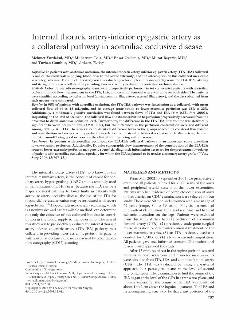
defined as lower-extremity perfusion. The percentage con-tribution of the ITA-IEA collateral to lower-extremity per-fusion was calculated using the formula: (IEA flow volume/CFA flow volume) � 100. Of the 128 sides in 64 patients,nine were excluded from final evaluation (4 had a CFAocclusion, 1 surgical revascularization, 2 amputation and 2ITA graft). Images from one patient are shown in Fig 1.
The influence of four separate factors on ITA-IEA CFADoppler flow data was examined:
1. levels of occlusion and refilling via collateral (abdominalaorta, common iliac artery, external iliac artery),
2. iliac artery occlusion as unilateral and bilateral,3. state of distal run-off as good (distal arteries open) and
poor (femoropopliteal occlusion or occlusion in at leasttwo arteries in the calf), and
4. severity of clinical symptoms as mild (Fontaine 1 and2A) and severe (Fontaine 2B to 4).
In each of the analyses, subgroups were compared interms of flow volume in the ITA-IEA collateral and itscontribution to the blood supply to the leg. The results arereported as mean � SD. Flow measurements from definedgroups were compared by Kruskal-Wallis one- way analysisof variance, the Mann-Whitney U test, and t tests. P � .05was considered to be indicative of a statistical difference.Linear regression methods were used to analyze the rela-tionships both between ITA and IEA flow, and betweenthe contribution of the ITA-IEA collateral pathway tolower-extremity perfusion and its flow.
RESULTS
IEA flow was reversed (in a caudal direction) in 72(95%) of 76 sides showing aortoiliac occlusion, normal inthree sides (4%), and occluded in one side (1%). In contrast,IEA flow was normal (in a cranial direction) in 40 (93%) of43 sides that did not show aortoiliac occlusion. Reverseflow was present in three sides (7%) without aortoiliacocclusion (aortic stenosis in 2; severe iliac stenosis in 1).Because the contribution of the ITA-IEA collateral was lessin these three sides with aortoiliac stenosis where reverseflow was observed in the IEA compared with sides withaortoiliac occlusion (average flow was 43 mL/min and theproportion of contribution to lower-extremity perfusionwas 11%), these three sides were not included in the study.In the remaining sides, the average ITA flow was 50 � 28mL/min in patients in the control group and 104 � 52mL/min in patients in the group with aortoiliac occlusion(P � .0001). Additionally, there was a statistically signifi-cant difference in ITA flow between the sides with unilat-eral iliac occlusion and the contralateral patent sides (87 �46 mL/min and 50 � 28 mL/min, respectively; P �.0002).
In patients with aortoiliac occlusion, the mean ITA-IEAcollateral flow was 66 � 48 mL/min and its contribution tolower-extremity perfusion was 38% � 23%. Furthermore, amoderately positive correlation was found between ITAflow and IEA flow (r � 0.55, P � .0001) (Fig 2), and local
linear regression showed a relationship between the pro-
the f
JOURNAL OF VASCULAR SURGERYVolume 43, Number 4 Yurdakul et al 709
portion of the contribution of the ITA-IEA collateral tolower-extremity perfusion and the ITA-IEA flow (Fig 3).Additionally, as seen on the graph, the flow volume of the
Fig 1. Right common iliac artery occlusion in a 65-yearcommon iliac artery. There is faint reconstitution of theiliac artery (arrow) that is due to partial washout of coninternal thoracic artery-inferior epigastric artery (ITA-IEartery demonstrates anastomosis between the ITA-IEAright external iliac artery. C, Color duplex ultrasonographmeasured as 126 mL/min in the IEA and 262 mL/min into lower-extremity perfusion is calculated to be 48%.
ITA-IEA collateral increased linearly up to a 50% level of
contribution to lower-extremity perfusion (85 mL/min),but after that level, it showed almost no change.
Mean flow volumes of the ITA and IEA decreased
an. A, Abdominal aortogram reveals occlusion of rightexternal iliac artery on the right via the deep circumflexaterial by unopacified blood from the ipsilateral of the
llateral. B, Selective angiography of the right subclavianw) via the superior epigastric artery, reconstituting thews monophasic spectral pattern in the IEA. The flow wasemoral artery, the contribution of the ITA-IEA collateral
-old mdistaltrast mA) co(arroy sho
according to the level of occlusion (aorta, common iliac

JOURNAL OF VASCULAR SURGERYApril 2006710 Yurdakul et al
artery, external iliac artery) from proximal to distal, andthere were statistically significant differences between flowvolumes at each of these levels (P � .0001 and P � .009,respectively) (Table I, Fig 4). I n contrast, although thecontribution of the ITA-IEA collateral flow to lower-
Fig 2. Correlation between internal thoracic artery (ITA) andinferior epigastric artery (IEA) blood flow (r � 0.55, P � .0001).
Fig 3. Scattergram illustrates the proportion of contribution ofthe internal thoracic artery (ITA)-inferior epigastric artery (IEA)collateral pathway to lower-extremity perfusion vs ITA-IEA flow.Local linear regression curve indicates that mean maximum flow is85 mL/min.
extremity perfusion also decreased from the proximal to
distal level of aortoiliac occlusion, the difference between thelevels was not significant (P � .311) (Table I, Fig 5). Therewas also no statistically significant difference between theocclusion levels concerning collateral flow volume and thecontribution to lower-extremity perfusion (Table II). Sim-ilarly, collateral flow volume and contribution to lower-extremity profusion were not significantly different whenunilateral vs bilateral occlusion of the iliac arteries, the stateof distal run-off being good or poor, or the clinical findingsbeing mild or severe were considered.
DISCUSSION
In aortoiliac occlusive disease, various collateral path-ways maintain blood flow to the lower extremities.6-12 TheITA-IEA pathway is one of these collaterals, but it mayeasily be overlooked owing to its origin distant from theaortoiliac segment. This collateral pathway involves theITA arising from the first part of subclavian artery connect-ing through the superior epigastric artery and the IEA andeventually to the external iliac artery (Fig 6). Because theITA is extensively used for CABG in patient populations athigh risk for associated atherosclerotic peripheral arterialdisease, preoperative evaluation of this collateral pathway isof significant importance.
Interruption of critical collaterals from ITA is consid-ered to be a major cause of acute limb-threatening ischemiain patients with aortoiliac occlusive disease.2,3 Acute limbischemia has also been reported in patients with aortoiliacocclusive disease after CABG.1-5 Acute limb-threateningischemia after cardiac operations may result from eventssuch as insufficient perfusion during cardiopulmonary by-pass, low cardiac output in the postoperative period, over-use of vasopressors, atheromatous embolization, postoper-ative thrombosis, and interruption of the ITA collateralpathway.3 Additionally, using the epigastric artery as flap inreconstructive surgery and performing a transverse abdom-inal incision in nonvascular procedures, which interruptsthe epigastric arteries, may result in serious leg ischemia forpatients with aortoiliac occlusion.4,11,13
When patients with aortoiliac disease are selected forCABG, selective ITA angiography or Doppler evaluation ofthe IEA likely should be done to assess the condition of theITA and IEA as a potential collateral pathway to the lowerextremity.2,13-16 On angiography, if ITA is found to be thecollateral to the ipsilateral external iliac artery or if there isDoppler evidence of reversal of IEA flow, it is recom-mended to avoid the use of the ITA or to perform asequential or staged revascularization of coronary and pe-ripheral arteries. However, opacification of the external iliacartery by the ITA-IEA collateral route on angiography orthe presence of reverse flow in the IEA on Doppler onlyindicates that the ITA-IEA collateral is functioning, andthese findings do not give information about the amount ofcollateral contribution to lower-extremity perfusion.
To our knowledge, no detailed study up to now hasevaluated the contribution of ITA-IEA collateral to lower-extremity perfusion in aortoiliac occlusion. In our study,
the contribution of this collateral to lower-extremity per-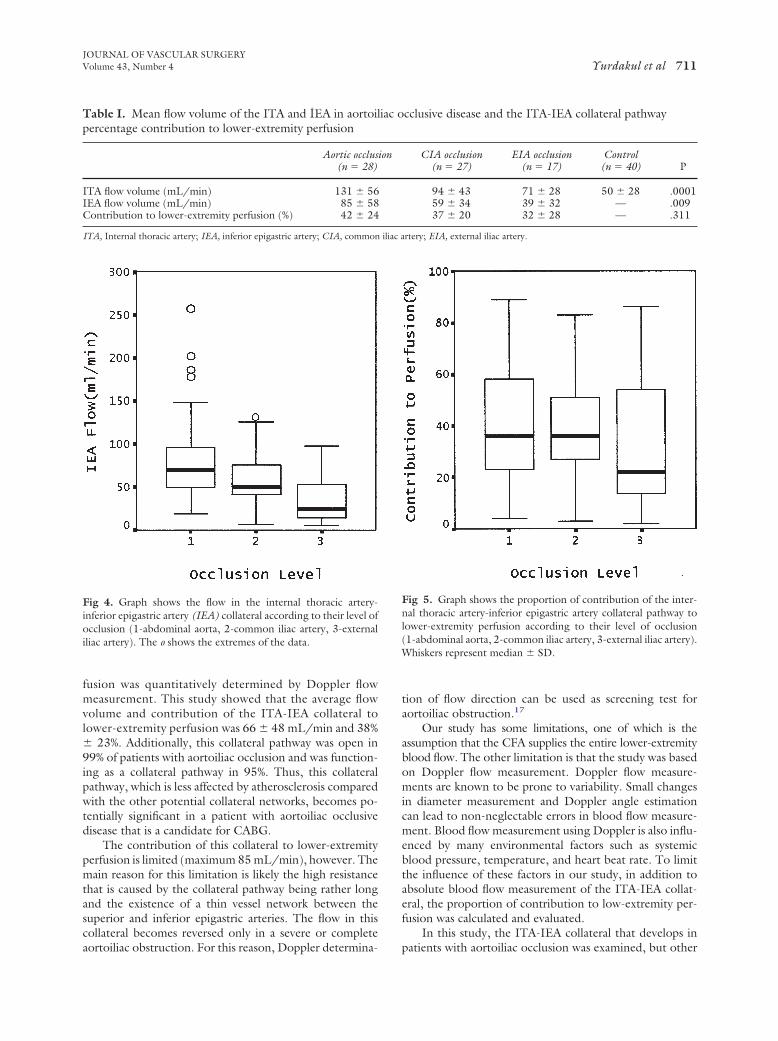
iliac a
JOURNAL OF VASCULAR SURGERYVolume 43, Number 4 Yurdakul et al 711
fusion was quantitatively determined by Doppler flowmeasurement. This study showed that the average flowvolume and contribution of the ITA-IEA collateral tolower-extremity perfusion was 66 � 48 mL/min and 38%� 23%. Additionally, this collateral pathway was open in99% of patients with aortoiliac occlusion and was function-ing as a collateral pathway in 95%. Thus, this collateralpathway, which is less affected by atherosclerosis comparedwith the other potential collateral networks, becomes po-tentially significant in a patient with aortoiliac occlusivedisease that is a candidate for CABG.
The contribution of this collateral to lower-extremityperfusion is limited (maximum 85 mL/min), however. Themain reason for this limitation is likely the high resistancethat is caused by the collateral pathway being rather longand the existence of a thin vessel network between thesuperior and inferior epigastric arteries. The flow in thiscollateral becomes reversed only in a severe or complete
Table I. Mean flow volume of the ITA and IEA in aortoilpercentage contribution to lower-extremity perfusion
Aortic occlus(n � 28)
ITA flow volume (mL/min) 131 � 56IEA flow volume (mL/min) 85 � 58Contribution to lower-extremity perfusion (%) 42 � 24
ITA, Internal thoracic artery; IEA, inferior epigastric artery; CIA, common
Fig 4. Graph shows the flow in the internal thoracic artery-inferior epigastric artery (IEA) collateral according to their level ofocclusion (1-abdominal aorta, 2-common iliac artery, 3-externaliliac artery). The o shows the extremes of the data.
aortoiliac obstruction. For this reason, Doppler determina-
tion of flow direction can be used as screening test foraortoiliac obstruction.17
Our study has some limitations, one of which is theassumption that the CFA supplies the entire lower-extremityblood flow. The other limitation is that the study was basedon Doppler flow measurement. Doppler flow measure-ments are known to be prone to variability. Small changesin diameter measurement and Doppler angle estimationcan lead to non-neglectable errors in blood flow measure-ment. Blood flow measurement using Doppler is also influ-enced by many environmental factors such as systemicblood pressure, temperature, and heart beat rate. To limitthe influence of these factors in our study, in addition toabsolute blood flow measurement of the ITA-IEA collat-eral, the proportion of contribution to low-extremity per-fusion was calculated and evaluated.
In this study, the ITA-IEA collateral that develops in
cclusive disease and the ITA-IEA collateral pathway
CIA occlusion(n � 27)
EIA occlusion(n � 17)
Control(n � 40) P
94 � 43 71 � 28 50 � 28 .000159 � 34 39 � 32 — .00937 � 20 32 � 28 — .311
rtery; EIA, external iliac artery.
Fig 5. Graph shows the proportion of contribution of the inter-nal thoracic artery-inferior epigastric artery collateral pathway tolower-extremity perfusion according to their level of occlusion(1-abdominal aorta, 2-common iliac artery, 3-external iliac artery).Whiskers represent median � SD.
iac o
ion
patients with aortoiliac occlusion was examined, but other

JOURNAL OF VASCULAR SURGERYApril 2006712 Yurdakul et al
collaterals were not. It is obvious, however, that an inter-action occurs between the collaterals that provide lower-extremity perfusion. If the other collaterals are well devel-oped, then the flow of the ITA-IEA collateral will be lesseffective in supplying perfusion to the lower extremities.This study found that the contribution of the ITA-IEAcollateral pathway to lower-extremity perfusion was higherin the section of proximal aortoiliac occlusion comparedwith distal occlusion, and this can be partially explained bythe fact that in proximal occlusion, more distal collateralscease to function. In other words, the ITA-IEA collateralpathway appears more important in aortic and commoniliac arterial occlusion, but in external iliac artery occlu-sions, the hypogastric collateral route likely has a high flowrate.
This study was not done with the aim of determiningthe threshold amount of collateral flow that would result insevere leg ischemia after the interruption of the ITA-IEAcollateral. However, in a patient with aortoiliac occlusion, ifthe ITA is used as a graft, the risk of acute ischemia developingin the lower extremity is expected to increase with the directproportion of the ITA-IEA collateral contribution tolower-extremity perfusion. This is because when the ITAis used as a graft, its function as a collateral would bedisrupted, and if it is a major collateral, then the othersecondary collaterals, whose contributions are likely verylow, would not compensate for the interrupted flow ofITA-IEA collateral route. Therefore, during the work-up ofpatients with aortoiliac occlusion who require CABG, notonly should the absolute quantity of the flow of this collat-eral to lower extremity be assessed but also its contributionto lower-extremity perfusion.
Although differences were noted in terms of ITA-IEAcollateral flow according to the levels of aortoiliac occlu-sion, there was also a significant overlap in flow volumebetween these groups. And, still more important, no sig-nificant differences were found in the proportion of contri-bution to lower-extremity perfusion. Furthermore, there
Table II. Comparison of groups according to theirangiographic and clinical findings
IEA flow volume(mL/min)
Contribution tolower-extremityperfusion (%)
Iliac occlusionUnilateral 54 � 34 35 � 23Bilateral 58 � 33 22 � 17
P � .599 P � .139Distal run-off
Good 61 � 47 37 � 24Poor 69 � 45 40 � 20
P � .563 P � .529Clinical finding
Mild 73 � 51 38 � 22Severe 49 � 38 36 � 25
P � .082 P � .659
IEA, Inferior epigastric artery.
were no differences in flow volume or contribution to
perfusion according to differences in the clinical findingsand distal run-off. Therefore, for the management an indi-vidual patient with aortoiliac occlusion, there do not appearto be clinical predictors of the importance of the ITA-IEApathway, and the contribution of this collateral to lower-extremity perfusion should be evaluated before CABG.Angiography may be used at this step; however, CDUevaluation has some convincing advantages, such as itsability to provide objective hemodynamic data regardingthe flow direction, flow volume, and perfusion.
CONCLUSION
In aortoiliac occlusion, the ITA-IEA collateral pathwayis an important route providing lower-extremity perfusion.Doppler sonographic flow measurements of the contribu-tion of the ITA-IEA route to lower-extremity perfusionmay provide beneficial diagnostic information necessary for
Fig 6. Schematic view of the collateral circulation via the internalthoracic and epigastric arteries in occlusion of the left common andexternal iliac arteries. ITA, Internal thoracic artery; SEA, superiorepigastric artery; IEA, inferior epigastric artery.
pretreatment evaluation of the patients with aortoiliac oc-

JOURNAL OF VASCULAR SURGERYVolume 43, Number 4 Yurdakul et al 713
clusion, especially those for whom the ITA is planned to beused as a graft.
AUTHOR CONTRIBUTIONS
Analysis and interpretation: MY, MT, EÖ, MB, TCData collection: MY, MT, EÖ, MBWriting the article: MY, MTCritical revision of the article: MY, MT, EÖ, MB, TCFinal approval of the article: MY, MT, EÖ, MB, TCStatistical analysis: MY, MTObtained funding: MY, MTOverall responsibility: MY
REFERENCES
1. Melissano G, Di Credico G, Chiesa R, Grossi A. The use of internalthoracic arteries for myocardial revascularization may produce acute legischemia in patients with concomitant Leriche’s syndrome. J Vasc Surg1996;24:698.
2. Dietzek AM, Goldsmith J, Veith FJ, Sanchez LA, Gupta SK, WengerterKR. Interruption of critical aortoiliac collateral circulation during non-vascular operations: a cause of acute limb-threatening ischemia. J VascSurg 1990;12:645-51; discussion, 652-3.
3. Kitamura S, Inoue K, Kawachi K, Morita R, Seki T, Taniguchi S, et al.Lower extremity ischemia secondary to internal thoracic-coronary ar-tery bypass grafting. Ann Thorac Surg 1993;56:157-9.
4. Tsui SS, Parry AJ, Large SR. Leg ischaemia following bilateral internalthoracic artery and inferior epigastric artery harvesting. Eur J Cardio-thorac Surg 1995;9:218-20.
5. Yapici F, Tuygun AG, Tarhan IA, Yilmaz M, Tuygun AK, Yapici N, et al.Limb ischemia due to use of internal thoracic artery in coronary bypass.Asian Cardiovasc Thorac Ann 2002;10:254-5.
structive arterial disease. Am J Roentgenol Radium Ther Nucl Med1957;77:296-311.
7. Bron KM. Thrombotic occlusion of the abdominal aorta. Associatedvisceral artery lesions and collateral circulation. Am J Roentgenol Ra-dium Ther Nucl Med 1966;96:887-95.
8. Caresano A. The collateral circulation in chronic occlusions of theabdominal aorta and of its terminal branches (anatomo-radiologicaldiscussion). J Cardiovasc Surg (Torino) 1966;7:297-310.
9. Edwards EA, Lemay M. Occlusion patterns and collaterals in arterio-sclerosis of the lower aorta and iliac arteries. Surgery 1955;38:950-63.
10. Chait A. The internal mammary artery: an overlooked collateral path-way to the leg. Radiology 1976;121:621-4.
11. Krupski WC, Sumchai A, Effeney DJ, Ehrenfeld WK. The importance ofabdominal wall collateral blood vessels. Planning incisions and obtain-ing arteriography. Arch Surg 1984;119:854-7.
12. Kim J, Won JY, Park SI, Lee do Y. Internal thoracic artery collateral tothe external iliac artery in chronic aortoiliac occlusive disease. Korean JRadiol 2003;4:179-83.
13. Hodge K, Yuen J, Moursi M, Eidt JF. Critical leg ischemia resultingfrom interruption of collaterals by harvest of the rectus abdominis freeflap: endovascular salvage. Ann Plast Surg 2000;45:427-30.
14. Arnold JR, Greenberg JD, Clements S. Internal mammary artery per-fusing the Leriche’s syndrome. Ann Thorac Surg 2000;69:1244-6.
15. Shimizu T, Hirayama T, Ikeda K, Ito S, Ishimaru S. Coronary revascu-larization with arterial conduits collateral to the lower limb. Ann ThoracSurg 1999;67:1783-5.
16. Parashara DK, Kotler MN, Ledley GS, Yazdanfar S. Internal mammaryartery collateral to the external iliac artery: an angiographic consider-ation prior to coronary bypass surgery. Cathet Cardiovasc Diagn 1994;32:343-5.
17. Kwaan JH, Connolly JE. Doppler assessment of the inferior epigastricartery flow patterns as a screening test for aortoiliac obstruction. Am JSurg 1979;137:250-1.
6. Figley MM, Muller RF. The arteries of the abdomen, pelvis, and thigh.I. Normal roentgenographic anatomy. II. Collateral circulation in ob- Submitted Jul 9, 2005; accepted Dec 8, 2005.
Authors requested to declare conditions of research funding
When sponsors are directly involved in research studies of drugs and devices, the editors will ask authors to clarify theconditions under which the research project was supported by commercial firms, private foundations, or government.Specifically, in the methods section, the authors should describe the roles of the study sponsor(s) and theinvestigator(s) in (1) study design, (2) conduct of the study, (3) data collection, (4) data analysis, (5) datainterpretation, (6) writing of the report, and (7) the decision regarding where and when to submit the report forpublication. If the supporting source had no significant involvement in these aspects of the study, the authors shouldso state.