Interleukin-21 Overexpression Dominates T Cell Response to ... · Interleukin-21 Overexpression...
7
Interleukin-21 Overexpression Dominates T Cell Response to Epstein- Barr Virus in a Fatal Case of X-Linked Lymphoproliferative Syndrome Type 1 Consuelo Ortega, a Orlando A. Estévez, b Silvia Fernández, a Rocío Aguado, b José M. Rumbao, c Teresa Gonzalez, d Juan L. Pérez-Navero, c Manuel Santamaría a,b Department of Immunology, School of Medicine, University of Córdoba, Córdoba, Spain a ; Clinical Immunology Unit, b Pediatric Unit, c and Pathology Unit, d Reina Sofía University Hospital, Córdoba, Spain Interleukin-21 (IL-21) is a cytokine whose actions are closely related to B cell differentiation into plasma cells as well as to CD8 cytolytic T cell effector and memory generation, influencing the T lymphocyte response to different viruses. X-linked lym- phoproliferative syndrome type 1 (XLP-1) is a primary immunodeficiency syndrome that is characterized by a high susceptibility to Epstein-Barr virus. We observed in a pediatric patient with XLP-1 that IL-21 was expressed in nearly all peripheral blood CD4 and CD8 T cells. However, IL-21 could not be found in the lymph nodes, suggesting massive mobilization of activated cells toward the infection’s target organs, where IL-21-producing cells were detected, resulting in large areas of tissue damage. CASE REPORT O ur patient was a 10-month-old Caucasian male, born full- term to a nonconsanguineous couple, who had a medical record of bronchiolitis and ear, nose, and throat infections. The child was referred to our Pediatric Infectious Disease Unit because of fever (38.5°C to 39.5°C) lasting 11 days, associated with a non- pruritic erythematous rash, tonsillitis, cervical lymphadenopa- thies, and hepatomegaly (3 cm below the costal edge). Neither splenomegaly nor abdominal lymph node enlargement was de- tected on admission. At this point, the blood values showed a white blood cell count of 20,900/l (lymphocytes, 72%), throm- bocytopenia (72,000/l), a raised C-reactive protein level, and a mild elevation of liver enzymes (aspartate aminotransferase [AST], 120 U/liter, alanine aminotransferase [ALT], 111 U/liter, and -glutamyl transferase [GGT], 324 U/liter). Specific IgM and IgG antibodies to cytomegalovirus (CMV) and Epstein-Barr virus (EBV) were found, with the latter at a high titer (anti-EBV IgG, 1/640). Viral loads were determined by means of PCRs. PCR values for EBV found in infectious mononucleosis (IMN) patients ranged from 6,541 copies/ml to 11,476 copies/ml. The value de- tected in the patient was as high as 50,368 copies/ml for EBV (human herpesvirus 4 [HHV-4]) but 600 copies/ml for CMV. EBV infection was diagnosed. The patient’s general condition worsened after 72 h, and he developed splenomegaly (14 cm) and basal right pneumonia, which was treated with 50 mg cefotaxime/kg of body weight intra- venously every 12 h in the absence of microbiological culture data. The fever disappeared, and the patient’s condition remained sta- ble for a week, but the fever returned (39.5°C) and was accompa- nied by pronounced jaundice (bilirubin, 4 mg/dl; AST, 843 U/liter; ALT, 339 U/liter; and GGT, 1,233 U/liter). High levels of ferritin (2,682 ng/ml) and plasma triglycerides (240 mg/dl), low levels of hemoglobin (7.3 g/dl), and a lymphocyte count of 50,000/l were detected. Within the next 24 h, severe thrombo- cytopenia occurred (40,000 platelets/l) along with general ton- ic-clonic seizures, brain front-lobe bleeding, and generalized ce- rebral edema (as observed on a computed tomography [CT] scan), which evolved in the subsequent 16 h to respiratory distress, hemodynamic shock, multiorgan failure, and exitus. The main clinical events that occurred in this X-linked lymphoproliferative syndrome type 1 (XLP-1) patient are represented in Fig. 1. XLP-1 is a primary immunodeficiency syndrome character- ized by a high susceptibility to Epstein-Barr virus (EBV) (1–3). The disease is caused by germ line mutations in the SH2D1A gene, which encodes the adaptor molecule signaling lymphocytic acti- vation molecule (SLAM)-associated protein (SAP) (4, 5). This protein modulates the signal transduction of SLAM family recep- tors in T lymphocytes, natural killer (NK) cells, and natural killer T (NKT) cells (6, 7), influencing their cytotoxic ability and cyto- kine regulation (8–10). The loss of a functional SAP results in both an impaired ability of cytotoxic cells to clear the EBV infection and overexpression of proinflammatory cytokines by T and NK cells (11). Interleukin 21 (IL-21) is a cytokine that is produced mainly by CD4 cells but also by CD8 lymphocytes in different human diseases (12, 13). In EBV-infected B cells, IL-21 induces the ex- pression of EBV genes, such as the latent membrane protein 1 (LMP1) gene, thus providing viral peptides that are recognizable by the immune system (14–16). Indeed, IL-21 is critical for CD8 T cell survival and memory generation (17–21), as well as for promoting the activity of CD8 T cell effectors during viral infec- tions (22–24), enhancing the cytotoxic response to virally infected cells by NK cells and CD8 T lymphocytes (25–27). Received 29 January 2013 Returned for modification 8 February 2013 Accepted 26 February 2013 Published ahead of print 6 March 2013 Address correspondence to Manuel Santamaría, [email protected]. O.A.E. and S.F. contributed equally to this article. Copyright © 2013, American Society for Microbiology. All Rights Reserved. doi:10.1128/CVI.00002-13 CASE REPORT May 2013 Volume 20 Number 5 Clinical and Vaccine Immunology p. 765–771 cvi.asm.org 765 on December 27, 2019 by guest http://cvi.asm.org/ Downloaded from
Transcript of Interleukin-21 Overexpression Dominates T Cell Response to ... · Interleukin-21 Overexpression...
Interleukin-21 Overexpression Dominates T Cell Response to Epstein-Barr Virus in a Fatal Case of X-Linked Lymphoproliferative SyndromeType 1
Consuelo Ortega,a Orlando A. Estévez,b Silvia Fernández,a Rocío Aguado,b José M. Rumbao,c Teresa Gonzalez,d Juan L. Pérez-Navero,c
Manuel Santamaríaa,b
Department of Immunology, School of Medicine, University of Córdoba, Córdoba, Spaina; Clinical Immunology Unit,b Pediatric Unit,c and Pathology Unit,d Reina SofíaUniversity Hospital, Córdoba, Spain
Interleukin-21 (IL-21) is a cytokine whose actions are closely related to B cell differentiation into plasma cells as well as to CD8�
cytolytic T cell effector and memory generation, influencing the T lymphocyte response to different viruses. X-linked lym-phoproliferative syndrome type 1 (XLP-1) is a primary immunodeficiency syndrome that is characterized by a high susceptibilityto Epstein-Barr virus. We observed in a pediatric patient with XLP-1 that IL-21 was expressed in nearly all peripheral bloodCD4� and CD8� T cells. However, IL-21 could not be found in the lymph nodes, suggesting massive mobilization of activatedcells toward the infection’s target organs, where IL-21-producing cells were detected, resulting in large areas of tissue damage.
CASE REPORT
Our patient was a 10-month-old Caucasian male, born full-term to a nonconsanguineous couple, who had a medical
record of bronchiolitis and ear, nose, and throat infections. Thechild was referred to our Pediatric Infectious Disease Unit becauseof fever (38.5°C to 39.5°C) lasting 11 days, associated with a non-pruritic erythematous rash, tonsillitis, cervical lymphadenopa-thies, and hepatomegaly (3 cm below the costal edge). Neithersplenomegaly nor abdominal lymph node enlargement was de-tected on admission. At this point, the blood values showed awhite blood cell count of 20,900/�l (lymphocytes, 72%), throm-bocytopenia (72,000/�l), a raised C-reactive protein level, and amild elevation of liver enzymes (aspartate aminotransferase[AST], 120 U/liter, alanine aminotransferase [ALT], 111 U/liter,and �-glutamyl transferase [GGT], 324 U/liter). Specific IgM andIgG antibodies to cytomegalovirus (CMV) and Epstein-Barr virus(EBV) were found, with the latter at a high titer (anti-EBV IgG,�1/640). Viral loads were determined by means of PCRs. PCRvalues for EBV found in infectious mononucleosis (IMN) patientsranged from 6,541 copies/ml to 11,476 copies/ml. The value de-tected in the patient was as high as 50,368 copies/ml for EBV(human herpesvirus 4 [HHV-4]) but �600 copies/ml for CMV.EBV infection was diagnosed.
The patient’s general condition worsened after 72 h, and hedeveloped splenomegaly (14 cm) and basal right pneumonia,which was treated with 50 mg cefotaxime/kg of body weight intra-venously every 12 h in the absence of microbiological culture data.The fever disappeared, and the patient’s condition remained sta-ble for a week, but the fever returned (39.5°C) and was accompa-nied by pronounced jaundice (bilirubin, �4 mg/dl; AST, 843U/liter; ALT, 339 U/liter; and GGT, 1,233 U/liter). High levels offerritin (2,682 ng/ml) and plasma triglycerides (240 mg/dl), lowlevels of hemoglobin (7.3 g/dl), and a lymphocyte count of�50,000/�l were detected. Within the next 24 h, severe thrombo-cytopenia occurred (�40,000 platelets/�l) along with general ton-ic-clonic seizures, brain front-lobe bleeding, and generalized ce-rebral edema (as observed on a computed tomography [CT]
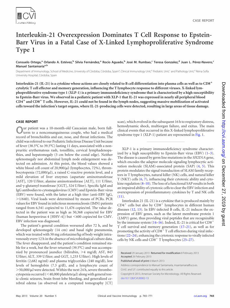
scan), which evolved in the subsequent 16 h to respiratory distress,hemodynamic shock, multiorgan failure, and exitus. The mainclinical events that occurred in this X-linked lymphoproliferativesyndrome type 1 (XLP-1) patient are represented in Fig. 1.
XLP-1 is a primary immunodeficiency syndrome character-ized by a high susceptibility to Epstein-Barr virus (EBV) (1–3).The disease is caused by germ line mutations in the SH2D1A gene,which encodes the adaptor molecule signaling lymphocytic acti-vation molecule (SLAM)-associated protein (SAP) (4, 5). Thisprotein modulates the signal transduction of SLAM family recep-tors in T lymphocytes, natural killer (NK) cells, and natural killerT (NKT) cells (6, 7), influencing their cytotoxic ability and cyto-kine regulation (8–10). The loss of a functional SAP results in bothan impaired ability of cytotoxic cells to clear the EBV infection andoverexpression of proinflammatory cytokines by T and NK cells(11).
Interleukin 21 (IL-21) is a cytokine that is produced mainly byCD4� cells but also by CD8� lymphocytes in different humandiseases (12, 13). In EBV-infected B cells, IL-21 induces the ex-pression of EBV genes, such as the latent membrane protein 1(LMP1) gene, thus providing viral peptides that are recognizableby the immune system (14–16). Indeed, IL-21 is critical for CD8�
T cell survival and memory generation (17–21), as well as forpromoting the activity of CD8� T cell effectors during viral infec-tions (22–24), enhancing the cytotoxic response to virally infectedcells by NK cells and CD8� T lymphocytes (25–27).
Received 29 January 2013 Returned for modification 8 February 2013Accepted 26 February 2013
Published ahead of print 6 March 2013
Address correspondence to Manuel Santamaría, [email protected].
O.A.E. and S.F. contributed equally to this article.
Copyright © 2013, American Society for Microbiology. All Rights Reserved.
doi:10.1128/CVI.00002-13
CASE REPORT
May 2013 Volume 20 Number 5 Clinical and Vaccine Immunology p. 765–771 cvi.asm.org 765
on Decem
ber 27, 2019 by guesthttp://cvi.asm
.org/D
ownloaded from
However, despite the well-known involvement of IL-21 in EBVinfection, direct evidence of its participation in the mechanismsleading to, or maintaining, the lymphoproliferative response toEBV infection in SAP-deficient (SAPneg) patients is lacking. Wereport here the results for the expression, production, and func-tion of IL-21 in a SAPneg pediatric patient with a fatal EBV infec-tion.
Human subjects. T cells from a SAPneg patient were analyzedduring a fatal evolution of EBV infection. In this study, sevenpediatric patients diagnosed with infectious mononucleosis andseven age- and sex-matched healthy donors were included to serveas controls. For this purpose, written informed consent from do-nors’ and patients’ parents as well as approval from the Institu-tional Review Board of The Reina Sofia University Hospital wasobtained.
Gene analysis procedure. Genomic DNA was extracted fromwhole peripheral blood using a Maxwell 16 blood DNA purifica-tion kit on a Maxwell DNA extraction device (Promega, USA). Allcoding exons and intronic boundaries of the PRF1, STX11,UNC13D, and SH2D1A genes were amplified by PCR. The PCRamplicons were purified with an illustra ExoStar one-step kit (GEHealthcare, USA); bidirectional fluorescence sequencing was per-formed with an ABI BigDye Terminator v3.1 cycle sequencing kit(Applied Biosystems, USA), and samples were run on an auto-mated ABI 3730 XL DNA analyzer.
Flow cytometry. For surface-directed staining, cells were incu-bated with relevant fluorochrome-conjugated mouse anti-humanmonoclonal antibodies (MAb) on ice for 30 min in the dark andwashed twice before analysis. Fluorescein isothiocyanate (FITC)-,phycoerythrin (PE)-, PE-Cy7-, antigen-presenting cell (APC)-, orAlexa-Fluor 647-conjugated anti-CD3, -CD4, or -CD8 MAb andisotype-matched control mouse IgG1 and IgG2 MAb were used asisotype controls (all from BD Biosciences, San José, CA, USA). Forintracellular staining, T cells from IMN patients and controls wereactivated with a lymphocyte activation cocktail consisting of 25ng/ml phorbol myristate acetate (PMA) and 1 �g/ml ionomycin(Sigma-Aldrich Spain) for 6 h at 37°C, prior to being stained,fixed, and permeabilized with the Cytofix/Cytoperm kit and a BDGolgi plug (BD Pharmingen, USA), according to the manufactur-er’s protocol; an inactivated control was included in the assays andtreated with brefeldin A (BD Pharmingen, San Diego, CA, USA)
only. For analysis of the XLP-1 patient’s T peripheral blood cells,the previous treatment was not performed, as they were found tobe already activated. The following anti-human MAb were used:anti-gamma interferon (anti-IFN-�) and anti-tumor necrosis fac-tor alpha (TNF-�) (both from BD Biosciences, USA), anti-IL-22(R&D Systems, Minneapolis, MN, USA), anti-IL-17 and anti-IL-21 (eBioscience, USA), and anti-IL-4, anti-IL-5, and anti-IL-10(BD Pharmingen, USA). Mouse IgG1 and IgG2 were used as iso-type controls. Flow cytometry was performed on a FACSCaliburflow cytometer (BD Biosciences, San José, CA, USA), and datawere analyzed using the Cell Quest Pro software (BD Biosciences,San José, CA, USA).
Immune cell isolation and cultures. Single-cell lymphocytesuspensions were isolated from peripheral blood mononuclearcells (PBMC), thymus, and lymph nodes by gradient centrifuga-tion with Ficoll-Paque (Sigma-Aldrich, Spain). Cells were cul-tured in complete medium, consisting of RPMI 1640 mediumcontaining 2 mM glutamine, 100 units/ml penicillin, and 100mg/ml streptomycin and supplemented with 10% fetal bovineserum (FBS) (all from BioWhittaker, Belgium) at 37°C in a hu-midified atmosphere containing 5% CO2. For some experiments,peripheral blood lymphocytes (PBL) from IMN patients and con-trols were stimulated in the presence of anti-CD3/CD28-coatedbeads (1 bead/cell) (human T-Expander CD3/CD28; InvitrogenDynal Biotech ASA, Norway) for 24 h. An EBV cell line was used asthe stimulus in experiments addressing the composition of IL-21-producing T cells in response to prolonged exposure to EBV-in-fected cells. Briefly, 106 PBL/well were cocultured with 106 irradi-ated (3,500 rads) EBV-infected cells/well. Cells were plated at afinal volume of 2 ml/well in 24-well culture plates (Nunc, Den-mark) for 15 days. At day 7 of incubation, T cells were restimu-lated with the same number of EBV cells after careful removal of 1ml of culture medium.
IL-21 production and proliferation assays. The productionand role of IL-21 in the proliferative response observed were stud-ied as previously described (13). Briefly, carboxyfluorescein suc-cinimidyl ester (CFSE; 5 �M; Sigma-Aldrich, Spain)-labeled Tcells (5 � 104 cells/well) were stimulated with anti-CD3/CD28-coated beads (human T-Expander; 1 bead/cell) in the absence orpresence of anti-IL-21 MAb (10 �g) in complete medium. After 3days, proliferation was determined on a FACSCalibur flow cytom-eter with CellQuest Pro software. IL-21 and IFN-� levels weredetermined in sera and 24-h culture supernatants from all studiedsubjects using enzyme-linked immunosorbent assays (ELISAs)according to manufacturer instructions (Med Systems, USA).
Immunohistochemistry. Paraffin-embedded sections frombrain and liver (4 �m) were deparaffinized and then hydratedwith graded alcohol solutions. Endogenous peroxidase wasblocked by incubation with 0.75% hydrogen peroxide for 15 min,followed by antigen unmasking with heated citrate buffer (pH 9).Sections were stained separately with anti-IL-21 at a 1/100 optimaldilution (Millipore, USA), mouse anti-human anti-CD4 MAb, oranti-CD8 MAb (Dako, United Kingdom) for 30 min. Slides weredeveloped with the EnVision FLEX�, mouse, high-pH link kit(Dako, United Kingdom) by incubation for 30 min at room tem-perature with the horseradish peroxidase anti-rabbit/anti-mousecomplex (Dako, United Kingdom) as described by the manufac-turer (Autostainer Link 48; Dako, United Kingdom). Staining wasperformed by incubating slides with diaminobenzidine as thechromogenic substrate for 5 min. To counterstain, we used
FIG 1 Clinical course of the XLP-1 patient from hospital inception.
Case Report
766 cvi.asm.org Clinical and Vaccine Immunology
on Decem
ber 27, 2019 by guesthttp://cvi.asm
.org/D
ownloaded from
EnVision FLEX hematoxylin (Dako, United Kingdom). As nega-tive controls for each staining, the same procedure as mentionedpreviously was followed except that isotype control MAb wasused.
On the basis of the clinical evolution as a fatal infectious mono-nucleosis, mutational analysis of the XLP-1 (SH2D1A) gene and offamilial hemophagocytic lymphohistiocytosis (FHLH)-relatedgenes (PRF1, STX11, and UNC13D) was performed. No defectswere detected in any of the FHLH-causative genes. However, atexon 2 of the SH2D1A gene, the nonsense mutation p.R55X wasdetected, a genetic variant previously reported as a disease-causingmutation (28).
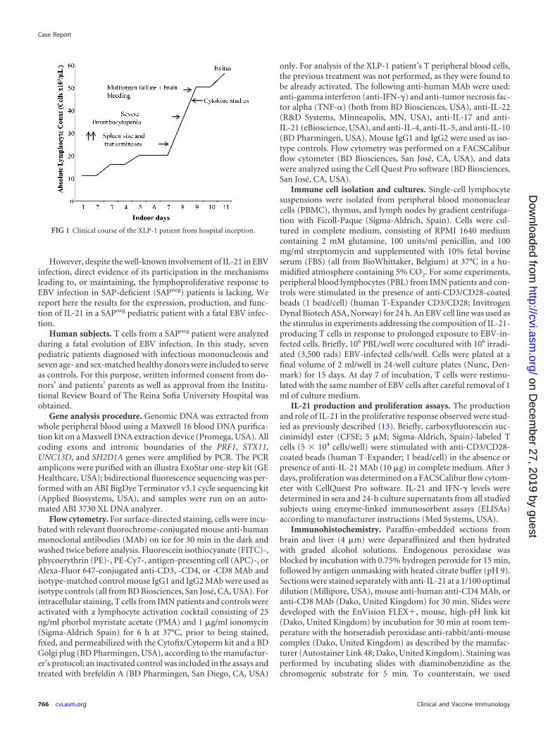
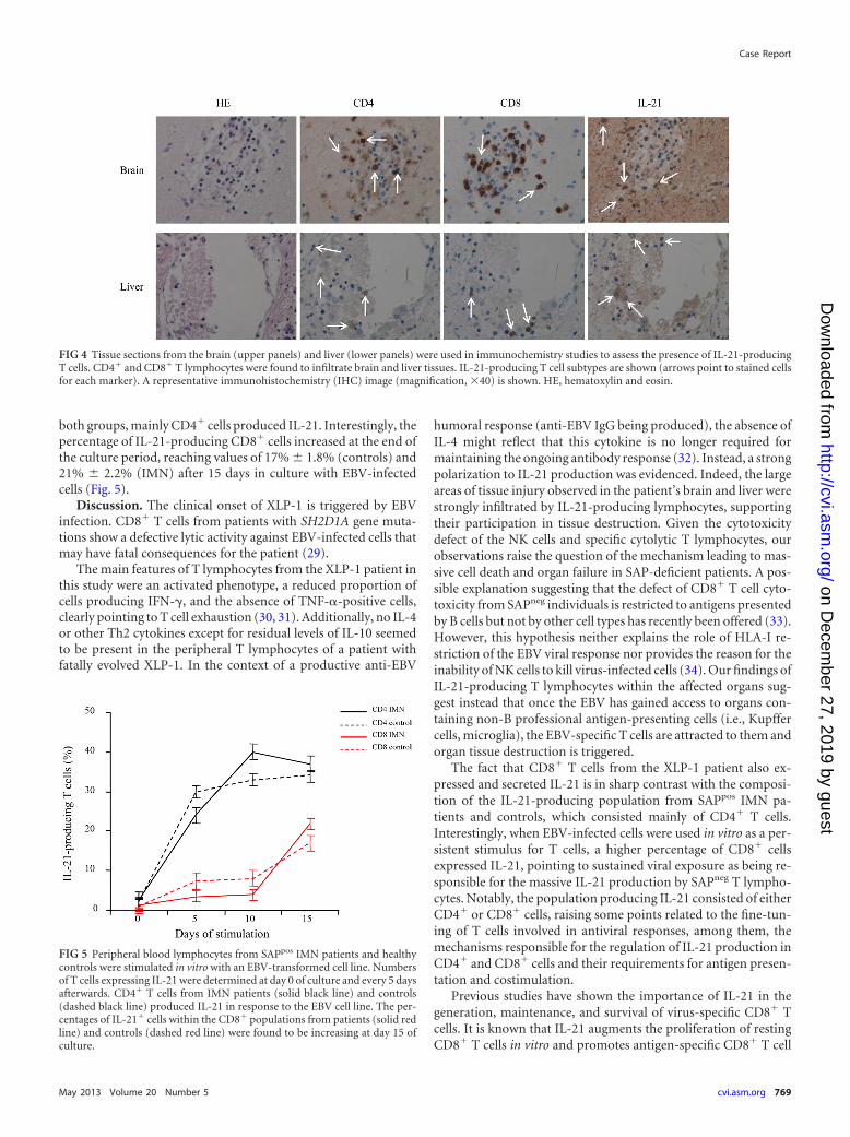
Ex vivo analysis of the T cell compartment of the SAPneg patientshowed that, in addition to the number of the patient’s circulatinglymphocytes being increased, 85% of his T cells expressed CD69,
denoting a T cell activation status that was not detected in EBV-infected SAP-positive (SAPpos) patients (Fig. 2, left column). Tofurther analyze this massive activation of T cells, the productionprofiles of the Th1 (IFN-�, TNF-�), Th2 (IL-4, IL-5, IL-10), andTh17 (IL-17, IL-21, IL-22) cytokine families were studied. Resultsshowed that 26% of CD4� cells and 47.8% of CD8� T lympho-cytes from the XLP-1 patient produced IFN-�. Notably, T cellsfrom the XLP-1 patient did not produce TNF-�, whereas CD4�
and CD8� cells from IMN patients produced TNF-� and, to alesser extent, IFN-� (15% of CD8� cells) (Fig. 2, two middle col-umns). Th2-related cytokine-producing cells were undetectable,except that IL-10 that was found in 7% of the total T cell popula-tion of the SAPneg patient (data not shown). Regarding the Th17family, expression of IL-21 was detected in most of the peripheralblood T lymphocytes from the XLP-1 patient. IL-21 was expressed
FIG 2 T cell activation was assessed by expression of CD69 (left column). Intracellular IFN-�, TNF-�, and IL-21 expression levels by CD4� and CD8� T cellsfrom an XLP-1 patient (n � 1) and IMN patients (n � 7) are depicted in the middle and right columns, respectively. The results of a representative experimentare shown.
Case Report
May 2013 Volume 20 Number 5 cvi.asm.org 767
on Decem
ber 27, 2019 by guesthttp://cvi.asm
.org/D
ownloaded from
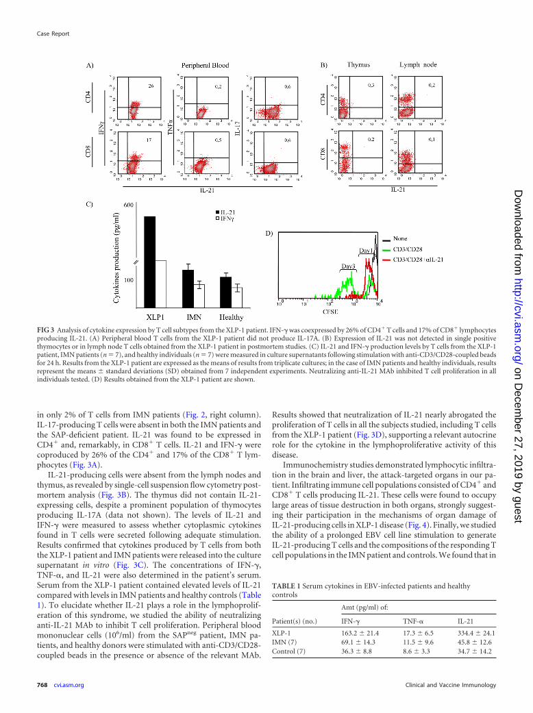
in only 2% of T cells from IMN patients (Fig. 2, right column).IL-17-producing T cells were absent in both the IMN patients andthe SAP-deficient patient. IL-21 was found to be expressed inCD4� and, remarkably, in CD8� T cells. IL-21 and IFN-� werecoproduced by 26% of the CD4� and 17% of the CD8� T lym-phocytes (Fig. 3A).
IL-21-producing cells were absent from the lymph nodes andthymus, as revealed by single-cell suspension flow cytometry post-mortem analysis (Fig. 3B). The thymus did not contain IL-21-expressing cells, despite a prominent population of thymocytesproducing IL-17A (data not shown). The levels of IL-21 andIFN-� were measured to assess whether cytoplasmic cytokinesfound in T cells were secreted following adequate stimulation.Results confirmed that cytokines produced by T cells from boththe XLP-1 patient and IMN patients were released into the culturesupernatant in vitro (Fig. 3C). The concentrations of IFN-�,TNF-�, and IL-21 were also determined in the patient’s serum.Serum from the XLP-1 patient contained elevated levels of IL-21compared with levels in IMN patients and healthy controls (Table1). To elucidate whether IL-21 plays a role in the lymphoprolif-eration of this syndrome, we studied the ability of neutralizinganti-IL-21 MAb to inhibit T cell proliferation. Peripheral bloodmononuclear cells (106/ml) from the SAPneg patient, IMN pa-tients, and healthy donors were stimulated with anti-CD3/CD28-coupled beads in the presence or absence of the relevant MAb.
Results showed that neutralization of IL-21 nearly abrogated theproliferation of T cells in all the subjects studied, including T cellsfrom the XLP-1 patient (Fig. 3D), supporting a relevant autocrinerole for the cytokine in the lymphoproliferative activity of thisdisease.
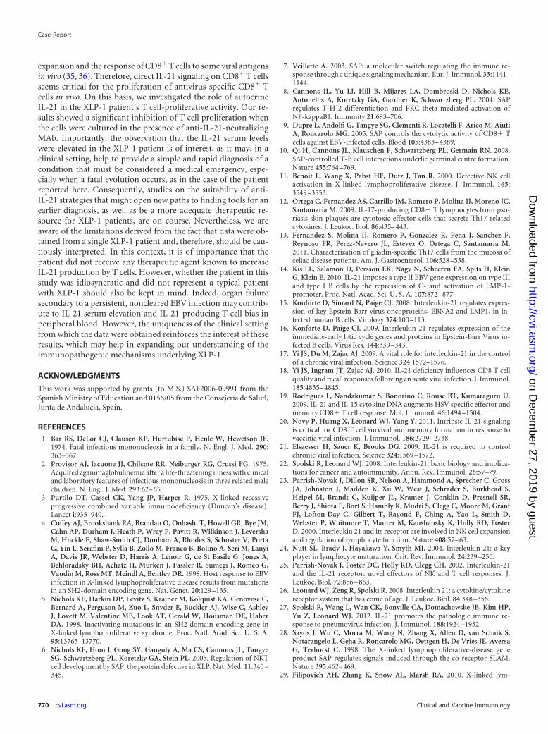
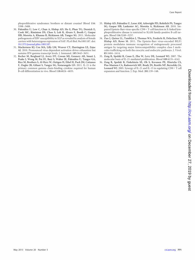
Immunochemistry studies demonstrated lymphocytic infiltra-tion in the brain and liver, the attack-targeted organs in our pa-tient. Infiltrating immune cell populations consisted of CD4� andCD8� T cells producing IL-21. These cells were found to occupylarge areas of tissue destruction in both organs, strongly suggest-ing their participation in the mechanisms of organ damage ofIL-21-producing cells in XLP-1 disease (Fig. 4). Finally, we studiedthe ability of a prolonged EBV cell line stimulation to generateIL-21-producing T cells and the compositions of the responding Tcell populations in the IMN patient and controls. We found that in
FIG 3 Analysis of cytokine expression by T cell subtypes from the XLP-1 patient. IFN-� was coexpressed by 26% of CD4� T cells and 17% of CD8� lymphocytesproducing IL-21. (A) Peripheral blood T cells from the XLP-1 patient did not produce IL-17A. (B) Expression of IL-21 was not detected in single positivethymocytes or in lymph node T cells obtained from the XLP-1 patient in postmortem studies. (C) IL-21 and IFN-� production levels by T cells from the XLP-1patient, IMN patients (n � 7), and healthy individuals (n � 7) were measured in culture supernatants following stimulation with anti-CD3/CD28-coupled beadsfor 24 h. Results from the XLP-1 patient are expressed as the means of results from triplicate cultures; in the case of IMN patients and healthy individuals, resultsrepresent the means standard deviations (SD) obtained from 7 independent experiments. Neutralizing anti-IL-21 MAb inhibited T cell proliferation in allindividuals tested. (D) Results obtained from the XLP-1 patient are shown.
TABLE 1 Serum cytokines in EBV-infected patients and healthycontrols
Patient(s) (no.)
Amt (pg/ml) of:
IFN-� TNF-� IL-21
XLP-1 163.2 21.4 17.3 6.5 334.4 24.1IMN (7) 69.1 14.3 11.5 9.6 45.8 12.6Control (7) 36.3 8.8 8.6 3.3 34.7 14.2
Case Report
768 cvi.asm.org Clinical and Vaccine Immunology
on Decem
ber 27, 2019 by guesthttp://cvi.asm
.org/D
ownloaded from
both groups, mainly CD4� cells produced IL-21. Interestingly, thepercentage of IL-21-producing CD8� cells increased at the end ofthe culture period, reaching values of 17% 1.8% (controls) and21% 2.2% (IMN) after 15 days in culture with EBV-infectedcells (Fig. 5).
Discussion. The clinical onset of XLP-1 is triggered by EBVinfection. CD8� T cells from patients with SH2D1A gene muta-tions show a defective lytic activity against EBV-infected cells thatmay have fatal consequences for the patient (29).
The main features of T lymphocytes from the XLP-1 patient inthis study were an activated phenotype, a reduced proportion ofcells producing IFN-�, and the absence of TNF-�-positive cells,clearly pointing to T cell exhaustion (30, 31). Additionally, no IL-4or other Th2 cytokines except for residual levels of IL-10 seemedto be present in the peripheral T lymphocytes of a patient withfatally evolved XLP-1. In the context of a productive anti-EBV
humoral response (anti-EBV IgG being produced), the absence ofIL-4 might reflect that this cytokine is no longer required formaintaining the ongoing antibody response (32). Instead, a strongpolarization to IL-21 production was evidenced. Indeed, the largeareas of tissue injury observed in the patient’s brain and liver werestrongly infiltrated by IL-21-producing lymphocytes, supportingtheir participation in tissue destruction. Given the cytotoxicitydefect of the NK cells and specific cytolytic T lymphocytes, ourobservations raise the question of the mechanism leading to mas-sive cell death and organ failure in SAP-deficient patients. A pos-sible explanation suggesting that the defect of CD8� T cell cyto-toxicity from SAPneg individuals is restricted to antigens presentedby B cells but not by other cell types has recently been offered (33).However, this hypothesis neither explains the role of HLA-I re-striction of the EBV viral response nor provides the reason for theinability of NK cells to kill virus-infected cells (34). Our findings ofIL-21-producing T lymphocytes within the affected organs sug-gest instead that once the EBV has gained access to organs con-taining non-B professional antigen-presenting cells (i.e., Kupffercells, microglia), the EBV-specific T cells are attracted to them andorgan tissue destruction is triggered.
The fact that CD8� T cells from the XLP-1 patient also ex-pressed and secreted IL-21 is in sharp contrast with the composi-tion of the IL-21-producing population from SAPpos IMN pa-tients and controls, which consisted mainly of CD4� T cells.Interestingly, when EBV-infected cells were used in vitro as a per-sistent stimulus for T cells, a higher percentage of CD8� cellsexpressed IL-21, pointing to sustained viral exposure as being re-sponsible for the massive IL-21 production by SAPneg T lympho-cytes. Notably, the population producing IL-21 consisted of eitherCD4� or CD8� cells, raising some points related to the fine-tun-ing of T cells involved in antiviral responses, among them, themechanisms responsible for the regulation of IL-21 production inCD4� and CD8� cells and their requirements for antigen presen-tation and costimulation.
Previous studies have shown the importance of IL-21 in thegeneration, maintenance, and survival of virus-specific CD8� Tcells. It is known that IL-21 augments the proliferation of restingCD8� T cells in vitro and promotes antigen-specific CD8� T cell
FIG 4 Tissue sections from the brain (upper panels) and liver (lower panels) were used in immunochemistry studies to assess the presence of IL-21-producingT cells. CD4� and CD8� T lymphocytes were found to infiltrate brain and liver tissues. IL-21-producing T cell subtypes are shown (arrows point to stained cellsfor each marker). A representative immunohistochemistry (IHC) image (magnification, �40) is shown. HE, hematoxylin and eosin.
FIG 5 Peripheral blood lymphocytes from SAPpos IMN patients and healthycontrols were stimulated in vitro with an EBV-transformed cell line. Numbersof T cells expressing IL-21 were determined at day 0 of culture and every 5 daysafterwards. CD4� T cells from IMN patients (solid black line) and controls(dashed black line) produced IL-21 in response to the EBV cell line. The per-centages of IL-21� cells within the CD8� populations from patients (solid redline) and controls (dashed red line) were found to be increasing at day 15 ofculture.
Case Report
May 2013 Volume 20 Number 5 cvi.asm.org 769
on Decem
ber 27, 2019 by guesthttp://cvi.asm
.org/D
ownloaded from
expansion and the response of CD8� T cells to some viral antigensin vivo (35, 36). Therefore, direct IL-21 signaling on CD8� T cellsseems critical for the proliferation of antivirus-specific CD8� Tcells in vivo. On this basis, we investigated the role of autocrineIL-21 in the XLP-1 patient’s T cell-proliferative activity. Our re-sults showed a significant inhibition of T cell proliferation whenthe cells were cultured in the presence of anti-IL-21-neutralizingMAb. Importantly, the observation that the IL-21 serum levelswere elevated in the XLP-1 patient is of interest, as it may, in aclinical setting, help to provide a simple and rapid diagnosis of acondition that must be considered a medical emergency, espe-cially when a fatal evolution occurs, as in the case of the patientreported here. Consequently, studies on the suitability of anti-IL-21 strategies that might open new paths to finding tools for anearlier diagnosis, as well as be a more adequate therapeutic re-source for XLP-1 patients, are on course. Nevertheless, we areaware of the limitations derived from the fact that data were ob-tained from a single XLP-1 patient and, therefore, should be cau-tiously interpreted. In this context, it is of importance that thepatient did not receive any therapeutic agent known to increaseIL-21 production by T cells. However, whether the patient in thisstudy was idiosyncratic and did not represent a typical patientwith XLP-1 should also be kept in mind. Indeed, organ failuresecondary to a persistent, noncleared EBV infection may contrib-ute to IL-21 serum elevation and IL-21-producing T cell bias inperipheral blood. However, the uniqueness of the clinical settingfrom which the data were obtained reinforces the interest of theseresults, which may help in expanding our understanding of theimmunopathogenic mechanisms underlying XLP-1.
ACKNOWLEDGMENTS
This work was supported by grants (to M.S.) SAF2006-09991 from theSpanish Ministry of Education and 0156/05 from the Consejería de Salud,Junta de Andalucía, Spain.
REFERENCES1. Bar RS, DeLor CJ, Clausen KP, Hurtubise P, Henle W, Hewetson JF.
1974. Fatal infectious mononucleosis in a family. N. Engl. J. Med. 290:363–367.
2. Provisor AJ, Iacuone JJ, Chilcote RR, Neiburger RG, Crussi FG. 1975.Acquired agammaglobulinemia after a life-threatening illness with clinicaland laboratory features of infectious mononucleosis in three related malechildren. N. Engl. J. Med. 293:62– 65.
3. Purtilo DT, Cassel CK, Yang JP, Harper R. 1975. X-linked recessiveprogressive combined variable immunodeficiency (Duncan’s disease).Lancet i:935–940.
4. Coffey AJ, Brooksbank RA, Brandau O, Oohashi T, Howell GR, Bye JM,Cahn AP, Durham J, Heath P, Wray P, Pavitt R, Wilkinson J, LevershaM, Huckle E, Shaw-Smith CJ, Dunham A, Rhodes S, Schuster V, PortaG, Yin L, Serafini P, Sylla B, Zollo M, Franco B, Bolino A, Seri M, LanyiA, Davis JR, Webster D, Harris A, Lenoir G, de St Basile G, Jones A,Behloradsky BH, Achatz H, Murken J, Fassler R, Sumegi J, Romeo G,Vaudin M, Ross MT, Meindl A, Bentley DR. 1998. Host response to EBVinfection in X-linked lymphoproliferative disease results from mutationsin an SH2-domain encoding gene. Nat. Genet. 20:129 –135.
5. Nichols KE, Harkin DP, Levitz S, Krainer M, Kolquist KA, Genovese C,Bernard A, Ferguson M, Zuo L, Snyder E, Buckler AJ, Wise C, AshleyJ, Lovett M, Valentine MB, Look AT, Gerald W, Housman DE, HaberDA. 1998. Inactivating mutations in an SH2 domain-encoding gene inX-linked lymphoproliferative syndrome. Proc. Natl. Acad. Sci. U. S. A.95:13765–13770.
6. Nichols KE, Hom J, Gong SY, Ganguly A, Ma CS, Cannons JL, TangyeSG, Schwartzberg PL, Koretzky GA, Stein PL. 2005. Regulation of NKTcell development by SAP, the protein defective in XLP. Nat. Med. 11:340 –345.
7. Veillette A. 2003. SAP: a molecular switch regulating the immune re-sponse through a unique signaling mechanism. Eur. J. Immunol. 33:1141–1144.
8. Cannons JL, Yu LJ, Hill B, Mijares LA, Dombroski D, Nichols KE,Antonellis A, Koretzky GA, Gardner K, Schwartzberg PL. 2004. SAPregulates T(H)2 differentiation and PKC-theta-mediated activation ofNF-kappaB1. Immunity 21:693–706.
9. Dupre L, Andolfi G, Tangye SG, Clementi R, Locatelli F, Arico M, AiutiA, Roncarolo MG. 2005. SAP controls the cytolytic activity of CD8� Tcells against EBV-infected cells. Blood 105:4383– 4389.
10. Qi H, Cannons JL, Klauschen F, Schwartzberg PL, Germain RN. 2008.SAP-controlled T-B cell interactions underlie germinal centre formation.Nature 455:764 –769.
11. Benoit L, Wang X, Pabst HF, Dutz J, Tan R. 2000. Defective NK cellactivation in X-linked lymphoproliferative disease. J. Immunol. 165:3549 –3553.
12. Ortega C, Fernandez AS, Carrillo JM, Romero P, Molina IJ, Moreno JC,Santamaria M. 2009. IL-17-producing CD8� T lymphocytes from pso-riasis skin plaques are cytotoxic effector cells that secrete Th17-relatedcytokines. J. Leukoc. Biol. 86:435– 443.
13. Fernandez S, Molina IJ, Romero P, Gonzalez R, Pena J, Sanchez F,Reynoso FR, Perez-Navero JL, Estevez O, Ortega C, Santamaria M.2011. Characterization of gliadin-specific Th17 cells from the mucosa ofceliac disease patients. Am. J. Gastroenterol. 106:528 –538.
14. Kis LL, Salamon D, Persson EK, Nagy N, Scheeren FA, Spits H, KleinG, Klein E. 2010. IL-21 imposes a type II EBV gene expression on type IIIand type I B cells by the repression of C- and activation of LMP-1-promoter. Proc. Natl. Acad. Sci. U. S. A. 107:872– 877.
15. Konforte D, Simard N, Paige CJ. 2008. Interleukin-21 regulates expres-sion of key Epstein-Barr virus oncoproteins, EBNA2 and LMP1, in in-fected human B cells. Virology 374:100 –113.
16. Konforte D, Paige CJ. 2009. Interleukin-21 regulates expression of theimmediate-early lytic cycle genes and proteins in Epstein-Barr Virus in-fected B cells. Virus Res. 144:339 –343.
17. Yi JS, Du M, Zajac AJ. 2009. A vital role for interleukin-21 in the controlof a chronic viral infection. Science 324:1572–1576.
18. Yi JS, Ingram JT, Zajac AJ. 2010. IL-21 deficiency influences CD8 T cellquality and recall responses following an acute viral infection. J. Immunol.185:4835– 4845.
19. Rodrigues L, Nandakumar S, Bonorino C, Rouse BT, Kumaraguru U.2009. IL-21 and IL-15 cytokine DNA augments HSV specific effector andmemory CD8� T cell response. Mol. Immunol. 46:1494 –1504.
20. Novy P, Huang X, Leonard WJ, Yang Y. 2011. Intrinsic IL-21 signalingis critical for CD8 T cell survival and memory formation in response tovaccinia viral infection. J. Immunol. 186:2729 –2738.
21. Elsaesser H, Sauer K, Brooks DG. 2009. IL-21 is required to controlchronic viral infection. Science 324:1569 –1572.
22. Spolski R, Leonard WJ. 2008. Interleukin-21: basic biology and implica-tions for cancer and autoimmunity. Annu. Rev. Immunol. 26:57–79.
23. Parrish-Novak J, Dillon SR, Nelson A, Hammond A, Sprecher C, GrossJA, Johnston J, Madden K, Xu W, West J, Schrader S, Burkhead S,Heipel M, Brandt C, Kuijper JL, Kramer J, Conklin D, Presnell SR,Berry J, Shiota F, Bort S, Hambly K, Mudri S, Clegg C, Moore M, GrantFJ, Lofton-Day C, Gilbert T, Rayond F, Ching A, Yao L, Smith D,Webster P, Whitmore T, Maurer M, Kaushansky K, Holly RD, FosterD. 2000. Interleukin 21 and its receptor are involved in NK cell expansionand regulation of lymphocyte function. Nature 408:57– 63.
24. Nutt SL, Brady J, Hayakawa Y, Smyth MJ. 2004. Interleukin 21: a keyplayer in lymphocyte maturation. Crit. Rev. Immunol. 24:239 –250.
25. Parrish-Novak J, Foster DC, Holly RD, Clegg CH. 2002. Interleukin-21and the IL-21 receptor: novel effectors of NK and T cell responses. J.Leukoc. Biol. 72:856 – 863.
26. Leonard WJ, Zeng R, Spolski R. 2008. Interleukin 21: a cytokine/cytokinereceptor system that has come of age. J. Leukoc. Biol. 84:348 –356.
27. Spolski R, Wang L, Wan CK, Bonville CA, Domachowske JB, Kim HP,Yu Z, Leonard WJ. 2012. IL-21 promotes the pathologic immune re-sponse to pneumovirus infection. J. Immunol. 188:1924 –1932.
28. Sayos J, Wu C, Morra M, Wang N, Zhang X, Allen D, van Schaik S,Notarangelo L, Geha R, Roncarolo MG, Oettgen H, De Vries JE, AversaG, Terhorst C. 1998. The X-linked lymphoproliferative-disease geneproduct SAP regulates signals induced through the co-receptor SLAM.Nature 395:462– 469.
29. Filipovich AH, Zhang K, Snow AL, Marsh RA. 2010. X-linked lym-
Case Report
770 cvi.asm.org Clinical and Vaccine Immunology
on Decem
ber 27, 2019 by guesthttp://cvi.asm
.org/D
ownloaded from
phoproliferative syndromes: brothers or distant cousins? Blood 116:3398 –3408.
30. Palendira U, Low C, Chan A, Hislop AD, Ho E, Phan TG, Deenick E,Cook MC, Riminton DS, Choo S, Loh R, Alvaro F, Booth C, GasparHB, Moretta A, Khanna R, Rickinson AB, Tangye SG. 2011. Molecularpathogenesis of EBV susceptibility in XLP as revealed by analysis of femalecarriers with heterozygous expression of SAP. PLoS Biol. 9:e1001187. doi:10.1371/journal.pbio.1001187.
31. Mackerness KJ, Cox MA, Lilly LM, Weaver CT, Harrington LE, ZajacAJ. 2010. Pronounced virus-dependent activation drives exhaustion butsustains IFN-gamma transcript levels. J. Immunol. 185:3643–3651.
32. Recher M, Berglund LJ, Avery DT, Cowan MJ, Gennery AR, Smart J,Peake J, Wong M, Pai SY, Baxi S, Walter JE, Palendira U, Tangye GA,Rice M, Brothers S, Al-Herz W, Oettgen H, Eibel H, Puck JM, CattaneoF, Ziegler JB, Giliani S, Tangye SG, Notarangelo LD. 2011. IL-21 is theprimary common gamma chain-binding cytokine required for humanB-cell differentiation in vivo. Blood 118:6824 – 6835.
33. Hislop AD, Palendira U, Leese AM, Arkwright PD, Rohrlich PS, TangyeSG, Gaspar HB, Lankester AC, Moretta A, Rickinson AB. 2010. Im-paired Epstein-Barr virus-specific CD8� T-cell function in X-linked lym-phoproliferative disease is restricted to SLAM family-positive B-cell tar-gets. Blood 116:3249 –3257.
34. Zuo J, Quinn LL, Tamblyn J, Thomas WA, Feederle R, Delecluse HJ,Hislop AD, Rowe M. 2011. The Epstein-Barr virus-encoded BILF1protein modulates immune recognition of endogenously processedantigen by targeting major histocompatibility complex class I mole-cules trafficking on both the exocytic and endocytic pathways. J. Virol.85:1604 –1614.
35. Zeng R, Spolski R, Casas E, Zhu W, Levy DE, Leonard WJ. 2007. Themolecular basis of IL-21-mediated proliferation. Blood 109:4135– 4142.
36. Zeng R, Spolski R, Finkelstein SE, Oh S, Kovanen PE, Hinrichs CS,Pise-Masison CA, Radonovich MF, Brady JN, Restifo NP, Berzofsky JA,Leonard WJ. 2005. Synergy of IL-21 and IL-15 in regulating CD8� T cellexpansion and function. J. Exp. Med. 201:139 –148.
Case Report
May 2013 Volume 20 Number 5 cvi.asm.org 771
on Decem
ber 27, 2019 by guesthttp://cvi.asm
.org/D
ownloaded from