Pharmacology of Interferon. Interferon Natural Interferons Man Made Interferons (Recombinant)

1
Whi
te P
aper
Background
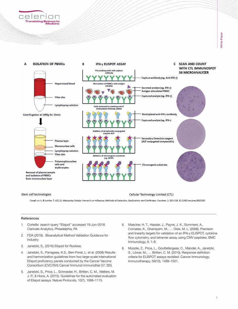
Immune Monitoring assays, such as Flow cytometry and Elispot (Enzyme-linked immunosorbent spot) have been utilized in the research arena for decades. Adapting such complex assays into the clinical realm has a host of challenges, with an increasing emphasis on compliance to industry standards in a regulated environment being at the forefront (FDA BMV guidelines, May 2018). An intensified focus on biomarkers in the drug development process in combination with technological advances has led to the growth in popularity of multifaceted assays such as Intracellular Cytokine Staining (ICS) and Elispot.
The Elispot assay provides a powerful tool in the development of new vaccines and novel immunotherapy agents. The emergence of global disease outbreaks has led to an expansion of studies focused on vaccine development for infectious agents. Breakthroughs in understanding the immune system in recent years have brought a new wave of treatments using immune system modulators, such as checkpoint inhibitors, and other ImmunoOncology treatment advances. The increase in references to Elispot has grown significantly since the 1990s (Figure 1.) with just over 1000 clinical trials utilizing Elispot as of July 2018 (Figure 2.)
Elispot was originally developed in the 1980s to measure secretion of antigen specific antibodies produced by B cells. The B-cell Elispot is still constantly changing with method improvements that increased sensitivity; this advancement allowed for the specific detection of rare
cell types, such as memory B cells. The T-cell Elispot quantifies the number of reactive antigen specific T-cells by detecting the secretion of INF-γ, other cytokines, or secreted molecules such as granzyme B. The T-cell Elispot is an effective technique to determine a patient’s immune response, and can be adapted to high throughput methods, along with other workflow measures, to provide a validated biomarker assay. The significance of the development of this assay is that it provides vital information to a sponsor in early clinical stages to make key decision in the drug development process such as vaccination schedule, formulation and adjuvant, or antigen selection.
Regulatory Guidance
Elispot and other immune monitoring assays such as intracellular cytokine staining (ICS) provide unique challenges as no reference material or gold standard can be utilized; it is important to note FDA Bioanalytical Method Validation guidance is not always applicable, or may need to be adapted to the unique properties of the assay. Numerous global harmonization studies have been carried out, creating optimized protocols and guidelines (Janetzki et al., 2008, 2015), as well as targets for precision and linearity (Maecker et al., 2008).
Key Validation Parameters
IFN-γ is the most common analyte measured with the Elispot assay in clinical studies. Utilizing optimized protocols and guidelines in established literature, a validation plan was developed for an IFN-γ Elispot. Important parameters in validating an Elispot assay for use in the bioanalytical context include precision, accuracy, specificity, limit of detection (LOD), and the linearity and range of the assay. Accuracy, defined as closeness of agreement to a true value, cannot be determined for the Elispot assay due to the lack of a reference standard or test that is able to provide an exact measurement of
Interferon-Gamma Elispot Assay: Unique Challenges of Validating Immune Monitoring Assays in a Regulated Environment
Wendy Adamowicz, Celerion
James Hnilo, Celerion
Curtis Sheldon, Celerion
Rafiqul Islam, Celerion
Figure 1. PubMED citations of Elispot by year.
Figure 2. Elispot in Clinical Trials by Indication (2017)
2
Whi
te P
aper
functional active antigen-specific cells in a given sample. Obtaining data on the accuracy of a laboratory’s performance can be achieved via participation in large proficiency panels that provide relative accuracy for a laboratory in comparison to many other laboratories testing the same samples. In bioanalysis, specificity refers to the ability to detect an analyte in the presence of potentially interfering substances. In the context of Elispot, the goal of specificity testing is to determine antigen-specific T-cells among the other components of the PBMC (peripheral blood mononuclear cells) sample. This can be achieved by examining donor response to the test antigen, medium control (background), and a negative control antigen. In this case, peptide pools corresponding to CEF (Cytomegalovirus, Epstein-Bar, and Flu) as well as CMV pp65 protein were the test antigens. The negative control antigen utilized was a peptide pool corresponding to human actin protein from skeletal muscle. The expected outcome for a negative control peptide and media (background) control is low or no reactivity (less than 10 spots) per well.
Precision in validation is essential to determining the variability of the assay. Precision was evaluated for intra-assay, inter-assay, including day to day and inter-operator variability. Precision is typically expressed using standard deviation, and Coefficient of Variation (% CV). Mathematically, as the spot number approaches zero the % CV will increase dramatically. Maecker et al., (2008) carefully evaluated precision for Elispot and found below 30 spots per well, % CV is not an effective measure of precision. As a result of this finding, standard deviation is reported as a measure of precision, when the spot count per well is below 30. The target precision is ≤ 50% CV when the number of spots per well is between 30 and 100, for donors with a mean number of spots greater than 100 spots/well, a % CV ≤ 25% is then utilized.
The limit of detection (LOD) for a cell based assay is generally considered 2-3 times the median background of the assay. This is an important parameter in Elispot as the LOD establishes a spot number below which spot counts are in the noise range of the assay and hence any statistical differences highlighted in that range are not meaningful. In contrast, the LLOQ (lower limit of quantification) is a term rarely used in in the field of Elispot. Due to the mathematical considerations of a high % CV at low spot numbers per well, statistical testing (Moodie et al., 2010) is recommended for samples that are below 30 spots per well and above the LOD. The ULOQ (upper limit of quantification) is defined as the maximum number of individual spots per well the Elispot plate reader software can discriminate. This can be achieved by counting spots using a series of cell dilutions treated with mitogen, or peptide for a donor with a very strong peptide response. By analyzing the linearity of the INF-γ response, the range of the assay can be determined by defining the cell densities where the results obtained are proportional. By carefully examining these aspects of the INF-γ Elispot Assay, parameters are determined that lead to consistent, precise measurement of INF-γ producing T-cells from PBMC samples.
Data Summary
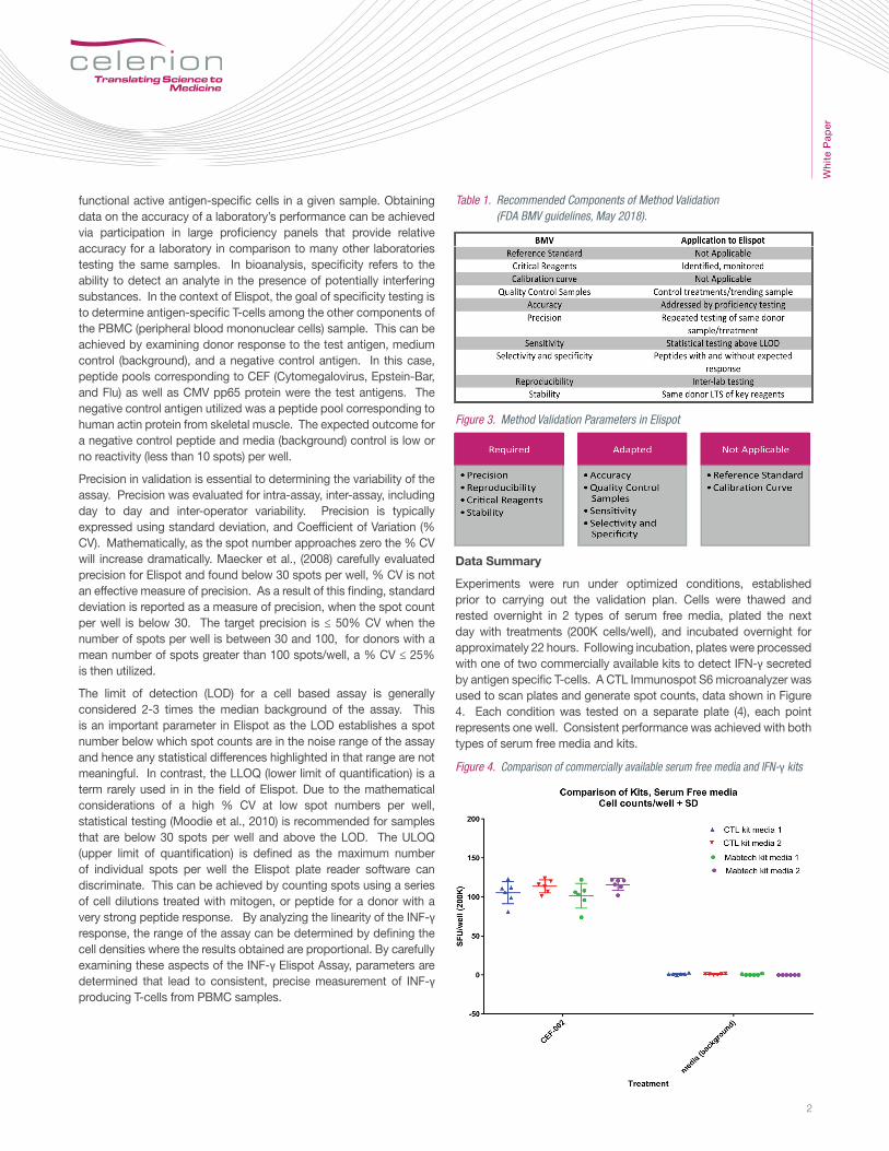
Experiments were run under optimized conditions, established prior to carrying out the validation plan. Cells were thawed and rested overnight in 2 types of serum free media, plated the next day with treatments (200K cells/well), and incubated overnight for approximately 22 hours. Following incubation, plates were processed with one of two commercially available kits to detect IFN-γ secreted by antigen specific T-cells. A CTL Immunospot S6 microanalyzer was used to scan plates and generate spot counts, data shown in Figure 4. Each condition was tested on a separate plate (4), each point represents one well. Consistent performance was achieved with both types of serum free media and kits.
Table 1. Recommended Components of Method Validation (FDA BMV guidelines, May 2018).
Figure 3. Method Validation Parameters in Elispot
Figure 4. Comparison of commercially available serum free media and IFN-γ kits
3
Whi
te P
aper
For validation, peptide pools (CEF and CMVpp65) corresponding to proteins in Cytomegalovirus, Epstein-Bar, and Influenza were utilized along with mitogen (PHA-L) to determine the number of INF-γ producing cells. Target criteria for precision were developed based on Maecker et al. as described above. Experiments addressing each component of the validation plan were carried out on cryopreserved PBMCs. Precision was evaluated by determining spot counts from PBMCs treated with CEF or CMVpp65 for 3 donors over two days with two operators. The mean percent CV for donors with spot count >100 was 12.5%, with an inter-assay range from 10.4% up to 13.9% CV (Table 2).
Specificity was assessed by comparing the response of 3 donors to CEF, pp65, and Actin peptide pools compared to media background control (Figure 5). PBMCs treated with negative control antigen, actin peptide pool, had no response, with similar spot counts to the media control well. This demonstrates that the INF-γ secretion in response to CEF is antigen specific, with minimal response to the negative control pool by cells in the PBMC cell mixture.
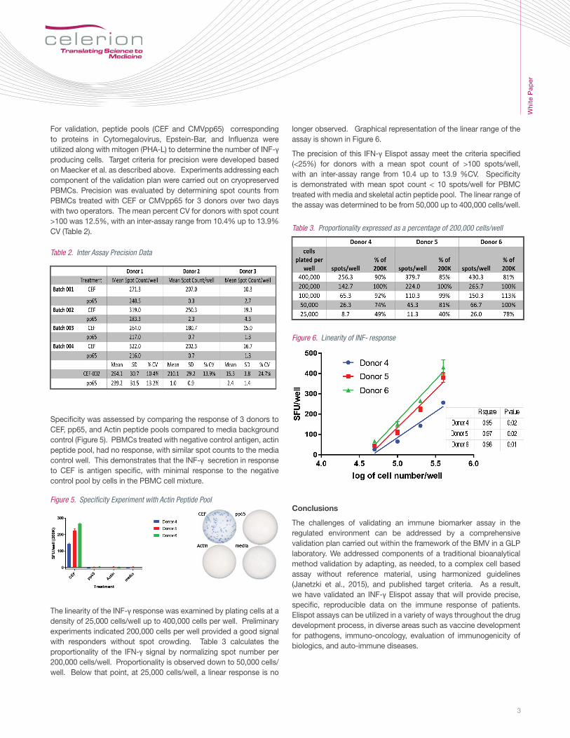
The linearity of the INF-γ response was examined by plating cells at a density of 25,000 cells/well up to 400,000 cells per well. Preliminary experiments indicated 200,000 cells per well provided a good signal with responders without spot crowding. Table 3 calculates the proportionality of the IFN-γ signal by normalizing spot number per 200,000 cells/well. Proportionality is observed down to 50,000 cells/well. Below that point, at 25,000 cells/well, a linear response is no
longer observed. Graphical representation of the linear range of the assay is shown in Figure 6.
The precision of this IFN-γ Elispot assay meet the criteria specified (<25%) for donors with a mean spot count of >100 spots/well, with an inter-assay range from 10.4 up to 13.9 %CV. Specificity is demonstrated with mean spot count < 10 spots/well for PBMC treated with media and skeletal actin peptide pool. The linear range of the assay was determined to be from 50,000 up to 400,000 cells/well.
Conclusions
The challenges of validating an immune biomarker assay in the regulated environment can be addressed by a comprehensive validation plan carried out within the framework of the BMV in a GLP laboratory. We addressed components of a traditional bioanalytical method validation by adapting, as needed, to a complex cell based assay without reference material, using harmonized guidelines (Janetzki et al., 2015), and published target criteria. As a result, we have validated an INF-γ Elispot assay that will provide precise, specific, reproducible data on the immune response of patients. Elispot assays can be utilized in a variety of ways throughout the drug development process, in diverse areas such as vaccine development for pathogens, immuno-oncology, evaluation of immunogenicity of biologics, and auto-immune diseases.
Table 2. Inter Assay Precision Data
Table 3. Proportionality expressed as a percentage of 200,000 cells/well
Figure 5. Specificity Experiment with Actin Peptide Pool
Figure 6. Linearity of INF- response
4
Whi
te P
aper
1. Cortellis search query “Elispot” accessed 19-Jun-2018. Clarivate Analytics, Philadelphia, PA
2. FDA (2018). Bioanalytical Method Validation Guidance for Industry
3. Janetzki, S., (2016) Elispot for Rookies.
4. Janetzki, S., Panageas, K.S., Ben-Porat, L. et al. (2008) Results and harmonization guidelines from two large-scale international Elispot proficiency panels conducted by the Cancer Vaccine Consortium (CVC/SVI) Cancer Immunol Immunother 57: 303.
5. Janetzki, S., Price, L., Schroeder, H., Britten, C. M., Welters, M. J. P., & Hoos, A. (2015). Guidelines for the automated evaluation of Elispot assays. Nature Protocols, 10(7), 1098–1115.
References
6. Maecker, H. T., Hassler, J., Payne, J. K., Summers, A., Comatas, K., Ghanayem, M., … Disis, M. L. (2008). Precision and linearity targets for validation of an IFN-γ ELISPOT, cytokine flow cytometry, and tetramer assay using CMV peptides. BMC Immunology, 9, 1–9.
6. Moodie, Z., Price, L., Gouttefangeas, C., Mander, A., Janetzki, S., Löwer, M., … Britten, C. M. (2010). Response definition criteria for ELISPOT assays revisited. Cancer Immunology, Immunotherapy, 59(10), 1489–1501.