Interference of paratuberculosis with the diagnosis of tuberculosis in a goat flock with a natural...
9
Interference of paratuberculosis with the diagnosis of tuberculosis in a goat flock with a natural mixed infection Julio A ´ lvarez a , Lucı ´a de Juan a , Javier Bezos a , Beatriz Romero a , Jose Luis Sa ´ez b , F.J. Reviriego Gordejo c , Vı ´ctor Briones a , Miguel A ´ ngel Moreno a , Ana Mateos a , Lucas Domı ´nguez a , Alicia Aranaz a, * a Laboratorio Visavet, Departamento de Sanidad Animal, Facultad de Veterinaria, Universidad Complutense de Madrid, 28040 Madrid, Spain b Subdireccio ´n General de Sanidad Animal, Direccio ´n General de Ganaderı ´a, Ministerio de Agricultura, Pesca y Alimentacio ´n, 28071 Madrid, Spain c European Commission, Health and Consumer Protection Directorate-General, Zootechnics, Froissart, 101 1040 Brussels, Belgium Received 6 July 2007; received in revised form 14 August 2007; accepted 17 August 2007 Abstract Detection of infected animals is a key step in eradication programs of tuberculosis. Paratuberculosis infection has been demonstrated to compromise the specificity of the diagnostic tests. However, its effect on their sensitivity has not been clarified. In the present study, skin tests and the interferon-gamma (IFN-g) assay were evaluated in a goat flock (n = 177) with a mixed tuberculosis–paratuberculosis infection in order to assess the possible effect of paratuberculosis on their sensitivity. Culture of mycobacteria was performed as the gold standard to determine the true infection status. All techniques showed lower sensitivities than previously described; the single intradermal tuberculin (SIT) test and the IFN-g assay detected 71% (62.4–78.6, 95% C.I.) of the infected animals; the single intradermal cervical comparative tuberculin (SICCT) test detected only 42.7% (34.1–51.7, 95% C.I.) of infected animals. The highest level of sensitivity was obtained when SIT test and IFN-g assay were combined in parallel (90.8%, 84.5–95.2, 95% C.I.). Sensitivities of the tests were also assessed by comparing animals suffering tuberculosis and animals with a mixed infection; tests were found to be more effective in the former group. Paratuberculosis seems to have a major effect in the sensitivity of the diagnostic tests under study, and therefore must be taken into account; in particular, the use of the SICCT test should be questioned when both tuberculosis and paratuberculosis are present. # 2007 Elsevier B.V. All rights reserved. Keywords: Tuberculosis; Paratuberculosis; Goat; Skin test; SIT; SICCT; IFN-g; Sensitivity www.elsevier.com/locate/vetmic Available online at www.sciencedirect.com Veterinary Microbiology 128 (2008) 72–80 * Corresponding author. Tel.: +34 91 3943721; fax: +34 91 3943795. E-mail address: [email protected] (A. Aranaz). 0378-1135/$ – see front matter # 2007 Elsevier B.V. All rights reserved. doi:10.1016/j.vetmic.2007.08.034
-
Upload
julio-alvarez -
Category
Documents
-
view
215 -
download
2
Transcript of Interference of paratuberculosis with the diagnosis of tuberculosis in a goat flock with a natural...

www.elsevier.com/locate/vetmic
Available online at www.sciencedirect.com
Veterinary Microbiology 128 (2008) 72–80
Interference of paratuberculosis with the diagnosis of tuberculosis
in a goat flock with a natural mixed infection
Julio Alvarez a, Lucıa de Juan a, Javier Bezos a, Beatriz Romero a, Jose Luis Saez b,F.J. Reviriego Gordejo c, Vıctor Briones a, Miguel Angel Moreno a,
Ana Mateos a, Lucas Domınguez a, Alicia Aranaz a,*
a Laboratorio Visavet, Departamento de Sanidad Animal, Facultad de Veterinaria,
Universidad Complutense de Madrid, 28040 Madrid, Spainb Subdireccion General de Sanidad Animal, Direccion General de Ganaderıa,
Ministerio de Agricultura, Pesca y Alimentacion, 28071 Madrid, Spainc European Commission, Health and Consumer Protection Directorate-General,
Zootechnics, Froissart, 101 1040 Brussels, Belgium
Received 6 July 2007; received in revised form 14 August 2007; accepted 17 August 2007
Abstract
Detection of infected animals is a key step in eradication programs of tuberculosis. Paratuberculosis infection has been
demonstrated to compromise the specificity of the diagnostic tests. However, its effect on their sensitivity has not been clarified.
In the present study, skin tests and the interferon-gamma (IFN-g) assay were evaluated in a goat flock (n = 177) with a mixed
tuberculosis–paratuberculosis infection in order to assess the possible effect of paratuberculosis on their sensitivity. Culture of
mycobacteria was performed as the gold standard to determine the true infection status. All techniques showed lower
sensitivities than previously described; the single intradermal tuberculin (SIT) test and the IFN-g assay detected 71%
(62.4–78.6, 95% C.I.) of the infected animals; the single intradermal cervical comparative tuberculin (SICCT) test detected
only 42.7% (34.1–51.7, 95% C.I.) of infected animals. The highest level of sensitivity was obtained when SIT test and IFN-g
assay were combined in parallel (90.8%, 84.5–95.2, 95% C.I.). Sensitivities of the tests were also assessed by comparing animals
suffering tuberculosis and animals with a mixed infection; tests were found to be more effective in the former group.
Paratuberculosis seems to have a major effect in the sensitivity of the diagnostic tests under study, and therefore must be taken
into account; in particular, the use of the SICCT test should be questioned when both tuberculosis and paratuberculosis are
present.
# 2007 Elsevier B.V. All rights reserved.
Keywords: Tuberculosis; Paratuberculosis; Goat; Skin test; SIT; SICCT; IFN-g; Sensitivity
* Corresponding author. Tel.: +34 91 3943721; fax: +34 91 3943795.
E-mail address: [email protected] (A. Aranaz).
0378-1135/$ – see front matter # 2007 Elsevier B.V. All rights reserved.
doi:10.1016/j.vetmic.2007.08.034

J. Alvarez et al. / Veterinary Microbiology 128 (2008) 72–80 73
1. Introduction
Caprine tuberculosis can be caused by Mycobac-
terium bovis and M. caprae (Gutierrez et al., 1995;
Aranaz et al., 2003); both pathogens represent a
serious concern because of the economic impact
and possible spread to bovine herds. Moreover, it
represents a potential source of infection to humans
through unpasteurised dairy products and direct
contact (Gutierrez et al., 1997; Kubica et al., 2003).
Paratuberculosis, caused by Mycobacterium avium
subsp. paratuberculosis (Map), also causes reduced
production. The involvement of Map in the develop-
ment of Crohn’s disease has not been demonstrated but
its zoonotic potential is still under study (Grant, 2005).
In Spain, several studies indicate that it is present in all
domestic ruminant species with variable prevalences
(Reviriego et al., 2000; de Juan et al., 2006a).
Several diagnostic tests are used to ascertain the
tuberculosis status. The single intradermal tuberculin
(SIT) test measures the cell mediated immune (CMI)
response to a M. bovis purified protein derivative
(PPD). As some of the proteins in PPD tuberculin are
shared among different mycobacterial species
(Aagaard et al., 2003), false positive reactions have
been described in animals exposed to other myco-
bacteria (de la Rua-Domenech et al., 2006). To limit
those false positive results, the single intradermal
comparative cervical tuberculin (SICCT) test com-
pares the response to the bovine PPD against an avian
PPD (Paterson et al., 1958). Therefore, specificity of
the test is increased, though its sensitivity might be
hampered, especially in adult animals (Collins, 2006).
The interferon-gamma (IFN-g) assay is an in vitro
alternative that has demonstrated a higher sensitivity
compared with the intradermal tuberculin tests (Wood
et al., 1992; Liebana et al., 1998; Gonzalez Llamazares
et al., 1999) and can detect infected animals at an earlier
stage (Gormley et al., 2006); therefore, it has been
proposed as an ancillary test in cattle (Council Directive
64/432/EEC, amended by (EC) No. 1226/2002 of 8 July
2002, Anon., 2002).
In certain areas of the European Union co-infection
with M. bovis or M. caprae and Map may occur in a
same herd. The effect of paratuberculosis in the
induction of false positive responses has been already
studied in cattle (Walravens et al., 2002; Dunn et al.,
2005), but little has been investigated about the
possible effect of a natural mixed tuberculosis–
paratuberculosis infection in the sensitivity of
diagnostic tests. Previous studies demonstrated a
decreased sensitivity of these tests when applied to
cattle previously infected with M. avium (Amadori
et al., 2002; Hope et al., 2005). In a preliminary study,
the sensitivity of SICCT and IFN-g test in a cattle herd
with dual tuberculosis–paratuberculosis infection
(Aranaz et al., 2006) was lower than in previous
reports, but the selection of animals may have resulted
in an artificially biased higher sensitivity.
No investigation regarding the sensitivity of
diagnostic tests in goat flocks with a dual infection
has been published, in spite of the possible impact on
control programs. The aim of the present study was to
evaluate the sensitivity of SIT, SICCT and IFN-g tests
in a goat flock with a natural tuberculosis–para-
tuberculosis infection in order to determine the
possible effect of paratuberculosis on their accuracy
in detecting tuberculosis infection.
2. Material and methods
2.1. Flock and design of the study
This study was performed on a flock of Spanish
Guadarrama breed with a previous diagnosis of both
M. caprae and Map infection. The flock was
composed of 177 goats older than 2 years that had
never been vaccinated against paratuberculosis.
Seroprevalence against Map, estimated by ELISA
(Mycobacterium paratuberculosis Antibody Test Kit,
ParachekTM Johne’s Absorbed EIA), was 64.4%. All
goats were analysed using the SIT test, the SICCT test
and the IFN-g detection assay. Animals were
euthanized and necropsied for the collection of
samples for bacteriology.
The goats included in this field trial were not
experimental animals. Handling of the animals,
sampling and euthanasia were performed by veter-
inarians according to Spanish Legislation.
2.2. Diagnostic tests
2.2.1. Skin tests
SIT and SICCT were performed after blood
sampling for IFN-g using official bovine and avian

J. Alvarez et al. / Veterinary Microbiology 128 (2008) 72–8074
PPDs (CZ Veterinaria, Porrino, Spain). Animals were
inoculated 0.1 mL containing 0.1 mg of bovine PPD
(2500 CTU) on the left side of the neck and 0.1 mL
(2500 IU) avian PPD on the right side. The results
were determined by measuring the increase of the
skin-fold thickness 72 h later.
Regarding the SIT test, animals were classified as
positive if a bovine reaction (increase of the skin-fold
thickness after the application of the bovine PPD) of
two or more millimetres or the presence of clinical
signs such as oedema, exudation, necrosis, pain or
inflammation at the injection site were observed
(severe interpretation).
Animals were considered positive for the SICCT
test if a positive bovine reaction of two or more
millimetres was observed which was greater than the
avian reaction, or if the presence of clinical signs at the
injection site of the bovine PPD were observed (severe
interpretation).
2.2.2. In vitro test (IFN-g test)
Blood samples were stimulated with the avian and
bovine PPDs used in the skin tests as previously
described (Liebana et al., 1998). Plasma was tested in
duplicate using a sandwich EIA kit for bovine IFN-g
(BovigamTM Bovine Gamma Interferon Test) accord-
ing to methods described by the supplier (Prionics,
Schlieren, Switzerland). The interpretation of the test
has been described elsewhere (Aranaz et al., 2006).
Two cut-off points were selected to evaluate the
sensitivity: the ‘‘severe interpretation’’ considered an
animal positive when the mean optical density (OD) of
its sample stimulated with bovine PPD minus the
mean OD of nil antigen was greater than 0.05, and
greater than the same value of the sample stimulated
with avian PPD; the ‘‘standard interpretation’’
declared an animal positive if the same data was
above 0.1. Interpretation of the test was also
performed just using the results of the stimulation
with bovine PPD to check if significant differences
could be observed.
2.3. Bacteriology
2.3.1. Tissue collection
Samples from lung and retropharyngeal, mediast-
inal and bronchial lymph nodes were taken for culture
of M. tuberculosis complex; samples from ileocecal
valve and adjacent tissue, and mesenteric lymph nodes
were taken for Map culture. All samples were stored at
�20 8C until analysis.
2.3.2. M. tuberculosis complex culture
Samples for culture included tuberculosis compa-
tible lesions and the adjacent areas, or a pool of the
collected tissues if no macroscopic lesion was
observed. Tissue samples were homogenized with
sterile distilled water and decontaminated with 0.35%
hexadecylpyridinium chloride (HPC) for 30 min
(Corner and Trajstman, 1988), centrifuged at
1068 � g for 30 min and cultured onto Coletsos and
0.2% (w/v) pyruvate-enriched Lowenstein–Jensen
media (bioMerieux Espana and Biomedics, Madrid,
Spain). Isolates were identified by staining for acid–
alcohol fastness and PCR amplification of Mycobac-
terium genus-specific 16S rRNA fragment and
MPB70 sequence (Wilton and Cousins, 1992). Isolates
were characterised using spoligotyping (Kamerbeek
et al., 1997).
2.3.3. Map culture
Samples of each animal were pooled, decontami-
nated using 0.75% HPC for 18 h (Greig et al., 1999)
and inoculated onto selective media (de Juan et al.,
2006b). Isolate identification was confirmed by
mycobactin-dependency, and specific PCR for the
detection of IS900 and f57 sequence (Coetsier et al.,
2000). Isolates were further characterised using the
PCR test described by Collins et al. (2002).
2.3.4. Classification of the animals according to
the culture results
To evaluate the performance of the diagnostic test
for tuberculosis depending on their infection status,
animals were classified using the culture result as
the gold standard. Animals that showed lesions
compatible with tuberculosis but had negative culture
results (n = 14) were excluded from the study to avoid
considering them as negative.
Secondly, in order to determine if paratuberculosis
infection could produce significant differences in the
reliability of the tests, tuberculosis-infected animals
were classified as (1) animals positive to tuberculosis:
all animals from which M. caprae was cultured
regardless of Map culture results, and (2) animals
positive to tuberculosis and paratuberculosis (tbc+/
J. Alvarez et al. / Veterinary Microbiology 128 (2008) 72–80 75
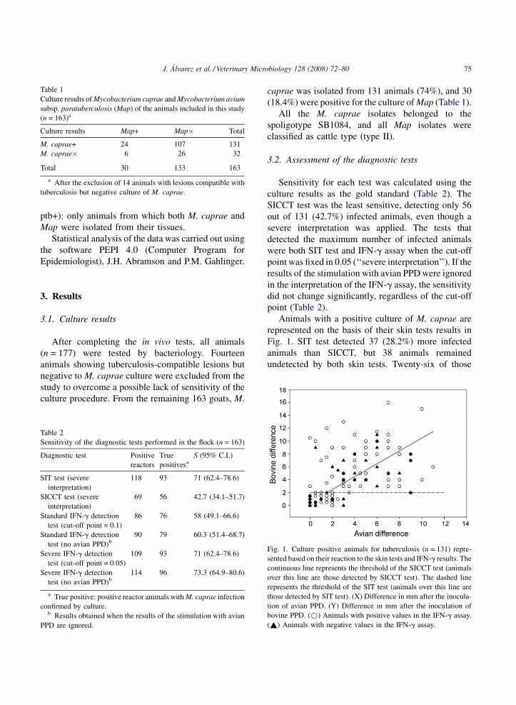
Table 1
Culture results of Mycobacterium caprae and Mycobacterium avium
subsp. paratuberculosis (Map) of the animals included in this study
(n = 163)a
Culture results Map+ Map� Total
M. caprae+ 24 107 131
M. caprae� 6 26 32
Total 30 133 163
a After the exclusion of 14 animals with lesions compatible with
tuberculosis but negative culture of M. caprae.
ptb+): only animals from which both M. caprae and
Map were isolated from their tissues.
Statistical analysis of the data was carried out using
the software PEPI 4.0 (Computer Program for
Epidemiologist), J.H. Abramson and P.M. Gahlinger.
3. Results
3.1. Culture results
After completing the in vivo tests, all animals
(n = 177) were tested by bacteriology. Fourteen
animals showing tuberculosis-compatible lesions but
negative to M. caprae culture were excluded from the
study to overcome a possible lack of sensitivity of the
culture procedure. From the remaining 163 goats, M.
Table 2
Sensitivity of the diagnostic tests performed in the flock (n = 163)
Diagnostic test Positive
reactors
True
positivesa
S (95% C.I.)
SIT test (severe
interpretation)
118 93 71 (62.4–78.6)
SICCT test (severe
interpretation)
69 56 42.7 (34.1–51.7)
Standard IFN-g detection
test (cut-off point = 0.1)
86 76 58 (49.1–66.6)
Standard IFN-g detection
test (no avian PPD)b
90 79 60.3 (51.4–68.7)
Severe IFN-g detection
test (cut-off point = 0.05)
109 93 71 (62.4–78.6)
Severe IFN-g detection
test (no avian PPD)b
114 96 73.3 (64.9–80.6)
a True positive: positive reactor animals with M. caprae infection
confirmed by culture.b Results obtained when the results of the stimulation with avian
PPD are ignored.
caprae was isolated from 131 animals (74%), and 30
(18.4%) were positive for the culture of Map (Table 1).
All the M. caprae isolates belonged to the
spoligotype SB1084, and all Map isolates were
classified as cattle type (type II).
3.2. Assessment of the diagnostic tests
Sensitivity for each test was calculated using the
culture results as the gold standard (Table 2). The
SICCT test was the least sensitive, detecting only 56
out of 131 (42.7%) infected animals, even though a
severe interpretation was applied. The tests that
detected the maximum number of infected animals
were both SIT test and IFN-g assay when the cut-off
point was fixed in 0.05 (‘‘severe interpretation’’). If the
results of the stimulation with avian PPD were ignored
in the interpretation of the IFN-g assay, the sensitivity
did not change significantly, regardless of the cut-off
point (Table 2).
Animals with a positive culture of M. caprae are
represented on the basis of their skin tests results in
Fig. 1. SIT test detected 37 (28.2%) more infected
animals than SICCT, but 38 animals remained
undetected by both skin tests. Twenty-six of those
Fig. 1. Culture positive animals for tuberculosis (n = 131) repre-
sented based on their reaction to the skin tests and IFN-g results. The
continuous line represents the threshold of the SICCT test (animals
over this line are those detected by SICCT test). The dashed line
represents the threshold of the SIT test (animals over this line are
those detected by SIT test). (X) Difference in mm after the inocula-
tion of avian PPD. (Y) Difference in mm after the inoculation of
bovine PPD. (*) Animals with positive values in the IFN-g assay.
(~) Animals with negative values in the IFN-g assay.

J. Alvarez et al. / Veterinary Microbiology 128 (2008) 72–8076
Table 3
Sensitivity obtained combining the diagnostic tests performed in the flock (n = 163)
Diagnostic tests in parallel Positive reactors True positivesa S (95% C.I.)
Severe SIT + standard IFN-g 142 115 87.8 (80.9–92.9)
Severe SIT + severe IFN-g 149 119 90.8 (84.5–95.2)
Severe SICCT + standard IFN-g 118 99 75.6 (67.3–82.7)
Severe SICCT + severe IFN-g 131 108 82.4 (76.5–89.8)
a True positive: animals with a M. caprae infection confirmed by culture.
Table 4
Agreement analysis between SIT test and IFN-g assay in the
tuberculosis-culture positive animals (n = 131)
IFN-g positive IFN-g negative
SIT positive 67 26
SIT negative 26 12
animals were detected by the IFN-g assay, and thus 12
remained undetected. Map infection was confirmed by
culture in 5 out of those 12 animals.
When the different tests are combined in parallel,
the best sensitivity was obtained when SIT and
‘‘severe’’ IFN-g assay are used together (90.8%)
(Table 3).
Results of SIT test and IFN-g assay in the
tuberculosis-infected animals (n = 131) are compared
Fig. 2. Culture positive animals for tuberculosis (n = 131) repre-
sented based on their reaction to the skin tests, and regression lines
of positive (dot-dashed line; y = 0.69x + 2.2) and negative (contin-
uous line; y = 0.64x + 1.9) animals in the IFN-g assay. The dashed
line represents the threshold of the SIT test (2 mm). (X) Difference
in mm after the inoculation of avian PPD. (Y) Difference in mm after
the inoculation of bovine PPD. (*) Animals with positive values in
the IFN-g assay. (~) Animals with negative values in the IFN-g
assay.
in Table 4. The agreement found between the two tests
was very low (Kappa = 0.036, C.I. 95%, �0.135 to
0.207). The same result was obtained when the IFN-g
assay results were compared with the SICCT readings
(Kappa = 0.036, C.I. 95%, �0.109 to 0.181). The
regression lines for positive and negative animals in
the IFN-g test were also determined and compared
(Fig. 2). Both lines showed a similar equation
(y = 0.69x + 2.2 and y = 0.64x + 1.9).
Following the criteria detailed above, results
obtained from all tuberculosis-infected animals
(n = 131) were compared with those from the sub-
group of animals with mixed infection (tbc+/ptb+)
(n = 24) (Table 5). SIT test showed a lower sensitivity
in the animals with the dual infection (54.2%) than
that achieved in the overall group (71%); differences
were found to be statistically significant (C.I. 95%).
SICCT test also performed worse in the tbc+/ptb+
group, but the differences were not found to be
significant (C.I. 95%). IFN-g assay showed a similar
sensitivity in both groups regardless the thres-
hold applied. The small differences found between
the two groups were not statistically significant
(C.I. 95%).
4. Discussion
Bovine tuberculosis is subject of control and
eradication programs in many countries worldwide;
these programs are based on the early detection and
removal of infected animals, and therefore they rely on
the sensitivity of available diagnostic tests. In this
sense, the presence of non-tuberculous mycobacteria
has been pointed out as a possible cause of
misdiagnosis (Lauzi et al., 2000; Dunn et al., 2005).
Caprine tuberculosis is also important, as it can
jeopardise the success of the programs for bovine
tuberculosis and represents a public health threat.

J. Alvarez et al. / Veterinary Microbiology 128 (2008) 72–80 77
Table 5
Sensitivities with confidence intervals (95%) of the tests in the groups formed based on culture results
Diagnostic test Tbc+a (n = 131) Tbc+/Ptb+b (n = 24)
Severe SIT 71 (62.4–78.6) 54.2 (33–74)
Severe SICCT 42.7 (34.1–51.7) 29.2 (13–51)
Standard IFN-g detection test (0.1) 58 (49.1–66.6) 58.3 (37–78)
Severe IFN-g detection test (0.05) 71 (62.4–78.6) 66.7 (45–84)
Severe SIT + standard IFN-g 87.8 (80.9–92.9) 75 (53–90)
Severe SIT + severe IFN-g 90.8 (84.5–95.2) 79.2 (58–93)
Severe SICCT + standard IFN-g 75.6 (67.3–82.7) 70.8 (49–87)
Severe SICCT + severe IFN-g 82.4 (76.5–89.8) 75 (53–90)
a Animals with M. caprae positive cultures, regardless Map culture results.b Animals with positive cultures of both M. caprae and Map.
In the present study, we have applied an adaptation
of the diagnostic tests used in cattle to a goat flock with
a mixed infection of tuberculosis and paratubercu-
losis. The sensitivity observed in all tests was lower
than results published by other authors for cattle
(Francis et al., 1978; Wood et al., 1992; Whipple et al.,
1995) and goats (Gutierrez et al., 1998; Liebana et al.,
1998). As there are no established criteria for the
interpretation of the skin tests in goats, in this study we
have adopted the criteria of the European legislation
for cattle but considering positive the inconclusive
reactors (animals with a bovine reaction of more than
2 mm and less than 4 mm with no clinical signs at the
injection site in the SIT test; and animals with a bovine
reaction of more than 2 mm but equal to or less than an
avian reaction and the absence of clinical signs in the
SICCT test). SIT test, under a severe interpretation,
showed the highest sensitivity, as it detected 71% of
the infected animals, which lies within previously
described sensitivity rates though on the lower side.
To avoid a possible lack of specificity of the SIT test,
the application of the SICCT test has been recom-
mended in areas where a high number of avian reactors
are expected (Monaghan et al., 1994). However, in this
study the SICCT test was an unreliable test, as it could
only detect 42.7% of the infected animals even when the
severe interpretation was applied. These data show that
SICCT test is not adequate for diagnosis of tuberculosis
in goats if tuberculosis and paratuberculosis coexist in
the same flock.
The low sensitivities observed in the present study
are most likely caused by a particular cause such as the
paratuberculosis co-infection. Another possible expla-
nation for the decreased sensitivity could be the
presence of high rates of anergic animals in the flock
that would not produce a detectable immune response.
However, this possibility was ruled out because a high
proportion of false negative reactors showed a
response to the stimulation with the avian PPD in
the SICCT test. Therefore, a plausible explanation was
the masking effect of the reaction against avian PPD in
animals with a mixed infection.
To confirm if paratuberculosis infection was
affecting the sensitivity of the tuberculosis diagnostic
tests, we have compared the results obtained from all
tuberculosis-infected animals with those from goats
with a mixed infection. This comparison revealed that
SIT test performed significantly better in the former
group (Table 5), as the differences between sensitiv-
ities in each group were statistically significant (C.I.
95%). This observation points out the possible effect
of the paratuberculosis co-infection in the perfor-
mance of the SIT test. The same remark can be done
regarding the SICCT test, though in this case the
differences found were not statistically significant
(C.I. 95%). However, this could be attributed to the
small size of the tbc+/ptb+ group.
The detection of IFN-g has been accepted as a
useful ancillary tool in the diagnosis of tuberculosis in
cattle. In our study, IFN-g assay with the cut-off point
fixed at 0.05 showed the same sensitivity as the SIT
test in the whole flock. It must be remarked that
ignoring the results of stimulation with avian PPD
allows the detection of a few more infected animals.
Thus, in situations of goat populations with a mixed
tuberculosis–paratuberculosis infection, the IFN-g
assay might show the same sensitivity but better
specificity than skin tests. In addition, it might be more
cost-effective to perform IFN-g assay stimulating the
lymphocytes only with bovine PPD. The cut-off value
J. Alvarez et al. / Veterinary Microbiology 128 (2008) 72–8078
of the ELISA for the IFN-g must be adjusted to obtain
a higher sensitivity (in our study, PPD minus the mean
OD of nil antigen greater than 0.05 instead of 0.1).
Interestingly, the sensitivity rates of the IFN-g assay
obtained in the two groups (tuberculosis-infected and
mixed infection animals) showed no statistically
significant differences (confidence level 95%), which
could imply that this test is not as influenced by a
concurrent paratuberculosis in a flock than the skin
tests. Adverse effects of the paratuberculosis on the
sensitivity of the tuberculosis diagnostic tests used in
this study (SIT, SICCTand IFN-g detection tests) were
also reported in cattle with a dual mycobacterial
infection (Aranaz et al., 2006).
Comparison between IFN-g assay and SIT test in
the infected animals revealed a low agreement,
meaning they behaved differently, detecting different
populations of infected animals. This is also observed
in Figs. 1 and 2, as the IFN-g test results are distributed
independently of the quantitative skin test results
throughout the whole graphic. This is in line with the
general assumption that some infected animals might
respond to only one test, so the population which is
positive to each test is not exactly the same (Neill
et al., 1994; Pollock et al., 2005; Aranaz et al., 2006),
and highlights the importance of the use of the IFN-g
assay as an ancillary test.
Twelve out of 131 infected animals (9.16%) did not
show any CMI response. They are represented in the
bottom left corner of Fig. 1. Map was isolated from
five of them. However, M. caprae was cultured from
the intestinal samples of other four animals, and
therefore could have overgrown a possible culture of
Map. No obvious cause for the lack of response, apart
from a concomitant Map infection, was found. These
animals represent a great risk, as they were not
detected by any diagnostic technique and therefore
would have maintained the tuberculosis infection in
the farm. On the other side, nine of these animals
showed macroscopic lesions compatible with tuber-
culosis, and therefore could have been detected in the
slaughterhouse.
5. Conclusion
- The sensitivities of the diagnostic tests were below
the values described in previous reports, and
paratuberculosis co-infection was considered the
most likely cause.
- T
he SICCT test was the least sensitive test, probablybecause the paratuberculosis infection induced an
important reaction to the avian PPD. Therefore, the
use of SICCT test in goats in areas where
tuberculosis and paratuberculosis are present should
be discouraged.
- S
kin tests and IFN-g detection assay detecteddifferent populations of infected animals. This
highlights the usefulness of IFN-g assay as an
ancillary test. In our study, IFN-g detection test
seemed to be more consistent regardless the
concomitant paratuberculosis infection as compared
to skin tests.
- S
ome infected animals (12/131, 9.16%) wereundetectable for the tuberculosis diagnostic tests
used in this study. Nine of these ‘‘undetectable’’
animals (75%) could have been detected in the
slaughterhouse because of their visible lesions; this
points out post-mortem inspection as a still essential
tool in eradication plans for tuberculosis.
Paratuberculosis represents a serious impairment
on the reliability of routine tuberculosis diagnosis tests
in goats, and should be taken into account in
eradication programs in areas with high paratubercu-
losis prevalences. Moreover, the impact of paratu-
berculosis in the sensitivity of these diagnostic tools in
cattle should also be addressed in further studies,
because of the economic and social consequences of a
failure of the diagnostic tests. Above all, a great deal of
common sense added to the use of the best diagnosis
tools and proper epidemiological studies are necessary
elements of the eradication programs, as the overall
assessment of the situation of each epidemiological
unit cannot rely exclusively on the results of the
diagnostic tests.
Acknowledgements
This research was funded by project AGL2004-
08092 of the Spanish Ministry of Science and
Technology, and by ParaTBTools (STREP 23106)
of the European Union. J. Alvarez was recipient of a
predoctoral grant assigned by the Spanish Ministry of
Education and Culture. The group is a partner of the

J. Alvarez et al. / Veterinary Microbiology 128 (2008) 72–80 79
coordination action ‘‘Veterinary European Network
on Mycobacteria (VENoMYC)’’ funded by the
European Union. We would like to thank C. Escribano,
L. Carbajo, J.L. Paramio, (Direccion General de
Ganaderıa, Spanish Ministry of Agriculture Fisheries
and Food), L. Sanchez, J. Carpintero, R. Dıaz,
(Direccion General de Agricultura y Desarrollo Rural,
Comunidad de Madrid), and B. Fernandez-Mardo-
mingo, O. Mınguez-Gonzalez, A. Grau and S.
Marques (Direccion General de Produccion Agrope-
cuaria, Junta de Castilla y Leon) for their continuous
encouragement.
We thank P. Diez de Tejada and J.M. Fernandez for
clinical assistance and appreciate the technical help of
F. Lozano and N. Moya. We are grateful to M. Gilmour
for careful revision of the manuscript.
References
Aagaard, C., Govaerts, M., Meng, O.L., Andersen, P., Pollock, J.M.,
2003. Genomic approach to identification of Mycobacterium
bovis diagnostic antigens in cattle. J. Clin. Microbiol. 41, 3719–
3728.
Amadori, M., Tagliabue, S., Lauzi, S., Finazzi, G., Lombardi, G.,
Telo, P., Pacciarini, L., Bonizzi, L., 2002. Diagnosis of Myco-
bacterium bovis infection in calves sensitized by mycobacteria
of the avium/intracellulare group. J. Vet. Med. B: Infect. Dis.
Vet. Public Health 49, 89–96.
Anon., 2002. Off. J. Eur. Union L179, 13–18.
Aranaz, A., Cousins, D., Mateos, A., Dominguez, L., 2003. Eleva-
tion of Mycobacterium tuberculosis subsp. caprae Aranaz et al.
1999 to species rank as Mycobacterium caprae comb. nov. sp.
nov. Int. J. Syst. Evol. Microbiol. 53, 1785–1789.
Aranaz, A., de Juan, L., Bezos, J., Alvarez, J., Romero, B., Lozano,
F., Paramio, J.L., Lopez-Sanchez, J., Mateos, A., Dominguez, L.,
2006. Assessment of diagnostic tools for eradication of bovine
tuberculosis in cattle co-infected with Mycobacterium bovis and
M. avium subsp. paratuberculosis. Vet. Res. 37, 593–606.
Coetsier, C., Vannuffel, P., Blondeel, N., Denef, J.F., Cocito, C.,
Gala, J.L., 2000. Duplex PCR for differential identification of
Mycobacterium bovis, M. avium, and M. avium subsp. para-
tuberculosis in formalin-fixed paraffin-embedded tissues from
cattle. J. Clin. Microbiol. 38, 3048–3054.
Collins, D.M., De Zoete, M., Cavaignac, S.M., 2002. Mycobacter-
ium avium subsp. paratuberculosis strains from cattle and sheep
can be distinguished by a PCR test based on a novel DNA
sequence difference. J. Clin. Microbiol. 40, 4760–4762.
Collins, J.D., 2006. Tuberculosis in cattle: strategic planning for the
future. Vet. Microbiol. 112, 369–381.
Corner, L.A., Trajstman, A.C., 1988. An evaluation of 1-hexade-
cylpyridinium chloride as a decontaminant in the primary iso-
lation of Mycobacterium bovis from bovine lesions. Vet.
Microbiol. 18, 127–134.
de Juan, L., Alvarez, J., Aranaz, A., Rodriguez, A., Romero, B.,
Bezos, J., Mateos, A., Dominguez, L., 2006a. Molecular epi-
demiology of Types I/III strains of Mycobacterium avium sub-
species paratuberculosis isolated from goats and cattle. Vet.
Microbiol. 115, 102–110.
de Juan, L., Alvarez, J., Romero, B., Bezos, J., Castellanos, E.,
Aranaz, A., Mateos, A., Dominguez, L., 2006b. Comparison of
four different culture media for isolation and growth of type II
and type I/III Mycobacterium avium subsp. paratuberculosis
strains isolated from cattle and goats. Appl. Environ. Microbiol.
72, 5927–5932.
de la Rua-Domenech, Goodchild, A.T., Vordermeier, H.M., Hewin-
son, R.G., Christiansen, K.H., Clifton-Hadley, R.S., 2006. Ante
mortem diagnosis of tuberculosis in cattle: a review of the
tuberculin tests, gamma-interferon assay and other ancillary
diagnostic techniques. Res. Vet. Sci. 81, 190–210.
Dunn, J.R., Kaneene, J.B., Grooms, D.L., Bolin, S.R., Bolin, C.A.,
Bruning-Fann, C.S., 2005. Effects of positive results for Myco-
bacterium avium subsp. paratuberculosis as determined by
microbial culture of feces or antibody ELISA on results of
caudal fold tuberculin test and interferon-gamma assay for
tuberculosis in cattle. J. Am. Vet. Med. Assoc. 226, 429–435.
Francis, J., Seiler, R.J., Wilkie, I.W., O’Boyle, D., Lumsden, M.J.,
Frost, A.J., 1978. The sensitivity and specificity of various
tuberculin tests using bovine PPD and other tuberculins. Vet.
Rec. 103, 420–425.
Gonzalez Llamazares, O.R., Gutierrez Martin, C.B., Alvarez, N.D.,
de la Puente Redondo, V.A., Dominguez, R.L., Rodriguez Ferri,
E.F., 1999. Field evaluation of the single intradermal cervical
tuberculin test and the interferon-gamma assay for detection and
eradication of bovine tuberculosis in Spain. Vet. Microbiol. 70,
55–66.
Gormley, E., Doyle, M.B., Fitzsimons, T., McGill, K., Collins, J.D.,
2006. Diagnosis of Mycobacterium bovis infection in cattle by
use of the gamma-interferon (Bovigam1) assay. Vet. Microbiol.
112, 171–179.
Grant, I.R., 2005. Zoonotic potential of Mycobacterium avium ssp.
paratuberculosis: the current position. J. Appl. Microbiol. 98,
1282–1293.
Greig, A., Stevenson, K., Henderson, D., Perez, V., Hughes, V.,
Pavlik, I., Hines, M.E., McKendrick, I., Sharp, J.M., 1999.
Epidemiological study of paratuberculosis in wild rabbits in
Scotland. J. Clin. Microbiol. 37, 1746–1751.
Gutierrez, M., Samper, S., Gavigan, J.A., Garcia Marin, J.F., Martin,
C., 1995. Differentiation by molecular typing of Mycobacterium
bovis strains causing tuberculosis in cattle and goats. J. Clin.
Microbiol. 33, 2953–2956.
Gutierrez, M., Samper, S., Jimenez, M.S., Van Embden, J.D., Marin,
J.F., Martin, C., 1997. Identification by spoligotyping of a
caprine genotype in Mycobacterium bovis strains causing human
tuberculosis. J. Clin. Microbiol. 35, 3328–3330.
Gutierrez, M., Tellechea, J., Garcia Marin, J.F., 1998. Evaluation of
cellular and serological diagnostic tests for the detection of
Mycobacterium bovis-infected goats. Vet. Microbiol. 62, 281–
290.
Hope, J.C., Thom, M.L., Villarreal-Ramos, B., Vordermeier, H.M.,
Hewinson, R.G., Howard, C.J., 2005. Exposure to Mycobacter-

J. Alvarez et al. / Veterinary Microbiology 128 (2008) 72–8080
ium avium induces low-level protection from Mycobacterium
bovis infection but compromises diagnosis of disease in cattle.
Clin. Exp. Immunol. 141, 432–439.
Kamerbeek, J., Schouls, L., Kolk, A., van, A.M., van, S.D., Kuijper,
S., Bunschoten, A., Molhuizen, H., Shaw, R., Goyal, M., van,
E.J., 1997. Simultaneous detection and strain differentiation of
Mycobacterium tuberculosis for diagnosis and epidemiology. J.
Clin. Microbiol. 35, 907–914.
Kubica, T., Rusch-Gerdes, S., Niemann, S., 2003. Mycobacterium
bovis subsp. caprae caused one-third of human M. bovis-asso-
ciated tuberculosis cases reported in Germany between 1999 and
2001. J. Clin. Microbiol. 41, 3070–3077.
Lauzi, S., Pasotto, D., Amadori, M., Archetti, I.L., Poli, G., Bonizzi,
L., 2000. Evaluation of the specificity of the gamma-interferon
test in Italian bovine tuberculosis-free herds. Vet. J. 160, 17–24.
Liebana, E., Aranaz, A., Urquia, J.J., Mateos, A., Dominguez, L.,
1998. Evaluation of the gamma-interferon assay for eradication
of tuberculosis in a goat herd. Aust. Vet. J. 76, 50–53.
Monaghan, M.L., Doherty, M.L., Collins, J.D., Kazda, J.F., Quinn,
P.J., 1994. The tuberculin test. Vet. Microbiol. 40, 111–124.
Neill, S.D., Cassidy, J., Hanna, J., Mackie, D.P., Pollock, J.M.,
Clements, A., Walton, E., Bryson, D.G., 1994. Detection of
Mycobacterium bovis infection in skin test-negative cattle with
an assay for bovine interferon-gamma. Vet. Rec. 135, 134–135.
Paterson, A.B., Stuart, P., Lesslie, I.W., 1958. The use of tests on
slaughterhouse cattle for estimating relative potencies of tuber-
culins and for the calculation of discrimination tests. J. Hyg.
(Lond.) 56, 1–18.
Pollock, J.M., Welsh, M.D., McNair, J., 2005. Immune responses
in bovine tuberculosis: towards new strategies for the diagnosis
and control of disease. Vet. Immunol. Immunopathol. 108,
37–43.
Reviriego, F.J., Moreno, M.A., Dominguez, L., 2000. Soil type as a
putative risk factor of ovine and caprine paratuberculosis ser-
opositivity in Spain. Prev. Vet. Med. 43, 43–51.
Walravens, K., Marche, S., Rosseels, V., Wellemans, V., Boelaert, F.,
Huygen, K., Godfroid, J., 2002. IFN-gamma diagnostic tests in
the context of bovine mycobacterial infections in Belgium. Vet.
Immunol. Immunopathol. 87, 401–406.
Whipple, D.L., Bolin, C.A., Davis, A.J., Jarnagin, J.L., Johnson,
D.C., Nabors, R.S., Payeur, J.B., Saari, D.A., Wilson, A.J., Wolf,
M.M., 1995. Comparison of the sensitivity of the caudal fold
skin test and a commercial gamma-interferon assay for diagnosis
of bovine tuberculosis. Am. J. Vet. Res. 56, 415–419.
Wilton, S., Cousins, D., 1992. Detection and identification of multi-
ple mycobacterial pathogens by DNA amplification in a single
tube. PCR Methods Appl. 1, 269–273.
Wood, P.R., Corner, L.A., Rothel, J.S., Ripper, J.L., Fifis, T.,
McCormick, B.S., Francis, B., Melville, L., Small, K., de
Witte, K., 1992. A field evaluation of serological and cellular
diagnostic tests for bovine tuberculosis. Vet. Microbiol. 31,
71–79.