Protein-protein interaction of Soybean Protein from Extrusion Processing

Nucleic Acids Research, Vol. 19, No. 11 2955
Interaction of protein SRP1 9 with signal recognitionparticle RNA lacking individual RNA-helices
Christian ZwiebDepartment of Molecular Biology, The University of Texas Health Center at Tyler, PO Box 2003,Tyler, TX 75710, USA
Received February 21, 1991; Revised and Accepted May 8, 1991
ABSTRACT
Derivatives of human SRP-RNA were constructed bysite-directed mutagenesis and tested for their ability tointeract with protein SRP19. An RNA missing helix 6barely interacts with SRP19, while the helix 8-deletionmutant retains much binding capability. A mutant RNAconsisting just of helix 6 also binds the protein, but notas well as the unaltered molecule. SRP19 interacts toa full extent with the fourth mutant RNA composed ofhelices 6, 7, 8 and a portion of helix 5. It is concludedthat helix 6-and not helix 8-is the major SRP19binding site. Helices 7, 8 and portions of helix 5contribute to the formation of a functional site. Theseresults agree with data suggesting a proximity of helix6 and the conserved part of SRP-RNA.
INTRODUCTIONThe majority of secretory proteins require signal recognitionparticle (SRP) for translocation from the cytosol into the lumenof the endoplasmic reticulum (ER). SRP is part of the secretoryapparatus which includes the signal peptide, ribosomes and theSRP-receptor in the ER-membrane (1, 2). SRP is a stablecytosolic ribonucleoprotein particle isolated and characterizedparticularly well from canine pancreas (3). It is composed of oneRNA molecule (SRP-RNA) with 300 nucleotides and of sixpolypeptides (SRP9, SRP14, SRP19, SRP68, SRP72 andSRP54). The RNA consists of eight helices and shows somedegree of tertiary structure (4, 5). It is contacted directly by allSRP-proteins, with the exception of SRP54. RNA-bound SRP19is required for assembly of SRP54 (3, 6).Mild digestion of the canine SRP with micrococcal nuclease
generates two subparticles. SRP19 is present in the larger one,together with the conserved part of helix 5, helices 6-8, theSRP72/68 heterodimer and SRP54 (7). SRP19 protects the distalloops of helices 6 and 8 from digestion by a-sarcin (8). Recently,an octamer sequence near the C-terminus was shown to berequired for interaction with SRP-RNA (9, 10).To identify the determinants of SRP-RNA required for binding
to SRP19 directly, individual helices were removed by sitedirected mutagenesis. RNAs were transcribed in vitro and testedfor their ability to interact with SRP19. I demonstrate that theprotein binds predominantly to helix 6. However, additionalelements from the conserved portion of SRP-RNA are requiredto form a fully functional binding site.
MATERIAL AND METHODSConstruction of phRPlasmid phR for synthesis of authentic human SRP-RNA wasconstructed by complete gene synthesis. Updated sequenceinformation was obtained by analysis of plasmid 7L30. 1 (11).The design of 16 oligonucleotides is shown in Figure la. 'Tritylon'-synthesis was accomplished on an Applied Biosystem DNAPCR-Mate using ,3-cyanoethyl-phosphoramidite chemistry.Oligonucleotides were purified and detritylated on purificationcartridges supplied by the manufacturer and dissolved at aconcentration of 8.5-nMol/ml. 2 ,l of each oligonucleotide wereadded to a 1.5 ml Eppendorf tube in a total reaction of 50 ,^lcontaining 50 mM Tris-HCl pH 9.0, 100 mM MgCl, 50 mMDTT, 0.4 mM ATP and 10 units T4 polynucleotide kinase.Incubation was for 20 min at 37°C. 2 ,ul 250 mM EDTA(pH 8.0) and 348 1A of TE (10 mM Tris-HCl pH 7.5, 1 mMEDTA), 100 mM NaCl was added. The sample was heated for5 min using a 300 ml beaker with boiling water. The beaker wasplaced at 4°C and left over night. The annealed DNA wasextracted once with phenol and chloroform, adjusted to 300 mMNaCl and precipitated by adding 3 volumes of ethanol andincubation at -70°C for 1 hr. The assembled gene was recoveredby centrifugation, washed once with 80% ethanol, dried anddissolved in 32 Al TE. Aliquots of the annealed oligonucleotideswere ligated to about 100 ng of EcoRI- and BamHI-digestedDNA of pUC18 for 3 hrs at room temperature and then placedat 4°C over night. The ligase was inactivated by a 10-minincubation at 70°C. The DNA was digested with KpnI for 20 minat 37°C to reduce the amount of circular pUC18 DNA.Competent E. coli DH5a cells (BRL) were transformed.Transformants were selected on LB plates containing 100 pg/mlampicillin. DNA of individual transformants was prepared,restricted with EcoRI and BamHI and analyzed by agarose gelelectrophoresis. A small number of clones containing insertedDNA was characterized by DNA sequencing with AMV reversetranscriptase (Stratagene) and a35S-dATP using the 24-mer M13reverse sequencing (-48) and the 17-mer M13 sequencing (-20)primers (New England Biolabs).
Construction of mutant plasmidsMutants pAH6, pAH8 were constructed as described above forphR with a subset of the phR-oligonucleotides and additionalmutant ones as indicated in Figure la. pH6 was assembled fromfour oligonucleotides as shown in Figure lb. The A35-mutation
.=) 1991 Oxford University Press

2956 Nucleic Acids Research, Vol. 19, No. 11
was obtained using the polymerase chain reaction (PCR) (12)wit mutagenic oligonucleotide b5 (ACTTAGTGCGGACAC-CCGATCTATAGTGCGTCGTATTAG) for the 5'- and b3(CAGGTCGACTCTAGAGGATCCACAGGCGCGATCCC-ACTAC) for the 3'-deletion. First, the 5'-deletion (pA5) wasconstructed with composite oligonucleotide a (TCCTGAAT-CTTCCCCTCCGTAGGCACCCCAGGCTTACACT) and b5.The PCR product was used to synthesize mutant DNA byamplification with oligonucleotides c (TCCTGAATCTTCCCC-TCCGT) and d (CGCCAGGGT-TTTCCCAGTCACGAC).Subsequently, the double deletion pA35 was obtained using pA5DNA and mutagenic oligonucleotide b3 with oligonucleotidesa, c and d. The nature of all mutant plasmids was verified byDNA-sequencing.
Synthesis of SRP- and mutant RNAsDNAs were digested with restriction enzymes DraI (phR, pAH6and pAH8) or BamHI (pH6 and p435), extracted with phenoland chloroform, concentrated by ethanol precipitation anddissolved in TE. RNA synthesis was initiated from thef7-promoter and carried out as described previously (9). Forsynthesis of radioactively labeled RNA, the concentration ofUTPwas reduced 50-fold and a32P-UTP (ICN, 25 Ci/mmole) wasadded. Aliquots of the RNAs were analyzed by electrophoresison 2% agarose and 6% polyacrylamide urea gels (13) anddissolved in water.
aEmEDR I 10 20 30 40 s0 60 70 30 90
1FCTAoC00II?AC300CC0T0C00TC I04 t & *
100 110 120 130 140 1l0 160 170 to0 190 200
Gt:OCTATOCklCGGTCISCCC I _t I. IATJf i t
A v
A v
AH8ANi
A
230 290 300 Dial Bman"
V
bw to0o 130
b H6 ?ATCTA OACrACTATACAI140 I50 160 BOHl
ff4CC _t &
Figure 1. Construction of phR for synthesis of authentic human SRP-RNA andmutant derivatives AH6, AH8 (a) and H6 (b). Sequences of huma SRP-RNAand mutant H6 are shown between EcoRI and BamHI restriction sites.T7-polymerase promoters (410) and the guanosines at the start of tncription(-) are indicated. Trnscripts are numbered on top of the sequences. The bordersbetween the syntheic oligonucleoides identical to the shown sequence are markedby arrows pointing up. Borders of oligonucleides with complementaiy sequencesare maked with afrows pointing down. Likewise, borders between oligonucleotidesused for construction of mutants are indicated by solid (AH6) or open triangles(AH8). Deleted regions are emphasized with a dark gray (AH6) and a light grayhorizontal bar (AH8).
Binding of SRP19 and C-terminal deletion mutantsBinding of SRP19 and mutants to the various RNAs wasmonitored by retardation of SRP19::RNA complexes on DEAE-Sepharose. A 50-pil aliquot of the translation mix was adjustedto 300 mM KOAc; 1 A1 water, 1 jig tRNA (E. coli tRNAPhe,Boehringer) or 1 gg SRP-RNA transcribed from phR-wt (14).The sample was incubated 30 min. at 25°C, spun in an airfugeat 30 psi for 15 mi. and the pellet (P) and a 5 I1 aliquot of thesupematant (S) were dissolved in 50 I1 SDS sample buffer. Themain portion of the supernatant was loaded on an 80 A1 DEAE-Sepharose column (Pharmaia) equilibraed with 300 mM KOAc,100 mM Tris-HCl pH 7.5. The column was washed four timeswith 160 p1 of 300 mM KOAc, 100 mM Tris-HCl pH 7.5(flowthrough, F) and four times with 160 1l of 2 M KOAc,100 mM Tris-HCl pH 7.5 (eluate, E). 70 p1 TCA was added tothe pooled fractions and samples were kept on ice for 30 min..Polypeptides were pelleted by a 10-mn. centrifugation,supernatants were removed, and pellets were dissolved in 100 Il1SDS sample buffer. SDS polyacrylamide gel electrophoresis ofaliquots of the samples was carried out as described above. Afterstaining and destaining, gels were dried and exposed to X-rayfilm. Individual bands were quantitated using exposure times inthe linear response range of a Abaton 300/GS scanner.SRP19 deletions were obtained by translation of run-off
transcripts generated by digestion of plasmids pACl and pAC2with HindH. Immunoprecipitation was carried out as described(9). The stability of RNAs in the binding assay was confirmedby adding uniformly 32P-labeled transcripts to the wheat germlysate and incubation at 25°C for 0, 10 and 30 min. Sampleswere extracted with phenol and chloroform, precipitated withethanol and analyzed by electrophoresis on 6% polyacrylamideurea gels.
RESULTSConstruction of plasmids and analysis of RNAsphR, a plasmid for transcription of authentic human SRP-RNAby T7-polymerase, was assembled by complete gene synthesis(14). Sequences of the annealed 16 overlapping syntheticoligonucleotides are indicated in Figure la. The number oftransformants containing only pUC18 was reduced by digestionof the ligation mixture with KpnI. About 10% of the clonesharbored stable inserts and about half of those contained theproper sequence. Human SRP-RNA was obtained by restrictingphR with DraI and transcription by T7-polymerase as describedin Material and Methods. Absorbance measurement at 260 nmand quantitation of ethidium bromide stained RNA after gelelectrophoresis showed that 100 to 150 molecules of RNA wereobtained from one plasmid molecule (not shown). Electrophoreticmobilities of in vitro transcribed SRP-RNA and of SRP-RNAisolated from canine SRP were identical (Figure 3a). Both RNAsbound efficiently to SRP19 (15, 9).To construct mutants pAH6 and pAH8, a subset of the phR-
oligonucleotides was used together with overlapping mutantoligonucleotides (Figure la). Mutations for the removal ofindividual SRP-RNA helices were chosen on the basis of secondarystructure information obtained by comparative sequence analysis(4). Helix 6 was removed in AH6; helix 8 was lacking in AH8.Their potulaed seodary stcu are shown in Figure 2. StableRNAs of the expected sizes were obained after restriction ofmutant DNAs with Dm1 and tanscription with T7-polymerase(Figure 3). pH6 was assembled from four oligonucleotides(Figure lb). To construct pA35, PCR-technology was used (12)with the oligonucleotides described in Material and Methods. The
'ATO
Nucleic Acids Research, Vol. 19, No. 11 2957
0 G-lSO0-CSRP-RNA C - GC-GU *GC-G
-C A6 A C6 G-C
U -A-G-C0-CcU Ac ua-G u AG
C-G- A-U4 :G2 uso A A A~\G U
%U' G AACAOGOC GAG'GCOG LUCGCU~ GUCCAGG CUGGGCUGUA0UGGCUAU AUGGGUGUCCCACU UCGGCA A
CCG CUCIJGCCC AGCGA CGUGUCC GACC00 5 u ,C~~AGCGA A CCUACAAUUCCGC GOGUGA UAGCJCGU0 AUC...O u-30 AACCGOC GACCUCUCUUAC.CCAUACCGUA 'C -GO" I~~~~~~~~C3 AcC-CG A
CA0UA A
CA -G-CU
C-G~~~~~~~~~~~~~~~~~~~CAA C
GcG AAA C A
cGGu 0~~ ~ ~~~~~~~~~~~~A o G
a-C 0U
U-~~~~ ~ ~ ~ ~ ~ ~ ~ ~ ~ ~ ~ ~ ~
0CACA AH8 A~ ~~~~~~~~~~~AGA A~~~~~~~~~~~~~~~~~~A
C-o~~~~ ~ ~ ~ ~ ~ ~ ~ ~ ~ ~ ~ ~~COCA
C-G A_C 20
U-A~~~~ ~ ~ ~ ~ ~ ~ ~ ~ ~ ~ ~ ~ ~~~uA-C-C-AGA AC CC ~~~~~~~~~~~~~ C-A-
CA CAACAA,CAC CCC AACA aAGCAAAAAUAAAA CCCAAC CACACAACCCC.AAACAC CGCACCC AACCA ~A ~AG
a C C CAACACCCGCAAAAHACAC AA ~AC A AC
A - A AAC AU
UACA U UA
CUAC AH6AC
C-A aC CCAC-A CUO
A
COCUUAC C CAACU C a a C a A CUUCOO CCaAOACAC CCA C0C AGOC U AUC A A G GU U ACC
A.ACAA CACACCC OACACUCACAUCACC A UAC AAUCCAC A GAC OCACACCACCCCA AA AGU
A.,C AAACAsoco ucoce0°OU vcuGc Ancccu^c^^oocoGouvuocu
UC ACC cu c oa8 c-°uO~CC
AACCa
AoAc.COAo U
-C AA2
c - ac -aC-0U *aC-0
H6 "_4ccU-AGcGc
uAA a
C oa,CG
cA^
A35 v-cG-C
-uAA-U
AU-
GA
Figure 2. Secondary structures of human SRP-RNA and AH6, AH8, H6 and A35. The secondary structure of human SRP-RNA is shown on top with large numbers2 to 8 for the seven helices (4). Arrows indicate hypersensitive cutting sites for micrococcal nuclease (7).
latter method was less time consuming and more efficient thancomplete gene synthesis. The corresponding RNAs (H6 and A35)were obtained after restriction of plasmid DNAs with BamHl andrun-off transcription (Figures 3).The stability of SRP-RNA and mutant transcripts under the
conditions of the binding assay was determined. RNAs werelabeled radioactively with a32P-UTP during transcription.Results shown in Figure 3b demonstrate that the integrity of theRNAs was not affected by incubation in the wheat germ cell-free system.
Binding of SRP19 to SRP-RNA and mutant RNAsBinding of human SRP19 to human SRP-RNA or mutant RNAswas monitored by retardation of SRP19::RNA complexes on
DEAE-Sepharose as described previously (9). Polypeptides wereanalyzed by SDS gel electrophoresis and autoradiography. SRP19was translated and labeled in the wheat germ cell-free systemin the presence of 35S-methionine and allowed to form acomplex with the various RNAs. At 300 mM potassium acetate,RNA-bound SRP19 was retarded on DEAE, from which it waseluted with 2 M potassium acetate. Figure 4 shows that-in thepresence of SRP-RNA-most of SRP19 was recovered in thehigh salt eluate. In the absence of SRP-RNA, virtually all theprotein bound to the ribosomal pellet or the flowthrough of theDEAE-colum (Figure 4).
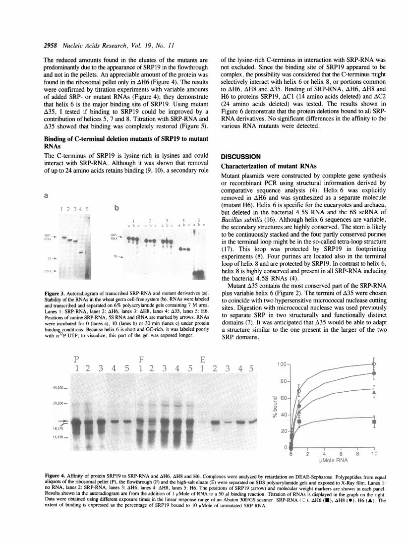
Binding of SRP19 to AH6 was greatly reduced, while AH8bound SRP19 efficiently, albeit not as well as the unmutated SRP-RNA. H6 also bound SRP19, yet with some reduced efficiency.
2958 Nucleic Acids Research, Vol. 19, No. 11
The reduced amounts found in the eluates of the mutants arepredominantly due to the appearance of SRP19 in the flowthroughand not in the pellets. An appreciable amount of the protein wasfound in the ribosomal pellet only in AH6 (Figure 4). The resultswere confirmed by titration experiments with variable amountsof added SRP- or mutant RNAs (Figure 4); they demonstratethat helix 6 is the major binding site of SRP19. Using mutantA35, I tested if binding to SRP19 could be improved by acontribution of helices 5, 7 and 8. Titration with SRP-RNA andA35 showed that binding was completely restored (Figure 5).
Binding of C-terminal deletion mutants of SRP19 to mutantRNAsThe C-terminus of SRP19 is lysine-rich in lysines and couldinteract with SRP-RNA. Although it was shown that removalof up to 24 amino acids retains binding (9, 10), a secondary role
a
b
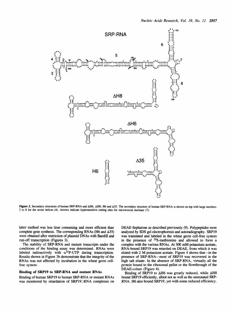
Figure 3. Autoradiogram of transcribed SRP-RNA and mutant derivatives (a).
Stability of the RNAs in the wheat germ cell-free system (b). RNAs were labeled
and transcribed and separated on 6% polyacrylamide gels containing 7 M urea.
Lanes 1: SRP-RNA, lanes 2: AH6, lanes 3: AH8, lanes 4: A35, lanes 5: H6.
Positions of canine SRP-RNA, 5S RNA and tRNA are mark-ed by arrows. RNAs
were incubated for 0 (lanes a), 10 (lanes b) or 30 min (lanes c) under protein
binding conditions. Because helix 6 is short and GC-rich, it was labeled poorly
with cx3 P-UTP; to visualize, this part of the gel was exposed longer.
of the lysine-rich C-terminus in interaction with SRP-RNA wasnot excluded. Since the binding site of SRP19 appeared to becomplex, the possibility was considered that the C-terminus mightselectively interact with helix 6 or helix 8, or portions commonto AH6, AH8 and A35. Binding of SRP-RNA, AH6, AH8 andH6 to proteins SRP19, AC1 (14 amino acids deleted) and AC2(24 amino acids deleted) was tested. The results shown inFigure 6 demonstrate that the protein deletions bound to all SRP-RNA derivatives. No significant differences in the affinity to thevarious RNA mutants were detected.
DISCUSSIONCharacterization of mutant RNAsMutant plasmids were constructed by complete gene synthesisor recombinant PCR using structural information derived bycomparative sequence analysis (4). Helix 6 was explicitlyremoved in AH6 and was synthesized as a separate molecule(mutant H6). Helix 6 is specific for the eucaryotes and archaea,but deleted in the bacterial 4.5S RNA and the 6S scRNA ofBacillus subtilis (16). Although helix 6 sequences are variable,the secondary structures are highly conserved. The stem is likelyto be continuously stacked and the four partly conserved purinesin the terminal loop might be in the so-called tetra-loop structure(17). This loop was protected by SRP19 in footprintingexperiments (8). Four purines are located also in the terminalloop of helix 8 and are protected by SRP19. In contrast to helix 6,helix 8 is highly conserved and present in all SRP-RNA includingthe bacterial 4.5S RNAs (4).
Mutant A35 contains the most conserved part of the SRP-RNAplus variable helix 6 (Figure 2). The termini of A35 were chosento coincide with two hypersensitive micrococcal nuclease cuttingsites. Digestion with micrococcal nuclease was used previouslyto separate SRP in two structurally and functionally distinctdomains (7). It was anticipated that A35 would be able to adapta structure similar to the one present in the larger of the twoSRP domains.
.; d,4
''.X1 _
d-M1M. 1 -
i 4.Cw)f.
mi- IsFF_.j1t _
_p '-_
Figure 4. Affinity of protein SRP19 to SRP-RNA and AH6, AH8 and H6. Complexes were analyzed by retardation on DEAE-Sepharose. Polypeptides from equal
aliquots of the ribosomal pellet (P), the flowthrough (F) and the high-salt eluate (E) were separated on SDS polyacrylamide gels and exposed to X-Ray film. Lanes 1:
no RNA, lanes 2: SRP-RNA, lanes 3: AH6, lanes 4: AH8, lanes 5: H6. The positions of SRP19 (arrow) and molecular weight markers are shown in each panel.
Results shown in the autoradiogram are from the addition of 1 itMole of RNA to a 50 /d binding reaction. Titration of RNAs is displayed in the graph on the right.
Data were obtained using different exposure times in the linear response range of an Abaton 300/GS scanner. SRP-RNA (C), zH6 (-), AH8 (0), H6 (A). The
extent of binding is expressed as the percentage of SRPl9 bound to 10 zIMole of unmutated SRP-RNA.
5.itF .e
.4
Nucleic Acids Research, Vol. 19, No. 11 2959
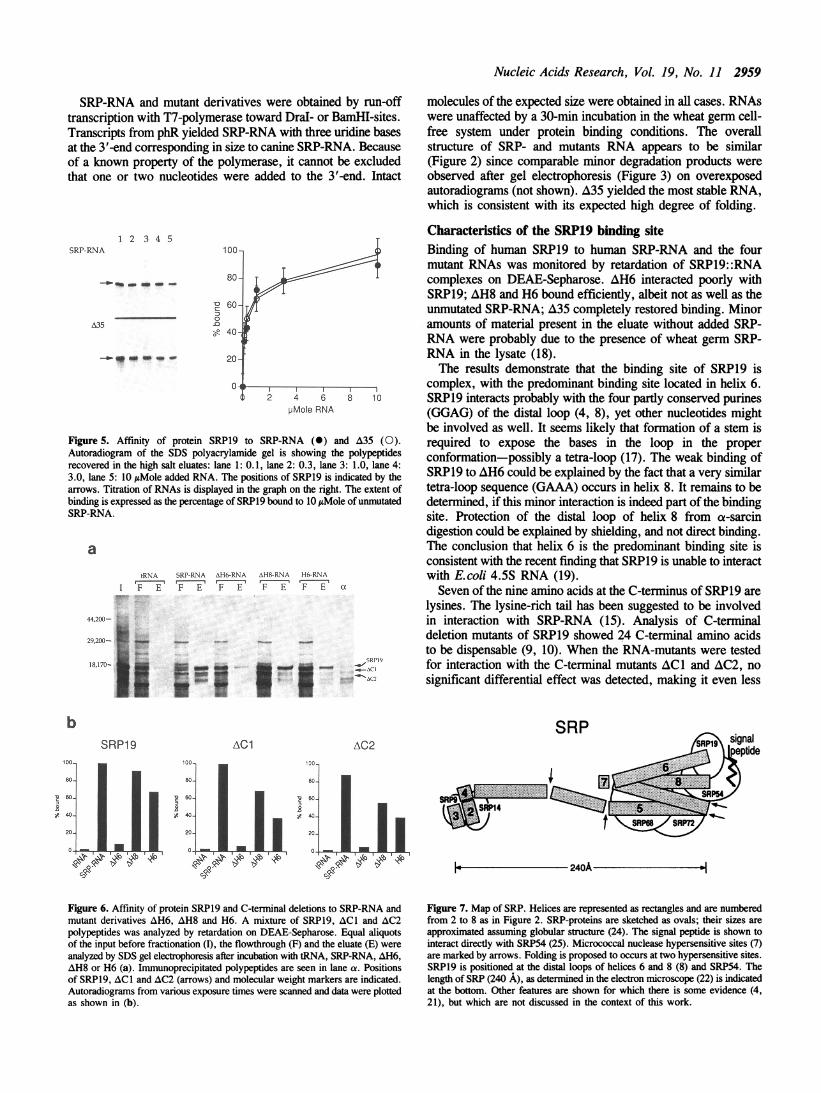
SRP-RNA and mutant derivatives were obtained by run-offtranscription with T7-polymerase toward Dral- or BamHI-sites.Transcripts from phR yielded SRP-RNA with three uridine basesat the 3'-end corresponding in size to canine SRP-RNA. Becauseof a known property of the polymerase, it cannot be excludedthat one or two nucleotides were added to the 3'-end. Intact
1 2 3 4 5SRP-RNA
-_ _ -m - -
0n0'11
A35
-*_* im - 20-
0-41~~~~~~~2 4 6 8 10
pMole RNA
Figure 5. Affinity of protein SRP19 to SRP-RNA (0) and A35 (0).Autoradiogram of the SDS polyacrylamide gel is showing the polypeptidesrecovered in the high salt eluates: lane 1: 0.1, lane 2: 0.3, lane 3: 1.0, lane 4:3.0, lane 5: 10 /AMole added RNA. The positions of SRP19 is indicated by thearrows. Titration of RNAs is displayed in the graph on the right. The extent ofbinding is expressed as the percentage of SRP19 bound to 10 joMole of unmutatedSRP-RNA.
atRNA
I F ESRP-RNA AH6-RNA AH8-RNA If--RNA
F E F E F E F E a
44,200- D
29200- .....
2PE2Wt....e.,.ffi.SR..1918,170- *a/I'Ia I *--C]
r - - _ -vso'? 1-c_ _i _ _ :~~~~~~~~~~~~~~~~C
* X w w*V
bSRP1 9 AC1
molecules of the expected size were obtained in all cases. RNAswere unaffected by a 30-min incubation in the wheat germ cell-free system under protein binding conditions. The overallstructure of SRP- and mutants RNA appears to be similar(Figure 2) since comparable minor degradation products wereobserved after gel electrophoresis (Figure 3) on overexposedautoradiograms (not shown). A35 yielded the most stable RNA,which is consistent with its expected high degree of folding.
Characteristics of the SRP19 binding siteBinding of human SRP19 to human SRP-RNA and the fourmutant RNAs was monitored by retardation of SRP19::RNAcomplexes on DEAE-Sepharose. AH6 interacted poorly withSRP19; AH8 and H6 bound efficiently, albeit not as well as theunmutated SRP-RNA; A35 completely restored binding. Minoramounts of material present in the eluate without added SRP-RNA were probably due to the presence of wheat germ SRP-RNA in the lysate (18).The results demonstrate that the binding site of SRP19 is
complex, with the predominant binding site located in helix 6.SRP19 interacts probably with the four partly conserved purines(GGAG) of the distal loop (4, 8), yet other nucleotides mightbe involved as well. It seems likely that formation of a stem isrequired to expose the bases in the loop in the properconformation-possibly a tetra-loop (17). The weak binding ofSRP19 to AH6 could be explained by the fact that a very similartetra-loop sequence (GAAA) occurs in helix 8. It remains to bedetermined, if this minor interaction is indeed part of the bindingsite. Protection of the distal loop of helix 8 from cx-sarcindigestion could be explained by shielding, and not direct binding.The conclusion that helix 6 is the predominant binding site isconsistent with the recent finding that SRP19 is unable to interactwith E.coli 4.5S RNA (19).Seven of the nine amino acids at the C-terminus of SRP19 are
lysines. The lysine-rich tail has been suggested to be involvedin interaction with SRP-RNA (15). Analysis of C-terminaldeletion mutants of SRP19 showed 24 C-terminal amino acidsto be dispensable (9, 10). When the RNA-mutants were testedfor interaction with the C-terminal mutants AC1 and AC2, nosignificant differential effect was detected, making it even less
SRPAC2 9 signal
2n tIeptide
SRPS,SR
SRPP14
Figure 6. Affinity of protein SRP19 and C-terminal deletions to SRP-RNA andmutant derivatives AH6, AH8 and H6. A mixture of SRP19, ACI and AC2polypeptides was analyzed by retardation on DEAE-Sepharose. Equal aliquotsof the input before fractionation (I), the flowthrough (F) and the eluate (E) wereanalyzed by SDS gel electrophoresis after incubation with tRNA, SRP-RNA, AH6,AH8 or H6 (a). Immunoprecipitated polypeptides are seen in lane ca. Positionsof SRP19, ACI and AC2 (arrows) and molecular weight markers are indicated.Autoradiograms from various exposure times were scanned and data were plottedas shown in (b).
Figure 7. Map of SRP. Helices are represented as rectangles and are numberedfrom 2 to 8 as in Figure 2. SRP-proteins are sketched as ovals; their sizes areapproximated assuming globular structure (24). The signal peptide is shown tointeract directly with SRP54 (25). Micrococcal nuclease hypersensitive sites (7)are marked by arrows. Folding is proposed to occurs at two hypersensitive sites.SRP19 is positioned at the distal loops of helices 6 and 8 (8) and SRP54. Thelength of SRP (240 A), as determined in the electron microscope (22) is indicatedat the bottom. Other features are shown for which there is some evidence (4,21), but which are not discussed in the context of this work.
I

2960 Nucleic Acids Research, Vol. 19, No. 11
probable that the lysine-rich tail functions in interaction withSRP-RNA.As shown in Figure 5, the full binding capacity was restored
with A35. This result supports the concept that-besideshelix 6-the conserved portion of SRP-RNA is important forbinding. Under the assumption that SRP19 is a globular protein,the distal loops of helix 6 and 8 must be close to each other tobe both protected, which could be accomplished by folding ofthe RNA in the vicinity of SRP19. This view is confirmed bycomparative sequence analysis (4) and molecular modeling (20,21) studies aimed to accommodate the RNA within thedimensions of SRP determined by electron microscopy. Ahypothetical folding design is shown in a map of SRP (Figure 7).To bring the distal loops of helices 6 and 8 closer, helices 5,6 and 8 were positioned parallel to each other by folding the RNAat two of the sites that are hypersensitive toward micrococcalnuclease digestion. Physical model building of the RNA showsthat as a consequence of such folding, its size and shape agreeswell with the dimensions (240 x 60 A) determined in the electronmicroscope (22). Only SRP-RNA and mutant A35 restorecomplete binding, presumably because they conform to thesuggested folding pattern. Binding of SRP19 could not beimproved using a mixture of equimolar amounts ofzH6 and H6(not shown) indicating that helix 6 must be placed properly inthe context of nucleotides located in the proximal parts ofhelices 6, 7 and 8. This region has been found to contain dynamicproperties (23) which were proposed to play a role in assemblingthe particle (5). It is possible-as was suggested recently (10)-that SRPl9 directly affects the formation of an RNA structureneeded for further assembly of SRP.
19. Ribes V., Romisch K., Giner A., Dobberstein B. and Tollervey D. (1990)Cell 63, 591-600
20. Zwieb C., and Schuler D. (1989) Biochem. and Cell Biol. 67, 434-44221. Zwieb C., unpublished22. Andrews D., Walter P. and Ottensmeyer P. (1987) EMBO J. 6, 3471-347723. Zwieb C. and Ullu, E. (1986) Nucleic Acids Res. 14, 4639-465724. Zwieb C. (1986) Endocyt. C. Res. 3, 41-5125. Kurzchalia T., Wiedmann M., Girshovich A., Bochkareva E., Bielka H.
and Rapoport T. (1986) Nature 320, 634-636
ACKNOWLEDGEMENTThis work was supported by a Biomedical Research SupportGrant, identification number 2-S07-RR-05958-04.
REFERENCES1. Walter P. and Lingappa V. (1986) Ann. Rev. Cell Biol. 2, 499-5162. Rapoport T.A. (1990) TIBS 15, 355-3583. Walter P. and Blobel G. (1983) Cell 34, 525-5334. Larsen N. and Zwieb C. (1991) Nucleic Acids Res. 19, 209-2155. Zwieb C. (1989) in: Prog. in Nucleic Acid Res. and Mol. Biol. 37, 207-2346. Siegel V. and Walter P. (1988) Cell 52, 39-497. Gundelfmger E.D., Krause E., Melli M. and Dobberstein B. (1983) Nucleic
Acids Res. 11, 7363-73748. Siegel V. and Walter P. (1988) Proc. Natl. Acad. Sci. U.S.A. 85, 1801-18059. Zwieb C., submitted
10. Romisch K., Webb J., Lingelbach K., Gausepohl H. and Dobberstein B.(1990) J. Cell Biol. 111, 1793-1802
11. Ullu E. and Weiner A.M. (1984) EMBO J. 3, 3303-331012. Nelson R.M. and Long G.L. (1989) Anal. Biochem. 180, 147-15113. Maniatis T., Fritsch E. and Sambrook J. (1982) in: Molecular Cloning, A
Laboratory Manual, Cold Spring Harbor Laboratory14. Romaniuk, P.J., deStevenson, I.L. and Wong, H.-H. A. (1987) Nucleic Acids
Res. 15, 2737-275515. Lingelbach K., Zwieb C., Webb J.R., Marshallsay C., Hoben P.J., Walter
P., and Dobberstein B. (1988) Nucleic Acids Res. 16, 9431-944216. Toschka H.Y., Struck J.C.R. and Erdmann V.A. (1989) Nucleic Acids Res.
17, 31-3617. Cheong C., Varani G. and Tinoco I. Jr. (1990) Nature (London) 346,
680-68218. Prehn S., Wiedmann M., Rapoport T.A., and Zwieb C. (1987) EMBO J.
6, 2093-2097