Interaction Between Two E3 ligases, NEDD8ylated Cullin and ...
32
University of Tennessee Health Science Center University of Tennessee Health Science Center UTHSC Digital Commons UTHSC Digital Commons Theses and Dissertations (ETD) College of Graduate Health Sciences 5-2016 Interaction Between Two E3 ligases, NEDD8ylated Cullin and Interaction Between Two E3 ligases, NEDD8ylated Cullin and HHARI HHARI Kheewoong Baek University of Tennessee Health Science Center Follow this and additional works at: https://dc.uthsc.edu/dissertations Part of the Enzymes and Coenzymes Commons, Medical Biochemistry Commons, and the Medical Immunology Commons Recommended Citation Recommended Citation Baek, Kheewoong (http://orcid.org/0000-0002-6853-4936), "Interaction Between Two E3 ligases, NEDD8ylated Cullin and HHARI" (2016). Theses and Dissertations (ETD). Paper 392. http://dx.doi.org/ 10.21007/etd.cghs.2016.0401. This Thesis is brought to you for free and open access by the College of Graduate Health Sciences at UTHSC Digital Commons. It has been accepted for inclusion in Theses and Dissertations (ETD) by an authorized administrator of UTHSC Digital Commons. For more information, please contact [email protected].
Transcript of Interaction Between Two E3 ligases, NEDD8ylated Cullin and ...

University of Tennessee Health Science Center University of Tennessee Health Science Center
UTHSC Digital Commons UTHSC Digital Commons
Theses and Dissertations (ETD) College of Graduate Health Sciences
5-2016
Interaction Between Two E3 ligases, NEDD8ylated Cullin and Interaction Between Two E3 ligases, NEDD8ylated Cullin and
HHARI HHARI
Kheewoong Baek University of Tennessee Health Science Center
Follow this and additional works at: https://dc.uthsc.edu/dissertations
Part of the Enzymes and Coenzymes Commons, Medical Biochemistry Commons, and the Medical
Immunology Commons
Recommended Citation Recommended Citation Baek, Kheewoong (http://orcid.org/0000-0002-6853-4936), "Interaction Between Two E3 ligases, NEDD8ylated Cullin and HHARI" (2016). Theses and Dissertations (ETD). Paper 392. http://dx.doi.org/10.21007/etd.cghs.2016.0401.
This Thesis is brought to you for free and open access by the College of Graduate Health Sciences at UTHSC Digital Commons. It has been accepted for inclusion in Theses and Dissertations (ETD) by an authorized administrator of UTHSC Digital Commons. For more information, please contact [email protected].

Interaction Between Two E3 ligases, NEDD8ylated Cullin and HHARI Interaction Between Two E3 ligases, NEDD8ylated Cullin and HHARI
Abstract Abstract RBR (RING1-in between RING-RING2) is a special type of E3 ubiquitin ligase containing three zinc-binding RING (Really Interesting New Gene) domains, while adopting mechanisms of HECT (Homologous to E6-AP Carboxyl Terminus) for substrate ubiquitination. Most well known RBRs include Parkin and HOIP, which are associated with Parkinson’s disease and innate immune deficiency. However, it is not well known how the RBR proteins gain activity, as they are known to be autoinhibited. Here I show that a specific F430A, E431A, E503A triple mutation of RBR protein HHARI (Human homologue of Ariadne) and its interaction with NEDD8ylated cullin RING ligase can both boost its activity and stabilize complex formation. Analytical size-exclusion chromatography, autoubiquitination, and electron microscopy reveal consistent behavior for this triple-mutant. Future structure-based studies will help elucidate the mechanism of the unsolved mystery of RBR activation and its interaction with NEDD8ylated cullin RING ligases.
Document Type Document Type Thesis
Degree Name Degree Name Master of Science (MS)
Program Program Biomedical Sciences
Research Advisor Research Advisor Brenda A. Schulman, Ph.D.
Keywords Keywords ubiquitin
Subject Categories Subject Categories Chemicals and Drugs | Enzymes and Coenzymes | Medical Biochemistry | Medical Immunology | Medical Sciences | Medicine and Health Sciences
This thesis is available at UTHSC Digital Commons: https://dc.uthsc.edu/dissertations/392

Interaction Between Two E3 Ligases, NEDD8ylated Cullin and HHARI
A Dissertation
Presented for
The Graduate Studies Council
The University of Tennessee
Health Science Center
In Partial Fulfillment
Of the Requirements for the Degree
Master of Science
From The University of Tennessee
By
Kheewoong Baek
May 2016

ii
Copyright © 2016 by Kheewoong Baek.
All rights reserved.

iii
ACKNOWLEDGEMENTS
I would like to thank Dr. Brenda Schulman for providing me the opportunity to
experience cutting edge science in structural biology.
I would also like to thank my committee members Dr. Eric Enemark and Dr. Julio
Cordero-Morales for providing great feedback, support, and for their time.
I would like to thank all the lab members of the Schulman Lab, they are fantastic
people fun to work with, and I learn a lot from everyone everyday.
I would also like to thank my parents back home giving me support in every way.

iv
ABSTRACT
RBR (RING1-in between RING-RING2) is a special type of E3 ubiquitin ligase
containing three zinc-binding RING (Really Interesting New Gene) domains, while
adopting mechanisms of HECT (Homologous to E6-AP Carboxyl Terminus) for substrate
ubiquitination. Most well known RBRs include Parkin and HOIP, which are associated
with Parkinson’s disease and innate immune deficiency. However, it is not well known
how the RBR proteins gain activity, as they are known to be autoinhibited. Here I show
that a specific F430A, E431A, E503A triple mutation of RBR protein HHARI (Human
homologue of Ariadne) and its interaction with NEDD8ylated cullin RING ligase can
both boost its activity and stabilize complex formation. Analytical size-exclusion
chromatography, autoubiquitination, and electron microscopy reveal consistent behavior
for this triple-mutant. Future structure-based studies will help elucidate the mechanism of
the unsolved mystery of RBR activation and its interaction with NEDD8ylated cullin
RING ligases.

v
TABLE OF CONTENTS
CHAPTER 1. INTRODUCTION .....................................................................................1
The Ubiquitin Pathway ....................................................................................................1
Cullin RING Ligases .......................................................................................................1 NEDD8ylation .................................................................................................................3 RBR E3 Ligases ...............................................................................................................3 Human Homolog of Ariadne (HHARI) ...........................................................................4 Interaction Between HHARI and Cullin RING Ligase ...................................................4
CHAPTER 2. METHODS.................................................................................................8
Protein Expression and Purification ................................................................................8
Biochemical Reactions and Assays .................................................................................8 Analytical Size-Exclusion Chromatography ...................................................................9 GraFix and Negative Staining ..........................................................................................9
CHAPTER 3. RESULTS .................................................................................................10
NEDD8ylated Cullin RING Ligases Activate HHARI .................................................10 NEDD8ylated Cullin RING Ligases Bind with HHARI ...............................................10
GraFix and Negative Staining ........................................................................................14
CHAPTER 4. DISCUSSION ..........................................................................................16
Microscopic Reversibility of Protein Complex .............................................................16
Stabilizing the Complex .................................................................................................16
Ub-VME as a Chemical Warhead .................................................................................17 Targets for Electron Microscopy ...................................................................................17
LIST OF REFERENCES ................................................................................................19
VITA..................................................................................................................................23

vi
LIST OF FIGURES
Figure 1-1. The Ubiquitin Pathway. ..................................................................................2
Figure 1-2. Human Homologue of Ariadne. ......................................................................5
Figure 1-3. FEE Mutation of HHARI. ...............................................................................6
Figure 3-1. FEE Mutation of HHARI and the Addition of N8Cul1Rbx1 Both
Activate HHARI. ..........................................................................................11
Figure 3-2. Analytical Size-Exclusion Chromatography Shows Interaction Between
N8Cul1Rbx1 and HHARI. ...........................................................................12
Figure 3-3. Interaction Between N8Cul3Rbx1 and HHARI. ..........................................13
Figure 3-4. Electron Microscopy of HHARI FEE, N8Cul1Rbx1, and Skp1/Fbw7. .......15

vii
LIST OF ABBREVIATIONS
CRL Cullin RING ligase
DTT 1,4-Dithiothreitol
HECT Homologous to E6-AP Carboxyl Terminus
HHARI Human homologue of Ariadne
NEDD8 Neural precursor cell expressed developmentally down-regulated 8
N8Cul1Rbx1 NEDD8ylated Cullin-1 Rbx1
N8Cul3Rbx1 NEDD8ylated Cullin-3 Rbx1
RBR RING1- in between RING- RING2
Rbx RING box protein
RING Really Interesting New Gene
WT Wild type
Ub Ubiquitin
Ub-VME Ubiquitin Vinyl Methyl Ester
1
CHAPTER 1. INTRODUCTION
The Ubiquitin Pathway
Ubiquitination is a post-translational covalent modification of protein by a 76
amino acid ubiquitin (Ub) forming an isopeptide bond between the ubiquitin C-terminal
glycine residue 76 and the substrate lysine. Ubiquitin can form monoubiquitin or
polyubiquitin chains linked by the N-terminal methionine or seven lysine residues (K6,
K11, K27, K29, K33, K48, K63) and its C-terminus GG is a required for its covalent
modification to proteins (Hodgins et al., 1992; Komander, 2009). Different types of
polyubiquitin chains allow participation of substrates in many important processes
including proteosomal/lysosomal degradation, transcription, signal transduction, and
protein trafficking (Komander and Rape, 2012).
In order to ubiquitinate target substrates, the pathway involves three sequential
mechanisms (Figure 1-1). First, the C-terminus of ubiquitin must be activated by acyl-
adenylation in the presence of ATP and Mg2+. The catalytic cysteine of the E1 activating
enzyme attacks the adenylated ubiquitin, producing a highly active E1~Ub thioester
complex (Schulman and Harper, 2009). Then, the E2 ubiquitin conjugating enzyme
accepts the ubiquitin from the E1 by transthioesterification. The ubiquitin-bound E2
interacts with the E3 ubiquitin ligase, which ultimately ubiquitinates target protein
substrates (Streich and Lima, 2014). This step largely divides into two major categories
of E3s. First, RING (Really New Interesting Gene) E3 ligases act as scaffolds for the
E2~Ub and substrates, where the ubiquitin directly transfers to the lysine of substrate
protein. Over 600 E3 ligases are known to be part of the RING family (Deshaies and
Joazeiro, 2009). Second, HECT (Homologous to E6-AP Carboxyl Terminus) E3 ligases
also act as scaffolds for the E2~Ub and substrates, but makes a thioester linked E3~Ub
intermediate, where the Ub from the E2 transfers to the catalytic cysteine of the HECT
E3, and then sequentially transfers the Ub to the substrate protein via its HECT domain
(Huibregtse et al., 1995). There is a hybrid type of E3 ligases called RBRs (RING1-in
between RING-RING2) where it contains RING domains but utilizes HECT
mechanisms. Overall in the human genome, there are two E1s, tens of E2s, and hundreds
of E3s that regulate ubiquitination of over thousands of substrates.
Cullin RING Ligases
Cullin RING ligases (CRL), a superfamily of RING E3s, are modular complexes
composed of a catalytic RING subunit bound to cullin repeats. The RING domain
interacts with two zinc ions by its cysteine and histidine residues to provide structural
stability for protein-protein interactions by producing a globular platform (Borden et al.,
1995). Each different cullins have variety of substrate receptors that allow participation
of cullin RING ligases in majority of cellular processes (Deshaies and Joazeiro, 2009).
Cul1, Cul2, Cul3, Cul4A, Cul4B, and Cul5 along with their substrate receptors and RING
box proteins (Rbx) yield around 500 different combinations of cullin RING ligases

2
Figure 1-1. The Ubiquitin Pathway.
Schematic of the ubiquitin pathway. Catalytic cysteine of E1 attacks adenylated
ubiquitin, producing a highly reactive complex of E1~Ub. By transthioesterification,
ubiquitin transfers from E1 to E2, and further onto substrate via 3 types of E3 ubiquitin
ligases: RING, HECT, and RBR.

3
(Zimmerman et al., 2010). Among the CRL family includes the well-known Skp1-Cul1-
F-box (SCF) ubiquitin ligase where the N-terminus of Cul1 binds Skp1, and the C-
terminus binds Rbx1 for E2 interaction. The F-box protein directly binds to Skp1 by the
F-box domain for substrate specificity (Cardozo and Pagano, 2004). The F-box proteins
also contain protein-protein interaction motifs such as leucine-rich repeats (LRR) or WD
repeats (Schulman et al., 2000). By different types of F-box proteins, the cullin RING
ligases undergo different conformations. For instance, Fbw7 can dimerize Cul1Rbx1
complex by its D-domain, which is highly conserved in F-box proteins with WD40 repeat
motifs (Tang et al., 2007). Cul3Rbx1 can also dimerize via MATH-BTB proteins
(Zhuang et al., 2009).
NEDD8ylation
Many ubiquitin like proteins (Ubl) exist to regulate protein functions. NEDD8
(neural-precursor-cell-expressed developmentally down-regulated 8), a Ubl, is essential
for the activity of cullin RING ligases. Human NEDD8 shares 60% sequence identity
with ubiquitin and has a NEDD8ylation cascade. By its Nedd8 E1 APPBP1-Uba3, Nedd8
E2 Ubc12, and Nedd8 E3 Rbx1/2, Nedd8 is ultimately attached onto its substrate cullin
(Huang et al., 2009). With its E3 RING box protein, NEDD8 conjugates onto the cullin
scaffold lysine that activates the cullin RING ligase (Lydeard et al., 2013). Cullin
NEDD8ylation enhances E2 recruitment by its selectivity towards ubiquitin charged E2s
and improves substrate ubiquitination (Saha and Deshaies, 2008). NEDD8 ligation also
causes a dramatic conformational change of the Cullin Rbx complex so that the E2~Ub
positions proximally to the substrate lysine (Duda et al., 2008; Scott et al., 2014). DCN1
also enhances cullin NEDD8ylation as an auxiliary E3 by binding its PONY (potentiating
NEDD8) domain with Cul1 and Ubc12. NEDD8ylated cullins are deNEDD8ylated by the
multiprotein complex COP9 signalosome (CSN) (Enchev et al., 2012; Lyapina et al.,
2001). Recent structural studies were able to observe the mechanism of CSN and its
cullin RING ligase regulation (Cavadini et al., 2016; Mosadeghi et al., 2016).
DeNEDD8ylation by CSN blocks substrate access and allows substrate receptor
exchange by CAND1. CAND1 can only bind to the unNEDD8ylated form of cullins and
this substrate receptor exchange allows cullin RING ligases to be involved in numerous
substrate ubiquitinations (Pierce et al., 2013; Schmidt et al., 2009).
RBR E3 Ligases
There is a special family of E3 ligases, called RBRs (RING1-in between RING-
RING2). RBRs are reported to be RING/HECT hybrid E3s. Unlike RING E3s, RBRs
produce an E3~Ub intermediate, where the ubiquitin makes a thioester link on the
catalytic cysteine of RING2, just as would occur on a HECT E3 (Wenzel et al., 2011).
RBR E3s contain 3 RING domains each coordinating two zinc ions: RING1, in-between-
RING, and RING2 (Hristova et al., 2009). However, the name today is controversial as
the RING2 does not have the canonical RING E3 structure with only a single cysteine. So
far, only 13 human RBRs and 2 yeast RBRs have been identified (Eisenhaber et al.,

4
2007). Most studied RBRs include the famous Parkinson’s disease related Parkin,
LUBAC (linear ubiquitin chain assembly complex), HOIP, HOIL-1L, Cul9, HHARI, and
Triad1 (Spratt et al., 2014). Studies show that RBRs adopt an autoinhibitory
conformation, where a specific domain masks the catalytic cysteine of RING2: Ubl
domain for Parkin and Ariadne domain for HHARI (Chaugule et al., 2011; Duda et al.,
2013; Trempe et al., 2013; Wauer and Komander, 2013). RBRs are also known to interact
with kinases. PINK1(PTEN induced putative kinase 1) is reported to phosphorylate
ubiquitin, sequentially activating parkin (Koyano et al., 2014).
Human Homolog of Ariadne (HHARI)
Human homolog of Ariadne, HHARI, is an E3 RBR ligase consisting of an N-
terminal acidic/glycine rich domain, UBA domain, RING1, IBR, RING2, and an Ariadne
domain (Figure 1-2). In recent years, Duda et al were able to solve the crystal structure
of HHARI, which provides tremendous insight into the mechanism of RBRs. The
structure explains how HHARI is autoinhibited, as 30% of the RING2 surface including
the catalytic cysteine is masked by the Ariadne domain (Figure 1-3). In order to study the
activation of HHARI, Duda et al performed various combinations of mutational analyses,
and discovered a set of mutations that significantly activates HHARI. Triple mutation of
F430A, E431A, and E503A significantly activated HHARI by a autoubiquitination assay,
which is known to be a good measurement of E3 ligase activity (Duda et al., 2013). Seen
in the structure (PDB ID: 4KBL), all three residues closely interact with the catalytic
cysteine residue 357, indicating that mutating these residues might facilitate opening of
the RING2 for ubiquitin access.
Interaction Between HHARI and Cullin RING Ligase
Kelsall et al screened combinations of two E3 ligases, one RBR and one RING,
for activity. With several combinations, they found that NEDD8ylated cullin RING
ligases and RBRs have interaction, specifically the two members of the Ariadne
subfamily HHARI and TRIAD. Autoubiquitination assays show that adding
NEDD8ylated Cul1Rbx1 activated wild type HHARI significantly, which is normally
autoinhibited (Kelsall et al., 2013). NEDD8ylation of cullin RING ligase is essential for
the activity of CRLs, and Kelsall et al were able to discover that only the NEDD8ylated
cullin RING ligases activate HHARI, and the unmodified form of CRLs have no effect.
In fact, modification of CRL by the ubiquitin like protein NEDD8 causes dramatic
reorientation of the structure allowing conformations capable of substrate
polyubiquitination (Duda et al., 2008). The interaction between these two types E3
ligases opens up possibilities of a whole new mechanism of the function of E3s. In fact,
another type of RBR Parc is known to be a fused form of Cul7 and an RBR Ariadne
gene, which indicates that interaction of cullins and HHARI might have specific
functions (Skaar et al., 2007).

5
Figure 1-2. Human Homologue of Ariadne.
(A) Domains of HHARI, human homolog of Ariadne. HHARI has an Acidic/Gly rich
domain, UBA domain, RING1, IBR, RING2, and an inhibitory Ariadne domain. (B)
Structure of HHARI. The Ariadne domain covers about 30% of RING2, containing the
catalytic site. Circles in yellow indicate zinc ions.
Data source: DOI: 10.2210/pdb4kbl/pdb
Duda, D.M., Olszewski, J.L., Schuermann, J.P., Kurinov, I., Miller, D.J., Nourse, A.,
Alpi, A.F., and Schulman, B.A. (2013). Structure of HHARI, a RING-IBR-RING
Ubiquitin Ligase: Autoinhibition of an Ariadne-Family E3 and Insights into Ligation
Mechanism. Structure 21, 1030-1041.

6
Figure 1-3. FEE Mutation of HHARI.
Sites of FEE mutation of HHARI. F430A, E431A, and E503A are sites closely related to
the catalytic cysteine C357. Circles in yellow indicate zinc ions.
Data Source: DOI: 10.2210/pdb4kbl/pdb
Duda, D.M., Olszewski, J.L., Schuermann, J.P., Kurinov, I., Miller, D.J., Nourse, A.,
Alpi, A.F., and Schulman, B.A. (2013). Structure of HHARI, a RING-IBR-RING
Ubiquitin Ligase: Autoinhibition of an Ariadne-Family E3 and Insights into Ligation
Mechanism. Structure 21, 1030-1041.

7
Based on these evidences, my primary goal is to study this complex of two E3
ligases structurally to understand its mechanism. As technology for electron microscopy
is on its prime time of development, I can take advantage of this to study the structural
interaction between the two proteins. Now, electron microscopy can produce 3D images
of macromolecules with resolutions close to that of X-ray crystallography or NMR
spectroscopy. Another groundbreaking technology of EM microscopy called GraFix,
which artificially fixes a complex during ultracentrifugation will facilitate greatly in
trying to stabilize the complex during electron microscopy. Capturing the NEDD8ylated
cullin and HHARI in action, along with the E2 and a potential substrate reveal the
molecular basis for a fundamentally novel mechanism of E3 ligases.

8
CHAPTER 2. METHODS
Protein Expression and Purification
All proteins used correspond to human sequences. Full length HHARI and
F430A, E431A, E503A triple mutant were both cloned into pGEX4T1 (GE) modified
with a TEV proteolytic cleavage site following GST. HHARI wt and FEE clones were
transformed to BL21 (DE3) Gold competent cells for expression in E.coli. Cultures were
grown in LB media in the presence of antibiotics at 37°C shaking at 200 rpm. At O.D. of
0.8, cells were induced with 0.6mM IPTG (isopropyl beta-D-thiogalactopyranoside) and
grown overnight at 16°C. Proteins were purified by glutathione affinity chromatography,
followed by overnight TEV proteolysis at 4°C to separate HHARI and GST. HHARI was
purified away from GST by anion exchange with a 5ml High trap Q column (GE), and
further purified by size exclusion chromatography in buffer conditions of 25mM HEPES
pH 7.5, 150mM NaCl, 1mM DTT. After size-exclusion chromatography, proteins were
pooled, concentrated, flash-frozen, and stored at -80°C. UbcH7, Uba1, and ubiquitin
used in the autoubiquitination assays along with APPBP1-Uba3, Ubc12, and Nedd8 used
in NEDD8ylation reactions were prepared in a similar manner.
Constructs of full-length Cul1 and Rbx1 were cloned into pFastBac vectors, with
untagged Cul1 and TEV cleavable GST-TEV-Rbx1. Baculoviruses were made with
bacmid DNA transformed into DH10alpha, and were amplified in Sf9 insect cells. Cul1
and Rbx1 was coexpressed in High Five insect cells, using viruses of Cul1 and GST-
Rbx1. Cul1Rbx1 was purified by glutathione affinity chromatography, followed by
overnight TEV proteolysis at 4°C. Further purification was done by cation exchange with
5ml High trap S column, and size-exclusion chromatography in buffer conditions of
25mM HEPES pH 7.5, 150mM NaCl, 1mM DTT. After size exclusion-chromatography,
proteins were pooled, concentrated, and flash-frozen, and stored at -80°C.
Biochemical Reactions and Assays
NEDD8ylation reactions of Cul1Rbx1 and Cul3Rbx1 were done by incubating
100nM APPBP1-Uba3, 1µM Ubc12, 12µM Cul1Rbx1, 25µM Nedd8, in 25mM HEPES
pH 7.8, 100mM NaCl, 2.5mM MgCl2, 1mM ATP, for 9 min at room temperature. The
reaction was done in 2ml volume, quenched with 10mM DTT. Sample was spun down
13,000rpm for 10 min and further purified by size-exclusion chromatography in 25mM
HEPES pH 7.5, 150mM NaCl, 1mM DTT.
Autoubiquitination reactions were performed by incubating 200nM Uba1, 1µM
UbcH7, 20µM Ubiquitin, 400nM HHARI, ±400nM N8Cul1Rbx1/N8Cul3Rbx1 in 50mM
Tris-HCl pH 7.6, 50mM NaCl, 10mM MgCl2, 5mM ATP at room temperature, and was
quenched with 2X SDS-PAGE sample buffer containing 20mM DTT each time point.
For reactions containing N8Cul1Rbx1/N8Cul3Rbx1, cullins were preincubated with
HHARI for 20 min on ice. Reactions were initiated by adding Uba1 and were run on 4%-

9
12% gradient gels (Invitrogen), stained overnight by SYPRO-Ruby stain, and visualized
by a Typhoon gel-imager.
Analytical Size-Exclusion Chromatography
Analytical size-exclusion chromatography was used to examine association
between HHARI and NEDD8ylated cullin RING ligases. Proteins were mixed at
equimolar concentrations of 10µM in 200µl volumes on ice for 30 min. Each mix was
spun down at 13000 rpm for 10 minutes and loaded onto a SD200 10/300 column (GE),
in buffer conditions of 25mM HEPES pH 7.5, 100mM NaCl, 1mM DTT. 500µl fractions
were collected and run on 10% SDS gels, and visualized by Coomassie staining.
GraFix and Negative Staining
Protein samples were mixed at equimolar concentrations of 10µM in 100µl.
N8Cul1Rbx1, Skp1Fbw7, HHARI FEE mutant were mixed on ice for 30 min. The
sample was spun down 35,000rpm for 15 hr in a 5ml 10%-30% (w/v) sucrose gradient
made by 25mM HEPES pH 7.5, 200mM NaCl, 1mM DTT. The sucrose gradient
contained 0.05% gluteraldehyde for fixation. 205µl fractions were taken from the top and
mixed with 5µl of 0.5M Sodium Aspartate pH 8.0 for each fraction. Fractions were
measured with 1x Biorad protein assay on a 96well plate, and desired fractions were
collected and flash frozen before use. 30µl of each fraction were used for making
negative stain grids and fixed with 2% Uranyl acetate on carbon film.
10
CHAPTER 3. RESULTS
NEDD8ylated Cullin RING Ligases Activate HHARI
I sought to understand the mechanisms of how E3 ligases gain activity,
specifically RBR type E3 ligases. Studies by Duda et al gave us insight into how the
F430A, E431A, E503A triple mutation helps activate HHARI. Further studies from
Kelsall et al showed that adding NEDD8ylated cullin RING ligase results in HHARI
activity. It might be possible that the interaction between the two E3 ligases might
produce an entire new form of E3 that has its own certain mechanism for substrate
ubiquitination. Therefore, I first tested whether the FEE mutation enhances interaction
with the NEDD8ylated cullins by looking at the autoubiquitination activity of HHARI.
HHARI FEE clearly showed activity compared to wild type HHARI by formation of
ubiquitin chains on HHARI (Figure 3-1A). Adding either NEDD8ylated Cul1Rbx1 or
Cul3Rbx1 both slightly activated HHARI WT. As HHARI FEE activates so quickly,
there is not a noticeable difference in autoubiquitination when NEDD8ylated cullins are
added (Figure 3-1B, C). However, it was clear from the wild type activation that the
NEDD8ylated cullins influence HHARI activation.
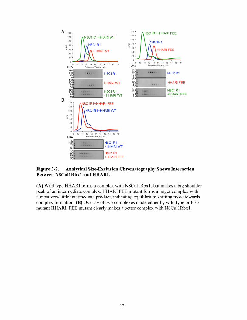
NEDD8ylated Cullin RING Ligases Bind with HHARI
In order to understand how the NEDD8ylated cullin RING ligases interact with
HHARI, I performed analytical size exclusion chromatography to assess complex
formation. If the interaction enhances HHARI activity, it is possible that the two proteins
have structural interaction. First, the complex formed with wild type HHARI and
N8Cul1Rbx1 produced a higher molecular weight complex, but also produced a big
shoulder peak that seemed to have a partial product. However, HHARI FEE and
N8Cul1Rbx1 made a larger complex, with a smaller shoulder peak (Figure 3-2A). Seen
by coomassie staining of gels run on same fractions, both WT or FEE HHARI make a
complex with N8Cul1Rbx1, but the FEE complex makes a larger form of almost two
fractions (Figure 3-2B). This suggests that it is possible the HHARI FEE makes a tighter
complex with N8Cul1Rbx1 compared to HHARI WT.
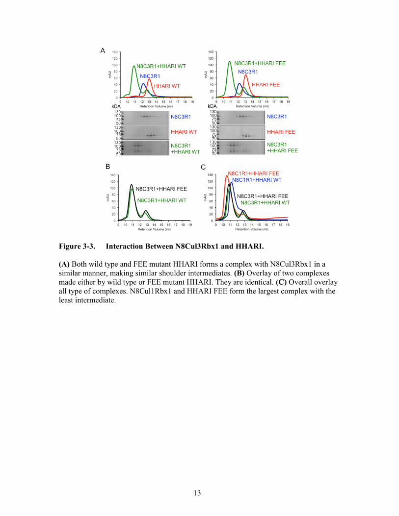
An analogous experiment showed that N8Cul3Rbx1 also helps activate HHARI
WT on its autoubiquitination. However, surprisingly there were no differences between
the sizes of the complexes made between N8Cul3Rbx1 and HHARI WT or HHARI FEE
(Figure 3-3A, B). Therefore, the N8Cul3Rbx1 did not differentiate whether HHARI has
the FEE mutation or not. Overlap of all peaks showed that the largest complex made was
between N8Cul1Rbx1 and HHARI FEE (Figure 3-3C).

11
Figure 3-1. FEE Mutation of HHARI and the Addition of N8Cul1Rbx1 Both
Activate HHARI.
(A) WT HHARI stays inactivate as no autoubiquitin chains are built by time. However,
FEE mutation of HHARI induces autoubiquitination activity starting from 0.5 min
building autoubiquitin chains, and almost fully saturated by 5min. (B) N8Cul1Rbx1
promotes wild type HHARI activation, while enhancing the activity of HHARI FEE
mutant. (C) N8Cul3Rbx1 also promotes wild type HHARI activation, while enhancing
the activity of HHARI FEE mutant. All assays are observed by SYPRO-Ruby staining.
12
Figure 3-2. Analytical Size-Exclusion Chromatography Shows Interaction
Between N8Cul1Rbx1 and HHARI.
(A) Wild type HHARI forms a complex with N8Cul1Rbx1, but makes a big shoulder
peak of an intermediate complex. HHARI FEE mutant forms a larger complex with
almost very little intermediate product, indicating equilibrium shifting more towards
complex formation. (B) Overlay of two complexes made either by wild type or FEE
mutant HHARI. FEE mutant clearly makes a better complex with N8Cul1Rbx1.
13
Figure 3-3. Interaction Between N8Cul3Rbx1 and HHARI.
(A) Both wild type and FEE mutant HHARI forms a complex with N8Cul3Rbx1 in a
similar manner, making similar shoulder intermediates. (B) Overlay of two complexes
made either by wild type or FEE mutant HHARI. They are identical. (C) Overall overlay
all type of complexes. N8Cul1Rbx1 and HHARI FEE form the largest complex with the
least intermediate.

14
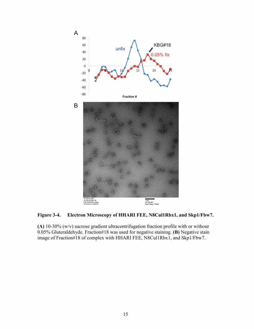
GraFix and Negative Staining
We sought to study the newly formed complex by structural studies. However,
due to the current limits of electron microscopy, a protein complex of less than 200 kDa
would not produce an ideal resolution. I tried to enhance contrast by making the complex
bigger by adding an adaptor protein of N8Cul1Rbx1. Skp1Fbw7 is a substrate adaptor
protein of N8Cul1Rbx1 that dimerizes, suggesting the entire complex might be able to
dimerize by Skp1Fbw7. However, adding Skp1Fbw7 to HHARI FEE and N8Cul1Rbx1
did not form a large complex when analyzed over size-exclusion chromatography (figure
not shown). Therefore, another way to generate a robust complex was to artificially trap
it. GraFix, a well known technique used to chemically lock a protein complex, was used
for making samples for electron microscopy (Kastner et al., 2008). This allowed me to
make a complex that contain all three proteins, HHARI FEE, N8Cul1Rbx1, and
Skp1Fbw7, possibly in a dimer form. The percentage of gluteraldehyde added, speed and
time of centrifugation were optimized, calculated by the theoretical molecular weight.
The peaks of Biorad protein assay traveled to an optimal fraction after ultracentrifugation
(Figure 3-4A).
Negative staining of samples of Fraction#18 produced semi-homogenous
molecular images (Figure 3-4B). We can definitely see the multiple circular shapes with
a hollow center, and cryo EM is currently in progress. If the results produce a high
resolution 3D image, it will give tremendous information on structural interaction
between NEDD8ylated cullins and RBRs.
15
Figure 3-4. Electron Microscopy of HHARI FEE, N8Cul1Rbx1, and Skp1/Fbw7.
(A) 10-30% (w/v) sucrose gradient ultracentrifugation fraction profile with or without
0.05% Gluteraldehyde. Fraction#18 was used for negative staining. (B) Negative stain
image of Fraction#18 of complex with HHARI FEE, N8Cul1Rbx1, and Skp1/Fbw7.

16
CHAPTER 4. DISCUSSION
Here, I studied the interaction between two E3 ligases, HHARI and N8Cul1Rbx1.
I showed that NEDD8ylated cullin RING ligases are able to make a complex with
HHARI. A specific set of HHARI mutations, F430A, E431A, and E503A, was not only
able to activate its autoubiquitination, but also produce a complex with N8Cul1Rbx1.
Understanding the mechanism of the two E3 ligases’ interaction may reveal a novel
mechanism of the ubiquitin pathway and providing structural insights will be the key.
Microscopic Reversibility of Protein Complex
Microscopic reversibility states that there is always a reverse reaction by time,
therefore a heterogenous substance can constantly exist in subgroups composed of
intermediate states for every process (Lewis, 1925). So far, the biggest challenge in our
efforts to make a stable complex with HHARI and N8Cul1Rbx1 was that whenever the
complex was run on size exclusion chromatography, it would produce a 70% bound
form, with a shoulder peak that would indicate an unbound form or an intermediate. This
resulted in many attempted failures on trying to crystallize the complex, and the crystals
obtained were only one part of the complex, either HHARI or NEDD8ylated Cul1Rbx1.
Therefore the microscopic reversibility of the complex of N8Cul1Rbx1 and HHARI WT
has been an issue as the constant association and dissociation produced an unstable
complex suitable for crystal formation. If we are able to somehow shift the equilibrium to
where it is more favorable in a bound form, that would enable us to study its structure
either through crystallography or electron microscopy.
Stabilizing the Complex
From preliminary studies (not shown), we know that the N-terminal 90 residues
of HHARI, which are highly acidic and glycine rich, are necessary for binding to
N8Cul1Rbx1. I believe the acidic regions are capable of making interactions but the
abundance of glycines makes the structure not stable enough for a single, specific
interaction. Therefore, this might be the reasoning behind the dynamic equilibrium state,
producing intermediate products. It was surprising to find out that introducing the F430A,
E431A, E503A triple mutation of HHARI was able to make a larger complex from size
exclusion chromatography, while having a very little shoulder peak. This indicated that
the equilibrium greatly shifted to the complex formation side and producing less
intermediate forms than previous experiments. It is possible that the triple mutation opens
up a secondary docking site for the NEDD8ylated cullin to bind. With the equilibrium
shift, I was able to gain confidence that HHARI FEE form a much more stable complex
compared to HHARI WT.

17
Ub-VME as a Chemical Warhead
Chemically modified ubiquitins were initially introduced as probes for reactivity
of deubiquitinating enzymes (DUB) (Borodovsky et al., 2002). In previous studies of
Parkin, chemically modified ubiquitin-vinyl sulfone (Ub-VS) was implemented as a
probe that covalently modifies the catalytic cysteine of Parkin without the help of E2
(Riley et al., 2013). Kelsall et al also implemented an electrophilic Ubiquitin-vinyl
methyl ester (Ub-VME) to find that the Ub-VME binds to the catalytic cysteine of
HHARI by either removing the Ariadne domain or the addition of NEDD8ylated cullins
(Kelsall et al., 2013). In fact, there have been structural studies using the Ub-VME to
study molecular contacts for ubiquitin recognition (Sheedlo et al., 2015). Implementing
Ub-VME on structural studies of the complex with NEDD8ylated cullins and HHARI
will have a modified ubiquitin bound to the active site cysteine, producing an active form
of the complex. Further on, if this active site bound Ub-VME interacts with multiple sites
on either HHARI or NEDD8ylated cullin, it can act as a chemical linker that locks the
complex in action.
Targets for Electron Microscopy
Currently, size is a very important factor in achieving high-resolution 3D images
for electron microscopy. Therefore, dimerizing the complex would significantly improve
EM studies. F-box protein Fbw7 of Cul1Rbx1 would theoretically produce a dimer. Also,
Cul3Rbx1 binds with a BTB protein that is known to interact with substrate adaptors.
Most importantly they are known to dimerize (Furukawa et al., 2003; Zhuang et al.,
2009). As Cul3Rbx1 also interacts with HHARI, dimerization of Cul3Rbx1 and its
substrate adaptor will likely cause dimerization of the potentially HHARI bound complex
as well, producing a sizable protein suitable for electron microscopy. However, according
to the analytical size-exclusion chromatography experiments (Figure 6B), N8Cul3Rbx1
did not make a difference in the interaction between either HHARI WT or FEE mutant.
Therefore, it is not clear whether N8Cul3Rbx1 would interact in a similar way with
N8Cul1Rbx1, but it will definitely give insight towards how the cullin RING ligases
interact with HHARI.
According to protein interaction studies, HHARI is seen to interact with Cul1,
Cul2, Cul3, and Cul4A, which can also be tried for structural studies. Another Ariadne
family protein TRIAD1 can also be used, as Kelsall et al used to study interactions with
Cul5. One option to study a complex between RBR and cullins would be Parc (Cul9) as
this is known as a cullin and Ariadne RBR fused protein. So far, there have been no
structural studies on this protein, which can potentially provide great insight on the cullin
RBR complex. From these studies I hope to learn a novel mechanism of two different E3
ligases working together. It is possible that the study might reveal a new category of E3
ligases, as there have been no studies on this interaction mechanism. Further on, as an E3
ligase, its substrate regulation will be most important in its participation in cellular
processes. It will be interesting to study what this new complex regulates downstream,
18
whether it will affect substrates of NEDD8ylated cullins, HHARI, or affect entirely new
substrates.

19
LIST OF REFERENCES
Borden, K.L., Boddy, M.N., Lally, J., O'Reilly, N.J., Martin, S., Howe, K., Solomon, E.,
and Freemont, P.S. (1995). The solution structure of the RING finger domain from the
acute promyelocytic leukaemia proto-oncoprotein PML. The EMBO journal 14, 1532-
1541.
Borodovsky, A., Ovaa, H., Kolli, N., Gan-Erdene, T., Wilkinson, K.D., Ploegh, H.L., and
Kessler, B.M. (2002). Chemistry-based functional proteomics reveals novel members of
the deubiquitinating enzyme family. Chemistry & biology 9, 1149-1159.
Cardozo, T., and Pagano, M. (2004). The SCF ubiquitin ligase: insights into a molecular
machine. Nat Rev Mol Cell Biol 5, 739-751.
Cavadini, S., Fischer, E.S., Bunker, R.D., Potenza, A., Lingaraju, G.M., Goldie, K.N.,
Mohamed, W.I., Faty, M., Petzold, G., Beckwith, R.E., et al. (2016). Cullin-RING
ubiquitin E3 ligase regulation by the COP9 signalosome. Nature 531, 598-603.
Chaugule, V.K., Burchell, L., Barber, K.R., Sidhu, A., Leslie, S.J., Shaw, G.S., and
Walden, H. (2011). Autoregulation of Parkin activity through its ubiquitin-like domain.
The EMBO journal 30, 2853-2867.
Deshaies, R.J., and Joazeiro, C.A. (2009). RING domain E3 ubiquitin ligases. Annu Rev
Biochem 78, 399-434.
Duda, D.M., Borg, L.A., Scott, D.C., Hunt, H.W., Hammel, M., and Schulman, B.A.
(2008). Structural insights into NEDD8 activation of cullin-RING ligases: conformational
control of conjugation. Cell 134, 995-1006.
Duda, D.M., Olszewski, J.L., Schuermann, J.P., Kurinov, I., Miller, D.J., Nourse, A.,
Alpi, A.F., and Schulman, B.A. (2013). Structure of HHARI, a RING-IBR-RING
Ubiquitin Ligase: Autoinhibition of an Ariadne-Family E3 and Insights into Ligation
Mechanism. Structure 21, 1030-1041.
Eisenhaber, B., Chumak, N., Eisenhaber, F., and Hauser, M.T. (2007). The ring between
ring fingers (RBR) protein family. Genome Biol 8, 209.
Enchev, R.I., Scott, D.C., da Fonseca, P.C., Schreiber, A., Monda, J.K., Schulman, B.A.,
Peter, M., and Morris, E.P. (2012). Structural basis for a reciprocal regulation between
SCF and CSN. Cell Rep 2, 616-627.
Furukawa, M., He, Y.J., Borchers, C., and Xiong, Y. (2003). Targeting of protein
ubiquitination by BTB-Cullin 3-Roc1 ubiquitin ligases. Nat Cell Biol 5, 1001-1007.

20
Hodgins, R.R., Ellison, K.S., and Ellison, M.J. (1992). Expression of a ubiquitin
derivative that conjugates to protein irreversibly produces phenotypes consistent with a
ubiquitin deficiency. The Journal of biological chemistry 267, 8807-8812.
Hristova, V.A., Beasley, S.A., Rylett, R.J., and Shaw, G.S. (2009). Identification of a
novel Zn2+-binding domain in the autosomal recessive juvenile Parkinson-related E3
ligase parkin. The Journal of biological chemistry 284, 14978-14986.
Huang, D.T., Ayrault, O., Hunt, H.W., Taherbhoy, A.M., Duda, D.M., Scott, D.C., Borg,
L.A., Neale, G., Murray, P.J., Roussel, M.F., et al. (2009). E2-RING expansion of the
NEDD8 cascade confers specificity to cullin modification. Molecular cell 33, 483-495.
Huibregtse, J.M., Scheffner, M., Beaudenon, S., and Howley, P.M. (1995). A family of
proteins structurally and functionally related to the E6-AP ubiquitin-protein ligase.
Proceedings of the National Academy of Sciences of the United States of America 92,
2563-2567.
Kastner, B., Fischer, N., Golas, M.M., Sander, B., Dube, P., Boehringer, D., Hartmuth,
K., Deckert, J., Hauer, F., Wolf, E., et al. (2008). GraFix: sample preparation for single-
particle electron cryomicroscopy. Nat Methods 5, 53-55.
Kelsall, I.R., Duda, D.M., Olszewski, J.L., Hofmann, K., Knebel, A., Langevin, F.,
Wood, N., Wightman, M., Schulman, B.A., and Alpi, A.F. (2013). TRIAD1 and HHARI
bind to and are activated by distinct neddylated Cullin RING ligase complexes. The
EMBO journal.
Komander, D. (2009). The emerging complexity of protein ubiquitination. Biochem Soc
Trans 37, 937-953.
Komander, D., and Rape, M. (2012). The ubiquitin code. Annu Rev Biochem 81, 203-
229.
Koyano, F., Okatsu, K., Kosako, H., Tamura, Y., Go, E., Kimura, M., Kimura, Y.,
Tsuchiya, H., Yoshihara, H., Hirokawa, T., et al. (2014). Ubiquitin is phosphorylated by
PINK1 to activate parkin. Nature 510, 162-166.
Lewis, G.N. (1925). A New Principle of Equilibrium. Proceedings of the National
Academy of Sciences of the United States of America 11, 179-183.
Lyapina, S., Cope, G., Shevchenko, A., Serino, G., Tsuge, T., Zhou, C., Wolf, D.A., Wei,
N., Shevchenko, A., and Deshaies, R.J. (2001). Promotion of NEDD-CUL1 conjugate
cleavage by COP9 signalosome. Science 292, 1382-1385.
Lydeard, J.R., Schulman, B.A., and Harper, J.W. (2013). Building and remodelling
Cullin-RING E3 ubiquitin ligases. EMBO Rep 14, 1050-1061.

21
Mosadeghi, R., Reichermeier, K.M., Winkler, M., Schreiber, A., Reitsma, J.M., Zhang,
Y., Stengel, F., Cao, J., Kim, M., Sweredoski, M.J., et al. (2016). Structural and kinetic
analysis of the COP9-Signalosome activation and the cullin-RING ubiquitin ligase
deneddylation cycle. eLife 5.
Pierce, N.W., Lee, J.E., Liu, X., Sweredoski, M.J., Graham, R.L., Larimore, E.A., Rome,
M., Zheng, N., Clurman, B.E., Hess, S., et al. (2013). Cand1 promotes assembly of new
SCF complexes through dynamic exchange of F box proteins. Cell 153, 206-215.
Riley, B.E., Lougheed, J.C., Callaway, K., Velasquez, M., Brecht, E., Nguyen, L., Shaler,
T., Walker, D., Yang, Y., Regnstrom, K., et al. (2013). Structure and function of Parkin
E3 ubiquitin ligase reveals aspects of RING and HECT ligases. Nat Commun 4, 1982.
Saha, A., and Deshaies, R.J. (2008). Multimodal activation of the ubiquitin ligase SCF by
Nedd8 conjugation. Molecular cell 32, 21-31.
Schmidt, M.W., McQuary, P.R., Wee, S., Hofmann, K., and Wolf, D.A. (2009). F-box-
directed CRL complex assembly and regulation by the CSN and CAND1. Molecular cell
35, 586-597.
Schulman, B.A., Carrano, A.C., Jeffrey, P.D., Bowen, Z., Kinnucan, E.R., Finnin, M.S.,
Elledge, S.J., Harper, J.W., Pagano, M., and Pavletich, N.P. (2000). Insights into SCF
ubiquitin ligases from the structure of the Skp1-Skp2 complex. Nature 408, 381-386.
Schulman, B.A., and Harper, J.W. (2009). Ubiquitin-like protein activation by E1
enzymes: the apex for downstream signalling pathways. Nat Rev Mol Cell Biol 10, 319-
331.
Scott, D.C., Sviderskiy, V.O., Monda, J.K., Lydeard, J.R., Cho, S.E., Harper, J.W., and
Schulman, B.A. (2014). Structure of a RING E3 Trapped in Action Reveals Ligation
Mechanism for the Ubiquitin-like Protein NEDD8. Cell 157, 1671-1684.
Sheedlo, M.J., Qiu, J., Tan, Y., Paul, L.N., Luo, Z.Q., and Das, C. (2015). Structural basis
of substrate recognition by a bacterial deubiquitinase important for dynamics of
phagosome ubiquitination. Proceedings of the National Academy of Sciences of the
United States of America 112, 15090-15095.
Skaar, J.R., Florens, L., Tsutsumi, T., Arai, T., Tron, A., Swanson, S.K., Washburn,
M.P., and DeCaprio, J.A. (2007). PARC and CUL7 form atypical cullin RING ligase
complexes. Cancer research 67, 2006-2014.
Spratt, D.E., Walden, H., and Shaw, G.S. (2014). RBR E3 ubiquitin ligases: new
structures, new insights, new questions. Biochem J 458, 421-437.
Streich, F.C., Jr., and Lima, C.D. (2014). Structural and functional insights to ubiquitin-
like protein conjugation. Annual review of biophysics 43, 357-379.

22
Tang, X., Orlicky, S., Lin, Z., Willems, A., Neculai, D., Ceccarelli, D., Mercurio, F.,
Shilton, B.H., Sicheri, F., and Tyers, M. (2007). Suprafacial orientation of the SCFCdc4
dimer accommodates multiple geometries for substrate ubiquitination. Cell 129, 1165-
1176.
Trempe, J.F., Sauve, V., Grenier, K., Seirafi, M., Tang, M.Y., Menade, M., Al-Abdul-
Wahid, S., Krett, J., Wong, K., Kozlov, G., et al. (2013). Structure of Parkin Reveals
Mechanisms for Ubiquitin Ligase Activation. Science 340, 1451-1455.
Wauer, T., and Komander, D. (2013). Structure of the human Parkin ligase domain in an
autoinhibited state. The EMBO journal.
Wenzel, D.M., Lissounov, A., Brzovic, P.S., and Klevit, R.E. (2011). UBCH7 reactivity
profile reveals parkin and HHARI to be RING/HECT hybrids. Nature 474, 105-108.
Zhuang, M., Calabrese, M.F., Liu, J., Waddell, M.B., Nourse, A., Hammel, M., Miller,
D.J., Walden, H., Duda, D.M., Seyedin, S.N., et al. (2009). Structures of SPOP-substrate
complexes: insights into molecular architectures of BTB-Cul3 ubiquitin ligases.
Molecular cell 36, 39-50.
Zimmerman, E.S., Schulman, B.A., and Zheng, N. (2010). Structural assembly of cullin-
RING ubiquitin ligase complexes. Curr Opin Struct Biol 20, 714-721.

23
VITA
Kheewoong Baek was born in 1990, Daegu, Korea. After receiving his high
school diploma from Hankuk Academy of Foreign Studies, Korea, he entered Rutgers
University in New Jersey. He graduated December of 2012 with a Bachelor of Arts in
Biological Sciences. Following February, he started a postbaccalaureate program at NIH
in the lab of Dr. Yihong Ye. 2014, he joined the school of Graduate Health Sciences at
University of Tennessee Health Science Center, carrying on his Masters studies in the lab
of Dr. Brenda A. Schulman at St.Jude Children’s Research Hospital. In May of 2016, he
will receive his Master of Science degree from the University of Tennessee.