Integumentary System 1. Introduction (Open your text to ...
15
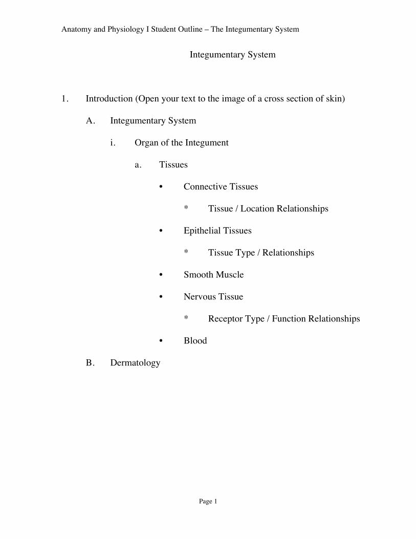
Anatomy and Physiology I Student Outline – The Integumentary System Page 1 Integumentary System 1. Introduction (Open your text to the image of a cross section of skin) A. Integumentary System i. Organ of the Integument a. Tissues • Connective Tissues * Tissue / Location Relationships • Epithelial Tissues * Tissue Type / Relationships • Smooth Muscle • Nervous Tissue * Receptor Type / Function Relationships • Blood B. Dermatology
Transcript of Integumentary System 1. Introduction (Open your text to ...
Anatomy and Physiology I Student Outline – The Integumentary System
Page 1
Integumentary System
1. Introduction (Open your text to the image of a cross section of skin)
A. Integumentary System
i. Organ of the Integument
a. Tissues
• Connective Tissues
* Tissue / Location Relationships
• Epithelial Tissues
* Tissue Type / Relationships
• Smooth Muscle
• Nervous Tissue
* Receptor Type / Function Relationships
• Blood
B. Dermatology

Anatomy and Physiology I Student Outline – The Integumentary System
Page 2

Anatomy and Physiology I Student Outline – The Integumentary System
Page 3
2. Functions of the Integument
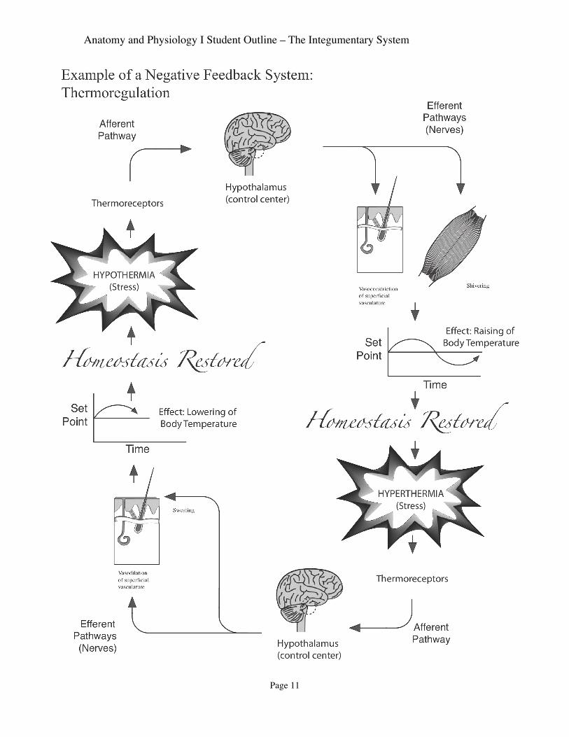
A. Thermoregulation (Review control mechanism from first lecture)
i. Superficial Vasculature
ii. Deep Vasculature
B. Protection from …
i. Abrasion
ii. UV Radiation
iii. Invasion from pathogens
iv. Desiccation
C. Receives stimuli
i. Receptor Types / Function Relationships
a. Touch Receptor
b. Pressure Receptors
c. Pain Receptors
d. Temperature Receptors
Anatomy and Physiology I Student Outline – The Integumentary System
Page 4
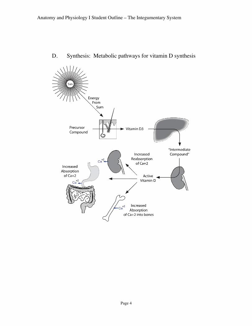
D. Synthesis: Metabolic pathways for vitamin D synthesis

Anatomy and Physiology I Student Outline – The Integumentary System
Page 5
E. Immunity
i. Tissue and Related Functions
3. Epidermis (Layer #1) (See diagrams in Text!)

Anatomy and Physiology I Student Outline – The Integumentary System
Page 6
A. General Characteristics
i. Keratinized Stratified Squamous Epithelium
ii. Avascular
iii. Basement Membrane
iv. Epidermal Ridges (and Dermal Papilla)
a. Two Functions
B. Cell Types and Desmosomes
i. Keratinocyte
• Desmosomes
• Keratinization

Anatomy and Physiology I Student Outline – The Integumentary System
Page 7
ii. Melanocyte
a. Process of Melanin Production
• Melanosomes
iii. Skin Color and Photoprotection of Keratinocytes (See Handout)
a. Melanin
• Melanin production processes
* Review of Transcription, translation ETC ETC
• Melanocyte
* Dendrites (“branches”)
• Melanosome
• Mechanisms of Melanin uptake by keratinocytes
* Receptor Mediated Endocytosis
* Exocytosis followed by phagocytosis
• Perinuclear protection of Chromatin
• Cell Signaling Control Mechanism by Keratinocytes
Anatomy and Physiology I Student Outline – The Integumentary System
Page 8
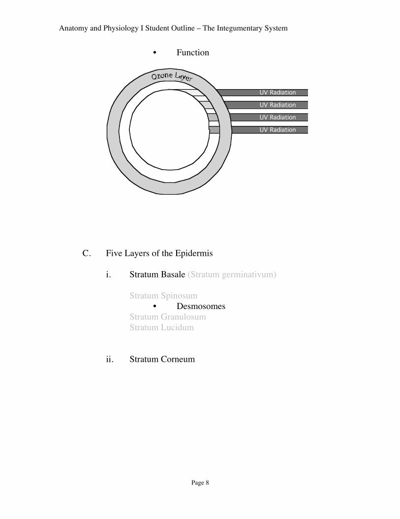
• Function
UV Radiation
UV Radiation
UV Radiation
UV Radiation
C. Five Layers of the Epidermis
i. Stratum Basale (Stratum germinativum)
Stratum Spinosum • Desmosomes
Stratum Granulosum Stratum Lucidum
ii. Stratum Corneum

Anatomy and Physiology I Student Outline – The Integumentary System
Page 9
D. Chemical Messangers Defined
• Hormone • Growth Factor (or colony stimulating factor)
3. Dermis
• Irregular Dense Connective Tissue
A. Regions of Dermis
i. Papillary Region
• Dermal Papillae
• Corpuscles of Touch
• Superficial Vasculature Functions
ii. Reticular Region
• Corpuscles of Pressure

Anatomy and Physiology I Student Outline – The Integumentary System
Page 10
B. General Characteristics of the Dermis
i. Fibers
ii. Vascular
iii. Muscle
iv. Nerve Fibers
v. Hair Follicles
vi. Sweat Glands
vii. Sebaceous Glands
4. Subcutaneous Layer (or region) of the Integument (also, Hypodermis)
A. Tissues
i. Adipose Connective Tissue
ii. Loose Areolar Connective Tissue
B. Functions
i. Thermoregulation
ii. Vascular Protection
iii. Adhesion
Anatomy and Physiology I Student Outline – The Integumentary System
Page 11

Anatomy and Physiology I Student Outline – The Integumentary System
Page 12
5. Accessory Structures
A. Hair
i. Hair follicle
ii. Shaft
iii. Arrector Pili Muscles
iv. Sebaceous Glands
v. Functions
Nails
i. Nail Bed
ii. Lunula
iii. Functions
B. Skin Glands
i. Sebaceous Glands (Oil glands)
• Holocrine Types
• Sebum
ii. Merocrine Sweat Glands
• Tubular
iii. Apocrine Sweat Gland
iii. Other Integumentary Glands a. Ceruminous Glands b. Mammary Glands
Homeostasis of Temperature Body – review in text

Anatomy and Physiology I Student Outline – The Integumentary System
Page 13
C. Aging and Hair
i. Hair Color
a. Gray Hair
b. White Hair
ii. Balding
a. Genetic Predisposition
b. Testosterone
6. Deep Wound Healing (ESSAY ALERT !! The outline below is partial. Take
good notes and see text for all important details).
A. Stabilization of Wound
i. An initial break damages dermal blood vessels and inserts
microorganisms
ii. Reflexive vasoconstriction reduces blood flow
iii. Platelets come in contact with collagen fibers and induce clotting
iv. Clot isolates bacterial, and further reduces blood lose
B. Inflammatory response
i. Mast cells and Basophile secrete histamine
ii. Histamine induces inflammation characterized first by vasodilatoin of
undamaged blood vessels

Anatomy and Physiology I Student Outline – The Integumentary System
Page 14
iii. Vasodilated vessels become porous allowing nutrients, oxygen and
other resources to enter damaged area.
iv. Oxygen inhibits anaerobic bacteria. Of particular importance here is
Clostridium tetani, causative agent of tetanus
v. Margination, Diapedesis, positive chemotaxis, and phagocytosis by
neutrophils
vi. Neutroophils secret Pyrogen secreted elevates local temperature for
further inhibition of bacterial growth.
vii. Monocytes marginate and diapedis. Once in interstitium, monocyte
differentiates into highly phagocytic macrophage
C. Injury Resolution
i. Stem cells within stratum basalis proliferate for Epidermis restoration
and repair of stratum corneum.
ii. Blood vessels begin grow for purposes of restoration of normal blood
flow
iii. Fibroblasts migrate into damaged area and secrete collagen “building
blocks” for restoration of collagen fibrils. This “knitting together of
the wound” results is restoration of Irregular Dense Connective Tissue
of Dermis.

Anatomy and Physiology I Student Outline – The Integumentary System
Page 15
iii. Epidermis mends
iv. Scab forms
v. Eosinophils digest old clot material by enzyme plasminogen.
D. Final Stages
i. Normal blood flow restored
ii. Bacterial and damaged tissue removed
iii. Irregularly placed collagen leaves scar
iv. Scab falls off.