Integrative Biology c0ib00131g - IT Services Help Siteusers.ox.ac.uk/~atdgroup/publications/Patel,...
31
Integrative Biology c0ib00131g The Q1 challenges of integrating molecular imaging into the optimization of cancer therapy G. S. Patel,* T. Kiuchi, K. Lawler, E. Ofo, G. Fruhwirth, M. Kelleher, E. Shamil, R. Zhang, P. R. Selvin, G. Santis, J. Spicer, N. Woodman, C. E. Gillett, P. R. Barber, B. Vojnovic, G. Ke´ri, T. Schaeffter, V. Goh, M. J. O’Doherty, P. A. Ellis and T. Ng* We wish to highlight our attempt to link nanometre scale protein oligomerisation/interaction events to whole body imaging. Q2 Please check this proof carefully. Our staff will not read it in detail after you have returned it. Translation errors between word-processor files and typesetting systems can occur so the whole proof needs to be read. Please pay particular attention to: tabulated material; equations; numerical data; figures and graphics; and references. If you have not already indicated the corresponding author(s) please mark their name(s) with an asterisk. Please e-mail a list of corrections or the PDF with electronic notes attached -- do not change the text within the PDF file or send a revised manuscript. Please bear in mind that minor layout improvements, e.g. in line breaking, table widths and graphic placement, are routinely applied to the final version. Please note that, in the typefaces we use, an italic vee looks like this: n, and a Greek nu looks like this: n. We will publish articles on the web as soon as possible after receiving your corrections; no late corrections will be made. Please return your final corrections, where possible within 48 hours of receipt, by e-mail to: [email protected] Reprints—Electronic (PDF) reprints will be provided free of charge to the corresponding author. Enquiries about purchasing paper reprints should be addressed via: http://www.rsc.org/publishing/journals/guidelines/paperreprints/. Costs for reprints are below: Reprint costs No of pages Cost (per 50 copies) First Each additional 2-4 £225 £125 5-8 £350 £240 9-20 £675 £550 21-40 £1250 £975 >40 £1850 £1550 Cost for including cover of journal issue: £55 per 50 copies Queries are marked on your proof like this Q1, Q2, etc. and for your convenience line numbers are indicated like this 5, 10, 15, ...
Transcript of Integrative Biology c0ib00131g - IT Services Help Siteusers.ox.ac.uk/~atdgroup/publications/Patel,...

Integrative Biology c0ib00131g
TheQ1 challenges of integrating molecular imaging into theoptimization of cancer therapy
G. S. Patel,* T. Kiuchi, K. Lawler, E. Ofo, G. Fruhwirth,M. Kelleher, E. Shamil, R. Zhang, P. R. Selvin, G. Santis,J. Spicer, N. Woodman, C. E. Gillett, P. R. Barber,B. Vojnovic, G. Keri, T. Schae!ter, V. Goh,M. J. O’Doherty, P. A. Ellis and T. Ng*
We wish to highlight our attempt to link nanometre scaleprotein oligomerisation/interaction events to whole bodyimaging.Q2
Please check this proof carefully. Our staff will not read it in detail after you have returned it. Translation errors betweenword-processor files and typesetting systems can occur so the whole proof needs to be read. Please pay particular attention to:tabulated material; equations; numerical data; figures and graphics; and references. If you have not already indicated thecorresponding author(s) please mark their name(s) with an asterisk. Please e-mail a list of corrections or the PDF with electronicnotes attached -- do not change the text within the PDF file or send a revised manuscript.
Please bear in mind that minor layout improvements, e.g. in line breaking, table widths and graphic placement, areroutinely applied to the final version.
Please note that, in the typefaces we use, an italic vee looks like this: n, and a Greek nu looks like this: n.
We will publish articles on the web as soon as possible after receiving your corrections; no late corrections will be made.
Please return your final corrections, where possible within 48 hours of receipt, by e-mail to: [email protected]
Reprints—Electronic (PDF) reprints will be provided free of charge to the corresponding author. Enquiries about purchasingpaper reprints should be addressed via: http://www.rsc.org/publishing/journals/guidelines/paperreprints/. Costs for reprints arebelow:
Reprint costs
No of pages Cost (per 50 copies)
First Each additional
2-4 £225 £1255-8 £350 £2409-20 £675 £55021-40 £1250 £975
>40 £1850 £1550
Cost for including cover of journal issue:£55 per 50 copies
Queries are marked on your proof like this Q1, Q2, etc. and for your convenience line numbers are indicated like this 5, 10, 15, ...

Queryreference
Query Remarks
Q1 For your information: You can cite this paper before thepage numbers are assigned with: (authors), Integr. Biol.,(year), DOI: 10.1039/c0ib00131g.
Q2 If the contents entry does not fit between the two horizontallines, then please trim the text and/or the title.
Q3 I have added an acknowledgement for Keppler and Tutt.Please check if this is ok or provide a replacement.
Q4 Please check references carefully. The DOIs of many of thereferences have been corrected and some references havebeen updated with years, volumes and page numberswhere possible.
Q5 Ref. 138: Please provide the following details: journal title.
Q6 Ref. 181: Please provide the following details: year ofpublication, volume number, page number(s).
Q7 Ref. 196: Can this reference be updated yet? Please supplydetails to allow readers to access the reference (forreferences where page numbers are not yet known, pleasesupply the DOI).
TheQ1 challenges of integrating molecular imaging into the optimization ofcancer therapy
G. S. Patel,*ab T. Kiuchi,ac K. Lawler,ad E. Ofo,ae G. Fruhwirth,a M. Kelleher,af
E. Shamil,a R. Zhang,g P. R. Selvin,g G. Santis,h J. Spicer,b N. Woodman,i
C. E. Gillett,ci P. R. Barber,j B. Vojnovic,jk G. Keri,lm T. Schae!ter,n V. Goh,o
M. J. O’Doherty,p P. A. Ellisb and T. Ng*ac
Received 23rd October 2010, Accepted 2nd March 2011
DOI: 10.1039/c0ib00131g
We review novel, in vivo and tissue-based imaging technologies that monitor and optimise cancer
therapeutics. Recent advances in cancer treatment centre around the development of targeted
therapies and personalisation of treatment regimes to individual tumour characteristics. However,
clinical outcomes have not improved as expected. Further development of the use of molecular
imaging to predict or assess treatment response must address spatial heterogeneity of cancer
within the body. A combination of di!erent imaging modalities should be used to relate the e!ect
of the drug to dosing regimen or e!ective drug concentration at the local site of action. Molecular
imaging provides a functional and dynamic read-out of cancer therapeutics, from nanometre to
whole body scale. At the whole body scale, an increase in the sensitivity and specificity
of the imaging probe is required to localise (micro)metastaticw foci and/or residual disease that
are currently below the limit of detection. The use of image-guided endoscopic biopsy can
produce tumour cells or tissues for nanoscopic analysis in a relatively patient-compliant manner,
thereby linking clinical imaging to a more precise assessment of molecular mechanisms. This
multimodality imaging approach (in combination with genetics/genomic information) could be
used to bridge the gap between our knowledge of mechanisms underlying the processes of
metastasis and tumour dormancy and routine clinical practice. Treatment regimes could therefore
be individually tailored both at diagnosis and throughout treatment, through monitoring of drug
pharmacodynamics providing an early read-out of response or resistance.
1. Introduction
Cancer therapies have evolved significantly in the past tenyears with the advent of targeted treatments designed to a
1
5
10
15
20
25
30
35
40
45
50
55
1
5
10
15
20
25
30
35
40
45
50
55
a Richard Dimbleby Department of Cancer Research, Randall Division & Division of Cancer Studies, Kings College London,Guy’s Medical School Campus, London, SE1 1UL, UK. E-mail: [email protected], [email protected]; Tel: +44(0) 20 7848 6174
bResearch Oncology, Division of Cancer Studies, Kings College London, Guys Campus, London, SE1 9RT, UKcBreakthrough Breast Cancer Research Unit, Research Oncology, King’s College London, Guy’s Medical School Campus,London, SE1 9RT, UK
dDepartment of Mathematics, King’s College London, Strand Campus, London, WC2R 2LS, UKeDepartment of Otolaryngology Head & Neck Surgery, Guys & St Thomas’ NHS Foundation Trust, London, SE1 1UL, UKfDepartment of Medical Oncology, St. George’s Hospital, London, SW17 0QT, UKgCenter for Biophysics and Computational Biology, Physics Department, and the Center of Physics of Living Cells,University of Illinois, Urbana-Champaign, USA
hDepartment of Asthma Allergy & Respiratory Science, Guy’s Hospital, London SE1 9RTiGuy’s & St Thomas’ Breast Tissue & Data Bank, King’s College London, Guy’s Hospital, London, SE1 9RT, UKjGray Institute for Radiation Oncology & Biology, University of Oxford, Old Road Campus Research Building, Roosevelt Drive,Oxford, OX3 7DQ, UKkRandall Division of Cell & Molecular Biophysics, Kings College London, London, UKlVichem Chemie Research Ltd., Herman Otto utca 15, Budapest, Hungarym Semmelweis University, Pathobiochemistry Research Group of Hungarian Academy of Science, 1444. Bp 8. POB 260, HungarynDivision of Imaging Sciences, Kings College London, London, SE1 7EH, UKoPaul Strickland Scanner Centre, Mount Vernon Hospital, Northwood, Middlesex, HA6 2RN, UKpPET Imaging Centre at St Thomas’ Hospital, Division of Imaging Sciences, Kings College London, London, SE1 7EH, UK
w Micrometastases were originally defined as small occult metastasesof less than 0.2 cm in diameter. Nowadays, the term refers to thespread of cancer cells in groups that are still so small they can only beseen under a microscope, and includes disseminated tumour cells thatare present in peripheral blood, bone marrow or lymph nodes.
This journal is !c The Royal Society of Chemistry 2011 Integr. Biol., 2011, ]]], 1–29 | 1
Integrative Biology Dynamic Article Links
Cite this: DOI: 10.1039/c0ib00131g
www.rsc.org/ibiology REVIEW ARTICLE

specific pathogenic process. Since the widespread adoption ofthe human epidermal growth factor (HER) inhibitor,trastuzumab (a monoclonal antibody to the extracellular domainof HER2) for HER2 overexpressing breast cancer, there has beena surge in the development of targeted, potential anti-cancerdrugs. During drug development, only one in 10000 compoundsscreened at the target localization stage will gain approval forclinical use. This process may take more than 10 years.1
Furthermore, once the drug is within the clinical sphere, clinicaloutcomes rarely meet initial expectations.
As an example, single-agent phase II studies of epidermalgrowth factor receptor (EGFR/HER1) inhibitors have shownresponse rates only of the order of 5–15% in non-smallcell lung cancer (NSCLC), head and neck squamous cellcarcinoma (HNSCC), and colorectal cancer.2 The tyrosine kinaseinhibitors (TKI), erlotinib and gefitinib, are targeted to EGFR,and approved for use in non-small cell lung cancer.3 However,response rates are less than 10% in unselected populations andoverexpression of EGFR does not correlate with response totreatment.4 Investigation of somatic gain-of-function mutationsin EGFR, led to the discovery of a missense mutation L858R inthe EGFR activation loop which facilitates gefitinib binding.5
This and other activating mutations are correlated with a muchimproved response rate to TKI therapy, and have helpedrevolutionise the treatment regimens for NSCLC patients.However, further mutations conferring resistance (especiallyT790M) can occur which render EGFR resistant to firstgeneration inhibitors.6 Multiple mutations that confer resistanceto the BCR-ABL tyrosine kinase inhibitor imatinib also exist inpatients with chronic myelogenous leukaemia (CML).7 In theseCML patients, the genetic basis of additional molecular changesthat occur and give rise to secondary drug resistance is frequentlyunknown. Thus understanding the molecular genotype does notprovide the complete explanation for resistance to molecule-targetedtherapies.
Here we propose that by combining di!erent modalities ofmolecular imaging we can begin to delineate and quantify thespecific molecular pathway alterations within the cancerpatient at a subcellular level. The cancer genome or proteomeis relatively plastic and can be reprogrammed, at di!erentstages of tumour development, to carry out various cellularprocesses such as proliferation, invasion and metastasis, orreversion to dormancy.8 This plasticity gives rise to spatialheterogeneity of cancer within the body and makes itchallenging to fully assess treatment response. Molecularimaging provides a solution by mapping the spatial response
of the tumour to treatment within the individual and thereby,to monitor progress throughout the patient journey.For instance, translational research may identify novel
biomarkers in the malignant phenotype, which can be imagedby radioligands or tracers, designed specifically to targetmolecules intrinsic to oncogenesis. Examples of imagingbiomarkers include tracers specific to hypoxia, angiogenesis,apoptosis and proliferation.9 Although novel imaging bio-markers may provide a non-invasive functional read-out ofthe malignant genome or proteome throughout treatment,there are many challenges in the integration of these bio-markers into clinical practice.
2. Overview of challenges in the implementation ofmolecular imaging for improving therapeutic e"cacy
Molecular imaging may provide an assessment of the temporaland spatial distribution of a probe or biomarker within adisease process. However the main challenge is to find the‘ideal’ imaging biomarker which should possess severalcharacteristics, in order to accurately assess the e!ect of atherapeutic intervention and fulfil clinical utility. These aresummarised in Box 1 below.10 Several key areas are furtherdiscussed in the subsequent subsections (2.1–2.4). In particu-lar, in terms of clinical imaging, the second point regarding theactivation state of a specific molecular target, which often maybe concentration-independent, is seldom addressed in theliterature. For this reason we have devoted a whole section(5) in this review to this topic.
Box 1. Ideal features of a molecular imaging biomarker
" Ability to detect specific changes at the molecular (in termsof concentration) level" Detection of the activated state of molecular target (which isindependent of concentration), e.g. ligand bound or receptordimerisation, as a read-out for monitoring drug e"cacy" Safe for human use" High sensitivity and specificity to distinguish target frombackground or confounding signals e.g. slow tumour washoutcompared to normal tissue to maintain good signal-to-noise ratio
1
5
10
15
20
25
30
35
40
45
50
55
1
5
10
15
20
25
30
35
40
45
50
55
Insight, innovation, integration
We review novel, in vivo and tissue-based imagingtechnologies that monitor and optimize cancer therapeutics.Clinical outcomes have lagged behind development oftargeted therapies. Combinations of imaging modalitiesshould be used to assess tumour spatial heterogeneity, treatmentresponse, and relate drug e!ects to dosing schedule.Molecular imaging provides a functional and dynamic
read-out of therapeutic response, from nanometre to whole
body scale. An increase in imaging probe sensitivity andspecificity is required to localise (micro) metastaticz foci thatare currently below systemic detection limits.Image-guided biopsy is key in the multimodality imaging
approach needed to bridge the gap between mechanisms under-lying pathological processes and clinical practice. Descriptionof novel pharmacodynamic endpoints using this approach canprovide early read-out of response or resistance.
z Micrometastases were originally defined as small occult metastasesof less than 0.2 cm in diameter. Nowadays, the term refers to thespread of cancer cells in groups that are still so small they can only beseen under a microscope, and includes disseminated tumour cells thatare present in peripheral blood, bone marrow or lymph nodes.
2 | Integr. Biol., 2011, ]]], 1–29 This journal is !c The Royal Society of Chemistry 2011
(continued )
Box 1. Ideal features of a molecular imaging biomarker
" Detection of established ‘on target’ drug e!ects in vivo toassess drug e"cacy and response" Appropriate ‘o!-rate’ or dissociation constant for adequate ima-ging but allows washout of biomarker prior to the next assessment." Rapid plasma clearance" Low hepatic excretion to visualise liver metastases" Rapid, simple chemical synthesis for tracer manufacture" Inexpensive
2.1 Sensitivity and specificity issues
A di"cult balance must be achieved whereby the tracer has ahigh a"nity for the target, which may be present in very small
amounts, but low a"nity for normal tissue to eliminate back-ground noise. For example, somatostatin analogues have beenin use for several decades to image gastroenteropancreatictumours, as they have a high a"nity for the somatostatinreceptor (SSR)-2 which is overexpressed by these tumours andin the central nervous system.11 Somatostatin is the ligand forSSRs but has a short half-life, which limits its use as animaging agent. Somatostatin analogues are more stable, yetbind as the native ligand, thus conferring high specificity andsensitivity to this imaging modality. The PET analogues[68Ga-DOTA, Tyr3]octreotide or [68Ga-DOTA, Tyr3]octreotateare emerging as the new standard.12 These analogues can belabelled with 90Y or 177Lu for use as radionuclide therapy withsuccess as a palliative treatment, thus translating imaging totherapeutics in a single step.13,14
The capacity of a biomarker to identify specific cells withinthe tumour population may describe tumour characteristics,
1
5
10
15
20
25
30
35
40
45
50
55
1
5
10
15
20
25
30
35
40
45
50
55
Gargi Patel, a medicaloncologist, completed her under-graduate medical degree at theUniversity of Cambridge with adistinction. She carried out herjunior doctor training at Guysand St. Thomas Hospital,amongst other London teachinguniversity hospitals. She inter-rupted her specialist training inorder to carry out a PhD atKings College London, usingFRET/FLIM assays in breastcancer, under Prof Tony Ng.She is in her second year, and
recently received the McElwain Fellowship from Cancer ResearchUK for her research.
G. S. Patel
Tai Kiuchi, a biophysicist,completed his PhD degree inthe Biophysical Laboratory(Prof. Kazuo Ohki) at theDepartment of Physics,Tohoku University, Japan.He has worked for four yearsas a postdoctoral fellow inthe Molecular BiologicalLaboratory (Prof. KensakuMizuno) at the Departmentof Life Sciences, TohokuUniversity. He was trained inmicroscopic imaging techniquesand molecular, biological
and biochemical techniques. Currently he is researching EGFRubiquitination signalling downstream of homo and heterodimersof the ErbB family using FRET/FLIM assays and molecularbiological experiments.
T. Kiuchi
Vicky Goh, Professor of ClinicalCancer Imaging, King’s CollegeLondon and HonoraryConsultant Radiologist, Guysand St Thomas’ NHSFoundation Trust, graduatedwith a First from University ofCambridge, underwent hertraining in medicine andradiology in London, a Fellow-ship in cross-sectional imaging inToronto, and was a ConsultantRadiologist at Mount VernonHospital, Northwood prior toher Chair. Her research
programme focuses on the clinical imaging of the tumour micro-environment (vascularity, hypoxia, and water di!usion) and tumourheterogeneity with CT, MRI (including DCE-MRI, BOLD-MRI,and DW-MRI) and PET–CT with particular reference togastrointestinal, renal and lung cancers.
V. Goh
Michael O’Doherty, a nuclearmedicine physician, with aspecial interest in clinicalapplications of PETCT.Professor of PET and NuclearMedicine in Imaging Sciencesat Kings College London andGuy’s and St Thomas’ NHSFoundation Trust.
M. J. O’Doherty
This journal is !c The Royal Society of Chemistry 2011 Integr. Biol., 2011, ]]], 1–29 | 3
such as resistance to treatment. For instance, CD133+/CXCR4+ tumour-initiator cells have been shown to undergoa 2-fold increase as a subpopulation (from 3.5% to 7.5% oftumour cells) following in vivo cisplatin treatment of lungtumour xenografts in mice, as an indication of the intrinsicresistance of this cell population to chemotherapy.15 Whilemolecular imaging of CXCR4 in a murine model of breastcancer metastasis with [64Cu]AMD3100 has recently beenpublished,16 it is likely that its ultimate clinical use will berestricted to imaging cancer metastases in a only a few organssuch as the lung, because there is a high uptake by normal stemcells at sites such as liver and bone marrow.
The relatively small numbers of ‘resistant’ or cancer stemcells within a tumour represents a further challenge to bio-marker sensitivity and whole body imaging. Potential cancerstem cells may constitute o1% of the tumour population.These cells may already be in the circulation, thus reducing thelikelihood of identification by the biomarker. Even if theimaging probe has a su"ciently high specific a"nity to bindto the target, significant signal amplification is likely to berequired in order to detect minimal target concentrations(typically in the nano- to pico-molar range).17 Imagingstrategies and methods to amplify target signal are discussedin a subsequent section (see section 4.6 Imaging drug resistancemechanisms including cancer stem cells).
2.2 Spatial heterogeneity issues
Further challenges are faced in the attempt to image tumourbiological responses at a microscopic scale, which willinevitably introduce another level of heterogeneity i.e. betweenindividual tumour cells. Furthermore, these imaging techniqueshave a limited depth of penetration and do not inform onwhole body distribution. There is scope to link whole bodyimaging (e.g. PET–CT) to imaging of pathological mechanismswithin the tumour cells at a nanometre scale. The enablingtechnologies linking these two imaging scales will includeimage-guidance by the co-registration of di!erent images
(e.g. PET and ultrasound (US)), to improve the currentaccuracy of sampling tumour-infiltrated lymph nodes, forinstance. Image-guided biopsy may complement whole bodyimaging by improving the accuracy of assessment of responseand recurrence, but is invasive. Tumour sites exhibiting poorresponse to therapy may be biopsied to define whether thesecells exhibit a clonal change or a change in receptor expression.For instance, the di!erence or discordance in protein expression(e.g. HER2 status18) on cancer cells between the primarytumour and distant metastatic sites may correlate with adi!erential sensitivity to treatment (to be expanded on furtherunder section 4.2 Use of imaging to characterise tumourheterogeneity).Cancer patient management is guided by the classification
of tumours into a variety of subtypes, representative of theirpathology and stage, as described by light microscopy, bio-markers derived from antigen-specific immunohistochemistry,mutation and cytogenic analysis, and gene microarray data.19
However, intratumour spatial heterogeneity may reducethe validity of this categorization. For example, nuclearpolymorphism represents one of several characteristics usedto determine grade in both invasive ductal carcinoma (IDC)and in situ breast cancer. Yet the commonly used gradingsystems do not recommend a minimum proportion of nucleithat need to be classed in the most marked pleomorphismgroup. How representative this is of the tumour as a whole isarguable. Conventional grading according to the modifiedScar!–Bloom–Richardson method assigns a score dependingon the highest level of nuclear atypia. Furthermore, detailedanalysis and subclassification of entire DCIS lesions, byimmunohistochemistry and microarray analysis, showedintratumoural biological diversity in 46% of all samples.20 Itis likely that histopathological analysis leads to an under-estimation of the total intratumour heterogeneity as only asmall percentage of the tissue is examined. The use ofendoscopic ultrahigh resolution optical coherence tomography/microscopy (resolutions of o4 mm axial and o2 mm
1
5
10
15
20
25
30
35
40
45
50
55
1
5
10
15
20
25
30
35
40
45
50
55
Professor Paul Ellis studiedmedicine at Otago Universityin New Zealand beforecompleting his fellowship inMedical Oncology and hispostgraduate Doctor ofMedicine research degree atthe Royal Marsden Hospitalin London. He is currentlyMedical Director for theSouth East London CancerNetwork. His major breastcancer research interestsinclude working with ProfessorTony Ng on the clinical
application of optical proteomics and novel clinical researchstrategies in the adjuvant and neoadjuvant setting.
P. A. Ellis
Tony Ng is the RichardDimbleby Professor ofCancer Research hhttp://www.dimblebycancercare.org/i,King’s College London. Hehas a mix of training/expertisein medicine, immunology, cancercell biology, biochemistry,optical imaging and cellbiophysics. Using FRETand FLIM techniques, hislaboratory has established,in live and fixed tumour cellsystems (including xenografts),imaging-based methods that
can monitor post-translational modifications and protein inter-actions, both in space and time. By combining imaging withbioinformatics and network modelling, we are now adopting amultidisciplinary approach to understand the cancer metastaticprocess and its immunological control.
T. Ng
4 | Integr. Biol., 2011, ]]], 1–29 This journal is !c The Royal Society of Chemistry 2011

transverse)21 is one of the imaging solutions that has been usedto overcome this issue of inadequate tumour sampling.
Although molecular imaging may help delineate intra- andinter-tumour heterogeneity, these findings may create challengingclinical implications. For example, the smallest volume of thetumour expressing the imaged target which may warrant achange of treatment, and the implications of determining themolecular profile of the biopsied material from e.g. thesecondary site, are issues which will have to be tackled in thiscontext, as we move towards an era of multimodality andmulti-scale cancer imaging.
2.3 Combining imaging of di!erent modalities and (length)scales to follow treatment response
An early potential role for molecular imaging in cancertherapeutics is the measurement of tumour response toanti-cancer drugs. Current technologies are limited as the unitof response assessment is anatomical (CT/MRI/US) or ameasure of metabolic activity which may be non-specific todrug activity (18F-FDG-PET). The imaging biomarker shouldbe able to delineate established ‘on target’ drug e!ects in vivo,so that treatment e"cacy and response may be assessed. Thepresence or concentration of a molecular target is notnecessarily a read-out of target activity, which is moreaccurately depicted, in the case of the HER receptor protein-tyrosine kinases, by protein dimerisation, which in turn leadsto phosphorylation and signal transduction via a variety ofintracellular signalling cascades.22 Except for one or tworecent examples,23,24 molecular parameters that delineateprotein target activities, such as protein dimerisation,phosphorylation and other intracellular signalling events,cannot be obtained by whole body imaging and are beststudied by specialised cellular and tissue imaging.25–29 Onthe basis of our and other colleagues’ research findings inthe HER field,26 we maintain that for monitoring clinicalresponse to therapies, molecular imaging would need to takeinto account the various processes of receptor activation,(e.g. ligand binding and dimerisation, which occurs at ananometre lengthscale) in order to provide an accurate,functional read-out of drug e"cacy. One of the key contributionswe wish to highlight is our attempt to link these nanometrescale protein oligomerisation/interaction events (nanoscopy)to whole body imaging. Repeated imaging of variousmodalities may be necessary at di!erent time points to obtainsurrogate markers for treatment response, for instancein a neoadjuvant trial setting. However, this imaging approachmay incur both financial costs and/or radiation dose concerns(for whole body imaging) as well as the requirement for repeataccess to cancer tissues or cells from patients (for nanoscopicanalysis) (discussed further in section 2.5 Radiation and financialissues).
A combination of imaging techniques, such as CT and PET orMR and PET may help delineate several di!erent pathwayssequentially or simultaneously. For example, clinical trials havedemonstrated significant modification and improvements toexternal beam radiotherapy planning with the use of CT–PETimaging, as discussed below (4.1 Combination strategies).However, further work is required prior to routine incorporation
of this modality into treatment planning. The greater challenge isin the incorporation of radiotracers other than FDG intotreatment planning. 18F-FDG-PET scanning has establishedbenefits in staging disease, as exemplified by numerous studiesin cervical cancer, lung cancer, intracranial tumours and inassessing lower gastrointestinal recurrence.30
18F-FDG-PET has also been shown to be of benefit in theearly assessment of response to therapy and as a prognosticmarker for survival, e.g. for NSCLC, oesaphageal cancer andlymphoma.31 The initial assessment of tumour uptake using asemiquantitative uptake value (SUV) is of interest as apredictor for individual patient survival despite varyingchemotherapy regimes.32 Molecular imaging of tissue materialfrom original biopsies may provide useful prognostic andpredictive information on tumour biology which may relateto SUV.Regardless of the issues surrounding use of combinatorial
imaging modalities discussed thus far, a further ubiquitouschallenge is present: the appropriate choice of biomarkerfor the diagnostic need. This will most likely vary betweendi!erent tumour types and may even vary between di!erentpatients. Until recently in vitro basic biological research hasestablished the mainstay of defining pathological biochemicaland gene expression pathways. Molecular imaging holds thepromise of evaluating physiological regulations of thesepathways within their micro-environment. However, manydi!erent proteins are likely to be involved in tumourdynamics. It is not possible to image all those involved andtherefore we must devise a strategy to elucidate key ‘nodes’within these networks for evaluation in the patient. In vitrocharacterisation of protein–protein interactions has beenintegrated to build signal networks to model carcinogenicpathways or response to drug treatment, for example forEGFR.33 ‘Nodes’ within these networks define key pathwayswhich are integral for carcinogenesis or as a target for therapy.These networks may be used to generate prognostic molecularpathways which can be interrogated in vivo using molecularimaging, as discussed below (section 5.5 Signalling networks toidentify optimal drug combinations).
2.4 A specific challenge in clinic-di"culties with quantificationof images
The quantification of imaging signals requires carefulconsideration. The measured 18F-FDG signal is the sumof 3 components: trapped intracellular 18F-FDG, as well asthe contribution from un-trapped 18F-FDG in intracellularand intravascular spaces. In particular the last two compo-nents are strongly related to flow related e!ects.34 The perfectbiomarker for PET based research would be a radioactive tracerthat is not rapidly metabolised and is trapped in the tumour ortumour environment, thus increasing its signal with time.Unfortunately the perfect marker does not exist. For instance,with a marker such as fluorothymidine, significant metabolismoccurs such that debate exists as to whether correction for themetabolites is required to assess proliferation, the function that isbeing measured. This also raises the question as to whether visualassessment, semiquantitative assessment or true quantitativeassessment is needed. Full quantitation increases the complexity
1
5
10
15
20
25
30
35
40
45
50
55
1
5
10
15
20
25
30
35
40
45
50
55
This journal is !c The Royal Society of Chemistry 2011 Integr. Biol., 2011, ]]], 1–29 | 5

of the examination, often requiring the acquisition of an inputfunction and scaling this input function to arterial bloodradioactivity measurements. If the tracer is metabolised thesemeasurements require correction for the amount of themetabolised product, thus altering the shape of the inputfunction. Most PET centres do not have this ability. Therefore,in order to translate research into routine practice, thetechnique needs to be simplified, using visual or semiquantitativemeasures e.g. semiquantitative uptake values. Even forthese simplified measurements standardised quality control(QC) and quality assurance (QA) is essential in order to enabledi!erent centres to assess the data using a common method.PET–CT has made major strides in establishment of commonQA/QC for clinical trials with FDG within Europe.35 Theseguidelines have provided a basic standard, but for more complexstudies, the level of QA and QC requires escalation beyond thesecriteria.
Further issues regarding the quantification of imagingsignals are faced within multinational biomarker studies.The Society of Nuclear Medicine in the United States ofAmerica (USA) has adopted similar guidelines to Europe.However, even within Europe, where there is a purportedcommon European Clinical Trials Directive, the application ofthe directive is variable. This particularly applies to theinvestigational medicinal product dossiers required as part ofthe clinical trial authorisation for new radiotracers. Theresearch and development process in the USA is di!erentand leads to di"culties developing major biomarker studiesfor patients around the world.
These limitations for PET–CT data acquisition are beingaddressed such that pan European studies are carried out withdata transfer to either a central facility for Europe or to ‘‘core’’labs in individual countries, thus enabling multicentre researchwith FDG. Data quality is improved further with attention todetail in image processing methods, data acquisition, phantomdata and daily, monthly and annual QC. The quantification ofalternate tracers to FDG depends on the manufacturing sitesavailable and the complexity of the image analysis to beperformed.
Quantification from CT and MRI techniques is also achallenge. The signal of both DCE-CT and DCE-MRI is thesum of both intravascular and extravascular contributions,and dependent on flow and rate of vascular leakage. The signalof DW-MRI is a!ected by intravascular flow, the extra-vascular space volume, presence of macromolecules, andcell density.36 Kinetic modelling approaches used for DCEtechniques may allow quantification of this signal but makeassumptions that may not necessarily hold in all cancer typesor normal tissue. Measurement robustness remains an issue;this is a!ected by acquisition technique, particularly wheresignal to noise is reduced, though less so where a percentagechange is being measured rather than absolute values. A furtherchallenge is translating this to the whole body level. Thecoverage of DCE techniques depends on spatial resolutionand temporal resolution, e.g. a typical coverage of 4 cm isachieved for a temporal resolution of less than 3 s forDCE-MRI for a single sequence.37 Whole body DW-MRIis being assessed for staging and response assessment butquantification remains exploratory.
2.5 Radiation and financial issues
Imaging based on ionising radiation, such as X-ray, CT, and
PET has a defined cancer risk and repeated imaging for
pharmacodynamic end-points may lead to unacceptable levels
of radiation exposure. However, this risk is still likely to besmall when related to the overall lifetime risk of cancer in a
normal population of 1 in 4 and the long term risks of
chemotherapy and radiotherapy. Clinical radiation experts
are cognisant of the issues related to radiation burden and insystems with financial constraints, keep a tight control over
the amount of imaging performed. Furthermore, there is
constant review, alongside manufacturers, of dose reduction
strategies to achieve the same result. Patient acceptabilityand feasibility for repeated imaging must be paramount,
and assessment of such must be included within the design
of imaging clinical trials. The number and type of imaginginterventions required must be rationalised in order to avoid
these patient and financial costs. Cumulative radiation
dose must be calculated for the entirety of the proposed
treatment regime for modalities such as CT and PET whichutilise ionising radiation. The radiation dose may be tempered in
a number of ways.A more targeted approach appropriate to the therapeutic
e!ects should be considered. For instance, the e"cacy ofexternal beam radiotherapy is attenuated in areas of tissuehypoxia. Areas of low oxygenation undergo less necrosis ontreatment as radiation-induced DNA damage is reliant uponoxygen. Hypoxia imaging, with 18F-FMISO has already beenshown to predict the response to radiotherapy in head andneck and NSCLC patients, and may play a role in the future indelineating disease for radiation boost, or reducing radio-therapy treatment in areas where it is likely to be ine!ectual.38
It is also possible that hypoxia imaging agents could be used toassess whether an improvement to tissue oxygenation has beenachieved by an intervention, thus reducing the requirement fora dose boost.The use of more specific imaging strategies for e"cient
response assessment should also be considered as alternativesto CT for anatomical response, in order to reduce totalradiation dose. If ionising radiation imaging strategies are tobe integrated into clinical practice, dose reduction strategiesshould be employed to maintain as low a radiation dose aspossible. The use of lower injected activity and longer imagingtimes may reduce radiation dose from PET with the same enddiagnostic result. The biologic e!ect of radiation dose ismeasured in millisieverts (mSv) and is calculated by multi-plication of radiation dose to organ, relative biologicale!ectiveness and a tissue weighting factor. The radiationdose for most fluorine labelled tracers using 3–400 MBq isapproximately 8 mSv for PET and 10 mSv for a body CT. Therisk of inducing fatal cancer is 0.05/Sv or for a standard FDGPET 18 mSv scan, is approximately 1 in 1000 (18 # 0.00005 =0.0009 or 1 in 1000). This figure has to be related to a 1 in 3natural lifetime risk for cancer. Cancer patients are at a higherlifetime risk for secondary malignancies due to anti-cancertherapy, e.g. radiation from external beam and internaldelivery, and chemotherapy. The radiation risk should beweighed up against the potential benefit of imaging.
1
5
10
15
20
25
30
35
40
45
50
55
1
5
10
15
20
25
30
35
40
45
50
55
6 | Integr. Biol., 2011, ]]], 1–29 This journal is !c The Royal Society of Chemistry 2011

Justification of the use of radiation in molecular imaging forroutine surveillance is equivocal, and is lacking for screening.39
An appropriate evidence base must be established for theimaging intervention to detect treatable disease at an earlystage and/or improve patient outcome, over and beyond therisk of secondary malignancy associated with the radiation.These databases have yet to be established for novel imagingstrategies such as molecular imaging/imaging with novel PETprobes.
A!ordability and availability are two further challenges tothe integration of molecular imaging to clinical practice.Nuclear imaging with PET is one of the most developedmodalities for molecular imaging, and may be eminentlytranslated to the clinic, due to the established use ofPET imaging in lymphoma and NSCLC, for example.40,41
However the routine use of PET imaging in a varietyof tumour types for diagnosis or staging is not supportedby evidence from randomised controlled trials (RCT),let alone the use of PET for screening. The currentstandard cost for a whole body 18F-FDG PET scan ishigh; approximately d1000 in the UK. If molecular imagingutilising PET is to be integrated into clinical practice, thismay entail multiple scans at an early stage in disease,thus increasing costs exponentially. Although a cost-benefitanalysis is preferable, in reality, it is di"cult for suchinterventions which may unpredictably change cancertherapy, and for which there are few RCTs to provide anevidence base.
The use of molecular imaging to select appropriate initialtherapy and accurately assess disease response has thepotential to reduce current costs significantly. For instanceine!ectual drugs may be stopped early, saving not only thedrug cost, but also producing health benefits in terms of drugtoxicities, quality of life, in-patient admissions, and may evenallow patients to return to work. For example, 18F-FDG-PETscanning has been shown to improve selection of patientsfor hepatic surgery of colorectal liver metastases.93 Thisstudy demonstrated a risk reduction in the number of futilelaparotomies from 45% to 28% using PET–CT compared toCT, potentially saving costs of surgical interventions, inpatientstays and patient morbidity. Further large RCTs are requiredto calculate the cost benefit of carrying out PET scans,especially as many of the imaging modalities discussed thusfar are research technologies. The ongoing Risk AdaptedTherapy for Hodgkin’s Lymphoma trial and a Cancer andLeukaemia Group B study are being carried out to address thebenefit of PET imaging in reducing treatment intensitycompared to standard high dose therapy or escalating therapyin those patients not responding to treatment, based on theFDG PET result. Data from studies such as these mayhelp quantify the cost benefit of PET imaging Within thesestudies, the acceptability and feasibility of multiple imagingfor the patient must also be addressed. Molecular imagingwith PET holds great promise in optimising cancer therapy,especially in the arena of surveillance and risk-adaptedtherapy.
In the following sections, we discuss examples of in vivo andin vitro molecular imaging modalities within the context of thechallenges described above.
3. Various established and investigational imagingmethods
Current imaging systems are based on the interaction ofelectromagnetic radiation or ultrasound waves with bodytissues or fluids. High frequency electromagnetic radiation inthe X-ray spectrum is ionising and may be tumourigenic initself.94 PET and nuclear medicine imaging systems havehigher functional sensitivity compared to magnetic resonanceimaging (MRI) which is more sensitive than X-ray systemssuch as CT.9 Examples of established and novel imagingtechniques are summarized in Table 1.Until recently modalities such as CT and MRI have been
used in diagnosis, staging and assessment of responseto treatment by measuring the volume of disease. Grossmacroscopic changes lag in time following alterations at themolecular level.95 The Response Evaluation Criteria in SolidTumours (RECIST) criteria, based on unidimensional tumourmeasurements, is the established method for assessing diseaseburden in clinical trials. However, structural changes may benon-specific, e.g. due to inflammation or malignancy. Forexample, the e"cacy of cytostatic targeted therapies cannotbe assessed on structural data alone.96
A combined approach, integrating the metabolic sensitivityof FDG PET with the anatomical spatial resolution of CT, isincreasingly used in clinical practice. This has been validatedfor use in staging, detection of residual disease, and to assessresponse to treatment.97 Using NSCLC as an example,PET–CT has improved staging accuracy, reduced futilethoracotomy rate and improved radiotherapy planning.98
Recently, Positron Emission Tomography Response CriteriaIn Solid Tumours (PERCIST) have been introduced99 whichcombines the quantification of anatomical changes (RECIST)with those developed by the EORTC PET response group.100
Although CT and MRI are mainly static techniques, emergingtechniques, such as dynamic contrast enhanced (DCE) CT,101
DCE-MRI102 and DW-MRI have allowed quantification ofvascularization and water di!usion respectively. Over the lastdecade, DCE-CT and DCE-MRI techniques have been exploredin Phase I and II studies of anti-angiogenic and vasculardisrupting agents to provide evidence of a mechanistic, anti-vascular e!ect.103 Studies employing DW-MRI for responseassessment are emerging.104 Multiparametric approaches forexample with MRI encompassing information on anatomy,perfusion, and cell density and proliferation has the potential too!er earlier, and more precise, information on treatmentresponse in the neoadjuvant setting than RECIST.105
4. Current state-of-the-art cancer imagingapplications
4.1 Combination strategies
In clinical practice, the combination of various modalities suchas PET and CT, have been shown to improve oncologicalimaging, especially for diagnosis, staging, responseassessment, guiding biopsy and radiotherapy planning. Inthe USA, PET–CT is now included in NCCN practiceguidelines in 21 cancers. In radiotherapy PET–CT has been
1
5
10
15
20
25
30
35
40
45
50
55
1
5
10
15
20
25
30
35
40
45
50
55
This journal is !c The Royal Society of Chemistry 2011 Integr. Biol., 2011, ]]], 1–29 | 7

1
5
10
15
20
25
30
35
40
45
50
55
1
5
10
15
20
25
30
35
40
45
50
55
Table 1 Current and promising research imaging modalities
Imaging modality Contrast agent Therapeutic intervention assessed Benefits
X-Ray based imagingComputed tomography(CT)
Density of varying body tissues(variable absorption of X-Rays)
All therapies, and routinely used in clinicallytrials as end-point (RECIST criteria). Usedfor image-guided biopsy and radiotherapyplanning
Routine practice, widelyavailable, standardized.Good spatial resolution
Dynamic contrastenhanced (DCE)-CT42,43
Iodinated agents (time-to-peakenhancement correlates withtumour perfusion and vascularpermeability)
Anti-angiogenic or antivascular agents,e.g. non small cell lung cancer (NSCLC)
Assesses on target drug e!ects, canbe incorporated into readilyavailable technology (CT)
CT colonography44 Tissue density (X-ray) andcontrast agents
Colonic screening -detects polyps 410 mm Less invasive compared tocolonoscopy, suitable for elderlypatients, concomitant staging
Full field digitalmammography45
Tissue density (X-ray) Used as screening tool for breast cancer, incombination with computer assisteddetection
Reduced radiation dose,improved sensitivity for densebreasts, tomosynthesis(3D visualization)
PET18F-FDG-PET $ CT Uptake of 18F-FDG, analogue
of endogenous glucoseResponse to imatinib in gastro-intestinalstromal tumours (GIST), prediction ofresponse to chemotherapy in NSCLC,oesophageal and colorectal cancer.46–48
Prognostic capacity in lung, oesophagealand thyroid cancer41,49
Clinically approved.
Quantification of tumour metabolicactivity possible by SUV.50
Predictive of treatment responsewith non-cytotoxic agents, e.g.imatinib. Improved biomarker forclinical response compared toRECIST
18F-FDG-positronemission mammography(PEM)
Uptake of 18F-FDG Identification of DCIS vs. invasive breastcancer
90% sensitivity for tumours lessthan 1 cm in size51
18F-FMISO-PET &18F-FAZA(Hypoxia imaging)
18F fluoro-misonidazolenucleoside (18F-FMISO), and18F-fluoroazomycin arabinoside(faster clearance compared to18F-FMISO)
Predicts treatment response forradiotherapy in NSCLC and head & neckcancer,38 clinical imaging of head and neckpatients.52
Identifies hypoxic tumour tissuewhich is resistant to DNAdamage by radiation orchemotherapy.
18F-FLT-PET(Proliferation imaging)
30-deoxy-30-18F-fluorothymidine(FLT) to infer rate of cellularproliferation
Response to chemotherapy in breast cancerand radiotherapy in pre-clinical models53
Non-invasive measurement ofproliferation, especially relevantfor non-cytotoxic drugs.(correlates with Ki-67)
18F-annexin V-PET(Apoptosis imaging)
18F-annexin V Apoptosis imaging in animal models54 Lower uptake in the liver, spleenand kidneys compared to99mTc-annexin V
18F-FES-PET 18F-fluoro-17boestradiol Response to tamoxifen in breast cancer55 May be able to delineatedi!erential expression of oestrogenreceptors in primary vs. metastaticdeposits
Acetate PET imaging 11C-acetate Well-di!erentiated hepatocellular cancer,brain carcinoma56
Labels relevant endogenouscompounds to monitor intrinsicbiological processes. Low renalexcretion, may be useful inurological cancer.
124I-PET 124I-antibody fragments, e.g.anti-HER2 antibodies and124I-annexin V
Anti-HER2 labelled diabody used to imageHER2 + ve xenograft57
Longer half-life (100.3 h)facilitates imaging, and matchesbiological half-life of antibodyfragments used for labelling, andallows imaging at late time-points
89Zr-PET 89Zr-antibodies,e.g.89Zr-U36(anti-CD44monoclonal antibody)
Stage and detect lymph node metastases inhead and neck cancer patients58
Long half-life (78.4 h), as above.May be better than 124I forinternalising antibodies as89Zr remains in the cell
68Ga-PET 68Ga-peptides, cancer stem cellsand antibodies, e.g.68Ga-Fab2-herceptin
RGD peptides (bind to avb3 integrins)image angiogenesis.59
Non-invasive monitoring ofangiogenesis. Clinical application inpatients with HER2 + ve tumours.
68Ga-Fab2-herceptin used to monitorHER2 as a target for Hsp-90 inhibitors,in clinical phase I trials60
64Cu-PET 64Cu-vascular endothelialgrowth factor
Imaging of angiogenic vasculature61 Images VEGF, angiogenesisregulator, and monitoring ofresponse to VEGF targeted drugs.
8 | Integr. Biol., 2011, ]]], 1–29 This journal is !c The Royal Society of Chemistry 2011

1
5
10
15
20
25
30
35
40
45
50
55
1
5
10
15
20
25
30
35
40
45
50
55
Table 1 (continued )
Imaging modality Contrast agent Therapeutic intervention assessed Benefits
Radiolabelled drugs 18F-desatinib, 18F-paclitaxel,tamoxifen, fluorouracil and13N-cisplatin62
Imaging of prostate xenografts with18F-desatinib.63 Pharmacokinetics oflabelled chemotherapeutics: biodistribution,metabolism, response, dosimetry.
Ideally combines treatment andimaging. May be used for thestudy of drug pharmacokinetics
MRIMRI Tissue relaxivity Used for locoregional staging e.g. breast
cancer, rectal cancer for staging and surgicalplanning. MRI is superior to US/mammography for assessing response totreatment64
Excellent soft tissue resolution,Non-ionising radiation(recommended for patients athigh-risk of radiation induced DNAmutations, e.g. BRCA1&265)
DCE-MRI Ga chelates (kinetic modellingassesses Ktrans, kep, ve, vp)
Predicts response to chemotherapy in breastcancer66 and chemoradiotherapy in rectaltumours,67 Assess e!ects of anti-angiogenicagents in early trials68
Dynamic studies possible utilizinga widely-available technology
Contrast enhanced(CE)-MRI
Targeted Ga chelates binding tocell surface markers ofangiogenesis (e.g.VEGF, avb3)
69
Assess pre-clinical e!ects70 Non-invasive assessment of theangiogenesis
CE-MRI Ultra small particle iron oxide(USPIO) accumulation inmacrophages
Evaluation of lymphatic drainage fordetection of micrometastasis, e.g. breast,bladder & prostate cancer71,72
100% sensitivity for LN mets inbreast cancer (in combination withFDG-PET)71
CE-MRI Targeted USPIOs, e.g. toannexin V,73 HER2 receptor74
or stem cell markers75
In vitro and pre-clinical in vivodemonstration of targeting agent only.76
Non-invasive assessment of keymetabolic processes inoncogenesis, and potential toassess response to cytotoxicchemotherapy or targeted agents.
Di!usion weightedimaging (DWI)-MRI
Water di!usion77 Biomarker of response to chemo-radiotherapy, time to progression and overallsurvival in malignant glioma,78 response toneoadjuvant chemotherapy in breast ca79
Whole body DWI-MRI maycompete with FDG-PET forevaluation of soft tissue and bonydisease.
Blood oxygen dependentMRI (BOLD MRI)
Blood oxygenation Surgical planning in cranial tumours80 Non-invasive imaging of tumourhypoxia, especially usefulintra-cranially81
MR spectroscopyProton MRS Proton (H)1, allows quantitation
of tissue metabolites containing(H)1, e.g. choline, amino acids,nucleotides, lipids
Prognostication and assessment of residualdisease in gliomas.82 Pilot studies in stagingbreast and prostate cancer83
Improved specificity andresolution, especially whencombined with MRI.
Fluorine Spectroscopy Fluorine 19F spectroscopyallows quantification ofexogenous 19F containingmolecules84
Measurement of chemotherapy response85 Quantitative information of druguptake
Spin hyperpolarisation Hyperpolarised metabolites oflabelled proteins.e.g.13C-pyruvate, acetate or urea
Pre-clinical in vivo measurement ofchemotherapy induced cell death using13C-pyruvate86
May be useful for non-specificmeasurement of treatmentresponse
Optical imagingBio-luminescence Overexpressed luminescent
protein, e.g. luciferaseRelapse and metastases in prostate cancerxenograft models87
Very sensitive with high spatialresolution.
Optical coherencetomography (OCT)/microscopy (OCM)
Varying reflection of low-coherence light from tissues
Di!erentiation of DCIS from invasivemalignancy intra-operatively88
Image resolution of o1 micron,represents tissue microarchitecturecomparable to histopathology
Fluorescence imaging Molecular probes which mayfluoresce in presence of targetprotein
Matrix-metalloproteinase activity in murinemodels89
Functional imaging of oncogenicprocess, can be translated to MRI
Forster resonance energytransfer (FRET)assays28,29
Interacting fluorescent probes E!ect of chemotherapy on caspaseactivity,90 multiphoton endoscopy91
Highly specific method offunctional imaging
UltrasoundConventional ultrasound(US)
Echogenicity of tissues Most commonly used in breast cancerdetection, staging, and for image-guidedbiopsy
Inexpensive, widely available,non-ionising radiation
US + microbubbletechnology
Contrast microbubble agents Detection of tumour angiogenesis in animalmodels92
Enhanced signal from tumourvasculature
Nuclear medicineConventionalradiolabelled ligands
131I, 111In, 99mTc, 67Ga Neuroendocrine imaging with e.g. MIBG,or radiolabelled octreotide13
Ligand specificity to receptorsoverexpressed on tumour
b-Particle emitter 90Y Peptide receptor radionuclide therapy11 Direct translation of imagingligand for therapeutic benefit.
This journal is !c The Royal Society of Chemistry 2011 Integr. Biol., 2011, ]]], 1–29 | 9

integrated into radiotherapy planning for NSCLC with amodification in the definition of gross tumour volume(GTV) treated and improvement in inter-observer variability.106
Similarly, PET–CT was found to improve GTV definitioncompared to CT alone for patients with pancreatic carcinoma,potentially reducing the risk of geographical misses.107 In thelatter study both scans were acquired separately and the data wasco-registered.
Various co-registration software programs have beenwritten in order to register pre-procedural scans with real-timescanning, as discussed for TRUS–MRI of the prostate(section 5.2). Briefly, matching landmarks on both studiesare graded, either manually, automatically, or both, in orderto match real-time needle positioning to anatomy from priorimages. Similar software application to PET–CT guidedbiopsy of intra-abdominal lesions has been to shown to befeasible.108 This area is particularly di"cult to characterise dueto motion artefacts of peri-diaphragmatic structures, e.g. liver,due to respiratory e!ort. Non-rigid algorithms can accommodatethis movement but require powerful computers and are labourintensive for routine procedures. Tatli et al. used rigidalgorithms and achieved technical feasibility for biopsy of liverlesions.108 Finally, in the case of PET–MR image fusion in softtissue sarcoma which lacks conspicuous anatomical features anddeviation from the rigid-body model, point-based PET–MRregistration using external markers is practical, reliable andaccurate to within approximately 5 mm towards the fiducialcentroid.109 Thus accurate targeting for biopsy is facilitated bythe co-registration of multiple image modalities.
Fundamental improvements in the way we apply imaging inclinic can potentially be achieved by combining imagingmodalities at di!erent resolutions. For instance, MRI-guidedclinical staging and presurgical planning may in the future becombined with intra-operative fluorescence-guided surgery,through the development and approval of nanoparticlesthat are dually labelled for in vivo fluorescence and MRimaging of proteases.110 Other examples include nanoparticlesthat contain a radionuclide (e.g. 18F) and a far redfluorochrome; with the latter being amenable to imagingwith fluorescence-mediated tomography in vivo, and at micro-scopic (sub-micron) resolution ex vivo.111 Promisingtechnologies have demonstrated the feasibility of combiningthree di!erent imaging modalities, PET, MRI and opticalimaging.112 This study demonstrates the advantages ofcombining these strategies, e.g. 50 times improvement in softtissue sensitivity compared to conventional MRI, thusdrastically reducing injected tracer volumes and rigorousprobe validation.
4.2 Use of imaging to characterise tumour heterogeneity
Disseminated tumour cells may exhibit a very di!erentphenotype to that of the primary tumour. They may consistof stem cells, which are resistant to treatment or may expressantigens which allow escape from immune surveillance inorder to seed at a distant site and establish metastases. Theassessment of tumour heterogeneity becomes imperative atmetastasis, as di!erential protein expression across tumourdeposits may have implications on the treatment regimes used.
One clinical example pertains to the overexpression ofthe HER2 receptor in a variety of tumour types, the mostprominent of which is breast cancer. It is overexpressed inapproximately 25% of patients and is associated with a poorprognosis.113 A number of therapeutic interventions have beendesigned in order to block the HER2 receptor, includingtrastuzumab and small membrane-penetrating molecules thatcompete with ATP at the intracellular tyrosine kinase domain,e.g. lapatinib. The decision to employ HER2 targeted treatmentsdepends upon overexpression of the receptor, as detected byimmunohistochemistry (IHC), or gene amplification as deter-mined by fluorescence in situ hybridization (FISH), usually onprimary tumour tissue alone. Comparison of the HER2 status ofprimary and metastatic lesions by IHC reveals significant dis-cordance: 127 out of 342 patients, 90 having a HER2 positivetumour but HER2 negative metastases, and 37 having a HER2negative primary tumour but HER2 positive metastases.114 Asimilar series has observed heterogeneity for HER2 amplificationwithin the primary tumour site.115 This discordance betweenprimary and metastatic tumour site could alter management ofmetastatic disease but is often not uncovered due to the di"cultyin obtaining repeated, invasive biopsies on patients with meta-static disease. An imaging modality which could characterize allsystemic lesions would greatly aid e!ective patient treatment.Radiolabelled tracers to the HER2 receptor have been
developed by labelling monoclonal antibodies, antibody andpeptide fragments for PET, SPECT and MRI imaging.Although full sized antibodies have been used, they are slowlycleared from the bloodstream due to their size. Thus labelledfragments are being developed.116 The most successful of theseto date are HER2 A"bodies and a fragment of trastuzumablabelled with 68Ga using DOTA (1,4,7,10-tetraazacyclododecane-1,4,7,10-tetraacetic acid) as the chelating group.117 PET imagingis preferred to SPECT as it is about an order of magnitude moresensitive, detecting molecules in the pmol L%1 range. A"bodiesare small non-immunoglobulin-a"nity proteins which are proventracers for molecular imaging.118 111In- and 68Ga- labelled HER2a"bodies have been used in patients to visualize HER2 positivemetastasis using PET and SPECT imaging, in 9 out of 11locations.119 Although this result is preliminary, the tracers werewell tolerated and comparable to 18F-FDG-PET. One of thepatients examined was on trastuzumab therapy, which did notinterfere with radioligand binding.These examples illustrate the feasibility of HER2 receptor
imaging in vivo. However, integration of this information intopatient management represents a further challenge. Forinstance, high uptake of 111In and 68Ga-a"bodies in thekidneys and liver exclude these important metastatic sitesfrom functional imaging. The spatial resolution of the imagesis su"cient to detect whether a HER2-positive metastasis ispresent or not, but not to delineate spatial heterogeneitywithin that sample. The presence of an established targetedtherapy such as trastuzumab indicates that HER2 detection,either in a primary or metastatic site, warrants treatmentwith the targeted drug. However, the proportion of HER2receptors detected within the tumour may be di"cult tostandardise and quantify in vivo, due to the limits of resolutionwith PET imaging. Current histopathological recommendationsdefine HER2 postivity as greater than 30% of cells exhibiting the
1
5
10
15
20
25
30
35
40
45
50
55
1
5
10
15
20
25
30
35
40
45
50
55
10 | Integr. Biol., 2011, ]]], 1–29 This journal is !c The Royal Society of Chemistry 2011

receptor on immunohistochemistry, and equivocal if greater than10%.120 However, until these guidelines were published,controversies existed in this established field regardingstandardised operating procedures, and proficiency testing withinthe laboratory. The direct translation of this definition to a3 dimensional in vivo sample is fraught with further di"culties.Even if potential HER2 receptor positivity is defined as 10% ofthe tumour volume on imaging, questions still remain. Does thisvolume refer to the total tumour volume within the patient orspecifically to the site where the receptor is detected?Furthermore, the clinical significance of the volume of HER2detected is not known as cancer databases have quantified HER2positivity from IHC or fluorescence in situ hybridisation (FISH)from tumour biopsies thus far. Large scale observational clinicalstudies are required to assess the prognostic significance ofvarying levels of HER2 receptor detection within a tumoursample on imaging, prior to establishment of guidelines regardingtreatment decisions.
By introducing our combined modality and multiscaleimaging approach, biological heterogeneity that exists bothwithin primary tumours and between primary and metastatictumours becomes a significant challenge for rationalisingtargeted therapies. For example, questions such as the smallestvolume (number of voxels) of the tumour expressing theimaged target that persists in a patient following targetedtherapy, to warrant a change of treatment, have yet to bedefined in this context. Having set out to describe the adventof novel imaging techniques, e.g. both radionuclide-based andnanoscopic imaging (section 5) of HER receptor, applicable towhole body and cells/excised tissues, we do not yet know thefull extent of the heterogeneity issues that may be brought tolight by these new techniques. It would be crucial in the future,however, to take into account the additional informationobtained using these techniques and then validate their usein informing treatment response or possible patientstratification.
4.3 Drug pharmacokinetics and pharmacodynamics
Drug pharmacokinetics (PK) describes the e!ect of the bodyon a drug, namely, liberation, absorption, distribution,metabolism, and excretion. Pharmacodynamics (PD) describesthe e!ect of the drug on the body, including therapeutic e!ectsand unwanted toxicities. These properties describe two keyfactors in drug therapeutics; namely, how much of the drug isreaching its target, and whether it is fulfilling its purpose.Definition of the relationship between PK and PD is essentialto the rational delivery and targeting of therapeutic agents,especially for those drugs with established molecular e!ects.Temporal delineation of drug pharmacodynamics can informon drug response, appropriate drug dosing regimens and canprovide an early assessment of resistance to therapy.
Traditional PK endpoints include invasive assessment ofdrug serum concentration by a variety of methods includingliquid chromatography and mass spectrometry, and PDendpoints are assessed on repeated tumour samples orsurrogate tissue.121 However, repeated tumour sample biopsiescan be challenging from the practical perspective. Biopsies areinvasive, and fixed in time and space. Many sites are not easily
accessible e.g. intracranial tumours or mediastinal lymphnodes. Repeated invasive biopsies whilst patients are ontherapy may confer patient morbidity and are not alwaysacceptable or feasible.PD endpoints are often defined by the maximum tolerated
(MTD) dose, which is determined in phase I clinical trials byexposing sequential patient cohorts to increasing doses of thedrug until the toxicities are intolerable. The MTD is describedas one dose level below the dose at which intolerable toxicityoccurs.122 Optimal biological dose (OBD) is arguably a morerational phase I trial endpoint in the case of targetedtherapies.123 OBD is defined by PD assessment of e!ectivetarget modulation, and may be attained at doses substantiallybelow MTD.The number of targeted novel agents available has increased
exponentially over the last decade, but the tools to assessreal-time function in vivo are awaiting more e!ective translation tothe clinic. Molecular imaging could be used to assess OBD andaid decision-making in terms of appropriate dosing schedule andregimen, thus reducing the need for multiple biopsies. These non-invasive markers could illustrate real-time patient heterogeneityand di!erential drug sensitivity, both at the drug developmentphase, and in routine practice.
4.4 Pharmacokinetic and pharmacodynamic imagingbiomarkers: potential applications and limitations
PET imaging, of either a directly labelled drug or an iso-tope-labelled ligand, has commonly been used in the assessment ofdrug PK and PD.124 The quantitative nature of PETallows determination of drug concentration in tissue, as low as1 # 10%12 mol L%1. The radionuclides commonly used for PET,e.g. carbon, nitrogen, or fluorine, may be incorporated intoalmost any drug for tracer synthesis, and the short physicalhalf-life of these tracers results in favourable radiation dosimetry.Chemotherapeutics and targeted drugs have been radiolabelled inorder to address their biodistribution and pharmacokinetics, e.g.111In-PEGylated liposomal vinorelbine, 64Cu-DOTA-cetuximabor 64Cu-DOTA-trastuzumab.125,126 These studies are also knownas ‘microdosing’, or phase 0 studies, whereby less than 1% of thetherapeutic dose is administered, so that toxicities are unlikely yetdrug half-life, rate of absorption and excretion can be measuredon repeat scans.127 However, the drugs do not achieve therapeutice"cacy, as shown by 111In-PEGylated liposomal vinorelbine in amurine model of colon carcinoma, and as the doses are so low,extrapolation to PK of the therapeutic dose can be di"cult. Forexample, first pass metabolism, gastrointestinal transportermechanisms, and plasma protein binding can all be very di!erentat such a low dose. The assessment of drug PK in vivo remains inthe pre-clinical arena. Although clinical translation holds thepotential to tailor dosing regimens according to individual patientmetabolism, significant further research is required in thepre-clinical arena, for instance, to improve chemical specificityor methods of extrapolation from microdosing studies.In terms of PD biomarkers, molecular imaging already
plays a role in the clinical field. For example, 18F-FDG-PETcan be used to predict response to platinum based chemo-therapy in patients with NSCLC, as discussed previously.47
This study used a non-specific radiolabelled tracer in order to
1
5
10
15
20
25
30
35
40
45
50
55
1
5
10
15
20
25
30
35
40
45
50
55
This journal is !c The Royal Society of Chemistry 2011 Integr. Biol., 2011, ]]], 1–29 | 11

assess tumour burden. However, more specific radioligandsare under development. 64Cu-DOTA-trastuzumab and89Zr-trastuzumab have been used to demonstrate thee!ects of heat shock protein-90 (hsp-90) inhibition on HER2expression.126,128 Hsp-90 is a chaperone for the receptortyrosine kinase HER2. Inhibition of hsp-90 allows ubiquitination,degradation and down-regulation of this oncogenic protein intumours overexpressing HER2.129 This example represents thedevelopment of an imaging biomarker to visualise the ‘on-target’e!ects of a drug, and real-time assessment of downstream in vivoe!ects. 89Zr-trastuzumab has been approved for use in humansand has already been trialled in patients with metastatic breastcancer. Therefore, this probe could be translated into the clinicalenvironment.130 This study included patients who were currentlyreceiving trastuzumab, and the authors did not find thatconcurrent treatment with the non-radiolabelled drug interferedwith detection rates.
However, there are several limitations. This study onmetastatic breast cancer patients clearly demonstrated highuptake of the radiolabelled drug in the liver, which precludesthe imaging of hepatic metastases. As this is a prime site formetastases from many tumour types, alternative tracerdevelopment may be necessary. A general limitation of thesetracers is the lack of in vivo chemical specificity. A radiolabelledtracer cannot always be distinguished from its radiolabelledmetabolites, thus confounding functional biomarker read-out.Accumulation of the tracer in tumour may depend on intrinsiccharacteristics, such as vascularisation and necrosis, as well astracer binding, complicating the result. Thus far, molecularimaging has been shown to improve clinical drug responseassessment, albeit in a non-specific manner.48 However, inorder to develop imaging pharmacokinetic and pharma-codynamic end-points, extensive clinical evaluation is requiredin order to assess whether the biomarkers measure drug e!ectsand whether this translates into a clinically meaningfulbenefit.131
4.5 Mechanisms of drug resistance
Cancer cells may exhibit drug resistance due to a variety ofmechanisms. Germline factors may contribute and includeexamples such as polymorphisms in MDR1, a gene encodingfor e#ux transporter p-glycoprotein, that limit the access ofdrugs to the site of action, and mutations in the tumoursuppressor gene, p53 that inhibit apoptosis.132 Chemo-therapeutic agents such as anthracyclines and taxanes arehypothesized to elicit drug resistance via mechanisms such asincreased drug e#ux, decreased drug influx, target modification,drug detoxification or modifications to apoptosis signallingpathways, increased drug inactivation, increased repair of DNAdamaged by chemotherapy and enhancement of alternativesurvival signalling pathways.133 Alternatively, cancer stem cellsmay exhibit inherent, epigenetic mechanisms of drug resistance,as discussed in a later section.134 Potential imaging biomarkers(both at whole body and subcellular levels) that detect andquantitatively monitor these resistance mechanisms may beinvaluable in implementing the concept of personalised medicine.
Currently available systemic treatment for cancer rarelyeradicates all disease as exemplified in the neo-adjuvant
setting. In a recent study the rate of pathological completeresponse after neo-adjuvant chemotherapy was quoted as 27%for basal-like, 36% for HER2 positive, and 7% for luminalsubtypes of breast cancers.135 Tailoring the treatment regimenemployed according to the molecular mechanisms of drugresistance may improve patient outcome, for both cytotoxicsand targeted therapeutics.
4.6 Imaging drug resistance mechanisms including cancer stemcells
P-glycoprotein (Pgp), a transporter protein, is a member of thesuperfamily of adenosine triphosphate (ATP) binding cassette(ABC) transporters. Pgp maintains chemical homeostasis,especially at protective sites, e.g. brain, testes, and can pumpcytotoxics out of the cell irrespective of concentrationgradient. Therefore it has been of interest as a biomarker forboth SPECT and PET imaging.136 However, Pgp activity isdi"cult to image directly as ligands are actively extruded fromthe cell. Therefore, Pgp activity is inferred by measuring theabsence of the radiolabeled substrate in a protected site,with or without a Pgp inhibitor. Several radiolabeled drugs,including chemotherapeutics such as 11[C]-paclitaxel and11[C]-daunorubicin, have been used in animal models but theonly drugs to progress to clinical evaluation are 11[C]-verapamiland 11[C]-loperamide. These tracers have been chosen dueto intrinsic chemical properties, such as high-signal to noiseratio and low signal contamination by their radiolabeledmetabolites.137 However, none have yet been used in patientswith drug resistant tumours. As studies have linked Pgpexpression to drug resistance and lower overall survival rates,this imaging approach may be key to assessing its functionin multidrug resistant cancer, and thus requires furtherdevelopment.It is likely that drug resistance of a small number of rare
cancer stem cells is naturally present before treatment withanticancer agents. The selective pressure of drug treatmentencourages clonal expansion of these cells. Sharma et al.examined the e!ects of supramaximal EGFR tyrosine kinaseinhibition in the PC9 non-small cell lung cancer cell line, whichcarries an EGFR activating mutation in exon 19.138 Exposureto the EGFR inhibitor, erlotonib at 50 times the treatmentdose (IC50) resulted in cell death for the majority of parentalcells. The small surviving proportion (B0.3%) of non-dividing, quiescent cells acquired non-mutational (genetic)resistance to the drug treatment (named drug tolerant persisters,DTPs). The cancer stem cell phenotype was pivotal to survival oferlotonib treatment. All DTPs express the cancer stem cellmarkers CD133 and CD24, whereas the parental PC9 tumourcells exhibit heterogeneous stem cell marker distribution, which isassociated with sensitivity to drug treatment. A synergistic e!ect ofusing erlotonib with HDAC inhibitors to eradicate the parentaland the majority of resistant DTP cell lines was demonstrated inthis study. In vivo assessment of the stem cell phenotype andinherent or acquired mutations conferring resistance to treatment,by subcellular imaging, could aid in the rational design oftreatment strategy to overcome these mechanisms.Real-time assessment of resistance is especially important in
the stem cell population as the drug-tolerant state may well be
1
5
10
15
20
25
30
35
40
45
50
55
1
5
10
15
20
25
30
35
40
45
50
55
12 | Integr. Biol., 2011, ]]], 1–29 This journal is !c The Royal Society of Chemistry 2011

reversible. For example, colorectal cancer patients who areresistant to the chemotherapy drug irinotecan may becomeresensitised to the drug on cetuximab (a monoclonal antibodyto EGFR) treatment139 A similar phenomenon has also beenshown in patients who exhibit primary or secondary resistanceto the chemotherapeutic, oxaliplatin. Treatment withcetuximab sensitises these patients to oxaliplatin.140 Theseobservations suggest a constant state of flux in the prevalenceof a variety of resistant ‘stem cells’. Repeated biopsies toidentify these cells may not be feasible or acceptable topatients. Therefore imaging of the mechanisms involved inthe reversible drug-tolerant state, is likely to be key to successfuleradication or control of tumour burden.
The development of a drug-tolerant state may be overcomeby the use of metronomic dosing schedules. This involves thecontinuous administration of classic chemotherapy agents atrelatively low, minimally toxic doses, with shorter or no drugfree breaks.141,142 Metronomic chemotherapy has been shownto inhibit tumour angiogenesis, but may also positivelymodulate the immune response against cancer cells and alsoinduce tumour dormancy.141 Metronomic chemotherapy canbe used in combination with conventional chemo-radiotherapyand/or targeted therapy, with positive responses reportedin hormone-resistant prostate cancer,143 recurrent ovariancancer,144 and recurrent /metastatic breast cancer,145 amongstothers. Such a dosing schedule may be employed for thosetumours in which a high proportion of resistant cells have beendemonstrated by molecular imaging. However, metronomictherapy is not yet established practice and further work isrequired in this field, to, for example, define the number of cellsmaking up the ‘resistant population’. This may be hindered bythe spatial resolution or sensitivity of the imaging modality aswell as tumour heterogeneity, as described above.
Whole body assessment of resistant or stem cell distributionfaces many challenges. These cells may be present in varioustumour sites in very small numbers, or in the circulation.Whole body, metabolic imaging systems such as FDG-PETare limited in spatial resolution to 2–5 mm, with a highsensitivity (10%11–10%12 mol L%1, i.e. picomolar).17 In orderto use metabolic imaging to detect stem cells above back-ground noise, the imaging probe would have to bind to severalmillion cells in close proximity. For example, within a 1 cm3
tumour deposit, one may expect over 1 billion cells. If, forinstance, stem cells occupy 1% of this volume, this wouldentail approximately 10 million cells which may be detected byPET, but only if these cells are within close proximity. If theprobe detects innate changes in DNA or mRNA, the numberof targets per cell would be reduced drastically to 1–2, or10–1000 per cell, respectively. It is entirely feasible that therealistic volume of cancer stem cells in the body may fall belowthe detection limit of whole body imaging.
This challenge may be circumvented in a number of ways.Amplification of the signal emitted from the reporter probecould allow detection of a much smaller number of cells,assuming the probe itself has a high a"nity, sensitivity andspecificity. The imaging probe itself may be amplified byavidin–biotin amplification or the attachment of largenumbers of radioactive molecules to the probe, assuming theprobe has a high specific activity.146 Sensitivity of the probe
may be increased using fluorescence dequenching but thisimaging modality has not yet been applied in vivo for thisindication.147
Furthermore, with the advent of gene therapy, stem cellshave been labelled, transplanted and monitored withnon-invasive imaging, in animal models.148–150 These methodsutilise imaging techniques such as MRI with nanoparticles or18F-FLT PET imaging of non-specific cell processes. Stem cellsare identified either at a known location, e.g. at sites oftransplantation or sites where they are known to reside, suchas the subventricular zone of the hippocampus for neural stemcells.149 The relevant technology for monitoring cancer stemcells in situ would require non-invasive imaging of anestablished stem cell marker or gene transduction with reportergene technology. Although the latter method has been applied,in vivo, e.g. for human mesenchymal stem cells in large animals150
or adenoviral mediated transgene expression in patients withhepatic malignancy,151 many challenges are associated with thistype of technology. The main issue surrounds the logistical andethical concerns regarding the transplantation of cancer cells intopatients, which we do not have scope to fully address here. Oneexample of clinical reporter gene use in a patient with grade IVglioblastoma, treated with cytotoxic CD8+ T cells geneticallyengineered to express the PET imaging reporter, illustrates anarea for potential clinical application.152 However, this technologyis in its infancy and much work is required before general clinicaluse can be considered.The identification of established cancer stem cell markers
could provide a more readily acceptable assay for identification ofstem cells in vivo. For example, CD133/prominin, a cancer stemcell surface marker, was identified in mouse xenograft models,using a fluorescent-labelled monoclonal antibody and quantitativefluorescence-based optical imaging.153 CD 133 is a glycosylatedtransmembrane protein which loses specific epitopes upondi!erentiation, thus enabling its use as a cancer stem cell markerfor brain tumours, pancreatic, colon, bronchial and prostatecancer amongst many others.154 Optical imaging of CSCs withinsubcutaneous xenografts was possible, as confirmed by FACSand immunohistochemical analysis. However, direct translationof this technology for tumours which are not anatomicallysuperficial would be di"cult. Although there are several potentialmethods of identifying CSCs within tumour bulk, those utilisingsignal amplification of established biomarkers, are most likely tobe applicable to clinical practice.The identification of CSCs poses significant challenges
to systemic imaging modalities, partly due to the limit ofsensitivity of resolution. Following, we discuss alternativeoptions for imaging at the micro- or nano-metre scale.
5. Protein oligomerisation/interactionimaging—preclinical and clinical applications
5.1 Linking whole body imaging modalities to micro-/nanoscopic imaging of subcellular mechanisms in vivo
Attempts have been made to adapt whole body imagingmodalities for imaging protein activity and function intumours. For example, the morpholino-[124I]-IPQA probewas developed to bind irreversibly to the ATP binding site
1
5
10
15
20
25
30
35
40
45
50
55
1
5
10
15
20
25
30
35
40
45
50
55
This journal is !c The Royal Society of Chemistry 2011 Integr. Biol., 2011, ]]], 1–29 | 13

of activated EGFR kinase, but not the inactive form, in orderto demonstrate specifically the active form of this oncogenicreceptor in vivo. PET imaging established an increased uptakein high EGFR expressing cell lines, in mouse xenografts,compared to low EGFR expressing lines.155 In addition, byexamining the kinetics of the radioactive decay duringwashout, this technique was shown to distinguish tumour cellsexpressing a constitutively activated variant of EGFR(EGFRvIII), from its wild type counterpart.
The interrogation of protein–protein interactions at thenanometre scale has been shown with PET imaging of a splitreporter in xenograft models.24 A genetically engineeredPET-reporter construct, encoding the herpes simplex virustype 1 thymidine kinase (HSV-tk), is split with the N- andC-termini attached to hypoxia-inducible factor-1a (HIF-1a)and the Von Hippel–Lindau (VHL) tumour suppressorprotein, respectively. Interaction between HIF and VHL, leadsto reconstitution of HSV-tk, which can be quantified bymicroPET using radiolabelled probes such as 18F-FHPG and18F-FHBG. However, as this strategy requires the injection ofa genetically engineered construct, it is not yet appropriate forin vivo imaging use.
MRI is an alternative to PET imaging at the cellular level.Iron oxide nanoparticles with super paramagnetic propertiesmay be used as contrast agents in MRI as they cause changesin the spin–spin relaxation times of neighbouring watermolecules.156 Surface modifications for conjugation ofradiolabelled chemicals or therapeutic agents, are easilycarried out on nanoparticles as they have a large surface area.Therefore these particles may be used as contrast agents, or todeliver targeted therapeutics to tumour sparing normaltissue.157 For example, an IgG antibody that is specific forthe truncated and constitutively active form of EGFR(EGFRvIII), which is only expressed in glioblastoma multi-forme, was conjugated to iron oxide nanoparticle and imagedin murine models, after convection enhanced delivery(CED).158 The targeted delivery of the antibody was alsotherapeutic since a significant decrease in glioblastoma cellsurvival was observed, alongside reduction in EGFR phos-phorylation on immunohistochemical analysis of these cells.
A chemical biology approach can provide the ‘‘tool-kit’’ forcombining di!erent imaging modalities to examine tumoursin vivo. For example, the matrix metalloproteinases (MMP)are involved in tumour invasion and metastasis. Fluorescentdendrimeric nanoparticles have been coated with activatablecell penetrating peptides (ACPPs) labelled with gadolinium, inorder to bind to, and visualize MMPs by fluorescence imagingand MRI.89 Active MMP-2 and MMP-9 on tumours werelocated in transgenic models, with fluorescence imaging andMRI. Fluorescent images detected post surgical residualtumour, and MRI was able to detect the spatial distributionof MMP within the tumour bulk. Often the tumourswere surrounded by bright edges on MRI, tunnelling intonormal tissue, thus visualizing the invasive and potentiallymetastatic process. Fluorescence imaging is limited interms of depth of penetration, and clinically, is only validatedfor use with superficial disease in vivo. This technologyis eminently applicable to the clinic for pre- or intra-operativeresection, and assessing metastatic potential for primary
tumours. For instance, a clinical application could involvedirect visualisation of tumour cell migration to sentinellymph nodes.
5.2 Linking whole body imaging to micro-/nanoscopic imagingof subcellular mechanisms on excised cancer tissues/cells
Whole body imaging of tumour pathophysiological processessuch as hypoxia, angiogenesis, apoptosis and proliferationvisualises disease processes with millimetre resolution. In orderto link these processes to molecular mechanisms which aremostly based on subcellular protein modifications (e.g. HER),such as dimerisation, phosphorylation and downstreamsignal events must be visualised. One approach is toutilise whole body imaging modalities such as CT, PET,MRI or US to delineate disease at a whole body level andguide biopsy to specific sites of interest, where subcellularprocesses may be assessed by, e.g. FRET or optical imaging.This strategy represents complementary information thatcompletes the description of the cancer molecular phenotype,when used with the in vivo methods described above, andthe associated development of multiple tracers as imagingbiomarkers.Image-guided percutaneous biopsy is a well-established
method in cancer diagnosis. For example, over the last 20years, routine methods for the diagnosis and staging of breastcancer have relied on percutaneous biopsy under ultrasoundor stereotactic mammographic guidance.159 Breast cancerscreening leads to a much higher detection rate of breastanomalies or microcalcifications. Most of these anomaliesare not palpable and require image guidance to obtaindiagnostic material. Image-guided biopsy has increased theaccuracy of non-operative diagnosis and di!erentiationbetween malignant and benign disease from 63% to 95%. Inthe meantime patient morbidity has decreased due to a reductionin the rates of open surgical biopsy. Ultrasound-guided biopsy isalso valuable in the neo-adjuvant setting in order to stage axillarylymph nodes for malignant infiltration. Fine-needle aspiration orcore biopsies are taken and the tissue assessed for nuclear orhistological grade, hormone receptor and HER2 status, in orderto plan surgery, chemotherapy and targeted treatment.Newer methods of imaging in this setting include proton MRSof biopsy material, which has prognostic significance.160
The chemical composition of cells may be measured frombiopsy specimens with this method. However, 2D spectroscopyis also being applied in vivo, as an adjunct to MRI inorder to delineate pathology in the whole breast at a subcellularlevel.161 This technology is in its infancy but could help tohighlight areas of interest for biopsy and to plan surgicalintervention.Another example of established image-guided biopsy
techniques is the use of transrectal ultrasound (TRUS) asthe gold standard method of visualising the prostate glandand determining potential malignant sites for biopsy.162
However, up to 30% of cancers are missed at initial biopsy.A multiparametric MRI approach,163 and co-registration ofMRI and TRUS is being used to improve diagnosis andtreatment, e.g. placement of brachytherapy beads. MRI hasbeen used to guide transrectal and transperineal prostate
1
5
10
15
20
25
30
35
40
45
50
55
1
5
10
15
20
25
30
35
40
45
50
55
14 | Integr. Biol., 2011, ]]], 1–29 This journal is !c The Royal Society of Chemistry 2011
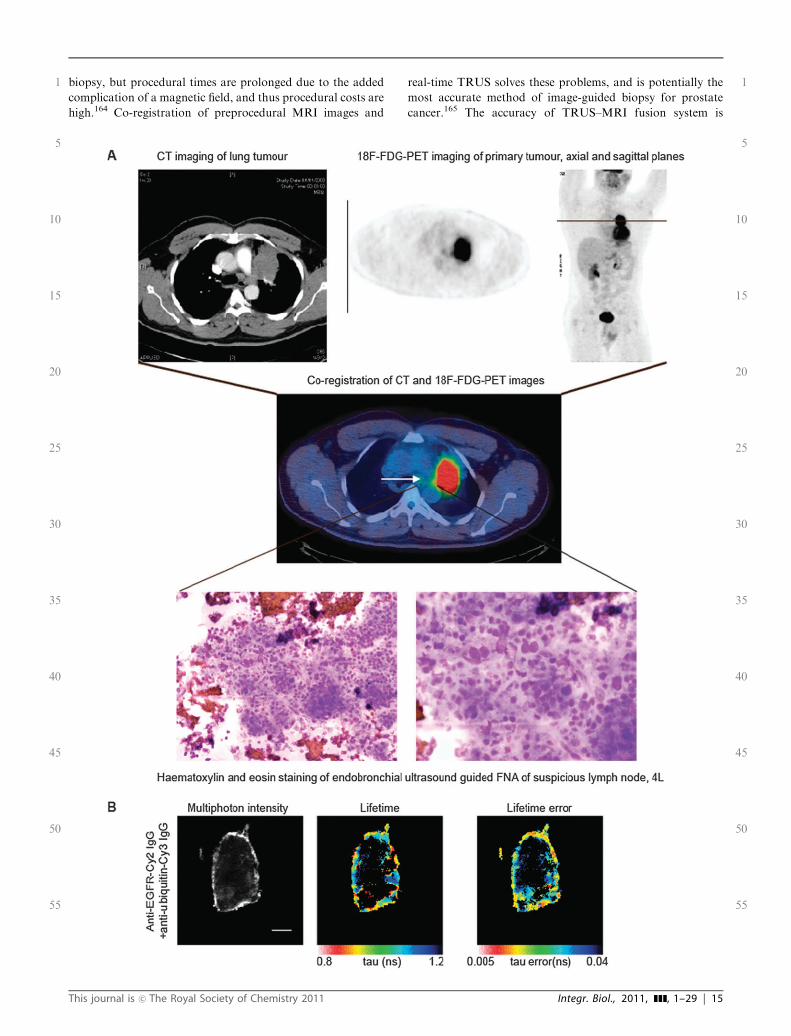
biopsy, but procedural times are prolonged due to the addedcomplication of a magnetic field, and thus procedural costs arehigh.164 Co-registration of preprocedural MRI images and
real-time TRUS solves these problems, and is potentially themost accurate method of image-guided biopsy for prostatecancer.165 The accuracy of TRUS–MRI fusion system is
1
5
10
15
20
25
30
35
40
45
50
55
1
5
10
15
20
25
30
35
40
45
50
55
This journal is !c The Royal Society of Chemistry 2011 Integr. Biol., 2011, ]]], 1–29 | 15

estimated to be 2.4 mm ($1.2 mm), which limits the detectionsize to lesions of 5 # 5 mm.166
These methods have been applied for initial cancer diagnosisand to plan surgery or radiotherapy. However, immunohisto-chemical information from the tissue obtained by image-guided biopsy, may also be used to guide treatment decisionsfor chemotherapy and targeted therapy. For example,percutaneous 18F-FDG-PET–CT guided bone biopsies havebeen shown to change the diagnostic staging, and thus alterplanned treatment in over half of the cancer patientsstudied.167 This group used repeated 18F-FDG-PET–CT scansto position a needle in order to biopsy metabolically activebone lesions which were deemed equivocal on routine stagingwith CT, MRI or 18F-FDG-PET.
Intended treatment was altered in 56% of patients (n= 20),with a variety of tumour types. For example, patients weretreated with palliative rather than curative intent, or withsystemic therapy rather than surgery. Included in this categorywere patients for whom the image-guided biopsies wereinvestigated for hormone receptor or HER2 status by immuno-histochemistry, which helped to decide on the appropriatetreatment with anti-oestrogen drugs and/or trastuzumab. Thisstudy illustrates the benefits of the combination of two imagingmodalities in order to correctly biopsy equivocal sites, which mayimpact on treatment decisions.
Using a similar principle, our group is developing a protocolin order to combine information from pre-procedural18F-FDG-PET scans with real time endobronchial ultrasoundguided transbronchial fine needle aspiration (EBUS-TBNA)of mediastinal and hilar lymph nodes in non-small cell lungcancer (Fig. 1). Smears and cell block preparations ofEBUS-TBNA aspirates can be screened for EGFR mutationsthat can render the tumour drug resistant, such as the T790Mmutation which can confer gefitinib resistance; as well as forquantifying protein–protein interaction of interest, e.g. EGFRubiquitination or EGFR heterodimerisation with HER2(which has been shown to prevent EGFR dephosphorylationand signal termination by endomembrane-bound proteintyrosine phosphatases (PTPs)169) by fluorescence lifetime imaging(FLIM)/Forster resonance energy transfer (FRET) assays (seethe following section). Subcellular imaging may be used toexamine potential di!erences in these signalling events, at siteswhich are positive and negative on imaging with 18F-FDG-PET.The success of treatment strategies may then be monitored (bothspatially and temporally) by the changes in specific tumour cell
mechanisms. In the future, molecular imaging may be carried outon image-guided biopsy material, in order to apply the righttreatment regimen to the right patient at the right time. The mainchallenge to this strategy would be the di"culty in choosing thesite for biopsy and the need to minimise multiple biopsies, andhence the need to improve the accuracy of currently availableimage-guidance techniques.
5.3 Subcellular imaging of pathologicalmechanisms—preclinical applications
Optical imaging, fluorescence and bioluminescence studiesbest describe subcellular protein–protein interactions and/orprotein modifications such as phosphorylation, at a highspatial resolution. For instance, Forster resonance energytransfer (FRET) imaging by fluorescence lifetime imaging(FLIM) has been used to quantify protein–protein interactionsat the nanometre scale, in vitro and in live cells/animals.27–29,170,171 This technique measures energy transferfrom an excited donor fluorophore to an acceptor molecule inclose proximity. In order for FRET to occur, molecules mustbe within nanometre proximity.172 Therefore this techniquemay be used to visualise protein–protein interactions or proteinmodifications within cells. FRET–FLIM technology can be usedto interrogate the malignant proteome, and its response totreatment, thus providing a functional insight which waspreviously the mainstay of analytical biochemistry. An exampleof application of FRET technology is the development of theso-called ‘Picchu’ (phosphorylation indicator of the CrkIIchimeric unit) FRET probe which can be used to monitor EGFRactivity in preclinical models.170 Preclinical experiments using thisFRET sensor demonstrated that the EGFR receptor remainsactive after endocytosis, until translocation to the perinuclearregion. This e!ect may be modulated by its ligand, epidermalgrowth factor (EGF) and by TKIs.Another example of using FRET imaging to monitor, in the
preclinical setting, the pharmacodynamic response to therapyin situ is the application of the probe SCAT3 to monitorcaspase-3 activity during tumour cell apoptosis subsequent tocisplatin or photodynamic (PDT) treatment.90 This techniquewas repeated at varying time-points in order to observe druge!ects on the tumour. Such an assessment of ‘on-target’ druge!ects could greatly improve response assessment. However,currently FRET quantification is limited to superficialtumours. Development of instruments combining endoscopiccellular resolution imaging with technology to quantify
1
5
10
15
20
25
30
35
40
45
50
55
1
5
10
15
20
25
30
35
40
45
50
55
Fig. 1 Endobronchial ultrasound guided transbronchial fine needle aspiration (EBUS-TBNA) to biopsy mediastinal lymph nodes in NSCLC. (A)
Co-registration of images from CT and 18F-FDG-PET combines spatial resolution of CT with the functional capacity of PET in order to stage this
patient with non-small cell lung cancer. The middle image shows FDG uptake in the left upper lobe primary tumour (red spot) and very low FDG
uptake in an adjacent left paratracheal lymph node, as demonstrated by the white arrow. The low FDG uptake in left paratracheal lymph node was
not deemed to be significant but EBUS-guided transbronchial needle aspiration (TBNA) showed evidence of metastatic infiltration by non-small
cell lung cancer (NSCLC). The H&E images (haematoxylin and eosin) show a mixed population of lymphocytes, but with a few groups of atypical
cells. Within these cells, a high nuclear to cytoplasmic ratio, angulated and prominent nucleoli suggest malignant transformation. (B) FRET/FLIM
assays are performed on samples obtained by EBUS-TBNA. EGFR ubiquitination is assessed by measuring FRET between anti-EGFR-Cy2 IgG
and anti-ubiquitin-Cy3 IgG. Interaction between Cy2 and Cy3 results in a shortening of the lifetime of Cy2, as seen in the pseudocolour lifetime
image. FRET e"ciency was calculated using the following equation in each pixel and averaged per each cell. FRET e"ciency = 1 % tda/tcontrol,where tda is the lifetime of cells stained with both anti-EGFR-Cy2 IgG and anti-ubiquitin-Cy3 IgG and tcontrol is the mean anti-EGFR-Cy2 lifetime
measured in the absence of acceptor. The lifetime error image on the far right illustrates the small error margins associated with this approach.
Analysis was done using Bayesian fitting methods.168 White scale bar represents 5 microns.
16 | Integr. Biol., 2011, ]]], 1–29 This journal is !c The Royal Society of Chemistry 2011

fluorescent lifetime and FRET, is underway and will increasepotential clinical applications.173
Although FRET probes may be able to quantify directprotein–protein interactions, within 5–10 nm proximity, thespatial organisation of signalling proteins may be over the10–250 nm scale on endosomal structures (including earlyendosomes to sorting endosomes and multivesicular bodies(MVBs)174). At this length scale, it may be di"cult to quantify,using FRET imaging, the inter-receptor distance withinhomo-oligomers of EGFR,175 and its heterodimer with othersignalling receptors such as the c-Met receptor tyrosinekinase.176,177 The spatial organisation of these receptors mayprovide significant insight into various mechanisms ofresistance, for example anti-EGFR treatment by the acquisitionof MET gene amplification.178 In order to address this issue, anovel technique, is being applied, to map the 3D position ofquantum-dot(QD)-labelled receptors in fixed breast cancer cells,within 3 nm accuracy, as shown in Fig. 2.
Modern super-resolution techniques can resolve thedistance between identical molecules to about 8–10 nm; asignificant improvement upon the conventional limit ofresolution in visible microscopy, 200–250 nm. Two of thetechniques are termed SHRIMP (single-molecule high resolutionimaging with photobleaching) or SHREC (single-molecule high-resolution colocalization). These techniques are both based on
FIONA (fluorescence imaging with one nanometre accuracy),which is able to localise a single fluorophore within 1 nmaccuracy. Despite the small size of the fluorophore (a fewnanometres in width), its position is limited by di!raction whichis approximately 250 nm. This is termed an airy function, andcan be approximated by a Gaussian function, as shown in thelower panel of Fig. 2. Recent advances in detectors allowdetection of the signal from an individual molecule so that thecentroid of the Gaussian function can be located to 1 nmaccuracy.SHRIMP and SHREC utilize FIONA to detect the
di!erence between two nearby Gaussian functions. In the caseof SHREC, one chooses two dyes whose emission spectra arewell separated from each other. Using an appropriate filter set,one can individually detect the location of each dye toFIONA-type accuracy. The di!erence in the centroids is theresolution.180 In SHRIMP, the two dyes are exactly the same, andone relies on one of the identical dyes being turned o!, generallyby random photobleaching. The dye which is still emitting islocated by FIONA to about one nanometre. The location of theoriginal dye is obtained by subtraction of the emissionimmediately after photobleaching, from the image just beforephotobleaching. The resolution is then the di!erence of theposition of the two centroids (Fig. 2). This procedure has beenshown to work for molecules separated by 10–20 nm.179
1
5
10
15
20
25
30
35
40
45
50
55
1
5
10
15
20
25
30
35
40
45
50
55
Fig. 2 Photobleaching as a method of colocalisation with SHRIMP (single-molecule high resolution imaging with photobleaching) based upon
fluorescence imaging with one nanometre accuracy (FIONA) SHRIMP techniques measure the distance between two dyes which are closer than
the di!raction-limit. In the case shown, the dyes can just be resolved; by conventional microscopy, they are 330 nm apart, fit by two Gaussians. In
order to determine this distance by SHRIMP, the sample is illuminated with light. Initially, fluorophores, for example, fluorophore F1 and
fluorophore F2, are bright, in total emitting with 2 units of intensity as shown on the graph in the lower panel (far left). Over time one of the
fluorophores (e.g. F2) photobleaches, the intensity decreases to approximately 1 unit, and one of the Gaussian ‘‘hills’’ disappears, as shown in the
middle graph. The position of the fluorophore which is still emitting, F1, can be determined to a few nanometres by fitting the centroid of the ‘‘hill’’.
The position of F1 can then be calculated by subtracting the image of both emitting—F1 + F2—minus the emission after the photobleaching-F2,
and fitting the centroid, as shown in the graph on the far right. The di!erence in the two centroids, i.e. 326 nm, is the resolution. This has shown to
be e!ective down to 10 nm between two dyes.179
This journal is !c The Royal Society of Chemistry 2011 Integr. Biol., 2011, ]]], 1–29 | 17

Delineation of the spatial organisation of receptors at thesubcellular level may help describe the tumour molecular pheno-type for selection of appropriate therapeutic agents.
5.4 Subcellular imaging of pathological mechanisms—clinicalapplications
We have established FRET–FLIM assays in cell line models ofcancer, fresh human tissues and formalin-fixed para"n-embedded tissue (FFPE), as well as dynamic deep tissueimaging of cancer cells in murine models.28 Fig. 3 demon-strates the translation of an in vitro protein–protein assay,measuring EGFR ubiquitination, in cell lines, to a dualantibody-based assay for quantification of EGFR ubiquitinationin tissue, using FRET/FLIM technology. These images demon-strate in vitro assessment of functional EGFR modifications, i.e.ubiquitination, which is associated with downregulation anddegradation of this receptor.129,181 Cell lines with varyingsusceptibility to EGFR degradation (panel B), mimic varyingtumour phenotypes. Thus, translation of these assays for use inpatient tumour tissue may delineate a group of patients who aremore likely to respond to drugs manipulating this pathway.
Besides whole tumour sections or image-guided biopsymaterial (Fig. 1), clinically these FRET–FLIM assays couldalso be applied to single metastatic cells, or the disseminating/circulating tumour cells (DTC/CTC), which confer a poorprognostic outcome in epithelial carcinomas, such as breast,lung and prostate.182 The current validated methods fordetection of DTCs rely on blood or bone sampling prior toimmunocytochemical or molecular analysis.183 Bone marrowinvolvement delineates the metastatic group more accuratelyin breast cancer patients, but bone marrow biopsy is invasive andthe cells obtained are often not viable.184 Circulating cancertumour cells in the blood have been identified by a varietyof immunological approaches, including identification ofepithelium-specific antigens, e.g. cytoskeleton-associatedcytokeratins, surface adhesion molecules, or growth factorreceptors, and by molecular PCR-based techniques. Thepresence or absence of DTCs in the blood both before and aftertreatment has been shown to correlate with treatmentresponse.185,186 Furthermore, the presence or absence ofradiological signs of disease progression can be combined withCTC counts in blood to improve the prediction of overall survivalin metastatic breast cancer patients undergoing therapy.186
The presence of micrometastases in the form of DTCs mayconfer a poor prognosis as the cells may exhibit the samecharacteristics as cancer stem cells. They are resistant totherapy disseminate and grow at alternative sites and expressmany of the same surface markers as cancer stem cells, e.g.CD44, cytokeratin 19, and EpCAM.187,188
Molecular signatures obtained from the analysis of CTCshave also highlighted certain markers which may predicttumour dormancy.188 Tumour dormancy is a phenomenonby which tumour cells evade eradication to become activemany years later. The immune system has been demonstratedto play an important role in both animal models and inpatients. For example, T-cell activation is strongly correlatedwith overall survival in patients with colon cancer, indepen-dent of primary tumour size or nodal status.189 Further
research is being carried out in order to characterise themalignant phenotype of these cells. This information maythen lead to clues as to how to target these resistant cells,and thus eradicate minimal residual disease. For example,DTCs in the bone marrow have been shown to overexpressurokinase-type plasminogen activator receptor and the extra-cellular matrix metalloproteinase inducer.190 These targets areamenable to both imaging and for therapeutic potential.Imaging modalities which can monitor these cells in vivo wouldbe of value to accurately gauge risk of relapse, and requirementfor adjuvant treatment.
5.5 Signalling networks to identify optimal drug combinations
The response to treatment and the number of therapiesrequired to eradicate a tumour has been mathematicallymodelled and integrated into signalling networks. These net-works take into account biological tumour characteristics,such as the level of cell turnover, the rate of mutations,increased tumour size, and biological fitness for clonal expansion,as described mainly, by in vitro research.191 Information derivedfrom in vivo molecular imaging techniques could feed into thesenetworks and greatly improve their capacity for prediction oftreatment response and to design optimal drug combinations.For example, a breast cancer patient who had kinome
sequencing that revealed approximately 100 000 somatic pointmutations would need 4–5 non-cross-resistant drugs to battleher cancer, according to this model.192 Computational modellinghas been used to create receptor tyrosine kinase co-activationnetworks to model the complex and dynamic interactionsinvolved in chemoresistance.193 Receptor co-activation describesthe simultaneous activation of two or more receptor tyrosinekinases in order to maintain robust intracellular signalling in theface of perturbations. For example, resistance to trastuzumabmay be conferred by the presence of a hetero-trimer of HER2,HER3 and insulin-like growth factor receptor (IGF-R).194 Simi-larly, co-activation of c-Met and EGFR leads to resistance toEGFR-tyrsoine kinase inhibition by, e.g. gefitinib.178 However, acombination strategy using inhibitors of c-Met, EGFR, andplatelet-derived growth factor receptor (PDGFR) is successfulin reducing cell viability compared to single-agent treatment, asmultiple co-activators are targeted.195 Although this is a viableapproach to chemoresistance, such a cocktail of drugs is unlikelyto be tolerated in patients. The attractiveness of such networks isthe ability to identify fragile points downstream of the activatorswhich may be specifically targeted to overcome resistance.Recently HER3 was identified as an example of a fragile node,as well as a co-activator, in animal models.176 However, thechallenge in translation of these models for clinical use lies in thepaucity of signalling data in humans. Networks constructed thusfar rely on in vitro or biochemical analysis of tumour tissue. Invivo subcellular imaging techniques are likely to be the next stepin providing the data required to further annotate these networkmaps for clinical use.Conversely, signalling networks developed in vitro have the
potential to identify novel imaging biomarkers for resistanceor response, in order to optimise the use of targeted treatments(a more detailed description of this approach can be found inref. 196). Table 1 illustrates a selection of the many and varied
1
5
10
15
20
25
30
35
40
45
50
55
1
5
10
15
20
25
30
35
40
45
50
55
18 | Integr. Biol., 2011, ]]], 1–29 This journal is !c The Royal Society of Chemistry 2011

biomarkers which may enter the clinical arena to optimisecancer therapy. However, the question of which biomarkershould be imaged for a particular patient remains unanswered.For example, an EGFR-centred protein network has been
constructed and probed using small interfering RNA (siRNA),in order to highlight protein–protein interactions which maycontribute to resistance or sensitivity to cancer therapy.33
Several proteins of interest were identified as potential
1
5
10
15
20
25
30
35
40
45
50
55
1
5
10
15
20
25
30
35
40
45
50
55
This journal is !c The Royal Society of Chemistry 2011 Integr. Biol., 2011, ]]], 1–29 | 19

regulators of response to treatment and therefore, may becandidates for development as imaging biomarkers. However,the complexity and robustness of protein signalling networkscombined with the intrinsic error rate associated with siRNAscreens means that these potential targets require thoroughvalidation in vivo prior to translation to the clinic.
In addition to application to signalling networks, clinicalimaging modalities may provide prognostic informationconcurrent with established prognostic tools, such as geneexpression signatures, in order to construct a mathematicalmodel for outcome prediction. The development of microarray-based gene expression signatures has enabled classification oftumour subtypes and association with clinical outcome, notablyin breast cancer.19,197 A 70-gene prognostic signature for lymphnode negative breast cancer patients is reported to provideprognostic information independent of clinicopathological scoresand with improved sensitivity and specificity for poor clinicaloutcome.198,199 A validation study of the 70-gene signaturereported 90% sensitivity for metastasis within 5 years, forexample, with specificity of 42%.199 The tumour heterogeneityobserved within intrinsic molecular subtypes of breast cancer isbeginning to be described as deregulated molecular pathways atthe gene expression level.200 Clinical imaging traits have beenused to partially reconstruct gene expression variation betweentumours.201 Association maps between clinical imaging featuresand gene expression variation have been constructed for CTimage traits in hepatocellular carcinoma202 and MRI traits inglioblastoma multiforme,203 suggesting that imaging traits canapproximately predict gene expression variation betweentumours.
6. Conclusions
We have summarised a variety of potential applications formolecular imaging, ranging from the nanometre to the wholebody scale, in the optimisation of cancer therapy; i.e. choosingthe right drug for the right patient at the right time. We havealso discussed some of the challenges faced in the integrationof molecular imaging into clinical practice.
Molecular imaging at a whole body level is unlikelyto be possible using a single imaging modality. Tumourheterogeneity and a di!erential response to treatmentrepresent a few of many characteristics influencing theeventual tumour phenotype, and thus, response to cancer
therapeutics. Whole body imaging is the only techniquecurrently available for the study of both primary tumourand metastases, both at diagnosis and to monitor responseto treatment, but may not provide vital information at themolecular level. Image-guided biopsy, co-registration ofcomplementary imaging modalities and appropriate bio-marker choice may provide optimal risk stratification andhelp overcome these challenges. The issues of radiationdose and financial cost are yet to be fully addressed.Appropriate choice of imaging technology, the combinationof modalities utilising ionising and non-ionising radiativesources and signal amplification may help alleviate someof the burden. Common quality assurance and controlmethods need to be developed in order to ensure a standardfor imaging which may impact on treatment choice. Thisbecomes increasingly di"cult with rapidly evolving technologyand whilst crossing national boundaries but is an area whichrequires future consideration.Clinical outcome and response to treatment has a
complex and multifaceted relationship to genotype andgene expression, dysregulation of signalling pathwaysvia gene expression, protein activity, protein–proteininteractions and tumour phenotypic traits. It may bepossible to extract this data from patients using molecularimaging amongst other established techniques. However,the challenge is to integrate datasets from each observablelevel of variation, from genotype to tumour phenotype, inorder to inform clinical management for the individualpatient.
Acknowledgements
The authors would like to thank M. Keppler and A. Tutt fortheir assistance in writing this review. Q3
Notes and references
1 Q4J. A. DiMasi, R. W. Hansen and H. G. Grabowski, The price ofinnovation: new estimates of drug development costs, J. HealthEcon., 2003, 22, 151–85, DOI: 10.1016/S0167-6296(02)00126-1.
2 M. Fukuoka, S. Yano, G. Giaccone, T. Tamura, K. Nakagawa,J. Y. Douillard, Y. Nishiwaki, J. Vansteenkiste, S. Kudoh,D. Rischin, R. Eek, T. Horai, K. Noda, I. Takata, E. Smit,S. Averbuch, A. Macleod, A. Feyereislova, R. P. Dong andJ. Baselga, Multi-institutional randomized phase II trialof gefitinib for previously treated patients with advanced
1
5
10
15
20
25
30
35
40
45
50
55
1
5
10
15
20
25
30
35
40
45
50
55
Fig. 3 Multiphoton FLIM measurements of the intermolecular FRET between ErbB1-GFP and ubiquitin-mRFP1. A. MCF cells were
transiently transfected with EGFR-eGFP in the presence or absence of mRFP1-ubiquitin to assess EGFR ubiquitination using a FRET-by-
FLIM assay. Cells were either EGF treated (100 ng ml%1, 30 min) or not. FRET between GFP and mRFP1 results in shortening of the fluorescence
lifetime (t) of GFP. FRET e"ciency was calculated using the following equation in each pixel and averaged per each cell. FRET e"ciency =
1 % tda/tcontrol, where tda is the fluorescence lifetime of cells co-expressing both EGFR-EGFP and mRFP1-ubiquitin and tcontrol is the mean
EGFR-EGFP lifetime measured in the absence of acceptor. B. Endogenous EGFR ubiquitination is assessed in the breast cancer cell lines, HCC
and MDA-MB-468. EGFR ubiquitination is increased in cells which highly degrade EGFR on EGF treatment (HCC) as opposed to those which
degrade EGFR less readily (MDA-MB-468). This is shown by the pseudocolour fluorescence lifetime maps. Here, FRET was measured between
Cy2 and m-RFP. (C) Translation of the above FRET-by-FLIM assays to dual fluorophore-labelled antibody assays for application to endogenous
protein interactions in tumour tissue. These Figures show the initial assessment of FRET e"ciency between anti-EGFR-Cy2 and anti-ubiquitin-
Cy3 in A431 cancer cells (with the established methodologies being applied to patient-derived cancer tissues). Anti-ubiquitin-Cy3
IgG concentration is at control level at 1ug ml%1, whereby no ubiquitin staining is seen (epifluorescence Cy3 image), versus optimal
concentration (10 ug ml%1), whereby a reduction in fluorescence lifetime of Cy2 is seen on the pseudocolour lifetime images. White scale bars
represent 5 microns.
20 | Integr. Biol., 2011, ]]], 1–29 This journal is !c The Royal Society of Chemistry 2011
non-small-cell lung cancer (The IDEAL 1 Trial) [corrected],J. Clin. Oncol., 2003, 21, 2237–46; E. E. Cohen, F. Rosen,W. M. Stadler, W. Recant, K. Stenson, D. Huo andE. E. Vokes, Phase II trial of ZD1839 in recurrent or metastaticsquamous cell carcinoma of the head and neck, J. Clin. Oncol.,2003, 21, 1980–7; L. B. Saltz, N. J. Meropol, P. J. Loehrer, Sr.,M. N. Needle, J. Kopit and R. J. Mayer, Phase II trial ofcetuximab in patients with refractory colorectal cancer thatexpresses the epidermal growth factor receptor, J. Clin. Oncol.,2004, 22, 1201–8; R. Perez-Soler, A. Chachoua, L. A. Hammond,E. K. Rowinsky, M. Huberman, D. Karp, J. Rigas, G. M. Clark,P. Santabarbara and P. Bonomi, Determinants of tumor responseand survival with erlotinib in patients with non-small-cell lungcancer, J. Clin. Oncol., 2004, 22, 3238–47.
3 L. Toschi and F. Cappuzzo, Understanding the new genetics ofresponsiveness to epidermal growth factor receptor tyrosinekinase inhibitors, Oncologist, 2007, 12, 211–20, DOI: 10.1634/theoncologist.12-2-211.
4 F. Cappuzzo, V. Gregorc, E. Rossi, A. Cancellieri, E. Magrini,C. T. Paties, G. Ceresoli, L. Lombardo, S. Bartolini, C. Calandri,M. de Rosa, E. Villa and L. Crino, Gefitinib in pretreatednon-small-cell lung cancer (NSCLC): analysis of e"cacy andcorrelation with HER2 and epidermal growth factor receptorexpression in locally advanced or metastatic NSCLC, J. Clin.Oncol., 2003, 21, 2658–63, DOI: 10.1200/JCO.2003.01.039;H. S. Parra, R. Cavina, F. Latteri, P. A. Zucali,E. Campagnoli, E. Morenghi, G. C. Grimaldi, M. Roncalli andA. Santoro, Analysis of epidermal growth factor receptorexpression as a predictive factor for response to gefitinib (‘Iressa’,ZD1839) in non-small-cell lung cancer, Br. J. Cancer, 2004, 91,208–12, DOI: 10.1038/sj.bjc.6601923.
5 J. G. Paez, P. A. Janne, J. C. Lee, S. Tracy, H. Greulich,S. Gabriel, P. Herman, F. J. Kaye, N. Lindeman, T. J. Boggon,K. Naoki, H. Sasaki, Y. Fujii, M. J. Eck, W. R. Sellers,B. E. Johnson and M. Meyerson, EGFR mutations in lungcancer: correlation with clinical response to gefitinib therapy,Science, 2004, 304, 1497–500, DOI: 10.1126/science.1099314;T. J. Lynch, D. W. Bell, R. Sordella, S. Gurubhagavatula,R. A. Okimoto, B. W. Brannigan, P. L. Harris, S. M. Haserlat,J. G. Supko, F. G. Haluska, D. N. Louis, D. C. Christiani,J. Settleman and D. A. Haber, Activating mutations in theepidermal growth factor receptor underlying responsiveness ofnon-small-cell lung cancer to gefitinib, N. Engl. J. Med., 2004,350, 2129–39, DOI: 10.1056/NEJMoa040938.
6 Z. Chen, J. Feng, J. S. Saldivar, D. Gu, A. Bockholt andS. S. Sommer, EGFR somatic doublets in lung cancer arefrequent and generally arise from a pair of driver mutationsuncommonly seen as singlet mutations: one-third of doubletsoccur at five pairs of amino acids, Oncogene, 2008, 27, 4336–43,DOI: 10.1038/onc.2008.71.
7 A. Hochhaus, S. Kreil, A. S. Corbin, P. La Rosee, M. C. Muller,T. Lahaye, B. Hanfstein, C. Schoch, N. C. Cross, U. Berger,H. Gschaidmeier, B. J. Druker and R. Hehlmann, Molecular andchromosomal mechanisms of resistance to imatinib (STI571)therapy, Leukemia, 2002, 16, 2190–6, DOI: 10.1038/sj.leu.2402741.
8 M. W. Teng, J. B. Swann, C. M. Koebel, R. D. Schreiber andM. J. Smyth, Immune-mediated dormancy: an equilibrium withcancer, J. Leukocyte Biol., 2008, 84, 988–93, DOI: 10.1189/jlb.1107774.
9 L. Fass, Imaging and cancer: a review, Mol. Oncol., 2008, 2,115–52, DOI: 10.1016/j.molonc.2008.04.001.
10 J. K. Willmann, N. van Bruggen, L. M. Dinkelborg andS. S. Gambhir, Molecular imaging in drug development,Nat. Rev. Drug Discovery, 2008, 7, 591–607, DOI: 10.1038/nrd2290.
11 K. H. Bohuslavizki, Somatostatin receptor imaging: currentstatus and future perspectives, J. Nucl. Med., 2001, 42,1057–8.
12 V. Ambrosini, D. Campana, P. Tomassetti, G. Grassetto,D. Rubello and S. Fanti, PET/CT with 68Gallium-DOTA-peptides in NET: An overview, Eur. J. Radiol., 2010, DOI:10.1016/j.ejrad.2010.07.022.
13 D. J. Kwekkeboom, B. L. Kam, M. van Essen, J. J. Teunissen,C. H. van Eijck, R. Valkema, M. de Jong, W. W. de Herder andE. P. Krenning, Somatostatin-receptor-based imaging and
therapy of gastroenteropancreatic neuroendocrine tumors, Endocr.Relat. Cancer, 2009, 17, R53–73, DOI: 10.1677/ERC-09-0078.
14 A. M. Groves, T. Win, S. B. Haim and P. J. Ell, Non-[18F]FDGPET in clinical oncology, Lancet Oncol., 2007, 8, 822–30, DOI:10.1016/S1470-2045(07)70274-7.
15 G. Bertolini, L. Roz, P. Perego, M. Tortoreto, E. Fontanella,L. Gatti, G. Pratesi, A. Fabbri, F. Andriani, S. Tinelli, E. Roz,R. Caserini, S. Lo Vullo, T. Camerini, L. Mariani, D. Delia,E. Calabro, U. Pastorino and G. Sozzi, Highly tumorigenic lungcancer CD133+ cells display stem-like features and are spared bycisplatin treatment, Proc. Natl. Acad. Sci. U. S. A., 2009, 106,16281–6, DOI: 10.1073/pnas.0905653106.
16 S. Nimmagadda, M. Pullambhatla, K. Stone, G. Green,Z. M. Bhujwalla and M. G. Pomper, Molecular imagingof CXCR4 receptor expression in human cancer xenograftswith [64Cu]AMD3100 positron emission tomography, CancerRes., 2010, 70, 3935–44, DOI: 10.1158/0008-5472.CAN-09-4396.
17 T. F. Massoud and S. S. Gambhir, Molecular imaging in livingsubjects: seeing fundamental biological processes in a new light,Genes Dev., 2003, 17, 545–80, DOI: 10.1101/gad.1047403.
18 U. Wilking, E. Karlsson, L. Skoog, T. Hatschek, E. Lidbrink,G. Elmberger, H. Johansson, L. Lindstrom and J. Bergh, HER2status in a population-derived breast cancer cohort: discordancesduring tumor progression, Breast Cancer Res. Treat., 2010,125, 553, DOI: 10.1007/s10549-010-1029-2.
19 C. Sotiriou and L. Pusztai, Gene-expression signatures in breastcancer, N. Engl. J. Med., 2009, 360, 790–800, DOI: 10.1056/NEJMra0801289.
20 D. C. Allred, Y. Wu, S. Mao, I. D. Nagtegaal, S. Lee,C. M. Perou, S. K. Mohsin, P. O’Connell, A. Tsimelzon andD. Medina, Ductal carcinoma in situ and the emergence ofdiversity during breast cancer evolution, Clin. Cancer Res.,2008, 14, 370–8, DOI: 10.1158/1078-0432.CCR-07-1127;W. J. Lennington, R. A. Jensen, L. W. Dalton and D. L. Page,Ductal carcinoma in situ of the breast. Heterogeneity of individuallesions, Cancer, 1994, 73, 118–24.
21 X. Chen, K. L. Reichenbach and C. Xu, Experimental andtheoretical analysis of core-to-core coupling on fiber bundleimaging, Opt. Express, 2008, 16, 21598–607, DOI: 10.1364/OE.16.021598; H. Inoue, S. E. Kudo and A. Shiokawa,Technology insight: Laser-scanning confocal microscopy andendocytoscopy for cellular observation of the gastrointestinaltract, Nat. Clin. Pract. Gastroenterol. Hepatol., 2005, 2, 31–7,DOI: 10.1038/ncpgasthep0072.
22 J. Baselga and S. M. Swain, Novel anticancer targets: revisitingERBB2 and discovering ERBB3, Nat. Rev. Cancer, 2009, 9,463–75, DOI: 10.1038/nrc2656.
23 H. Fan-Minogue, Z. Cao, R. Paulmurugan, C. T. Chan,T. F. Massoud, D. W. Felsher and S. S. Gambhir, Noninvasivemolecular imaging of c-Myc activation in living mice, Proc. Natl.Acad. Sci. U. S. A., 2010, 107, 15892, DOI: 10.1073/pnas.1007443107; A. De, P. Ray, A. M. Loening andS. S. Gambhir, BRET3: a red-shifted bioluminescence resonanceenergy transfer (BRET)-based integrated platform for imagingprotein–protein interactions from single live cells and livinganimals, FASEB J., 2009, 23, 2702–9, DOI: 10.1096/fj.08-118919.
24 T. F. Massoud, R. Paulmurugan and S. S. Gambhir, A molecularlyengineered split reporter for imaging protein–protein interactionswith positron emission tomography, Nat. Med., 2010, 16, 921–6,DOI: 10.1038/nm.2185.
25 G. Tarcic, S. K. Boguslavsky, J. Wakim, T. Kiuchi, A. Liu,F. Reinitz, D. Nathanson, T. Takahashi, P. S. Mischel, T. Ng andY. Yarden, An unbiased screen identifies DEP-1 tumor suppressor asa phosphatase controlling EGFR endocytosis, Curr. Biol., 2009, 19,1788–98; M. Parsons, J. Monypenny, S. M. Ameer-Beg,T. H. Millard, L. M. Machesky, M. Peter, M. D. Keppler,G. Schiavo, R. Watson, J. Cherno!, D. Zicha, B. Vojnovic andT. Ng, Spatially distinct binding of Cdc42 to PAK1 and N-WASP inbreast carcinoma cells, Mol. Cell. Biol., 2005, 25, 1680–95, DOI:10.1128/MCB.25.5.1680-1695.2005; R. F. Carvalho, M. Beutler,K. J. Marler, B. Knoll, E. Becker-Barroso, R. Heintzmann, T. Ngand U. Drescher, Silencing of EphA3 through a cis interaction withephrinA5, Nat. Neurosci., 2006, 9, 322–30; D. Romano,D. Matallanas, G. Weitsman, C. Preisinger, T. Ng and W. Kolch,
1
5
10
15
20
25
30
35
40
45
50
55
1
5
10
15
20
25
30
35
40
45
50
55
This journal is !c The Royal Society of Chemistry 2011 Integr. Biol., 2011, ]]], 1–29 | 21

Proapoptotic Kinase MST2 Coordinates Signaling Crosstalkbetween RASSF1A, Raf-1, and Akt, Cancer Res., 2010, 70,1195–203; T. Ng, D. Shima, A. Squire, P. I. Bastiaens,S. Gschmeissner, M. J. Humphries and P. J. Parker, PKCalpharegulates beta1 integrin-dependent cell motility through associationand control of integrin tra"c, EMBO J., 1999, 18, 3909–23, DOI:10.1093/emboj/18.14.3909; T. Niault, I. Sobczak, K. Meissl,G. E. Weitsman, D. Piazzolla, K. Ehrenreiter, M. Hamerl,I. Moarefi, T. Leung, O. Carugo, T. Ng and M. Baccarini, FromAutoinhibition to Inhibition in Trans: the Raf-1 Regulatory DomainInhibits Rok-a Kinase Activity, J. Cell Biol., 2009, 187, 335–342;N. Anilkumar, M. Parsons, R. Monk, T. Ng and J. C. Adams,Interaction of fascin and protein kinase Calpha: a novel intersectionin cell adhesion and motility, EMBO J., 2003, 22, 5390–402, DOI:10.1093/emboj/cdg521; K. Makrogianneli, L. M. Carlin,M. D. Keppler, D. R. Matthews, E. Ofo, A. Coolen,S. M. Ameer-Beg, P. R. Barber, B. Vojnovic and T. Ng, Integratingreceptor signal inputs that influence small Rho GTPase activationdynamics at the immunological synapse, Mol. Cell. Biol., 2009, 29,2997–3006; L. M. Carlin, K. Makrogianneli, M. Keppler,G. O. Fruhwirth and T. Ng, Visualisation of signalling in immunecells, Methods Mol. Biol., 2010, 616, 97–113, DOI: 10.1007/978-1-60761-461-6_7; D. R. Matthews, L. M. Carlin, E. Ofo, P. R. Barber,B. Vojnovic, M. Irving, T. Ng and S. M. Ameer-Beg, Time-lapseFRET microscopy using fluorescence anisotropy, J. Microsc., 2010,237, 51–62, DOI: 10.1111/j.1365-2818.2009.03301.x; M. Beutler,K. Makrogianneli, R. J. Vermeij, M. Keppler, T. Ng, T. M. Jovinand R. Heintzmann, satFRET: estimation of Forster resonanceenergy transfer by acceptor saturation, Eur. Biophys. J., 2008, 38,69–82, DOI: 10.1007/s00249-008-0361-5; S. J. Heasman,L. M. Carlin, S. Cox, T. Ng and A. J. Ridley, Coordinated RhoAsignaling at the leading edge and uropod is required for T celltransendothelial migration, J. Cell Biol., 2010, 190, 553–63, DOI:10.1083/jcb.201002067.
26 E. M. Bublil, G. Pines, G. Patel, G. Fruhwirth, T. Ng andY. Yarden, Kinase-mediated quasi-dimers of EGFR, FASEB J.,2010, 24, 4744, DOI: 10.1096/fj.10-166199.
27 T. Ng, M. Parsons, W. E. Hughes, J. Monypenny, D. Zicha,A. Gautreau, M. Arpin, S. Gschmeissner, P. J. Verveer,P. I. Bastiaens and P. J. Parker, Ezrin is a downstreame!ector of tra"cking PKC-integrin complexes involved inthe control of cell motility, EMBO J., 2001, 20, 2723–41, DOI:10.1093/emboj/20.11.2723; J. W. Legg, C. A. Lewis, M.Parsons, T. Ng and C. M. Isacke, A novel PKC-regulatedmechanism controls CD44 ezrin association and directionalcell motility, Nat. Cell Biol., 2002, 4, 399–407, DOI: 10.1038/ncb797.
28 M. T. Kelleher, G. Fruhwirth, G. Patel, E. Ofo, F. Festy,P. R. Barber, S. M. Ameer-Beg, B. Vojnovic, C. Gillett,A. Coolen, G. Keri, P. A. Ellis and T. Ng, The potential ofoptical proteomic technologies to individualize prognosis andguide rational treatment for cancer patients, Targeted Oncol.,2009, 4, 235–52, DOI: 10.1007/s11523-009-0116-y.
29 T. Ng, A. Squire, G. Hansra, F. Bornancin, C. Prevostel,A. Hanby, W. Harris, D. Barnes, S. Schmidt, H. Mellor,P. I. Bastiaens and P. J. Parker, Imaging protein kinase Calphaactivation in cells, Science, 1999, 283, 2085–9.
30 H. J. Choi, W. Ju, S. K. Myung and Y. Kim, Diagnosticperformance of computer tomography, magnetic resonanceimaging, and positron emission tomography or positron emissiontomography/computer tomography for detection of metastaticlymph nodes in patients with cervical cancer: meta-analysis,Cancer Sci., 2010, 101, 1471–9, DOI: 10.1111/j.1349-7006.2010.01532.x; T. C. Kwee and R. M. Kwee, CombinedFDG-PET/CT for the detection of unknown primary tumors:systematic review and meta-analysis, Eur. Radiol., 2009, 19,731–44, DOI: 10.1007/s00330-008-1194-4.
31 L. Pan, P. Gu, G. Huang, H. Xue and S. Wu, Prognosticsignificance of SUV on PET/CT in patients with esophagealcancer: a systematic review and meta-analysis, Eur. J.Gastroenterol. Hepatol., 2009, 21, 1008–15, DOI: 10.1097/MEG.0b013e328323d6fa; H. M. Quarles van U!ord, H. vanTinteren, S. G. Stroobants, Riphagen, II and O. S. Hoekstra,Added value of baseline 18F-FDG uptake in serial 18F-FDG PETfor evaluation of response of solid extracerebral tumors to
systemic cytotoxic neoadjuvant treatment: a meta-analysis,J. Nucl. Med., 2010, 51, 1507–16, DOI: 10.2967/jnumed.110.075457; L. S. Poulou, L. Thanos and P. D. Ziakas,Unifying the predictive value of pretransplant FDG PET inpatients with lymphoma: a review and meta-analysis of publishedtrials, Eur. J. Nucl. Med. Mol. Imaging, 2009, 37, 156–62, DOI:10.1007/s00259-009-1258-y.
32 M. Paesmans, T. Berghmans, M. Dusart, C. Garcia, C.Hossein-Foucher, J. J. Lafitte, C. Mascaux, A. P. Meert,M. Roelandts, A. Scherpereel, V. Terrones Munoz andJ. P. Sculier, Primary tumor standardized uptake value measuredon fluorodeoxyglucose positron emission tomography is ofprognostic value for survival in non-small cell lung cancer: updateof a systematic review and meta-analysis by the European LungCancer Working Party for the International Association for theStudy of Lung Cancer Staging Project, J. Thorac. Oncol., 2010, 5,612–9, DOI: 10.1097/JTO.0b013e3181d0a4f5.
33 I. Astsaturov, V. Ratushny, A. Sukhanova, M. B. Einarson,T. Bagnyukova, Y. Zhou, K. Devarajan, J. S. Silverman,N. Tikhmyanova, N. Skobeleva, A. Pecherskaya, R. E. Nasto,C. Sharma, S. A. Jablonski, I. G. Serebriiskii, L. M. Weiner andE. A. Golemis, Synthetic lethal screen of an EGFR-centerednetwork to improve targeted therapies, Sci. Signaling, 2010,3, ra67, DOI: 10.1126/scisignal.2001083.
34 W. A. Weber, Use of PET for monitoring cancer therapy and forpredicting outcome, J. Nucl. Med., 2005, 46, 983–95.
35 R. Boellaard, M. J. O’Doherty, W. A. Weber, F. M. Mottaghy,M. N. Lonsdale, S. G. Stroobants, W. J. Oyen, J. Kotzerke,O. S. Hoekstra, J. Pruim, P. K. Marsden, K. Tatsch,C. J. Hoekstra, E. P. Visser, B. Arends, F. J. Verzijlbergen,J. M. Zijlstra, E. F. Comans, A. A. Lammertsma, A. M.Paans, A. T. Willemsen, T. Beyer, A. Bockisch, C. Schaefer-Prokop, D. Delbeke, R. P. Baum, A. Chiti and B. J. Krause,FDG PET and PET/CT: EANM procedure guidelines fortumour PET imaging: version 1.0, Eur. J. Nucl. Med.Mol. Imaging, 2009, 37, 181–200, DOI: 10.1007/s00259-009-1297-4.
36 A. Szafer, J. Zhong, A. W. Anderson and J. C. Gore,Di!usion-weighted imaging in tissues: theoretical models, NMRBiomed., 1995, 8, 289–96; C. H. Sotak, Nuclear magneticresonance (NMR) measurement of the apparent di!usioncoe"cient (ADC) of tissue water and its relationship to cellvolume changes in pathological states, Neurochem. Int., 2004,45, 569–82, DOI: 10.1016/j.neuint.2003.11.010; A. R. Padhani,G. Liu, D. M. Koh, T. L. Chenevert, H. C. Thoeny, T. Takahara,A. Dzik-Jurasz, B. D. Ross, M. Van Cauteren, D. Collins,D. A. Hammoud, G. J. Rustin, B. Taouli and P. L. Choyke,Di!usion-weighted magnetic resonance imaging as a cancerbiomarker: consensus and recommendations, Neoplasia, 2009,11, 102–25.
37 R. G. Kierkels, W. H. Backes, M. H. Janssen, J. Buijsen,R. G. Beets-Tan, P. Lambin, G. Lammering, M. C. Oellers andH. J. Aerts, Comparison between perfusion computed tomo-graphy and dynamic contrast-enhanced magnetic resonanceimaging in rectal cancer, Int. J. Radiat. Oncol., Biol., Phys.,2010, 77, 400–8, DOI: 10.1016/j.ijrobp.2009.05.015; V. Goh,M. Dattani, J. Farwell, J. Shekhdar, E. Tam, S. Patel, J. Juttla,I. Simcock, J. Stirling, H. Mandeville, E. Aird and P. Hoskin,Radiation dose from volumetric helical perfusion CT of thethorax, abdomen or pelvis, Eur. Radiol., 2010, DOI: 10.1007/s00330-010-1997-y.
38 S. M. Eschmann, F. Paulsen, M. Reimold, H. Dittmann, S. Welz,G. Reischl, H. J. Machulla and R. Bares, Prognostic impact ofhypoxia imaging with 18F-misonidazole PET in non-small celllung cancer and head and neck cancer before radiotherapy,J. Nucl. Med., 2005, 46, 253–60.
39 H. Schoder and M. Gonen, Screening for cancer with PET andPET/CT: potential and limitations, J. Nucl. Med., Suppl., 2007,48(Suppl 1), 4S–18S.
40 M. E. Juweid and B. D. Cheson, Positron-emission tomographyand assessment of cancer therapy, N. Engl. J. Med., 2006, 354,496–507, DOI: 10.1056/NEJMra050276; P. Seam, M. E. Juweidand B. D. Cheson, The role of FDG-PET scans in patients withlymphoma, Blood, 2007, 110, 3507–16, DOI: 10.1182/blood-2007-06-097238.
1
5
10
15
20
25
30
35
40
45
50
55
1
5
10
15
20
25
30
35
40
45
50
55
22 | Integr. Biol., 2011, ]]], 1–29 This journal is !c The Royal Society of Chemistry 2011

41 R. J. Downey, T. Akhurst, M. Gonen, A. Vincent, M. S. Bains,S. Larson and V. Rusch, Preoperative F-18 fluorodeoxyglucose-positron emission tomography maximal standardized uptakevalue predicts survival after lung cancer resection, J. Clin. Oncol.,2004, 22, 3255–60, DOI: 10.1200/JCO.2004.11.109.
42 K. A. Miles, Functional CT imaging in oncology, Eur. J. Radiol.,2003, 13(Suppl 5), M134–8.
43 Q. S. Ng, V. Goh, H. Fichte, E. Klotz, P. Fernie, M. I. Saunders,P. J. Hoskin and A. R. Padhani, Lung cancer perfusion atmulti-detector row CT: reproducibility of whole tumor quantitativemeasurements, Radiology, 2006, 239, 547–53, DOI: 10.1148/radiol.2392050568.
44 J. Yee, G. A. Akerkar, R. K. Hung, A. M. Steinauer-Gebauer,S. D. Wall and K. R. McQuaid, Colorectal neoplasia:performance characteristics of CT colonography for detectionin 300 patients, Radiology, 2001, 219, 685–92.
45 E. D. Pisano, C. Gatsonis, E. Hendrick, M. Ya!e, J. K. Baum,S. Acharyya, E. F. Conant, L. L. Fajardo, L. Bassett,C. D’Orsi, R. Jong and M. Rebner, Diagnostic performance ofdigital versus film mammography for breast-cancer screening,N. Engl. J. Med., 2005, 353, 1773–83, DOI: 10.1056/NEJMoa052911.
46 C. H. Holdsworth, R. D. Badawi, J. B. Manola, M. F. Kijewski,D. A. Israel, G. D. Demetri and A. D. Van den Abbeele, CT andPET: early prognostic indicators of response to imatinib mesylatein patients with gastrointestinal stromal tumor, AJR, Am. J.Roentgenol., 2007, 189, W324–30, DOI: 10.2214/AJR.07.2496;C. Y. Wong, J. Schmidt, J. S. Bong, S. Chundru, L. Kestin,D. Yan, I. Grills, M. Gaskill, V. Cheng, A. A. Martinez andD. Fink-Bennett, Correlating metabolic and anatomic responsesof primary lung cancers to radiotherapy by combined F-18 FDGPET-CT imaging, Radiat. Oncol., 2007, 2, 18, DOI: 10.1186/1748-717X-2-18; H. Kato, M. Fukuchi, T. Miyazaki,M. Nakajima, N. Tanaka, T. Inose, H. Kimura, A. Faried,K. Saito, M. Sohda, Y. Fukai, N. Masuda, R. Manda,H. Ojima, K. Tsukada, N. Oriuchi, K. Endo, T. Nonaka,M. Shioya, H. Ishikawa, H. Sakurai, T. Nakano andH. Kuwano, Prediction of response to definitive chemoradio-therapy in esophageal cancer usingpositron emission tomography,Anticancer Res., 2007, 27, 2627–33.
47 L. F. de Geus-Oei, H. F. van der Heijden, E. P. Visser,R. Hermsen, B. A. van Hoorn, J. N. Timmer-Bonte,A. T. Willemsen, J. Pruim, F. H. Corstens, P. F. Krabbe andW. J. Oyen, Chemotherapy response evaluation with 18F-FDGPET in patients with non-small cell lung cancer, J. Nucl. Med.,2007, 48, 1592–8, DOI: 10.2967/jnumed.107.043414.
48 L. F. de Geus-Oei, H. W. van Laarhoven, E. P. Visser,R. Hermsen, B. A. van Hoorn, Y. J. Kamm, P. F. Krabbe,F. H. Corstens, C. J. Punt and W. J. Oyen, Chemotherapyresponse evaluation with FDG-PET in patients with colorectalcancer, Ann. Oncol., 2008, 19, 348–52, DOI: 10.1093/annonc/mdm470.
49 N. Rizk, R. J. Downey, T. Akhurst, M. Gonen, M. S. Bains,S. Larson and V. Rusch, Preoperative 18[F]-fluorodeoxyglucosepositron emission tomography standardized uptake values predictsurvival after esophageal adenocarcinoma resection, Ann. Thorac.Surg., 2006, 81, 1076–81, DOI: 10.1016/j.athoracsur.2005.09.063;R. J. Robbins, Q. Wan, R. K. Grewal, R. Reibke, M. Gonen,H. W. Strauss, R. M. Tuttle, W. Drucker and S. M. Larson,Real-time prognosis for metastatic thyroid carcinoma based on2-[18F]fluoro-2-deoxy-D-glucose-positron emission tomographyscanning, J. Clin. Endocrinol. Metab., 2006, 91, 498–505, DOI:10.1210/jc.2005-1534.
50 S. C. Huang, Anatomy of SUV. Standardized uptake value, Nucl.Med. Biol., 2000, 27, 643–6, DOI: 10.1016/S0969-8051(00)00155-4.
51 W. A. Berg, I. N. Weinberg, D. Narayanan, M. E. Lobrano,E. Ross, L. Amodei, L. Tafra, L. P. Adler, J. Uddo, W. Stein, 3rdand E. A. Levine, High-resolution fluorodeoxyglucose positronemission tomography with compression (‘‘positron emissionmammography’’) is highly accurate in depicting primary breastcancer, Breast J., 2006, 12, 309–23, DOI: 10.1111/j.1075-122X.2006.00269.x.
52 M. Souvatzoglou, A. L. Grosu, B. Roper, B. J. Krause, R. Beck,G. Reischl, M. Picchio, H. J. Machulla, H. J. Wester andM. Piert, Tumour hypoxia imaging with [18F]FAZA PET in head
and neck cancer patients: a pilot study, Eur. J. Nucl. Med. Mol.Imaging, 2007, 34, 1566–75, DOI: 10.1007/s00259-007-0424-3.
53 L. Kenny, R. C. Coombes, D. M. Vigushin, A. Al-Nahhas,S. Shousha and E. O. Aboagye, Imaging early changes inproliferation at 1 week post chemotherapy: a pilot study in breastcancer patients with 30-deoxy-30-[18F]fluorothymidine positronemission tomography, Eur. J. Nucl. Med. Mol. Imaging, 2007,34, 1339–47, DOI: 10.1007/s00259-007-0379-4; Y. J. Yang,J. S. Ryu, S. Y. Kim, S. J. Oh, K. C. Im, H. Lee, S. W. Lee,K. J. Cho, G. J. Cheon and D. H. Moon, Use of 30-deoxy-30-[18F]fluorothymidine PET to monitor early responses to radiationtherapy in murine SCCVII tumors, Eur. J. Nucl. Med. Mol.Imaging, 2006, 33, 412–9, DOI: 10.1007/s00259-005-0011-4.
54 K. J. Yagle, J. F. Eary, J. F. Tait, J. R. Grierson, J. M. Link,B. Lewellen, D. F. Gibson and K. A. Krohn, Evaluation of18F-annexin V as a PET imaging agent in an animal model ofapoptosis, J. Nucl. Med., 2005, 46, 658–66.
55 H. M. Linden, S. A. Stekhova, J. M. Link, J. R. Gralow,R. B. Livingston, G. K. Ellis, P. H. Petra, L. M. Peterson,E. K. Schubert, L. K. Dunnwald, K. A. Krohn andD. A. Manko!, Quantitative fluoroestradiol positron emissiontomography imaging predicts response to endocrinetreatment inbreast cancer, J. Clin. Oncol., 2006, 24, 2793–9, DOI: 10.1200/JCO.2005.04.3810.
56 C. L. Ho, S. C. Yu and D. W. Yeung, 11C-acetate PET imaging inhepatocellular carcinoma and other liver masses, J. Nucl. Med.,2003, 44, 213–21; T. Ohtani, H. Kurihara, S. Ishiuchi, N. Saito,N. Oriuchi, T. Inoue and T. Sasaki, Brain tumour imaging withcarbon-11 choline: comparison with FDG PET and gadolinium-enhanced MR imaging, Eur. J. Nucl. Med. Mol. Imaging, 2001,28, 1664–70, DOI: 10.1007/s002590100620.
57 M. K. Robinson, M. Doss, C. Shaller, D. Narayanan,J. D. Marks, L. P. Adler, D. E. Gonzalez Trotter andG. P. Adams, Quantitative immuno-positron emission tomo-graphy imaging of HER2-positive tumor xenografts with aniodine-124 labeled anti-HER2 diabody, Cancer Res., 2005, 65,1471–8, DOI: 10.1158/0008-5472.CAN-04-2008.
58 P. K. Borjesson, Y. W. Jauw, R. de Bree, J. C. Roos,J. A. Castelijns, C. R. Leemans, G. A. van Dongen andR. Boellaard, Radiation dosimetry of 89Zr-labeled chimericmonoclonal antibody U36 as used for immuno-PET in headand neck cancer patients, J. Nucl. Med., 2009, 50, 1828–36,DOI: 10.2967/jnumed.109.065862.
59 X. Zhang, Z. Xiong, Y. Wu, W. Cai, J. R. Tseng, S. S. Gambhirand X. Chen, Quantitative PET imaging of tumor integrin ab3expression with 18F-FRGD2, J. Nucl. Med., 2006, 47, 113–21.
60 P. M. Smith-Jones, D. B. Solit, T. Akhurst, F. Afroze, N. Rosenand S. M. Larson, Imaging the pharmacodynamics of HER2degradation in response to Hsp90 inhibitors, Nat. Biotechnol.,2004, 22, 701–6, DOI: 10.1038/nbt968; D. B. Solit, S. P. Ivy,C. Kopil, R. Sikorski, M. J. Morris, S. F. Slovin, W. K. Kelly,A. DeLaCruz, T. Curley, G. Heller, S. Larson, L. Schwartz,M. J. Egorin, N. Rosen and H. I. Scher, Phase I trial of17-allylamino-17-demethoxygeldanamycin in patients withadvanced cancer, Clin. Cancer Res., 2007, 13, 1775–82, DOI:10.1158/1078-0432.CCR-06-1863; P. M. Smith-Jones, D. Solit,F. Afroze, N. Rosen and S. M. Larson, Early tumor response toHsp90 therapy using HER2 PET: comparison with 18F-FDGPET, J. Nucl. Med., 2006, 47, 793–6.
61 M. V. Backer, Z. Levashova, V. Patel, B. T. Jehning, K. Cla!ey,F. G. Blankenberg and J. M. Backer, Molecular imaging ofVEGF receptors in angiogenic vasculature with single-chainVEGF-based probes, Nat. Med., 2007, 13, 504–9, DOI:10.1038/nm1522.
62 K. A. Kurdziel, D. O. Kiesewetter, R. E. Carson, W. C. Eckelmanand P. Herscovitch, Biodistribution, radiation dose estimates, andin vivo Pgp modulation studies of 18F-paclitaxel in nonhumanprimates, J. Nucl. Med., 2003, 44, 1330–9; T. Inoue, E. E. Kim,S. Wallace, D. J. Yang, F. C. Wong, P. Bassa, A. Cherif,E. Delpassand, A. Buzdar and D. A. Podolo!, Positron emissiontomography using [18F]fluorotamoxifen to evaluate therapeuticresponses in patients with breast cancer: preliminary study,Cancer Biother. Radiopharm., 1996, 11, 235–45; M. Moehler,A. Dimitrakopoulou-Strauss, F. Gutzler, U. Raeth,L. G. Strauss and W. Stremmel, 18F-labeled fluorouracil positron
1
5
10
15
20
25
30
35
40
45
50
55
1
5
10
15
20
25
30
35
40
45
50
55
This journal is !c The Royal Society of Chemistry 2011 Integr. Biol., 2011, ]]], 1–29 | 23

emission tomography and the prognoses of colorectal carcinomapatients with metastases to the liver treated with 5-fluorouracil,Cancer, 1998, 83, 245–53, DOI: 10.1002/(SICI)1097-0142(19980715)83:2o245::AID-CNCR743.0.CO;2-P;J. Z. Ginos, A. J. Cooper, V. Dhawan, J. C. Lai, S. C. Strother,N. Alcock and D. A. Rottenberg, [13N]cisplatin PET toassess pharmacokinetics of intra-arterial versus intravenouschemotherapy for malignant brain tumors, J. Nucl. Med., 1987,28, 1844–52.
63 S. M. Larson and H. Schoder, New PET tracers for evaluation ofsolid tumor response to therapy, Q. J. Nucl. Med. Mol. Imaging,2009, 53, 158–66.
64 M. Bhattacharyya, D. Ryan, R. Carpenter, S. Vinnicombe andC. J. Gallagher, Using MRI to plan breast-conserving surgeryfollowing neoadjuvant chemotherapy for early breast cancer, Br.J. Cancer, 2008, 98, 289–93, DOI: 10.1038/sj.bjc.6604171.
65 E. Warner, H. Messersmith, P. Causer, A. Eisen, R. Shumak andD. Plewes, Systematic review: using magnetic resonance imagingto screen women at high risk for breast cancer, Ann. Intern. Med.,2008, 148, 671–9; M. O. Leach, MRI for breast cancer screening,Ann. Oncol., 2006, 17(Suppl 10), x325–31, DOI: 10.1093/annonc/mdl281.
66 A. R. Padhani, C. Hayes, L. Assersohn, T. Powles, A. Makris,J. Suckling, M. O. Leach and J. E. Husband, Prediction ofclinicopathologic response of breast cancer to primarychemotherapy at contrast-enhanced MR imaging: initial clinicalresults, Radiology, 2006, 239, 361–74, DOI: 10.1148/radiol.2392021099.
67 M. L. George, A. S. Dzik-Jurasz, A. R. Padhani, G. Brown,D. M. Tait, S. A. Eccles and R. I. Swift, Non-invasive methods ofassessing angiogenesis and their value in predicting response totreatment in colorectal cancer, Br. J. Surg., 2001, 88, 1628–36,DOI: 10.1046/j.0007-1323.2001.01947.x.
68 M. O. Leach, K. M. Brindle, J. L. Evelhoch, J. R. Gri"ths,M. R. Horsman, A. Jackson, G. C. Jayson, I. R. Judson,M. V. Knopp, R. J. Maxwell, D. McIntyre, A. R. Padhani,P. Price, R. Rathbone, G. J. Rustin, P. S. Tofts, G. M. Tozer,W. Vennart, J. C. Waterton, S. R. Williams and P. Workman,The assessment of antiangiogenic and antivascular therapies inearly-stage clinical trials using magnetic resonance imaging: issuesand recommendations, Br. J. Cancer, 2005, 92, 1599–610, DOI:10.1038/sj.bjc.6602550.
69 K. M. Brindle, Molecular imaging using magnetic resonance: newtools for the development of tumour therapy, Br. J. Radiol., 2003,76(Spec. No. 2), S111–7; P. M. Winter, S. D. Caruthers,A. Kassner, T. D. Harris, L. K. Chinen, J. S. Allen,E. K. Lacy, H. Zhang, J. D. Robertson, S. A. Wickline andG. M. Lanza, Molecular imaging of angiogenesis in nascent Vx-2rabbit tumors using a novel a(n)b3-targeted nanoparticle and 1.5tesla magnetic resonance imaging, Cancer Res., 2003, 63,5838–43.
70 P. M. Winter, A. H. Schmieder, S. D. Caruthers, J. L. Keene,H. Zhang, S. A. Wickline and G. M. Lanza, Minute dosages ofalpha(nu)beta3-targeted fumagillin nanoparticles impair Vx-2tumor angiogenesis and development in rabbits, FASEB J.,2008, 22, 2758–67, DOI: 10.1096/fj.07-103929.
71 T. W. Stadnik, H. Everaert, S. Makkat, R. Sacre, J. Lamote andC. Bourgain, Breast imaging. Preoperative breast cancer staging:comparison of USPIO-enhanced MR imaging and 18F-fluoro-deoxyglucose (FDC) positron emission tomography (PET)imaging for axillary lymph node staging-initial findings,Eur. Radiol., 2006, 16, 2153–60, DOI: 10.1007/s00330-006-0276-4.
72 W. M. Deserno, M. G. Harisinghani, M. Taupitz, G. J. Jager,J. A. Witjes, P. F. Mulders, C. A. Hulsbergen van deKaa, D. Kaufmann and J. O. Barentsz, Urinary bladdercancer: preoperative nodal staging with ferumoxtran-10-enhanced MR imaging, Radiology, 2004, 233, 449–56, DOI:10.1148/radiol.2332031111; M. G. Harisinghani, J. Barentsz,P. F. Hahn, W. M. Deserno, S. Tabatabaei, C. H. van de Kaa,J. de la Rosette and R. Weissleder, Noninvasive detection ofclinically occult lymph-node metastases in prostate cancer, N.Engl. J. Med., 2003, 348, 2491–9, DOI: 10.1056/NEJMoa022749.
73 M. Zhao, D. A. Beauregard, L. Loizou, B. Davletov andK. M. Brindle, Non-invasive detection of apoptosis using
magnetic resonance imaging and a targeted contrast agent, Nat.Med., 2001, 7, 1241–4, DOI: 10.1038/nm1101-1241.
74 D. Artemov, N. Mori, B. Okollie and Z. M. Bhujwalla, MRmolecular imaging of the Her-2/neu receptor in breast cancer cellsusing targeted iron oxide nanoparticles, Magn. Reson. Med.,2003, 49, 403–8, DOI: 10.1002/mrm.10406.
75 W. J. Rogers, C. H. Meyer and C. M. Kramer, Technologyinsight: in vivo cell tracking by use of MRI, Nat. Clin. Pract.Cardiovasc. Med., 2006, 3, 554–62, DOI: 10.1038/ncpcardio0659.
76 E. A. Schellenberger, D. Hogemann, L. Josephson andR. Weissleder, Annexin V-CLIO: a nanoparticle for detectingapoptosis by MRI, Acad. Radiol., 2002, 9(Suppl 2), S310–1.
77 D. Le Bihan, C. Poupon, A. Amadon and F. Lethimonnier,Artifacts and pitfalls in di!usion MRI, J. Magn. Reson. Imaging,2006, 24, 478–88, DOI: 10.1002/jmri.20683.
78 D. A. Hamstra, T. L. Chenevert, B. A. Mo!at, T. D. Johnson,C. R. Meyer, S. K. Mukherji, D. J. Quint, S. S. Gebarski, X. Fan,C. I. Tsien, T. S. Lawrence, L. Junck, A. Rehemtulla andB. D. Ross, Evaluation of the functional di!usion map as anearly biomarker of time-to-progression and overall survival inhigh-grade glioma, Proc. Natl. Acad. Sci. U. S. A., 2005, 102,16759–64, DOI: 10.1073/pnas.0508347102.
79 M. D. Pickles, P. Gibbs, M. Lowry and L. W. Turnbull, Di!usionchanges precede size reduction in neoadjuvant treatment of breastcancer, Magn. Reson. Imaging, 2006, 24, 843–7, DOI: 10.1016/j.mri.2005.11.005.
80 E. J. Vlieger, C. B. Majoie, S. Leenstra and G. J. Den Heeten,Functional magnetic resonance imaging for neurosurgicalplanning in neurooncology, Eur. Radiol., 2004, 14, 1143–53,DOI: 10.1007/s00330-004-2328-y.
81 S. P. Li, N. J. Taylor, A. Makris, M. L. Ah-See, M. J. Beresford,J. J. Stirling, J. A. d’Arcy, D. J. Collins and A. R. Padhani,Primary Human Breast Adenocarcinoma: Imaging andHistologic Correlates of Intrinsic Susceptibility-weighted MRImaging before and during Chemotherapy, Radiology, 2010,257, 643, DOI: 10.1148/radiol.10100421.
82 J. Oh, R. G. Henry, A. Pirzkall, Y. Lu, X. Li, I. Catalaa,S. Chang, W. P. Dillon and S. J. Nelson, Survival analysis inpatients with glioblastoma multiforme: predictive value ofcholine-to-N-acetylaspartate index, apparent di!usion coe"cient,and relative cerebral blood volume, J. Magn. Reson. Imaging,2004, 19, 546–54, DOI: 10.1002/jmri.20039; R. G. Henry,D. B. Vigneron, N. J. Fischbein, P. E. Grant, M. R. Day,S. M. Noworolski, J. M. Star-Lack, L. L. Wald, W. P.Dillon, S. M. Chang and S. J. Nelson, Comparison ofrelative cerebral blood volume and proton spectroscopy inpatients with treated gliomas, AJNR Am. J. Neuroradiol., 2000,21, 357–66.
83 J. Kurhanewicz, M. G. Swanson, S. J. Nelson andD. B. Vigneron, Combined magnetic resonance imaging andspectroscopic imaging approach to molecular imaging of prostatecancer, J. Magn. Reson. Imaging, 2002, 16, 451–63, DOI: 10.1002/jmri.10172; U. Sharma, A. Mehta, V. Seenu andN. R. Jagannathan, Biochemical characterization of metastaticlymph nodes of breast cancer patients by in vitro1H magneticresonance spectroscopy:a pilot study, Magn. Reson. Imaging,2004, 22, 697–706, DOI: 10.1016/j.mri.2004.01.037.
84 G. M. Lanza, P. M. Winter, A. M. Neubauer, S. D. Caruthers,F. D. Hockett and S. A. Wickline, 1H/19F magnetic resonancemolecular imaging with perfluorocarbon nanoparticles, Curr.Top. Dev. Biol., 2005, 70, 57–76, DOI: 10.1016/S0070-2153(05)70003-X.
85 C. A. Presant, W. Wolf, V. Waluch, C. Wiseman, P. Kennedy,D. Blayney and R. R. Brechner, Association of intratumoralpharmacokinetics of fluorouracil with clinical response, Lancet,1994, 343, 1184–7.
86 S. E. Day, M. I. Kettunen, F. A. Gallagher, D. E. Hu, M. Lerche,J. Wolber, K. Golman, J. H. Ardenkjaer-Larsen andK. M. Brindle, Detecting tumor response to treatment usinghyperpolarized 13C magnetic resonance imaging and spectro-scopy, Nat. Med., 2007, 13, 1382–7, DOI: 10.1038/nm1650.
87 D. E. Jenkins, S. F. Yu, Y. S. Hornig, T. Purchio andP. R. Contag, In vivo monitoring of tumor relapse and metastasisusing bioluminescent PC-3M-luc-C6 cells in murine models ofhuman prostate cancer, Clin. Exp. Metastasis, 2003, 20, 745–56.
1
5
10
15
20
25
30
35
40
45
50
55
1
5
10
15
20
25
30
35
40
45
50
55
24 | Integr. Biol., 2011, ]]], 1–29 This journal is !c The Royal Society of Chemistry 2011

88 S. A. Boppart, B. E. Bouma, C. Pitris, G. J. Tearney,J. F. Southern, M. E. Brezinski and J. G. Fujimoto, Intra-operative assessment of microsurgery with three-dimensionaloptical coherence tomography, Radiology, 1998, 208, 81–6.
89 E. S. Olson, T. Jiang, T. A. Aguilera, Q. T. Nguyen, L. G. Ellies,M. Scadeng and R. Y. Tsien, Activatable cell penetrating peptideslinked to nanoparticles as dual probes for in vivo fluorescence andMR imaging of proteases, Proc. Natl. Acad. Sci. U. S. A., 2010,107, 4311–6, DOI: 10.1073/pnas.0910283107.
90 F. Zhou, D. Xing, S. Wu and W. R. Chen, Intravital imaging oftumor apoptosis with FRET probes during tumor therapy, Mol.Imaging Biol., 2009, 12, 63–70, DOI: 10.1007/s11307-009-0235-y.
91 J. C. Jung and M. J. Schnitzer, Multiphoton endoscopy, Opt.Lett., 2003, 28, 902–4.
92 G. E. Weller, M. K. Wong, R. A. Modzelewski, E. Lu,A. L. Klibanov, W. R. Wagner and F. S. Villanueva, Ultrasonicimaging of tumor angiogenesis using contrast microbubblestargeted via the tumor-binding peptide arginine-arginine-leucine,Cancer Res., 2005, 65, 533–9.
93 T. J. Ruers, B. Wiering, J. R. van der Sijp, R. M. Roumen,K. P. de Jong, E. F. Comans, J. Pruim, H. M. Dekker,P. F. Krabbe and W. J. Oyen, Improved selection of patientsfor hepatic surgery of colorectal liver metastases with (18)F-FDGPET: a randomized study, J. Nucl. Med., 2009, 50, 1036–41, DOI:10.2967/jnumed.109.063040.
94 D. A. Pierce and D. L. Preston, Risks from low doses ofradiation, Science, 1996, 272, 632–3.
95 M. Atri, New technologies and directed agents for applications ofcancer imaging, J. Clin. Oncol., 2006, 24, 3299–308, DOI:10.1200/JCO.2006.06.6159.
96 P. Therasse, S. G. Arbuck, E. A. Eisenhauer, J. Wanders,R. S. Kaplan, L. Rubinstein, J. Verweij, M. Van Glabbeke,A. T. van Oosterom, M. C. Christian and S. G. Gwyther, Newguidelines to evaluate the response to treatment in solid tumors.European Organization for Research and Treatment of Cancer,National Cancer Institute of the United States, National CancerInstitute of Canada, J. Natl. Cancer Inst., 2000, 92, 205–16;P. Therasse, E. A. Eisenhauer and J. Verweij, RECIST revisited:a review of validation studies on tumour assessment, Eur. J.Cancer, 2006, 42, 1031–9, DOI: 10.1016/j.ejca.2006.01.026.
97 T. Beyer, D. W. Townsend and T. M. Blodgett, Dual-modalityPET/CT tomography for clinical oncology, Q. J. Nucl. Med.,2002, 46, 24–34.
98 D. Lardinois, W. Weder, T. F. Hany, E. M. Kamel, S. Korom,B. Seifert, G. K. von Schulthess and H. C. Steinert, Staging ofnon-small-cell lung cancer with integrated positron-emissiontomography and computed tomography, N. Engl. J. Med.,2003, 348, 2500–7, DOI: 10.1056/NEJMoa022136; B. Fischer,U. Lassen, J. Mortensen, S. Larsen, A. Loft, A. Bertelsen,J. Ravn, P. Clementsen, A. Hogholm, K. Larsen,T. Rasmussen, S. Keiding, A. Dirksen, O. Gerke, B. Skov,I. Ste!ensen, H. Hansen, P. Vilmann, G. Jacobsen, V. Backer,N. Maltbaek, J. Pedersen, H. Madsen, H. Nielsen andL. Hojgaard, Preoperative staging of lung cancer with combinedPET-CT, N. Engl. J. Med., 2009, 361, 32–9, DOI: 10.1056/NEJMoa0900043.
99 R. L. Wahl, H. Jacene, Y. Kasamon and M. A. Lodge, FromRECIST to PERCIST: Evolving Considerations for PETresponse criteria in solid tumors, J. Nucl. Med., 2009,50(Suppl 1), 122S–50S, DOI: 10.2967/jnumed.108.057307.
100 H. Young, R. Baum, U. Cremerius, K. Herholz, O. Hoekstra,A. A. Lammertsma, J. Pruim and P. Price, Measurement ofclinical and subclinical tumour response using [18F]-fluorodeoxy-glucose and positron emission tomography: review and 1999EORTC recommendations. European Organization for Researchand Treatment of Cancer (EORTC) PET Study Group, Eur. J.Cancer, 1999, 35, 1773–82, DOI: 10.1016/S0959-8049(99)00229-4.
101 T. S. Koh, C. H. Thng, S. Hartono, P. S. Lee, S. P.Choo, D. Y. Poon, H. C. Toh and S. Bisdas, Dynamiccontrast-enhanced CT imaging of hepatocellular carcinoma incirrhosis: feasibility of a prolonged dual-phase imaging protocolwith tracer kinetics modeling, Eur. Radiol., 2009, 19, 1184–96,DOI: 10.1007/s00330-008-1252-y; S. Bisdas, G. N. Konstantinou,P. S. Lee, C. H. Thng, J. Wagenblast, M. Baghi and T. S. Koh,Dynamic contrast-enhanced CT of head and neck tumors:
perfusion measurements using a distributed-parameter tracerkinetic model. Initial results and comparison with deconvolution-based analysis, Phys. Med. Biol., 2007, 52, 6181–96, DOI:10.1088/0031-9155/52/20/007.
102 M. L. Ah-See, A. Makris, N. J. Taylor, M. Harrison,P. I. Richman, R. J. Burcombe, J. J. Stirling, J. A. d’Arcy,D. J. Collins, M. R. Pittam, D. Ravichandran andA. R. Padhani, Early changes in functional dynamicmagnetic resonance imaging predict for pathologic response toneoadjuvant chemotherapy in primary breast cancer, Clin. CancerRes., 2008, 14, 6580–9, DOI: 10.1158/1078-0432.CCR-07-4310;P. L. Choyke, A. J. Dwyer and M. V. Knopp, Functional tumorimaging with dynamic contrast-enhanced magnetic resonanceimaging, J. Magn. Reson. Imaging, 2003, 17, 509–20, DOI:10.1002/jmri.10304.
103 H. W. van Laarhoven, W. Fiedler, I. M. Desar, J. J. van Asten,S. Marreaud, D. Lacombe, A. S. Govaerts, J. Bogaerts, P. Lasch,J. N. Timmer-Bonte, A. Lambiase, C. Bordignon, C. J. Punt,A. Heerschap and C. M. van Herpen, Phase I clinical andmagnetic resonance imaging study of the vascular agentNGR-hTNF in patients with advanced cancers (EuropeanOrganization for Research and Treatment of Cancer Study16041), Clin. Cancer Res., 2010, 16, 1315–23, DOI: 10.1158/1078-0432.CCR-09-1621; J. Baar, P. Silverman, J. Lyons, P. Fu,F. Abdul-Karim, N. Ziats, J. Wasman, P. Hartman, J. Jesberger,L. Dumadag, E. Hohler, R. Leeming, R. Shenk, H. Chen,K. McCrae, A. Dowlati, S. C. Remick and B. Overmoyer, Avasculature-targeting regimen of preoperative docetaxel with orwithout bevacizumab for locally advanced breast cancer: impacton angiogenic biomarkers, Clin. Cancer Res., 2009, 15, 3583–90,DOI: 10.1158/1078-0432.CCR-08-2917; T. Meyer, A. M. Gaya,G. Dancey, M. R. Stratford, S. Othman, S. K. Sharma,D. Wellsted, N. J. Taylor, J. J. Stirling, L. Poupard,L. K. Folkes, P. S. Chan, R. B. Pedley, K. A. Chester,K. Owen, J. A. Violet, A. Malaroda, A. J. Green, J. Buscombe,A. R. Padhani, G. J. Rustin and R. H. Begent, A phase I trial ofradioimmunotherapy with 131I-A5B7 anti-CEA antibody incombination with combretastatin-A4-phosphate in advancedgastrointestinal carcinomas, Clin. Cancer Res., 2009, 15,4484–92, DOI: 10.1158/1078-0432.CCR-09-0035; O. M. Hahn,C. Yang, M. Medved, G. Karczmar, E. Kistner, T. Karrison,E. Manchen, M. Mitchell, M. J. Ratain and W. M. Stadler,Dynamic contrast-enhanced magnetic resonance imagingpharmacodynamic biomarker study of sorafenib in metastaticrenal carcinoma, J. Clin. Oncol., 2008, 26, 4572–8, DOI: 10.1200/JCO.2007.15.5655; Q. S. Ng, V. Goh, J. Milner, M. R. Stratford,L. K. Folkes, G. M. Tozer, M. I. Saunders and P. J. Hoskin,E!ect of nitric-oxide synthesis on tumour blood volume andvascular activity: a phase I study, Lancet Oncol., 2007, 8, 111–8,DOI: 10.1016/S1470-2045(07)70001-3; Q. S. Ng, V. Goh,D. Carnell, K. Meer, A. R. Padhani, M. I. Saunders andP. J. Hoskin, Tumor antivascular e!ects of radiotherapycombined with combretastatin a4 phosphate in human non-small-cell lung cancer, Int. J. Radiat. Oncol., Biol., Phys., 2007,67, 1375–80, DOI: 10.1016/j.ijrobp.2006.11.028.
104 D. A. Hamstra, C. J. Galban, C. R. Meyer, T. D. Johnson,P. C. Sundgren, C. Tsien, T. S. Lawrence, L. Junck, D. J. Ross,A. Rehemtulla, B. D. Ross and T. L. Chenevert, Functionaldi!usion map as an early imaging biomarker for high-gradeglioma: correlation with conventional radiologic response andoverall survival, J. Clin. Oncol., 2008, 26, 3387–94, DOI: 10.1200/JCO.2007.15.2363; S. Kim, L. Loevner, H. Quon, E. Sherman,G. Weinstein, A. Kilger and H. Poptani, Di!usion-weightedmagnetic resonance imaging for predicting and detecting earlyresponse to chemoradiation therapy of squamous cell carcinomasof the head and neck, Clin. Cancer Res., 2009, 15, 986–94, DOI:10.1158/1078-0432.CCR-08-1287; Y. Cui, X. P. Zhang, Y. S. Sun,L. Tang and L. Shen, Apparent di!usion coe"cient: potentialimaging biomarker for prediction and early detection of responseto chemotherapy in hepatic metastases, Radiology, 2008, 248,894–900, DOI: 10.1148/radiol.2483071407; W. Reichardt,E. Juettner, M. Uhl, D. V. Elverfeldt and U. Kontny,Di!usion-weighted imaging as predictor of therapy response inan animal model of Ewing sarcoma, Invest. Radiol., 2009, 44,298–303, DOI: 10.1097/RLI.0b013e31819dcc84.
1
5
10
15
20
25
30
35
40
45
50
55
1
5
10
15
20
25
30
35
40
45
50
55
This journal is !c The Royal Society of Chemistry 2011 Integr. Biol., 2011, ]]], 1–29 | 25

105 A. R. Padhani and K. A. Miles, Multiparametric imaging oftumor response to therapy, Radiology, 2010, 256, 348–64, DOI:10.1148/radiol.10091760.
106 H. Ashamalla, S. Rafla, K. Parikh, B. Mokhtar, G. Goswami,S. Kambam, H. Abdel-Dayem, A. Guirguis, P. Ross andA. Evola, The contribution of integrated PET/CT to the evolvingdefinition of treatment volumes in radiation treatment planning inlung cancer, Int. J. Radiat. Oncol., Biol., Phys., 2005, 63, 1016–23,DOI: 10.1016/j.ijrobp.2005.04.021.
107 E. Topkan, A. A. Yavuz, M. Aydin, C. Onal, F. Yapar andM. N. Yavuz, Comparison of CT and PET-CT based planning ofradiation therapy in locally advanced pancreatic carcinoma,J. Exp. Clin. Cancer Res., 2008, 27, 41, DOI: 10.1186/1756-9966-27-41.
108 S. Tatli, V. H. Gerbaudo, M. Mamede, K. Tuncali, P. B. Shynand S. G. Silverman, Abdominal masses sampled at PET/CT-guided percutaneous biopsy: initial experience with registrationof prior PET/CT images, Radiology, 2010, 256, 305–11, DOI:10.1148/radiol.10090931.
109 E. J. Somer, P. K. Marsden, N. A. Benatar, J. Goodey,M. J. O’Doherty and M. A. Smith, PET-MR image fusion insoft tissue sarcoma: accuracy, reliability and practicality ofinteractive point-based and automated mutual informationtechniques, Eur. J. Nucl. Med. Mol. Imaging, 2003, 30, 54–62.
110 E. S. Olson, T. Jiang, T. A. Aguilera, Q. T. Nguyen, L. G. Ellies,M. Scadeng and R. Y. Tsien, Activatable cell penetrating peptideslinked to nanoparticles as dual probes for in vivo fluorescence andMR imaging of proteases, Proc. Natl. Acad. Sci. U. S. A., 2010,107, 4311–6, DOI: 10.1073/pnas.0910283107.
111 M. Nahrendorf, E. Keliher, B. Marinelli, P. Waterman,P. F. Feruglio, L. Fexon, M. Pivovarov, F. K. Swirski,M. J. Pittet, C. Vinegoni and R. Weissleder, Hybrid PET-opticalimaging using targeted probes, Proc. Natl. Acad. Sci. U. S. A.,2010, 107, 7910–5, DOI: 10.1073/pnas.0915163107.
112 M. Nahrendorf, H. Zhang, S. Hembrador, P. Panizzi,D. E. Sosnovik, E. Aikawa, P. Libby, F. K. Swirski andR. Weissleder, Nanoparticle PET-CT imaging of macrophagesin inflammatory atherosclerosis, Circulation, 2008, 117, 379–87,DOI: 10.1161/CIRCULATIONAHA.107.741181.
113 D. J. Slamon, G. M. Clark, S. G. Wong, W. J. Levin, A. Ullrichand W. L. McGuire, Human breast cancer: correlation of relapseand survival with amplification of the HER-2/neu oncogene,Science, 1987, 235, 177–82.
114 E. E. Lower, E. Glass, R. Blau and S. Harman, HER-2/neuexpression in primary and metastatic breast cancer, Breast CancerRes. Treat., 2009, 113, 301–6, DOI: 10.1007/s10549-008-9931-6.
115 M. Brunelli, E. Manfrin, G. Martignoni, K. Miller, A. Remo,D. Reghellin, S. Bersani, S. Gobbo, A. Eccher, M. Chilosi andF. Bonetti, Genotypic intratumoral heterogeneity in breastcarcinoma with HER2/neu amplification: evaluation accordingto ASCO/CAP criteria, Am. J. Clin. Pathol., 2009, 131, 678–82,DOI: 10.1309/AJCP09VUTZWZXBMJ.
116 Y. Tang, J. Wang, D. A. Scollard, H. Mondal, C. Holloway,H. J. Kahn and R. M. Reilly, Imaging of HER2/neu-positiveBT-474 human breast cancer xenografts in athymic mice using(111)In-trastuzumab (Herceptin) Fab fragments, Nucl. Med.Biol., 2005, 32, 51–8, DOI: 10.1016/j.nucmedbio.2004.08.003.
117 T. A. Smith, Towards detecting the HER-2 receptor andmetabolic changes induced by HER-2-targeted therapies usingmedical imaging, Br. J. Radiol., 2010, 83, 638–44, DOI: 10.1259/bjr/31053812.
118 A. Orlova, V. Tolmachev, R. Pehrson, M. Lindborg, T. Tran,M. Sandstrom, F. Y. Nilsson, A. Wennborg, L. Abrahmsen andJ. Feldwisch, Synthetic a"body molecules: a novel class of a"nityligands for molecular imaging of HER2-expressing malignanttumors, Cancer Res., 2007, 67, 2178–86, DOI: 10.1158/0008-5472.CAN-06-2887.
119 R. P. Baum, V. Prasad, D. Muller, C. Schuchardt, A. Orlova,A. Wennborg, V. Tolmachev and J. Feldwisch, Molecular ima-ging of HER2-expressing malignant tumors in breast cancerpatients using synthetic vIn- or 68Ga-labeled a"body molecules,J. Nucl. Med., 2010, 51, 892–7, DOI: 10.2967/jnumed.109.073239.
120 A. C. Wol!, M. E. Hammond, J. N. Schwartz, K. L. Hagerty,D. C. Allred, R. J. Cote, M. Dowsett, P. L. Fitzgibbons,W. M. Hanna, A. Langer, L. M. McShane, S. Paik,
M. D. Pegram, E. A. Perez, M. F. Press, A. Rhodes,C. Sturgeon, S. E. Taube, R. Tubbs, G. H. Vance, M. van deVijver, T. M. Wheeler and D. F. Hayes, American Society ofClinical Oncology/College of American Pathologists guidelinerecommendations for human epidermal growth factor receptor2 testing in breast cancer, J. Clin. Oncol., 2007, 25, 118–45, DOI:10.1200/JCO.2006.09.2775.
121 P. Workman, Pharmacokinetics and cancer: successes, failuresand future prospects, Cancer Surv., 1993, 17, 1–26; B. M. Seddonand P. Workman, The role of functional and molecular imagingin cancer drug discovery and development, Br. J. Radiol., 2003,76(Spec. No. 2), S128–38.
122 D. M. Potter, Adaptive dose finding for phase I clinical trials ofdrugs used for chemotherapy of cancer, Stat. Med., 2002, 21,1805–23, DOI: 10.1002/sim.1141.
123 M. J. Ratain, R. L. Schilsky, B. A. Conley and M. J. Egorin,Pharmacodynamics in cancer therapy, J. Clin. Oncol., 1990, 8,1739–53.
124 A. J. Fischman, N. M. Alpert and R. H. Rubin, Pharmacokineticimaging: a noninvasive method for determining drug distributionand action, Clin. Pharmacokinet., 2002, 41, 581–602, DOI:10.2165/00003088-200241080-00003.
125 T. H. Chow, Y. Y. Lin, J. J. Hwang, H. E. Wang, Y. L. Tseng,V. F. Pang, R. S. Liu, W. J. Lin, C. S. Yang and G. Ting,Therapeutic e"cacy evaluation of 111In-labeled PEGylatedliposomal vinorelbine in murine colon carcinoma with multi-modalities of molecular imaging, J. Nucl. Med., 2009, 50,2073–81, DOI: 10.2967/jnumed.109.063503; W. Cai, K. Chen,L. He, Q. Cao, A. Koong and X. Chen, Quantitative PET ofEGFR expression in xenograft-bearing mice using 64Cu-labeledcetuximab, a chimeric anti-EGFR monoclonal antibody, Eur. J.Nucl. Med. Mol. Imaging, 2007, 34, 850–8, DOI: 10.1007/s00259-006-0361-6.
126 G. Niu, Z. Li, Q. Cao and X. Chen, Monitoring therapeuticresponse of human ovarian cancer to 17-DMAG by noninvasivePET imaging with (64)Cu-DOTA-trastuzumab, Eur. J. Nucl.Med. Mol. Imaging, 2009, 36, 1510–9, DOI: 10.1007/s00259-009-1158-1.
127 I. R. Wilding and J. A. Bell, Improved early clinical developmentthrough human microdosing studies, Drug Discovery Today,2005, 10, 890–4, DOI: 10.1016/S1359-6446(05)03509-9.
128 T. H. Oude Munnink, M. A. Korte, W. B. Nagengast,H. Timmer-Bosscha, C. P. Schroder, J. R. Jong, G. A. Dongen,M. R. Jensen, C. Quadt, M. N. Hooge and E. G. Vries,(89)Zr-trastuzumab PET visualises HER2 downregulation bythe HSP90 inhibitor NVP-AUY922 in a human tumour xeno-graft, Eur. J. Cancer, 2010, 46, 678–84, DOI: 10.1016/j.ejca.2009.12.009.
129 A. Citri, I. Alroy, S. Lavi, C. Rubin, W. Xu, N. Grammatikakis,C. Patterson, L. Neckers, D. W. Fry and Y. Yarden,Drug-induced ubiquitylation and degradation of ErbB receptortyrosine kinases: implications for cancer therapy, EMBO J., 2002,21, 2407–17, DOI: 10.1093/emboj/21.10.2407.
130 E. C. Dijkers, T. H. Oude Munnink, J. G. Kosterink,A. H. Brouwers, P. L. Jager, J. R. de Jong, G. A. van Dongen,C. P. Schroder, M. N. Lub-de Hooge and E. G. de Vries,Biodistribution of 89Zr-trastuzumaband PET imaging ofHER2-positive lesions in patients with metastatic breast cancer, Clin.Pharmacol. Ther., 2010, 87, 586–92, DOI: 10.1038/clpt.2010.12.
131 R. Katz, Biomarkers and surrogate markers: an FDA perspective,NeuroRx, 2004, 1, 189–95.
132 M. M. Gottesman, How cancer cells evade chemotherapy:sixteenth Richard and Hinda Rosenthal Foundation AwardLecture, Cancer Res., 1993, 53, 747–54; S. W. Lowe,H. E. Ruley, T. Jacks and D. E. Housman, p53-dependentapoptosis modulates the cytotoxicity of anticancer agents, Cell,1993, 74, 957–67, DOI: 10.1016/0092-8674(93)90719-7.
133 S. Kraljevic, M. Sedic, M. Scott, P. Gehrig, R. Schlapbach andK. Pavelic, Casting light on molecular events underlyinganti-cancer drug treatment: what can be seen from the proteomicspoint of view?, Cancer Treat. Rev., 2006, 32, 619–29, DOI:10.1016/j.ctrv.2006.09.002.
134 A. Trumpp and O. D. Wiestler, Mechanisms of Disease: cancerstem cells[mdash]targeting the evil twin, Nat. Clin. Pract. Oncol.,2008, 5, 337–347; R. M. Glasspool, J. M. Teodoridis and
1
5
10
15
20
25
30
35
40
45
50
55
1
5
10
15
20
25
30
35
40
45
50
55
26 | Integr. Biol., 2011, ]]], 1–29 This journal is !c The Royal Society of Chemistry 2011

R. Brown, Epigenetics as a mechanism driving polygenic clinicaldrug resistance, Br. J. Cancer, 2006, 94, 1087–1092.
135 L. A. Carey, E. C. Dees, L. Sawyer, L. Gatti, D. T. Moore,F. Collichio, D. W. Ollila, C. I. Sartor, M. L. Graham andC. M. Perou, The triple negative paradox: primary tumorchemosensitivity of breast cancer subtypes, Clin. Cancer Res.,2007, 13, 2329–34, DOI: 10.1158/1078-0432.CCR-06-1109;V. C. Hodgkinson, G. L. Eagle, P. J. Drew, M. J. Lind andL. Cawkwell, Biomarkers of chemotherapy resistance in breastcancer identified by proteomics: current status, Cancer Lett.,2010, 294, 13–24, DOI: 10.1016/j.canlet.2010.01.036.
136 P. Kannan, C. John, S. S. Zoghbi, C. Halldin, M. M. Gottesman,R. B. Innis and M. D. Hall, Imaging the function of P-glycoproteinwith radiotracers: pharmacokinetics and in vivo applications, Clin.Pharmacol. Ther., 2009, 86, 368–77, DOI: 10.1038/clpt.2009.138.
137 M. Fellner, W. Dillenburg, H. G. Buchholz, N. Bausbacher,M. Schreckenberger, F. Renz, F. Rosch and O. Thews, Assessingp-Glycoprotein (Pgp) Activity in vivoUtilizing (68)Ga-Schi! BaseComplexes, Mol. Imaging Biol., 2010, DOI: 10.1007/s11307-010-0410-1; C. M. Gomes, A. J. Abrunhosa, E. K. Pauwels andM. F. Botelho, P-glycoprotein versusMRP1 on transport kineticsof cationic lipophilic substrates: a comparative study using[99mTc]sestamibi and [99mTc]tetrofosmin, Cancer Biother.Radiopharm., 2009, 24, 215–27, DOI: 10.1089/cbr.2008.0539;N. H. Hendrikse and W. Vaalburg, Imaging of P glycoproteinfunction in vivo with PET, Novartis Found. Symp., 2002, 243,137–45, DOI: 10.1002/0470846356.ch10; discussion 145-8, 180-5.
138 S. V. Sharma, D. Y. Lee, B. Li, M. P. Quinlan, F. Takahashi,S. Maheswaran, U. McDermott, N. Azizian, L. Zou,M. A. Fischbach, K.-K. Wong, K. Brandstetter, B. Wittner,S. Ramaswamy, M. Classon and J. Settleman, A Chromatin-Mediated Reversible Drug-Tolerant State in Cancer CellSubpopulations,Q5 2010, 141, 69–80.
139 D. Cunningham, Y. Humblet, S. Siena, D. Khayat, H. Bleiberg,A. Santoro, D. Bets, M. Mueser, A. Harstrick, C. Verslype,I. Chau and E. Van Cutsem, Cetuximab monotherapy andcetuximab plus irinotecan in irinotecan-refractory metastaticcolorectal cancer, N. Engl. J. Med., 2004, 351, 337–45, DOI:10.1056/NEJMoa033025.
140 J. Souglakos, A. Kalykaki, L. Vamvakas, N. Androulakis,K. Kalbakis, S. Agelaki, N. Vardakis, M. Tzardi,A. P. Kotsakis, J. Gioulbasanis, D. Tsetis, G. Sfakiotaki,D. Chatzidaki, D. Mavroudis and V. Georgoulias, Phase II trialof capecitabine and oxaliplatin (CAPOX) plus cetuximab inpatients with metastatic colorectal cancer who progressed afteroxaliplatin-based chemotherapy, Ann. Oncol., 2007, 18, 305–10,DOI: 10.1093/annonc/mdl392.
141 E. Pasquier, M. Kavallaris and N. Andre, Metronomicchemotherapy: new rationale for new directions, Nat. Rev. Clin.Oncol., 2010, 7, 455–65, DOI: 10.1038/nrclinonc.2010.82;R. S. Kerbel and B. A. Kamen, The anti-angiogenic basis ofmetronomic chemotherapy, Nat. Rev. Cancer, 2004, 4, 423–36,DOI: 10.1038/nrc1369.
142 G. Gasparini, Metronomic scheduling: the future of chemo-therapy?, Lancet Oncol., 2001, 2, 733–40, DOI: 10.1016/S1470-2045(01)00587-3.
143 R. Lord, S. Nair, A. Schache, J. Spicer, N. Somaihah, V. Khooand H. Pandha, Low dose metronomic oral cyclophosphamidefor hormone resistant prostate cancer: a phase II study, J. Urol.,2007, 177, 2136–40, DOI: 10.1016/j.juro.2007.01.143; discussion2140.
144 J. M. Jurado, A. Sanchez, B. Pajares, E. Perez, L. Alonso andE. Alba, Combined oral cyclophosphamide and bevacizumab inheavily pre-treated ovarian cancer, Clin. Transl. Oncol., 2008, 10,583–6, DOI: 10.1007/s12094-008-0254-7.
145 M. Colleoni, A. Rocca, M. T. Sandri, L. Zorzino, G. Masci,F. Nole, G. Peruzzotti, C. Robertson, L. Orlando, S.Cinieri, B. F. de, G. Viale and A. Goldhirsch, Low-doseoral methotrexate and cyclophosphamide in metastatic breastcancer: antitumor activity and correlation with vascular endo-thelial growth factor levels, Ann. Oncol., 2002, 13, 73–80;S. Dellapasqua, F. Bertolini, V. Bagnardi, E. Campagnoli,E. Scarano, R. Torrisi, Y. Shaked, P. Mancuso, A. Goldhirsch,A. Rocca, E. Pietri and M. Colleoni, Metronomic cyclo-phosphamide and capecitabine combined with bevacizumab in
advanced breast cancer, J. Clin. Oncol., 2008, 26, 4899–905, DOI:10.1200/JCO.2008.17.4789.
146 S. F. Rosebrough, Two-step immunological approaches forimaging and therapy, Q. J. Nucl. Med., 1996, 40, 234–51.
147 C. Bremer, S. Bredow, U. Mahmood, R. Weissleder andC. H. Tung, Optical imaging of matrix metalloproteinase-2activity in tumors: feasibility study in a mouse model, Radiology,2001, 221, 523–9.
148 G. Lucignani and M. Rodriguez-Porcel, In vivo imaging for stemcell therapy: new developments and future challenges, Eur. J.Nucl. Med. Mol. Imaging, 2011, 38, 400–5, DOI: 10.1007/s00259-010-1695-7; M. Rodriguez-Porcel, In vivo imaging andmonitoring of transplanted stem cells: clinical applications, Curr.Cardiol. Rep., 2010, 12, 51–8, DOI: 10.1007/s11886-009-0073-1;R. Schafer, R. Bantleon, R. Kehlbach, G. Siegel, J. Wiskirchen,H. Wolburg, T. Kluba, F. Eibofner, H. Northo!, C. D. Claussenand H. P. Schlemmer, Functional investigations on humanmesenchymal stem cells exposed to magnetic fields and labeledwith clinically approved iron nanoparticles, BMCCell Biol., 2010,11, 22, DOI: 10.1186/1471-2121-11-22; T. Higuchi, M. Anton,A. Saraste, K. Dumler, J. Pelisek, S. G. Nekolla, F. M. Bengeland M. Schwaiger, Reporter gene PET for monitoring survival oftransplanted endothelial progenitor cells in the rat heart afterpretreatment with VEGF and atorvastatin, J. Nucl. Med., 2009,50, 1881–6, DOI: 10.2967/jnumed.109.067801.
149 M. A. Rueger, H. Backes, M. Walberer, B. Neumaier, R. Ullrich,M. L. Simard, B. Emig, G. R. Fink, M. Hoehn, R. Graf andM. Schroeter, Noninvasive imaging of endogenous neural stemcell mobilization in vivo using positron emission tomography,J. Neurosci., 2010, 30, 6454–60, DOI: 10.1523/JNEUROSCI.6092-09.2010.
150 J. K. Willmann, R. Paulmurugan, M. Rodriguez-Porcel,W. Stein, T. J. Brinton, A. J. Connolly, C. H. Nielsen,A. M. Lutz, J. Lyons, F. Ikeno, Y. Suzuki, J. Rosenberg,I. Y. Chen, J. C. Wu, A. C. Yeung, P. Yock, R. C. Robbinsand S. S. Gambhir, Imaging gene expression in human mesenchymalstem cells: from small to large animals, Radiology, 2009, 252, 117–27,DOI: 10.1148/radiol.2513081616.
151 I. Penuelas, G. Mazzolini, J. F. Boan, B. Sangro, J. Marti-Climent, M. Ruiz, J. Ruiz, N. Satyamurthy, C. Qian,J. R. Barrio, M. E. Phelps, J. A. Richter, S. S. Gambhir andJ. Prieto, Positron emission tomography imaging of adenoviral-mediated transgene expression in liver cancer patients, Gastro-enterology, 2005, 128, 1787–95, DOI: 10.1053/j.gastro.2005.03.024.
152 S. S. Yaghoubi, M. C. Jensen, N. Satyamurthy, S. Budhiraja,D. Paik, J. Czernin and S. S. Gambhir, Noninvasive detection oftherapeutic cytolytic T cells with 18F-FHBG PET in a patient withglioma, Nat. Clin. Pract. Oncol., 2009, 6, 53–8, DOI: 10.1038/ncponc1278.
153 C. Tsurumi, N. Esser, E. Firat, S. Gaedicke, M. Follo, M. Behe,U. Elsasser-Beile, A. L. Grosu, R. Graeser and G. Niedermann,Non-invasive in vivo imaging of tumor-associated CD133/prominin, PLoS One, 2010, 5, e15605, DOI: 10.1371/journal.pone.0015605.
154 A. Weigmann, D. Corbeil, A. Hellwig and W. B. Huttner,Prominin, a novel microvilli-specific polytopic membraneprotein of the apical surface of epithelial cells, is targeted toplasmalemmal protrusions of non-epithelial cells, Proc. Natl.Acad. Sci. U. S. A., 1997, 94, 12425–30; K. Kemper,M. R. Sprick, M. de Bree, A. Scopelliti, L. Vermeulen,M. Hoek, J. Zeilstra, S. T. Pals, H. Mehmet, G. Stassi andJ. P. Medema, The AC133 epitope, but not the CD133 protein,is lost upon cancer stem cell di!erentiation, Cancer Res., 2010, 70,719–29, DOI: 10.1158/0008-5472.CAN-09-1820; S. K. Singh,I. D. Clarke, M. Terasaki, V. E. Bonn, C. Hawkins, J. Squireand P. B. Dirks, Identification of a cancer stem cell in humanbrain tumors, Cancer Res., 2003, 63, 5821–8; G. Ferrandina,M. Petrillo, G. Bonanno and G. Scambia, Targeting CD133antigen in cancer, Expert Opin. Ther. Targets, 2009, 13, 823–37,DOI: 10.1517/14728220903005616.
155 A. Pal, A. Glekas, M. Doubrovin, J. Balatoni, M. Namavari,T. Beresten, D. Maxwell, S. Soghomonyan, A. Shavrin,L. Ageyeva, R. Finn, S. M. Larson, W. Bornmann andJ. G. Gelovani, Molecular imaging of EGFR kinase activity in
1
5
10
15
20
25
30
35
40
45
50
55
1
5
10
15
20
25
30
35
40
45
50
55
This journal is !c The Royal Society of Chemistry 2011 Integr. Biol., 2011, ]]], 1–29 | 27

tumors with 124I-labeled small molecular tracer and positronemission tomography, Mol. Imaging Biol., 2006, 8, 262–77, DOI:10.1007/s11307-006-0049-0.
156 J. M. Perez, L. Josephson and R. Weissleder, Use of magneticnanoparticles as nanosensors to probe for molecular interactions,ChemBioChem, 2004, 5, 261–4, DOI: 10.1002/cbic.200300730.
157 H. K. Sajja, M. P. East, H. Mao, Y. A. Wang, S. Nie andL. Yang, Development of multifunctional nanoparticles fortargeted drug delivery and noninvasive imaging of therapeutice!ect, Curr. Drug Discovery Technol., 2009, 6, 43–51.
158 C. G. Hadjipanayis, R. Machaidze, M. Kaluzova, L. Wang,A. J. Schuette, H. Chen, X. Wu and H. Mao, EGFRvIIIantibody-conjugated iron oxide nanoparticles for magneticresonance imaging-guided convection-enhanced delivery andtargeted therapy of glioblastoma, Cancer Res., 2010, 70,6303–12, DOI: 10.1158/0008-5472.CAN-10-1022.
159 E. A. O’Flynn, A. R. Wilson and M. J. Michell, Image-guidedbreast biopsy: state-of-the-art, Clin. Radiol., 2010, 65, 259–70,DOI: 10.1016/j.crad.2010.01.008.
160 C. E. Mountford, R. L. Somorjai, P. Malycha, L. Gluch, C. Lean,P. Russell, B. Barraclough, D. Gillett, U. Himmelreich,B. Dolenko, A. E. Nikulin and I. C. Smith, Diagnosis andprognosis of breast cancer by magnetic resonance spectroscopyof fine-needle aspirates analysed using a statistical classificationstrategy, Br. J. Surg., 2001, 88, 1234–40, DOI: 10.1046/j.0007-1323.2001.01864.x.
161 C. Mountford, S. Ramadan, P. Stanwell and P. Malycha, ProtonMRS of the breast in the clinical setting, NMR Biomed., 2009, 22,54–64, DOI: 10.1002/nbm.1301.
162 B. Turkbey, P. A. Pinto and P. L. Choyke, Imaging techniques forprostate cancer: implications for focal therapy, Nat. Rev. Urol.,2009, 6, 191–203, DOI: 10.1038/nrurol.2009.27.
163 D. L. Langer, T. H. van der Kwast, A. J. Evans, J. Trachtenberg,B. C. Wilson and M. A. Haider, Prostate cancer detection withmulti-parametric MRI: logistic regression analysis of quantitativeT2, di!usion-weighted imaging, and dynamic contrast-enhancedMRI, J. Magn. Reson. Imaging, 2009, 30, 327–34, DOI: 10.1002/jmri.21824.
164 R. C. Susil, C. Menard, A. Krieger, J. A. Coleman,K. Camphausen, P. Choyke, G. Fichtinger, L. L. Whitcomb,C. N. Coleman and E. Atalar, Transrectal prostate biopsy andfiducial marker placement in a standard 1.5 T magnetic resonanceimaging scanner, J. Urol., 2006, 175, 113–20, DOI: 10.1016/S0022-5347(05)00065-0; A. V. D’Amico, C. M. Tempany,R. Cormack, N. Hata, M. Jinzaki, K. Tuncali, M. Weinsteinand J. P. Richie, Transperineal magnetic resonance image guidedprostate biopsy, J. Urol., 2000, 164, 385–7, DOI: 10.1016/S0022-5347(05)67366-1.
165 A. K. Singh, J. Kruecker, S. Xu, N. Glossop, P. Guion,K. Ullman, P. L. Choyke and B. J. Wood, Initial clinicalexperience with real-time transrectal ultrasonography-magneticresonance imaging fusion-guided prostate biopsy, BJU Int., 2008,101, 841–5, DOI: 10.1111/j.1464-410X.2007.07348.x; B. Turkbey,S. Xu, J. Kruecker, J. Locklin, Y. Pang, M. Bernardo,M. J. Merino, B. J. Wood, P. L. Choyke and P. A. Pinto,Documenting the location of prostate biopsies with imagefusion, BJU Int., 2011, 107, 53, DOI: 10.1111/j.1464-410X.2010.09483.x.
166 S. Xu, J. Kruecker, B. Turkbey, N. Glossop, A. K. Singh,P. Choyke, P. Pinto and B. J. Wood, Real-time MRI-TRUSfusion for guidance of targeted prostate biopsies, Comput. AidedSurg., 2008, 13, 255–64, DOI: 10.3109/10929080802364645.
167 B. Klaeser, J. Wiskirchen, J. Wartenberg, T. Weitzel,R. A. Schmid, M. D. Mueller and T. Krause, PET/CT-guidedbiopsies of metabolically active bone lesions: applications andclinical impact, Eur. J. Nucl. Med. Mol. Imaging, 2010, 37, 2027,DOI: 10.1007/s00259-010-1524-z.
168 P. R. B. Mark I. Rowley, Anthony Coolen and Borivoj Vojnovic,in BIOS, SPIE Photonics West, The Moscone Center, SanFrancisco, California, USA, 2011, pp. 7903–74.
169 M. O!terdinger and P. I. Bastiaens, Prolonged EGFR signalingby ERBB2-mediated sequestration at the plasma membrane,Tra"c, 2008, 9, 147–55.
170 R. E. Itoh, K. Kurokawa, A. Fujioka, A. Sharma, B. J. Mayerand M. Matsuda, A FRET-based probe for epidermal growth
factor receptor bound non-covalently to a pair of syntheticamphipathic helixes, Exp. Cell Res., 2005, 307, 142–52, DOI:10.1016/j.yexcr.2005.02.026.
171 A. Y. Ting, K. H. Kain, R. L. Klemke and R. Y. Tsien,Genetically encoded fluorescent reporters of protein tyrosinekinase activities in living cells, Proc. Natl. Acad. Sci. U. S. A.,2001, 98, 15003–8, DOI: 10.1073/pnas.211564598; J. Zhang,R. E. Campbell, A. Y. Ting and R. Y. Tsien, Creating newfluorescent probes for cell biology, Nat. Rev. Mol. Cell Biol.,2002, 3, 906–18, DOI: 10.1038/nrm976; R. B. Sekar andA. Periasamy, Fluorescence resonance energy transfer (FRET)microscopy imaging of live cell protein localizations, J. Cell Biol.,2003, 160, 629–33, DOI: 10.1083/jcb.200210140; M. Peter,S. M. Ameer-Beg, M. K. Hughes, M. D. Keppler, S. Prag,M. Marsh, B. Vojnovic and T. Ng, Multiphoton-FLIMquantification of the EGFP-mRFP1 FRET pair for localizationof membrane receptor-kinase interactions, Biophys. J., 2005, 88,1224–37, DOI: 10.1529/biophysj.104.050153; J. R. Morris,C. Boutell, M. Keppler, R. Densham, D. Weekes,A. Alamshah, L. Butler, Y. Galanty, L. Pangon, T. Kiuchi,T. Ng and E. Solomon, The SUMO modification pathway isinvolved in the BRCA1 response to genotoxic stress, Nature,2009, 462, 886–90, DOI: 10.1038/nature08593; A. Kong,V. Calleja, P. Leboucher, A. Harris, P. J. Parker andB. Larijani, HER2 oncogenic function escapes EGFR tyrosinekinase inhibitors via activation of alternative HER receptors inbreast cancer cells, PLoS One, 2008, 3, e2881, DOI: 10.1371/journal.pone.0002881; G. Fruhwirth, D. R. Matthews, A. Brock,M. Keppler, B. Vojnovic, T. Ng, S. M. Ameer-Beg, presented inpart at SPIE BIOS/Photonics West 2009, Yea.
172 M. Peter and S. M. Ameer-Beg, Imaging molecular interactionsby multiphoton FLIM, Biol. Cell, 2004, 96, 231–6, DOI: 10.1016/j.biolcel.2003.12.006; T. Forster, in Modern Quantum Chemistry,ed. Sinanoglu, Academic Press, New York, 1965, vol. III,pp. 93–137.
173 G. O. Fruhwirth, S. Ameer-Beg, R. Cook, T. Watson, T. Ng andF. Festy, Fluorescence lifetime endoscopy using TCSPC for themeasurement of FRET in live cells, Opt. Express, 2010, 18,11148–58, DOI: 10.1364/OE.18.011148.
174 S. Urbe, J. McCullough, P. Row, I. A. Prior, R. Welchman andM. J. Clague, Control of growth factor receptor dynamics byreversible ubiquitination, Biochem. Soc. Trans., 2006, 34, 754–6,DOI: 10.1042/BST0340754.
175 M. J. Clague and S. Urbe, Multivesicular bodies, Curr. Biol.,2008, 18, R402–4, DOI: 10.1016/j.cub.2008.02.068.
176 B. Schoeberl, E. A. Pace, J. B. Fitzgerald, B. D. Harms, L. Xu,L. Nie, B. Linggi, A. Kalra, V. Paragas, R. Bukhalid,V. Grantcharova, N. Kohli, K. A. West, M. Leszczyniecka,M. J. Feldhaus, A. J. Kudla and U. B. Nielsen, Therapeuticallytargeting ErbB3: a key node in ligand-induced activation oftheErbB receptor-PI3K axis, Sci. Signaling, 2009, 2, ra31, DOI:10.1126/scisignal.2000352.
177 A. Guo, J. Villen, J. Kornhauser, K. A. Lee, M. P. Stokes,K. Rikova, A. Possemato, J. Nardone, G. Innocenti, R. Wetzel,Y. Wang, J. MacNeill, J. Mitchell, S. P. Gygi, J. Rush,R. D. Polakiewicz and M. J. Comb, Signaling networksassembled by oncogenic EGFR and c-Met, Proc. Natl. Acad.Sci. U. S. A., 2008, 105, 692–7, DOI: 10.1073/pnas.0707270105.
178 J. A. Engelman, K. Zejnullahu, T. Mitsudomi, Y. Song,C. Hyland, J. O. Park, N. Lindeman, C. M. Gale, X. Zhao,J. Christensen, T. Kosaka, A. J. Holmes, A. M. Rogers,F. Cappuzzo, T. Mok, C. Lee, B. E. Johnson, L. C. Cantleyand P. A. Janne, MET amplification leads to gefitinib resistancein lung cancer by activating ERBB3 signaling, Science, 2007, 316,1039–43, DOI: 10.1126/science.1141478.
179 M. P. Gordon, T. Ha and P. R. Selvin, Single-moleculehigh-resolution imaging with photobleaching, Proc. Natl. Acad.Sci. U. S. A., 2004, 101, 6462–5, DOI: 10.1073/pnas.0401638101.
180 E. Toprak and P. R. Selvin, New fluorescent tools for watchingnanometer-scale conformational changes of single molecules,Annu. Rev. Biophys. Biomol. Struct., 2007, 36, 349–69, DOI:10.1146/annurev.biophys.36.040306.132700; L. S. Churchman,Z. Okten, R. S. Rock, J. F. Dawson and J. A. Spudich, Singlemolecule high-resolution colocalization of Cy3 and Cy5 attachedto macromolecules measures intramolecular distances through
1
5
10
15
20
25
30
35
40
45
50
55
1
5
10
15
20
25
30
35
40
45
50
55
28 | Integr. Biol., 2011, ]]], 1–29 This journal is !c The Royal Society of Chemistry 2011
time, Proc. Natl. Acad. Sci. U. S. A., 2005, 102, 1419–23, DOI:10.1073/pnas.0409487102.
181 Y. Mosesson, G. B. Mills and Y. Yarden, Derailed endocytosis:an emerging feature of cancer, Nat. Rev. Cancer, 2008, 8, 835–50;K. Shtiegman, B. S. Kochupurakkal, Y. Zwang, G. Pines,A. Starr, A. Vexler, A. Citri, M. Katz, S. Lavi, Y. Ben-Basat,S. Benjamin, S. Corso, J. Gan, R. B. Yosef, S. Giordano andY. Yarden, Defective ubiquitinylation of EGFR mutants of lungcancer confers prolonged signaling, Oncogene, 2007.
182Q6 O. Zach and D. Lutz, Tumor cell detection in peripheral bloodand bone marrow, Curr. Opin. Oncol., 2006, 18, 48–56, DOI:10.1097/01.cco.0000198973.51615.fa.
183 K. Pantel, R. H. Brakenho! and B. Brandt, Detection, clinicalrelevance and specific biological properties of disseminatingtumour cells, Nat. Rev. Cancer, 2008, 8, 329–40, DOI: 10.1038/nrc2375.
184 S. Braun, F. D. Vogl, B. Naume, W. Janni, M. P. Osborne,R. C. Coombes, G. Schlimok, I. J. Diel, B. Gerber, G. Gebauer,J. Y. Pierga, C. Marth, D. Oruzio, G. Wiedswang,E. F. Solomayer, G. Kundt, B. Strobl, T. Fehm, G. Y. Wong,J. Bliss, A. Vincent-Salomon and K. Pantel, A pooled analysis ofbone marrow micrometastasis in breast cancer, N. Engl. J. Med.,2005, 353, 793–802, DOI: 10.1056/NEJMoa050434.
185 S. Nagrath, L. V. Sequist, S. Maheswaran, D. W. Bell, D. Irimia,L. Ulkus, M. R. Smith, E. L. Kwak, S. Digumarthy,A. Muzikansky, P. Ryan, U. J. Balis, R. G. Tompkins,D. A. Haber and M. Toner, Isolation of rare circulating tumourcells in cancer patients by microchip technology, Nature, 2007,450, 1235–9, DOI: 10.1038/nature06385.
186 G. T. Budd, M. Cristofanilli, M. J. Ellis, A. Stopeck, E. Borden,M. C. Miller, J. Matera, M. Repollet, G. V. Doyle,L. W. Terstappen and D. F. Hayes, Circulating tumor cells versusimaging–predicting overall survival in metastatic breast cancer,Clin. Cancer Res., 2006, 12, 6403–9, DOI: 10.1158/1078-0432.CCR-05-1769.
187 M. Balic, H. Lin, L. Young, D. Hawes, A. Giuliano,G. McNamara, R. H. Datar and R. J. Cote, Most earlydisseminated cancer cells detected in bone marrow of breastcancer patients have a putative breast cancer stem cell phenotype,Clin. Cancer Res., 2006, 12, 5615–21, DOI: 10.1158/1078-0432.CCR-06-0169.
188 K. Pantel, C. Alix-Panabieres and S. Riethdorf, Cancer micro-metastases,Nat. Rev. Clin. Oncol., 2009, 6, 339–51, DOI: 10.1038/nrclinonc.2009.44.
189 J. Galon, A. Costes, F. Sanchez-Cabo, A. Kirilovsky, B. Mlecnik,C. Lagorce-Pages, M. Tosolini, M. Camus, A. Berger, P. Wind,F. Zinzindohoue, P. Bruneval, P. H. Cugnenc, Z. Trajanoski,W. H. Fridman and F. Pages, Type, density, and location ofimmune cells within human colorectal tumors predict clinicaloutcome, Science, 2006, 313, 1960–4, DOI: 10.1126/science.1129139.
190 A. Hemsen, L. Riethdorf, N. Brunner, J. Berger, S. Ebel,C. Thomssen, F. Janicke and K. Pantel, Comparative evaluationof urokinase-type plasminogen activator receptor expression inprimary breast carcinomas and on metastatic tumor cells, Int. J.Cancer, 2003, 107, 903–9, DOI: 10.1002/ijc.11488; N. Reimers,K. Zafrakas, V. Assmann, C. Egen, L. Riethdorf, S. Riethdorf,J. Berger, S. Ebel, F. Janicke, G. Sauter and K. Pantel, Expression ofextracellular matrix metalloproteases inducer on micrometastatic andprimary mammary carcinoma cells, Clin. Cancer Res., 2004, 10,3422–8, DOI: 10.1158/1078-0432.CCR-03-0610.
191 N. L. Komarova and D. Wodarz, Drug resistance in cancer:principles of emergence and prevention, Proc. Natl. Acad. Sci.U. S. A., 2005, 102, 9714–9, DOI: 10.1073/pnas.0501870102.
192 C. L. Sawyers, Calculated resistance in cancer, Nat. Med., 2005,11, 824–5, DOI: 10.1038/nm0805-824.
193 A. M. Xu and P. H. Huang, Receptor tyrosine kinasecoactivation networks in cancer, Cancer Res., 2010, 70,3857–60, DOI: 10.1158/0008-5472.CAN-10-0163.
194 X. Huang, L. Gao, S. Wang, J. L. McManaman, A. D. Thor,X. Yang, F. J. Esteva and B. Liu, Heterotrimerization of the growthfactor receptors erbB2, erbB3, and insulin-like growth factor-ireceptor in breast cancer cells resistant to herceptin, Cancer Res.,2010, 70, 1204–14, DOI: 10.1158/0008-5472.CAN-09-3321.
195 J. M. Stommel, A. C. Kimmelman, H. Ying, R. Nabioullin,A. H. Ponugoti, R. Wiedemeyer, A. H. Stegh, J. E.Bradner, K. L. Ligon, C. Brennan, L. Chin and R. A. DePinho,Coactivation of receptor tyrosine kinases a!ects the response oftumor cells to targeted therapies, Science, 2007, 318, 287–90,DOI: 10.1126/science.1142946.
196 G. Fruhwirth, L. Fernandez, G. Weitsman, G. S. Patel,M. T. Kelleher, A. Brock, S. Poland, D. R. Matthews, G. Keri,P. R. Barber, B. Vojnovic, S. M. Ameer-Beg, K. Lawler,A. Coolen, F. Fraternali and T. Ng, The use of Forster resonanceenergy transfer (FRET) to understand human proteomefunction: from studying networks to intravital imaging ofcancer metastasis. ChemPhysChem, ChemPhysChem, 2011, inpress.
197 Q7T. Sorlie, R. Tibshirani, J. Parker, T. Hastie, J. S.Marron, A. Nobel,S. Deng, H. Johnsen, R. Pesich, S. Geisler, J. Demeter, C. M. Perou,P. E. Lonning, P. O. Brown, A. L. Borresen-Dale and D. Botstein,Repeated observation of breast tumor subtypes in independent geneexpression data sets, Proc. Natl. Acad. Sci. U. S. A., 2003, 100,8418–23, DOI: 10.1073/pnas.0932692100; J. S. Parker, M. Mullins,M. C. Cheang, S. Leung, D. Voduc, T. Vickery, S. Davies,C. Fauron, X. He, Z. Hu, J. F. Quackenbush, I. J. Stijleman,J. Palazzo, J. S. Marron, A. B. Nobel, E. Mardis, T. O. Nielsen,M. J. Ellis, C. M. Perou and P. S. Bernard, Supervised risk predictorof breast cancer based on intrinsic subtypes, J. Clin. Oncol., 2009, 27,1160–7, DOI: 10.1200/JCO.2008.18.1370; Z. Hu, C. Fan, D. S. Oh,J. S. Marron, X. He, B. F. Qaqish, C. Livasy, L. A. Carey,E. Reynolds, L. Dressler, A. Nobel, J. Parker, M. G. Ewend,L. R. Sawyer, J. Wu, Y. Liu, R. Nanda, M. Tretiakova, A. RuizOrrico, D. Dreher, J. P. Palazzo, L. Perreard, E. Nelson, M. Mone,H. Hansen, M. Mullins, J. F. Quackenbush, M. J. Ellis,O. I. Olopade, P. S. Bernard and C. M. Perou, The molecularportraits of breast tumors are conserved across microarrayplatforms, BMC Genomics, 2006, 7, 96, DOI: 10.1186/1471-2164-7-96.
198 L. J. van’t Veer, H. Dai, M. J. van de Vijver, Y. D. He, A. A. Hart,M. Mao, H. L. Peterse, K. van der Kooy, M. J. Marton,A. T. Witteveen, G. J. Schreiber, R. M. Kerkhoven, C. Roberts,P. S. Linsley, R. Bernards and S. H. Friend, Gene expression profilingpredicts clinical outcome of breast cancer, Nature, 2002, 415, 530–6,DOI: 10.1038/415530a.
199 M. Buyse, S. Loi, L. van’t Veer, G. Viale, M. Delorenzi,A. M. Glas, M. S. d’Assignies, J. Bergh, R. Lidereau, P. Ellis,A. Harris, J. Bogaerts, P. Therasse, A. Floore, M. Amakrane,F. Piette, E. Rutgers, C. Sotiriou, F. Cardoso and M. J. Piccart,Validation and clinical utility of a 70-gene prognostic signaturefor women with node-negative breast cancer, J. Natl. Cancer Inst.,2006, 98, 1183–92, DOI: 10.1093/jnci/djj329.
200 H. Y. Chuang, E. Lee, Y. T. Liu, D. Lee and T. Ideker, Network-based classification of breast cancer metastasis, Mol. Syst. Biol.,2007, 3, 140, DOI: 10.1038/msb4100180.
201 A. M. Rutman and M. D. Kuo, Radiogenomics: creating a linkbetween molecular diagnostics and diagnostic imaging, Eur. J.Radiol., 2009, 70, 232–41, DOI: 10.1016/j.ejrad.2009.01.050.
202 E. Segal, C. B. Sirlin, C. Ooi, A. S. Adler, J. Gollub, X. Chen,B. K. Chan, G. R. Matcuk, C. T. Barry, H. Y. Chang andM. D. Kuo, Decoding global gene expression programs in livercancer by noninvasive imaging, Nat. Biotechnol., 2007, 25,675–80, DOI: 10.1038/nbt1306.
203 M. Diehn, C. Nardini, D. S. Wang, S. McGovern, M. Jayaraman,Y. Liang, K. Aldape, S. Cha and M. D. Kuo, Identification ofnoninvasive imaging surrogates for brain tumor gene-expressionmodules, Proc. Natl. Acad. Sci. U. S. A., 2008, 105, 5213–8, DOI:10.1073/pnas.0801279105.
1
5
10
15
20
25
30
35
40
45
50
55
1
5
10
15
20
25
30
35
40
45
50
55
This journal is !c The Royal Society of Chemistry 2011 Integr. Biol., 2011, ]]], 1–29 | 29