Rééducation proprioceptive sur plateau de Freeman versus ...
Upload
truongthuyCategory
view
232download
0
J. exp. Biol. 12S, 193-217 (1987) 193Printed in Great Britain © The Company of Biologists Limited 1987
INTEGRATION OF WING PROPRIOCEPTIVE ANDDESCENDING EXTEROCEPTIVE SENSORY INPUTS BY
THORACIC INTERNEURONES OF THE LOCUST
BY ROBERT C. ELSON*
Department of Zoology, University of Cambridge, Downing Street,Cambridge CB2 3EJ, England
Accepted 20 October 1986
SUMMARY
1. The campaniform sensilla on the wings of the locust are strain-sensitivemechanoreceptors that provide proprioceptive feedback about wing forces, particu-larly aerodynamic lift, experienced during flight. They can be excited by imposeddeformations of the wing, including those caused by imposed wing twisting. Theafferents of the single subcostal group of sensilla on the hindwing had the samedirectional selectivity for supinating twist and shared the properties of a dynamicsensitivity and adaptation. A group of strain-sensitive mechanoreceptors with similarproperties, presumably campaniform sensilla, is also found in the forewings.
2. Four types of thoracic interneurones influenced by these factors were recordedand stained intracellularly. The response of interneurone 5AA to imposed de-formations of the hindwing ipsilateral to its soma is determined by excitatorychemical synaptic input from the campaniform sensilla. Interneurone and sensillahave a common directional selectivity and optimal stimulus, and similar qualitativedynamics of response. Each spike of individual afferents is followed at short,constant latency by an excitatory postsynaptic potential (EPSP) in the interneurone,even at instantaneous frequencies of about 90 Hz. Physiological evidence isconsistent with direct, chemically mediated synaptic inputs from campaniformsensilla afferents.
3. Interneurone 5AA is also excited by a short-latency, chemical synaptic inputfrom the ocelli when lights are turned off. EPSPs could be elicited by light-offstimuli to the median and contralateral, but not the ipsilateral, ocelli. In addition,the interneurone is excited when the head is moved relative to the thorax.
4. The other three interneurones respond to strains in more than one wing.Inputs are derived from specific combinations of wings, with the sign of responsedepending on the neurone and the particular wing. Interneurones 3AA and 1AA arealso phasically excited by light-off stimuli. In 1AA this response was shown tooriginate from the ocelli. Median and contralateral, but not ipsilateral, ocelli couldevoke EPSPs. This neurone was also excited by imposed head movements.
5. It is argued that the interneurones described here at suited to monitor liftproduction in particular wings and its pattern among several wings. Convergence of
•Present address: Department of Physiology, University of Bristol, School of VeterinaryScience, Park Row, Bristol BS1 5LS, England.
Key words: multimodal, convergence, flight, interneurones, locust.
194 R. C. ELSON
ocellar and head-motion inputs implies a function in the exteroceptive detection andcorrection of flight instability. It is inferred that these thoracic interneurones may actas the nexus for several different feedback pathways, proprioceptive andexteroceptive, which modulate flight motor output.
INTRODUCTION
Sensory information has a powerful influence upon the neural control of postureand locomotion. This information comprises feedback from proprioceptors, whichmonitor the state and performance of effectors, and feedback from equilibriumreceptors (where present) and exteroceptors, which can register the animal's positionand movement in space or relative to the environment. Integration of these signals isvital for the production of appropriate motor responses if stability is to be maintainedand deviations from the intended course corrected. The need for such integration isclearly seen in swimming or flying animals, where defective motor performance, orturbulence in the medium, will cause instability if not detected and corrected. Locustflight is a relatively simple locomotor behaviour that is under these constraints. Aslocusts possess no specialized inertial organs, information about position and move-ment in space is provided entirely by exteroceptors. The modification of flight motoractivity by both proprio- and exteroception is well documented in behaviouralstudies (e.g. Weis-Fogh, 1949; Goodman, 1965; D. M. Wilson, 1968; Wendler,1974; Pearson, Reye & Robertson, 1983; for reviews see Wendler, 1983; Reichert,1985). Its stereotyped motor pattern and identifiable central neurones make thissystem an advantageous subject for physiological analysis (reviewed by Burrows,1976<2, 1977). Recent studies have elucidated some aspects of the interneuronalorganization of the flight pattern generator (Robertson & Pearson, 1982, 1983,1985), so that it is now feasible to ask how sensory feedback is processed byinterneurones that may be involved with flight activity.
Proprioceptors on the wings of a flying locust must register the forces which act inthe wing during flight. In particular they must monitor the aerodynamic forceexperienced by the wing. This force provides the flying animal with vertical lift andforward thrust. Registration of this force is important, as any fluctuations in itsgeneration by one wing, or the pattern of its generation amongst different wings, mayquickly lead to instability. The wing campaniform sensilla are the probable sensors(Gettrup & Wilson, 1964; Gettrup, 1965, 1966; Wilson, 1964; Pringle, 1976). Acampaniform sensillum is a mechanoreceptor which detects strains in the exoskeletalcuticle through deformations of its cuticular cap, which in turn excite the singleunderlying sensory neurone (Pringle, 1938a,b; Moran, Chapman & Ellis, 1971). Inflight, the sensilla react to forces encountered at each wingbeat, and can register thelift produced by their wing. Behavioural studies have shown that they are essentialfor the active regulation of lift during flight (Gettrup & Wilson, 1964; Gettrup,1966). However, interneurones that process feedback from these receptors have notbeen studied.
Multimodal flight interneurones 195
Exteroceptive feedback is also important in flight stabilization. Deviations fromstraight flight can be detected by reference to the relative wind, or to the image of thesurroundings (see Reichert, 1985), as provided by the compound eyes and ocelli(simple eyes) (Goodman, 1965; M. Wilson, 1978; Taylor, 1981a). The three ocelli(one located medially on the head, the other two laterally and on opposite sides) areluminance detectors that are suited for rapid detection of apparent movements of thehorizon; pitching down and rolling are signalled by dimming of the median and alateral ocellus, respectively (Wilson, 1978; Stange & Howard, 1979). The ex-teroreceptive signal is conveyed by large, descending 'deviation detector neurones'(Reichert, Rowell & Griss, 1985) to the thorax. Here the descending neuronessynapse in specific combinations on thoracic interneurones which may modulate theflight motor output to produce appropriate compensatory changes in wingbeat(Simmons, 1980; Taylor, 1981a,fc; Rowell & Pearson, 1983; Reichert & Rowell,1985; Reichert et al. 1985). A second control route involves an initial correctiverepositioning of the head following a course deviation. Deviation causes a misalign-ment of head and thorax that is detected by proprioceptors of the neck, which thenelicit a flight compensation by the wings that restores the alignment (Goodman,1959, 1965; Taylor, 1981a,b). However, it is not known how exteroceptive feedbackis integrated with proprioception from the wings.
In this paper I report an analysis of the responses of four types of thoracicinterneurone that receive potent input from the wing campaniform sensilla. Thisprovides a first characterization of interneurones which integrate inputs from theseimportant receptors. One type is shown to be a first-order sensory interneurone forcampaniform afferents from a hindwing. The results allow an insight into howinterneurones may register the forces experienced by the two pairs of wings. I alsoshow that ocellar and possibly neck inputs can converge on these same interneurones.The pattern of convergence of specific proprioceptive and exteroceptive inputs isdescribed, and its possible functional significance discussed in terms of the control offlight stability.
MATERIALS AND METHODS
Adult locusts, Schistocerca gregaria (ForskSl), were obtained from a crowdedculture at the Department of Zoology, Cambridge.
Electrophysiological techniques
Two types of acute preparation were used to record from, and stain, neurones ofthe meso- and metathoracic ganglia. In the first, locusts were mounted ventralsurface uppermost with the legs firmly restrained but the wings outspread. Thesternum was removed and the pleura spread slightly. This preparation was used torecord from the soma of interneurone 5AA. In the second, locusts were restraineddorsal surface uppermost with wings spread. The body cavity was opened by a dorsalmidline cut from the third abdominal segment to just behind the head. This type ofpreparation was used for neuropilar recordings from the other interneurones. In both

196 R. C. ELSON
preparations the posterior edge of the pronotum was trimmed to allow the forewingsto unfold freely; the hindwings were prevented from passive refolding by restrainingpins.
Touching or brushing the wings can be an auditory stimulus. Therefore, care wastaken to distinguish wing proprioceptive responses from auditory reactions, and insome experiments metathoracic nerves 6 (carrying the auditory afferents) were cutbilaterally. Similarly, in some experiments meso- and metathoracic nerves 1D2(sometimes the whole nerve ID) were cut bilaterally to eliminate input from theproprioceptors of the wing hinge, and the tegulae of all four wings were cauterized orexcised, leaving intact the afferents of sensilla located on or in the wing, running innerve 1C.
The meso- and metathoracic ganglia were stabilized upon a wax-coated platinumplatform, which also acted as the reference electrode. The tracheal supply was leftintact and the thorax perfused continuously with locust saline (Usherwood &Grundfest, 1965) at room temperature. The ganglionic sheath was softened byapplying protease (Sigma type XIV, approximately 1 % w/v in saline) for 2—5 min.Glass microelectrodes had resistances, measured in saline, of 40-80 MQ when filledwith 2moll~1 potassium acetate, or 100-200MQ when filled with 0-lmolP1
hexamminecobalt (III) chloride (Brogan & Pitman, 1981) for intracellular staining.Neurones were filled using positive current pulses of 3—lOnA, 500 ms long, at 1 Hzfor between 5 and 30 min. After injection, the cobalt was allowed to diffuse for30—60 min before precipitation with ammonium sulphide and intensification withsilver (Bacon & Altman, 1977). Drawings of neurones in whole-mounts were madeusing a camera lucida.
Spikes of sensory neurones were recorded by one of two methods: in the ventralpreparation the tips of a pair of electrolytically sharpened tungsten pin electrodeswere placed in the subcostal vein of the hindwing, near the campaniform sensilla.This technique seems to give recordings of only a fraction of the sensilla, presumablythose sensory neurones near to the electrodes. These electrodes were also used tostimulate the campaniform sensilla electrically. In other preparations, hook elec-trodes were placed under nerve 1C.
Sensory stimulation
Mechanical stimuli were applied to the wings manually, using a fine paintbrush ora glass probe, or by a servomotor that twisted the wing about its longitudinal axis (seeFig. 1A). Head movements were produced by pushing with a probe.
Simple light-off and light-on responses were evoked by the movement of a bar ofshadow across the eyes and ocelli. Whole-field changes in illumination were effectedby a small torch bulb located about 5 cm in front of the locust in a darkenedlaboratory. Changes in light emission were monitored by an apposed photosensitivediode. Light-on and light-off stimuli were also applied to individual ocelli, using highbrightness, green light-emitting diodes (LEDs) whose light was delivered via plasticlight piping (Dupont 'Crofon', diameter 0-5 mm), or apposed miniature green LEDswhose aperture was restricted to 2mm. Voltage pulses produced light pulses,
Multimodal flight interneurones 197
rectangular in time course of intensity, whose onset and offset followed those of thevoltage at latencies of less than 0-04ms. The peak emission wavelength was 565 nm(the spectral sensitivity of ocellar receptors contains a broad secondary peak whosemaximum occurs at 515 nm; Wilson, 1978).
All experiments were carried out at room temperature (17-25 °C). The results arebased on 20 impalements of interneurone 5AA, including five dye-fills; one im-palement and fill of 3AA; four of 1AA, including two fills; and one impalement andfill of 1AB.
RESULTS
Response of campaniform sensilla to deformation of the zving
Campaniform sensilla are concentrated in one group of 60—70 receptors on theventral posterior face of the subcostal vein at the base of the hindwing (Fig. lAi).The forewing has two groups on the subcosta, a distal group homologous inorganization and position to the one on the hindwing and a more proximal group ofabout 20 sensilla, and also some sensilla on the most anterior costal vein (Gettrup,1966). The following account refers only to the hindwing group, but a group ofafferents from the forewing, presumably those of the homologous subcostalcampaniform sensilla, responded very similarly (R. C. Elson, in preparation).
The hindwing sensilla spiked in response to bending the hindwing (deflecting thetip upwards or downwards relative to the base) and to twisting the wing (specifically,its anterior, more rigid section) about its longitudinal axis (Fig. lAii). Twisting thewing so that its leading edge was elevated (supination) evoked an increase in the rateof spiking (Fig. IB, Sup.), whereas twisting in the opposite direction (pronation)caused a decrease (Fig. IB, Pro.) These stimuli elicited qualitatively similar re-sponses in many campaniform afferents, recorded in the wing sensory nerve (nerve1C), although estimates of the number of active units were difficult. Favourablerecordings allowed the discrimination of units by spike amplitude, and showed thatunits with small spikes discharged at smaller amplitudes of twist, compared withlarger ones (Fig. 1C, arrowheads). Units of all sizes showed adaptation by spiking athigher frequencies during a supinating twist movement than during the subsequentmaintained twist in ramp-hold stimulation (Fig. ID). The frequency of spikes insingle small units initially adapted rapidly during steady deformation, but thenprogressively more slowly (Fig. IE). Larger units spiked in response to twistingmovement but fired at much lower frequency during steady twist (Fig. ID).
Twisting stimuli were used extensively in this study since, in addition to theintrinsic interest of the twist response, this form of deformation is readily applied andcontrolled and is relatively selective in action: no other receptors associated with thewing showed this sensitive, directionally selective response to supination [althoughthe stretch receptor of the wing-hinge can be excited by twisting (see Pfau, 1983) itdischarged only a few spikes during this stimulation].

198 R. C. ELSON
150
100
v
50
Pro.
400 ms
10 15 17Time (s)
Fig. 1

Multimodal flight interneurones 199
Thoracic interneurones sensitive to wing strains: morphology and criteria forselection
The morphologies of four types of thoracic interneurone that responded sensi-tively to imposed wing strains, particularly imposed wing twist, are shown in Fig. 2.These neurones were selected on the basis of their reactions to wing deformation, andwere characterized by: (i) specific, low-threshold sensory responses - changes insynaptic or spike activity were evoked by small deformations of specific wings (carewas taken to ensure that responses were not secondary effects of motor reactionstriggered by the stimulus); and (ii) consistent, non-habituating responses (in inter-neurone 5AA, up to stimulus repetition rates of at least 10Hz - data not shown).Three of these interneurone types, however, responded also to sensory stimuli ofother modalities (see below).
In order to name these cells without functional bias, a modified version ofRobertson & Pearson's (1983) nomenclature for thoracic interneurones was pro-visionally adopted. Interneurones were given a three-character designation, in whichthe first digit signifies the presence or absence of an axon and its course relative to thesoma, while the remaining characters specify the neurone within its class (seeRobertson & Pearson, 1983, for more detail). To avoid unintentional overlaps withthe designations of interneurones found by other workers, the final two characters ofthe provisional names given here are alphabetical, not numerical.
It is not possible to say unequivocally that any of these four cell types is a uniqueindividual. However, dye-fills of interneurone 5AA (Fig. 2A) in five preparationsrevealed a neurone with very consistent morphology, indicating the occurrence ofeither a single identified cell or a small cluster of equivalent cells. Microelectrodesearches for the soma of 5AA revealed that it was surrounded by the somata of
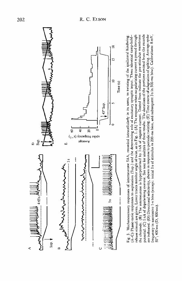
Fig. 1. Qualitative characteristics of the response of campaniform sensilla afferents toimposed twisting deformations of the hindwing. (Ai) Dorsal aspect of a right hindwing.The location of the row of campaniform sensilla (CS) on the posterior ventral surface ofthe subcosta (the second most anterior wing vein) is indicated by the row of stars. Alsoshown is the longitudinal axis (dotted line) about which the anterior part of the wing wastwisted by rotation applied distally (curved arrows), and the vannal fold (broken line,arrowed) about which the anterior part articulated with the posterior. (Aii) A schematicview, from anterior and above, of a right hindwing. Curved arrows show the directions oftwisting: Sup., supination, Pro., pronation. Double-headed arrows mark the cross-section of the wing midway along its length, showing how it is rotated relative to the baseduring twisting. (B-D) The spikes of campaniform sensilla afferents from the hindwing,recorded in nerve 1C (upper traces). Lower traces show the angle of wing twisting,imposed at the distal end. (B) Directional selectivity. Afferents spike during supinatingtwist, but are silenced by pronating twist. (C) Different units, distinguished by spikeamplitude, have different sensory thresholds. A small unit (single arrowhead) is excitedto spike at a smaller degree of twisting than a larger unit (double arrowhead).(D) Adapting responses. Spike frequencies of afferents are higher during twistingmovement (ramp) than during subsequent maintained twist. The same units are arrowedas in C. (E) Time course of adaptation of a small unit. Average spike frequency insuccessive 1-s bins (except initial 2 s of stimulus, where bins are 200 ms) during a ramp-hold-release twist stimulus. Adaptation is initially rapid but becomes progressivelyslower.

200 R. C. ELSON
5AA
1AA
Fig. 2

Multimodal flight interneurones 201
different interneurones, so that the number of cells in such a cluster must be verysmall, and may be only one.
In the dorsoventral dimension of the ganglia, the somata are located ventrally, butthe arborization of all four types is confined almost exclusively within the dorsal halfof the neuropile. Lateral branching (particularly the axon collaterals of 5AA and3AA) runs just below the dorsal ganglionic sheath. In other respects the cells,morphologies are diverse. 5AA is an ascending interganglionic interneurone, origi-nating in the metathoracic ganglion and having axon collaterals in the contralateralneuropiles of the meta- and mesothoracic ganglia (Fig. 2A; mesothoracic projectionnot shown). 3AA (Fig. 2B) is also interganglionic with contralateral axon collaterals,but originates in the mesothoracic ganglion and descends (metathoracic projectionnot known). 1AA (Fig. 2C) and 1AB (Fig. 2D) are both unilateral localinterneurones.
Proprioceptive input to interneurone SAAfrom hindwing campaniform sensilla
Interneurone 5AA responded to imposed deformations of the hindwing ipsilateralto its soma (Fig. 3). No response was evoked by mechanical stimuli applied to theother wings. Movement of the whole ipsilateral hindwing, e.g. elevation or de-pression, evoked no synaptic input or changes in spike frequency unless itsecondarily distorted the wing cuticle, whereas deformations were still effective whenthe wing was immobilized at the wing-hinge. Effective deformations includedbending the wing and twisting it about its longitudinal axis. The response to twistingwas directionally selective. Supination caused the neurone to depolarize and spike(Fig. 3A), while twisting in the pronating direction caused a decrease in the rate ofspiking (Fig. 3A,C, end of stimuli). Alternatively supinating and pronating the wingtherefore evoked a clear directionally selective response, and revealed the occurrenceof increased depolarizing input in the supinating phase compared with the pronatingphase (Fig. 3D).
Ramp-hold stimuli revealed a phasic-tonic reaction to supinating twist (Fig. 3A).The frequency of spikes during the ramp equalled or exceeded that at the start of thesteady twist, and during the maintained twist the spike frequency slowly declined.Adaptation in response to steady twist was initially rapid but became progressivelyslower (Fig. 3E). Hyperpolarizing the neurone by injected negative current abol-ished spiking and revealed a barrage of EPSPs whose envelope paralleled the timecourse of the spiking response to the same stimulus (Fig. 3B). Injection of depolar-izing current failed to reveal any inhibitory postsynaptic potentials (IPSPs) associ-ated with the stimulus, including the pronating ramp at the end (Fig. 3C). Thuscessation of spiking at the end of the supination, as also during pronation stimuli
Fig. 2. The morphology of (A) interneurone 5AA; (B) interneurone 3AA; (C) inter-neurone 1AA; and (D) interneurone 1AB. 5AA (A) is situated in the metathoracicganglion, the other three cells (B-D) in the mesothoracic ganglion. Interneurones werestained by intracellular cobalt injection, followed by silver intensification, and drawn inwhole-mount from the dorsal aspect. The dashed lines indicate the border of theneuropile. Lateral nerves 1, 3, 4 and 5 are labelled.
D
Sup.
\ 1 43O
sup.
--
0 5
10
15
18
Tim
e (s
)
Fig
. 3.
Rle
chan
osen
sory
res
pons
es o
f in
tern
euro
ne S
AA
, re
cord
ed i
ntra
cell
ular
ly i
n it
s so
ma,
to
twis
ting
of
the
ipsi
late
ral
hind
win
g.
(A-C
) P
hasi
c-to
nic
resp
onse
s to
sup
inat
ion
(Sup
.) a
nd t
he d
eter
min
ing
role
of
exci
tato
ry s
ynap
tic
inpu
t. T
hre
e id
enti
cal
ram
p-ho
ld-
rele
ase
stim
uli a
re g
iven
. Low
er tr
aces
mon
itor
ang
le o
f tw
ist,
as
in F
ig.
1. (A
) T
he
resp
onse
whe
n no
pol
ariz
ing
curr
ent i
s pa
ssed
thr
ough
th
e el
ectr
ode.
(B
) T
he
inte
rneu
rone
is
hype
rpol
ariz
ed b
y 1 nA
of
nega
tive
cur
rent
. D
otte
d li
nes
indi
cate
the
pre
-sti
mul
us m
embr
ane
pote
ntia
l. (
C)
1 nA
of
depo
lari
zing
cur
rent
. S
ee te
xt f
or a
naly
sis
of t
hese
res
ults
. Th
e du
rati
ons
of t
he p
orti
ons
omit
ted
from
the
rec
ords
ar
e in
dica
ted.
(D
) D
irec
tion
al s
elec
tivi
ty, s
how
n in
res
pons
e to
per
iodi
c tw
isti
ng. (
E) T
ime
cour
se o
f ad
apta
tion
of
spik
ing.
Ave
rage
sp
~k
e fr
eque
ncy
in s
ucce
ssiv
e 1-
s bin
s (e
xcep
t: i
niti
al 1
s of
sti
mul
us i
n 20
0-m
s bi
ns, a
nd s
ubse
quen
t 3
s in
500
-ms
bins
). C
alib
rati
ons:
8 m
V,
SO0;
400
ms
(D,
800m
s).
Multimodal flight interneurones 203
(data not illustrated), was due to withdrawal of excitatory synaptic input. Hyper-polarizing current augmented, while depolarizing current diminished, the amplitudeof the summed synaptic input underlying responses to identical supinating stimuli(Fig. 3B,C), implying that the input was predominantly chemical in nature.
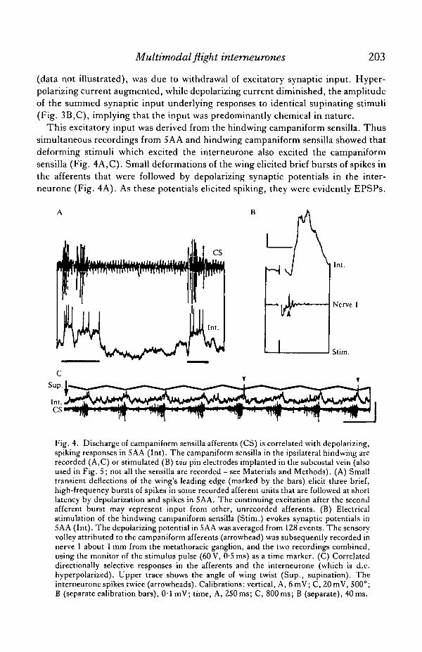
This excitatory input was derived from the hindwing campaniform sensilla. Thussimultaneous recordings from 5AA and hindwing campaniform sensilla showed thatdeforming stimuli which excited the interneurone also excited the campaniformsensilla (Fig. 4A,C). Small deformations of the wing elicited brief bursts of spikes inthe afferents that were followed by depolarizing synaptic potentials in the inter-neurone (Fig. 4A). As these potentials elicited spiking, they were evidently EPSPs.
CS
HInt.
Nerve 1
Stim.
Fig. 4. Discharge of campaniform sensilla afferents (CS) is correlated with depolarizing,spiking responses in 5AA (Int). The campaniform sensilla in the ipsilateral hindwing arerecorded (A,C) or stimulated (B) via pin electrodes implanted in the subcostal vein (alsoused in Fig. 5; not all the sensilla are recorded - see Materials and Methods). (A) Smalltransient deflections of the wing's leading edge (marked by the bars) elicit three brief,high-frequency bursts of spikes in some recorded afferent units that are followed at shortlatency by depolarization and spikes in 5AA. The continuing excitation after the secondafferent burst may represent input from other, unrecorded afferents. (B) Electricalstimulation of the hindwing campaniform sensilla (Stim.) evokes synaptic potentials in5AA (Int). The depolarizing potential in SAA was averaged from 128 events. The sensoryvolley attributed to the campaniform afferents (arrowhead) was subsequently recorded innerve 1 about 1 mm from the metathoracic ganglion, and the two recordings combined,using the monitor of the stimulus pulse (60 V, 0-5 ms) as a time marker. (C) Correlateddirectionally selective responses in the afferents and the interneurone (which is d.c.hyperpolarized). Upper trace shows the angle of wing twist (Sup., supination). Theinterneurone spikes twice (arrowheads). Calibrations: vertical, A, 6mV; C, 20 mV, 500°;B (separate calibration bars), 0 1 mV; time, A, 250ms; C, 800ms; B (separate), 40 ms.
204 R. C. ELSON
Deformations of the wing base that strongly excited the interneurone also caused thecampaniform sensilla, which are located in that region, to spike at high frequencies(not illustrated). Compound depolarizing potentials could also be evoked when thecampaniform sensilla were stimulated electrically (e.g. Fig. 4B). Potentials followedstimulus pulses at constant latency and without decrement at frequencies up to 10 Hz(the highest frequency tested). These results are consistent with the commonresponses of afferents and interneurone to controlled twisting stimuli. Thus, the timecourse of adaptation was qualitatively similar in both (cf. Figs IE, 3E). Periodictwisting of the wing showed common directional sensitivity. Excitatory synapticinput to the interneurone during the supinating phase correlated with the occurrenceof activity in the afferents in the same phase (Fig. 4C).
Short-latency connections of campaniform sensilla afferents to 5AA
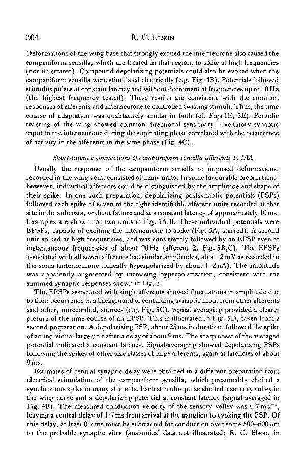
Usually the response of the campaniform sensilla to imposed deformations,recorded in the wing vein, consisted of many units. In some favourable preparations,however, individual afferents could be distinguished by the amplitude and shape oftheir spike. In one such preparation, depolarizing postsynaptic potentials (PSPs)followed each spike of seven of the eight identifiable afferent units recorded at thatsite in the subcosta, without failure and at a constant latency of approximately 10 ms.Examples are shown for two units in Fig. 5A,B. These individual potentials wereEPSPs, capable of exciting the interneurone to spike (Fig. 5A, starred). A secondunit spiked at high frequencies, and was consistently followed by an EPSP even atinstantaneous frequencies of about 90Hz (afferent 2, Fig. 5B,C). The EPSPsassociated with all seven afferents had similar amplitudes, about 2 mV as recorded inthe soma (interneurone tonically hyperpolarized by about l-2nA). The amplitudewas apparently augmented by increasing hyperpolarization, consistent with thesummed synaptic responses shown in Fig. 3.
The EPSPs associated with single afferents showed fluctuations in amplitude dueto their occurrence in a background of continuing synaptic input from other afferentsand other, unrecorded, sources (e.g. Fig. 5C). Signal averaging provided a clearerpicture of the time course of an EPSP. This is illustrated in Fig. 5D, taken from asecond preparation. A depolarizing PSP, about 25 ms in duration, followed the spikeof an individual large unit after a delay of about 9 ms. The sharp onset of the averagedpotential indicated a constant latency. Signal-averaging showed depolarizing PSPsfollowing the spikes of other size classes of large afferents, again at latencies of about9 ms.
Estimates of central synaptic delay were obtained in a different preparation fromelectrical stimulation of the campaniform sensilla, which presumably elicited asynchronous spike in many afferents. Each stimulus pulse elicited a sensory volley inthe wing nerve and a depolarizing potential at constant latency (signal averaged inFig. 4B). The measured conduction velocity of the sensory volley was 0-7 ms"1,leaving a central delay of 1 •! ms from arrival at the ganglion to evoking the PSP. Ofthis delay, at least 0-7 ms must be subtracted for conduction over some 500-600/imto the probable synaptic sites (anatomical data not illustrated; R. C. Elson, in
Multitnodal flight intemeurones 205
preparation). This yields an estimated central synaptic delay of 1 ms (error range atleast ±0-3 ms), comparable to that reported for monosynaptic connections betweenanother flight afferent and motor neurones in the locust (Burrows, 1975). Takentogether, these data are therefore consistent with an interpretation that some, atleast, of the campaniform sensilla afferents make monosynaptic connections with5AA.
Exteroceptive and other inputs to 5AA
In addition to receiving input from the campaniform sensilla of the ipsilateralhindwing, 5AA also responded to specific sensory stimuli of other modalities.
CS
Fig. 5. Connections between individual campaniform afferents (CS, extracellular spikes,upper traces) and interneurone 5AA (Int., intracellular, lower traces). (A-C) The sweepis triggered by the spikes of selected individual afferents, from the same preparation.(A) Each spike of a single unit, termed afferent 1, is followed by an EPSP (arrowhead) ata constant latency. One EPSP triggers a spike in the interneurone (star). (B) An EPSP(arrowhead) follows the spike of afferent 2 at a constant latency. (C) Each spike ofafferent 2 is followed by an EPSP (arrowheads), even at high instantaneous frequency.Another unit, afferent 3, is also followed by an EPSP (star) in this example. Changes inthe amplitude of the EPSP of afferent 2 are probably due to changes in postsynapticmembrane potential and conductance. (D) In another preparation, signal averaging (64events) reveals a small depolarizing potential following the spike of an individual largeafferent.
206 R. C. ELSON
Ocellar stimulation
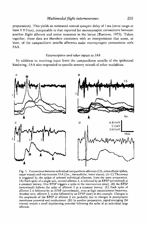
Light-off evoked a reliable, phasic burst of EPSPs and high-frequency spikes in5AA (Fig. 6A). Gradual light-off sets revealed many small EPSPs in the response
A Illuminate whole head
Fig. 6. Exteroceptive input to 5AA: responses to light-on and light-off. (A-D)Responses to changes in the intensity of light incident on the whole head (monitored by aphotosensitive diode, whose output is displayed in lower traces of A, B and C). (A) Phasicdepolarization and spiking in response to light-off. (B) The light-off response comprisesseveral summating EPSPs. (C) Tonic depolarizing current injected into the interneuronereduces the amplitude of the synaptic input at light-off, suggesting that it is chemicallymediated. (D) Triggering the oscilloscope sweep from rapid light-off stimuli (as in A)reveals that the depolarizing synaptic input occurs at a constant latency (several tracesoverlaid). (E-I) Stimulation of individual ocelli. (E) Light-off at the ocellus on the sidecontralateral to the soma elicits depolarizing potentials (vertical arrowheads). There is noresponse to light-on. (F) These potentials follow the light-off stimulus (at arrowhead) at aconstant latency. Several sweeps are overlaid. (G) Depolarizing potentials (arrowheads)evoked by light-off stimuli to the median ocellus. (H) These potentials also follow light-off (at arrowhead) at a constant latency. (I) In contrast, no such potentials are evoked bylight-off stimuli (at arrowhead) to the ipsilateral ocellus (a higher frequency of EPSPsoccurs in the background synaptic activity of this record; these are not correlated with thestimulus, and probably derive from the hindwing sensilla). All records except B are fromthe same experiment. Calibrations: voltage, 8mV (except B, 10mV); time, A,C,E,G,400ms; B, 200ms; D,F,H,I, 67ms).

Multimodal flight interneurones 207
(Fig. 6B). These potentials were apparently conventional chemical EPSPs, as d.c.depolarization of the interneurone reduced the amplitude of the summed synapticinput at light-off (Fig. 6C). For a given light pulse, the latency from light-off to theinitial depolarizing response was constant (Fig. 6D). Light-offsets from bright lightgave latencies as short as 35 ms, but latency increased as the size and steepness of thelight-offset was decreased. Light-on elicited no consistent response, but couldterminate a train of EPSPs triggered by a closely preceding light-off stimulus(Fig. 6A). The movement of black or white spots or stripes, against plain whitebackgrounds or the contrast-rich background of the laboratory, also evoked noresponse. These results do not preclude the possibility of a visual input from thecompound eyes in response to an appropriate stimulus. However, simple light-offstimuli to either compound eye, delivered by a light-emitting diode (see Materialsand Methods), evoked no response in the interneurone.
Stimulation of individual ocelli contralateral (Fig. 6E) and medial (Fig. 6G) to thesoma could elicit one or two EPSPs at short, constant latency of about 35 ms for eachlight-off (contralateral, Fig. 6F; median, Fig. 6H), i.e. at the same latency as thewhole-field off response (Fig. 6D). In contrast, stimulation of the ipsilateral ocelluselicited no such PSPs (Fig. 61).
Head movement
Interneurone 5AA was also powerfully excited by imposed displacements of thehead relative to the thorax. Small (2-3 mm) brief tilting movements elicited a phasicburst of spikes (Fig. 7A), and sudden head deflections to a new position evoked anadapting train of spikes (Fig. 7B). Imposed rolling movements of the head were alsoexcitatory, but directional responses were not studied. The spikes were underlain bydiscernible EPSPs, showing that the excitatory input was received in the meta-thoracic ganglion and not in anterior ganglia, to which 5AA projects. These
ATilt head
800 ms
A TWind on Off
Fig. 7. Interneurone SAA is excited by movement of the locust's head relative to thethorax (which was fixed). (A) Brisk spiking responses to imposed downward tilting of thehead (stimuli marked by bars). (B) Evidence for a phasic-tonic reaction. A suddenhead deflection is produced by a strong frontal airstream whose duration is shownschematically in the lower trace (the response is unlikely to be due primarily to wind: theantennae were amputated and the cephalic wind-hairs covered by Vaseline; and weakerwind stimuli did not elicit this response). Voltage calibration: A, 8mV; B, 4mV.
208 R. C. ELSON
responses were not related to touching any particular head structure and could beelicited by other stimuli, for example, by deflections of the head caused by a wind jet(where the cephalic wind-hairs were covered by Vaseline and the antennae ampu-tated, thus gTeatly attenuating any response to the air movement itself; here,however, thoracic wind-hairs might also have been stimulated). The responsespersisted undiminished in darkness (tested immediately after darkening, so that littledark adaptation would be expected). This suggests that a large part of this reactionwas not visual, but proprioceptive, e.g. from mechanoreceptors of the neck region.Responses were apparently not mediated by receptors on the cervical sclerites(Goodman, 1959), as touching these structures with a probe in one experimentelicited no response.
Other inputs
Interneurone 5AA received a weak auditory input. Loud claps elicited smalldepolarizing potentials. Lightly touching the tarsi or femorotibial joints of theipsilateral legs also evoked depolarizing potentials and spikes.
Other interneurones
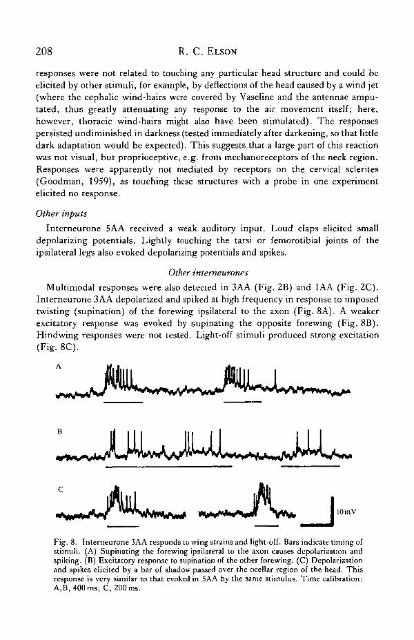
Multimodal responses were also detected in 3AA (Fig. 2B) and 1AA (Fig. 2C).Interneurone 3AA depolarized and spiked at high frequency in response to imposedtwisting (supination) of the forewing ipsilateral to the axon (Fig. 8A). A weakerexcitatory response was evoked by supinating the opposite forewing (Fig. 8B).Hindwing responses were not tested. Light-off stimuli produced strong excitation(Fig. 8C).
*w-wV< ^Vta«ww ********* W I 10 mV
Fig. 8. Interneurone 3AA responds to wing strains and light-off. Bars indicate timing ofstimuli. (A) Supinating the forewing ipsilateral to the axon causes depolarization andspiking. (B) Excitatory response to supination of the other forewing. (C) Depolarizationand spikes elicited by a bar of shadow passed over the ocellar region of the head. Thisresponse is very similar to that evoked in 5AA by the same stimulus. Time calibration:A,B, 400ms; C, 200ms.
Multitnodal flight interneurones 209
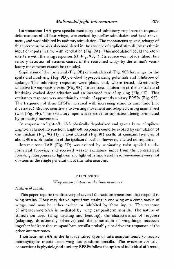
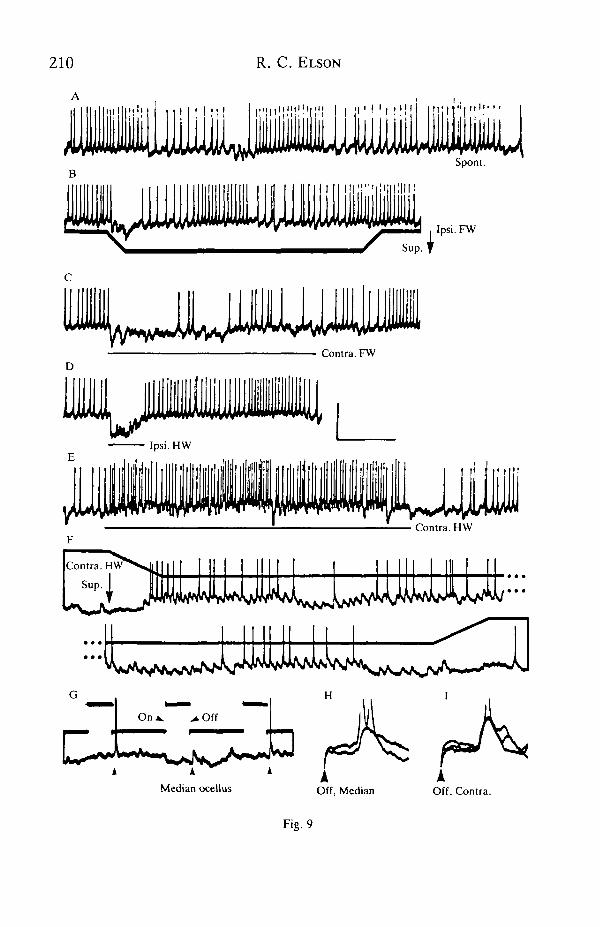
Interneurone 1AA gave specific excitatory and inhibitory responses to imposeddeformations of all four wings, was excited by ocellar stimulation and head move-ment, and was inhibited by auditory stimulation. The spontaneous spike discharge ofthis interneurone was also modulated in the absence of applied stimuli, by rhythmicinput or inputs in time with ventilation (Fig. 9A). This modulation could thereforeinterfere with the wing responses (cf. Fig. 9B,F). Its source was not identified, butsensory detection of stresses caused in the restrained wings by the animal's venti-latory movements cannot be excluded.
Supination of the ipsilateral (Fig. 9B) or contralateral (Fig. 9C) forewings, or theipsilateral hindwing (Fig. 9D), evoked hyperpolarizing potentials and inhibition ofspiking. The inhibitory responses were phasic and, where tested, directionallyselective for supinating twist (Fig. 9B). In contrast, supination of the contralateralhindwing evoked depolarization and an increased rate of spiking (Fig. 9E). Thisexcitatory response was underlain by a train of apparently unitary EPSPs (Fig. 9F).The frequency of these EPSPs increased with increasing stimulus amplitude (notillustrated), showed sensitivity to twisting movement and adapted during maintainedtwist (Fig. 9F). This excitatory input was selective for supination, being terminatedby pronating movement.
In response to light-off, 1AA phasically depolarized and gave a burst of spikes.Light-on elicited no reaction. Light-off responses could be evoked by stimulation ofthe median (Fig. 9G,H) or contralateral (Fig. 91) ocelli, at constant latencies ofabout 40 ms. Stimulation of the ipsilateral ocellus, however, elicited no response.
Interneurone 1AB (Fig. 2D) was excited by supinating twist applied to theipsilateral forewing and received weaker excitatory input from the contralateralforewing. Responses to light-on and light-off stimuli and head movements were notobvious in the single penetration of this interneurone.
DISCUSSION
Wing sensory inputs to the interneurones
Nature of inputs
This paper reports the discovery of several thoracic interneurones that respond towing strains. They may derive input from strains in one wing or a combination ofwings, and may be either excited or inhibited by these inputs. The responseof interneurone 5AA is mediated by wing campaniform sensilla. The nature ofstimulation used (wing twisting and bending), the characteristics of response(adapting, directionally selective) and the elimination of wing-hinge receptorstogether indicate that campaniform sensilla probably also drive the responses of theother interneurones.
Interneurone 5AA is the first identified type of interneurone found to receivemonosynaptic inputs from wing campaniform sensilla. The evidence for suchconnections is physiological: unitary EPSPs follow the spikes of individual afferents,
210 R. C. ELSON
Illl1III 1 Mi l ' 1 !
ill!
111ni
1• II
•
I > ! I , , l i |
ililMISpont.
Ipsi.FW
Median ocellusA
Off, Median
Fig. 9
Multimodal flight intemeurones 211
1:1, at short, constant latency and at high instantaneous frequencies. This behaviourwould not be expected if spiking neurones were interposed. Non-spiking neuronesmight possibly be interposed, but none have yet been found to receive direct inputsfrom mechanosensory afferents in the locust (Siegler & Burrows, 1983). Theestimated central synaptic delay is brief, about 1 ms. Anatomical evidence from thelight microscope is also consistent with direct connectivity: the afferent projectionoccupies the same localized, identified regions of neuropile as parts of the inter-neurone's arborization (R. C. Elson, in preparation).
Monosynaptic inputs to interganglionic intemeurones from another flight pro-prioceptor, the wing-hinge stretch receptor, were reported recently by Pearson et al.(1983). This and other evidence (e.g. Callec, Guillet, Pichon & Boistel, 1971; Bacon& Murphey, 1984; Hustert, 1985; this paper) indicates that mechanosensory af-ferents of insects can connect directly to interganglionic intemeurones, as well as tospiking local intemeurones (Siegler & Burrows, 1983). In comparison, the existenceof monosynaptic connections from mechanoreceptor afferents to intersegmentalintemeurones in the crayfish, is well established (Calabrese, 1976; Zucker, 1972).
For the other intemeurones described, there are no physiological data concerningthe directness of afferent input. However, anatomy precludes direct input fromcertain wings. The afferents of the campaniform sensilla do not ascend or projectcontralaterally (Tyrer & Altman, 1974), so that the hindwing responses in 1AA, andthe contralateral wing responses in 1AA and 1AB, must be mediated by other,intercalated intemeurones. Interestingly, the contralateral hindwing input to 1AA isderived from a presynaptic neurone that has 5AA-like response properties (seeFig. 9F). lAA's ipsilateral inhibitory input is also probably indirect, as no mechano-sensory afferents are yet known to produce monosynaptic inhibition in centralneurones (Calabrese, 1976; Siegler & Burrows, 1983).
It cannot be excluded that other, unidentified, strain-detecting receptors mightcontribute inputs to these intemeurones as well. One such class could be the small-axoned multipolar sensilla dispersed within the wing cuticle (Knyazeva, 1970).
Fig. 9. Responses of 1AA to deformations of all four wings and to ocellar stimulation.(Ipsi., ipsilateral; Contra., contralateral; FW, forewing; HW, hindwing). (A) In theabsence of sensory stimulation there is a slow, spontaneous (Spont.) rhythm of spikesapparently in time with ventilation. (B) Supination (Sup.) of the ipsilateral forewing(angle of twisting monitored in the lower trace) evokes a transient inhibition. Note IPSPsduring the twisting movement. (C-E) Stimulation by a hand-held probe. Bars show theduration of imposed stimuli. (C) Supinating the contralateral forewing evokes IPSPs.(D) IPSPs are also evoked by deforming the ipsilateral hindwing. (E) Supinating thecontralateral hindwing excites the interneurone. (F) Apparently unitary EPSPs in theresponse to contralateral hindwing supination (Sup., angle of twist in upper trace).The duration of the omitted portion is 3-8 s. (G-I) Stimulation of individual ocelli.(G) Light-on and light-off stimuli (monitored in upper trace) to the median ocellus elicitphasic EPSPs and spikes at off, but no response at on. (H) EPSPs follow the off stimulus(at arrowhead) to the median ocellus at a constant latency. (I) as H, but for thecontralateral ocellus. Initial deflection is an artefact of the pulse offset. Calibrations:vertical, A,E-G, 8mV; B-D, lOmV; H,I, 4mV; B, 100°; F, 50°; time, 800ms (exceptF, 400ms; H,I ,67ms).
212 R. C. ELSON
Functional significance
The possible functioning of the interneurones in flight must be inferred from theirresponse properties and those of the sensory neurones which drive them. In theflying animal the mechanical stimulation of the campaniform sensilla results from theforces acting on and in the wing. Behavioural studies indicate that a principalstimulus for these receptors is the aerodynamic force (lift and thrust) generated bytheir wing (Gettrup & Wilson, 1964; Gettrup, 1965, 1966; Pringle, 1976). Theproprioceptive response of the interneurones may therefore be largely determined bythis force. A neurone with excitatory input from just one wing (e.g. 5AA) willprogressively depolarize and spike at higher frequency as the intensity of activity inthe campaniform sensilla increases. In flight it would provide a simple monitor of theresultant strain perceived by the campaniform sensilla, and thus the aerodynamicforce, in that wing. A neurone deriving excitatory input from the sensilla of morethan one wing (e.g. 3AA) would allow an interneurone to monitor the sum of theforce experienced by those wings. Where an interneurone derives a combination ofexcitatory and inhibitory inputs, from the sensilla of several wings (e.g. 1AA), thenet excitation will depend on the balance of these inputs. Here the interneuronecould monitor the pattern of strains among the wings, and therefore their relativeaerodynamic contributions. The development of an imbalance of forces could also bedetected. A comparable monitoring of the pattern of proprioceptive inputs fromseveral appendages occurs in decapod crustaceans. Joint proprioceptors in each legdetect the movements caused by body displacements relative to the substrate. Thedirection of displacement is abstracted from the pattern of movement among the legsand determines the direction of an equilibrium reflex. Some sensory interneuroneswith opposite bilateral inputs from leg proprioceptors, which could detectcomponents of these displacements, have been found in lobsters (Schone, Neil, Stein& Carlstead, 1976; Priest, 1983; Neil et al. 1984).
Although the cells described here probably represent only a fraction of the totalnumber of interneurones with sensitive reactions to wing strains, they do indicate thetypes of proprioceptive responses which can occur, and how they could function inflight.
Extervceptive input to the interneurones
The synaptic responses evoked in interneurones 5AA and 1AA by ocellar stimu-lation resemble those seen in a small population of thoracic interneurones, describedby Rowell and co-workers (Rowell & Pearson, 1983; Reichert & Rowell, 1985), thatare postsynaptic to ocellar units that descend the nerve cord from the brain, andoccur at comparable latencies. The descending interneurones respond to light-offstimulation of specific ocelli by firing one or two rapidly conducted spikes at constantlatencies (Patterson & Goodman, 1974; Simmons, 1980; Rowell & Pearson, 1983;Reichert et al. 1985; Rowell & Reichert, 1986). The thoracic interneurones de-scribed here, therefore, probably also derive their ocellar inputs from these descend-ing units, and may similarly receive direct connections; if the input is not direct, it
Multimodal flight interneurones 213
must be mediated via non-spiking neurones, or neurones which themselves arecaused to spike at consistent latencies. The response of 3AA to light-off stimuli isvery similar to those of 5AA and 1AA, and may also be ocellar, though this was nottested.
Interneurones 5AA and 1AA also resemble the thoracic interneurones describedabove in the asymmetry of ocellar input (EPSPs from the medial and one lateralocellus). In the thoracic interneurones of Rowell & Pearson (1983) and Reichert &Rowell (1985), the asymmetric input patterns derive from particular combinations ofpresynaptic descending units, and have functional significance for the detection offlight instability. Lateral ocelli can detect rolling, the medial ocellus, pitching. Theocelli mediate compensatory reactions which would correct the perceived deviationfrom straight, stable flight (Wilson, 1978; Stange & Howard, 1979; Taylor,1981a,b). The pattern of ocellar input to 5AA and 1AA, as to the thoracic inter-neurones above, implies that they will respond optimally to particular rotations in therolling and pitching planes. Modulation of activity in the thoracic interneurones,some of which are premotor, is thought to underlie compensatory changes in flightmotor output (Reichert & Rowell, 1985; Reichert et al. 1985). These cells havetherefore been termed 'steering interneurones' (Reichert, 1985). The asymmetricocellar inputs to 5AA and 1AA are suggestive of some function in steering reactionsfor those neurones also.
Interneurones 5AA, 3AA and 1AA resemble steering interneurones in two furtherrespects: first, in morphology — both types arborize in the most dorsal regions ofneuropile (Rowell & Pearson, 1983; R. C. Elson, in preparation; note especially theclose structural resemblance of 1AA to Rowell & Pearson's thoracic interneurone116, and 3AA to their 315); second, in other sensory responses — both types havemultimodal reactions, including tactile and auditory inputs (Reichert & Rowell,1985). All these similarities suggest that the interneurones of this paper andpreviously described steering interneurones are overlapping categories and may besampled from the same population. The results then have two implications. First, asthe descending interneurones and steering cells also respond to appropriate com-pound eye, wind-hair and antennal stimuli that would signal flight deviations(Reichert et al. 1985), these types of response should be looked for in the type ofneurones reported here. (The lack of a clear wind response in 5AA - see legend ofFig. 7 - may reflect the relative insensitivity of the descending neurones to weak windstimuli and the inhibition of their wind response during bright illumination, as oftenused in the preparations described here: cf. Rowell & Reichert, 1986.) Second,proprioceptive inputs from wing campaniform sensilla may also occur in previouslydescribed steering interneurones, and this type of convergence should be studiedfurther.
Some of the same interneurones, described here, that receive ocellar input can alsobe excited by imposed movements of the locust's head. In 5AA (and probably also in1AA) a major part of this response is probably proprioceptive, and detects headmotion rather than specifically touch or pressure on the head. Detection of headmisalignment with the thorax, resulting from corrective repositioning of the
214 R. C. ELSON
head, elicits compensatory flight reactions in a second pathway for exteroceptiveresponses to course deviations (see Introduction). It is tempting to ascribe such afunction to the responses in 5AA and 1 AA. The receptors underlying these responsesand the reaction of the intemeurones to active head movements must receive study,however, before behavioural function can be inferred.
Specific convergence of proprioceptive and exteroceptive inputs
The convergence of specific proprioceptive and exteroceptive inputs suggests thatthe same intemeurones that could be active during the exteroceptive adjustment offlight could also participate when adjustments are signalled by proprioceptivefeedback. Proprioceptive input from the wings is involved in the coordination of themovements of individual wings and the ensemble of four wings (Burrows, 19766;Mohl, 1985a,b,c). This coordination is important for the maintenance of flightstability, as each wing has its own set of motor neurones and is capable ofindependent fluctuations in performance. In flying locusts where exteroceptivefeedback has been disabled, this coordination appears as correlations in the activity ofmotor neurones of different wings. Correlation patterns commonly link activity incontralateral and heterosegmental wings (Mohl, 1985a). The intemeurones de-scribed here project contralaterally and intersegmentally, or have bilateral andheterosegmental wing inputs. In this way the feedback they derive about force (liftand thrust) generation in various wings (which, if irregular or unbalanced, will causeinstability) could be used in the coordination of compensatory motor changes amongthe wings, leading to correction of the instability.
Intemeurones of this type could therefore be used by several feedback pathways inthe production of appropriate flight adjustments. Different sensory inputs thatrequire a common motor response may converge (see Olberg, 1981). For instance,the ocellar inputs to 5AA (excitation at dimming the median and contralateral ocelli)would signal downward pitch and contralateral roll (i.e. rolling downwards on thecontralateral side, upwards on the ipsilateral side). The interneurone can also detectipsilateral roll of the head (i.e. rolling downwards on the ipsilateral side), a mismatchwith the thorax that might occur during repositioning of the head following acontralateral roll by the whole locust. Similarly, 5AA would detect an increase in liftgeneration by the ipsilateral hindwing, which, if uncompensated, would causedownward pitching and contralateral rolling. In each case the appropriate flightcompensation, which the interneurone might signal, would be the generation of arestoring torque that included upward pitching and ipsilateral rolling components.
Similar convergence of modalities occurs in the equilibrium reactions of otherarthropods and of vertebrates. Unintended body movements are detected byequilibrium receptors (functional analogues of the exteroceptors used by the flyinglocust) and by limb proprioceptors. The two sensory systems commonly interact inthe control of posture. These interactions seem to involve integration at a segmental(spinal) level, analogous to the suggested role of the intemeurones described here. Indecapod crustaceans, feedback signals from statocyst and leg proprioceptors (seqi

Multimodal flight intemeurones 215
above) interact in the production of appropriate righting responses and compen-satory eye movements, suggesting that they converge in appropriate patterns(Schone et al. 1976; Schone, Neil, Scapini & Dreissman, 1983; Neil et al. 1984;Hisada & Neil, 1985). The convergence is thought to occur at segmental, premotorintemeurones, as yet unidentified (Hisada & Neil, 1985). In vertebrates there is acontinual interaction between vestibular inputs and those from proprioceptors andcutaneous afferents. The lateral vestibulospinal tract excites limb extensor motorneurones and flexor la inhibitory intemeurones, spinal neurones which are alsoexcited by the extensor la afferents (Grillner, Hongo & Lund, 1970; Hultbom, Illert& Santini, 1976).
To understand more fully the functions of the types of interneurone describedhere, we need to know more about the range of sensory inputs to individualneurones, and what outputs they have. Some reflex effects of wing campaniformsensilla on flight motor neurones are known (Wendler, 1978; Horsmann, 1981; R. C.Elson, in preparation) in which these cells might participate, but the particularmotor effects of single intemeurones remain unknown. It is clear, however, thatsingle thoracic intemeurones can receive both ocellar and head-motion signals(implicating them in exteroceptive flight control), and combine these with pro-prioceptive feedback about wing forces. These multimodal flight intemeurones and,by inference, analogous steering intemeurones, may be the nexus of several parallelfeedback loops (see Rowell & Pearson, 1983) which converge in specific patterns.This utilization of single intemeurones by different pathways reflects the economy ofdesign in the locust's nervous system.
Supported by an MRC (UK) studentship. I thank Dr M. Burrows, University ofCambridge, for his supervision and encouragement, the use of the facilities of hislaboratory, and valuable criticism of this paper.
REFERENCES
BACON, J. & ALTMAN, J. S. (1977). A silver intensification method for cobalt-filled neurones inwholemount preparations. Brain Res. 138, 359-363.
BACON, J. & MURPHEY, R. K. (1984). Receptive fields of cricket giant intemeurones are related totheir dendritic structure. J . Physiol., Land. 352, 601-623.
BROGAN, R. T. & PITMAN, R. M. (1981). Axonal regeneration in an identified insect motoneurone.J. Physiol., Lond. 319, 34-35P.
BURROWS, M. (1975). Monosynaptic connexions between wing stretch receptors and flightmotoneurones of the locust.,7. exp. Biol. 62, 189-219.
BURROWS, M. (1976a). Neural control of flight in the locust. lnNeural Control of Locomotion (ed.R. M. Hermann, S. Grillner, P. S. G. Stein & D. G. Stuart), pp. 419-438. New York: PlenumPress.
BURROWS, M. (19766). The influence of sensory inflow on the flight system of the locust. InPerspectives in Experimental Biology, vol. 1, Zoology (ed. P. S. Davies), pp. 399-409. Oxford,New York: Pergamon Press.
BURROWS, M. (1977). Flight mechanisms of the locust. In Identified Neurons and Behaviour ofArthropods (ed. G. Hoyle), pp. 339-356. New York: Plenum Press.
CALABRESE, R. L. (1976). Crayfish mechanoreceptive interneurons. I. The nature of ipsilateralexcitatory inputs.X comp. Physiol. 105, 83-102.

216 R. C. ELSON
CALLEC, J. J., GUILLET, J. C , PICHON, Y. & BOISTEL, J. (1971). Further studies in synaptictransmission in insects. II. Relations between sensory information and its synaptic integration atthe level of a single giant axon in the cockroach. J. exp. Biol. 55, 123-149.
GETTRUP, E. (1965). Sensory mechanisms in locomotion: the campaniform sensilla of the insectwing and their function during flight. Cold Spring Harb. Symp. quant. Biol. 30, 615-622.
GETTRUP, E. (1966). Sensory regulation of wing twisting in locusts. J'. exp. Biol. 44, 1-16.GETTRUP, E. & WILSON, D. M. (1964). The lift control reaction of flying locusts. J . exp. Biol. 41,
183-190.GOODMAN, L. J. (1959). Hair receptors in locusts. Hair plates on the first cervical sclerites of the
Orthoptera. Nature, Lond. 183, 1106-1107.GOODMAN, L. J. (1965). The role of certain optomotor reactions in regulating stability in the
rolling plane during flight in the desert locust, Schistocerca gregaria. J. exp. Biol. 42, 382-407.GRJLLNER, S., HONGO, T. & LUND, S. (1970). The vestibulospinal tract. Effects on alpha-
motoneurones in the lumbosacral spinal cord in the cat. Expl Brain Res. 10, 94—120.HISADA, M. & NEIL, D. M. (1985). The neuronal basis of equilibrium behaviour in decapod
crustaceans. In Coordination of Motor Behaviour (ed. B. M. H. Bush & F. Clarac), Soc. exp.Biol. Seminar 24, 229-248. Cambridge: Cambridge University Press.
HORSMANN, U. (1981). Flugrelevante Afferenzen und ihre Verarbeitung bei der Wander-heuschrecke (Locusta migratoria, L.). Diplomarbeit, Universitat zu Koln.
HULTBORN, H., ILLERT, M. & SANTINI, M. (1976). Convergence on interneurones mediating thereciprocal la inhibition of motoneurones. III. Effects from supraspinal pathways. Ada physiol.scand. 96, 368-391.
HuSTERT, R. (1985). Multisegmental integration and divergence of afferent information fromsingle afferent hairs in a cricket. J . exp. Biol. 118, 209-227.
KNYAZEVA, N. I. (1970). Receptors of the wing apparatus regulating the flight of the migratorylocust, Locusta migratoria L. (Orthoptera, Acrididae). Ent. Rev. 49, 311-317.
MOHL, B. (1985a). The role of proprioception in locust flight control. I. Asymmetry and couplingwithin the time pattern of motor units. J. comp. Physiol. 156, 93-101.
MOHL, B. (19856). The role of proprioception in locust flight control. II. Information signalled byforewing stretch receptor during flight. .7- comp. Physiol. 156, 103-116.
MOHL, B. (1985C). The role of proprioception in locust flight control. I II . The influence of afferentstimulation of the stretch receptor nerve. J. comp. Physiol. 156, 281—291.
MORAN, D. T., CHAPMAN, K. M. & ELLIS, R. A. (1971). The fine structure of cockroachcampaniform sensilla. J . Cell Biol. 48, 155-173.
NEIL, D. M., PRIEST, T. D., MIYAN, J. A., WOTHERSPOON, R. M. & SCHONE, H. (1984). Co-
ordinated equilibrium responses at two joints in the spiny lobster antenna in relation to thepattern of movements imposed upon the legs. J. comp. Physiol. 155, 351-363.
OLBERG, R. (1981). Parallel encoding of direction of wind, head, abdomen and visual patternmovement by single interneurons in the dragonfly. J. comp. Physiol. 142, 27-41.
PATTERSON, J. A. & GOODMAN, L. J. (1974). Relationship between ocellar units in the ventralnerve cord and ocellar pathways in the brain of Schistocerca gregaria. J. comp. Physiol. 95,251-262.
PEARSON, K. G., REYE, D. N. & ROBERTSON, R. M. (1983). Phase-dependent influence of wingstretch receptors on flight rhythm in the locust. J. Neurophysiol. 49, 1168-1181.
PFAU, H. K. (1983). Mechanik und sensorische Kontrolle der Flugel-Pronation und Supination.In Physiology and Biophysics ofInsect Flight, vol. II (ed. W. Nachtigall). pp. 61-77. Stuttgart:Fischer.
PRJEST, T. D. (1983). An equilibrium reflex in decapod Crustacea mediated by basal legproprioceptors. Ph.D. thesis, University of Glasgow.
PRINGLE, J. W. S. (1938a). Proprioception in insects. I. A new type of mechanical receptor fromthe palps of the cockroach. J . exp. Biol. 15, 101-113.
PRINGLE, J. W. S. (19386). Proprioception in insects. II. The action of the campaniform sensillaon the legs. J . exp. Biol. 15, 114-131.
PRINGLE, J. W. S. (1976). The muscles and sense organs involved in insect flight. In RoyalEntomological Society Symposium no. 7, Insect Flight (ed. R. C. Rainey), pp. 3-15. Oxford^Blackwell.

Multimodal flight interneurones 217
REICHERT, H. (1985). The cellular basis of sensorimotor coordination in the flight control systemof the locust. In Coordination of Motor Behaviour (ed. B. M. H. Bush & F. Clarac), pp.121-140. Cambridge: Cambridge University Press.
REICHERT, H. & ROWELL, C. H. F. (1985). Integration of nonphaselocked exteroceptiveinformation in the control of rhythmic flight in the locust..7. Neurophysiol. 53, 1201-1218.
REICHERT, H., ROWELL, C. H. F. & GRISS, C. (1985). Course correction circuitry translatesfeature detection into behavioural action in locusts. Nature, Lond. 315, 142-144.
ROBERTSON, R. M. & PEARSON, K. G. (1982). A preparation for the intracellular analysis ofneuronal activity during flight in the locust. J. comp. Physiol. 146, 311-320.
ROBERTSON, R. M. & PEARSON, K. G. (1983). Interneurons in the flight system of the locust:distribution, connections and resetting properties. J. comp. Neural. 215, 33-50.
ROBERTSON, R. M. & PEARSON, K. G. (1985). Neural circuits in the flight system of the locust.J. Neurophysiol. 53, 110-128.
ROWELL, C. H. F. &PEARSON, K. G. (1983). Ocellar input to the flight motor system of the locust:structure and function. J. exp. Biol. 103, 265-288.
ROWELL, C. H. F. & REICHERT, H. (1986). Three descending interneurons reporting deviationfrom course in the locust. II. Physiology. J. comp. Physiol. 158, 775-794.
SCHONE, H., NEIL, D. M., SCAPINI, F. & DREISSMAN, G. (1983). Interaction of substrate, gravityand visual cues in the control of compensatory eye responses in the spiny lobster, Palinurusvulgaris.J. comp. Physiol. 150, 23-30.
SCHONE, H., NEIL, D. M., STEIN, A. & CARLSTEAD, M. K. (1976). Reactions of the spiny lobster,Palinurus vulgans, to substrate tilt (l).jf. comp. Physiol. 107, 113-128.
SIEGLER, M. V. S. & BURROWS, M. (1983). Spiking local interneurons as primary integrators ofmechanosensory information in the locust. J. Neurophysiol. 50, 1281-1295.
SIMMONS, P. J. (1980). A locust wind and ocellar brain neurone. J . exp. Biol. 85, 281-294.STANGE, G. & HOWARD, J. (1979). An ocellar dorsal light response in a dragonfly. J . exp. Biol. 83,
351-355.TAYLOR, C. P. (1981a). Contribution of compound eyes and ocelli to steering of locusts in flight.
I. Behavioural analysis. J. exp. Biol. 93, 1-18.TAYLOR, C. P. (19816). Contribution of compound eyes and ocelli to steering of locusts in flight.
II. Timing changes in flight motor units, jf. exp. Biol. 93, 19-31.TYRER, N. M. & ALTMAN, J. S. (1974). Motor and sensory neurones in a locust demonstrated using
cobalt chloride. J . comp. Neurol. 157, 117-138.USHERWOOD, P. N. R. & GRUNDFEST, H. (1965). Peripheral inhibition in skeletal muscle of
insects. J. Neurophysiol. 28, 497-518.WEIS-FOGH, T. (1949). An aerodynamic sense organ stimulating and regulating flight in locusts.
Nature, Lond. 164, 873-874.WENDLER, G. (1974). The influence of proprioceptive feedback on locust flight coordination.
J. comp. Physiol. 88, 173-200.WENDLER, G. (1978). The possible role of fast wing reflexes in locust flight. Naturwissenschaften
65,65.WENDLER, G. (1983). The locust flight system: functional aspects of sensory input and methods of
investigation. In Physiology and Biophysics of Insect Flight, vol. II (ed. W. Nachtigall),pp. 113-125. Stuttgart: Fischer.
WILSON, D. M. (1964). The origin of the flight motor command in grasshoppers. In Neural Theoryand Modelling (ed. R. Reiss), pp. 331-345. Stanford, California: Stanford University Press.
WILSON, D. M. (1968). Inherent asymmetry and reflex modulation of the locust flight motorpattern. J. exp. Biol. 48, 631-641.
WILSON, M. (1978). The functional organisation of locust ocelli. J . comp. Physiol. 124, 297-316.ZUCKER, R. S. (1972). Crayfish escape behavior and central synapses. I. Neural circuit exciting
lateral giant fibre. J . Neurophysiol. 35, 599-620.