Integrated Master in Dental Medicine
47
Integrated Master in Dental Medicine OSSEOINTEGRATION OF ZIRCONIA IMPLANTS SYSTEMATIC REVIEW AND META-ANALYSIS Soraia Dores Supervisor: Professor Doutor João Paulo Tondela Co-Supervisor: Professor Doutor Francisco Caramelo 2018
Transcript of Integrated Master in Dental Medicine

Integrated Master in Dental Medicine
OSSEOINTEGRATION OF ZIRCONIA IMPLANTS SYSTEMATIC REVIEW AND META-ANALYSIS
Soraia Dores Supervisor: Professor Doutor João Paulo Tondela Co-Supervisor: Professor Doutor Francisco Caramelo
2018

Integrated Master in Dental Medicine
OSSEOINTEGRATION OF ZIRCONIA IMPLANTS SYSTEMATIC REVIEW AND META-ANALYSIS
Soraia D.*, Caramelo F.**, Tondela JP.**
* Dentistry Student at the Faculty of Medicine, University of Coimbra. E-mail: [email protected]
**Assistant Professor, Dentistry Department, Faculty of Medicine, University of Coimbra
Dentistry Department of the Faculty of Medicine of the University of Coimbra,
Avenida Bissaya Barreto, Bloco de Celas, 3000-075 Coimbra
Tel.: +351 239484183
Fax: +351 239402910
Coimbra, Portugal
2018


IV
Summary
I. Abstract / Resumo
II. List of abbreviations and acronyms
III. Introduction
1. Osseointegration: a major factor to implant stability
2. State of the art: implant evolution
IV. Materials and Methods
V. Results
VI. Discussion
VII. Conclusions
VIII. Implications for clinical practice
IX. Acknowledgements
X. Bibliography
XI. Appendix
XII. Index

V
I - Abstract
Introduction: Osseointegration is one of the most primordial factors in implant rehabilitation.
Although osseointegration of titanium implants is considered a reference of implantology
some disadvantages of them as the potential presence of allergic reactions or the dark color
that may compromise aesthetics when there is an unfavorable mucosa, have led to the
development of alternatives. Zirconia, a well studied bioinert structure derived from the
Zirconium metal, has been used in medicine and dentistry. Zirconia implants may be an
alternative due to the aesthetics, biocompatibility and high fracture resistance. Objective:
This systematic review and meta-analysis aimed to evaluate the survival and success rates
of zirconia implants in humans. Methodology: A PICO question was defined "In patients
subjected to tooth replacement with single unit zirconia implant does the survival rates do
compare to single unit titanium implant?", followed by a search in primary databases
PubMed / MEDLINE, Cochrane and Embase with the following keywords: "dental
implantation, osseointegrated", "implantation, osseointegrated dental", "osseointegrated
dental implantation", "osseointegration", "zirconium", "titanium", "dental implants", "dental
implants, single tooth", "single tooth dental implants", "dental implantation, endosseous";
using the appropriate boolean operators, "OR" and "AND". Wherever possible the MeSH
terms were used. The search criteria did not include a time limit or restrictions on the
language or type of publication. Results: A total of 1465 articles were obtained from which 71
were selected for full text reading, after exclusion of the duplicates and reading the title and
abstract. Of these articles, nine were included on this systematic review. The outcomes
evaluated were the survival rate, the success rate and the Marginal Bone Level; others
parameters were extracted to complement the review. Discussion: Recently, the option for
zirconia implants has increased exponentially due to the inherent characteristics of the
material. Regarding the parameters, survival and success rates as well as the Marginal Bone
Level, translation of an effective osseointegration, present values very similar to that of
titanium implants. Conclusion: Considering the limits of this systematic review, it is possible
to conclude that zirconia implants can be a safe and viable option, and an alternative to
titanium implants in single unit implant-supported restorations. However, more multicentre
randomized clinical trials with scientific quality and validity, with a larger follow-up, are
needed to accurately prove the success of these implants.
Key-words: osseointegration; zirconia implant; success rates; survival rates; marginal bona
level

VI
Resumo Introdução: A osteointegração é um dos factores preponderantes no sucesso da reabilitação
com implantes. A osteointegração dos implantes de titânio é considerada a referência da
implantologia; contudo, algumas desvantagens dos mesmos como a potencial presença de
reacções alérgicas ou a sua cor escura que pode comprometer a estética quando existam
condições desfavoráveis da mucosa, levaram ao desenvolvimento de alternativas. A
zircónia, uma estrutura bionerte bem estudada que deriva do metal Zircónio, tem sido usada
amplamente na Medicina e na Medicina Dentária. Os implantes em zircónia podem constituir
essa alternativa devido à sua estética, biocompatibilidade e elevada resistência à fractura.
Objectivo: Este trabalho pretende fazer uma revisão sistemática e meta-análise para
avaliação das taxas de sobrevivência e sucesso dos implantes de zircónia, em humanos.
Metodologia: Foi definida uma questão PICO "Em pacientes submetidos à substituição
dentária com um implante unitário de zircónia, as taxas de sobrevivência podem-se
comparar com a dos implantes unitários de titânio?", seguida de uma pesquisa nas bases de
dados primárias da PubMed/MEDLINE, Cochrane e Embase com as seguintes palavras-
chave: "dental implantation, osseointegrated", "implantation, osseointegrated dental",
"osseointegrated dental implantation", "osseointegration", "zirconium", "titanium", "dental
implants", "dental implants, single tooth", "single tooth dental implants", "dental implantation,
endosseous"; com os conectores boleanos "AND" e "OR". Sempre que possível foram
utilizados os termos MeSH. Os critérios de pesquisa não incluíram um limite cronológico
nem foram feitas restrições quanto à língua ou tipo de publicação. Resultados: Obtiveram-se
um total de 1465 artigos dos quais foram seleccionados 71 para leitura integral, após
exclusão dos duplicados e leitura do título e abstract. Desses artigos, nove foram incluídos
para esta revisão sistemática. Os parâmetros avaliados foram a taxa de sobrevivência, a
taxa de sucesso e o nível ósseo marginal e foram extraídos ainda outros que permitiram
enriquecer a revisão. Discussão: Recentemente, a opção por implantes de zircónia
aumentou exponencialmente devido às características oferecidas pelo material.
Relativamente aos parâmetros, as taxas de sobrevivência e sucesso, assim como o nível
óseo marginal, tradução de uma osteointegração efectiva, apresentam valores muito
semelhantes à dos implantes de titânio. Conclusão: Considerando os limites desta revisão, é
possível concluir que os implantes de zircónia podem constituir uma opção segura e viável,
e uma alternativa aos implantes de titânio. Contudo, são necessários mais ensaios clínicos
randomizados multicêntricos com qualidade e validade científica, apresentando um
seguimento maior, para provar com exactidão o sucesso destes implantes.

VII
Palavras-chave: osteointegração; implante de zircónia; taxas de sucesso; taxas de
sobrevivência; nível ósseo marginal

VIII
II - List of abbreviations and acronyms
BIC - Bone Implant Contact
FDPs - Fixed Dental Prostheses
FGF - Fibroblast Growth Factor
HIF-1 - Hypoxia Inducible Factor
LTD - Low-Temperature Degradation
MBL - Marginal Bone Level
Mg-PSZ - Magnesium Partially Stabilized Zirconia
PMN - Polymorphonuclear Leucocytes
RCTs - Randomized Clinical Trials
VEGF - Vascular Endothelial Growth Factor
Y-TZP - Yttria-Stabilized Tetragonal Zirconia Polycrystal
ZTA - Zirconia-Thougened Alumina

9
III - Introduction
1. Osseointegration: a major factor to implant stability Since ages, people have the concern, because of a functional or aesthetic care, of replacing
teeth that were lost. In ancient Egypt, people used animal teeth as the first attempts to
replace missing teeth. Ivory was the material of choice, and it was mechanically shaped to
look like a tooth. Similar implantation procedures were also found in South America and
Europe. (1) The continuous evolution of implant dentistry, as in dentistry, made possible that
currently we can replace missing teeth from other materials that became so popular because
of it features, like the biocompatibility.
Implant dentistry has passed from an experimental treatment to a highly predictable and
long-term option to treat fully or partially edentulous patients. When compared with
conventional fixed or removable prostheses, it offers a significant functional and biologic
advantages that turn this rehabilitation an excellent treatment option. Nowadays, it is the best
treatment option for many patients and it became the first line of choice for so many dentists
and patients.
This change was initiated 50 years ago by the discovery that implants made of commercially
pure titanium could achieve anchorage in the bone with functional and direct bone-to-implant
contact (BIC). In the 1960s, Branemark, the most important pioneer of modern implant
dentistry from the University of Gothenburg (Sweden), and coworkers performed the first
preclinical and clinical studies and discovered this anchorage when blocks of titanium placed
into the femur of rabbit got ankylosed with the surrounding bone and could not be retrieved.(2)
Schroeder, from the University of Bern (Switzerland), and coworkers started to examine the
tissue integration. Both were the pioneers that provided evidence for direct bone apposition
on the surface of titanium, a phenomenon later termed "osseointegration", which is today
widely accepted. A few years later, he reported as well the soft tissues reactions to titanium

10
implants. (1,2)
Surface characteristics of an implant have become an important point because of the
decisive influence on the cascade of the osseointegration. Modifications of these surfaces
were found to influence both the speed and the process of osseointegration. It is necessary
to understand the mechanisms by which bone may be formed on an implant surface.
Osseointegration is in a comprehensive way "a direct structural and functional connection
between ordered, living bone and the surface of a load-bearing implant", which is based on
the principle of bone regeneration and on the osteoconductivity of the biomaterial. (3)
Intraoral bone healing of an implant wound comprises four phases: the hemostasis (minutes
to hours), the inflammatory phase (hours to days), the proliferative phase (days to weeks)
and finally the remodeling phase (approximately 3 weeks and lasts for years). (4) Cells have
an important role in this process.
Hemostasis phase begins with the dental implant drill that will allow the insertion of the
implant and which cause a surgical trauma. The implant surface interacts with water
molecules and ions, which are followed by plasma proteins like albumin, present at high
concentrations in blood, globulins or fibrin. These will be slowly replaced by proteins with
lower concentrations such as vitronectin or fibronectin. Cells are able to attach to the implant
surface, after proteins absorption and initial coating of the surface implant with blood
proteins. (5)
The inflammatory phase begins with the degranulation of platelets which will lead to a
release of cytokines. The innate host defense systems are activated and consists of
molecular and cellular elements: polymorphonuclear leucocytes (PMN) and macrophages.
PMN have different roles, such as kill bacteria through reactive radicals or secrete digestive
enzymes like collagenase and elastase. They have short-lived and are replaced by
lymphocytes and macrophages. (5)

11
It is necessary to attempt to a cleanest possible surgical work as well as antibacterial
measures including antibiosis and local disinfection, to limit this phase and to move as
quickly as possible into the proliferative phase. Macrophages have the potential to eliminate
bacterias but can act as a switch to the inflammatory phase, by secreting angiogenic and
fibrogenic growth factors.
New extracellular matrix and angiogenesis will characterize the proliferative phase. The first
one is ensured by the fibroblast growth factor (FGF) that produce metalloproteinases and
insoluble cellular fibronectin and other soluble proteins of the extracellular matrix like
collagens, vitronectin, and others. Hypoxia will stimulate angiogenesis. That process occurs
because of the macrophages that are attracted by hypoxia and an intracellular transcription
factor called hypoxia inducible factor (HIF-1) will stimulate the expression of vascular
endothelial growth factor (VEGF) which stimulates the production of the endothelial cell.
Newly formed vessels, which are a prerequisite for osteogenesis, will connect to existing
blood vessels. New bone can only forms when a blood vessel in not far than 200 µm away.
This process of new bone formation close to a new blood vessel is designated angiogenetic
osteogenesis. (5)
It is important to understand that an osteoblast does not directly attach to the surface of the
implant, but to the protein layer on the top of the implant. An insoluble cellular fibronectin is
produced by cellular attachment. Osteoprogenitor cell becomes secretory active when firm
attachment to the surface and it is called osteoblast. Osteoblast starts to express osteocalcin
and alkaline phosphatase as a molecular marker. The new bone formation begins with the
secretion of a collagen matrix by osteoblasts. (4, 7)
Dental implant present primary stability after implant insertion. In particular, mechanical
implant stability is regarded as a prerequisite for the short- and long- term clinical success of
osseointegrated implants. (3) This primary stability, which provides a passively stabilized
implant, is given by the bone wound through friction being mechanical, not biological.
Woven bone will be the first bone to forms and the primary bone contacts will be supplement
by a newly formed secondary bone contacts, building up of the secondary stability. (3)

12
Usually, this happens along the existing bone surfaces and the implant surface. The woven
bone is characterized by the fact that its collagen fibers are randomly oriented and not
parallel. Because of being a process of intramembranous ossification, the bone formation is
starting by the secretion of collagen type III. This matrix is subsequently mineralized by
hydroxyapatite.
Osteoclast has an important role during the remodelling phase. First, they start to create
space for the new bone formation and remove primary bone contacts. The new bone is
called lamellar bone, named after the parallel orientation of its collagen fibers under polarized
light. (3)
It is important to highlight that the so-called bone-implant contact can decrease during the
remodelling phase and the balance between osteoclasts and osteoblasts have to be
maintained. This phase only finishes when most woven bone and old bone from the primary
bone contact is removed and replaced by newly bone.
The unique mechanical and biological properties of bone are due to its nanostructural
architecture, making it rigid enough to resist pressure and traction forces while maintaining
elasticity. (3)
Another notion that has to be pointed is the concept of distance and contact osteogenesis.
Osborn and Newesley in 1980 described those terms and refer, essentially, two different
phenomena by which bone can become juxtaposed to the implant surface. In distance
osteogenesis, new bone is formed on the surfaces of bone in peri-implant site. (4)
In contact osteogenesis, new bone forms first on the implant surfaces, which must become
colonized first by a population of osteogenic cells before initiation of bone matrix formation. (4)

13
2. State of art - implant evolution
Till the 1980s, implant therapy was mostly used in fully edentulous patients. After the first
clinical publications, that appeared around 1990, partially edentulous patients have become
the dominant patient group. Consequently, and because of the differences between the
research teams concerning the implant surfaces or material, industry answered by producing
and improving dental implants, enhancing physical, mechanical, chemical and even optical
properties, until today. (2)
Titanium implants have so far been the material of choice in implant dentistry. However, the
potential to development undesirable allergic reactions, cellular sensitization, galvanic
current formation and aesthetics gray hue, in particular for titanium alloys, in the presence of
thin mucosal biotype, (1, 8) have raised demands for more aesthetic and biocompatible implant
material.
In order to improve aesthetic, aluminum oxide (Al2O3) was the first ceramic material used in
implant dentistry, due to its good osteointegrative properties. In follow-up examinations after
10 years, the success of those implants was between 87 and 92,5%. (1) However, these
systems, that were used particularly for immediate implantation in the areas where chewing
forces were relatively weak, were retired from the market for apparent mechanical weakness
and due to inadequate osseointegration resulting in a poor clinical outcome. (1, 10)
Yttrium-stabilized tetragonal zirconia polycrystals (Y-TZP), a well-studied bioinert structure,
appear to offer advantages over aluminum oxide for dental implants because of their higher
fracture resilience, higher flexural strength, low temperature conductance, esthetic with tooth-
like color and biocompatibility. (11-13)
Zirconia, which is a Zirconium oxide, was isolated the first time in an impure form by Jons
Jakob Berzelius in 1824. Initially, zirconia was used in Medicine in various orthopedic
surgical procedures like ball heads for total hip replacements. Later it was introduced in
14
dentistry and has received an exponent interest as a dental material. (8) From crown to
aesthetic orthodontic brakets, it has been an exponent demand to use zirconia-based dental
implants, especially for aesthetics, which is an advantage over titanium. It was only in 1987,
that the first ceramic implant known as the Sigma implant (Sandhause, Incermed, Lausanne,
Switzerland) was developed by Sandhaus.
There are two major ways to found zirconia in the pure form: a crystalline or amorphous
form. The amorphous form, which is bluish-black powder, will be refined and treated at high
temperatures to obtain the crystalline zirconia. Concerning the structure of zirconia, there are
three crystalline phases, depending on the temperature: monoclinic (M), tetragonal (T) and
cubic (C). Till 1170°C, the M phase is stable and above this mark, it changes to T phase with
5% decrease in volume. At 2370°C the C phase starts appearing. It is important to remark
that upon cooling stress generates and causes it to become unstable at room temperature
(M phase). (8, 10, 13)
Various stabilizing oxides [16 mol% magnesia (MgO), 16mol% of limestone (CaO) or 8 mol%
Yttria (Y2O3)] are added to zirconia, what remarkably increases the crack resistance,
fracture toughness, and longevity of zirconia endosseous implant. Zirconia, for biomedical
purpose, can be classified into three types: magnesium partially stabilized zirconia (Mg-PSZ),
Y-TZP and zirconia-thougened alumina (ZTA). To improve the resistance of zirconia to low-
temperature degradation (LTD) and "ageing" and to enhance more mechanical strength, high
wear resistance, fracture tougness, by means of the improvement in the durability and
stability of zirconia crystals, alumina has been added to Y-TZP in low quantities (0.25 wt%) to
yield ZTA. (13)
3Y-TZP, which is formed by doping zirconia with 2-3 mol% of Y2O3, are the mainly form used
in dental applications, like in zirconia implants. This type of zirconia, among zirconia
ceramics, present the highest toughness and strenght. (8, 10, 13)
Besides the numerous modification methods that have been proposed to enhance
mechanical properties of zirconia some surface modifications to enhance osseointegration as
been proposed as well. These changes includes optimization of surface microroughness with

15
sandblasting and/or acid etching, sinterization of particles onto the implant surface, bioactive
coatings like calcium phosphate, and laser surface modifications. (12)
By far, histomorphometric analysis has been the gold standard in evaluating the bone-
implant interface. (12, 14) However, only in cases of failure it is possible to apply those
techniques to humans and evaluate them. In vivo investigations were designed with animal
models to draw some clinical implications on the performance in humans.
Sivaraman et al. (8) also refer that animal and humans’ clinical studies have evaluated and
confirmed the deposition of newly formed mature bone. In addition, they have also revealed
the osteoconductive nature in of zirconia implant surfaces with no cytotoxic, oncogenic or
mutagenic effects on the bone cells and fibroblasts after implantation into muscles or bones.
Further, cell culture studies demonstrated that zirconia surface is well tolerated by
osteoblasts and integrates into bone tissue. (12) Gahlert et al. confirmed that the increased
surface roughness of sandblasted and acid etched zirconia implants not only has an
important influence on bone integration but also is associated with increased removal torque
strength and bone stability in minipigs. (12) Scarano et al., on an experimental study in New
Zealand male rabbits, demonstrated a good bone response to zirconia implants with a
percentage of Bone Implant Contact (BIC) of nearly 68,4% at four weeks. Dubruille et al.
compared the BIC of three different materials and found BIC to be 54% for titanium, 64,6% to
zirconia and 68% for alumina, which translates on no statistically significant difference
between the three types of implants. (12)
In a recent systematic review, for the BIC analyses of titanium and zirconia implants, values
varied between 0% after one week in pig maxillae and 89,09% after 90 weeks in rabbit
femurs when evaluating both materials. When the data of both materials were evaluated
separately, it was shown that titanium implants has a BIC of 60,70% and zirconia implants
showed a 3,47% lower BIC. (10, 14) However, it is still not defined what values of the BIC are
better.
Recently the demand for zirconia-based implant system is rising tremendously due to the
features offered by the material that allows to obtain excellent in vivo results and may elict

16
also good clinical resuts The objective of the present systematic review was to assess the
survival rates of single unit zirconia implants when compared to single unit titanium implants.

17
IV - Materials and Methods
Protocol and Registration
This systematic review was performed and reported as prescribed by the Preferred
Reporting Items for Systematic Review (PRISMA) guidelines, to answer the following
focused question: In patients subjected to tooth replacement with single unit zirconia implant
does the survival rates do compare to single unit titanium implant?
Initially, a population intervention/exposure comparison outcome (PICO) assessment
worksheet was used to define the topic and plan the search strategy considering:
• Population: Patients subjected to tooth replacement with a unitary implant
• Intervention: Rehabilitation with a single unit zirconia implant
• Comparison: Single unit titanium implant
• Outcome: Survival rates of single unit zirconia implant
Eligibility criteria
The inclusion criteria for this systematic review were studies with implant survival rates and
measures of the marginal bone level, only in humans, patients subjected to tooth
replacement with a single unit implant, the number of implants and patients ≥15 and a mean
observation period of at least 12 months. To reach a higher level of evidence, it was the
option to only include studies with a prospective design (RCTs and prospective clinical trial).
Furthermore, eligible data considered were patients' mean age, implant design (1 or 2
pieces), type of loading and time of implant placement.

18
Exclusion criteria to the focused question were in vitro studies, titanium alloys and alumina-
toughened zirconia implants, retrospective studies, pilot studies, case series studies, and
other languages than english, spanish, portuguese or french.
Information sources and search strategy
A detailed search strategy was developed for the identification of all the prospectively
designed human studies reporting on implant therapy with zirconia implants in online
electronic databases (PubMed, Embase and Cochrane).
This search was complemented by hand searches and expert recommendations. For
possible additional studies, all reference lists of selected papers were scanned. No further
search was performed after the last executed update, which was on 30 of April, 2018.
The search strategy was designed and established by the review author (SD). The review
author realized the searches based on the identified medical subject headings (MeSH)
search terms. To perform the search in the databases, the terms were applied using the
appropriate Boolean operators, "OR" and "AND". The complete set of search terms used on
MEDLINE/PubMed, Cochrane and Embase are described on figure 1.

19
Figure 1. Complete set of terms used on MEDLINE/PubMed, Cochrane and Embase, respectively.
Study selection
Restrictions were applied relating to the type of studies included, which included all studies
with a prospective design (randomized controlled trials - RCTs, prospective cohort studies,
prospective case-control studies, and prospective case series). All the others were
automatically excluded from the review. After the removal of the duplicate records and
reading titles and abstracts, one shortlisted studies were included for a full-text analysis. The
author extracted data from the studies meeting the inclusion criteria.
MEDLINE/PubMed
(dental implantation, osseointegrated[MeSH Terms]) OR implantation, osseointegrated dental[MeSH Terms] OR osseointegrated dental implantation[MeSH Terms] OR osseointegration[MeSH Terms]) AND (zirconium[MeSH Terms] AND titanium[MeSH Terms]) AND (dental implants[MeSH Terms]) OR dental implants, single tooth[MeSH Terms] OR single tooth dental implants[MeSH Terms]) AND (dental implantation, endosseous[MeSH Terms])
Cochrane Library
["Dental Implantation, Endosseous" (MeSH descriptor) OR "Osseointegration" (MeSH descriptor)] AND "Zirconium" (MeSH descriptor) AND ["Dental Implants" (MeSH descriptor) OR "Dental Implants, Single-Tooth" (MeSH descriptor)]
Embase
'osseointegration'/exp AND ('zirconium oxide'/exp OR 'titanium'/exp) AND ('tooth implantation'/exp OR 'tooth implant'/exp OR 'single tooth implant'/exp)

20
Data extraction and management
The review author extracted data using Microsoft Excel spreadsheets for cataloging the
extracted information. When in doubt, concerning the extracted data, the second review
author (JPT) was contacted by email or personally.
Risk of bias and quality assessment of the included studies
The methodological quality of the included RCTs was evaluated using the Cochrane
Collaboration's tool for assessing the risk of bias. Data was extract regarding sequence
generation, allocation concealment, blinding of participants and personnel, blinding of
outcome assessment, completeness of outcome data, selective outcome reporting and risk
of other potential sources of bias. Studies were classified as low risk (all domains with low
risk), moderate risk (unclear risk of bias for one or more key domains) and high risk (one or
more key domains with high risk of bias).
As part of the data extraction process, the qualitative assessment of the included prospective
clinical trials was analyzed with the Newcastle-Ottawa Scale, regarding the selection,
comparability, and outcome.
Summary measures and synthesis of results
To be able to answer the PICO question, the primary outcome measure set was the implant
survival rate. Our secondary outcome measure was the marginal bone level (MBL,
millimeters). The review author (SD) also extracted information necessary according to the
inclusion criteria: number of patients, number of implants, and observation period (months).
Then, others measures were extracted to complement the review: mean age (years), type of
the implant, pieces, loading, bone regeneration and placement of the implant.

21
A meta-analysis was performed for our first and second outcome, only at 12 months of
follow-up. For the survival rate, the first outcome, 7 included studies were used. Two studies (15, 16) were excluded from this meta-analysis because they did not made reference to 12
months survival rate. For the MBL, the second outcome, all studies were included because
they reported the same outcome measures at 12 months. Also a statistical sub-analysis was
performed to verify if there was statistically significant influence regarding MBL, the type of
loading and bone regeneration. Forest plot were used for graphic presentation of the results
for main parameters.

22
V - Results
Study selection
In the PRISMA flow diagram (figure 2) are described the details of the original search. The
systematic database search retrieved a total of 1465 records. More specifically, the
electronic search in MEDLINE/PubMed, Embase and Cochrane yielded 1373, 73 and 19,
respectively. After removing duplicates, a total of 1390 articles were screened for title and
abstract. After exclusion of articles irrelevant for this systematic review, 24 were selected for
full-text analysis. Of these, 15 records could not be included in this review and reasons could
be found on appendix A.
The articles for this systematic review consisted of nine publications: two RCTs (Cannizaro et
al, 2010; Payer et al, 2015), three prospective cohort study (Kohal et al, 2012; Gahlert et al,
2015; Grassi et al, 2015), two prospective case series (Kohal et al, 2013; Payer et al, 2013),
one prospective cohort investigation (Spies et al, 2018) and one prospective cohort clinical
trial (Jung et al, 2015). The results of the included and analysed articles can be found on the
table 1.
Figure 2. Flow diagram showing the entire identification and inclusion process, by the Preferred
Reporting Items for Systematic Reviews and Meta-analysis (PRISMA).
Inde
ntifi
catio
nS
cree
ning
In
clud
ed
Elig
ibili
ty
MEDLINE/PubMed (n= 1373)
Embase (n= 73)
Cochrane (n= 19)
Records after duplicates removed (n= 1390)
Full-text articles obtained (n= 24)
Articles removed by title and abstract (n= 1366)
Studies included in the meta-analysis (n=9)
Full-text articles excluded (n= 15)

23
Tabl
e 1:
Res
ults
of t
he In
clud
ed a
nd A
naly
zed
Artic
les.

24
Con
tinua
tion
of th
e ta
ble
1.

25
Risk of bias/quality assessment of the included studies
The representation of the risk of bias is described in the figure 3.
Figure 3: Representation of the risk of bias: RCT evaluated through the Cochrane Collaboration Tools
and the Newcastle-Ottawa Scale to evaluated the prospective clinical trials.
The two randomized controlled trials (16, 17) were classified as high risk of bias because they
both had a high risk of bias for one or more key domains, according with the Cochrane
Collaboration Tools. So, it is important to have in attention when evaluating statistically both
studies because they present a high risk of bias.
A star system was used to judged a study on three broad perspectives (the selection of the
study groups, the comparabilitity between the groups and the ascertainment of either the
exposure or outcome of interest.) For the risk of bias analyzed with the Newcastle-Ottawa
Scale most of the cases, the evaluation resulted in 6 stars. Two studies were classified with 8
stars. (20, 22) According to that scale, all the included studies presented with a low risk of bias.
Study characteristics
The 9 articles included a total of 348 patients with a mean age ranging from 38 (17) to 53,6
years (21) The follow-up was between 12 months (18, 19, 21, 23) and 60 months. (20) One study
presented a 3 years follow-up period. (22)

26
After analyzed the studies, two types of implants could be identified: an implant design as a
single-piece and a two-piece implant that was used only in one study. (15) Studies also
included different loading concepts (immediate/no immediate) with provisional restorations
being or not out of occlusion, and different type of restorations (single-tooth restored with
single-crowns or fixed dental prostheses). There were also two types of implant placement:
immediately after extraction (type 1) or in healed sites (type 4).
Implant Survival
A total of 416 implants were at the data baseline but only 399 implants completed the 12
months of follow-up of the nine studies. The survival rate ranged between 85% (17) and
100%. (20) Based on the authors, the early period after implant placement was when they
identified most implant failure. (16-18, 20, 23)
Our meta-analysis resulted on a 12 months survival rate of 98% (95% confidence interval
[95% CI]: 96% to 99%). There was no heterogeneity between the outcomes evaluated [Test
for Heterogeneity: Q(df = 7) = 5.5405, p-val = 0.5943; I2 = 0.09%]. The forest-plot of the
survival rates at 12 months of follow-up is represented on figure 4.

27
Figure 4: Forest-plot of the survival rates at 12 months of follow-up. 95% Cl, 95% Condifence Interval
Marginal Bone Level
The MBL was evaluated at follow-ups and measured on standardized intraoral digital
radiographs. The authors refered that the distance was measured from "the implant shoulder
to the crestal bone margin". The values at 12 months ranged from 0,60 mm (SD: 0,57 mm) (22) to 2,27 mm (SD: 1,00 mm). (16)
The meta-analysis for the second outcome resulted in a MBL of −1.13mm (95% confidence
interval [95% CI]: −1.45 to −0.80). The test for heterogeneity revealed heterogeneity between
the 9 included studies [Test for Heterogeneity: Q(df = 9) = 90.6093, p-val < .0001; I2 =
95,82%]. As it is a loss of bone, in other words the bone decrease, the values were
expressed in negative in the meta-analysis. The forest-plot of the MBL measures at 12
months of follow-up is represented on figure 5.

28
Figure 5: Forest-plot of the MBL measures at 12 months of follow-up. 95% Cl, 95% Condifence Interval
Type of restorations
The nine studies mentioned different type of restorations. Six of these studies addressed
single-tooth replacements all with cemented all-ceramic crowns (15-20) one study considered
3-unit FDPs and all-ceramic three-unit bridges were cemented (21) and two studies included
both types, also with cemented all-ceramic crowns and FDPs. (22, 23)
Implant Temporization and Loading
There were two types of implant temporization: seven studies performed immediate
temporization (16-18, 20-23) and two performed no immediate temporization. (15, 19) Provisional

29
acrylic restorations were used in six of the studies performing immediate temporization of the
implants. (17, 18, 20-23) Payer et al. (16) preferred to restore the implants with all-ceramic
CAD/CAM provisionals crowns. The two studies which option was a non immediate
temporization, made the definitive restoration in a delayed load concept after 11 to 13 weeks (19) and in a late load concept after 4 to 6 months after implant placement. (15)
Concerning the implant loading, only one study referred to an immediately or non
immediately loaded prosthesis. (17) Grassi et al. (19) applied an immediate loading concept.
The others studies opted for leaving off the provisionals out of occlusion. The authors state
that all "centric and eccentric contact points were removed to avoid any excessive forces on
the implant". However, Jung et al. (23) even if he tried to prevent the excessive occlusal and
lateral loads, he opted for slight occlusal contacts.
A sub-analysis was performed to verified if there was statistically significant influence
regarding MBL and the type of loading. The statistical analysis resulted in a MBL of −1.18
mm and -0.94 mm (95% confidence interval [95% CI]: −1.58 to −0.78 and -1.41 to -0.47), to
immediate and no immediate loading, respectively. The test for heterogeneity revealed
heterogeneity between the 8 included studies which have an immediate loading and the 2
with no immediate loading [Test for Heterogeneity (immediate): Q(df = 7) = 85.9317, p-val <
.0001; I2 = 96.92% and Test for Heterogeneity (no immediate): Q(df = 1) =4.5795, p-val =
0.0324; I2 = 78.16%]. The forest-plot of the sub-analysis to verified if there was statistically
significant influence regarding immediate loading or no immediate loading and MBL at 12
months of follow-up are represented on figure 6 and 7, respectively.

30
Figure 6: Forest-plot of the sub-analysis to verify if there was statistically significant influence
regarding immediate loading and MBL. 95% Cl, 95% Confidence Interval.
Figure 7: Forest-plot of the sub-analysis to verify if there was statistically significant influence
regarding no immediate loading and MBL. 95% Cl, 95% Confidence Interval.

31
Implant Placement
Only one study inserted the implant immediately after extraction (type 1 implant
placement).(16) In this study, a total of 20 implants were inserted in single-tooth gaps, which
11 were in the maxilla and 9 in the mandible. Four studies inserted implants in healed sites
(type 3 or 4 implant placement) only. (15, 19, 22, 23) Gahlert et al. (19) referred a time of at last 8
weeks after tooth extraction (type 3 implant placement); the others four studies used all the
techniques (type 1, 3 or 4 implant placement). (17, 18, 20, 21) So, a total of 56 implants were
placed immediately after extraction (type 1) and 360 implants were inserted in partial or
completely healed sites. Grassi et al. (20) used both techniques (type 1 or 4 implant
placement) and referred that in healed sites the survival rate was 100% and in immediately
after extraction the rate decrease to 93,75%. Placement in healed sites can be a promising
parameter because of the enhancing of a better primary stability that can translate in a better
initial osseointegration.
A sub-analysis was not performed because a forest-plot could not be performed with the
results of the placement type. This is because different studies use both techniques (type 1,
3 or 4 implant placement) and with a different number of implants depending on the
technique. It would not be a reliable comparison.
Bone Regeneration
Of the nine studies, only two did not apply any type of bone regeneration. (15, 16) Both studies
state that patients with the need for bone augmentation were an exclusion criteria. Gahlert et
al. (19) was the only study to mention that a major augmentation with autogenous bone was
necessary at least 3 months before implant surgery, in 31,8% of the cases and in addition, it
was also performed a minor bone augmentation (synthetic bone grafts). Minor bone
augmentation were necessary in two others studies (17, 20), with Grassi et al. (20) performing
that bone regeneration at seven implant sites and three in the study of Cannizzaro et al. (17)
The others studies referred to guided bone regeneration procedures with bovine bone or
autogenous bone covered with a resorbable membrane. (18, 21-23)

32
A sub-analysis was also performed here to verified if there was statistically significant
influence regarding MBL and procedures of bone regeneration. The statistical analysis
resulted in a MBL of −0.98mm and -1.73mm (95% confidence interval [95% CI]: −1.24 to
−0.71 and -2.78 to -0.68), to the use of bone regeneration or not, respectively. The test for
heterogeneity revealed heterogeneity between the 8 included studies which have bone
regeneration procedures and no immediate loading [Test for Heterogeneity (bone
regeneration): Q(df = 7) = 45.4958, p-val < .0001; I2 = 93.35% and Test for Heterogeneity (no
bone regeneration): Q(df = 1)= 13.8095, p-val = 0.0002 I2 = 92.76%]. The forest-plot of the
sub-analysis to verified if there was statistically significant influence regarding bone
regeneration procedures or no bone regeneration procedures and MBL at 12 months of
follow-up are represented on figure 8 and 9, respectively.
Figure 8: Forest-plot of the sub-analysis to verify if there was statistically significant influence
regarding bone regeneration procedures and MBL. 95% Cl, 95% Confidence Interval.

33
Figure 9: Forest-plot of the sub-analysis to verify if there was statistically significant influence
regarding no bone regeneration procedures and MBL. 95% Cl, 95% Confidence Interval.

34
VI - Discussion
Due to the aggressive marketing that exists today, it becomes necessary to know how we
can improve our clinical results. Successful clinical results depend on different parameters in
the treatment. So, clinicians and patients should have the opportunity to choose which type
of implant to use to assure patient satisfaction and successful clinical outcomes, with an
implant in function without complications and with good aesthetics. Titanium implants have
been considered the gold standard. However, due to some disadvantages, like the color and
the allergic reactions in some patients, new alternatives have arisen, as the zirconia
implants.
The primary outcome measure set was the implant survival rate. The meaning of survival is
the implant being in situ independently of the modifications that can occur during the
observation period. It is therefore necessary to be judicious when referring to this parameter
and its values, regarding its implications in our clinical practice. Branemark et al. reported the
first retrospective clinical studies, which included the rates of osseointegrated implants. In
completely edentulous arches, the authors referred survival rates of 78% and 86% in the
maxilla and mandible after 15 years of function, respectively. (24)
In this systematic review the rates range between 85% (17) and 98,6% (22), in a follow-up of 12
months. Cannizzaro and co-authors (17) obtained this survival rate when they placed the
provisional crown in immediate occlusion with the opposite dentition. They also referred that
80% of failed implants were placed immediately after tooth extraction (four out of the five).
This type of failures is considered as biological failure, which could be divided on early
(failure to establish osseointegration, like in case of immediate loading) or late (failure to
maintain the established osseointegration). Kohal et al. placed 5 implants immediately after
extraction and 61 in healed sites (18) and 5 implants immediately after extraction and 51 in
healed sites. (21) However, the authors did not discriminate in the 2012 publication (18) what
the type of implant placement of the 3 that were lost. In the 2013 paper (21), they referred that
one implant, which was placed immediately, failed after 21 days. Grassi et al. (20) placed 16
immediately and 16 in healed sites. In this prospective clinical cohort study, one immediate
implant was lost after 3 months, making a survival rate of 93,75% and 100% in immediately
and in healed sites, respectively. Not many studies evaluated both type of placement
(immediately after extraction or in healed sites) with zirconia implants, making more difficult
to compare the two types. Spies et al. (22) obtained the higher survival rate of 98,5%, after 3

35
years in function. One implant was lost 5 weeks after implantation due to a loss of
osseointegration. Payer et al. (15), in a randomized controlled clinical trial, was the only one to
evaluated two-piece zirconia implants compared to titanium implants. The survival rate was
93,3%, showing no significant difference in the clinical outcome between zirconia and
titanium implants. Clinically and radiologically all implants were osseointegrated. To our
knowledge, it is the only study that placed two-pieces zirconia implants; the values showed
no difference between titanium and zirconia implants but comparing to one piece zirconia
implants the survival rate was lower. Nevertheless, this two-pieces systems, can afford some
advantages like the sub-gingival placement (submerged implant) that can afford lower initial
forces on the implant and, consenquentely, translate a better initial osseointegration. More
data are necessary to draw final conclusions about this two-pieces zirconia implants, which
can be very promising.
Our goal was to have as a primary outcome the implant success rate, which is a more
reliable parameter for clinical practice, but the different studies did not refer it or used
different classification criteria to set the implant success. Despite the use of different
classifications, there is no clinical implication, because the major criterion that is to keep the
implant in the mouth within well established parameters is covered by all classifications.
However, it was not possible to extract data appropriately and to compare between them all
the studies about the success rates. Success is classified differentely among authors.
Ostman et al. (26) classified as success grade I (bone loss ≥ 2 mm) and success grade II
(bone loss ≥ 3 mm) and Albrektsson et al. (27) classified as grade I when there are no clinical
and radiographic signs of pathology and ≤ 2 mm MBL at the first year of follow-up and grade
II if it had no clinical and radiographic signs of pathology and > 2 mm MBL during the first
year of follow-up.
Four studies did not mention the success rates of the zirconia implants. (16, 17, 22, 23) Grassi et
al. (20) referred an optimal success was observed for all implants (96,9%) of 30 surviving
implants (29 showed success grade I and 1 success grade II). Others two studies (15, 19) also
referred that the implant success was the same as implant survival rates (97,6% and 93,3%,
respectively). Kohal et al. make reference to the success rates and despite the high survival
rates of 95,4% (18) and 98,2% (21), when they consider the MBL as a success criteria, 66% of
the patients were success grade I and 86% success grade II (18) and 60% of the patients
were success grade I and 72% success grade II, at the 1-year follow-up. (21)

36
The MBL, which was our secondary outcome measure, is an important criteria to classify an
excellent clinical result and the maintenance of periodontal health, being a translation of an
effective osseointegration. The goal is to have a minimal long-term loss of marginal
perimplant bone levels. According to Buser et al.(2), during the first five years, it is well
accepted that crestal bone loss is an inevitable phenomenon. For successful osteointegrated
implants, in the first year, a marginal bone loss is between 1-1.5 mm after placement and a
bone loss of 0.2 mm annually can be expected. However presently, advance in vitro and
clinical research guided us to a new world of new implants designs and materials, so these
values tend to decrease.
To evaluate the MBL a radiographic analysis is required. To make sure that all radiographs
were taken in the same position, two techniques were used and all studies reffered an
paralleling technique used to radiograph. Of the 9 included studies, 8 studies reffered or the
use of an holder beam aiming device (15, 19, 20) or a customized radiographic stents attached to
the cone of the radiographic source. (18, 21, 23, 25) One study did not make reference to the
technique used to ensure the same position. (17) The MBL was measured using different
softwares. A critical aspect is that different techniques and software used may lead to
discrepancies in the measures of the MBL, between the different studies. In the same study,
imperceptible errors can always occur. Also the three-dimensional aspect is not evaluated in
a periapical radiography. The validity of this radiographic analysis in these cases is therefore
insufficient for a completely reliable evaluation.
Despite the cause of crestal bone loss is an unanswered question, it is generally pointed as
an adaptive response to loading and surgical trauma. This bone loss can compromise the
results and lead to implant failure, with a risk of implant loss, making necessary to identify the
type of implants (implant material, implant design, etc.) that better avoid or minimize this
crestal bone loss.
In this systematic review, the values at 12 months ranged from 0,60 mm (SD: 0,57 mm) (22) to
1,95 mm (SD: 1,71 mm) (21). Spies et al. (22), who evaluated the mean marginal bone loss at 3
years of follow-up after the final prosthetic restoration, referred a value of 0.70 mm (SD: 0,72
mm). The authors also reported that between the implant surgery and the insertion of the
final restoration they observed the largest marginal bone loss. (22) Forty eight implants

37
received a single-crown and eleven a fixed dental prosthesis, reporting marginal bone loss of
0,67 mm (SD: 0,61 mm) and 0,44 mm (SD: 0,43 mm) at 12 months and 0,73 mm (SD: 0,77
mm) and 0,64 mm (SD: 0,62 mm) at 3 years of follow-up. Also in this prospective cohort
study, 13% and 56% of the implants gained marginal bone and lost less than 1 mm,
respectively. Jung et al. (23) in a prospective cohort clinical trial made the same comparison
and reported on 49 implants who received a single-crown a value of 0,66 mm (SD: 0,61 mm)
and on 22 implants who received a fixed dental prosthesis a value of 0,44 mm (SD: 0,42
mm), at 12 months of follow-up. Kohal et al. (21), in a prospective case series, stated the
higher value of the MBL, from implant insertion to 12 months of follow-up on the 25 patients
evaluated. They referred that 2% of the patients gained some bone; however, in 40%, 28%
and 12% of the patients lost more than 2 mm, 3 mm and 4 mm of bone, respectively. They
found a correlation between the flap design and bone loss (17), where implants placed with no
flap (4.52 mm; SD=1.5 mm) or with the punch technique (4.13 mm; SD= 3.5 mm) showed
more bone loss comparatively with a flapped approach (1.66 mm; SD= 1.8 mm). Grassi et al. (20) showed values for the MBL at 12 months and 60 months (5 years) with statistically
significant difference (p<0.0001), which were 0,83 mm (SD= 0,24 mm) and 1,23 mm (SD=
0,29 mm), respectively. At 12 months of follow-up, 5 implants lost 1.1 to 2.0 mm of bone; this
number increased to 12 implants having lost this amount of bone at 60 months. In this
prospective study, no significant difference was found between the intervention timing at 12
and 60 months of follow-up ((immediate: 0,88 mm (SD= 0.22 mm) and 1,29 mm (SD= 0,25
mm); healed sites: 0,78mm (SD= 0,26 mm) and 1,17 mm (SD= 0,33 mm)). Payer et al. (15,16)
also referred values of MBL at 24 months; in the prospective case series of 2013 publication (16), the mean bone loss after 24 months was 1.29 mm on 20 zirconia implants, not reaching
further significant differences compared to initial implant insertion (p > 0.05). In the study of
2015 (15), the value was 0.19 mm higher (SD= 1,05 mm), but only 7 zirconia implants were
evaluated at 24 months.
Our goal was also to try to evaluate with a sub-analysis if there was statistically significant
influence regarding MBL and the type of loading and bone regeneration procedures. The
heterogeneity was too bigger to allow a reliable conclusion. However, it was possible to
verified that the values of MBL were lower when bone regeneration procedures and no
immediate loading were evaluated. Despite this, it is important to pay attention that only two
studies were analysed to the no immediate loading. A sub-analysis regarding the survival
rate was also thought out. However, we would obtain results with also a huge heterogeneity
and the focus of the meta-analysis would be lost. So, we only opted to make a sub-analysis

38
regarding the MBL, an important criteria to translate an effective osseointegration
Some values should be taken as goals for zirconia implants. To our clinical practice, it is
important to guide us according to the success rates. Titanium alloys have success rates of
92-98% after 10 years, which could be a promising goal to zirconia implants too. Survival
rate, that was our first outcome, is also an important clinical factor. Promising rates ranging
from 85% to 100% were reported in this systematic review, so we could be ambitious and
point to values around 98% on a 12 months follow-up. However, it is necessary to take into
account all the conditions and parameters that can change these rates and the question of
clinical survival and its clinical implications. The MBL, our second outcome, is also an
important factor to translate an effective osseointegration. The values at 12 months ranged
from 0,60 mm to 2,27 mm in this review, with a statistical analysis referring an MBL of 1,13
mm, values that are among those considered to have successful osseointegrated implants.

39
VII - Conclusion
It is important to emphasize that the strategy showed could result in a loss of relevant data.
An inadvertent exclusion of relevant articles may be provided by the conjugation of MeSH
terms and the boolean connectors. So, a combination of MeSH terms and text words should
be used in different databases to ensure the best search quality
Recently, the option for zirconia implants has increased exponentially due to the
characteristics offered by the material. Survival and success rates along with marginal bone
level (MBL), which translates into an effective osseointegration, present values very similar to
titanium implants. Although these rates are promising to 1 year and comparable to 2 years of
follow-up, more studies with a longer follow-up are required, which present not only the
survival rates of zirconia implants but also their success rates.
The conclusion values for the survival rate and for the MBL are 98% and 1,13 mm at the 12-
month follow-up, respectively. Maintaining these values in the coming years and consequent
data makes zirconia implants very promising for clinical application.
VIII - Implications for Clinical Practice
Despite this systematic review was guided by the recommended guidelines, a lack of higher
scientific evidence and a short follow-up of the included studies must be taken into account.
A high level of evidence can provide results that will later be more reliable in clinical practice.
Also, a larger follow-up will allow us to have a more correct long-term notion of clinical
outcomes.
All included studies only made a reference for single-tooth gaps or/and 3-unit fixed dental
prostheses. The showed outcomes are similar to those of titanium implants making zirconia

40
implants a valuable option in particular clinical cases. More studies with different kind of
rehabilitations and a considerable longer follow-up period are necessary to expand the
clinical practice with zirconia implants.
A point to have in consideration is that outcomes could be modified by each patient individual
biology. The experience of the dentist and the patient compliance are crucial to ensure a
good prognosis. It is, also, important to highlight that each patient has a different need and a
good planning should always be taken into account, in order to obtain the best and more
reliable clinical result.
This systematic review does not make reference on surface modifications that aimed to
improve osseointegration. Subtle changes on the surface implant, as etching, sintering,
coating or sandblasting, can have a high impact on bone apposition, so particular attention
has to be paid to this in future reviews.

41
IX - Acknowledgements
Em primeiro lugar, quero agradecer ao meu orientador, Professor Doutor João Paulo
Tondela, pela disponibilidade e incentivo que foram fundamentais para realizar esta
monografia. A sua análise crítica e reflexões, assim como toda a sua transmissão de
conhecimentos científicos, foram imprenscidíveis para a realização deste trabalho e para
todo o meu crescimento ao longo deste ano. Eternamente grata por todo o apoio.
Ao Professor Francisco Caramelo, meu co-orientador, agradeço pela disponibilidade que foi
prontamente disponibilizada e por todo o contributo para a realização deste trabalho.
Aos docentes e colegas, com quem tive a oportunidade de conviver durante todo o meu
percurso académico. Um agradecimento especial aos que me acompanharam de perto e
sempre me incentivaram e apoiaram.
À minha mãe e aos meus irmãos, que durante este percurso académico estiveram sempre
ao meu lado e me apoiaram de forma incondicional. Nunca serei suficientemente grata por
todo o amor que me dão, de forma tão verdadeira e pura.
À Carolina, por todo o amor, apoio e equilíbrio que a tornam num porto seguro. Obrigada por
me fazeres crescer em todas as vertentes que acreditamos serem possíveis.
A ti, Coimbra... Sempre!

42
X - Bibliography
1. Kubasiewicz-Ross P, Dominiak M, Gedrange T, Botzenhart UU. Zirconium: The
material of the future in modern implantology. Adv Clin Exp Med. 2017;26(3):533-7.
2. Buser D, Sennerby L, De Bruyn H. Modern implant dentistry based on
osseointegration: 50 years of progress, current trends and open questions. Periodontol 2000.
2017;73(1):7-21.
3. Bosshardt DD, Chappuis V, Buser D. Osseointegration of titanium, titanium alloy and
zirconia dental implants: current knowledge and open questions. Periodontol 2000.
2017;73(1):22-40.
4. Davies JE. Mechanisms of endosseous integration. Int J Prosthodont.
1998;11(5):391-401.
5. Terheyden H, Lang NP, Bierbaum S, Stadlinger B. Osseointegration--communication
of cells. Clin Oral Implants Res. 2012;23(10):1127-35.
6. Degidi M, Artese L, Scarano A, Perrotti V, Gehrke P, Piattelli A. Inflammatory
infiltrate, microvessel density, nitric oxide synthase expression, vascular endothelial growth
factor expression, and proliferative activity in peri-implant soft tissues around titanium and
zirconium oxide healing caps. J Periodontol. 2006;77(1):73-80.
7. Davies JE. Understanding peri-implant endosseous healing. J Dent Educ.
2003;67(8):932-49.
8. Sivaraman K, Chopra A, Narayan AI, Balakrishnan D. Is zirconia a viable alternative
to titanium for oral implant? A critical review. J Prosthodont Res. 2017.
9. Al Qahtani WM, Schille C, Spintzyk S, Al Qahtani MS, Engel E, Geis-Gerstorfer J, et
al. Effect of surface modification of zirconia on cell adhesion, metabolic activity and
proliferation of human osteoblasts. Biomed Tech (Berl). 2017;62(1):75-87.
10. Pieralli S, Kohal RJ, Lopez Hernandez E, Doerken S, Spies BC. Osseointegration of
zirconia dental implants in animal investigations: A systematic review and meta-analysis.
Dent Mater. 2017.
11. Ozkurt Z, Kazazoglu E. Zirconia dental implants: a literature review. J Oral Implantol.
2011;37(3):367-76.
12. Hafezeqoran A, Koodaryan R. Effect of Zirconia Dental Implant Surfaces on Bone
Integration: A Systematic Review and Meta-Analysis. Biomed Res Int. 2017;2017:9246721.
13. Chen YW, Moussi J, Drury JL, Wataha JC. Zirconia in biomedical applications. Expert
Rev Med Devices. 2016;13(10):945-63.

43
14. Pieralli S, Kohal RJ, Jung RE, Vach K, Spies BC. Clinical Outcomes of Zirconia
Dental Implants: A Systematic Review. J Dent Res. 2017;96(1):38-46.
15. Payer M, Heschl A, Koller M, Arnetzl G, Lorenzoni M, Jakse N. All-ceramic restoration
of zirconia two-piece implants--a randomized controlled clinical trial. Clin Oral Implants Res.
2015;26(4):371-6.
16. Payer M, Arnetzl V, Kirmeier R, Koller M, Arnetzl G, Jakse N. Immediate provisional
restoration of single-piece zirconia implants: a prospective case series - results after 24
months of clinical function. Clin Oral Implants Res. 2013;24(5):569-75.
17. Cannizzaro G, Torchio C, Felice P, Leone M, Esposito M. Immediate occlusal versus
non-occlusal loading of single zirconia implants. A multicentre pragmatic randomised clinical
trial. Eur J Oral Implantol. 2010;3(2):111-20.
18. Kohal RJ, Knauf M, Larsson B, Sahlin H, Butz F. One-piece zirconia oral implants:
one-year results from a prospective cohort study. 1. Single tooth replacement. J Clin
Periodontol. 2012;39(6):590-7.
19. Gahlert M, Kniha H, Weingart D, Schild S, Gellrich NC, Bormann KH. A prospective
clinical study to evaluate the performance of zirconium dioxide dental implants in single-tooth
gaps. Clin Oral Implants Res. 2016;27(12):e176-e84.
20. Grassi FR, Capogreco M, Consonni D, Bilardi G, Buti J, Kalemaj Z. Immediate
occlusal loading of one-piece zirconia implants: five-year radiographic and clinical evaluation.
Int J Oral Maxillofac Implants. 2015;30(3):671-80.
21. Kohal RJ, Patzelt SB, Butz F, Sahlin H. One-piece zirconia oral implants: one-year
results from a prospective case series. 2. Three-unit fixed dental prosthesis (FDP)
reconstruction. J Clin Periodontol. 2013;40(5):553-62.
22. Balmer M, Spies BC, Vach K, Kohal RJ, Hammerle CHF, Jung RE. Three-year
analysis of zirconia implants used for single-tooth replacement and three-unit fixed dental
prostheses: A prospective multicenter study. Clin Oral Implants Res. 2018.
23. Jung RE, Grohmann P, Sailer I, Steinhart YN, Feher A, Hammerle C, et al. Evaluation
of a one-piece ceramic implant used for single-tooth replacement and three-unit fixed partial
dentures: a prospective cohort clinical trial. Clin Oral Implants Res. 2016;27(7):751-61.
24. Adell R, Eriksson B, Lekholm U, Brånemark PI, Jemt T. Long-term follow-up study of
osseointegrated implants in the treatment of totally edentulous jaws. Int J Oral Maxillofac
Implants. 1990;5(4):347-59.
25. Osman RB, Payne AG, Duncan W, Ma S. Zirconia implants supporting overdentures:
a pilot study with novel prosthodontic designs. Int J Prosthodont. 2013;26(3):277-81.
26. Van Dooren E, Calamita M, Calgaro M, et al. Mechanical, biological and clinical
aspects of zirconia implants. Eur J Esthet Dent. 2012;7(4):396-417.

44
27. Blaschke C, Volz U. Soft and hard tissue response to zirconium dioxide dental
implants--a clinical study in man. Neuro Endocrinol Lett. 2006;27 Suppl 1:69-72.
28. Pirker W, Kocher A. Immediate, non-submerged, root-analogue zirconia implants
placed into single-rooted extraction sockets: 2-year follow-up of a clinical study. Int J Oral
Maxillofac Surg. 2009;38(11):1127-1132.
29. Oliva J, Oliva X, Oliva JD. Five-year success rate of 831 consecutively placed
Zirconia dental implants in humans: a comparison of three different rough surfaces. Int J Oral
Maxillofac Implants. 2010;25(2):336-344.
30. Borgonovo AE, Censi R, Vavassori V, et al. Evaluation of the success criteria for
zirconia dental implants: a four-year clinical and radiological study. Int J Dent.
2013;2013:463073.
31. Borgonovo AE, Fabbri A, Vavassori V, Censi R, Maiorana C. Multiple teeth
replacement with endosseous one-piece yttrium-stabilized zirconia dental implants. Med Oral
Patol Oral Cir Bucal. 2012;17(6):e981-987.
32. Turk AG, Ulusoy M, Toksavul S, Guneri P, Koca H. Marginal bone loss of two implant
systems with three different superstructure materials: a randomised clinical trial. J Oral
Rehabil. 2013;40(6):457-463.
33. Gahlert M, Burtscher D, Pfundstein G, Grunert I, Kniha H, Roehling S. Dental zirconia
implants up to three years in function: a retrospective clinical study and evaluation of
prosthetic restorations and failures. Int J Oral Maxillofac Implants. 2013;28(3):896-904.
34. Osman RB, Payne AG, Duncan W, Ma S. Zirconia implants supporting overdentures:
a pilot study with novel prosthodontic designs. Int J Prosthodont. 2013;26(3):277-281.
35. Brull F, van Winkelhoff AJ, Cune MS. Zirconia dental implants: a clinical,
radiographic, and microbiologic evaluation up to 3 years. Int J Oral Maxillofac Implants.
2014;29(4):914-920.
36. Cionca N, Muller N, Mombelli A. Two-piece zirconia implants supporting all-ceramic
crowns: a prospective clinical study. Clin Oral Implants Res. 2015;26(4):413-418.
37. Roehling S, Woelfler H, Hicklin S, Kniha H, Gahlert M. A Retrospective Clinical Study
with Regard to Survival and Success Rates of Zirconia Implants up to and after 7 Years of
Loading. Clin Implant Dent Relat Res. 2016;18(3):545-558.
38. Siddiqi A, Kieser JA, De Silva RK, Thomson WM, Duncan WJ. Soft and Hard Tissue
Response to Zirconia versus Titanium One-Piece Implants Placed in Alveolar and Palatal
Sites: A Randomized Control Trial. Clin Implant Dent Relat Res. 2015;17(3): 483-496.
39. Spies BC, Balmer M, Patzelt SB, Vach K, Kohal RJ. Clinical and Patient-reported
Outcomes of a Zirconia Oral Implant: Three-year Results of a Prospective Cohort
Investigation. J Dent Res. 2015;94(10):1385-1391.

45
40. Jank S, Hochgatterer G. Success Rate of Two-Piece Zirconia Implants: A
Retrospective Statistical Analysis. Implant Dent. 2016;25(2):193-198.
41. Kohal RJ, Schwindling FS, Bachle M, Spies BC. Peri-implant bone response to
retrieved human zirconia oral implants after a 4-year loading period: A histologic and
histomorphometric evaluation of 22 cases. J Biomed Mater Res B Appl Biomater.
2016;104(8):1622-1631.
42. Östman PO, Hellman M, Albrektsson T, Sennerby L. 2007. Direct loading of Nobel
Direct and Nobel Perfect one-piece implants: a 1-year prospective clinical and radiographic
study. Clin Oral Implants Res. 18(4):409–418.
43. Albrektsson T, Zarb G, Worthington P, Eriksson AR. 1986. The long-term effi- cacy of
currently used dental implants: a review and proposed criteria of success. Int J Oral
Maxillofac Implants. 1(1):11–25.
46
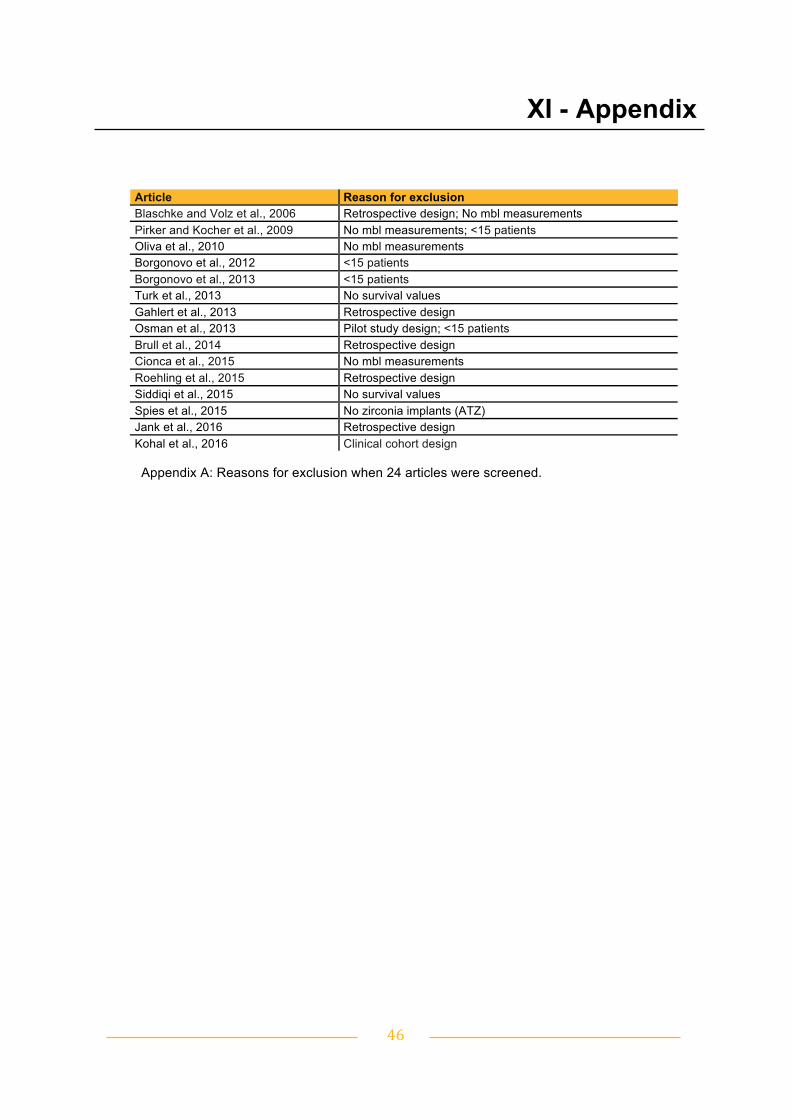
XI - Appendix
Appendix A: Reasons for exclusion when 24 articles were screened.
Article Reason for exclusion Blaschke and Volz et al., 2006 Retrospective design; No mbl measurements Pirker and Kocher et al., 2009 No mbl measurements; <15 patients Oliva et al., 2010 No mbl measurements Borgonovo et al., 2012 <15 patients Borgonovo et al., 2013 <15 patients Turk et al., 2013 No survival values Gahlert et al., 2013 Retrospective design Osman et al., 2013 Pilot study design; <15 patients Brull et al., 2014 Retrospective design Cionca et al., 2015 No mbl measurements Roehling et al., 2015 Retrospective design Siddiqi et al., 2015 No survival values Spies et al., 2015 No zirconia implants (ATZ) Jank et al., 2016 Retrospective design Kohal et al., 2016 Clinical cohort design

47
XII - Index
I. Abstract / Resumo V
II. List of abbreviations and acronyms VIII
III. Introduction 9
1. Osseointegration: a major factor to implant stability 9
2. State of the art: implant evolution 13
IV. Materials and Methods 17
V. Results 22
VI. Discussion 34
VII. Conclusions 39
VIII. Implications for clinical practice 39
IX. Acknowledgements 41
X. Bibliography 42
XI. Appendix 46
XII. Index 47