InTech-Use of Non Invasive Brain Stimulation in Stroke
25
8 Use of Non-Invasive Brain Stimulation in Stroke Bulent Turman 1 and Sultan Tarlaci 2 1 School of Medicine, Bond University, 2 Özel Ege Sa ğlõk Hospital, Izmir 1 Australia 2 Turkey 1. Introduction Stroke is the leading cause of permanent disability in the Western world (Kolominsky-Rabas et al., 2001). Clinically, stroke is defined as a neurological deficit of cerebrovascular cause that persists beyond 24 hours. The clinical outcome of a stroke depends on which part of the brain is injured and how severely it is affected. The most common symptom of a stroke is sudden weakness or numbness of the face, arm or leg, most often on one side of the body. Other symptoms include: confusion, difficulty in speech production or comprehension; visual deficits; difficulty walking, dizziness, loss of balance or coordination; severe headache with no known cause; fainting or unconsciousness. The clinical presentation is closely associated with the affected artery, which is occluded by a clot or plaque (ischemic stroke), or ruptured (hemorrhagic stroke), and the extent of tissue infarct (Amarenco et al., 2009). In recent decades, the introduction of thrombolysis and the establishment of stroke units in hospitals have led to a significant reduction of mortality rate after stroke (Howard et al., 2001). However, declining mortality rate has resulted in increased proportion of patients to be left with moderate to severe disability, affecting their daily activities. It is now well- established that early rehabilitation provides more effective recove ry of function than would occur in the natural course of recovery (Maulden et al., 2005). However, in most cases this recovery is still incomplete. Up to 60% of patients still have impaired manual dexterity six months after the onset of stroke (Kolominsky-Rabas et al., 2001; Kwakkel et al., 2002). Advances in neuroscience in the last two decades unequivocally established the brain’s capacity to reorganize itself (Nudo, 2007). This has led to the development of various techniques that could potentially improve the rehabilitation of stroke patients. Most widely researched and experimented non-invasive techniques are transcranial magnetic stimulation (TMS) and transcranial direct current stimulation (tDCS). 2. Non-invasive brain stimulation methods 2.1 Transcranial Magnetic Stimulation (TMS) In the 1980’s Merton and Morton (1980) were able to stimulate the human brain non- invasively by means of transcranial electrical stimulation (TES). However, contraction of scalp muscles and activation of nociceptive fibres evoked intense unpleasant pain sensation. www.intechopen.com
Transcript of InTech-Use of Non Invasive Brain Stimulation in Stroke

7/23/2019 InTech-Use of Non Invasive Brain Stimulation in Stroke
http://slidepdf.com/reader/full/intech-use-of-non-invasive-brain-stimulation-in-stroke 1/25
8
Use of Non-Invasive Brain Stimulation in Stroke
Bulent Turman1 and Sultan Tarlaci2 1School of Medicine, Bond University,
2Özel Ege Sa ğlõk Hospital, Izmir1 Australia
2Turkey
1. Introduction
Stroke is the leading cause of permanent disability in the Western world (Kolominsky-Rabaset al., 2001). Clinically, stroke is defined as a neurological deficit of cerebrovascular causethat persists beyond 24 hours. The clinical outcome of a stroke depends on which part of thebrain is injured and how severely it is affected. The most common symptom of a stroke issudden weakness or numbness of the face, arm or leg, most often on one side of the body.Other symptoms include: confusion, difficulty in speech production or comprehension;visual deficits; difficulty walking, dizziness, loss of balance or coordination; severe headachewith no known cause; fainting or unconsciousness. The clinical presentation is closelyassociated with the affected artery, which is occluded by a clot or plaque (ischemic stroke),or ruptured (hemorrhagic stroke), and the extent of tissue infarct (Amarenco et al., 2009).
In recent decades, the introduction of thrombolysis and the establishment of stroke units inhospitals have led to a significant reduction of mortality rate after stroke (Howard et al., 2001). However, declining mortality rate has resulted in increased proportion of patients tobe left with moderate to severe disability, affecting their daily activities. It is now well-established that early rehabilitation provides more effective recovery of function than wouldoccur in the natural course of recovery (Maulden et al., 2005). However, in most cases thisrecovery is still incomplete. Up to 60% of patients still have impaired manual dexterity sixmonths after the onset of stroke (Kolominsky-Rabas et al., 2001; Kwakkel et al., 2002).
Advances in neuroscience in the last two decades unequivocally established the brain’scapacity to reorganize itself (Nudo, 2007). This has led to the development of varioustechniques that could potentially improve the rehabilitation of stroke patients. Most widelyresearched and experimented non-invasive techniques are transcranial magnetic stimulation(TMS) and transcranial direct current stimulation (tDCS).
2. Non-invasive brain stimulation methods
2.1 Transcranial Magnetic Stimulation (TMS)
In the 1980’s Merton and Morton (1980) were able to stimulate the human brain non-invasively by means of transcranial electrical stimulation (TES). However, contraction ofscalp muscles and activation of nociceptive fibres evoked intense unpleasant pain sensation.
www.intechopen.com
7/23/2019 InTech-Use of Non Invasive Brain Stimulation in Stroke
http://slidepdf.com/reader/full/intech-use-of-non-invasive-brain-stimulation-in-stroke 2/25
Topics in Neuromodulation Treatment168
Introduction of TMS by Barker, Jalinous and Freeston in 1985 instantly attracted moreattention over electrical stimulation, as in this method the current in the coil could activatecortical structures without causing pain. In the following two decades there have beensignificant advances in this method both technologically and scientifically.
2.1.1 Basics of TMS
TMS is based on the concept of electromagnetic induction. It involves the generation of abrief but strong magnetic field capable of activating cortical elements in the brain ofconscious subjects without causing pain (Wasserman et al., 2008). This magnetic field isderived from a changing primary electric current circulating in a coil which then passesthrough the skull to induce a secondary electric current capable of altering the neurons’transmembrane potential. Rapid depolarization of the membrane leads to action potentialgenerations (Figure 1).
Fig. 1. TMS setup. A brief pulsed electric current passes through the coil, which results in arapidly changing magnetic field that is perpendicular to the coil’s surface. This magneticfield passes through the skull and scalp, and generates an electric field flowing in theopposite direction to the flow of current in the coil. This current leads to the activation ofexcitable structures in the brain tissue (from Fyre et al., 2008).
The extent of activation within the cortex during magnetic stimulation is influenced by a
number of variables, including the coil shape and its position over the head (Tingset al
.2005); number, intensity and frequency of pulses; output waveforms (monophasic vs.biphasic); induced current direction; and the anatomy of the region stimulated. For example,a circular shaped coil generates a relatively large and diffuse magnetic field over the brain,whereas a figure-of-eight (butterfly) coil produces a more focalized field (Wassermann et al., 2008). More recently introduced coils, such as the double-cone coil and H-coil, weredesigned to stimulate deeper structures within the brain (Hayward et al., 2007; Zangen et al.,
2005). However, in general the depth of stimulation is restricted to 2-3 cm below the scalpand the stimulated area within the cortex to around 1-3 cm2. Increasing the stimulationintensity to activate deeper brain regions would result in wider and stronger stimulation ofmore superficial areas.
www.intechopen.com

7/23/2019 InTech-Use of Non Invasive Brain Stimulation in Stroke
http://slidepdf.com/reader/full/intech-use-of-non-invasive-brain-stimulation-in-stroke 3/25
Use of Non-Invasive Brain Stimulation in Stroke 169
2.1.2 TMS techniques
A number of different stimulation techniques and paradigms have been introduced over thepast two decades. Initially single-pulse TMS was used to primarily evaluate the excitability
changes of the motor cortex and its output. It is still widely used to determine the bestlocation (hot-spot) of recorded muscles within the motor homunculus and theactive/passive motor thresholds, and to assess the effect of interventions on variousintracortical influences (Wassermann et al., 2008). Paired-pulse TMS utilizes two individualmagnetic pulses, separated by a variable inter-stimulus interval (ISI). This method is used toevaluate the intracortical influences of magnetic stimulation, such as short- and long-interval intra-cortical inhibition (SICI and LICI) and intra-cortical facilitation (ICF) (forreview see Reis et al., 2008). The ICF of a test motor evoked potential (MEP) elicited from thetarget muscle can be observed at ISIs of 6-25 ms, using a subthreshold conditioning stimulus(CS) to influence the response to a subsequent suprathreshold test stimulus (TS) (Kujirai etal., 1993). This effect tends to become stronger with increasing CS intensity and weaker with
increasing TS intensity. A SICI on the other hand, can be observed when a subthreshold CSsuppresses the MEP evoked in response to the suprathreshold TS if the interval between thestimuli is 5 ms or less (Kujirai et al., 1993). In LICI a suprathreshold CS strong enough toproduce an MEP in the target muscle could suppress an MEP to a later stimulus of the sameintensity if the ISI was 50-200 ms.
Paired associative stimulation (PAS) technique involves applying pairs of peripheral andcentral stimuli repeatedly (Stefan et al. 2000). When around 100 peripheral electrical stimuliand central TMS pulses are paired at an ISI of 25 ms over 30 min, the cortical excitabilityincreases. At an ISI of 10 ms a reduced cortical excitability is observed.
Technical advances in magnetic stimulator and coil designs led to more recent TMS
techniques based on delivery of a series of pulses by means of multiple capacitors. Thismethod, referred to as “repetitive transcranial magnetic stimulation” (rTMS), enabledresearchers and clinicians to explore the potential benefits of TMS in clinical conditions(Pascual-Leone et al. 1994; Wassermann et al., 2008; Hoogendam et al., 2010).
2.1.3 Clinical and diagnostic applications of TMS
Since its introduction, TMS has been used to measure and evaluate the motor evokedpotential (MEP) responses from target muscles and commonly applied as a non-invasivetool to clinically evaluate aspects of sensorimotor cortex and pyramidal tract function (Chenet al., 2008). Motor threshold (MT) measurements are useful in determining the level of
excitability within the motor cortex. MT is defined as the lowest stimulation required for asingle pulse to produce a criterion amplitude MEP on a pre-specified fraction of consecutivetrials (Wassermann et al., 1998). MT measurements are also useful in establishing andfollowing-up the hemispheric differences in clinical conditions, such as stroke. MEPamplitude and onset latency measurements are also useful parameters in the assessment andcomparison of motor cortex excitability and its output. For example, in pathologiesinvolving upper motor neurons, such as multiple sclerosis, MEP amplitudes are oftenreduced or absent, and central motor conduction times are prolonged (Cruz-Martínez et al. 2000). Somatosensory information processing at the cortical level is also influenced by TMSand can be evaluated by psychophysical measurements, such as vibration detectionthresholds (Morley et al. 2007).
www.intechopen.com

7/23/2019 InTech-Use of Non Invasive Brain Stimulation in Stroke
http://slidepdf.com/reader/full/intech-use-of-non-invasive-brain-stimulation-in-stroke 4/25
Topics in Neuromodulation Treatment170
Cerebral hemispheres exert various influences on each other through interhemisphericconnections. Therefore, TMS could be useful for investigating inter-hemispheric dynamicswhich can be investigated using paired-pulse TMS. In this paradigm, a conditioning stimulus is applied to one hemisphere, followed by a test stimulus applied to the other. Although a
number of studies have reported some complex and inconsistent interhemisphericfacilitatory influences dependent on background motor activity, coil position andconditioning stimulus intensity (Hanajima et al. 2001, Chowdhury & Matsunami, 2002),more consistent effects are observed in interhemispheric inhibition. The response to the teststimulus can be inhibited by the conditioning stimulus at inter-stimulus interval range of 6-50 ms (Ferbert et al., 1992; Daskalakis et al. 2002). These transcallosal effects appear to beimportant in influencing the cortical excitability. For example, interhemispheric inhibitionabnormalities have been found in patients with amyotrophic lateral sclerosis (Karandreas etal., 2007).
Another method, called “triple stimulation technique (TST)”, delivers a single magnetic pulse
in association with two timed peripheral electrical pulses and is used to evaluate theintegrity of neuronal pathways by means of collision (Magistris et al. 1999). It is reportedthat in amyotrophic lateral sclerosis patients TST provides a quantitative tool for assessingthe upper motor neuron conduction failure and when used together with silent periodmeasurements provides a sensitive diagnostic tool (Attarian et al., 2007).
In short, TMS has been shown to be an important non-invasive diagnostic tool forevaluation of certain aspects of motor cortex function and its output. In clinical settings TMScould therefore be a useful tool to determine subclinical presentations in which clear clinicalsigns are not yet present or indecisive.
The most talked about adverse effect of magnetic brain stimulation is the induction of
seizures. A number of cases of accidental seizures induced by rTMS have been reportedover the years (total of 16 cases from 1998 to 2008). However, given the large number ofsubjects and patients who have undergone rTMS in over 3,000 published studies, it issuggested that the risk of rTMS to induce seizures is very low (Rossi et al., 2009).Comprehensive screening of participants with regards to medication and predisposition toseizures will certainly further eliminate the possibility of this adverse effect.
2.1.4 Therapeutic applications of TMS
Since the introduction of repetitive stimulation capable stimulators, rTMS has beenincreasingly investigated and applied as a therapeutic tool. Using ‘simple’ rTMS, in which a
series of regularly repeated magnetic pulses are delivered in trains and then separated byconstant inter-train intervals, it is possible to induce changes to the excitability of motorcortex that outlast the stimulation period from several minutes up to 30 minutes (Touge etal., 2001; Peinemann et al., 2004). In this method, stimulation frequency plays a crucial role inproducing selective changes in motor cortex excitability. Overall, low frequency (< 5Hz)rTMS results in suppression of corticospinal excitability, while high frequency (≥5 Hz)stimulation leads to facilitatory after-effects (for review see Siebner & Rothwell, 2003).
Another form of repetitive stimulation involves patterned stimuli. Theta-burst stimulation (TBS) is a burst of three to five pulses at high frequency (30-100 Hz) delivered at a repeatedfrequency (usually 5 Hz). This method has been shown to be safe and effective in producing
www.intechopen.com

7/23/2019 InTech-Use of Non Invasive Brain Stimulation in Stroke
http://slidepdf.com/reader/full/intech-use-of-non-invasive-brain-stimulation-in-stroke 5/25
Use of Non-Invasive Brain Stimulation in Stroke 171
changes in the excitability of motor systems (Huang & Rothwell, 2004). The typical form ofTBS contains three pulse 50Hz bursts given every 200 ms (i.e. at 5 Hz) at the stimulusintensity of 80% of active motor threshold. When a 2-sec train of TBS is given every 10 sec(intermittent TBS – iTBS), the cortical excitability is enhanced due to a long-term potentiation
(LTP) like effect. Conversely, when bursts are given every 200 ms continuously withoutinterruption (continuous TBS – cTBS), the cortical excitability is suppressed due to a long-term depression (LTD) like effect (Huang et al., 2005).
Fig. 2. Theta bust stimulation patterns. A usual TBS contains 3-pulse 50Hz bursts givenevery 200 ms. When a 2-sec train of TBS is given every 10 sec (iTBS), the cortical excitabilityis enhanced, while the excitability is suppressed when bursts are given every 200 mscontinuously (cTBS) (from Huang, 2010).
More recently, quadripulse stimulation (QPS) has been introduced as a patterned rTMS
protocol in which repeated trains of four mono-phasic pulses are separated by inter-
stimulus intervals of 1.5-1250 ms to produce facilitation (at short intervals) or inhibition
(at longer intervals). This protocol appears to induce long-term changes in cortical
excitability, probably through a modulatory action on intracortical excitatory circuits
(Hamada et al., 2008).Over the years, many studies have investigated the therapeutic use of rTMS in psychiatric
disorders, particularly in depression. For this clinical condition many stimulation paradigms
and durations have been trialled. At the end of 2008, the United States Food and Drug
Administration (FDA) approved the NeuroStar TMS Therapy System™ for “the treatment of Major Depressive Disorder in adult patients who have failed to achieve satisfactory improvement from one prior antidepressant medication at or above the minimal effective dose and duration in thecurrent episode”. However, there is still no consensus on the treatment protocols and
durations, and the efficacy, tolerability, cost and inconvenience of TMS over
electroconvulsive therapy and medication are still debatable (Rasmussen, 2011).
www.intechopen.com
7/23/2019 InTech-Use of Non Invasive Brain Stimulation in Stroke
http://slidepdf.com/reader/full/intech-use-of-non-invasive-brain-stimulation-in-stroke 6/25
Topics in Neuromodulation Treatment172
Other clinical conditions in which rTMS has been investigated as a therapeutic tool includeamyotrophic lateral sclerosis (ALS), dystonia, migraine and stroke. Studies on ALS patientsrevealed some promising preliminary data. However, recent studies have demonstrated alack of significant long-term beneficial effects of rTMS on neurological deterioration in ALS
(Dileone et al., 2011).
Both inhibitory (low frequency) and excitatory (high frequency) rTMS over the primarymotor cortex (M1) appear to reduce chronic pain. A number of studies have assessed theefficacy of rTMS in patients with drug-resistant chronic pain of various causes and a meta-analysis showed that rTMS was associated with a significant reduction in pain (Lima andFregni, 2008). Analgesic effects were also shown after stimulation of other cortical areas,such as the prefrontal cortex. However, as the induced effects are relatively short duration,the therapeutic use of rTMS in chronic pain is limited, unless repeated sessions over severalweeks are considered (for review see Lefaucheur et al., 2008).
2.2 Transcranial Direct Current Stimulation (tDCS)
Transcranial direct current stimulation (tDCS) is a non-invasive, low-cost and easy-to-usetechnique that has the potential to modify cortical excitability and behavior in a range ofclinical and experimental conditions. Historically, strong electrical currents have beendelivered to patients for the relief of headache and epilepsy using torpedo electric fish(Kellaway, 1946). Since the rediscovery of tDCS about 10 years ago, interest in this methodhas grown significantly.
2.2.1 Basics of tDCS
The constant direct current delivered to the brain in tDCS is caused simply by positioning
the two poles of an electric battery-based stimulator to the brain (Nitsche & Paulus, 2000). In
order to stimulate the motor cortical region, the stimulating (active) electrode is placed overthe motor cortex (M1) and the reference electrode over the contralateral supraorbital ridge
or the neck region. More accurate stimulation of a representation within M1, such as thehand area, could be achieved after TMS assessment of the hand area’s “hot spot”. Two
surface conductive rubber electrodes (sized 25 cm2 - 35 cm2) attached to the device are
usually placed inside sponges soaked in NaCl solution. The sponge-electrodes are then
placed and kept on their desired region by a non-conducting rubber band, which is strappedfirmly around the subject’s head (Figure 3). Current intensities used during sessions vary
between 1 mA - 2 mA and are commonly applied for 10 to 20 minutes.
Physical modeling of currently available stimulators suggests that only around 50% of theapplied current is actually delivered to the brain tissue. The remaining current is shuntedacross the scalp following the path of least resistance towards the other electrode (Mirandaet al., 2006). However, the portion of the current which does eventually reach the brain canbe sufficient in altering neuronal activity (Wagner et al., 2007). The current delivered bytDCS cannot directly generate action potentials in cortical neurons, as the electric field in thebrain tissue is not capable of inducing a rapid depolarization (Nitscheet al., 2008). Therefore,tDCS might be considered a neuromodulatory intervention. The electric field modifies theexcitability of exposed cells by a tonic depolarization or repolarization of their restingmembrane potential by only few millivolts. Evidence that the effects of anodal stimulation
www.intechopen.com
7/23/2019 InTech-Use of Non Invasive Brain Stimulation in Stroke
http://slidepdf.com/reader/full/intech-use-of-non-invasive-brain-stimulation-in-stroke 7/25
Use of Non-Invasive Brain Stimulation in Stroke 173
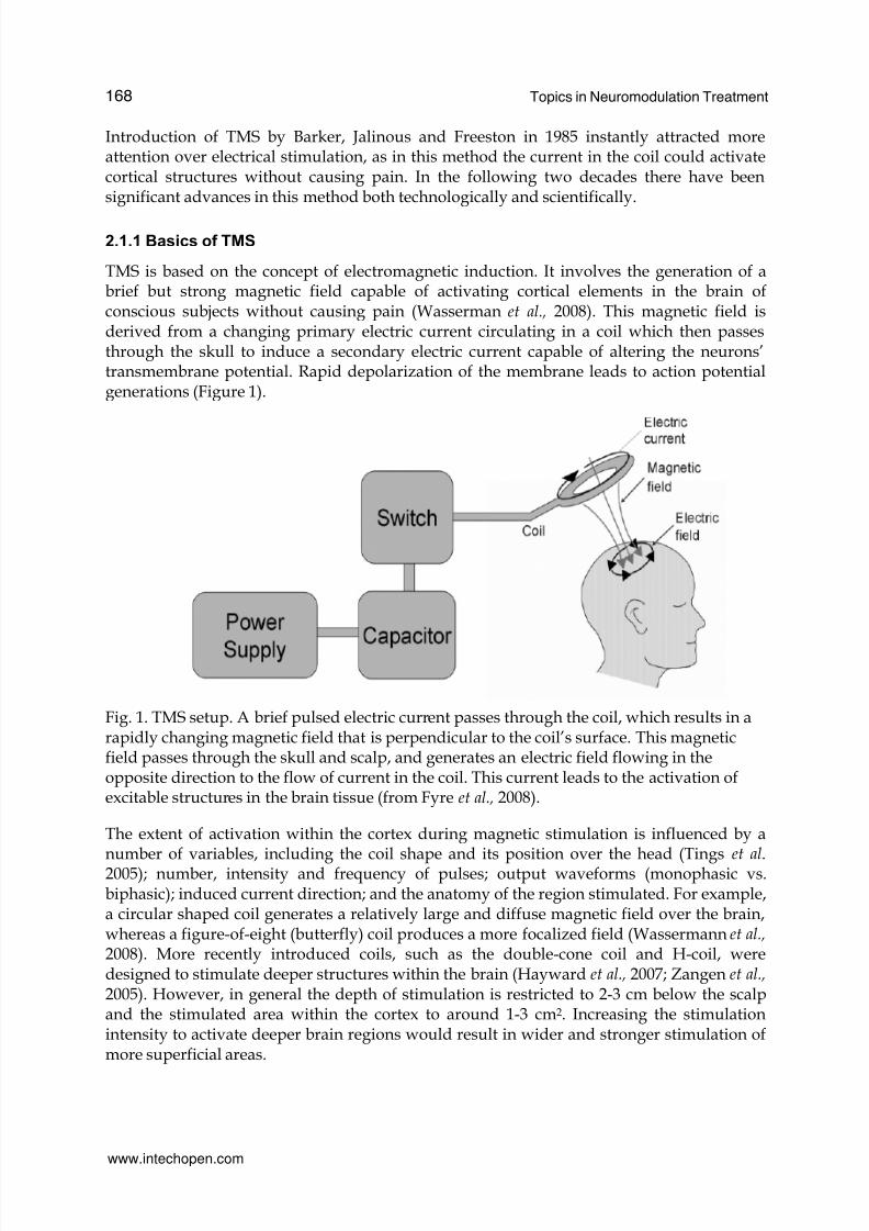
Fig. 3. tDCS setup and montage. (A) The setup using a battery-operated direct currentstimulator connected with two electrodes. One electrode (active) is positioned over C3(corresponding to the precentral gyrus), and the reference electrode is positioned over thecontralateral supraorbital region. If current flows from C3 to the supraorbital region, then the
tissue underlying C3 is subjected to anodal (increase in excitability) stimulation. If current isreversed, then the tissue underlying C3 is subjected to cathodal (decrease in excitability)stimulation. (B) Regional cerebral blood increases in the motor region underlying the electrodepositioned over C3 after anodal stimulation. Regional cerebral blood was determined using anon-invasive arterial spin-labeling technique (from Schlaug et al., 2008).
appear to be solely dependent on changes in membrane potential comes from studies usingpharmacological agents. For example, while calcium channel blocker flunarizine reducesand the sodium channel blocker carbamezipine abolishes the effects of anodal stimulation,NMDA receptor antagonist dextromethorphane does not alter current-generated excitabilitychanges (Nitsche et al., 2003a). In terms of the effects of tDCS on cortical interneurons,
anodal tDCS does not modify the TMS measures of either glutaminergic interneurons(intracortical facilitation – ICF) or GABAergic interneurons (short-interval cortical inhibition– SICI); suggesting that GABAergic or glutaminergic interneuronal pools are notsignificantly modulated (Nitsche et al., 2005). During cathodal stimulation, blockade ofcalcium or sodium channels does not alter the effects of tDCS, suggesting ahyperpolarisation of neurons generated by tDCS itself (Nitsche et al., 2003a). However, ICFand the input/output curve for TMS motor threshold were modulated during cathodalstimulation (Nitsche et al., 2005), suggesting that the membrane potential of glutaminergicinterneurones, rather than pyramidal neurons, is modulated by tDCS (for review see Stagg& Nitsche, 2011). Overall, the evidence so far suggests that the modulation observed withtDCS are shaped by a combination of non-synaptic mechanisms, which alter the resting
www.intechopen.com

7/23/2019 InTech-Use of Non Invasive Brain Stimulation in Stroke
http://slidepdf.com/reader/full/intech-use-of-non-invasive-brain-stimulation-in-stroke 8/25
Topics in Neuromodulation Treatment174
membrane potential of neurons, and synaptic mechanisms, which alter the signalingstrength of neurons.
2.2.2 Variables in the application of tDCS
The current density in the tissue is the quotient of current strength and electrode size.Hence, stimulation efficacy can be augmented by either increasing the current strength orreducing the electrode size (Nitsche & Paulus, 2000). Furthermore, the duration ofstimulation sessions also affects the strength of the tDCS induced response; longer sessiondurations result in prolonged after-effects (Nitsche & Paulus, 2001; Nitsche et al., 2003b).
The direction of current flow is another parameter that influences the electrical stimulationeffects. The population of neurons exposed to the electrical field and the shifts in theirmembrane potential depend mainly on the positions of the electrodes and their polarity. IntDCS, both positive electrode (anode) and negative electrode (cathode) are used for
stimulation. In this circuit the current flows from the cathode to the anode. The positioningof these electrodes on the scalp is important in determining the overall effects elicited in theunderlying cortex. For example, during anodal tDCS of the primary motor cortex, the anodeis generally placed over the primary motor cortex (M1) and the cathode over thecontralateral supraorbital region. In this montage most studies report an increase in thecortico-motor excitability (Nitsche & Paulus, 2001; Jeffery et al., 2007). Conversely, reversingthe current flow (cathodal stimulation) generally diminishes the cortical excitability(Ardolino et al., 2005).
In the literature, so far over 100 studies with tDCS in healthy and patient populations havebeen published; with no serious side effects. At the start of stimulation, most subjects reporta slight itching sensation, which then normally fades. It is possible to reduce or avoid thissensation by ramping the current up and down at the beginning and end of session. Poreisz et al., (2007) in a group of 567 subjects, reported most commonly a mild tingling sensation(~70%), moderate fatigue (~35%) and slight itching under the electrode (~30%), and in ≤10%of cases, headache, nausea and insomnia. Other studies, for example evidence of neuronaldamage as assessed by serum neuron-specific enolase, MRI measures of edema usingcontrast-enhanced and diffusion-weighted MRI measures, EEG waveform analyses andneuropsychological measures, reported no evidence of neural damage or brain pathology(for review see Stagg & Nitsche, 2011).
The large size of stimulating electrodes could result in the stimulation of a larger corticalregion then intended. Furthermore, as the reference electrode is not physiologically inert
because of current flow between electrodes, there might be modulatory effects in remotebrain areas. Therefore, other brain regions and structures between electrodes should betaken into consideration during the application of tDCS. Moreover, modulations of corticalexcitability can be focused by reducing the size of the stimulating electrode and byincreasing the size of the reference electrode (Nitsche et al., 2007). An extracephalic (e.g. neckregion) reference could be used to avoid the undesirable effects of two electrodes withopposite polarities over the brain (Nitsche & Paulus, 2000).
As subjects only occasionally experience any sensation related to the stimulation, controlledplacebo sessions could be conducted without the need for additional equipment orattachments. During sham stimulation the stimulator can be initially ramped up (around 10
www.intechopen.com

7/23/2019 InTech-Use of Non Invasive Brain Stimulation in Stroke
http://slidepdf.com/reader/full/intech-use-of-non-invasive-brain-stimulation-in-stroke 9/25
Use of Non-Invasive Brain Stimulation in Stroke 175
sec), and after a 30 sec period of stimulation it can be slowly turned down (within 10 sec). Withthis method placebo and real stimulation sessions are indistinguishable (Gandiga et al., 2006).It should be noted that with motor cortex stimulation, strong cognitive effort by the subjectunrelated to the stimulated area, as well as strong activation of the stimulated motor cortex by
voluntary prolonged muscle contraction abolishes the effects of tDCS (Antal et al., 2007)
2.2.3 Time course and after-effects of tDCS
With short duration (seconds) tDCS, changes in cortical excitability are observed during thestimulation period, but these effects do not outlast the stimulation itself (Nitsche & Paulus,2000). However, when applied for several minutes longs lasting excitability shifts areproduced. For example, around 10 minutes of tDCS can produce stable effects for up to anhour (Nitsche & Paulus, 2001).
The changes in cortical output measures that outlast a tDCS session are dependent on
membrane depolarization. The after-effects induced by anodal stimulation could beabolished by calcium or sodium channel blockers or prolonged by NMDA receptor agonists(Nitsche et al., 2003a, 2004). Results from other studies using TMS mediated measures and
neuropharmacological applications suggest that the after-effects of anodal tDCS aredependent on modulation of both GABAergic and glutaminergic synapses, and these effectsare modulated by acetylcholine, serotonin and catecholamines (for review see Stagg &Nitsche, 2011).
In order to achieve relatively stable changes in cortical function, repeated sessions of tDCS isnecessary. For example, recently it was reported that tDCS enhances motor skill acquisitionover multiple days through an effect on consolidation (Reis et al., 2009). However, the
optimal number and duration of sessions, as well as intersession intervals will depend onthe objective of the study or therapeutic application, and requires more research.
2.2.4 Therapeutic applications of tDCS
tDCS has been shown to have beneficial effects in a wide range of clinical pathologies; suchas refractory epilepsy (Fregni et al., 2006), stroke (Fregni et al., 2005; Hummel et al., 2005) andvarious pain conditions (for review see O’Connell et al., 2011), as well as psychiatricconditions, like depression and addiction (Arul-Anandam & Loo 2009; Utz et al., 2010).However, the measurable effects induced in a single session are usually short lived. Withrepeated session tDCS, growing number of clinical trials is reporting long-term benefits, in
particular for depression. For example, in a recent double-blind clinical trial with 40 patientswith major depression, significantly large reductions in depression scores were reportedafter dorsolateral prefrontal cortex (DLPFC) anodal tDCS applied for 10 sessions during a 2-
week period (Boggio et al., 2008). These results suggest promising potential for tDCS as anantidepressant treatment.
3. Use of non-invasive brain stimulation in stroke
Following stroke, the neuroplastic changes within the brain lead to reorganization that isattributable to spontaneous recovery of function. Possible mechanisms of suchreorganization include; axonal and/or dendritic regeneration or sprouting, reorganization
www.intechopen.com

7/23/2019 InTech-Use of Non Invasive Brain Stimulation in Stroke
http://slidepdf.com/reader/full/intech-use-of-non-invasive-brain-stimulation-in-stroke 10/25
Topics in Neuromodulation Treatment176
within the lesioned cortical region by means of synaptic modulation, and remapping offunctional representations from the lesioned region onto neighboring unaffected areassurrounding the lesion or homologous areas within the unaffected hemisphere.
During local ischemia various cytotoxic and metabolic reactions result in the loss ofstructural and functional integrity of neural tissue (Schallert et al., 2000). However, earlyrepair mechanisms, such as expression of developmental proteins and other substrates ofmolecular plasticity, as well as structural changes, such as regeneration and sprouting,modulation of synaptic plasticity, changes in cortical excitability due to neurotransmitteralterations take place locally and in remote areas of the brain (Witte & Stoll 1997). There isincreasing evidence that suggests functional reorganization in both hemispheres. Functionalmagnetic resonance imaging (fMRI) studies reveal bilateral activation in recovered strokepatients (Gerloff et al., 2006; Nair et al., 2007).
A network of cortical and subcortical areas constitutes the motor system. The final motor
output is determined by complex interactions between multiple excitatory and inhibitorycircuits within and between these areas. After stroke, the balance in this system could bevitally disturbed as a result of damage to neurons or their fibers within the white matterwhich connects these areas. For example, in recovered stroke cases, magnetic stimulationover the dorsal premotor cortex, the superior parietal lobe, as well as the primary motorcortex results in significant interference with recovered finger movement performance(Lotze et al., 2006). Furthermore, experimental results using TMS in stroke patients suggestthat the motor output from the lesioned hemisphere could be further reduced bypathologically enhanced inhibitory influences from the intact hemisphere (Murase et al.,2004; Duque et al., 2005; Hummel & Cohen, 2006). Although the exact mechanism of thisinterhemispheric interaction is still unclear, the possibility that suppressing the inhibitory
influences exerted by the intact hemisphere could improve recovery has gained interest inrecent years.
As stated earlier, depending on the stimulation parameters, cortical excitability can bereduced (inhibition) or enhanced (facilitation). Therefore, non-invasive brain stimulationcould accelerate, facilitate or potentiate the functional recovery process and provide betterrehabilitation outcomes. TMS and tDCS are the most extensively researched methods instroke recovery and rehabilitation (for review see Nowak et al., 2010). These techniques notonly cause a local change in cortical excitability, but can also evoke changes within remoteparts of the cortical motor system, hence improve recovery after stroke (Nowak et al., 2008;Ameli et al., 2009; Grefkes et al., 2010).
3.1 Application of TMS in stroke
In the last decade a number of studies using rTMS in stroke patients have been conducted.These include, single session interventions, in which patients are assessed before and afterrTMS and longer term treatment strategies in which patients are given daily sessions ofrTMS for up to two weeks. In multiple session interventions rTMS is usually combined withconventional physical therapy to assess and compare the benefits of rTMS in rehabilitation(for review see Khedr & Abo-El Fetoh, 2010; Nowak et al., 2010). Majority of these studieshave been conducted in chronic stroke patients whose baseline performance is likely to bestable, compared to acute and subacute stroke cases.
www.intechopen.com

7/23/2019 InTech-Use of Non Invasive Brain Stimulation in Stroke
http://slidepdf.com/reader/full/intech-use-of-non-invasive-brain-stimulation-in-stroke 11/25
Use of Non-Invasive Brain Stimulation in Stroke 177
So far, there have been over twenty clinical studies conducted using low or high frequencysimple rTMS, or theta-burst stimulation (TBS) of the lesioned or intact hemisphere in acuteor chronic patients (for review see Khedr & Abo-El Fetoh, 2010). For example, Koganemaruet al., (2010) used 5 Hz rTMS of the upper-limb area of the primary motor cortex, combined
with extensor motor training, and suggested that combining motor training with rTMS canfacilitate use-dependent plasticity and achieve functional recovery of motor impairmentsthat cannot be accomplished by either intervention alone. Overall, rTMS gives a 10–30%improvement over sham in a range of performance measures, from simple reaction times totimed behavioral tests. In addition, the effects of multiple session intervention tend to besimilar in size but longer lasting than those seen in single session trials.
Even more complex intervention protocols, by stimulating multiple target areas have beentrialed. For example, in a group of thirty chronic stroke patients, comparison of unilateraland bilateral rTMS (1 Hz over intact hemisphere and 10 Hz over affected hemisphere)revealed improved motor training effect on the paretic hand after bilateral rTMS (Takeuchi
et al., 2008).
Although still relatively few, there are studies conducted to investigate the possible benefitsof rTMS in other disabilities associated with stroke; such as, dysphagia, aphasia andhemispatial neglect. The underlying concept of rTMS treatment is based on “upregulating”the lesioned hemisphere or “downregulating” the intact hemisphere (for review see Platz &Rothwell, 2010). Altered connectivity within the cortex as a result of stroke influences themodulatory effects of afferent inputs (Tarlaci et al., 2010). Therefore, combining TMSintervention with afferent inputs, such as vibrotactile stimuli could also be effective.
Overall, the application of rTMS as a therapeutic tool is still in its infancy. According toavailable evidence, cortical magnetic stimulation could be an effective method forimproving functional recovery of acute and chronic stroke. Table 1 summarizes the studiesundertaken using rTMS. Although the majority of results report improvements in variousbehavioral functions, the overall methodology remains to be optimized, in particularregarding the number and duration of rTMS sessions, the site, frequency and intensity ofstimulation and the exact timing of rTMS application after stroke
3.2 Application of tDCS in stroke
Human studies using electrical brain stimulation can be divided into invasive and non-invasive. The invasive method principally involves implantation of epidural electrodesthrough a small craniotomy around a “hot spot” within the perilesional area determinedby fMRI. Cortical stimulation is then applied together with physical therapy. Initialcortical stimulation feasibility studies in combination with a motor rehabilitation trainingtargeting the affected arm and hand reported significant improvements compared tocontrol patients receiving only rehabilitation (Brown et al., 2006; Levy et al., 2008).However, in a subsequent larger, multi-center study (Everest Clinical Trial) involving 174chronic stroke patients (implant and control groups) who underwent six weeks of upperlimb rehabilitation, the outcome measures did not meet its primary efficiency end-point at4-week follow-up, with improvement of 30% in both implant and control groups (Harvey
& Winstein, 2009). It is clear that more basic and clinical research into the efficacy ofinvasive cortical electrical stimulation is needed.
www.intechopen.com

7/23/2019 InTech-Use of Non Invasive Brain Stimulation in Stroke
http://slidepdf.com/reader/full/intech-use-of-non-invasive-brain-stimulation-in-stroke 12/25
Topics in Neuromodulation Treatment178
Study StimulationSide
Lesionlocation
Time ofstroke
Stimulus Behavioral resultson affected hand
Khedr et al.,
2005
IL 26 cortical,
26 subcortical
acute 3 Hz, 10 daily
stimulation
sessions
Improved hand
function
(ScandinavianStroke Scale,
National
Institute of Health
Stroke Scale
Scale, Barthel
index)
Mansur et al.,
2005
CL 10 subcortical subacute,
chronic
1 Hz Shortened simple
and choice
reaction times,
improvement ofhand function
(Purdue Pegboard
Test)
Takeuchi et
al., 2005
CL 20 subcortical chronic 1 Hz Improved peak
pinch
acceleration
Boggio et al.,
2006
CL one
subcortical
chronic 1 Hz Improved hand
function (clinical
testing), no change
in spasticity(modified
Ashworth scale for
spasticity)
Fregni et al.,
2006
CL 2 cortical,
13 subcortical
chronic 1Hz , 5 daily
stimulation
sessions
Shortening of
simple and choice
reaction times,
improvement of
hand function
(Jebsen-Taylor
Hand FunctionTest, Purdue
Pegboard Test)
Kim et al.,
2006
IL 5 cortical,
10 subcortical
chronic 10 Hz Improved
movement
accuracy and
movement time
(sequential
finger movement
task)
www.intechopen.com

7/23/2019 InTech-Use of Non Invasive Brain Stimulation in Stroke
http://slidepdf.com/reader/full/intech-use-of-non-invasive-brain-stimulation-in-stroke 13/25
Use of Non-Invasive Brain Stimulation in Stroke 179
Study StimulationSide
Lesionlocation
Time ofstroke
Stimulus Behavioral resultson affected hand
Malcom et al.,
2007
IL 11 cortical,
8 subcortical
chronic 20 Hz,
followed by
constraintinduced
movement
therapy, 10
daily
stimulation
sessions
Improved hand
function (Wolf
Motor FunctionTest, Motor
Activity Log) after
constraint induced
movement
therapy, no
additive effect of
rTMS
Talelli et al.,2007
CL 3 cortical,3 subcortical
chronic continuoustheta burst
stimulation
No change inacceleration and
amount of peakgrip force
Talelli et al.,2007
IL 3 cortical,3 subcortical
chronic intermittenttheta burststimulation
Improvedmovement speed,no effect on peakgrip force
Nowak et al.,2008
CL 15 subcortical subacute 1 Hz Improvedgraspingmovements(kinematic motionanalysis)
Takeuchi etal., 2008
CL 20 subcortical chronic 1 Hz,stimulationsession withmetronome-pacedpinchingbetweenindexfinger andthumb
Improved of pinchacceleration andpeak pinch force
Dafotakis etal., 2008 CL 12 subcortical subacute,chronic 1 Hz Improved timingand efficiency ofgrasping(kinetic motionanalysis)
Liepert et al.,2007
CL 12 subcortical acute 1 Hz No change in peakgrip force,improved handfunction(Nine Hole PegTest)
www.intechopen.com

7/23/2019 InTech-Use of Non Invasive Brain Stimulation in Stroke
http://slidepdf.com/reader/full/intech-use-of-non-invasive-brain-stimulation-in-stroke 14/25
Topics in Neuromodulation Treatment180
Study StimulationSide
Lesionlocation
Time ofstroke
Stimulus Behavioral resultson affected hand
Kirton et al.,2008
CL 10 childrenwith
subcorticalstroke
chronic 1 Hz, 8 dailystimulation
sessions
Improved handfunction
(Melbourneassessment ofupper extremityfunction)
Carey et al.,2009
CL 1 subcortical,1 cortical
chronic 1 Hz primedby 6Hz
Improved handfunction (clinicaltesting)
Carey et al.,2009
CL 10 cortical chronic 1 Hz primedby 6Hz
No change in handfunction (clinicaltesting);transientlydeteriorated verballearning (HopkinsVerbal LearningTest-Revised)
Khedr et al.,2009
IL 48 subcorticaland cortical
acute 3 Hz or 10Hz, 5 dailystimulationsessions
Improved handfunction 1,2,3 and12 months afterrTMS
Yozbatiran etal., 2009
IL No detailedinformation
subacute,chronic
20 Hz Improved gripstrength,
improved handfunction (Ninehole peg test)
Koganemaruet al., 2010
IL 9 subcortical chronic 5 Hz Betterimprovement ofextensormovement when
rTMS is combinedwith extensormotor training
Grefkes et al.,2010
CL 11 subcortical subacute 1 Hz Improved handfunctionsupported by fMRI
Table 1. A summary of studies and their outcomes conducted with rTMS in stroke patientsIL: ipsilesional, CL: contralesional. Time of stroke after symptom onset; acute: < 1month,subacute 1-6 months, chronic > 6 months (modified from Nowak et al., 2010).
Introduction of tDCS as a research tool a decade ago also attracted attention for its clinicalapplication in stroke. tDCS would have advantages over direct cortical stimulation bystimulating a wider region of brain involving not only the primary motor cortex but alsopremotor, supplementary motor and somatosensory areas, all of which have been
www.intechopen.com

7/23/2019 InTech-Use of Non Invasive Brain Stimulation in Stroke
http://slidepdf.com/reader/full/intech-use-of-non-invasive-brain-stimulation-in-stroke 15/25
Use of Non-Invasive Brain Stimulation in Stroke 181
Study Stimulationside
Lesionlocation
Time ofstroke
Stimulus Behavioral results onaffected hand
Fregni etal., 2005
IL 2 cortical,4 subcortical
chronic anodal Improved hand function(Jebsen-Taylor Hand
Function Test)Fregni etal., 2005
CL 3 cortical,3 subcortical
chronic cathodal Improved hand function(Jebsen-Taylor HandFunction Test)
Hummeland Cohen.,2005
IL 1 subcortical chronic anodal Improved hand function(Jebsen-Taylor HandFunction Test, peak pinchforce), shortened simplereaction times
Hummel etal., 2005
IL 1 cortical,5 subcortical
chronic anodal Improved hand function(Jebsen-Taylor Hand
Function Test)Hummel etal., 2006
IL No detailedinformation
chronic anodal Shortened simplereaction time, increasedpeak pinch force
Boggio etal., 2007
IL 1 subcortical chronic anodal Improved hand function(Jebsen-Taylor HandFunction Test)
Boggio etal., 2007
CL 9 subcortical chronic cathodal, 5dailystimulationsessions
Improved hand function(Jebsen-Taylor HandFunction Test)
Hesse et al.,2007 IL 8 cortical,2 subcortical acute,subacute anodal,followed byrobotassistedarm training, 6dailystimulationsessions
Improved hand function(Jebsen-Taylor HandFunction Test, MedicalResearch Council score)
Celnik etal., 2009
IL 9 corticalandsubcortical
chronic anodal,followed byperipheralnervestimulation tothe affectedhand and a keypressing task
Improved key pressingtask performance
Lindenberg et al., 2010
IL 20 corticalandsubcortical
chronic bihemispheric(anodal on IL,cathodal onCL)
Improved key pressingtask performance
Table 2. A summary of studies and their outcomes conducted with tDCS in stroke patientsIL: ipsilesional, CL: contralesional. Time of stroke after symptom onset; acute: < 1month,subacute 1-6 months, chronic > 6 months (modified from Nowak et al., 2010).
www.intechopen.com

7/23/2019 InTech-Use of Non Invasive Brain Stimulation in Stroke
http://slidepdf.com/reader/full/intech-use-of-non-invasive-brain-stimulation-in-stroke 16/25
Topics in Neuromodulation Treatment182
implicated in the recovery process (Nair et al., 2007). Furthermore, as a non-invasivetechnique, tDCS is less risky, portable and flexible in its montage parameters. Studiesinvestigating the effects of anodal tDCS of the lesioned hemisphere on rehabilitationmeasures suggest limited benefits of this intervention. For example, Hummel et al. (2005,
2006) reported beneficial effects of anodal tDCS on reaction times and a set of handfunctions that mimic activities of daily living in the paretic hand of patients with chronicstroke. However, in a study involving robot-assisted arm training during anodal tDCS of tenstroke patients, the arm function of only three patients improved significantly (Hesse et al.,2007). Based on the concept of modulation of corticomotor excitability by peripheral sensoryinputs (Kaelin-Lang et al., 2002), Celnik et al., (2009) investigated the effects of tDCS andperipheral nerve stimulation (PNS) on motor training in chronic stroke patients and
reported a significant facilitatory effect of combing tDCS with PNS compared with eachintervention alone.
In recent years, most clinical studies have been designed with the concept of
interhemispheric competition. Hence, abnormal interhemispheric inhibition is thehypothetical model for these experimental therapies. It is possible to modulate corticalexcitability within motor areas of the lesioned and intact hemispheres by means of tDCS, aswell as rTMS. These modulatory influences may induce synaptic plasticity and/or interferewith maladaptive processes that could develop after stroke. Although still limited, studiesso far with cathodal stimulation of the intact hemisphere and/or anodal stimulation of thelesioned hemisphere suggest improvements in hand function (Fregni et al., 2005; Boggio etal., 2007; Lindenberg et al., 2010).
In summary, research on the efficacy of tDCS as a therapeutic intervention is wellunderway. Table 2 summarizes the cases, stimulation protocols and outcomes of tDCS
studies in stroke patients. It is clear that more clinical data are required to establish efficientprotocols, including the optimal stimulation locations, dose, duration and frequency oftreatment.
4. Controversies
In a recent review, the key opinion leaders in the area of brain stimulation identified andaddressed the controversial aspects of “therapeutic” cortical stimulation in stroke (Hummel et al., 2008). These controversies include the following:
1. Mechanism of effect: Increased cortical excitability with brain stimulation suggests plasticchanges in glutaminergic and GABAergic intracortical networks, resembling themechanism of LTP-like changes at the cellular level. However, these assumptions areindirect and have not been proven directly. With regards to inhibitory stimulation ofthe intact hemisphere to suppress transcallosal inhibition, clinical reports areencouraging, but still there are relatively few studies and the exact neuronal mechanismof this interhemispheric interaction is not clear.
2. Site of stimulation: There is evidence of beneficial clinical outcomes from stimulation ofthe lesioned, as well as the intact hemisphere. Although theoretically susceptibility toseizures with lesioned hemisphere stimulation is possible, so far no such incident hasbeen reported. Other possible adverse effects include the excitotoxicity and metabolicchanges in the vicinity of the lesion due to induced hyperexcitability and the current
www.intechopen.com
7/23/2019 InTech-Use of Non Invasive Brain Stimulation in Stroke
http://slidepdf.com/reader/full/intech-use-of-non-invasive-brain-stimulation-in-stroke 17/25
Use of Non-Invasive Brain Stimulation in Stroke 183
shunting effects of the scar tissue within brain. In this regard, targeting the intact ratherthan the lesioned hemisphere as the site of stimulation could have advantages.However, if post-stroke reorganizational changes leading to functional recovery are, atleast in part, due to inputs originating from the intact hemisphere, reducing the activity
of this region with excitability-decreasing stimulation could have unintendedconsequences and lead to impaired performance of the paretic hand (Lotze et al., 2006).Interaction between multiple cortical areas, such as premotor and supplementary areas,and the posterior parietal cortex during motor performance makes these regions apossible target for up-regulation or down-regulation during stroke recovery. However,our understanding of the role and interaction of these areas is still limited, and morebasic research is necessary.
3. Type of stimulation and its parameters: Although epidural electrical stimulation has
advantages over non-invasive methods due to its proximity to the cortical tissue, still
more patient data is needed to establish its benefits. In terms of practical use, tDCS is
advantageous over TMS because it is safer, easier to apply, portable and well-tolerated
by patients. It is also a cheaper option as a device. However, technological advances
and expending markets will certainly lead to cheaper and more portable magnetic
stimulators in the near future.
Currently, most stimulation parameters for stroke patients are based on the
effectiveness of polarity, electrode/coil size, stimulus amplitude, frequency, duration,
and session repetition and interval reported in previous studies, in particular in healthy
subjects. As more data become available on the efficacy of clinical studies using
different parameters, eventually consensus on this controversy will be reached.
4. Combining stimulation techniques: Studies so far indicate that stimulation alone might not
produce significant improvement in motor function. If combined with other
interventional techniques, such as peripheral nerve stimulation (Celnik et al., 2009),better outcomes could be achieved. However, studies that combined brain stimulation
with constrained-induced movement therapy (Malcolm et al., 2007) or robot-aided
training (Hesse et al., 2007) failed to show clear additive effects. Clearly, more clinical
studies are needed in order to determine which combinations could produce better
clinical outcomes of motor function.
5. Commencement of stimulation: As mentioned earlier, most clinical studies are conducted
on chronic stroke patients (>6 months). Although in the chronic stage the deficits are
stable and it is easier to assess motor function, within the brain the scar tissue has
already formed and natural reorganizational changes have occurred. On the other
hand, interference during the acute stage when there is NMDA-induced calcium influx,which might be involved in neuronal toxicity, could result in unintended changes in the
brain. Several studies report dynamic changes in neural activation patterns within both
lesioned and intact hemispheres during the functional recovery process (for review see
Hummel et al., 2008). Therefore, as we better understand the exact mechanisms of post-
stroke reorganization, it will be easier to determine the optimal commencement times
for intervention by non-invasive brain stimulation. There are a number of variable
factors that can influence the magnitude and direction of plastic changes induced
during and after non-invasive brain stimulation. These include; age, sex, genetic profile,
regular daily activity level, attention, use of neuropharmacological drugs and time of
www.intechopen.com

7/23/2019 InTech-Use of Non Invasive Brain Stimulation in Stroke
http://slidepdf.com/reader/full/intech-use-of-non-invasive-brain-stimulation-in-stroke 18/25
Topics in Neuromodulation Treatment184
day (for review see Ridding & Ziemann, 2010). Future therapeutic application of brain
stimulation will most likely be part of personalized medicine which takes into account
all these variable factors.
6. Effect size: Reports so far on the effectiveness of brain stimulation on various motor
tasks indicate an improvement of only 10-30% over placebo (for review see Khedr &Abo-El Fetoh, 2010). The transient nature of these improvements is also a shortcomingand raises the question that if these outcomes are obvious improvements to dailyactivities of patients. As the controversies outlined above are resolved in time, the effectsize of clinical measures will also improve and produce accepted meaningful functionalimprovements after stroke.
5. Conclusion
In the last two decades, non-invasive brain stimulation techniques have been increasinglyemployed as a therapeutic tool in the rehabilitation of stroke patients. However, these
methods are still experimental and there are many questions and unknowns to be addressedbefore agreed intervention prescriptions are determined for optimal and desired outcomes.In conclusion, non-invasive brain stimulation techniques are novel and promising but still intheir infancy as universally accepted clinical tools.
6. References
Amarenco, P., Bogousslavsky, J., Caplan, L.R., Donnan, G.A. & Hennerici, M.G. (2009).Classification of stroke subtypes. Cerebrovasc Dis. 27, 493-501.
Ameli, M., Grefkes, C., Kemper, F., Riegg, F.P., Rehme, A.K., Karbe, H., Fink, G.R. & Nowak,D.A. (2009). Differential effects f high-frequency repetitive transcranial magneticstimulation over ipsilesional primary motor cortex in cortical and subcorticalmiddle cerebral artery stroke. Ann Neurol. 66, 298-309.
Antal, A., Terney, D., Poreisz, C. & Paulus, W. (2007). Towards unravelling task- relatedmodulations of neuroplastic changes induced in the human motor cortex. Eur JNeurosci. 26, 2687-2691.
Ardolino, G., Bossi, B., Barbieri, S. & Priori, A. (2005). Non-synaptic mechanisms underliethe after-effects of cathodal transcutaneous direct current stimulation of the humanbrain. J Physiol. 568, 653-663.
Arul-Anandam, A. & Loo, C. (2009). Transcranial direct current stimulation: A new tool forthe treatment of depression? J Affective Disorders. 117, 137-145.
Attarian, S., Verschueren, A. & Pouget, J. (2007). Magnetic stimulation including the triple-stimulation technique in amyotrophic lateral sclerosis. Muscle Nerve. 36, 55-61.Barker, A., Jalinous, R. & Freeston, I. (1985). Non-invasive magnetic stimulation of human
motor cortex, Lancet, 1(8437), 1106-1107.Boggio, P.S., Nunes, A., Rigonatti, S.P., Nitsche, M.A., Pascual-Leone, A., & Fregni, F. (2007).
Repeated sessions of noninvasive brain DC stimulation is associated with motorfunction improvement in stroke patients. Restor Neurol Neurosci. 25, 123-129.
Boggio, P.S., Rigonatti, S.P., Ribeiro, R.B., Myczkowski, M.L., Nitsche, M.A., Pascual-Leone,A., & Fregni, F. (2008). A randomized, double-blind clinical trial on the efficacy ofcortical direct current stimulation for the treatment of major depression. Int JNeuropsychopharmacol. 11, 249–254.
www.intechopen.com

7/23/2019 InTech-Use of Non Invasive Brain Stimulation in Stroke
http://slidepdf.com/reader/full/intech-use-of-non-invasive-brain-stimulation-in-stroke 19/25
Use of Non-Invasive Brain Stimulation in Stroke 185
Brown, J.A., Lutsep, H.L., Weinand, M. & Cramer, S.C. (2006). Motor cortex stimulation forthe enhancement of recovery from stroke: a prospective, multicenter safety study.Neurosurgery. 58, 464-473.
Celnik, P., Paik, N.J., Vandermeeren, Y., Dimyan, M. &Cohen, LG. (2009). Effects of
combined peripheral nerve stimulation and brain polarization on performance of amotor sequence task after chronic stroke. Stroke. 40, 1764-1771.
Chen, R., Cros, D., Curra, A., Di Lazzaro, V., Lefaucheur, J., Magistris, M.R., Mills, K.,Rösler, K.M., Triggs, W.J., Ugawa, Y. & Ziemann, U. (2008). The clinical diagnosticutility of transcranial magnetic stimulation: Report of an IFCN committee. ClinNeurophysiol. 119, 504-532.
Chowdhury, S.A. & Matsunami, K.I. (2002). GABA-B-related activity in processing oftranscallosal response in cat motor cortex. J Neurosci Res. 68, 489-495.
Cruz—Martínez, A., González-Orodea, J.I., López Pajares, R. & Arpa, J. (2000). Disability inmultiple sclerosis. The role of transcranial magnetic stimulation. Electromyogr ClinNeurophysiol. 40, 441-447.
Daskalakis, Z.J., Christensen, B.K., Fitzgerald, P.B., Roshan, L. & Chen, R. (2002). Themechanisms of interhemispheric inhibition in the human motor cortex. J Physiol543, 317–326.
Dileone, M., Profice, P., Pilato, F., Ranieri, F., Capone, F., Musumeci, G., Florio, L., Di Iorio,R. & Di Lazzaro, V. (2011). Repetitive transcranial magnetic stimulation for ALS.CNS & Neurological Disorders Drug Targets. 9, 331-334.
Duque, J., Hummel, F., Celnik, P., Murase, N., Mazzocchio, R. & Cohen L.G. (2005).Transcallosal inhibition in chronic subcortical stroke. Neuroimage. 28, 940 –946.
Ferbert, A., Priori, A., Rothwell, J.C., Day, B.L., Colebatch, J.G. & Marsden, C.D. (1992).Interhemispheric inhibition of the human motor cortex. J Physiol 453, 525–546.
Fregni, F., Boggio, P., Mansur, C., Wagner, T., Ferreira, M., Lima, M., Rigonatti, S.P.,Marcolin, M.A., Freedman, S.D., Nitshe, M.A. & Pascual-Leone, A. (2005).Transcranial direct current stimulation of the unaffected hemisphere in strokepatients. Neuroreport. 16, 1551–1555.
Fregni, F., Thome-Souza, S., Nitsche, M.A., Freedman, S.D., Valente, K.D., Pascual-Leone, A.(2006). A controlled clinical trial of cathodal DC polarization in patients withrefractory epilepsy. Epilepsia. 47, 335–42.
Fyre, R., Rotenberg, A., Ousley, M. & Pascual-Leone, A. (2008). Transcranial magneticstimulation in child neurology. J Child Neurol. 23, 79-96.
Gandiga, P.C., Hummel, F.C. & Cohen, L.G. (2006). Transcranial DC stimulation (tDCS): atool for double-blind sham-controlled clinical studies in brain stimulation. Clin
Neurophysiol. 117, 845-850.Gerloff, C., Bushara, K., Sailer, A., Wassermann, E.M., Chen, R., Matsuoka, T., Waldvogel,
D., Wittenberg, G.F., Ishii, K., Cohen, L.G. & Hallett, M. (2006). Multimodalimaging of brain reorganization in motor areas of the contralesional hemisphere ofwell recovered patients after capsular stroke. Brain. 129, 791-808.
Grefkes, C., Nowak, D.A., Wang, L.E., Dafotakis, M., Eickhoff, S.B. & Fink, G.R. (2010).Modulating cortical connectivity in stroke patients by rTMS assessed with fMRIand dynamic causal modeling. Neuroimage. 50, 233-242.
Hamada, M., Terao, Y. Hanajima, R., Shirota, Y., Nakatani-Enomoto, S., Furubayashi, T.,Matsumoto, H. & Ugawa, Y. (2008). Bidirectional long-term motor cortical plasticity
www.intechopen.com
7/23/2019 InTech-Use of Non Invasive Brain Stimulation in Stroke
http://slidepdf.com/reader/full/intech-use-of-non-invasive-brain-stimulation-in-stroke 20/25
Topics in Neuromodulation Treatment186
and metaplasticity induced by quadripulse transcranial magnetic stimulation. JPhysiol. 586, 3927–3947.
Hanajima, R., Ugawa, Y., Machii, K., Mochizuki, H., Terao, Y., Enomoto, H., Furubayashi, T.,Shiio, Y., Uesugi, H. & Kanazawa, I. (2001). Interhemispheric facilitation of the
hand motor area in humans. J Physiol. 531, 849–859.Harvey, R.L. & Winstein, C.J. (2009) Everest Trial Group. Design for the Everest randomized
trial of cortical stimulation and rehabilitation for arm function following stroke.Neurorehabil Neural Repair . 23, 32-44.
Hayward, G., Mehta, M.A., Harmer, C., Spinks, T.J., Grasby, P.M. & Goodwin, G.M. (2007).Exploring the physiological effects of double-cone coil TMS over the medial frontalcortex on the anterior cingulate cortex: an H2(15)O PET study. Eur J Neurosci. 25,2224-2233.
Hesse, S., Werner, C., Schonhardt, E.M., Bardeleben, A., Jenrich, W. & Kirker, S.G. (2007).Combined transcranial direct current stimulation and robot-assisted arm trainingin subacute stroke patients: a pilot study. Restor Neurol Neurosci. 25, 9-15.
Hoogendam, J.M., Ramakers, G.M. & Di Lazzaro V. (2010). Physiology of repetitivetranscranial magnetic stimulation of the human brain. Brain Stimul. 3, 95-118.
Howard, G., Howard, V.J., Katholi, C., Oli, M.K. & Huston, S. (2001). Decline in USstrokemortality: an analysis of temporal patterns by sex, race, and geographic region.Stroke. 32, 2213- 2220
Huang, Y. & Rothwell, J. (2004). The effect of short duration bursts of high frequency, lowintensity transcranial magnetic stimulation on the human motor cortex. ClinNeurophysiol. 115, 1069-1075.
Huang, Y. (2010). The modulation of cortical motor circuits and spinal reflexes using thetaburst stimulation in healthy and dystonic subjects. Restor Neurol Neurosci. 28, 449-
457.Huang, Y., Edwards, M., Rounis, E., Bhatia, K. & Rothwell, J. (2005). Theta burst stimulationof the human motor cortex. Neuron. 45, 201-206.
Hummel, F.C. & Cohen, L.G. (2006). Non-invasive brain stimulation: a new strategy toimprove neurorehabilitation after stroke? Lancet Neurol. 5, 708 –712.
Hummel, F., Celnik, P., Giraux, P., Floel, A., Wu, W., Gerloff, C., & Cohen, L.G. (2005).Effects of non-invasive cortical stimulation on skilled motor function in chronicstroke. Brain. 128, 490–499.
Hummel, F.C., Celnik, P., Pascual-Leone, A., Fregni, F., Byblow, W.D., Buetefisch, C.M.,Rothwell, J., Cohen, L.G. & Gerloff, C. (2008). Controversy: Noninvasive andinvasive cortical stimulation show efficacy in treating stroke patients. Brain Stimul.
1, 370–382.Hummel, F.C., Voller, B., Celnik, P., Floel, A., Giraux, P., Gerloff, C. & Cohen, L.G. (2006).
Effects of brain polarization on reaction times and pinch force in chronic stroke.BMC Neuroscience. 7, 73.
Jeffery, D.T., Norton, J.A., Roy, F.D. & Gorassini, M.A. (2007). Effects of transcranial directcurrent stimulation on the excitability of the leg motor cortex. Exp Brain Res. 182,281–287.
Kaelin-Lang, A., Luft, A.R., Sawaki, L., Burstein, A.H., Sohn, Y.H. & Cohen, L.G. (2002).Modulation of human corticomotor excitability by somatosensory input. J Physiol.540, 623–633.
www.intechopen.com

7/23/2019 InTech-Use of Non Invasive Brain Stimulation in Stroke
http://slidepdf.com/reader/full/intech-use-of-non-invasive-brain-stimulation-in-stroke 21/25
Use of Non-Invasive Brain Stimulation in Stroke 187
Karandreas, N., Papadopoulou, M., Kokotis, P., Papapostolou, A., Tsivgoulis & G.,Zambelis, T. (2007). Impaired interhemispheric inhibition in amyotrophic lateralsclerosis. Amyotroph Lateral Scler . 8, 112-118.
Kellaway, P. (1946). The part played by electric fish in the early history of bioelectricity and
electrotherapy. Bull Hist Med. 20, 112–37.Khedr, E.M. & Abo-El Fetoh, N. (2010). Short- and long-term effect of rTMS on motor
function recovery after ischemic stroke. Restor Neurol Neurosci. 28, 545-559.Koganemaru, S., Mima, T., Thabit, M.N., Ikkaku, T., Shimada, K., Kanematsu, M.,
Takahashi, K., Fawi, G., Takahashi, R., Fukuyama, H. & Domen, K. (2010).Recovery of upper-limb function due to enhanced use-dependent plasticity inchronic stroke patients. Brain. 133, 3373-3384.
Kolominsky-Rabas, P.L.,Weber, M., Gefeller, O., Neundörfer, B.& Heuschmann, P.U. (2001).Epidemiology of ischemic stroke subtypes according to the TOAST criteria:incidence, recurrence, and long-term survival in ischemic stroke subtypes: apopulation-based study. Stroke. 32, 2735-2740.
Kujirai, T., Caramia, M.D., Rothwell, J.C., Day, B.L., Thompson, P.D., Ferbert, A., Wroe, S.,Asselman, P. & Marsden, C.D. (1993). Corticocortical inhibition in human motorcortex. J Physiol. 471, 501-519.
Kwakkel, G., Kollen, B.J. & Wagenaar RC. (2002). Long-term effects of intensity of upper andlower limb training following stroke: a randomised trial. J Neurol NeurosurgPsychiatry. 72, 473-479.
Lefaucheur, J.P., Antal, A., Ahdab, R., de Andrade, D.C., Fregni, F., Khedr, E.M., Nitsche, M.and Paulus, W. (2008). The use of repetitive transcranial magnetic stimulation(rTMS) and transcranial direct current stimulation (tDCS) to relieve pain. BrainStimul. 1, 337–344.
Levy, R., Ruland, S., Weinand, M., Lowry, D., Dafer, R. & Bakay, R. (2008). Corticalstimulation for the rehabilitation of patients with hemiparetic stroke: a multicenterfeasibility study of safety and efficacy. J Neurosurg. 108, 707-714.
Lima, M.C. & Fregni, F. (2008). Motor cortex stimulation for chronic pain: systemic reviewand meta-analysis of the literatura. Neurology. 70, 2329-2337.
Lindenberg, R. Renga, V. Zhu, L.L. Nair, D. & Schlaug, G. (2010). Bihemispheric brainstimulation facilitates motor recovery in chronic stroke patients. Neurology. 75,2176-2184.
Lotze, M., Markert, J., Sauseng, P., Hoppe, J., Plewnia, C. & Gerloff, C. (2006). The role ofmultiple contralesional motor areas for complex hand movements after internalcapsular lesion. J Neurosci. 26, 6096-6102.
Malcolm, M.P., Triggs, W.J., Light, K.E., Gonzalez Rothi, L.J., Wu, S., Reid, K. & Nadeau, S.E.(2007). Repetitive transcranial magnetic stimulation as an adjunct to constraint-induced therapy: an exploratory randomized controlled trial. Am J Phys MedRehabil. 86, 707–715.
Magistris, M.R., Rösler, K.M., Truffert, A., Landis, T. & Hess, C.W. (1999). A clinical study ofmotor evoked potentials using a triple stimulation technique. Brain. 122, 265-279.
Maulden, S.A., Gassaway, J., Horn, S.D., Smout, R.J. & DeJong, G. (2005). Timing ofinitiation of rehabilitation after stroke. Arch Phys Med Rehabil. 86. S34-S40.
Merton, P. & Morton, H. (1980). Stimulation of the cerebral cortex in the intact humansubject. Nature, 285, 227.
www.intechopen.com

7/23/2019 InTech-Use of Non Invasive Brain Stimulation in Stroke
http://slidepdf.com/reader/full/intech-use-of-non-invasive-brain-stimulation-in-stroke 22/25
Topics in Neuromodulation Treatment188
Miranda, P.C., Lomarev, M. & Hallett, M. (2006). Modeling the current distribution duringtranscranial direct current stimulation. Clin Neurophysiol. 117, 1623–1639.
Morley, J. W., Vickery, R.M., Stuart, M. & Turman A.B. (2007). Suppression of vibrotactilediscrimination by transcranial magnetic stimulation of primary somatosensory
cortex. Eur J Neurosci. 26, 1007-1010.Murase, N., Duque, J., Mazzocchio, R. & Cohen L.G. (2004). Influence of interhemispheric
interactions on motor function in chronic stroke. Ann Neurol. 55, 400–409.Nair, D.G., Hutchinson, S., Fregni, F., Alexander, M., Pascual-Leone, A. & Schlaug, G. (2007).
Imaging correlates of motor recovery from cerebral infarction and theirphysiological significance in well-recovered patients. Neuroimage.34, 253-263.
Nitsche, M. & Paulus, W. (2000). Excitability changes induced in the human motor cortex byweak transcranial direct current stimulation. J Physiol. 527, 633–639.
Nitsche, M. & Paulus, W. (2001). Sustained excitability elevations induced by transcranialDC motor cortex stimulation in humans. Neurology. 57, 1899–1901.
Nitsche, M., Cohen, L.G., Wassermann, E., Priori, A., Lang, N., Antal, A., Paulus, W.,
Hummel, F., Boggio, P.S., Fregni, F. & Pascual-Leone, A. (2008). Transcranial directcurrent stimulation: state of the art 2008. Brain Stimul. 1, 206–223.
Nitsche, M.A., Doemkes, S., Karaköse, T., Antal, A., Liebetanz, D., Lang, N., Tergau, F. &Paulus, W. (2007). Shaping the effects of transcranial direct current stimulation ofthe human motor cortex. J Neurophysiol. 97, 3109-3117.
Nitsche, M.A., Fricke, K., Henschke, U., Schlitterlau, A., Liebetanz, D., Lang, N., Henning, S.,Tergau, F. & Paulus, W. (2003a). Pharmacological modulation of cortical excitabilityshifts induced by transcranial direct current stimulation in humans. J Physiol. 553,293–301.
Nitsche, M.A., Jaussi, W., Liebetanz, D., Lang, N., Tergau, F. &, Paulus, W. (2004).
Consolidation of human motor cortical neuroplasticity by D-cycloserine.Neuropsychopharmacology. 29, 1573–1578.Nitsche, M.A., Schauenburg, A., Lang, N., Liebetanz, D., Exner, C., Paulus, W. & Tergau, F.
(2003b). Facilitation of implicit motor learning by weak transcranial direct currentstimulation of the primary motor cortex in the human. J Cogn Neurosci. 15, 619–626.
Nitsche, M.A., Seeber, A., Frommann, K., Klein, C.C., Nitsche, M.S., Rochford, C., Liebetanz,D., Lang, N., Antal, A., Paulus, W. & Tergau F. (2005). Modulating parameters ofexcitability during and after transcranial direct current stimulation of the humanmotor cortex. J Physiol. 568, 291–303.
Nowak, D.A., Bösl, K., Podubeckà, J. & Carey, J.R. (2010). Noninvasive brain stimulationand motor recovery after stroke. Restor Neurol Neurosci. 28, 531-544.
Nowak, D.A., Grefkes, C., Dafotakis, M., Eickhoff, S., Küst, J., Karbe, H. & Fink, G.R. (2008).Effects of low-frequency repetitive transcranial magnetic stimulation of thecontralesional primary motor cortex on movement kinematics and neural activityin subcortical stroke. Arch Neurol. 65, 741-747.
Nudo, R.J. (2007). Postinfarct cortical plasticity and behavior recovery.Stroke. 38, 840-845.O'Connell, N.E., Wand, B.M., Marston, L., Spencer, S. & DeSouza, L.H. (2011). Non-invasive
brain stimulation techniques for chronic pain. Cochrane Database Syst Rev. Vol.6,2011.
www.intechopen.com
7/23/2019 InTech-Use of Non Invasive Brain Stimulation in Stroke
http://slidepdf.com/reader/full/intech-use-of-non-invasive-brain-stimulation-in-stroke 23/25
Use of Non-Invasive Brain Stimulation in Stroke 189
Pascual-Leone, A., Valls-Solé, J., Wassermann, E.M. & Hallett , M. (1994). Responses torapid-rate transcranial magnetic stimulation of the human motor cortex. Brain.117,847–858.
Platz, T. & Rothwell, J.C. (2010). Brain stimulation and brain repair – rTMS: from animal
experiment to clinical trials – what do we know? Restor Neurol Neurosci. 28, 387-398Peinemann, A., Reimer, B., Loer, C., Quartarone, A., Munchau, A., Conrad, B. & Siebner,
H.R. (2004). Long-lasting increase in corticospinal excitability after 1800 pulses ofsubthreshold 5 Hz repetitive TMS to the primary motor cortex. Clin Neurophysiol.115, 1519-1526.
Poreisz, C., Boros, K., Antal, A. & Paulus, W. (2007). Safety aspects of transcranial directcurrent stimulation concerning healthy subjects and patients. Brain Res Bull. 72,208–214.
Rasmussen, K. (2011). Some considerations in choosing electroconvulsive therapy versustranscranial magnetic stimulation for depression. The Journal of ECT . 27, 51-54.
Reis, J., Schambra, H.M., Cohen, L.G., Buch, E.R., Fritsch, B., Zarahn, E., Celnik, P.A. &
Krakauer, J.W. (2009). Noninvasive cortical stimulation enhances motor skillacquisition over multiple days through an effect on consolidation. Proc Natl AcadSci U S A. 106, 1590–1595.
Reis, J., Swayne, O.B., Vandermeeren, Y., Camus, M., Dimyan, M.A., Harris-Love, M., Perez,M.A., Ragert, P., Rothwell, J.C. & Cohen, L.G. (2008). Contribution of transcranialmagnetic stimulation to the understanding of cortical mechanisms involved inmotor control, J Physiology. 586, 325-3351.
Ridding, M.C. & Ziemann, U. (2010). Determinants of the induction of cortical plasticity bynon-invasive brain stimulation in healthy subjects. J Physiol. 588, 2291–2304.
Rossi, S., Hallett, M., Rossini, P.M., Pascual-Leone, A. & The Safety of TMS Consensus
Group. (2009). Safety, ethical considerations, and application guidelines for the useof transcranial magnetic stimulation in clinical practice and research. ClinNeurophysiol. 120, 2008-2039.
Schallert, T., Leasure, J.L. & Kolb, B. (2000). Experience-associated structural events,subependymal cellular proliferative activity, and functional recovery after injury tothe central nervous system. J Cereb Blood Flow Metab. 20, 1513-1528.
Schlaug, G., Renga, V. & Nair, D. (2008). Transcranial direct current stimulation in strokerecovery. Arch Neurol. 65, 1571-1576.
Siebner, H.R. & Rothwell, J. (2003). Transcranial magnetic stimulation: new insights intorepresentational cortical plasticity. Exp Brain Res. 148, 1-16.
Stagg, C.J. & Nitsche, M.A. (2011). Physiological basis of transcranial direct current
stimulation. The Neuroscientist. 17, 37-53.Stefan, K., Kunesch, E., Cohen, L,G., Benecke, R. & Classen, J. (2000). Induction of plasticity
in the human brain by paired associative stimulation. Brain 123, 572-584.Takeuchi, N., Tada, T., Toshima, M., Takayo, C., Matsuo, Y. & Ikoma, K. (2008). Inhibition of
the unaffected motor cortex by 1 Hz repetitive transcranial magnetic stimulationenhances motor performance and training effect of the paretic hand in patientswith chronic stroke. J Rehabil Med. 40, 298-303.
Tarlaci, S., Turman, B., Uludag, B. & Ertekin, C. (2010). Differential effects of peripheralvibration on motor-evoked potentials in acute stages of stroke. Neuromodulation:Technology at the Neural Interface. 13, 232-237.
www.intechopen.com

7/23/2019 InTech-Use of Non Invasive Brain Stimulation in Stroke
http://slidepdf.com/reader/full/intech-use-of-non-invasive-brain-stimulation-in-stroke 24/25
Topics in Neuromodulation Treatment190
Tings, T., Lang, N., Tergau, F., Paulus, W. & Sommer, M. (2005). Orientation-specific fastrTMS maximizes corticospinal inhibition and facilitation. Exp Brain Res. 163, 3233-333.
Touge, T., Gerschlager, W., Brown, P. & Rothwell, J.C. (2001). Are the after-effects of low-
frequency rTMS on motor cortex excitability due to changes in the efficacy ofcortical synapses? Clin Neurophysiol. 112, 2138–2145.
Utz, K.S., Dimova, V., Oppenländer, K. & Kerkhoff, G. (2010). Electrified minds:Transcranial direct current stimulation (tDCS) and Galvanic Vestibular Stimulation(GVS) as methods of non-invasive brain stimulation in neuropsychology – Areview of current data and future implications. Neuropsychologia. 48, 2789-2810.
Valls-Sole, J., Pascual-Leone, A., Wassermann, E.M. & Hallett, M. (1992). Human motorevoked responses to paired transcranial magnetic stimuli. Electroencep ClinNeurophysiol. 85, 355-364.
Wagner, T., Fregni, F., Fecteau, S., Grodzinsky, A., Zahn, M. & Pascual-Leone, A. (2007).Transcranial direct current stimulation: a computer-based human model study.
Neuroimage. 35, 1113–1124.Wassermann, E.M. (1998). Risk and safety of repetitive transcranial magnetic stimulation:
report and suggested guidelines from the International Workshop on the Safety ofRepetitive Magnetic Stimulation. Electroencep Clin Neurophysiol. 108, 1-16.
Wassermann, E.M., Epstein, C., Ziemann, U., Walsh, V., Paus, T. & Lisanby, S. (eds) (2008).The Oxford Handbook of Transcranial Stimulation, Oxford University Press, Oxford.
Witte, O.W. & Stoll, G. (1997). Delayed and remote effects of focal cortical infarctions:secondary damage and reactive plasticity. In: Freund HJ, Sabel BA, Witte OW, eds.Brain Plasticity, Volume 73: Advances in Neurology. Philadelphia, PA: Lippincott-Raven, 207–227.
Zangen, A., Roth, Y., Voller, B. & Hallett, M. (2005). Transcranial magnetic stimulation ofdeep brain regions: evidence for efficacy of the H-coil. Clin Neurophysiol. 116, 775-779.
www.intechopen.com

7/23/2019 InTech-Use of Non Invasive Brain Stimulation in Stroke
http://slidepdf.com/reader/full/intech-use-of-non-invasive-brain-stimulation-in-stroke 25/25
Topics in Neuromodulation Treatment
Edited by Dr. José Carrillo-Ruiz
ISBN 978-953-51-0395-0
Hard cover, 190 pages
Publisher InTech
Published online 23, March, 2012
Published in print edition March, 2012
InTech Europe
University Campus STeP Ri
Slavka Krautzeka 83/A51000 Rijeka, Croatia
Phone: +385 (51) 770 447
Fax: +385 (51) 686 166
www.intechopen.com
InTech China
Unit 405, Office Block, Hotel Equatorial Shanghai
No.65, Yan An Road (West), Shanghai, 200040, China
Phone: +86-21-62489820
Fax: +86-21-62489821
"Topics in Neuromodulation Treatment" is a book that invites to the reader to make an update in this important
and well-defined area involved in the Neuroscience world. The book pays attention in some aspects of the
electrical therapy and also in the drug delivery management of several neurological illnesses including the
classic ones like epilepsy, Parkinson's disease, pain, and other indications more recently incorporated to this
important tool like bladder incontinency, heart ischemia and stroke. The manuscript is dedicated not only to the
expert, but also to the scientist that begins in this amazing field. The authors are physicians of different
specialties and they guarantee the clinical expertise to provide to the reader the best guide to treat the patient.
How to reference
In order to correctly reference this scholarly work, feel free to copy and paste the following:
Sultan Tarlaci and Bulent Turman (2012). Use of Non-Invasive Brain Stimulation in Stroke, Topics in
Neuromodulation Treatment, Dr. José Carrillo-Ruiz (Ed.), ISBN: 978-953-51-0395-0, InTech, Available from:
http://www.intechopen.com/books/topics-in-neuromodulation-treatment/use-of-non-invasive-brain-stimulation-
in-stroke