
INSTITUT FÜR BIOMEDIZINISCHE TECHNIK (ST. INGBERT ...€¦ · to be used on a cross-institute...
218
INSTITUT FÜR BIOMEDIZINISCHE TECHNIK (ST. INGBERT, SULZBACH, MÜNSTER/WOLBECK) 2014 JAHRESBERICHT
Transcript of INSTITUT FÜR BIOMEDIZINISCHE TECHNIK (ST. INGBERT ...€¦ · to be used on a cross-institute...

I N S T I T U T F Ü R B I O M E D I Z I N I S C H E T E C H N I K ( S T. I N G B E R T, S U L Z B A C H , M Ü N S T E R / W O L B E C K )
2014
JAHRESBERICHT

Titelbild
Mobiles epidemiologisches Diagnostiklabor
bei der Probenahme in Münster, Januar 2013
(Foto: Bernd Müller).
Cover picture
Mobile epidemiological diagnostics laboratory
taking samples in Münster, January 2013
(Photo: Bernd Müller).

F R A U N H O F E R - I N S T I T U T F Ü R B I O M E D I Z I N I S C H E T E C H N I K
20132014
JAHRESBERICHTANNUAL REPORT

Im siebenundzwanzigsten Jahr seines Bestehens nimmt das Institut für Biomedizinische Technik
in der Reihung der nunmehr 67 Fraunhofer-Institute einen Platz mittleren Alters und mittlerer
Größe sowie einzigartiger Ausrichtung mit seinem biomedizinisch-biotechnologischen For-
schungsfeld ein. Aufgrund des ausschließlich projektbezogenen Fraunhofer-Forschungsmodells
und anders als bei Instituten der Max-Planck-Gesellschaft, Helmholtz- und Leibniz-Gemein-
schaft, werden die Forschungs- und Geschäftsfelder nicht nur durch die Akkumulation von
Wissen, Wachstum und exzellenten Forschungsergebnissen erweitert, sondern von externen
Aufträgen bestimmt. Dies erfordert eine frühzeitige Kursbestimmung als auch kritische Über-
prüfung und Ordnung der Forschungsgebiete und Institutsstrukturen – eine aktive strategische
Justage entsprechend den nationalen und internationalen Anforderungen aus der Wirtschaft
und Wissenschaft.
Es gibt jedoch noch andere Gesichtspunkte, die im institutionellen Strategieprozess Beachtung
finden müssen: Kurz nach der Gründung eines Instituts stehen in der Regel sehr großzügige
Mittel für die Erarbeitung der Kernkompetenzen zur Verfügung und spielen die Frage der
Altersstruktur als auch die Zahl der permanenten Stellen eine untergeordnete Rolle. Dies ändert
sich bereits in den ersten zehn Jahren der Existenz eines Instituts, die das IBMT in den 90er Jah-
ren durchlaufen hat. Nicht wenige Forschungseinrichtungen müssen ihr ursprüngliches Portfolio
der Nachfrage aus der Wirtschaft und den globalen Bedürfnissen anpassen, einen anderen
Kundenkreis als angedacht finden, gewinnen und halten, der die erforderliche Trag fähigkeit
und Nachhaltigkeit der Geschäftsfelder garantiert, soweit dies in der sich überaus schnell entwi-
ckelnden globalen Wirtschaft und bei der wachsenden nationalen wie internationalen Konkur-
renz möglich ist.
Die Wahl der »Biomedizinischen Technik« und die Etablierung des IBMT als Geräteentwickler
und Technologie-Provider hat sich ohne Einschränkungen für das Institut bewährt, erwies sich
als weitgehend krisenfest und bietet Anwendungen für nahezu alle Technologien, die in den
letzten 50 Jahren entwickelt wurden und den Medizintechnikmarkt erobert haben. Das IBMT
hat in zwei Jahrzehnten kontinuierlicher strategischer Investition mit einem Gesamtbetrag von
mehr als 50 Millionen Euro durch Kernkompetenzen in den Feldern der Labortechnologien,
In-vitro-Automatisierung von Zellkulturen, der medizinisch-biotechnologischen Etablierung
humaner Stammzellen, dem Biobanking und der Kryotechnologie, einem Baukasten passiver
und aktiver Implantate, der Ultraschallanwendung (von der Transducerfertigung bis zum Ima-
ging und der Unterwasserakustik), der zellfreien Produktion von Bioreagenzien, einer IvD-Platt-
form sowie pharmakologischer Testmodelle die Voraussetzung für die erforderlichen Medizin-
technikzulassungen und Zertifizierungen erworben. Gleichzeitig hat das Mutterinstitut im
Saarland Aufbauarbeit zur Ausbildung neuer Fraunhofer-Keimzellen im Ausland (den USA und
China) als auch in verschiedenen, in der Fraunhofer-Gesellschaft bislang noch unterrepräsen-
tierten Bundesländern, wie Brandenburg und Schleswig-Holstein, geleistet.
V O R W O R T
2

In the twenty-seventh year of its existence, the Institute for Biomedical Engineering takes its
place alongside what are now 67 Fraunhofer institutes as a research establishment of middle
age and medium size with a unique orientation in the field of biomedical and biotechnological
research. Due to the exclusively project-oriented Fraunhofer research model, and unlike insti-
tutes of the Max-Planck-Gesellschaft or the Helmholtz and Leibniz communities, the research
and business fields are extended not only by the accumulation of knowledge, growth and
excellent research results, but determined by external orders. This requires an early determina-
tion of the course as well as critical examination and organization of the research areas and
institute structures – an active strategic alignment in accordance with the national and interna-
tional requirements from business and science.
There are, however, other aspects that have to be taken into account in the institutional strat-
egy process: Shortly after the foundation of an institute there are generally very generous funds
available for working on the core competences, and the question of the age structure and the
number of permanent positions plays a subordinate role. This changes already within the first
ten years of the existence of an institute, a phase which the IBMT went through in the 1990s.
Many research institutes have to adapt their original portfolio to the demands of business and
the global necessities; they have to identify, win and hold a different circle of clients to what
was originally intended in order to guarantee the necessary feasibility and sustainability of the
business areas insofar as this is even possible in a rapidly developing global economy and
against growing national and international competition.
The choice of "Biomedical Engineering" and the establishment of the IBMT as a device devel-
oper and technology provider has been an unmitigated success for the institute, and has
proved to be largely crisis-resistant, offering applications for almost all the technologies devel-
oped over the last 50 years that have conquered the medical engineering market. In two
de cades of continuous strategic investment with a sum total of more than € 50 million, the
IBMT has acquired the core competence for the necessary medical engineering licensing and
certification in the fields of laboratory technologies, in vitro automation of cell cultures, the
medical-biotechnological establishment of human stem cells, biobanking and cryotechnology, a
building set of passive and active implants, ultrasound applications (from transducer production
to imaging and submarine acoustics), cell-free production of bioreagents, an IvD platform as
well as pharmacological test models. At the same time the parent institute in Saarland has been
involved in development work for the creation of new Fraunhofer germ cells abroad (the USA
and China) as well as in various German Länder that are still underrepresented in the Fraun-
hofer-Gesellschaft such as Brandenburg and Schleswig-Holstein.
F O R E W O R D
3

V O R W O R T
Im Jahre 2013 stand die Institutsleitung gemeinsam mit der Führungsebene der Abteilungsleiter
und Arbeitsgruppenleiter vor der grundlegenden Frage, entweder ein möglichst großes, sich
inhaltlich verbreiterndes und weiter wachsendes dezentrales Institut der Fraunhofer-Gesellschaft
anzustreben, oder sich in einem stringenten Prozess geographisch wie inhaltlich zu fokussieren.
In einem intensiven Strukturierungsprozess in enger Abstimmung mit dem Vorstand der Fraun-
hofer-Gesellschaft haben wir uns für eine Fokussierung und Bündelung unserer Kompetenzen
und Aktivitäten und den Ausbau des saarländischen Standorts entschieden. Dies war möglich
durch eine sich von Seiten der Bundesländer bietende Möglichkeit, einen Teil der gegründeten
Einrichtungen in die allgemeine Strategie des Bundes und der Fraunhofer-Gesellschaft einzu-
bringen. Sowohl die Einrichtung für Marine Biotechnologie in Lübeck als auch die Potsdam-
Golmer Niederlassung konnten in die Eigenständigkeit überführt werden, wenngleich die erar-
beiteten Kompetenzen in einer Vielzahl gemeinsamer Projekte weiterhin institutsübergreifend
genutzt werden.
Das IBMT will sich frühzeitig und zukünftig stärker als uns das in der Konstellation des Jahres
2013 möglich gewesen wäre, in den fortschreitenden Prozess der Personalisierung der Technik
einbringen. Über die Anbindung an die Medizin und die pharmakologische Forschung war das
IBMT von Beginn an in den Prozess der personalisierten Diagnostik und Therapie involviert. Wir
erkennen nunmehr einen allgemeinen Trend in nahezu allen Bereichen der Gesellschaft hin zu
einer Personalisierung der sich verbreitenden Technologieplattformen von der Computer- über
die Kommunikationstechnik bis in den häuslichen Bereich und den öffentlichen sowie privaten
Verkehr. Derzeit befinden wir uns in einer Situation des gesellschaftlichen wie industriellen
Überangebots von Möglichkeiten und technischen Lösungen in nahezu allen Anwendungsfel-
dern, die zu einer für den Einzelnen unüberschaubaren Breite technischer Varianten und
umsetzbarer Lösungen führt. Zukünftig erscheint es nicht sinnvoll, die Zahl dieser Möglichkei-
ten, insbesondere aber schwer integrierbare Insellösungen auszubauen und Lösungsvarianten
in beliebiger Breite anzubieten, sondern vielmehr von dem, was bereits existiert, das optimal an
die Person und Bedürfnisse Angepasste auszuwählen. Wir brauchen nicht nur persönliche Soft-
ware, sondern auch flexible personalisierte Hardware-Lösungen. Hier geht es ebenfalls vor-
dringlich um die Nachhaltigkeit, Bezahlbarkeit und Angemessenheit der Lösungen, die häufig
medizinische, geriatrische und Wellness-Aspekte einschließen werden. Um in diesem Prozess als
Fraunhofer-Institut erfolgreich tätig sein zu können, bedarf es mehr als nur der Technologie-
plattform eines einzelnen Instituts. Wir verfügen in der Fraunhofer-Gesellschaft über ein welt-
weit einzigartiges Technologieportfolio, wie es nicht einmal von Großkonzernen permanent
4

In 2013 the institute directors and the management on the department and working group lev-
els were faced with a fundamental decision: to strive to achieve a large and growing, decentral-
ized institute of the Fraunhofer-Gesellschaft with ever broadening content, or to focus itself
geographically and in terms of content in a rigorous process. In an intensive structuring process
in close coordination with the management board of the Fraunhofer-Gesellschaft we decided
to opt for the focussing and bundling of our competences and activities and for the expansion
of the Saarland location. This was possible due to an option offered by the Länder to integrate
part of the existing locations in the general strategy of the government and the Fraunhofer-
Gesellschaft. Both the Research Institution for Marine Biotechnology in Lübeck as well as the
Potsdam-Golm branch were made independent, while the acquired competences will continue
to be used on a cross-institute basis in a wide range of joint projects.
The IBMT wants to be involved at an early stage and to a greater degree than was possible in
the constellation of 2013 in the ongoing process of personalization in technology. Through its
engagement in medicine and pharmacological research, from the very beginning the IBMT was
involved in the process of personalized diagnosis and therapy. We can identify a general trend
in almost all areas of society towards a personalization of the broadening technology platforms,
from computer to communications technology, right up to the domestic area as well as public
and private transport. At present we are in a situation of social and industrial oversupply of
possibilities and technical solutions in almost all application fields, leading to a breadth of tech-
nical variations and implementable solutions that is barely comprehensible for the individual. In
the long term it does not appear to make sense to extend the number of these options, in par-
ticular insular solutions that are difficult to integrate, and to offer an indefinite range of solu-
tion variants, but rather to choose options from what already exists and adapt them to the
optimum for the person and the requirements. We need not only personalized software, but
also flexible, personalized hardware solutions. Here it is also mainly a question of the sustain-
ability, affordability and appropriateness of the solutions which often include medical, geriatric
and wellness aspects. In order to be successful in this process as a Fraunhofer institute requires
more than just the technology platform of an individual institute. In the Fraunhofer-Gesellschaft
we have a technology portfolio which is unique worldwide and which even major corporate
F O R E W O R D
5

und aktualisiert vorgehalten werden kann. Gemäß dem Fraunhofer-Modell können die Institute
über diese Technologieplattformen allerdings nicht frei von Institut zu Institut verfügen. Die
wechselseitige Nutzung erfolgt strikt auf der Basis einer fairen Geschäftsgrundlage und gegen-
seitiger Finanzierung der Leistungen. Es gilt daher die Institute nicht nur technisch nach außen,
sondern auch untereinander lukrativ zu machen. Die bisher über permanentes Wachstum ent-
wickelte Fraunhofer-Gesellschaft muss nun entsprechend der Altersstruktur ihrer Institute, ihrer
erreichten Größe, Leistungsfähigkeit und verfügbaren Technologieplattformen in eine neue
Phase eintreten, in der Dopplungen zu vermeiden und Synergien zu befördern sind. Damit wer-
den ein dynamischer, aber nicht unkontrollierter Aufbau der Kompetenzen, die Aufrechterhal-
tung der bereits vorhandenen Technologieplattformen, vor allem aber die breite Verfügbarkeit
und Sichtbarmachung für unsere Kunden noch besser als bisher erfolgen.
Dem vorliegenden Bericht des Instituts für Biomedizinische Technik können Sie in diesem Sinne
eine Vielzahl von Angeboten entnehmen und bekommen über den vorliegenden Jahresbericht
2013/2014, wie wir hoffen, ein Gefühl und guten Einblick in unser gegenwärtiges und zukünf-
tiges Forschungsprofil.
Wir danken allen Kunden und Auftraggebern, die den Erfolg unseres Instituts in der Vergan-
genheit maßgeblich bestimmt haben und erwarten Ihre Aufträge und Anregungen mit Inter-
esse und überaus motiviert auch im kommenden Jahr. Den Mitarbeiterinnen und Mitarbeitern
an allen Standorten des IBMT sei an dieser Stelle herzlichst für die geleistete Arbeit gedankt.
St. Ingbert, im September 2014
V O R W O R T
Prof. Dr. Günter R. Fuhr
Institutsleiter des Fraunhofer IBMT
Prof. Dr. Heiko Zimmermann
Institutsleiter des Fraunhofer IBMT
6

groups would not be able to maintain on a permanent and updated basis. In accordance with
the Fraunhofer model, however, the institutes cannot freely use these technology platforms
from institute to institute. The mutual usage takes places strictly on the basis of a fair business
foundation and mutual financing of the work. It is, therefore, necessary to make the institutes
lucrative not only technically in relation to the outside, but also among themselves. Having
developed up to now on the basis of permanent growth, the Fraunhofer-Gesellschaft must
now, in line with the age structure of its institutes, its size, capacities and available technology
platforms, enter a new phase in which double work is avoided and synergies promoted. This
means we will be more successful than was hitherto the case in achieving a dynamic but not
uncontrolled development of the competences, in maintaining the existing technology plat-
forms and, above all, in ensuring the broad availability and the visualization for our customers.
In the current Annual Report of the Institute for Biomedical Engineering 2013/2014 you will
find a wide range of offers and receive, we hope, an impression and a good overview of our
current and future research profile.
We would like to thank all customers and clients who have had a determining influence on the
success of our institute in the past, and look forward to your orders and feedback in the com-
ing year with great interest and motivation. A warm thank you also to the staff at all of the
IBMT locations.
St. Ingbert, September 2014
F O R E W O R D
Prof. Dr. Günter R. Fuhr
Head of the Fraunhofer IBMT
Prof. Dr. Heiko Zimmermann
Head of the Fraunhofer IBMT
7

I N H A L T
Vorwort 2
U N S E R P R O F I L 11
Portfolio 13
Von gestern bis heute – Historische Entwicklung 16
Blick voraus – Ausbau des Standorts Sulzbach 22
Kurzportrait 26
Einbindung in Universitäten und Hochschulen 30
Einbindung in die Fraunhofer-Gesellschaft 32
Kuratorium 34
Das Institut in Zahlen 36
Organisation und Ansprechpartner 38
M E N S C H E N U N D M O M E N T E 49
Wissenschaftliche Ereignisse und Preise des Jahres 50
D E R K U N D E I M M I T T E L P U N K T 65
Das Forschungs- und Dienstleistungsangebot 66
U N S E R A N G E B O T –
A U S G E W Ä H L T E F O R S C H U N G S -
E R G E B N I S S E U N D A N W E N D U N G E N 72
Zellbiologie & Angewandte Virologie 75
Biokompatible Hybridkeramiken als Knochenersatz 84
Ultraschall 91
Ultraschall-Plattform zur Präzisionssteigerung von
Diagnose- und Therapiesystemen 98
»CSonic«-MBES – Kompaktes Fächer-Echolot für den
Einsatz in Tauchfahrzeugen 102
Labor- & Informationstechnologie 109
Mobile Einheiten für epidemiologische Anwendungen 120
»Förderprojekt eEFA Düren« im Rahmen der Dürener
Arbeitsgemeinschaft Integrierte Versorgung (DAGIV) 128
Medizintechnik & Neuroprothetik 139
Implantierbare, elastische nanofunktionalisierte
Polysiloxan-Strukturen für Anwendungen in der
Neuroprothetik (elaN) 146
Biomedizinische Mikrosysteme 153
Myoplant – Drahtlose Energie- und Datenübertragung für
die hochsensible Ansteuerung einer Handprothese mittels
implantierter Impulsableitung an der Muskulatur 160
Medizinische Biotechnologie 165
Automatisierung der Generierung und Expansion von
humanen pluripotenten Stammzellen 171
F A K T E N T E I L 177
Messe- und Veranstaltungsspiegel 178
Wissenschaftliche Veröffentlichungen 180
Promotionen, Diplom-, Master- und Bachelorarbeiten 180
Personalia 184
Publikationen und Vorträge 186
Patente 209
Impressum 213
Anfahrt 214
8

C O N T E N T S
Foreword 3
O U R P R O F I L E 11
Portfolio 14
From yesterday to today – historical development 17
Looking ahead – expansion of the IBMT location
in Sulzbach 25
Brief portrait 27
Integration in universities and colleges 31
Integration in the Fraunhofer-Gesellschaft 33
Advisory board 34
The institute in facts and figures 36
Organization and contacts 38
P E O P L E A N D M O M E N T S 49
Scientific events and awards of the year 51
T H E C U S T O M E R A T T H E C E N T R E 65
The research and service offers 67
O U R O F F E R – S E L E C T E D R E S E A R C H
R E S U L T S A N D A P P L I C A T I O N S 72
Cell Biology & Applied Virology 75
Biocompatible hybrid ceramics as bone substitute 85
Ultrasound 91
Ultrasound platform to increase the precision
of diagnosis and therapy systems 99
"CSonic" MBES – compact multibeam echo sounder
for use in diving vehicles 103
Laboratory & Information Technology 109
Mobile units for epidemiological applications 121
"Funding Project eEFA Düren" (Förderprojekt eEFA Düren)
within the framework of the Düren Working Group
"Integrated Healthcare Provision" (DAGIV) 129
Medical Engineering & Neuroprosthetics 139
Implantable, elastic nano-functionalized polysiloxane
structures for applications in neuroprosthetics (elaN) 147
Biomedical Microsystems 153
Myoplant – wireless energy and data transmission for
the highly sensitive actuation of a hand prosthesis by
means of implanted impulse derivation at the muscle 161
Medical Biotechnology 165
Automation of the generation and expansion
of human pluripotent stem cells 173
F A C T S S E C T I O N 177
Fairs and Events 178
Scientific Publications 180
Doctorates, diploma, master and bachelor theses 180
Staff 184
Publications and presentations 186
Patents 209
Imprint 213
How to find us 214
9

Blick auf den entstehenden Erweiterungsbau in Sulzbach
im September 2014 (Foto: Frank Obergrießer).
View of the new extension building in Sulzbach
in September 2014 (Photo: Frank Obergrießer).
10

Portfolio
Von gestern bis heute – Historische Entwicklung
Blick voraus – Ausbau des Standorts Sulzbach
Kurzportrait
Einbindung in Universitäten und Hochschulen
Einbindung in die Fraunhofer-Gesellschaft
Kuratorium
Das Institut in Zahlen
Organisation und Ansprechpartner
Portfolio
From yesterday to today – historical development
Looking ahead – expansion of the IBMT location in Sulzbach
Brief portrait
Integration in universities and colleges
Integration in the Fraunhofer-Gesellschaft
Advisory Board
The institute in facts and figures
Organization and contacts
UNSER PROFILOUR PROFILE
11

STANDORTE LOCATIONS
U N S E R P R O F I L
Saarland: Mutterinstitut in
St. Ingbert (v. l. n. r. Villa, Neu-
bau und Hauptgebäude).
Saarland: Parent institute in
St. Ingbert (from left to right:
villa, new building and main
building).
Saarland: Industriestandort in Sulzbach. Saarland: Industrial location in Sulzbach.
Nordrhein-Westfalen: Anmietungen im Technologiehof Münster.
North Rhine-Westphalia: Leased premises in the Technologiehof Münster.
Nordrhein-Westfalen: Biobank in Wolbeck (Fotos: Bernd Müller).
North Rhine-Westphalia: Biobank in Wolbeck (Photos: Bernd Müller).
12

PORTFOLIO
O U R P R O F I L E
Agierend im internationalen Wachstumsmarkt der Life Sciences
und Medizin/Medizintechnik, versteht sich das Fraunhofer-Insti-
tut für Biomedizinische Technik (IBMT) seit seiner Gründung im
Jahr 1987/1992 vornehmlich als Technologieentwickler und
Gerätehersteller für Kunden aus aller Welt. Als Gründungsmit-
glied im heute sechs Institute und eine Einrichtung umfassenden
Life Science-Verbund der Fraunhofer-Gesellschaft arbeitet es eng
verzahnt mit seinen Kunden aus der Wirtschaft sowie öffentli-
chen und privaten Auftraggebern zusammen. Die Institutsstrate-
gie ist ausgerichtet auf die Gebiete der Biomedizin-/Medizin-
technik (insbesondere nicht- und minimalinvasive sowie
miniaturisierte Verfahren), Biotechnologie, Implantate, Kryotech-
nologie sowie Biobanken und Stammzellforschung. Zukunfts-
weisende, automatisierbare Labortechnologien, die Entwicklung
mobiler Speziallabore (S3, GMP, GCLP, etc.) und Informations-
technologien für Health Care-Lösungen runden das Portfolio des
Fraunhofer IBMT ab. Die jahrzehntelange Expertise auf biotech-
nologisch-medizinischen Forschungs- und Entwicklungsfeldern
erlaubt es auch eine Vielzahl rein technischer Aufgaben zu lösen.
In diesem Zusammenhang sind ultraschallbasierte Füllstandsmes-
sungen, Spezialtransducer für akustische Anwendungen, aber
auch Mikroelektroden und miniaturisierte Manipulationssysteme
sowie automatisierte In-vitro-Kulturapparaturen zu nennen.
Gut ausbalanciert zwischen Grundlagen- und Anwendungsfor-
schung unterstützt das Institut den »gelebten« Technologie-
transfer in die Medizin, Biotechnologie, Labortechnik, Nahrungs-
mittel-, chemische und pharmazeutische Industrie und
Umwelttechnik wie auch in weitere Bereiche der produzierenden
Industrie und wissensintensiven Dienstleistung. Das Fraunhofer
IBMT arbeitet langjährig erfolgreich auf dem Gebiet der Stamm-
zellforschung und erhielt bis heute als einziges Institut der
Fraunhofer-Gesellschaft Genehmigungen (Nr. 18, 19 und 44)
des Robert-Koch-Instituts zur Einfuhr und wissenschaftlichen
Nutzung humaner embryonaler Stammzellen. In den letzten Jah-
ren sind die Herstellung und Charakterisierung/Expansion indu-
zierter Pluripotenter Stammzellen (iPS) hinzugekommen. Das Ins-
titut ist im Rahmen eines europäischen Großprojekts am Aufbau
einer internationalen iPS-Zellbank beteiligt.
Kernkompetenzen des Fraunhofer IBMT sind:
– Biomedizintechnik/Medizintechnik
– molekulare und zelluläre Biotechnologie
– Nano(bio)technologie und molekulare Diagnostik/Therapie
– Kryo(bio)technologie von Kryoprozeduren bis zur Kryo-
mikroskopie
– Stammzellforschung und Zelldifferenzierung
– Tissue Engineering und Entwicklung neuer In-vitro-Kultur-
systeme
– Neuroprothetik und technische Implantatkomponenten
– Konzeption und Aufbau kleiner, mittlerer und großer
Bio banken
– (mobile) Labortechnologie, neue Konzepte drahtloser
Energieversorgung
– medizinische und technische Ultraschallanwendungen
– autonome Tiefseesysteme und bildgebende Akustik
– Sensorfertigungstechnik/Mikrosystemtechnik
– telemetrische Daten- und Energieübertragung
– multilokale Sensorik verbunden durch Kommunikationstechnik
– Gesundheitsinformationssysteme/Medizinische Netze
Der Technologietransfer aus der Grundlagenforschung wird ent-
lang der Innovationsschiene über die wissenschaftlich-technische
Beratung, Machbarkeitsstudie, Prototypentwicklung, Feldtests
bis hin zur Fertigungstechnologie realisiert. Ausgründungen des
IBMT übernehmen bei Bedarf die Systemfertigung als Service-
leistung, so dass eine schnellstmögliche Umsetzung der Wün-
sche unserer Kunden bis hin zum Markt gegeben ist. Weitere
Geschäftsfelder stellen die Beratung von Venture Capital (VC)-
Gesellschaften, die Erarbeitung von Studien und Gutachten
sowie die Begleitung von Start-up-Unternehmen dar. Das IBMT
ist im Saarland sowie seit Anfang 2012 auch in Nordrhein-West-
falen in Münster tätig. Die Akquisition und Kundenbetreuung
erfolgen weltweit.
13

PORTFOLIO
U N S E R P R O F I L
Das Institut finanziert sich über Forschungs- und Entwicklungs-
aufträge öffentlicher und privater (hauptsächlich industrieller)
Auftraggeber. Die enge Verbindung einer breiten technischen
Kompetenz mit tiefgründigem Wissen auf medizinisch-biologi-
schem Gebiet sowie die Verfügbarkeit modernster Technologien,
von der Mikrosystemtechnik und Nanotechnologie bis zur IT und
Simulation, verleiht ihm eine herausragende Stellung in Europa.
Das IBMT wurde mit seiner Gründung das 45. Institut in der
Gemeinschaft von inzwischen 67 Fraunhofer-Instituten.
Operating in the international growth markets for life sciences
and medicine/medical engineering, since its foundation in
1987/1992 the Fraunhofer Institute for Biomedical Engineering
(IBMT) has worked primarily as a technology developer and
device manufacturer for customers from all over the world. As
a founding member of the Life Sciences Group of the Fraun-
hofer-Gesellschaft, which now comprises six institutes and one
research establishment, the Fraunhofer IBMT cooperates
closely with its industrial customers as well as public and pri-
vate customers. The IBMT's strategy is focused on the areas of
biomedical/medical engineering (especially non-invasive and
minimally invasive as well as miniaturized technologies), bio-
technology, implants, cryotechnology, biobanks and stem cell
research. Trend-setting automated laboratory technologies,
the development of mobile special laboratories (S3, GMP,
GCLP, etc.) and information technologies for healthcare solu-
tions round off the portfolio of the Fraunhofer IBMT. Decades
of expertise in biotechnological and medical research and
development fields also allows us to solve a variety of purely
technical tasks. This includes ultrasound-based level metering,
special transducers for acoustic applications, but also micro-
electrodes and miniaturized manipulation systems as well as
automated in vitro culture devices.
With a good balance between basic and applied research, the
institute promotes the "lived" technology transfer in medicine
and biotechnology, laboratory technology, food, chemical and
pharmaceutical industries and environmental technology as
well as in other areas of industry and knowledge-intensive ser-
vices. For many years, the Fraunhofer IBMT has been working
in the field of stem cell research and is still the only institute of
the Fraunhofer-Gesellschaft to obtain licences (No. 18, 19 and
44) of the Robert-Koch-Institut to import and use human
embryonic stem cells for scientific purposes. In recent years
this has been extended to the production and characteriza-
tion/expansion of induced pluripotent stem cells (iPS). The
institute is involved as part of a major European project in
building an international iPS cell bank.
14
Core competences of the Fraunhofer IBMT are:
– biomedical/medical engineering
– molecular and cellular biotechnology
– nano(bio)technology and molecular diagnostics/therapy
– cryo(bio)technology from cryoprocedures to cryomicroscopy
– stem cell research and cell differentiation
– tissue engineering and development of new in vitro culture
systems
– neuroprosthetics and technical implant components
– design and construction of small, medium-sized and large
biobanks
– (mobile) laboratory technologies, new concepts for wireless
energy supply
– medical and technical ultrasound applications
– autonomous deep-sea systems and acoustic imaging
– sensor manufacturing/microsystems technology
– telemetric data and energy transmission
– multi-local sensors connected by communications
technology
– health information systems/medical networks
The technology transfer from basic research is carried out
along the innovation rail ranging from scientific-technical con-
sulting, feasibility study, prototype development, field testing,
right up to manufacturing technology. Spin-off companies of
the IBMT take over, as necessary, system manufacturing as a
service, so that the fastest possible implementation of our cus-
tomers´ needs right up to market readiness is assured. Further
business segments include the consulting for venture capital
(VC) companies, the compilation of studies and expert assess-
ments as well as support for start-up companies. The IBMT has
been active in Saarland and, since early 2012, in North Rhine-
Westphalia (Münster). Acquisition and customer care take
place on a global basis.
The institute is funded through research and development
contracts of public and private (mainly industrial) customers.
The conjunction of broad technological expertise and pro-
found knowledge in the fields of medicine and biology as well
as the availability of state-of-the-art technologies from
microsystems technology and nanotechnology, right up to IT
and simulation, gives it an outstanding position in Europe.
When it was founded, the IBMT was the 45th institute in the
community of now 67 Fraunhofer institutes.
O U R P R O F I L E
15

VON GESTERN BIS HEUTE – HISTORISCHE ENTWICKLUNG
U N S E R P R O F I L
Das Fraunhofer IBMT ist die Keimzelle der Medizintechnik in der
Fraunhofer-Gesellschaft und das führende Institut in diesem
wichtigen Feld mit klinischen und industriellen Anwendungen in
der Gemeinschaft der Fraunhofer-Institute.
Mit der Gründung des Instituts für Biomedizinische Technik bzw.
eines Vorläufers durch Prof. Dr. Klaus Gersonde im Jahre 1987
verfolgte die Fraunhofer-Gesellschaft das Ziel, natur- und ingeni-
eurwissenschaftliche Forschung, moderne Technik und den Tech-
nologietransfer im Bereich der klinischen Forschung im Saarland
in Zusammenarbeit mit den Universitätskliniken in Homburg/
Saar, der Universität des Saarlandes und Instituten der Helm-
holtz-, der Leibniz-Gemeinschaft als auch der Max-Planck-Gesell-
schaft voranzutreiben. Gründungsdirektor Prof. Dr. Klaus Ger-
sonde folgte 1987 einem Ruf auf den neu eingerichteten
Lehrstuhl für Medizintechnik im Fachbereich Klinische Medizin
der Medizinischen Fakultät der Universität des Saarlandes und
übernahm zugleich als Kodirektor des Fraunhofer-Instituts für
Zerstörungsfreie Prüfverfahren (IZFP) die Leitung des Vorläufers
des IBMT, der Hauptabteilung Medizintechnik in St. Ingbert, die
sich aufgrund ihrer stetigen Entwicklung bereits 1992 als selbst-
ständiges Fraunhofer-Institut für Biomedizinische Technik (IBMT)
etablierte. Das Gründungsinstitut hat seinen Sitz in St. Ingbert
(Saarland).
Am 01. April 2001 fand der altersbedingte Wechsel im Direkto-
rium des Fraunhofer IBMT statt. Prof. Dr. Günter Rolf Fuhr über-
nahm die Institutsleitung und folgte zum gleichen Datum vom
Lehrstuhl für Membranphysiologie an der Humboldt-Universität
zu Berlin (seit 1993, bei paralleler Vertretung des Lehrstuhls für
Experimentelle Biophysik seit 2000) einem Ruf auf den Lehrstuhl
für Biotechnologie und Medizintechnik an der Medizinischen
Fakultät der Universität des Saarlandes. Professor Fuhr ist sowohl
Mitglied in der Medizinischen Fakultät als auch kooptiert in der
Fakultät Physik und Mechatronik, Mitglied des Zentrums für Bio-
informatik sowie kooptiertes Mitglied der Mathematisch-Natur-
wissenschaftlichen Fakultät der Humboldt-Universität zu Berlin.
Er promovierte 1981 auf dem Gebiet der Photomorphogenese
höherer Pflanzen, 1985 habilitierte er sich in der Biophysik. Im
Jahr 1999 gründete er ein Zentrum für Biophysik und Bioinfor-
matik an der Humboldt-Universität zu Berlin, dessen erster
geschäftsführender Direktor er bis zu seinem Eintritt in die
Fraunhofer-Gesellschaft war.
Im November 2012 wurde Prof. Dr. Heiko Zimmermann vom
Vorstand der Fraunhofer-Gesellschaft als zweiter Institutsleiter in
Vorbereitung der Übernahme der IBMT-Führung nach dem
altersbedingten Ausscheiden von Professor Fuhr im Jahr 2016
berufen. Er studierte Physik in Würzburg und Berlin, promovierte
auf dem Gebiet der Biophysik, absolvierte eine Juniorprofessur
und wurde 2008 auf eine W3-Professur an der Universität des
Saarlandes berufen. Über seinen Lehrstuhl für Molekulare und
Zelluläre Biotechnologie/Nanotechnologie ist das IBMT nunmehr
auch direkt mit der Fakultät für Chemie, Pharmazie, Bio- und
Werkstoffwissenschaften verbunden.
Prof. Dr. Hagen von Briesen vernetzt das Fraunhofer IBMT mit
der Medizinischen Fakultät der Universität des Saarlandes, Fach-
bereich Experimentelle Hämatologie. Eine weitere Professur für
Biomedizinische Technik, besetzt durch Prof. Dr. Klaus-Peter
Hoffmann, Leiter der Abteilung Medizintechnik & Neuroprothe-
tik in St. Ingbert, verbindet das IBMT mit der Hochschule für
Technik und Wirtschaft des Saarlandes (htw saar). Auf dieser
Basis verfügt das Institut über eine exzellente Ausbildungs- und
Hochschulkompetenz, was sich in einer Vielzahl von Graduie-
rungsarbeiten (vom Master über die Promotion bis zur Juniorpro-
fessur und Habilitation) und einem regen wissenschaftlichen
Leben niederschlägt.
Im Jahr 1994 wurde in konsequenter Weiterentwicklung des seit
Institutsgründung praktizierten Technologietransfers die IBMT-
Außenstelle Sulzbach/Saar etabliert, in der als erste die Arbeits-
gruppe Sensorfertigung ihre Tätigkeit aufnahm. Heute arbeiten
dort Biobanken, Kryoelektronik- und Geräteentwicklungsgrup-
pen Seite an Seite mit Immunologen, Molekularbiologen und
Biophysikern. Im Jahr 1997 formten sich am Standort Sulzbach/
Saar die Biomedizinischen Kompetenzzentren, so dass ein Kno-
16

FROM YESTERDAY TO TODAY – HISTORICAL DEVELOPMENT
The Fraunhofer IBMT is the nucleus of medical engineering in
the Fraunhofer-Gesellschaft and the leading institute in this
important field, with clinical and industrial applications in the
community of the Fraunhofer institutes.
With the foundation of the Institute for Biomedical Engineer-
ing, i.e. a precursor thereof, by Prof. Dr. Klaus Gersonde in
1987, the Fraunhofer-Gesellschaft was pursuing the aim of
advancing natural science and engineering research, modern
technology and the technology transfer in the field of clinical
research in Saarland in cooperation with the university clinics
in Homburg/Saar, the University of Saarland and the institutes
of the Helmholtz and Leibniz communities as well as the Max-
Planck-Gesellschaft. Founding director, Prof. Dr. Klaus Ger-
sonde, in 1987, followed a call to the newly established Chair
of Medical Engineering in the Department of Clinical Medicine
at the Medical Faculty of the University of Saarland and, at the
same time, took over as co-director of the Fraunhofer Institute
for Non-destructive Testing (IZFP) the management of the pre-
cursor of the IBMT, the Division of Medical Engineering in St.
Ingbert. Due to its steady development, in 1992 this depart-
ment established itself as an independent Fraunhofer Institute
for Biomedical Engineering (IBMT). The founding institute is
based in St. Ingbert (Saarland).
On April 1, 2001 there were changes in the management
board of the Fraunhofer IBMT due to age reasons. Prof. Dr.
Günter R. Fuhr became the head of the institute and, at the
same time, followed a call from the Chair of Membrane Physi-
ology at the Humboldt University Berlin (since 1993, with par-
allel representation of the Chair of Experimental Biophysics
since 2000) to the Chair for Biotechnology and Medical Tech-
nology at the Medical Faculty of the University of Saarland.
Professor Fuhr is a member of both the Medical Faculty and
co-opted in the Faculty of Physics and Mechatronics, a mem-
ber of the Centre for Bioinformatics and co-opted member of
the Faculty of Mathematics and Natural Sciences of the Hum-
boldt University Berlin. He received his PhD in 1981 in the field
of photomorphogenesis in higher plants; in 1985 he habili-
tated in biophysics. In 1999 he founded the Centre for Bio-
physics and Bioinformatics at the Humboldt University Berlin,
and served as its first executive director until he joined the
Fraunhofer-Gesellschaft.
In November 2012, Prof. Dr. Heiko Zimmermann was
appointed by the executive board of the Fraunhofer-Gesell-
schaft as the second head of institute. He will take over the
direction of the Fraunhofer IBMT after the retirement of Pro-
fessor Fuhr in 2016. He studied physics in Würzburg and Ber-
lin, received his doctorate in the field of biophysics, completed
a Junior Professorship and was appointed in 2008 to a W3
professorship at the University of Saarland. Through its Chair
of Molecular and Cellular Biotechnology/Nanotechnology,
IBMT is now connected directly with the Faculty of Chemistry,
Pharmacy, Biosciences and Materials Sciences.
Prof. Dr. Hagen von Briesen links the Fraunhofer IBMT with the
Medical Faculty of the University of Saarland, Department of
Experimental Haematology. Another professorship for biomed-
ical engineering, occupied by Prof. Dr. Klaus-Peter Hoffmann,
Head of the Department of Medical Engineering & Neuropros-
thetics in St. Ingbert, links the IBMT with the Hochschule für
Technik und Wirtschaft des Saarlandes – University of Applied
Sciences of Saarland (htw saar). On this basis the institute has
an excellent training and college competence, which is mani-
fested in a wide range of graduation works (from master
degrees to doctorates right up to junior professorships and
habilitation) and a busy scientific life.
In 1994, the IBMT branch Sulzbach/Saar was established in
constant further development of the technology transfer prac-
ticed since the foundation of the institute – here the first
working group (Sensor Manufacturing Technology) started its
activities. Today, biobanks, cryoelectronics and device manu-
facturing groups work side by side with immunologists, molec-
ular biologists and biophysicists. In 1997, several biomedical
Centres of Competence were established in Sulzbach to form
O U R P R O F I L E
17

U N S E R P R O F I L
tenpunkt für den Wissenstransfer entstand, der vielen Kunden
geholfen hat, den für ihr Problem geeignetsten Partner zu
finden.
Das 1996 in den USA gegründete und kontinuierlich entwickelte
Fraunhofer-IBMT Technology Center Hialeah (FTeCH) in Miami
(Florida) wurde im Jahr 2004 nach überaus erfolgreichem
Wachstum ausgegliedert und unter der Schirmherrschaft der
City of Hialeah in die Selbstständigkeit überführt. Diese Aus-
gründung des IBMT auf dem amerikanischen Kontinent war der
erfolgreiche Abschluss einer mehr als zehnjährigen internationa-
len Profilbildung des Instituts. Im Laufe des Jahres 2006 konnte
als Ergebnis der langjährigen US-Erfahrungen und -Kontakte des
IBMT das erste Großprojekt der Bill & Melinda Gates Foundation
akquiriert werden. Inzwischen wird das vierte Projekt der Gates
Foundation am IBMT bearbeitet, bei einem Gesamtfördervolu-
men von über 15 Mio $. Damit ist das Fraunhofer IBMT vermut-
lich der größte Drittmittelempfänger der Gates Foundation in
Deutschland.
Unter der Leitung von Prof. Dr. Nai-Teng Yu (The Hong Kong
University of Science and Technology, HKUST) wurde mit Wir-
kung vom 01. Oktober 1998 die IBMT-Repräsentanz China in
Shenzhen, Guangdong, ins Leben gerufen (FTeCS), die als weite-
rer Bestandteil des IBMT-Netzwerks die Verbindungen zu Pro-
vinzregierungen und rasch aufstrebenden Industrien in China
aufbaute. Im Jahr 2000 wurden die China-Aktivitäten durch das
Fraunhofer-IBMT Technology Center in Xiamen (FTeCX) abgerun-
det. Inzwischen hat das IBMT so viele chinesische Partner gefun-
den, dass die direkte Kooperation effektiver ist und das Repre-
sentative Office der Fraunhofer-Gesellschaft in Peking die
Vermittlung der Partner und Forschungsaufgaben übernommen
hat.
Im November 1998 folgte die Gründung der Arbeitsgruppe
Molekulare Bioanalytik in Potsdam-Rehbrücke als einer neuen
Außenstelle des Fraunhofer IBMT in Brandenburg. Für die Stand-
ortwahl waren die Nähe zum Institut für Biochemie der Universi-
tät Potsdam, an dem bereits seit Jahren erfolgreich Biosensoren
zur Marktreife entwickelt wurden, und zum schnell wachsenden
Markt der Biotechnologie im Raum Berlin-Brandenburg von ent-
scheidender Bedeutung. Diese Arbeitsgruppe entwickelte sich im
Jahr 2000 zur Abteilung Molekulare Bioanalytik & Bioelektronik
und wurde mit der im Jahr 2001 vom Lehrstuhl des neuen Insti-
tutsleiters eingebrachten Arbeitsgruppe Medizinische Biotechno-
logie & Biochips an der Humboldt-Universität zu Berlin, einge-
bettet in das Zentrum für Biophysik & Bioinformatik, zur
Arbeitsgruppe Medizinische Biotechnologie (AMBT) der Fraun-
hofer-Gesellschaft zusammengefasst. Für diese zunächst noch
dezentralen Arbeitsgruppen wurde ein Teilinstitut des IBMT als
Neubau in Golm bei Potsdam errichtet. Der Spatenstich erfolgte
am 30. August 2004, das Richtfest am 22. Juni 2005, der
Umzug Mitte Oktober 2006 und die Einweihung am 09. Mai
2007. Das Forschungs- und Entwicklungsspektrum der beiden
Abteilungen ergänzt sich zu einem Kompetenz-Cluster für Bio-
chipsysteme und Nanobiotechnologie. Der Institutsteil Potsdam-
Golm wurde im Jahr 2007 um die Abteilung Nanobiotechnolo-
gie & Nanomedizin und die BMBF-Nachwuchsgruppe
Biomimetische Materialien & Systeme sowie die vom RZPD über-
nommene Arbeitsgruppe Biodatenbanken/CRIP erweitert. Über
eine Nachwuchsforschergruppe konnte weiterhin im Jahr 2010
am Standort Potsdam-Golm ein Laborkomplex zur Entwicklung
der »zellfreien Biotechnologie« in Betrieb genommen werden,
der sich danach zu einer eigenständigen Abteilung mit drei
Arbeitsgruppen entwickelt hat.
Um die regional an den Standorten Leipzig, Halle und Golm vor-
handenen Fraunhofer-Kompetenzen in Bereich Life Sciences syn-
ergetisch zu konzentrieren, beschloss der Vorstand der Fraunho-
fer-Gesellschaft, die überaus erfolgreichen Forschungsaktivitäten
in Golm zum 01. Juli 2014 in das Fraunhofer IZI mit Hauptsitz in
Leipzig zu integrieren.
Damit schließt das IBMT eine weitere Gründung und Pionierar-
beit beim Aufbau der neuen Bundesländer über die Erweiterung
des Wissenschaftsparks Potsdam-Golm ab und konzentriert sich
nun wieder auf die Region Saar-Lor-Lux und neue Felder der Bio-
technologie und Medizintechnik.
18

a cluster for technology transfer that helped many customers
to find the most suitable partner for their specific problem.
Founded in 1996 in the U.S. and continuously developed since
then, the Fraunhofer IBMT Technology Centre Hialeah (FTeCH)
in Miami (Florida) was outsourced in 2004 after extremely suc-
cessful growth and made independent under the auspices of
the City of Hialeah. This outsourcing of IBMT on the American
continent was the successful completion of more than ten
years of international profile building by the institute. During
the year 2006, as a result of many years of U.S. experience
and contacts of the IBMT, the first major project of the Bill &
Melinda Gates Foundation could be acquired. In the meantime
the IBMT is working on the fourth project of the Gates Foun-
dation with a total funding of over $ 15 million. Thus, the
Fraunhofer IBMT is probably the largest third-party funding
recipient of the Gates Foundation in Germany.
Under the direction of Prof. Dr. Nai-Teng Yu (The Hong Kong
University of Science and Technology, HKUST), with effect
from October 1, 1998, IBMT´s China representative office in
Shenzhen, Guangdong (FTeCS) was established, which, as a
further component of the IBMT network, built connections to
provincial governments and the rapidly emerging industries in
China. In 2000, the China activities were rounded off by the
Fraunhofer IBMT Technology Centre in Xiamen (FTeCX). Mean-
while, the IBMT has found so many Chinese partners that
direct cooperation is more effective, and the representative
office of the Fraunhofer-Gesellschaft has taken over the place-
ment of partners and research tasks in Beijing.
In November 1998, this was followed by the establishment of
the working group Molecular Bioanalytics in Potsdam-Reh-
brücke as a new branch of the Fraunhofer IBMT in Branden-
burg. For selection of the location, the proximity to the Insti-
tute of Biochemistry at the University of Potsdam, where for
many years biosensors have been successfully developed to
market maturity, and to the rapidly growing biotechnology
market in the Berlin-Brandenburg region, were of decisive
importance. This working group developed to become the
Department of Molecular Bioanalytics & Bioelectronics in the
year 2000, and was joined with the working group Medical
Biotechnology & Biochips initiated by the chair of the new
head of the institute at the Humboldt University Berlin and
embedded in the Centre for Biophysics & Bioinformatics, to
form the working group Medical Biotechnology (AMBT) of the
Fraunhofer-Gesellschaft. For these initially decentralized work-
groups, a branch institute of the IBMT was newly built in Golm
near Potsdam. The sod-turning ceremony took place on
August 30, 2004, the topping out ceremony on June 22,
2005, the move in mid-October 2006 and the inauguration on
May 09, 2007. The research and development spectrum of the
two departments merged to form a competence cluster for
biochip systems and nanobiotechnology. The Potsdam-Golm
section was extended in 2007 by the Department of Nanobio-
technology & Nanomedicine and the BMBF Junior Research
Group Biomimetic Materials & Systems as well as the working
group Biodatabases/CRIP assumed by the RZPD. With a group
of young researchers it was also possible to start up, in 2010,
a lab complex for the development of "cell-free biotechnol-
ogy" at the Potsdam-Golm location, which later developed to
become a separate department with three working groups.
In order to synergetically focus the regionally existing Fraun-
hofer expertise in the life sciences sector at the sites in Leipzig,
Halle and Golm, the executive board of the Fraunhofer-
Gesellschaft decided to integrate the highly successful research
activities in Golm as of July 1, 2014 into the Leipzig-based
Fraunhofer IZI.
The Fraunhofer IBMT thus completes a further foundation and
pioneer work in the reconstruction of the New Länder with
the expansion of the Science Park Potsdam-Golm and is now
focusing again on the Saar-Lor-Lux region and new fields of
biotechnology and medical engineering.
O U R P R O F I L E
19

U N S E R P R O F I L
Gemeinsam mit dem damaligen saarländischen Ministerpräsi-
denten Peter Müller eröffnete die Fraunhofer-Gesellschaft unter
der Präsidentschaft von Professor Hans-Jörg Bullinger am 09.
September 2003 in Sulzbach/Saar die Kryoforschungsbank
. Damit nahm das Fraunhofer-Institut für Biomedizini-
sche Technik (IBMT) nach dem Zentrum für Kryobiotechnologie
& Kryobiophysik eine zweite Einheit zur Entwicklung einer den
Anforderungen der zukünftigen Biotechnologie und Medizin
entsprechende Technologieplattform in Betrieb. Aufgabe der
ursprünglichen »Europäischen Kryoforschungsbank«, dem heuti-
gen »Fraunhofer BioArchiv«, ist es, wertvolle und einzigartige
Zellsammlungen (Bioressourcen) aus den verschiedensten Berei-
chen der Biowissenschaften zu unterstützen und anzulegen
sowie moderne automatisierbare Technologien zu entwickeln
und diese dritten Nutzern in angewandter Form zu demonstrie-
ren und zur Verfügung zu stellen. Auf bislang mehr als 1 200
Quadratmetern sind Kryolagertanks mit einem Nettovolumen
von jeweils bis zu 1 400 Litern installiert und mit Proben gefüllt.
Am 14. September 2007 konnten in einem zweiten Kryohallen-
teil in Sulzbach eine weitere Kryobank für die Bill & Melinda
Gates-Stiftung konzipiert und nach nur einem Jahr Projekt- und
Bauzeit in Betrieb genommen werden. Seither können die am
AIDS-Programm der Gates Foundation beteiligten Wissenschaft-
ler aus aller Welt Bioreagenzien mit dem Ziel der Entwicklung
von HIV-Impfstoffen in einer tiefgekühlten Bibliothek ablegen
und sich bei Bedarf schicken lassen. Durch den im Jahr 2014 fer-
tiggestellten Erweiterungsbau kommen weitere 1 000 Quadrat-
meter an nutzbarer Biobankfläche und universell einsetzbare
Laboreinheiten der Klassen S1 bis S3 hinzu.
Im Jahr 2004 gründete das Fraunhofer IBMT eine Projektgruppe
an der Universität zu Lübeck unter der Leitung von Herrn Prof.
Dr. Charli Kruse, die sich vor allem mit der Gewinnung von
Stammzellen aus marinen Organismen, Differenzierungsproto-
kollen, aber auch neuen Formen der Aquakultur beschäftigen
sollte. Über eine Reihe gemeinsamer Patente wurde die Grund-
lage für die Entwicklung einer Fraunhofer-gemäßen Forschungs-
einheit gelegt. Gemeinsam mit dem Land Schleswig-Holstein
entstand an der Lübecker Universität die Keimzelle eines Instituts
der »Marinen Biotechnologie«. Nach einer achtjährigen Aufbau-
phase unter der Leitung von Prof. Dr. Günter R. Fuhr wurde die
Projektgruppe zur Fraunhofer-Einrichtung befördert, die nun
unter der Leitung von Prof. Dr. Charli Kruse einen Institutsneu-
bau auf dem Universitätscampus erhält und 2013 in die Eigen-
ständigkeit überführt wurde. Das IBMT kann mit Stolz darauf
verweisen, im Life Science-Verbund ein Institut mit der größten
Ausstrahlung und ein Katalysator für die Technologieentwick-
lung in allen Feldern der Biotechnologie gewesen zu sein.
Im Jahr 2011 bewarb sich das Fraunhofer IBMT um die vom
Bundesministerium für Umwelt, Naturschutz und Reaktorsicher-
heit (BMU) ausgeschriebene Umweltprobenbank (Humane Pro-
ben) und erhielt den Zuschlag. Seit dem Jahr 2012 betreibt das
Fraunhofer IBMT im Rahmen dieses Langzeitprojekts einen zwei-
ten Biobankstandort mit großer Lagerkapazität in Münster/Wol-
beck.
Als neue Auslandsaktivität ist die Gründung einer Arbeitsgruppe
und Außenstelle des Fraunhofer IBMT in Chile im Jahr 2013 zu
nennen. Unter dem Schirm von Fraunhofer Chile, initiiert über
Prof. Dr. Rainer Fischer, Institutsleiter des Fraunhofer IME in
Aachen, gegründet durch den Vorstand der Fraunhofer-Gesell-
schaft im Jahr 2011, werden unter der Leitung von Prof. Dr.
Heiko Zimmermann Bioreagenzien aus marinen Algen vor Ort
gewonnen und veredelt. Als ein Produkt ist hochreines biokom-
patibles Alginat zu nennen. In Chile betreibt das IBMT Labore
und entwickelt kommerzielle Verwertungen für marine Pro-
dukte, wie Bioreagenzien.
Diese stark komprimierte Beschreibung der nationalen und inter-
nationalen Aktivitäten des IBMT zeigt in typischer Form die
Umsetzung des Fraunhofer-Modells zur Förderung der ange-
wandten Forschung und die Bandbreite unserer FuE-Ansätze.
20

In the presence of the former Prime Minister of Saarland, Peter
Müller, the Fraunhofer-Gesellschaft, under the patronage of its
President Professor Hans-Jörg Bullinger, opened the Cryo
Research Bank in Sulzbach/Saar on September 9,
2013. Thus, following the Centre for Cryobiotechnology &
Cryophysics, the Fraunhofer Institute for Biomedical Engineer-
ing (IBMT) took on a second unit for the development of a
technology platform appropriate to all requirements of future
biotechnology and medicine. The object of the original "Euro-
pean Cryo Research Bank", today's "Fraunhofer BioArchive",
is to support and store valuable and unique collections of cells
(bioresources) from numerous areas of life sciences and medi-
cine, as well as to develop modern automatable technologies
and demonstrate and provide them in applied form to third-
party users. So far, cryostorage tanks with a volume of up to
1,400 litres each have been installed and fi lled with samples
on an area of more than 1,200 square metres. On September
14, 2007 another cryobank for the Bill & Melinda Gates Foun-
dation was designed and started in a second part of the cryo-
hall in Sulzbach after only one year of project and construction
work. Since then, scientists from all over the world participat-
ing in the Gates Foundation AIDS program can store biore-
agents with the aim of developing HIV vaccines in a deep-
freeze library and have it sent as needed. Through the
extension building completed in 2014, 1,000 square metres
will be added to the usable area for biobanks and universally
usable laboratory units of the class S1 to S3.
In 2004, the Fraunhofer IBMT established a project group at
the University of Lübeck led by Prof. Dr. Charli Kruse to deal
primarily with the extraction of stem cells from marine organ-
isms, differentiation protocols, but also new forms of aquacul-
ture. Through a series of joint patents, the basis was laid for
the development of a Fraunhofer-specifi c research unit.
Together with the State of Schleswig-Holstein, the nucleus of
an institute in the fi eld of "Marine Biotechnology" was cre-
ated at the University of Lübeck. After an eight-year develop-
ment phase under the direction of Prof. Dr. Günter R. Fuhr, the
project group was promoted as a Fraunhofer research estab-
lishment, which now, under the direction of Prof. Dr. Charli
Kruse, is receiving a new institute building on the university
campus and became an independent institute in 2013. Within
the Fraunhofer Group for Life Sciences the IBMT can proudly
claim to have been the institute with the greatest impact and
a catalyst for technology development in all fi elds of biotech-
nology.
In 2011, the Fraunhofer IBMT applied for the Environmental
Specimen Bank (Human Samples) tendered by the German
Federal Ministry for the Environment, Nature Conservation,
Building and Nuclear Safety (BMU) and was awarded the con-
tract. Since 2012, the Fraunhofer IBMT has been operating a
second biobank site with large storage capacities in Münster/
Wolbeck as part of a long-term project.
New foreign activity includes the establishment of a working
group and a branch facility of the Fraunhofer IBMT in Chile in
2013. Under the umbrella of Fraunhofer Chile, initiated by
Prof. Dr. Rainer Fischer, Head of the Fraunhofer IME in Aachen,
and established by the executive board of the Fraunhofer-
Gesellschaft in 2011, bioreagents are extracted and refi ned
from marine algae onsite under the direction of Prof. Dr. Heiko
Zimmermann. These are used, for example, to produce high-
purity biocompatible alginate. In Chile, the Fraunhofer IBMT
operates laboratories and develops commercial applications
for marine products such as bioreagents.
This highly condensed description of the national and interna-
tional activities of the Fraunhofer IBMT provides some typical
examples of the implementation of the Fraunhofer model for
the promotion of applied research and the wide range of our
R&D approaches.
O U R P R O F I L E
21

Die Entwicklung des Standorts Sulzbach des Fraunhofer IBMT
ist für die Zukunft des Instituts von großer Bedeutung und
trägt der Strukturentwicklung der Saar-Lor-Lux-Region Rech-
nung. Der Standortausbau ist auf das Biomaterialbanking in
Verbindung mit der dafür unabdingbaren Automatisierung der
In-vitro-Kultur tierischer und humaner Zellen sowie der mole-
kularen und zellulären Diagnostik für die Forschung und indus-
trielle Anwendung in der Biotechnologie und Medizin ausge-
richtet. Die Grundlage hierfür legten die beiden Kryogroß-
banken »Eurocryo Saar« und das »Cryorepository« der Bill &
Melinda Gates Foundation.
Im Zeitraum von 2008 bis 2014 wurden/werden folgende
Maßnahmen durchgeführt:
– Schaffung einer erweiterbaren und innovativen Infrastruktur
für die Überführung von Forschungsergebnissen in eine
industrielle Nutzung am Standort Sulzbach.
– Ausdehnung der vorhandenen Lagerkapazität von Kryoban-
ken und Aufnahme weiterer Biomaterialbanken.
– Eröffnung neuer Felder der zellulären Biotechnologie und
Medizin (In-vitro-Kultursysteme sowie molekulare und geno-
mische Diagnostik) über Pilotprojekte und deren Vorberei-
tung bezüglich einer industriellen Umsetzung.
– Erarbeitung eines Konzepts für die Bauplanung und Realisie-
rung eines Zentrums für medizinisch ausgerichtete Biotech-
nologie, das es ermöglicht, eine langfristige und nachhaltige
Biotechnologieinfrastruktur in Form eines Gebäudekomple-
xes im Kernbereich Europas am Standort Sulzbach in Zusam-
menarbeit mit der Fraunhofer-Gesellschaft aufzubauen (BIO-
MAT Center).
Im Jahr 2009 erfolgten die Vergabeverfahren für die Architek-
tenleistungen und Ingenieurdienstleistungen. Im vierten Quar-
tal 2009 fanden mit den im Vergabeverfahren ermittelten
Büros erste Planungsgespräche statt. Es hatten sich ca. 50
Büros beworben. Der Entwurf des Architekturbüros hammes-
krause, Stuttgart, wurde schließlich zur Realisierung ausge-
wählt. Durch eine transparente und, trotz der Größe, geradezu
filigrane Fassadenstruktur auf der dem Besucher zugewandten
Front, wird der zuvor nüchterne Sechziger-Jahre-Industriebau
in ein, seiner Funktion und dem institutionellen Inhalt geschul-
detes, modernes Gebäude überführt. Die übrigen Ansichten
behalten ihren nüchternen, klar strukturierten Industriehallen-
charakter, um das seit über 40 Jahren bestehende Ortsbild
nicht grundlegend zu verändern. Im Oktober 2011 begannen
die Abrissarbeiten und der Aufbau der neuen Räumlichkeiten.
Der Baufortschritt in den Jahren 2012 und 2013 mit den neu
geschaffenen Lichtinnenhöfen ließ die Großzügigkeit des
architektonischen Entwurfs bereits erahnen. Die gute Struktu-
rierung und die Vielseitigkeit der Räumlichkeiten erleichtern
die spätere Nutzung der Büros, Labore und Funktionalflächen.
Der Bezug wird planmäßig im Jahr 2014 erfolgen.
BLICK VORAUS – AUSBAU DES STANDORTS SULZBACH
U N S E R P R O F I L
22

1 2
3 Blick in den Durchgangsbe-
reich mit Einsicht in die Kryo-
halle (rechts) am 28. August
2014 (Foto: Annette Maurer).
4 Erweiterungsfläche Kryolager-
bereich am 28. August 2014
(Foto: Annette Maurer).
3 View of the passage area and
the cryo-hall (right) on August
28, 2014 (Photo: Annette
Maurer).
4 Expansion area for cryo sto-
rage on August 28, 2014 (Photo:
Annette Maurer).
1 Vorderansicht September
2014 (Foto: Frank Obergrießer).
2 Gesamtperspektive Front-
bereich (Architekturbüro
hammeskrause, Stuttgart).
1 Front view September 2014
(Photo: Frank Obergrießer).
2 Total perspective of the front
area (architects' office hammes-
krause, Stuttgart).
3 4
O U R P R O F I L E
23

5 Eines der drei Atrien mit
Zugang zu künftigen Büros am
28. August 2014.
6 Blick in eine der Teeküchen.
7 Treppenaufgang im Atrium.
5 One of the three atriums with
access to future offices on
August 28, 2014.
6 View into one of the kitchens.
7 Staircase in the atrium.
8 Sicherheitswerkbänke in
der neu errichteten
Erweiterungsfl äche S3-Labor.
9 Neue S2-Laborflächen im
Erweiterungsbau
(Fotos: Annette Maurer).
8 Safety cabinets in the
newly constructed extension
surface S3 laboratory.
9 New S2-laboratory space in
the extension
(Photos: Annette Maurer).
5
8 9
6 7
8
U N S E R P R O F I L
24
LOOKING AHEAD – EXPANSION OF THE IBMT LOCATION IN SULZBACH
The development of the Fraunhofer IBMT location in Sulzbach
is of great importance for the future of the institute, and
reflects the structural development in the Saar-Lor-Lux region.
The site expansion is focused on biomaterial banking in con-
nection with the automation of in vitro cultures of animal and
human cells – which is indispensable for this purpose – as well
as molecular and cellular diagnostics for research and indus-
trial applications in biotechnology and medicine. The basis for
this was created by two large cryobanks: "Eurocryo Saar" and
the "Cryorepository" of the Bill & Melinda Gates Foundation.
From 2008 until 2014 the following tasks have been/will be
completed:
– Establishment of an upscalable and innovative infrastructure
for transferring research results in industrial usage at the
Sulzbach location.
– Extension of the existing storage capacity of cryobanks and
addition of further biomaterial banks.
– Establishment of new fields in cellular biotechnology and
medicine (in vitro culture systems as well as molecular and
genomic diagnostics) on the basis of pilot projects and their
preparation with regard to industrial implementation.
– Elaboration of a concept for construction planning and reali-
zation of a centre for medically oriented biotechnology that
will allow the development of a long-term and sustainable
biotechnology infrastructure in the form of a complex of
buildings in the centre of Europe at the Sulzbach location in
cooperation with the Fraunhofer-Gesellschaft (BIOMAT
Centre).
The tendering procedure for the architectural and engineering
services took place in 2009. In the fourth quarter of 2009, the
first planning discussions were held with the offices selected in
the tendering procedure. About 50 offices had applied. The
design proposed by the Stuttgart-based architects’ office
hammeskrause was finally selected for implementation. Due to
a transparent and, despite its size, almost delicate facade
structure on the front area facing the visitor, the previously
sober industrial building from the 1960s has been converted
into a modern building that is in line with its function and
institutional content. The other views retain their sober, clearly
structured industrial hall character in order not to change fun-
damentally the town’s appearance existing for more than 40
years. Demolition work and construction of the new premises
began in October 2011. The construction progress in the years
2012 and 2013 with the newly created atriums already gave a
hint of the generosity of the architectural design. The good
structuring and the versatility of the premises facilitate the
subsequent use of offices, laboratories and functional areas.
Occupation of the new building is scheduled for 2014.
O U R P R O F I L E
25

Das Fraunhofer IBMT versteht sich vornehmlich als Technolo-
gie- und Geräteentwickler und befasst sich in seinen Schwer-
punkten mit Themen wie der Ankopplung technischer Mikro-
systeme an biologische Komponenten wie Zellen und Gewebe,
der molekularen und zellulären Biotechnologie mit medizini-
schen Zielstellungen, der Nano(bio)technologie, der Biokom-
patibilitätsprüfung, Biobanken- und Kryobiotechnologie, Bio-
chipentwicklung, aber auch der Lasermedizin, der
Mikrosystemtechnik (Mikrosensorik, Mikroaktorik und Signal-
verarbeitung), der Ultraschalltechnik, Sensorfertigungstechnik
sowie multilokalen Sensorik verbunden durch Kommunikati-
onstechnik, Gesundheitstelematik, telemetrischen Daten- und
Energieübertragung und der magnetischen Resonanz, Bildge-
bung und Spektroskopie. Die dafür notwendigen Grundlagen-
kenntnisse werden projektgebunden komplettiert und in
Kooperation mit der Industrie durch Auftragsentwicklungen in
Produkte umgesetzt und dann zur Serienreife gebracht. Die
Bandbreite der Tätigkeiten umfasst die Untersuchung techno-
logischer Grundlagen, die Entwicklung von Komponenten und
Systemen bis zur Ausführung von Demonstrationsanlagen für
die industrielle Praxis. Nicht nur die medizintechnische Indust-
rie und Biotechnologie-Unternehmen, sondern auch andere
technische Bereiche wie die Polymer- und keramische Indust-
rie, Halbleiterhersteller, Umwelttechnik, Hydraulikindustrie,
Lebensmittelindustrie, Haus- und Klimatechnik, Prozess- und
Prozessüberwachungstechnik, Fertigungs- und Automatisie-
rungstechnik sowie Materialprüftechnik finden im IBMT Bera-
tung und problemspezifische Lösungen. Machbarkeitsstudien,
Prototypentwicklung sowie die Einführung von Kleinserien und
permanenten Sensorfertigungslinien bieten die Grundlage für
erfolgreiche Verbesserungen und Innovationen. Auf einer Flä-
che von über 8 000 Quadratmetern entwickelt das Fraunhofer
IBMT im benachbarten Industriepark Sulzbach-Neuweiler neue
Techniken zur flexiblen Fertigung von Sensoren und Kryo-
equipment, die es kleinen und mittleren Unternehmen ermög-
lichen, z. B. Ultraschall- und Mikrosensoren zu marktfähigen
Kosten herzustellen. Regionale und überregionale Kunden
werden in ihrer Wettbewerbsfähigkeit auf dem europäischen
Markt durch das IBMT gefördert.
Ein weiteres wichtiges Zukunftsfeld wurde bereits 1994 mit
den verstärkten Aktivitäten im Bereich der Medizintelematik
erschlossen. Neue Ansätze in der individuellen Versorgung von
Patienten durch telemedizinische Dienste werden in zukunfts-
weisenden Telematikprojekten im Bereich Gesundheitsinfor-
mationssysteme und Arzt/Arzt- sowie Arzt/Krankenhaus-
Vernetzung umgesetzt. Aus diesen Arbeiten haben sich in der
Zwischenzeit hochkompetente Abteilungen entwickelt, die mit
Sachkenntnis und flexibel auf Kundenwünsche eingehen
können.
Mit der 2003 am Standort Sulzbach/Saar eröffneten Kryofor-
schungsbank begann die Entwicklung einer Technologieplatt-
form für die künftige Biotechnologie und Medizin. Die Auf-
gabe des heutigen »Fraunhofer BioArchivs« besteht in der
Anlage wertvoller und einzigartiger Zellsammlungen (Biores-
sourcen) aus den verschiedensten Bereichen der Biowissen-
schaften sowie der Entwicklung und Demonstration moderner
automatisierbarer Technologien. Die Lebendablage von Zellsus-
pensionen erlaubt eine Vermehrung zu jedem späteren Zeit-
punkt, insbesondere aber auch retrospektive Untersuchung
von Proben. Nach Jahrzehnten kann noch nach Genen, Mak-
romolekülen, Krankheiten, Erregern, Kontamination, ja sogar
nach Dingen gesucht werden, für die heute noch nicht einmal
die Methoden oder die Kenntnisse existieren. Die Anlage einer
Zellbank ist somit die umfangreichste, vollständigste Doku-
mentation der Eigenschaften von Bioproben. Die Kryobankan-
lage trägt neben der Forschungsaufgabe den Charakter einer
Demonstrationsbank für neue Technologien, insbesondere
auch für industrielle Nutzer und die öffentliche Hand. Die im
Rahmen eines Projekts der Bill & Melinda Gates-Stiftung ent-
standene Kryobank und S3-Labore wurden über die Jahre in
vier Projekten der Gates Foundation gefördert. Das IBMT ist
damit der größte Fördermittelempfänger dieser Industriestif-
tung in Deutschland.
Mehr als zehn Jahre interdisziplinäre Forschung in der Kryobio-
technologie haben am Fraunhofer-Institut für Biomedizinische
Technik (IBMT) zu dem international technologisch führenden
KURZPORTRAIT
U N S E R P R O F I L
26

The Fraunhofer IBMT sees itself mainly as a technology and
device developer, and focuses on areas such as the coupling of
technical microsystems to biological components such as cells
and tissue, molecular and cellular biology for medical pur-
poses, nano(bio)technology, biocompatibility testing, biobanks
and cryobiotechnology, biochip development, but also laser
medicine, microsystem technology (microsensors, microactua-
tors and signal processing), ultrasound technology, sensor pro-
duction technology as well as multilocal sensors connected by
communications technology, healthcare telemetry, telemetric
data and energy transmission, magnetic resonance, imaging
and spectroscopy. The basic know-how necessary for this is
built up on a project-related basis and implemented in pro-
ducts in cooperation with industry on the basis of contract
developments and then brought to serial production maturity.
The bandwidth of activities covers the investigation of basic
technological principles, the development of components and
systems, right up to the execution of demonstration systems
for industrial practice. The IBMT provides consulting and prob-
lem-specific solutions not only for the medical engineering
industry and biotechnology companies, but also for other
technical areas such as the polymer and ceramics industry,
semiconductor manufacturers, environmental technology,
hydraulics industry, food industry, home systems and air qual-
ity and control systems, process and process monitoring tech-
nology, production and automation technology, as well as
material testing technology. Feasibility studies, prototype
development as well as the introduction of small series and
permanent sensor production lines provide the basis for suc-
cessful improvements and innovations. On an area of over
8,000 square metres in the neighbouring industrial campus
Sulzbach-Neuweiler, the Fraunhofer IBMT is developing new
technologies for the flexible production of sensors and cryo-
equipment that will allow small and medium-sized companies
to produce, for example, ultrasound and microsensors at mar-
ket-oriented costs. Regional and supraregional customers are
promoted by the IBMT in their ability to compete on the Euro-
pean market.
Work began on another important field for the future in 1994
with the increased activities in the area of health telematics.
New approaches to individual healthcare provision to patients
using telemedical services are being implemented in trendset-
ting telematics projects in the area of healthcare information
systems and doctor/doctor and doctor/hospital networking. In
the meantime, highly competent departments have developed
out of this work that can respond flexibly and expertly to cus-
tomers' needs.
With the cryo research bank opened in 2003 at the Sulzbach/
Saar location, development of a technology platform for
future biotechnology and medicine was started. The task of
the present-day "Fraunhofer BioArchive" consists in archiving
valuable and unique cell collections (bioresources) from
numerous areas of biological sciences as well as the develop-
ment and demonstration of modern automatable technolo-
gies. The keeping of live cell suspensions means that they can
be multiplied at any later time, and, in particular, that samples
can be retrospectively examined. Even after decades they can
be examined for genes, macromolecules, diseases, pathogens,
contamination, and even for things for which the necessary
methods or the know-how do not yet exist. Creating a cell
bank thus represents the most extensive and complete docu-
mentation of the properties of biosamples. Alongside the
research task, the cryobank also serves as a demonstration
bank for new technologies, in particular for industrial users
and the public sector. The cryobank and S3 laboratories devel-
oped within the framework of a project of the Bill & Melinda
Gates Foundation have been funded over the years in four
projects of the Gates Foundation. The IBMT is thus the largest
funding recipient of this industrial foundation in Germany.
More than ten years of interdisciplinary research in cryobio-
technology led the Fraunhofer Institute for Biomedical Engi-
neering (IBMT) to the internationally and technologically lead-
BRIEF PORTRAIT
O U R P R O F I L E
27
Biobankenverbund, dem »Fraunhofer-BioArchiv« und der
Gründung der »Gemeinschaft Deutscher Kryobanken (GDK)«,
geführt. Die realisierte und kommerziell verfügbare Biobanken-
Technologieplattform basiert auf mehr als 40 Patentfamilien
des Fraunhofer IBMT. Das Fraunhofer IBMT ist Systemanbieter
für therapeutisch und diagnostisch ausgerichtete Biobanken
und bietet Partnern aus Industrie und Forschung die Entwick-
lung neuer Kryomedien, optimierte Einfrierprotokolle, Auf-
tragslagerung sowie Planung und Validierung von Kryobanken
an.
»Umweltprobenbank – Humanproben«
Die Umweltprobenbank des Bundes bildet ein zentrales Ele-
ment der Umweltbeobachtung in Deutschland. Seit mehr als
30 Jahren liefert sie dem Bundesministerium für Umwelt,
Naturschutz, Bau und Reaktorsicherheit (BMUB) eine wichtige
wissenschaftliche Grundlage, um Maßnahmen im Umwelt-
und Naturschutz ergreifen und deren Erfolg kontrollieren zu
können. Die Umweltprobenbank ist eine permanente Einrich-
tung des BMUB und arbeitet unter der Ägide des Umweltbun-
desamtes (UBA).
Die Arbeitsgruppe Umweltprobenbank – Humanproben des
Fraunhofer-Instituts für Biomedizinische Technik (IBMT) sam-
melt im Auftrag des UBA seit Januar 2012 jährlich an vier
Standorten in der Bundesrepublik (Münster, Halle, Ulm, Greifs-
wald) Blut- und Urinproben von jeweils 120 freiwilligen Pro-
bandinnen und Probanden für die Umweltprobenbank des
Bundes. Jährlich gewinnt das Fraunhofer IBMT somit über
13 000 Einzelproben, die für die Untersuchung der Belastung
des Menschen durch Umweltschadstoffe eingesetzt werden
können. Ein Teil der Proben wird im Anschluss an die Probe-
nahme auf klinische Parameter (wie z. B. den Cholesteringe-
halt) hin analysiert. Eine analytische Erstcharakterisierung im
Hinblick auf chemische Belastungen wird vom Institut und der
Poliklinik für Arbeits-, Sozial- und Umweltmedizin (IPASUM)
der Friedrich-Alexander-Universität Erlangen-Nürnberg durch-
geführt. Der Großteil der jährlich gesammelten Proben wird
jedoch vom Fraunhofer IBMT für eine spätere retrospektive
Analyse auf umweltrelevante Chemikalien und Verbindungen
in kryokonservierter Form unbefristet und veränderungsfrei in
der Umweltprobenbank gelagert.
Die Humanproben der Umweltprobenbank des Bundes erlau-
ben einen Überblick über die umweltbedingte Schadstoffbe-
lastung des Menschen. Die wiederholte Untersuchung von
vergleichbaren Personengruppen in regelmäßigen Zeitabstän-
den ermöglicht die langfristige Verfolgung von Schadstoff-
trends, die von grundlegender Bedeutung für die Entwicklung
von gesetzlichen Maßnahmen und deren Erfolgskontrolle sind.
Mit der zeitlich unbefristeten Kryokonservierung der gesam-
melten Proben und den damit gegebenen veränderungsfreien
Bedingungen wird zudem die Voraussetzung geschaffen, auch
zu späteren Zeitpunkten rückblickende Untersuchungen
durchzuführen oder Untersuchungen mit neueren und mögli-
cherweise sensibleren Messtechniken zu wiederholen. Somit
lassen sich auch noch nach Jahrzehnten retrospektiv Substan-
zen nachweisen, die zum Zeitpunkt der Einlagerung der Pro-
ben noch nicht bekannt oder analysierbar waren bzw. bislang
nicht für bedeutsam gehalten wurden.
U N S E R P R O F I L
28

ing alliance of biobanks, the "Fraunhofer BioArchive", and to
the establishment of the "Association of German Cryobanks"
(Gemeinschaft Deutscher Kryobanken, GDK). The realized and
commercially available biobank technology platform is based
on more than 40 patent families of the Fraunhofer IBMT. The
Fraunhofer IBMT is a system provider for therapeutically and
diagnostically oriented biobanks, and offers partners from
industry and research the development of new cryomedia,
optimized freezing protocols, contract storage as well as plan-
ning and validation of cryobanks.
"Environmental Specimen Bank – Human Samples"
The German Environmental Specimen Bank (ESB) is a central
element of environmental monitoring in Germany. For more
than 30 years it has been providing the Federal Ministry for
the Environment, Nature Conservation, Building and Nuclear
Safety (BMUB) with an important scientific basis for measures
in environmental protection and nature conservation, and for
the control of such measures. The Environmental Specimen
Bank is a permanent facility of the BMUB and works under the
aegis of the Federal Environment Agency (UBA).
Since January 2012, the working group Environmental Speci-
men Bank – Human Samples of the Fraunhofer Institute for
Biomedical Engineering (IBMT) has been collecting blood and
urine samples every year from 120 voluntary subjects at four
locations in Germany (Münster, Halle, Ulm, and Greifswald) for
the German Environmental Specimen Bank. This provides more
than 13,000 individual samples every year for the Fraunhofer
IBMT, which can be used to monitor the burden on human
beings of environmental toxics. Once they are taken, some of
the samples are analyzed for clinical parameters (e.g. choles-
terol level). An analytical initial characterization with regard to
chemical contamination is carried out by the Institute and
Poly clinic for Occupational, Social and Environmental Medicine
(IPASUM) of the Friedrich Alexander University of Erlangen-
Nuremberg. Most of the samples collected annually, however,
are stored indefinitely and without any changes in cryopre-
served form in the Environmental Specimen Bank for later ret-
rospective analysis for environmentally relevant chemicals and
compounds.
The human samples of the German Environmental Specimen
Bank allow an overview of the environment-related toxic con-
tamination of humans. The repeated analysis of comparable
groups of persons at regular intervals makes it possible to
track long-term contamination trends which are of fundamen-
tal importance for the development of legislative measures
and controlling their success. The indefinite cryopreservation
of the collected samples and the maintenance of unchanged
conditions also create the basis for carrying out retrospective
analyses at later dates or for repeating analyses with newer
and possibly more sensitive measurement techniques. This
means that even decades later it may be possible to identify
substances retrospectively which at the time of the samples
being put into storage were as yet unknown or not analysable,
or formerly not regarded as significant.
O U R P R O F I L E
29

Prof. Dr. Günter R. Fuhr
Universität des Saarlandes
Fachbereich Klinische Medizin (Medizinische Fakultät)
Kooptiertes Mitglied in den Naturwissenschaftlich-Technischen
Fakultäten II und III
Mitglied des Zentrums für Bioinformatik
Lehrstuhl für Biotechnologie und Medizintechnik
sowie
Kooptiertes Mitglied der Mathematisch-Naturwissenschaft lichen
Fakultät I der Humboldt-Universität zu Berlin
Prof. Dr. Heiko Zimmermann
Universität des Saarlandes
Fakultät Chemie, Pharmazie, Bio- und Werkstoffwissenschaften
(Naturwissenschaftlich-Technische Fakultät III)
Lehrstuhl für Molekulare und Zelluläre Biotechnologie/
Nanotechnologie
Prof. Dr. Hagen von Briesen
Universität des Saarlandes
Medizinische Fakultät
Fachgebiet Experimentelle Hämatologie
Prof. Dr. Klaus-Peter Hoffmann
Hochschule für Technik und Wirtschaft des Saarlandes (htw saar)
Fachbereich Elektrotechnik
Gründungsprofessur für Biomedizinische Technik
EINBINDUNG IN UNIVERSITÄTEN UND HOCHSCHULEN
U N S E R P R O F I L
30

Prof. Dr. Günter R. Fuhr
University of Saarland
Faculty of Clinical Medicine (Medical Faculty)
Co-opted member in the Faculties of Natural Science and
Technology II and III
Member of the Centre for Bioinformatics
Chair of Biotechnology and Medical Engineering
and
Co-opted member of the Faculty of Mathematics and Natural
Sciences I of the Humboldt University Berlin
Prof. Dr. Heiko Zimmermann
University of Saarland
Faculty of Chemistry, Pharmacy, Biosciences and Material
Sciences (Faculty of Natural Sciences and Technology III)
Chair of Molecular and Cellular Biotechnology/
Nano technology
Prof. Dr. Hagen von Briesen
University of Saarland
Medical Faculty
Specialized in Experimental Haematology
Prof. Dr. Klaus-Peter Hoffmann
Hochschule für Technik und Wirtschaft des Saarlandes –
University of Applied Sciences of Saarland (htw saar)
Faculty of Electrical Engineering
Founding professorship for Biomedical Engineering
INTEGRATION IN UNIVERSITIES AND COLLEGES
O U R P R O F I L E
31

Forschen für die Praxis ist die zentrale Aufgabe der Fraunhofer-
Gesellschaft. Die 1949 gegründete Forschungsorganisation
betreibt anwendungsorientierte Forschung zum Nutzen der
Wirtschaft und zum Vorteil der Gesellschaft. Vertragspartner
und Auftraggeber sind Industrie- und Dienstleistungsunter-
nehmen sowie die öffentliche Hand.
Die Fraunhofer-Gesellschaft betreibt in Deutschland derzeit 67
Institute und selbstständige Forschungseinrichtungen. Rund
23 000 Mitarbeiterinnen und Mitarbeiter, überwiegend mit
natur- oder ingenieurwissenschaftlicher Ausbildung, erarbeiten
das jährliche Forschungsvolumen von 2 Milliarden Euro. Davon
fallen 1,7 Milliarden Euro auf den Leistungsbereich Vertrags-
forschung. Über 70 Prozent dieses Leistungsbereichs erwirt-
schaftet die Fraunhofer-Gesellschaft mit Aufträgen aus der
Industrie und mit öffentlich finanzierten Forschungsprojekten.
Knapp 30 Prozent werden von Bund und Ländern als Grundfi-
nanzierung beigesteuert, damit die Institute Problem lösungen
entwickeln können, die erst in fünf oder zehn Jahren für Wirt-
schaft und Gesellschaft aktuell werden.
Internationale Niederlassungen sorgen für Kontakt zu den
wichtigsten gegenwärtigen und zukünftigen Wissenschafts-
und Wirtschaftsräumen.
Mit ihrer klaren Ausrichtung auf die angewandte Forschung
und ihrer Fokussierung auf zukunftsrelevante Schlüsseltechno-
logien spielt die Fraunhofer-Gesellschaft eine zentrale Rolle im
Innovationsprozess Deutschlands und Europas. Die Wirkung
der angewandten Forschung geht über den direkten Nutzen
für die Kunden hinaus: Mit ihrer Forschungs- und Entwick-
lungsarbeit tragen die Fraunhofer-Institute zur Wettbewerbsfä-
higkeit der Region, Deutschlands und Europas bei. Sie fördern
Innovationen, stärken die technologische Leistungsfähigkeit,
verbessern die Akzeptanz moderner Technik und sorgen für
Aus- und Weiterbildung des dringend benötigten wissen-
schaftlich-technischen Nachwuchses.
Ihren Mitarbeiterinnen und Mitarbeitern bietet die Fraunhofer-
Gesellschaft die Möglichkeit zur fachlichen und persönlichen
Entwicklung für anspruchsvolle Positionen in ihren Instituten,
an Hochschulen, in Wirtschaft und Gesellschaft. Studierenden
eröffnen sich aufgrund der praxisnahen Ausbildung und Erfah-
rung an Fraunhofer-Instituten hervorragende Einstiegs- und
Entwicklungschancen in Unternehmen.
Namensgeber der als gemeinnützig anerkannten Fraunhofer-
Gesellschaft ist der Münchner Gelehrte Joseph von Fraunhofer
(1787–1826). Er war als Forscher, Erfinder und Unternehmer
gleichermaßen erfolgreich.
EINBINDUNG IN DIE FRAUNHOFER-GESELLSCHAFT
U N S E R P R O F I L
1 Joseph von Fraunhofer
(1787–1826).
32

Research of practical utility lies at the heart of all activities pur-
sued by the Fraunhofer-Gesellschaft. Founded in 1949, the
research organization undertakes applied research that drives
economic development and serves the wider benefit of society.
Its services are solicited by customers and contractual partners
in industry, the service sector and public administration.
At present, the Fraunhofer-Gesellschaft maintains 67 institutes
and independent research units. The majority of the more than
23,000 staff are qualified scientists and engineers, who work
with an annual research budget of 2 billion euros. Of this sum,
more than 1.7 billion euros is generated through contract
research. More than 70 percent of the Fraunhofer-Gesell-
schaft’s contract research revenue is derived from contracts
with industry and from publicly financed research projects.
Almost 30 percent is contributed by the German federal and
Länder governments in the form of base funding, enabling the
institutes to work ahead on solutions to problems that will not
become acutely relevant to industry and society until five or
ten years from now.
Affiliated international research centers and representative
offices provide contact with the regions of greatest impor-
tance to present and future scientific progress and economic
development.
With its clearly defined mission of application-oriented
research and its focus on key technologies of relevance to the
future, the Fraunhofer-Gesellschaft plays a prominent role in
the German and European innovation process. Applied
research has a knockon effect that extends beyond the direct
benefits perceived by the customer: Through their research
and development work, the Fraunhofer Institutes help to rein-
force the competitive strength of the economy in their local
region, and throughout Germany and Europe. They do so by
promoting innovation, strengthening the technological base,
improving the acceptance of new technologies, and helping to
train the urgently needed future generation of scientists and
engineers.
As an employer, the Fraunhofer-Gesellschaft offers its staff the
opportunity to develop the professional and personal skills
that will allow them to take up positions of responsibility
within their institute, at universities, in industry and in society.
Students who choose to work on projects at the Fraunhofer
Institutes have excellent prospects of starting and developing a
career in industry by virtue of the practical training and experi-
ence they have acquired.
The Fraunhofer-Gesellschaft is a recognized non-profit organi-
zation that takes its name from Joseph von Fraunhofer (1787–
1826), the illustrious Munich researcher, inventor and entre-
preneur.
INTEGRATION IN THE FRAUNHOFER-GESELLSCHAFT
1 Joseph von Fraunhofer
(1787–1826).
1
O U R P R O F I L E
33
Das Kuratorium des Fraunhofer IBMT besteht aus hochkaräti-
gen Ärzten und Wissenschaftlern sowie Entscheidungsträgern
aus Industrie und Wirtschaft, Politik, den Landesbehörden und
dem jeweilig amtierenden Präsidenten der Universität des
Saarlandes und dem Rektor der Hochschule für Technik und
Wirtschaft des Saarlandes. Es berät die Institutsleitung
sowie den Vorstand und bewertet jährlich die Leistungen des
Instituts.
Mitglieder des Kuratoriums sind:
– Prof. Dr. Emmeran Gams, ehemals Direktor der Klinik für
Thorax- und Kardiovaskularchirurgie der Heinrich-Heine-
Universität, Düsseldorf
– Prof. Dr. Hartmut Juhl, Geschäftsführer, Indivumed GmbH,
Hamburg
– Prof. Dr. Volker Linneweber, Präsident der Universität des
Saarlandes, Saarbrücken
– Prof. Dr. Michael Menger, Direktor, Abteilung für Chirurgi-
sche Forschung, Medizinische Fakultät, Universität des Saar-
landes, Homburg/Saar
– Prof. Dr. Nikolaus Müller-Lantzsch, Präsident der Gesell-
schaft für Virologie, Emeritus der Universität des Saarlandes,
Institut für Virologie, Homburg/Saar
– Bernd Pfeil, Vizepräsident Central Europe Sales + Marke-
ting, EBV Elektronik GmbH & Co. KG, Poing
– Dr. Susanne Reichrath, Beauftragte der Ministerpräsidentin
für Hochschulen, Wissenschaft und Technologie, Staats-
kanzlei des Saarlandes, Saarbrücken
– Prof. Dr. Wolrad Rommel, Rektor der Hochschule für Tech-
nik und Wirtschaft des Saarlandes, Saarbrücken
– Dipl.-Ing. Otmar Peter Schön (Vorsitzender), Geschäfts-
führender Gesellschafter, Fa. Hydac Technology GmbH,
Sulzbach/Saar
– Dr.-Ing. Harald Stallforth, Mitglied der Geschäftsleitung,
Forschung & Entwicklung, Aesculap AG & Co. KG, Tuttlingen
– Prof. Dr. Michael Stuke, Max-Planck-Institut für Biophysi-
kalische Chemie, Laser Materials Processing, Göttingen
The advisory board of the Fraunhofer IBMT consists of high-
quality doctors and scientists as well as decision-makers from
industry and economy, politics, local governments, the respec-
tive current President of the University of Saarland and the
Rector of the Hochschule für Technik und Wirtschaft des Saar-
landes – University of Applied Sciences of Saarland. It advises
the institute management and the executive board and makes
an annual evaluation of the performance of the institute.
Members of the advisory board are:
– Prof. Dr. Emmeran Gams, former Director of the Clinic for
Thoracic and Cardiovascular Surgery, Heinrich Heine Univer-
sity, Düsseldorf
– Prof. Dr. Hartmut Juhl, CEO Indivumed GmbH, Hamburg
– Prof. Dr. phil. Volker Linneweber, President of the Univer-
sity of Saarland, Saarbrücken
– Prof. Dr. Michael Menger, Director of the Department of
Surgery Research, Medical Faculty, University of Saarland,
Homburg/Saar
– Prof. Dr. Nikolaus Müller-Lantzsch, President of the
Society for Virology, Emeritus of the University of Saarland,
Institute for Virology, Homburg/Saar
– Bernd Pfeil, Vice President of Central Europe Sales +
Marketing, EBV Elektronik GmbH & Co. KG, Poing
– Dr. Susanne Reichrath, Delegate of the Prime Minister for
Universities, Science and Technology, State Chancellery of
Saarland, Saarbrücken
– Prof. Dr. Wolrad Rommel, Rector of the Hochschule für
Technik und Wirtschaft des Saarlandes – University of
Applied Sciences of Saarland, Saarbrücken
– Dipl.-Ing. Otmar Peter Schön, Managing Director, Hydac
Technology GmbH, Sulzbach/Saar (Chairman)
– Dr.-Ing. Harald Stallforth, Executive Vice President,
Research & Development, Aesculap AG & Co. KG, Tuttlingen
– Prof. Dr. Michael Stuke, Max Planck Institute for Biophysi-
cal Chemistry, Laser Materials Processing, Göttingen
KURATORIUM ADVISORY BOARD
U N S E R P R O F I L
34

Überreichung der Ernennungs-
urkunden an einen der zwei neu
aufgenommenen Kuratoren in
der Kuratoriumssitzung am 04.
Dezember 2013 in St. Ingbert.
V. l. n. r. Prof. Dr. Günter Fuhr,
Handover of the nomination
certificates for one of the new
members of the advisory board
at the advisory board meeting on
December 4, 2013 in St. Ingbert.
From left to right Prof. Dr.
Günter Fuhr, Dipl.-Kfm. Andreas
Dipl.-Kfm. Andreas Meuer
(Vertreter des Vorstands der
Fraunhofer-Gesellschaft),
Prof. Dr. Wolrad Rommel
(seit 2013 neuer Kurator),
Dipl.-Ing. Otmar Schön.
Meuer (representative of the
executive board of the Fraunho-
fer-Gesellschaft), Prof. Dr. Wolrad
Rommel (new advisory board
member since 2013), Dipl.-Ing.
Otmar Schön.
O U R P R O F I L E
35

DAS INSTITUT IN ZAHLEN
THE INSTITUTE IN FACTS AND FIGURES
U N S E R P R O F I L
Betriebsergebnis
Der saarländische IBMT-Gesamthaushalt betrug im Jahr 2013
16,7 Mio. € und in 2014 17 Mio. €.
Investionen
Seit Gründung des Instituts wurden 50 Mio. € in Geräte und
Laborausstattungen investiert.
Verwaltungsleitung
Dipl.-Volksw. Steffen Fiebig
Telefon: +49 (0) 6894/980-104
Operative result
The total Saarland IBMT budget amounted to € 16.7 million in
2013 and € 17 million in 2014.
Investments
Since the institute was founded, € 50 million has been
invested in devices and laboratory equipment.
Head of Administration
Dipl.-Volksw. Steffen Fiebig
Telephone: +49 (0) 6894/980-104
1993 1994 1995 1996 1997 1998 1999 2000 2001 2002 2003 2004 2005 2006 2007 2008 2009 2012 2014201320112010
0
1
2
3
4
5Mio. €
E N T W I C K L U N G D E R I N V E S T I T I O N E N V O N 1 9 9 3 B I S 2 0 1 4 .
I N V E S T M E N T T R E N D S F R O M 1 9 9 3 T O 2 0 1 4 .
36

Taking samples for the German Environmental Specimen Bank in
the mobile epidemiological diagnostic laboratory in 2013
(Photo: Bernd Müller).
Probenahme für das Umweltbundesamt im mobilen epidemio-
logischen Diagnostiklabor 2013 (Foto: Bernd Müller).
O U R P R O F I L E
37

U N S E R P R O F I L
ORGANISATION UND ANSPRECHPARTNER ORGANIZATION AND CONTACTS
Das Institut ist seinen Arbeitsgebieten entsprechend in zwei
Hauptabteilungen: Zellbiologie & Angewandte Virologie und
Ultraschall sowie acht Abteilungen gegliedert: Biobanken &
Kryolager, Bioprozesstechniken & Nanotechnologie, Medizini-
scher Ultraschall, Technischer Ultraschall, Labor- & Informati-
onstechnologie, Medizintechnik & Neuroprothetik, Biomedizi-
nische Mikrosysteme und Medizinische Biotechnologie am
Standort St. Ingbert. Die Abteilungen werden als eigenstän-
dige »Profit«- und »Cost«-Zentren geführt.
In line with its areas of work, the institute is divided into two
divisions: Cell Biology & Applied Virology and Ultrasound, as
well as eight departments: Biobanks & Cryorepositories,
Bioprocess Technologies & Nanotechnology, Medical Ultra-
sound, Technical Ultrasound, Laboratory & Information Tech-
nology, Medical Engineering & Neuroprosthetics, Biomedical
Microsystems and Medical Biotechnology at the St. Ingbert
location. The departments are run as independent profit and
cost centres.
Prof. Dr. Günter R. Fuhr
+49 (0) 6894/980-100 [email protected]
Sekretariat/AssistenzSecretaries/Assistants
Andrea Pichler+49 (0) 6894/[email protected]
Ulrike Scheuermann+49 (0) 6894/980-108 [email protected]
Presse und ÖffentlichkeitsarbeitPress and Public Relations
Dipl.-Phys. Annette Eva Maurer +49 (0) 6894/980-102 [email protected]
Prof. Dr. Heiko Zimmermann
Institutsleitung des IBMT / Head of the IBMT
VerwaltungsleitungHead of Administration
Dipl.-Volksw. Steffen Fiebig+49 (0) 6894/[email protected]
38
Prof. Dr. Hagen von Briesen+49 (0) 6894/[email protected]
Biobanken & Kryolager (mit Außenstelle Münster/Wolbeck) Biobanks & Cryorepositories (including Münster/Wolbeck branch)
Prof. Dr. Hagen von Briesen+49 (0) 6894/[email protected]
Umweltprobenbank des Bundes – Humanproben German Environmental Specimen Bank – Human Samples
Dr. Dominik Lermen+49 (0) 6894/[email protected]
HIV Specimen Cryorepository (Bill & Melinda Gates Foundation) HIV Specimen Cryorepository (Bill & Melinda Gates Foundation)
Dr. Anja Germann+49 (0) 6897/[email protected]
CRYO-BREHM – Zellbank für Wildtiere CRYO-BREHM – Cellbank for Wildlife
Dr. Dominik Lermen+49 (0) 6894/[email protected]
Bioprozesstechniken & Nanotechnologie Bioprocess Technologies & Nanotechnology
Prof. Dr. Hagen von Briesen+49 (0) 6894/[email protected]
Präklinische Nanobiotechnologie Preclinical Nanobiotechnology
Dr. Sylvia Wagner+49 (0) 6894/[email protected]
In-vitro-Kulturtechniken In Vitro Culture Technologies
Dr. Erwin Gorjup+49 (0) 6894/[email protected]
Prüflaboratorien & Qualitätssicherung Test Laboratories & Quality Assurance
Prof. Dr. Hagen von Briesen+49 (0) 6894/[email protected]
Zellbiologie & Angewandte Virologie / Cell Biology & Applied Virology
(Haupt-)Abteilungen und Arbeitsgruppen / Divisions, Departments and Working Groups
O U R P R O F I L E
39

U N S E R P R O F I L
Medizinischer Ultraschall Medical Ultrasound
Dipl.-Ing. Steffen Tretbar+49 (0) 6894/[email protected]
Ultraschallsysteme/Klinische Anwendungen Ultrasound Systems/Clinical Applications
Dipl.-Ing. Steffen Tretbar+49 (0) 6894/[email protected]
Biomedizinische Ultraschallforschung Biomedical Ultrasound Research
Dr. Marc Fournelle+49 (0) 6894/[email protected]
Hochfrequente Piezosysteme High-Frequency Piezosystems
Dr. Frank Tiefensee+49 (0) 6894/[email protected]
Zulassung medizinischer Systeme Regulatory Affairs Medical Systems
Dipl.-Ing. Steffen Tretbar+49 (0) 6894/[email protected]
Technischer Ultraschall Technical Ultrasound
Dipl.-Ing. Christian Degel+49 (0) 6894/[email protected]
Technische Ultraschallsysteme Technical Ultrasound Systems
Dipl.-Ing. Peter Weber+49 (0) 6894/[email protected]
Wandlerentwicklung Transducer Development
Dipl.-Ing. Franz Josef Becker (komm.)+49 (0) 6894/980-202 oder +49 (0) 6897/[email protected]
Fertigungstechnologie (ISO 9001 & 13485) Manufacturing Technology (ISO 9001 & 13485)
Thomas Trautmann (komm.)+49 (0) 6897/[email protected]
Mechanische Werkstätten Precision Engineering
Rainer Luck+49 (0) 6894/[email protected]
Ultraschall / Ultrasound
40

Labor- & Informationstechnologie Laboratory & Information Technology
Dipl.-Ing. Peter Wambsganß+49 (0) 6897/[email protected]
Labortechnologien Laboratory Technologies
Dipl.-Phys. Daniel Schmitt+49 (0) 6894/[email protected]
Labor- & Kryomechatronik Laboratory & Cryomechatronics
Dr. Frank Ihmig+49 (0) 6897/[email protected]
Medizinische Netze Medical Networks
Dipl.-Phys. Bertram Bresser+49 (0) 6894/[email protected]
Gesundheitsinformationssysteme Health Information Systems
Dipl.-Inform. Stephan Kiefer+49 (0) 6894/[email protected]
Medizintechnik & Neuroprothetik Medical Engineering & Neuroprosthetics
Prof. Dr. Klaus-Peter Hoffmann+49 (0) 6894/[email protected]
Neuromonitoring Neuromonitoring
Dipl.-Ing. Roman Ruff+49 (0) 6894/[email protected]
Neuroprothetik Neuroprosthetics
Dr. Wigand Poppendieck+49 (0) 6894/[email protected]
Silikontechnologie Silicone Technology
Prof. Dr. Klaus-Peter Hoffmann+49 (0) 6894/[email protected]
O U R P R O F I L E
41

U N S E R P R O F I L
Biomedizinische Mikrosysteme Biomedical Microsystems
Dr. Thomas Velten+49 (0) 6894/[email protected]
Mikrosensorik & Mikrofluidik Microsensors & Microfluidics
Dipl.-Ing. Thorsten Knoll+49 (0) 6894/[email protected]
Biotelemetrie Biotelemetry
Dr. Carsten Müller +49 (0) 6894/[email protected]
Aktive Implantate Active Implants
Dipl.-Ing. Andreas Schneider+49 (0) 6894/[email protected]
Medizinische Biotechnologie Medical Biotechnology
Dr. Julia Neubauer (komm.)+49 (0) 6894/[email protected]
Kryokonservierung & Zellkultur-Automatisierung Cryopreservation & Cell Culture Automation
Dr. Julia Neubauer+49 (0) 6894/[email protected]
Biomedizinische Optik Biomedical Optics
Dr. Frank Stracke+49 (0) 6894/[email protected]
42

O U R P R O F I L E
Automatisierte Biobanken: Gemeinsam mit industriellen Partnern
werden automatisierte Anlagen für die Kryolagerung von wertvol-
lem Probenmaterial unter Einhaltung der Kühlkette konzipiert und
entwickelt (Foto: Annette Maurer).
Automated biobanks: In cooperation with industrial partners,
automated systems for the cryostorage of valuable sample material
without interruptions in the refrigeration chain are conceived and
developed (Photo: Annette Maurer).
43

U N S E R P R O F I L
INSTITUT FÜR BIOMEDIZINISCHE TECHNIK (IBMT) INSTITUTE FOR BIOMEDICAL ENGINEERING (IBMT)
Ensheimer Straße 48 66386 St. Ingbert Tel.: +49 (0) 6894/980-0 Fax: +49 (0) 6894/980-400http://www.ibmt.fraunhofer.de
Industriestraße 5 66280 Sulzbach Tel.: +49 (0) 6897/9071-40 Fax: +49 (0) 6897/9071-49
Mendelstraße 11 48149 MünsterTel.: +49 (0) 251/980-2500 Fax: +49 (0) 251/980-2509
I N S T I T U T S L E I T U N G / H E A D O F I N S T I T U T E Prof. Dr. G. R. Fuhr / Prof. Dr. H. Zimmermann
Presse & Öffentlichkeit / Press & Public RelationsDipl.-Phys. A. Maurer
Stabsstelle / Staff Position Innovationsstrategie & Neue Technologien Innovation Strategy & New Technologies
Dr. B. Kleffner
Verwaltung / AdministrationDipl.-Volksw. S. Fiebig
Personal / Human Resources
Hauptsitz Main Institute
St. Ingbert/Sulzbach
Außenstelle / Branch LabMünster/Wolbeck
ÜBERSICHT ÜBER DIE STANDORTE DES IBMT OVERVIEW OF IBMT LOCATIONS
44

Aachen
Euskirchen
Sankt Augustin
Schmallenberg
OberhausenDortmund
Paderborn
Braunschweig
Hannover
Bremen
Itzehoe
Lübeck
I B M T I N S T I T U T S T E I L M Ü N S T E R
Rostock
Berlin
Golm Teltow
MagdeburgCottbus
Halle
Dresden
JenaIlmenau Chemnitz
Frankfurt
WürzburgDarmstadt
KaiserslauternErlangen
Wertheim
NürnbergH A U P T I N S T I T U T S T . I N G B E R T
I B M T K R Y O F O R -S C H U N G S B A N KS U L Z B A C H
Saarbrücken
Karlsruhe Pfinztal
Stuttgart
Freiburg
Efringen-Kirchen
Freising
München
Holzkirchen
O U R P R O F I L E
45

U N S E R P R O F I L
HAUPTSITZ ST. INGBERT UND STANDORT SULZBACH
INSTITUT FÜR BIOMEDIZINISCHE TECHNIK (IBMT)
HAUPTSITZ SANKT INGBERT / SULZBACH
BIOBANKEN & KRYOLAGER
Prof. Dr. H. von Briesen
Umweltprobenbank des Bundes –
Humanproben Dr. D. Lermen
HIV Specimen Cryo-repository (B & M Gates Foundation)
Dr. A. Germann
CRYO-BREHMZellbank
für WildtiereDr. D. Lermen
BIOPROZESSTECH-NIKEN & NANO-TECHNOLOGIE
Prof. Dr. H. von Briesen
Präklinische Nanobio-
technologieDr. S. Wagner
In-vitro-Kultur-techniken
Dr. E. Gorjup
ULTRASCHALL
MEDIZINISCHERULTRASCHALL
Dipl.-Ing. S. Tretbar
Ultraschallsysteme/Klinische
AnwendungenDipl.-Ing. S. Tretbar
Biomedizinische Ultraschall-forschung
Dr. M. Fournelle
HochfrequentePiezosystemeDr. F. Tiefensee
Zulassungmedizinischer
SystemeDipl.-Ing. S. Tretbar
TECHNISCHERULTRASCHALL
Dipl.-Ing. C. Degel
Technische UltraschallsystemeDipl.-Ing. P. Weber
Wandler-entwicklung
Dipl.-Ing. F. J. Becker (komm.)
Fertigungs- technologie
(ISO 9001 & 13485)T. Trautmann (komm.)
MechanischeWerkstätten
R. Luck
LABOR- & INFORMATIONS- TECHNOLOGIE
Dipl.-Ing. P. Wambsganß
LabortechnologienDipl.-Phys. D. Schmitt
Labor- & Kryomechatronik
Dr. F. Ihmig
Medizinische NetzeDipl.-Phys. B. Bresser
Gesundheits-informations-
systemeDipl.-Inform. S. Kiefer
MEDIZINTECHNIK & NEUROPROTHETIK
Prof. Dr. K.-P. Hoffmann
NeuromonitoringDipl.-Ing. R. Ruff
NeuroprothetikDr. W. Poppendieck
SilikontechnologieProf. Dr.
K.-P. Hoffmann
BIOMEDIZINISCHEMIKROSYSTEME
Dr. T. Velten
Mikrosensorik &Mikrofluidik
Dipl.-Ing. T. Knoll
BiotelemetrieDr. C. Müller
Aktive ImplantateDipl.-Ing.
A. Schneider
MEDIZINISCHE BIOTECHNOLOGIE
Dr. J. Neubauer (komm.)
Kryokonservierung & Zellkultur-
AutomatisierungDr. J. Neubauer
Biomedizinische Optik
Dr. F. Stracke
Hauptabteilung Abteilung Arbeitsgruppe Prüfeinrichtung
I N S T I T U T S L E I T U N G Prof. Dr. G. R. Fuhr / Prof. Dr. H. Zimmermann
Außenstelle Münster/Wolbeck
ZELLBIOLOGIE & ANGEWANDTE
VIROLOGIEProf. Dr.
H. von Briesen
Prüflaboratorien &Qualitätssicherung
Prof. Dr. H. von Briesen
46

HEADQUARTERS IN ST. INGBERT AND SULZBACH LOCATION
INSTITUTE FOR BIOMEDICAL ENGINEERING (IBMT)
MAIN INSTITUTE SANKT INGBERT / SULZBACH
BIOBANKS &CRYOREPOSITORIES
Prof. Dr. H. von Briesen
Environmental Specimen Bank – Human Samples
Dr. D. Lermen
HIV Specimen Cryo-repository (B & M Gates Foundation)
Dr. A. Germann
CRYO-BREHMGerman Cellbank
for WildlifeDr. D. Lermen
BIOPROCESS TECHNOLOGIES &
NANOTECHNOLOGYProf. Dr.
H. von Briesen
PreclinicalNanobiotechnology
Dr. S. Wagner
In vitro CultureTechnologiesDr. E. Gorjup
ULTRASOUND
MEDICALULTRASOUND
Dipl.-Ing. S. Tretbar
Ultrasound Systems/Clinical Applications
Dipl.-Ing. S. Tretbar
Biomedical Ultrasound Research
Dr. M. Fournelle
High-FrequencyPiezosystemsDr. F. Tiefensee
Regulatory AffairsMedical Systems
Dipl.-Ing. S. Tretbar
TECHNICALULTRASOUND
Dipl.-Ing. C. Degel
TechnicalUltrasound Systems
Dipl.-Ing. P. Weber
Transducer Development
Dipl.-Ing. F. J. Becker (comm.)
ManufacturingTechnology
(ISO 9001 & 13485)T. Trautmann (comm.)
Precision Engineering
R. Luck
LABORATORY & INFORMATIONTECHNOLOGY
Dipl.-Ing. P. Wambsganß
Laboratory Technologies
Dipl.-Phys. D. Schmitt
Laboratory & Cryomechatronics
Dr. F. Ihmig
Medical NetworksDipl.-Phys. B. Bresser
Health InformationSystems
Dipl.-Inform. S. Kiefer
MEDICAL ENGINEE-RING & NEURO-PROSTHETICS
Prof. Dr. K.-P. Hoffmann
NeuromonitoringDipl.-Ing. R. Ruff
NeuroprostheticsDr. W. Poppendieck
Silicone TechnologyProf. Dr.
K.-P. Hoffmann
BIOMEDICALMICROSYSTEMS
Dr. T. Velten
Microsensors &Microfluidics
Dipl.-Ing. T. Knoll
BiotelemetryDr. C. Müller
Active ImplantsDipl.-Ing.
A. Schneider
MEDICALBIOTECHNOLOGY
Dr. J. Neubauer (comm.)
Cryopreservation & Cell Culture Automation
Dr. J. Neubauer
Biomedical OpticsDr. F. Stracke
Division Department Working Group Test Facility
H E A D O F I N S T I T U T E Prof. Dr. G. R. Fuhr / Prof. Dr. H. Zimmermann
Branch Lab Münster/Wolbeck
CELL BIOLOGY & APPLIED VIROLOGY
Prof. Dr. H. von Briesen
O U R P R O F I L E
Test Laboratories &Quality Assurance
Prof. Dr. H. von Briesen
47

System für die automatisierte Zellkultur zur Herstellung von HIV-
Env-Pseudotyp-Viren im Rahmen des HIV Specimen Cryorepository
(HSC) als gemeinsame Entwicklung mit TECAN Group Ltd., gefördert
durch die Bill & Melinda Gates Foundation und die Saarländische
Landesregierung (Foto: Bernd Müller).
System for automated cell culture for the production of HIV enve-
lope pseudotyped viruses within the framework of the HIV Specimen
Cryorepository (HSC) as a joint development with TECAN Group Ltd.,
funded by the Bill & Melinda Gates Foundation and the Government
of Saarland (Photo: Bernd Müller).
48

Wissenschaftliche Ereignisse und Preise Scientific Events and Awards
MENSCHEN UND MOMENTEPEOPLE AND MOMENTS
49

Laboreröffnung Fraunhofer IBMT Chile September 2013
Als neue Auslandsaktivität ist die Gründung einer Arbeits-
gruppe und Außenstelle des Fraunhofer IBMT in Chile im Jahr
2013 zu nennen. Unter dem Schirm von Fraunhofer Chile, ini-
tiiert über Prof. Dr. Rainer Fischer, Institutsleiter des Fraunhofer
IME in Aachen, gegründet durch den Vorstand der Fraunhofer-
Gesellschaft im Jahr 2011, werden unter der Leitung von Prof.
Dr. Heiko Zimmermann Bioreagenzien aus marinen Algen vor
Ort gewonnen und veredelt. Als ein Produkt ist hochreines
biokompatibles Alginat zu nennen. In Chile betreibt das IBMT
Labore und entwickelt kommerzielle Verwertungen für marine
Produkte, wie Bioreagenzien.
DGBMT Preis der Stiftung Familie Klee
Die Deutsche Gesellschaft für Biomedizinische Technik schreibt
in Gemeinschaft mit der Stiftung Familie Klee jährlich den
DGBMT Preis der Stiftung Familie Klee zur Förderung des wis-
senschaftlichen Nachwuchses aus. Verliehen wird der Preis im
Rahmen eines Wettbewerbs wissenschaftlicher Arbeiten mit
folgenden Schwerpunkten:
– biomedizinische Technik als interdisziplinäres Fach,
– ingenieurwissenschaftliche Lösungen aktueller klinischer Pro-
bleme,
– naturwissenschaftliche Beiträge für Diagnostik oder
Therapie.
Wolfgang Bost aus der Arbeitsgruppe Biomedizinische Ultra-
schallforschung des Fraunhofer IBMT hat mit seiner Disserta-
tion »Hochauflösende Optoakustische Bildgebung« bei der
Vergabe des Preises der Stiftung Familie Klee am 19. Septem-
ber 2013 den 2. Platz erreicht.
1 V. l. n. r.: Prof. Dr. Jaime
Meruane, Vice-President of
Universidad Catolica del Norte,
Capus, Coquimbo, Prof. Dr.
Jorge Tabilo, President of
Universidad Catolica del Norte,
Prof. Dr. Heiko Zimmermann,
Institutsleiter Fraunhofer IBMT,
Dr. Wolfgang Schuch, General
Manager Fraunhofer Chile
Research, Fraunhofer Center for
Systems Biotechnology.
1
M E N S C H E N U N D M O M E N T E
50

P E O P L E A N D M O M E N T S
Laboratory opening of the Fraunhofer IBMT Chile in
September 2013
New foreign activity includes the establishment of a working
group and a branch facility of the Fraunhofer IBMT in Chile in
2013. Under the umbrella of Fraunhofer Chile, initiated by
Prof. Dr. Rainer Fischer, Head of the Fraunhofer IME in Aachen,
and established by the executive board of the Fraunhofer-
Gesellschaft in 2011, bioreagents are extracted and refined
from marine algae onsite under the direction of Prof. Dr. Heiko
Zimmermann. These are used, for example, to produce high-
purity biocompatible alginate. The Fraunhofer IBMT operates
laboratories in Chile and develops commercial applications for
marine products such as bioreagents.
DGBMT award of the Klee Family Foundation
Every year, the German Society for Biomedical Engineering, in
cooperation with the Family Klee Foundation, grants the
DGBMT Award of the Family Klee Foundation to promote
young scientific talent. The award is granted within the frame-
work of a scientific competition with the following focal
points:
– biomedical engineering as an interdisciplinary subject
– engineering solutions to current clinical problems
– natural-scientific contributions to diagnosis or therapy.
Wolfgang Bost from the working group Biomedical Ultrasound
Research of the Fraunhofer IBMT achieved second place at this
year's Family Klee Foundation awards on the 19th of Septem-
ber 2013 for his dissertation "High-Resolution Optoacoustic
Imaging".
1 From left to right: Prof. Dr.
Jaime Meruane, Vice-President
of the Universidad Catolica del
Norte, Capus, Coquimbo, Prof.
Dr. Jorge Tabilo, President of the
Universidad Catolica del Norte,
Prof. Dr. Heiko Zimmermann,
Head of the Fraunhofer IBMT,
Dr. Wolfgang Schuch, General
Manager Fraunhofer Chile
Research , Fraunhofer Centre for
Systems Biotechnology.
51

Schiffstaufe »Joseph von Fraunhofer« – Fraunhofers
erstes Forschungsschiff für Expeditionsfahrten
Am 15. Juli 2014 wurde das erste Forschungsschiff der Fraun-
hofer-Gesellschaft in den Media Docks am Lübecker Hafen fei-
erlich auf den Namen »Joseph von Fraunhofer« getauft. Dr.
Gabriele Neugebauer, die Ehefrau des Präsidenten der Fraun-
hofer-Gesellschaft, Professor Reimund Neugebauer, übernahm
die Taufpatenschaft.
Das Schiff vom Typ Baltic Trawler 42, Baujahr 2007, wurde in
den Jahren 2012 bis 2013 als Forschungsschiff eingerichtet
und umgebaut und wird im Sinne der Vereinszwecke der
Fraunhofer-Gesellschaft gemeinsam vom Fraunhofer-Institut
für Biomedizinische Technik IBMT und der Fraunhofer EMB
betrieben und für kooperative Forschungs- und Entwicklungs-
arbeiten genutzt.
Die »Joseph von Fraunhofer« beinhaltet in ihren 14 m Länge
modernstes technisches Equipment und ein Labor für For-
schungsfahrten zunächst auf der Ostsee. Das Forschungsschiff
ist ideal ausgerüstet, um die am Fraunhofer IBMT entwickelten
Spezialsonare (Fächer-Echolote, Sidescanner, Sediment-Sonare)
in realer Umgebung zu testen. Die Lage des Schiffes in der
Ostsee erlaubt den Zugang zu hochinteressanten Bereichen für
den Test von IBMT-Spezialsystemen für die ultraschallbasierte
Bestimmung von Fischbeständen (Arten-, Größen- und
Geschlechtsbestimmung im Hinblick auf die Biodiversität).
Ebenso ermöglicht der Liegeplatz Lübeck den schnellen Einsatz
in Bereichen, in denen Alt-Munition (sogenannte UXOs –
Unexploded Ordnance) nach dem zweiten Weltkrieg entsorgt
wurde. Diese kann mit den Sonaren des Fraunhofer IBMT und
dem Oberflächenfahrzeug »Hydrocrawler« genau detektiert
und identifiziert werden. Die Einsatzmöglichkeiten sind zahl-
reich, z. B. können neben der Entnahme von Bodenproben,
Fischen, Algen, Plankton und Muscheln auch Messungen der
Wassertemperatur, des Druckes und der Leitfähigkeit durchge-
führt werden. Der am Heck angebaute Spezialkran dient der
Wasserung technischen Equipments, wie z. B. der am Fraun-
hofer IBMT entwickelten Sonare, Tauchfahrzeuge (AUVs)
sowie Oberflächenfahrzeuge.
Das Schiff hat seinen Liegeplatz im Lübecker Hafen.
1 Ansichten des Forschungs-
schiffs »Joseph von Fraunhofer«.
2 AUV des Fraunhofer IBMT.
3 Dr. Gabriele Neugebauer bei
der Schiffstaufe.
M E N S C H E N U N D M O M E N T E
1
52

Launch of the "Joseph von Fraunhofer" – Fraunhofer's
first research ship for expeditions
On July 15, 2014 the first research ship of the Fraunhofer-
Gesellschaft was ceremoniously launched in the Media Docks
at Lübeck Harbour and christened the "Joseph von Fraun-
hofer" by Dr. Gabriele Neugebauer, wife of the President of
the Fraunhofer-Gesellschaft, Professor Reimund Neugebauer.
Between 2012 and 2013, the ship, a Baltic Trawler 42 built in
2007, was converted and fitted out as a research ship and is
operated jointly, in accordance with the association’s aims, for
cooperative research and development purposes by the Fraun-
hofer-Gesellschaft as well as the Fraunhofer Institute for Bio-
medical Engineering (IBMT) and the Fraunhofer EMB.
With a length of 14 metres, the "Joseph von Fraunhofer" has
state-of-the-art technical equipment and a laboratory for
research expeditions, initially in the Baltic Sea. The research
ship is perfectly equipped to test the special sonar devices
(multibeam echo sounder, side scanner, sediment sonars)
developed at the Fraunhofer IBMT in a real environment. The
location of the ship in the Baltic Sea allows access to highly
interesting areas for testing IBMT special systems for the ultra-
sound-based determination of fish stocks (species, size and
gender determination with respect to biodiversity). Being
docked at Lübeck also allows rapid access to areas where old
ammunition (so-called UXOs – Unexploded Ordnance) was
dumped after the Second World War. This can be precisely
detected and identified with the sonar devices of the Fraun-
hofer IBMT and the surface vehicle "Hydrocrawler". There is a
wide range of possible missions for the ship. For example,
alongside the recovery of soil samples, fish, algae, plankton
and shellfish, water temperature, pressure and conductivity
can be measured. The special crane at the stern can be used
for the lowering of technical equipment into the water such as
the echo sounders, diving vehicles (AUVs) and surface vehicles
developed at the Fraunhofer IBMT.
The ship is docked in Lübeck Harbour.
1 Views of the research ship
"Joseph von Fraunhofer".
2 AUV of the Fraunhofer IBMT.
3 Dr. Gabriele Neugebauer at
the launch of the ship.
32
P E O P L E A N D M O M E N T S
53

M E N S C H E N U N D M O M E N T E
Ersteinsätze des mobilen epiLabors
Nach der Vorstellung im November 2012 auf der MEDICA
Messe in Düsseldorf war das mobile epiLabor am 25.–26.
Januar 2013 zum ersten Mal im echten Einsatz. In Münster,
Westfalen, fand die Probenahme für den Humanteil der
Umweltprobenbank des Bundes (UPB) statt, erstmalig kom-
plett in einem mobilen Labor. Anamnese, Zahnstatus und Blut-
entnahme wurden in den Behandlungsbereichen durchge-
führt, die Analyse der Blut- und Urinproben im angrenzenden
Laborbereich. Insgesamt konnten so bei den Probenahmen an
jeweils zwei Tagen 150 Probanden von 10–12 an Bord arbei-
tenden Personen betreut werden. Alle notwendigen Materia-
lien und Geräte werden im Fahrzeug mitgeführt. Die Proben
wurden bereits im Fahrzeug in einem Lagertank tiefgefroren.
Der Kryotank mit allen gesammelten Proben wurde im Fahr-
zeug zu seinem endgültigen Bestimmungsort gebracht, ohne
die Kühlkette jemals zu unterbrechen. Bei der Probenahme in
Münster war es nur eine kurze Fahrt zum Archiv der Umwelt-
probenbank im Süden von Münster, Westfalen. Bei den fol-
genden Probenahmen in Halle (Saale) vom 19.–20. März
2013, Greifswald vom 20.–21. April 2013 und Ulm im Mai
2013 wurde es eine längere Reise.
»Labor der Zukunft« des Fraunhofer IBMT – Neue Weichenstellung in der Technologiepolitik
Forschung für und mit Unternehmen – Wertschöpfungen im Saarland halten
54
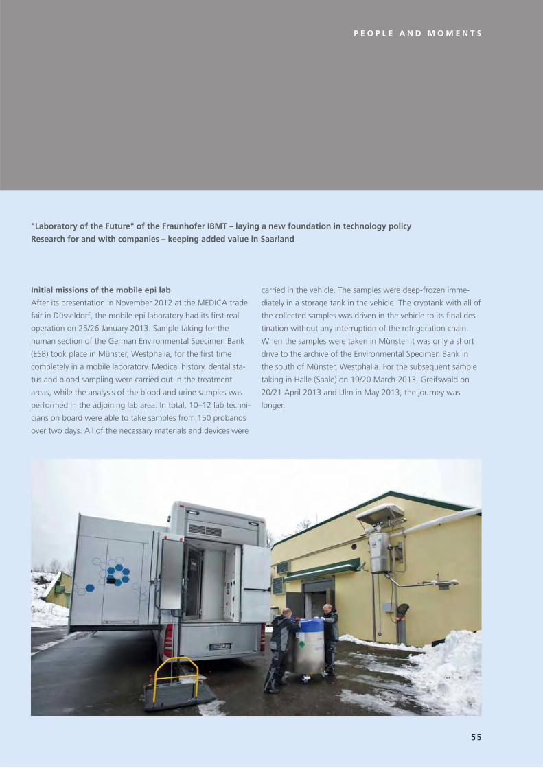
P E O P L E A N D M O M E N T S
Initial missions of the mobile epi lab
After its presentation in November 2012 at the MEDICA trade
fair in Düsseldorf, the mobile epi laboratory had its first real
operation on 25/26 January 2013. Sample taking for the
human section of the German Environmental Specimen Bank
(ESB) took place in Münster, Westphalia, for the first time
completely in a mobile laboratory. Medical history, dental sta-
tus and blood sampling were carried out in the treatment
areas, while the analysis of the blood and urine samples was
performed in the adjoining lab area. In total, 10–12 lab techni-
cians on board were able to take samples from 150 probands
over two days. All of the necessary materials and devices were
carried in the vehicle. The samples were deep-frozen imme-
diately in a storage tank in the vehicle. The cryotank with all of
the collected samples was driven in the vehicle to its final des-
tination without any interruption of the refrigeration chain.
When the samples were taken in Münster it was only a short
drive to the archive of the Environmental Specimen Bank in
the south of Münster, Westphalia. For the subsequent sample
taking in Halle (Saale) on 19/20 March 2013, Greifswald on
20/21 April 2013 and Ulm in May 2013, the journey was
longer.
"Laboratory of the Future" of the Fraunhofer IBMT – laying a new foundation in technology policy
Research for and with companies – keeping added value in Saarland
55

M E N S C H E N U N D M O M E N T E
Messeauftritte des mobilen epiLabors
MEDTEC Europe 2013
Das Fraunhofer IBMT und Bischoff + Scheck zeigten auf der
MEDTEC Europe 2013 in Stuttgart vom 26.–28. Februar 2013
das Flaggschiff der Initiative »Labor der Zukunft« in Halle 5,
Stand E20.
Medical Fair India 2013
Das »Labor der Zukunft« stellte vom 08.–10. März 2013 auf
der Medical Fair India, Delhi, aus.
64. Internationale Saarmesse 2013
Anlässlich des Technologietages der Saarländischen Landesre-
gierung am 13. April 2013 auf der 64. Internationalen Saar-
messe in Halle 1, Stand 107 stellte sich das Projekt »Labor der
Zukunft« vor.
LABOTEC SUISSE 2013
Vom 15.–16. Mai 2013 war das mobile Labor in Beaulieu Lau-
sanne, Schweiz zu besichtigen.
HOSPITALAR 2013
Gemeinsam mit den saarländischen Industriepartnern Medical
Technology Promedt Consulting GmbH und Woll Maschinen-
bau GmbH stellte das »Labor der Zukunft« auf der HOSPITA-
LAR 2013, die vom 21.–24. Mai 2013 in der brasilianischen
Wirtschaftsmetropole São Paulo stattfand, aus. »Labor der
Zukunft« ist offizieller Partner der Initiative »Research in Ger-
many – Land of Ideas«.
BIOTECHNICA 2013
Fraunhofer IBMT und BioKryo GmbH stellten gemeinsam unter
dem Thema »Labor der Zukunft« vom 08.–10. Oktober 2013
auf der BIOTECHNICA in Hannover aus. Hauptthemen waren
das Biobanking und mobile Laborlösungen. Der Auftritt fand
im Rahmen des Gemeinschaftsstandes der Fraunhofer-Gesell-
schaft in Halle 9, Stand E09 statt.
MEDICA 2013
Das »Labor der Zukunft« stellte im Rahmen des Saarland-
Gemeinschaftsstandes auf der MEDICA in Düsseldorf vom
20.–23.11. 2013, Halle 7a, Stand B09 aus.
56

P E O P L E A N D M O M E N T S
Trade fair appearances of the mobile epi lab
MEDTEC Europe 2013
The Fraunhofer IBMT and Bischoff + Scheck showed the flag-
ship of the initiative "Laboratory of the Future" at the MED-
TEC Europe 2013 in Stuttgart from 26 to 28 February 2013 in
Hall 5, Stand E20.
Medical Fair India 2013
The "Laboratory of the Future" was on show at the Medical
Fair India in Delhi from 8 to 10 March 2013.
64th International Saar Trade Fair 2013
The project "Laboratory of the Future" was showcased on the
occasion of the technology day of the Saarland State Govern-
ment on April 13, 2013 at the 64th International Saar Trade
Fair in Hall 1, Stand 107.
LABOTEC SUISSE 2013
From 15 to 16 May 2013 the mobile lab was on show in Beau-
lieu Lausanne, Switzerland.
HOSPITALAR 2013
In cooperation with the Saarland industrial partners Medical
Technology Promedt Consulting GmbH and Woll Maschinen-
bau GmbH, the "Laboratory of the Future" was shown at the
HOSPITALAR 2013, which took place from 21 to 24 May 2013
in the Brazilian commercial metropolis São Paulo. "Laboratory
of the Future" is an official partner of the initiative "Research
in Germany – Land of Ideas".
BIOTECHNICA 2013
Fraunhofer IBMT and BioKryo GmbH participated jointly in the
BIOTECHNICA trade fair from 8 to 10 October 2013 in Hano-
ver under the heading of "Laboratory of the Future". The
main themes were biobanking and mobile lab solutions. The
appearance took place within the framework of the joint
stand of the Fraunhofer-Gesellschaft in Hall 9, Stand E09.
MEDICA 2013
The "Laboratory of the Future" was shown at the MEDICA in
Düsseldorf from 20 to 23 November 2013 within the frame-
work of the joint Saarland stand in Hall 7a, Stand B09.
57

M E N S C H E N U N D M O M E N T E
»Labor der Zukunft« – Impfmobil auf Tour im Saarland
Anlässlich der Grippesaison 2013/2014 kam das mobile epide-
miologische Labor in der Zeit vom 08.-16. November 2013 in
einer vom Ministerium für Soziales, Gesundheit, Frauen und
Familie des Saarlandes unterstützten Impfaktion der Ärzte-
kammer des Saarlandes und der Kassenärztlichen Vereinigung
Saarland zum Einsatz. Besuchte Städte waren Neunkirchen/
Saar, Saarlouis und Saarbrücken. Neben Informationen zur
Impfung und der Möglichkeit, vor Ort geimpft zu werden,
demonstrierte das Fraunhofer IBMT im Laborbereich des Trucks
Stationen einer medizinischen Probe bis zum Laborergebnis.
"Laboratory of the Future" – mobile vaccination station
on tour in Saarland
In time for the flu season 2013/2014 the mobile epidemiologi-
cal laboratory was in operation from 8 to 16 November 2013
in a vaccination campaign carried out by the Medical Council
of Saarland and the Association of Statutory Health Insurance
Physicians (Kassenärztliche Vereinigung) Saarland and spon-
sored by the Ministry for Social Affairs, Health, Women and
Family. The mobile lab made stops in Neunkirchen/Saar, Saar-
louis and Saarbrücken. Alongside information on vaccinations
and the possibility of receiving vaccinations onsite, the Fraun-
hofer IBMT also demonstrated the various stations of a medi-
cal sample right up to the lab result in the laboratory section
of the truck.
1 Auf dem Foto impft Dr. Joachim Meiser (Stellvertretender Vorsit-
zender des Vorstandes der KV Saar) Gaby Schäfer (Staatssekretärin
im Ministerium für Soziales, Gesundheit, Frauen und Familie).
1 In the photo Dr. Joachim Meiser (Vice-chairman of the executive
board of the KV Saar) is vaccinating Gaby Schäfer (State Secretary in
the Ministry for Social Affairs, Health, Women and Family).
1
58

P E O P L E A N D M O M E N T S
Mobiles epiLabor auf Vorstellungstour im Saarland
Parallel zur Impfkampagne stand das mobile Labor des Fraun-
hofer IBMT allen Bürgerinnen und Bürgern zur Besichtigung
offen. In Neunkirchen, Saarlouis und Saarbrücken nutzten
viele Interessierte die Gelegenheit, ein medizinisch-analytisches
Labor zu besichtigen. An den Vormittagen wurde ein spezielles
Programm für Schüler geboten. Über 160 Schüler aus sieben
Oberstufen-Kursen hatten die Gelegenheit, im Labor selbst
Hand anzulegen und Details zum Einsatzzweck und zum Auf-
bau der mobilen Einheit zu erfahren.
Mobile epi lab on introduction tour in Saarland
Parallel to the vaccination campaign, the mobile lab of the
Fraunhofer IBMT was also open to citizens for viewing. In
Neunkirchen, Saarlouis and Saarbrücken numerous people
took the opportunity of learning about a medical-analytical
laboratory. A special program for schoolchildren was offered in
the mornings. More than 160 pupils from seven advanced
level courses had the opportunity of gaining hands-on experi-
ence with medical-analytical instruments and of learning
more about the purpose and the technical installations of the
mobile unit.
59

M E N S C H E N U N D M O M E N T E
Routinebetrieb des mobilen epiLabors in 2014
Das mobile Labor machte vom 24.-26. Januar 2014 zum zwei-
ten Mal Station in Münster (Westfalen). Wie schon im Vorjahr
wurden innerhalb von 2 Tagen bei mehr als 120 Probanden
Daten zum expositionsrelevanten Verhalten aufgenommen,
der Status von Zahnfüllungen ermittelt und die Abnahme von
Blut durchgeführt. Die entnommenen Proben wurden ohne
Zeitverzögerung vor Ort aufbereitet und analysiert und schließ-
lich im integrierten Lagertank zur Langzeitlagerung in der
Umweltprobenbank eingefroren. Insgesamt wurden diese
Tätigkeiten im Fahrzeug zeitgleich von bis zu 12 Mitarbeitern
umgesetzt. Nachdem bei den vergangenen Probenahmen in
2013 die Abläufe etabliert und durch ein Qualitätsmanage-
mentsystem optimiert wurden, wird nun 2014 die Akkreditie-
rung nach DIN EN ISO / IEC 17025 erstmalig für ein mobiles
Labor angestrebt. Weitere Stationen im Rahmen des Einsatzes
für die Umweltprobenbank des Bundes waren Halle/Saale
(18.-19. März 2014), Greifswald (26.-27. April 2014) und Ulm
(10.-11. Mai 2014). Im Herbst wird das mobile epiLabor im
Rahmen von verschiedenen Impfkampagnen bundesweit im
Einsatz sein.
Routine operation of the mobile epi lab in 2014
The mobile lab was stationed in Münster (Westphalia) for the
second time from 24 to 26 January 2014. Just as in the previ-
ous year, data on exposure-relevant behaviour was taken over
two days from more than 120 probands, the status of dental
fillings was determined and blood samples were taken. The
samples were immediately prepared and analyzed onsite, and
then frozen in the integrated cryotank for long-term storage in
the Environmental Specimen Bank. All of these activities were
carried out by up to 12 lab workers simultaneously in the vehi-
cle. After the workflow had been established in previous sam-
ple takings in 2013, and optimized by a quality management
system, for 2014 the accreditation in accordance with DIN EN
ISO / IEC 17025 is expected for the first time for a mobile lab.
Further stops during the mission of the German Environmental
Specimen Bank were Halle/Saale (18-19 March 2014),
Greifswald (26-27 April 2014) and Ulm (10-11 May 2014). In
autumn the mobile epi lab will be in operation as part of vari-
ous vaccination campaigns nationwide.
60

P E O P L E A N D M O M E N T S
Gründung »Labor der Zukunft e. V.« – Saarland wird
Ehrenmitglied
Als Startschuss für eine nachhaltige und mit der Schaffung von
Arbeitsplätzen verbundene Nutzung von innovativen Labor-
technologien im Saarland bezeichnete Ministerpräsidentin
Annegret Kramp-Karrenbauer die Vereinsgründung »Labor der
Zukunft e. V.«. Initiator ist das Fraunhofer-Institut für Biomedi-
zinische Technik (IBMT) in St. Ingbert.
Annegret Kramp-Karrenbauer: »Durch die Ehrenmitgliedschaft
des Saarlandes im Verein Labor der Zukunft e. V. dokumentiert
die Landesregierung ihr nachhaltiges Interesse an der Weiter-
entwicklung völlig neuartiger Labortechniken und den damit
verbundenen Perspektiven für den saarländischen Technologie-
und Innovationsstandort.«
Grundlage für die Vereinsgründung ist das von der Staatskanz-
lei mit rund 6,5 Millionen Euro unterstützte Projekt »Labor der
Zukunft« des Fraunhofer IBMT. Seit 2009 entwickelt eine Tech-
nologieinitiative unter Führung des Fraunhofer IBMT im Saar-
land innovative Technologien für biologische, medizinisch-
analytische, chemische und physikalische Labore. Gemeinsam
mit kleinen und mittelständischen Unternehmen und unter-
stützt von der Landesregierung steht die Initiative »Labor der
Zukunft« für Entwurf, Entwicklung und Förderung der zukünf-
tigen Generation von innovativer Labortechnologie.
Im Gegensatz zu anderen Technologiebereichen benötigen
Bau und Betrieb moderner Labore die vertikale Integration von
Handwerk über Ingenieurtätigkeit bis hin zur Grundlagenfor-
schung. Die Herausforderung besteht darin, High- und Low-
Tech-Zulieferer in die Lage zu versetzen, in einem Projekt
zusammenzuarbeiten. Dies erfordert einen Paradigmenwechsel
in der Technologieforschung und -förderung sowie die Integra-
tion der kompletten Wertschöpfungskette von der anwen-
dungsorientierten Forschung bis hin zum potenziellen End-
nutzer. Beispielhaft für diese Zusammenarbeit ist das seit 2013
u. a. für die Umweltprobenbank des Bundes erfolgreich im
Einsatz befindliche und völlig neuartige mobile epiLabor des
Fraunhofer IBMT (Labor-Truck).
Zur Etablierung einer gemeinsamen und nachhaltigen Organi-
sationsstruktur haben sich die Kernakteure dazu entschlossen,
einen Verein mit dem Namen »Labor der Zukunft e. V.« zu
gründen. Die Handwerkskammer des Saarlandes, das Zentrum
für Mechatronik und Automatisierungstechnik (ZeMA), das
Fraunhofer IBMT und die saarländische Landesregierung for-
men dabei gemeinsam mit den Firmen CETECOM ICT Services,
Woll Maschinenbau, Biokryo und Medizinisches Versorgungs-
zentrum Saar den lokalen Kern des Vereins, der seinen Sitz in
Sulzbach hat. Die überregional angesiedelten Firmen EBV Elek-
tronik (Poing, Bayern), Bischoff + Scheck (Rheinmünster,
Baden-Württemberg) und Tecan Deutschland (Crailsheim,
Baden-Württemberg) komplettieren die Gründungsmitglieder
in den Innovationsfeldern der Labortechnologie: Elektronik,
Mobilität und Automatisierung.
Der Verein hat die Schaffung von Synergien zur Festigung der
noch jungen, sich entwickelnden Strukturen in der Region
zum Ziel. Dabei wird gemeinsam mit allen Akteuren der bisher
entwickelte Wissensvorsprung auf dem Zukunftsfeld mobiler
Labortechnologie weiter ausgebaut und somit dem Saarland
zukünftig eine Schlüsselstellung in einem zukunftsträchtigen
Technologiebereich zuteil.
Der offizielle Gründungsakt des Vereins »Labor der Zukunft
e. V.« fand am Montag, den 03. Februar 2014, um 16.00 Uhr,
in den Räumen der Staatskanzlei des Saarlandes (Foyer des
Palais Röder) statt.
61

M E N S C H E N U N D M O M E N T E
Gründungsakt am 03. Februar
2014 in der Staatskanzlei, Palais
Röder, Saarbrücken.
Am Tisch v. l. n. r.: Jürgen
Lennartz, Chef der Staatskanzlei,
Prof. Dr. Heiko Zimmermann,
Institutsleiter Fraunhofer IBMT
und Vorsitzender des Vereins
»Labor der Zukunft e. V.«
Hintere Reihe v. l. n. r.: Karl-Josef
Schmitt, Technischer Leiter Woll
Maschinenbau, Bernd Pfeil, Vice
President Sales & Marketing
Central Europe EBV, Bernd
Foundation act on February 3,
2014 in the State Chancellery,
Palais Röder, Saarbrücken.
At the table from left to right:
Jürgen Lennartz, Head of the
State Chancellery, Prof. Dr. Heiko
Zimmermann, Head of the
Fraunhofer IBMT and Chairman
of the association Labor der
Zukunft e. V.
Back row from left to right: Karl-
Josef Schmitt, Technical Director
Woll Maschinenbau, Bernd Pfeil,
Vice President Sales & Marketing
Central Europe EBV, Bernd
Bischoff, Geschäftsführer
Bischoff + Scheck, Volker Scheck,
Geschäftsführer Bischoff +
Scheck, Dr. Reinhard Stute, Ärzt-
licher Leiter MVZ Saar, Jochen
Flackus, Geschäftsführer ZeMA,
Georg Brenner, Hauptgeschäfts-
führer Handwerkskammer,
Daniel Schmitt, Fraunhofer
IBMT, Gerhard Schirra,
Geschäftsführer CETECOM ICT,
Heribert Hess, Tecan Deutsch-
land, Dr. Vincent von Walcke-
Wulffen, Geschäftsführer
BioKryo GmbH.
Bischoff, Managing Director
Bischoff + Scheck, Volker Scheck,
Managing Director Bischoff +
Scheck, Dr. Reinhard Stute, Medi-
cal Director MVZ Saar, Jochen
Flackus, Managing Director
ZeMA, Georg Brenner, Managing
Director Chamber of Trades and
Crafts, Daniel Schmitt, Fraun-
hofer IBMT, Gerhard Schirra,
Managing Director CETECOM
ICT, Heribert Hess, Tecan
Deutschland, Dr. Vincent von
Walcke-Wulffen, Managing
Director BioKryo GmbH.
62

P E O P L E A N D M O M E N T S
Foundation of the Labor der Zukunft e. V. – Saarland
becomes honorary member
Prime Minister Annegret Kramp-Karrenbauer described the
foundation of the association "Labor der Zukunft e. V." as the
starting shot for the sustainable use of innovative lab technol-
ogies in Saarland that will create jobs. The initiator is the
Fraunhofer Institute for Biomedical Engineering (IBMT) in
St. Ingbert.
Annegret Kramp-Karrenbauer: "Through the honorary mem-
bership of the Saarland in the association Labor der Zukunft
e.V., the state government is documenting its sustained inter-
est in the further development of completely innovative lab
technologies and the related perspectives for Saarland as a
technology and innovation location."
The basis for the foundation of the association is the project
"Laboratory of the Future" of the Fraunhofer IBMT, which is
supported by the State Chancellery with around € 6.5 million.
Since 2009 a technology initiative led by the Fraunhofer IBMT
in Saarland has been developing innovative technologies for
biological, medical-analytical, chemical and physical laborato-
ries. In cooperation with small and medium-sized enterprises,
and supported by the state government, the initiative "Labo-
ratory of the Future" stands for design, development and
funding of the future generation of laboratory technology.
Unlike other technology areas, the building and operation of
modern laboratories requires the vertical integration of crafts
and trades, from engineering right up to basic research. The
challenge consists in enabling high- and low-tech suppliers to
collaborate in a project. This requires a paradigm change in
technology research and funding as well as the integration of
the complete value-added chain from application-oriented
research right up to the potential end user. An example of this
cooperation is the completely innovative mobile epi lab of the
Fraunhofer IBMT (lab truck), which has been working success-
fully since 2013 for, among others, the German Environmental
Specimen Bank.
In order to establish a joint and sustainable organizational
structure, the main parties decided to found an association
called "Labor der Zukunft e. V.". The Chamber of Trades and
Crafts of Saarland, the Centre for Mechatronics and Automa-
tion Technology (Zentrum für Mechatronik und Automa-
tisierungstechnik, ZeMA), the Fraunhofer IBMT and the Saar-
land State Government along with the companies CETECOM
ICT Services, Woll Maschinenbau, Biokryo und Medizinisches
Versorgungszentrum Saar form the local core of the associa-
tion, which is based in Sulzbach. The supraregionally operating
companies EBV Elektronik (Poing, Bavaria), Bischoff + Scheck
(Rheinmünster, Baden-Württemberg) and Tecan Deutschland
(Crailsheim, Baden-Württemberg) round off the list of founder
members in the innovation fields of lab technology: electron-
ics, mobility and automation.
The objective of the association is the creation of synergies to
consolidate the still young, developing structures in the region.
For this purpose, all of the participants will cooperate to fur-
ther expand the advances in knowledge achieved so far in the
future-oriented field of mobile laboratory technology, thus giv-
ing Saarland a key role in a future-oriented technology field.
The official foundation act of the association "Labor der
Zu kunft e.V." took place on Monday, February 3, 2014 at
4 pm in the rooms of the State Chancellery of Saarland (Foyer
of Palais Röder).
63

Nicht größer als eine Getränkedose: Das miniaturisierte Inkubator-
Mikroskop ist eine platzsparende und kostengünstige Lösung, um
Zeitraffersequenzen von Zellen in Kultur zu erfassen
(Foto: Thorsten Knoll).
No larger than a beverage can: the miniaturized incubator
microscope is a space-saving and cost-efficient solution for
the analysis of time-lapse sequences of cells in culture
(Photo: Thorsten Knoll).
64

Das Forschungs- und Dienstleistungsangebot
Institutsspezifische Angebote zur Vertragsforschung
Verträge und Patentvereinbarungen
Kunden
Kontakt und weitere Informationen
The research and service offers
Institute-specific offers for contract research
Contracts and patent agreements
Customers
Contact and further information
DER KUNDE IM MITTELPUNKTTHE CUSTOMER AT THE CENTRE
65

D E R K U N D E I M M I T T E L P U N K T
Institutsspezifische Angebote zur Vertragsforschung
Arbeitsweise:
FuE-Projekte werden in Phasen erfolgsorientiert ausgeführt,
beginnend mit einer technischen Marktstudie, daraus abgelei-
tet die Machbarkeitsstudie, über die Prototypentwicklung und
den Feldtest (klinische Studie) bis hin zur Entwicklung von
kostenoptimierten Fertigungstechniken und Technologieent-
wicklungen. Zur Servicefertigung von Sensoren und Mikrosys-
temen können Firmen benannt werden.
Praxisbezug:
Die Bearbeitung der Projekte am Fraunhofer-Institut für Biome-
dizinische Technik (IBMT) erfolgt in enger Abstimmung mit
dem jeweiligen Kunden, um den größtmöglichen Praxisbezug
herzustellen. Die Kundennähe ist ein Charakteristikum und
eine wichtige Voraussetzung, um den Bedürfnissen des Mark-
tes aus der Grundlagenforschung heraus gerecht zu werden.
Flexibilität:
Die konkrete Form, die Ausrichtung und der Umfang der Pro-
jektarbeiten richten sich nach den Anforderungen und Vorstel-
lungen des Kunden oder Auftraggebers.
Synergie:
Die Einordnung in die Forschungsstrategie der Fraunhofer-
Gesellschaft mit ihren 67 Forschungseinrichtungen und dem
im Jahr 2001 gegründeten Life Sciences-Verbund, der inzwi-
schen sechs Fraunhofer-Institute (IBMT, IGB, IME, ITEM, IVV
und IZI) und eine Fraunhofer-Einrichtung (EMB) umfasst,
schafft Synergieeffekte. Fachkenntnisse aus unterschiedlichs-
ten Forschungsfeldern können in Kooperationen genutzt wer-
den und erlauben eine kompetente Bearbeitung auch multi-
disziplinärer Fragestellungen. Durch Kooperationsverträge
werden für IBMT-Kunden vollständige Wertschöpfungsketten
angeboten.
Qualität:
Liefertreue und Zuverlässigkeit prägen die Arbeiten des Fraun-
hofer-Instituts für Biomedizinische Technik. Die Erstellung eines
Pflichtenhefts, in Zusammenarbeit mit dem Kunden, gewähr-
leistet die inhaltlich korrekt abgestimmte und zeitlich ange-
messene Bearbeitung der Projekte.
Preiswürdigkeit:
Forschungs- und Entwicklungsaufträge werden auf Selbstkos-
tenbasis durchgeführt. Das IBMT ist als Institut der Fraunhofer-
Gesellschaft eine gemeinnützige Einrichtung und finanziert die
notwendige anwendungsorientierte Forschung und Vorlauffor-
schung weitgehend unter Mitwirkung öffentlicher Auftrag-
geber.
FuE-Ergebnis:
Nach erfolgter Bearbeitung eines FuE-Auftrags wird dem Kun-
den das Ergebnis zur Verfügung gestellt.
Vertraulichkeit:
Anfragen werden auf Wunsch des Kunden absolut vertraulich
behandelt.
66

T H E C U S T O M E R A T T H E C E N T R E
Institute-specific offers for contract research
How IBMT Works:
R&D projects are carried out on a success-oriented basis in
phases, starting with a technical market research study which
forms the basis for the feasibility study, prototype develop-
ment and field testing (clinical study), right up to the develop-
ment of cost-optimized manufacturing technologies. Compa-
nies can be named for service and production of sensors and
microsystems.
Practical relevance:
The projects are carried out at the Fraunhofer Institute for Bio-
medical Engineering (IBMT) in close contact with the respec-
tive customer to guarantee the greatest possible practical rele-
vance. Customer proximity is a characteristic and an important
requirement in order to meet the needs of the market with
basic research.
Flexibility:
The concrete form, the orientation and scope of project work
are based on the requirements and wishes of the customer or
client.
Synergy:
The integration into the research strategy of the Fraunhofer-
Gesellschaft with its 67 research institutes and the Life Sci-
ences Group founded in 2001 which, in the meantime,
includes six Fraunhofer institutes (IBMT, IGB, IME, ITEM, IVV
und IZI) and one Fraunhofer research establishment (EMB) cre-
ates synergies. Expertise from different research fields can be
used in cooperation to allow the competent processing of
multidisciplinary issues. Complete value-added chains are
offered for IBMT customers on the basis of cooperation agree-
ments.
Quality:
On-time delivery and reliability characterize the work of the
Fraunhofer Institute for Biomedical Engineering. The compila-
tion of the specifications in cooperation with the client ensures
that the contents are correctly coordinated and that projects
are dealt with in a timely manner.
Value for money:
Research and development contracts are carried out on a cost
basis. As an institute of the Fraunhofer-Gesellschaft, the IBMT
is a non-profit organization and finances the necessary appli-
cation-oriented research and preliminary research mainly from
funds of public clients.
R&D results:
After completion of an R&D project, the results are made
available to the customer.
Confidentiality:
Inquiries will be kept strictly confidential at the request of the
customer.
67

D E R K U N D E I M M I T T E L P U N K T
P R O J E K T A N Z A H L
A N W E N D U N G S O R I E N T I E R T E G R U N D L A G E N F O R S C H U N G
Z I E L
Wissenschaftlich-technische Beratung
Machbarkeits-studie
Prototyp-Entwicklung
FeldtestsFertigungs-technologie
Markt
– existierendes
Know-how,
– Literaturstudien,
– Patentrecherche,
– Expertisen,
– Marktanalysen
– Studien,
– Simulation,
– Experimente
– Spezialmodule,
– mechanische
Baugruppen,
– Elektronik,
– Verbindungs-
techniken,
– Spezialsoftware,
– Definition von
Testkriterien
– Test unter Feld-
bedingungen,
– Evaluierung,
– Zuverlässigkeits-
analyse,
– Optimierung,
– Testunter-
stützung
– Produktent-
wicklung,
– Optimierung der
Produktions-
technologie,
– Kleinserien-
produktion,
– Technologie-
transfer
Qualitäts-
sicherung
Phasenmodell:
Die Projektarbeit erfolgt im Fraunhofer-Institut für Biomedizini-
sche Technik wie folgt: Am Beginn eines Projekts steht eine
wissenschaftlich-technische Beratung. Hierbei können anhand
des existierenden Know-hows sowie mittels Literatur-, Patent-
und Marktrecherchen die möglichen Probleme des Projekts
aufbereitet und das Projektrisiko abgeschätzt werden. Darauf
folgt eine Machbarkeitsstudie, die das Projekt spezifiziert und
den Aufwand beurteilt. Eine Laborprototyp-Entwicklung dient
dem praktischen Funktionsnachweis in Form eines Demonstra-
tors. Diese Phase mündet in die Feldprototyp-Entwicklung, an
deren Ende umfangreiche Tests stehen. Das Redesign, die
Technologieoptimierung, die Kleinserienfertigung und der
Technologietransfer sind Elemente der Produktionsvorberei-
tung. Begleitend leistet das Fraunhofer-Institut für Biomedizini-
sche Technik auch Hilfestellung bei Marketing und Qualitätssi-
cherung. Dies steht im Dienste des Produktionsanlaufs und der
Risikominimierung im Rahmen der Fertigung. Der Kunde hat
die Möglichkeit, seinen Auftrag entsprechend dieser Phasen
ein- und aufzuteilen und am Ende jeder einzelnen Stufe neu
zu entscheiden, ob es sich für ihn lohnt, in die nächste Phase
einzutreten. Dieses Kriterium erleichtert dem Kunden wie auch
dem IBMT die Auftragsvergabe bzw. -annahme und führt zu
überschaubaren, kalkulierbaren Projektzeiten und Projekt-
kosten.
Risikominimierte Produktentwicklung.
68

T H E C U S T O M E R A T T H E C E N T R E
N U M B E R O F P R O J E C T S
A P P L I C A T I O N - O R I E N T E D B A S I C R E S E A R C H
G O A L
Scientific-Technical Consulting
Feasibility Study
PrototypeDesign
Field TestsProduction Technology
Market
– existing know-
how,
– literature
analysis,
– patent studies,
– expertises,
– market analysis
– studies,
– simulations,
– experiments
– special modules,
– mechanical
design,
– electronics,
– interconnecting
technologies,
– dedicated
software,
– definition of
test criteria
– test under field
constraints,
– evaluation,
– reliability
analysis,
– optimization,
– test support
– final product
design,
– optimization
of production
technology,
– small volume
production,
– transfer of tech-
nology
Quality
Assurance
Phase Model:
Project work is carried out in the Fraunhofer Institute for Bio-
medical Engineering as follows. The project starts with a scien-
tific-technical consultation process. The possible problems of
the project can be examined here and the project risk esti-
mated on the basis of existing know-how, as well as literature,
patent and market research. This is followed by a feasibility
study, which specifies the project and estimates the costs. A
laboratory prototype development provides practical function
verification in the form of a demonstrator. This phase leads to
the field prototype development at the end of which extensive
tests are carried out. The redesign, technology-optimization,
small-scale production and technology transfer are elements
of pre-production. The IBMT also assists in quality assurance
and marketing of the product. This provides support for the
production start-up and helps to minimize risks during produc-
tion. The customer has the option of dividing up his order on a
phase-to-phase basis and can decide at the end of each phase,
whether it is worth starting the next phase. This criterion helps
the customer as well as the IBMT in granting or accepting the
order, and leads to manageable, predictable project schedules
and costs.
Risk-Minimised Product Development.
69

Verträge und Patentvereinbarungen
Vertragsabschluss:
Faire und verlässliche Vertragsbedingungen für den Kunden
sind das oberste Gebot. Dabei werden die Wissenschaftler und
Ingenieure von einer erfahrenen Vertragsabteilung innerhalb
der Fraunhofer-Gesellschaft unterstützt.
Nutzungsrechte:
Über die Nutzungsrechte an den in der Auftragsbearbeitung
entstandenen Patenten verfügt allein der Kunde. Nach den
Wünschen des Kunden werden individuelle Vereinbarungen
getroffen. Das IBMT wird durch mehr als fünf renommierte
Patentanwaltskanzleien vertreten.
Koordination:
Das Fraunhofer-Institut für Biomedizinische Technik ist erfah-
ren in der Koordination komplexer Verbundvorhaben und
übergeordneter Leitprojekte. In diesem Zusammenhang wer-
den administrative und koordinative Aufgaben übernommen
und eine gute Kommunikation zwischen den Projektpartnern
im Verbund sichergestellt, um Reibungsverluste zu minimieren.
Schulungen:
Als Dienstleistung für den Kunden bietet das IBMT auch die
Schulung von Mitarbeitern im Hinblick auf die Einführung
neuer Verfahren und Technologien an. Diese kann direkt vor
Ort im Betrieb des Kunden erfolgen.
Qualitätssicherung:
Die Wissenschaftler und Entwicklungsingenieure des Fraunho-
fer-Instituts für Biomedizinische Technik arbeiten nach den
Regeln des modernen Projektmanagements. Die Projekte und
Arbeiten unterliegen einer sorgfältigen und permanenten
Überprüfung nach Zeit und Kosten und sind auf einen erfolg-
reichen Projektabschluss hin ausgerichtet. Computerunter-
stütztes Projekt-Controlling begleitet jeden Einzelauftrag.
Fördermöglichkeiten:
Die Fraunhofer-Gesellschaft hilft dem Kunden dabei, alle Mög-
lichkeiten der Projektförderung auszuschöpfen. Eine langjäh-
rige Erfahrung bei der Beantragung von Fördermitteln der
Europäischen Union, des Bundesministeriums für Bildung und
Forschung BMBF oder anderer Zuwendungsgeber unterstützt
den Kunden in Fragen der Finanzierung von Forschungs-
projekten.
Kunden
Neben Auftraggebern aus dem biomedizinischen und medizin-
technischen Bereich sowie der Biotechnologie gehören auch
Auftraggeber anderer Industriesparten (Umwelttechnik,
Labortechnik, Biologie, Chemie, Pharmazie, Materialtechnik,
Kfz-Technik, Hydraulik, Maschinenbau, Anlagenbau, Sensor-
systeme) zu den Kunden des Fraunhofer-Instituts für Biomedi-
zinische Technik. Das IBMT arbeitet seit seiner Gründung mit
Unternehmen unterschiedlicher Größen zusammen.
Kontakt und weitere Informationen
Bitte rufen Sie uns an, wenn Sie Fragen haben, weitere Infor-
mationen oder ein konkretes Angebot wünschen. Publikatio-
nen und Broschüren senden wir Ihnen gerne zu. Besuchen Sie
unsere Internetseiten: http://www.ibmt.fraunhofer.de.
Fraunhofer-Institut für Biomedizinische Technik IBMT
Ensheimer Straße 48
66386 St. Ingbert
Telefon: +49 (0) 6894/980-0
Fax: +49 (0) 6894/980-400
Presse und Öffentlichkeitsarbeit
Dipl.-Phys. Annette Maurer
Telefon: +49 (0) 6894/980-102
D E R K U N D E I M M I T T E L P U N K T
70

Contracts and patent agreements
Contracts:
Fair and reliable contractual terms for customers are the
utmost priority. The scientists and engineers are supported by
an experienced contract department within the Fraunhofer-
Gesellschaft.
Usage rights:
The customer has the exclusive usage rights to the patents
arising out of the execution of the order. According to the cus-
tomer's wishes, individual agreements are reached. The IBMT
is represented by more than five established patent attorney
offices.
Coordination:
The Fraunhofer IBMT is experienced in the coordination of
complex joint projects and superordinate management pro-
jects. This means that administrative and coordinative tasks are
assumed and good communication between the joint project
partners is ensured in order to avoid friction losses.
Training:
IBMT offers training for the staff with regard to the introduc-
tion of new processes and technologies as a service for cus-
tomers. This training may take place at the customer´s prem-
ises.
Quality Assurance:
The scientists and development engineers at the Fraunhofer
IBMT work according to the rules of modern project manage-
ment. The projects and works are subject to careful and con-
tinuous assessment in terms of time and costs and geared
towards a successful conclusion of the project. Each individual
order is accompanied by computer-aided project controlling.
Funding options:
The Fraunhofer-Gesellschaft helps the customer to exhaust all
possibilities of project funding. Many years of experience in
applying for funding from the European Union, the German
Federal Ministry for Education, Research and Technology
(BMBF) or other donors support the customer in matters of
research project funding.
Customers
Alongside clients from the fields of biomedical and medical
engineering as well as biotechnology, clients from other indus-
trial sectors (environmental technology, laboratory technology,
biology, chemistry, pharmaceuticals, materials technology,
automotive technology, hydraulics, mechanical engineering,
plant construction, sensor systems) are also among the cus-
tomers of the Fraunhofer Institute for Biomedical Engineering.
Since its foundation, the IBMT has been working with compa-
nies of different sizes.
Contact and further information
Please call us if you have any queries or require further infor-
mation or a concrete offer. We will be happy to send you the
relevant publications and brochures. Visit our website at:
http://www.ibmt.fraunhofer.de.
Fraunhofer Institute for Biomedical Engineering (IBMT)
Ensheimer Strasse 48
66386 St. Ingbert
Germany
Telephone: +49 (0) 6894/980-0
Fax: +49 (0) 6894/980-400
Press and public relations
Dipl.-Phys. Annette Maurer
Telephone: +49 (0) 6894/980-102
T H E C U S T O M E R A T T H E C E N T R E
71

Zellbiologie & Angewandte Virologie
– Biobanken & Kryolager
– Bioprozesstechniken & Nanotechnologie
– Prüflaboratorien & Qualitätssicherung
Ultraschall
– Medizinischer Ultraschall
– Technischer Ultraschall
Labor- & Informationstechnologie
Medizintechnik & Neuroprothetik
Biomedizinische Mikrosysteme
Medizinische Biotechnologie
Cell Biology & Applied Virology
– Biobanks & Cryorepositories
– Bioprocess Technologies & Nanotechnology
– Test Laboratories & Quality Assurance
Ultrasound
– Medical Ultrasound
– Technical Ultrasound
Laboratory & Information Technology
Medical Engineering & Neuroprosthetics
Biomedical Microsystems
Medical Biotechnology
UNSER ANGEBOT – AUSGEWÄHLTE FORSCHUNGS ERGEBNISSE UND ANWENDUNGEN
OUR OFFER – SELECTED RESEARCH RESULTS AND APPLICATIONS
72

Automated cryostorage: In collaboration with industrial partners the
Fraunhofer IBMT designs and develops automated systems for the
cryostorage of valuable sample material without interruptions in the
refrigeration chain (Photo: Annette Maurer).
Automatisierte Kryolagerung: Gemeinsam mit industriellen Partnern
konzipiert und entwickelt das Fraunhofer IBMT automatisierte Anla-
gen für die Kryolagerung von wertvollem Probenmaterial unter Ein-
haltung der Kühlkette (Foto: Annette Maurer).
73

U N S E R A N G E B O T – A U S G E W Ä H L T E F O R S C H U N G S E R G E B N I S S E U N D A N W E N D U N G E N
Lager Wolbeck: Einlagerung von Humanproben der Umweltproben-
bank des Bundes in Kryotanks zur späteren retrospektiven Analyse
(Foto: Bernd Müller).
Wolbeck repository: Storage of human samples of the German Envi-
ronmental Specimen Bank (Umweltprobenbank des Bundes, UPB) in
cryotanks for later, retrospective analysis (Photo: Bernd Müller).
74

O U R O F F E R – S E L E C T E D R E S E A R C H R E S U L T S A N D A P P L I C A T I O N S
ZELLBIOLOGIE & ANGEWANDTE VIROLOGIECELL BIOLOGY & APPLIED VIROLOGY
Angebote, Ergebnisse und Produkte der Abteilung
Biobanken & Kryolager
Umweltprobenbank des Bundes – Humanproben
HIV Specimen Cryorepository (B & M Gates Foundation)
CRYO-BREHM – Zellbank für Wildtiere
Angebote, Ergebnisse und Produkte der Abteilung
Bioprozesstechniken & Nanotechnologie
Präklinische Nanobiotechnologie
In-vitro-Kulturtechniken
Prüflaboratorien & Qualitätssicherung
Projektbeispiel: Biokompatible Hybridkeramiken als
Knochenersatz
Ausstattung
Offers, results and products of the Biobanks &
Cryorepositories Department
Environmental Specimen Bank – Human Samples
HIV Specimen Cryorepository (B & M Gates Foundation)
CRYO-BREHM – German Cell Bank for Wildlife
Offers, results and products of the Bioprocess
Technologies & Nanotechnology Department
Preclinical Nanobiotechnology
In vitro Culture Technologies
Test Laboratories & Quality Assurance
Project example: Biocompatible hybrid ceramics as
bone substitute
Equipment
75

Verbesserte und neuartige Zellkulturtechniken und die darauf
aufbauenden analytischen Messverfahren müssen bei der
rasanten biotechnologischen Entwicklung von zukunftsori-
entierten therapeutischen Konzepten Schritt halten. Hierbei
gewinnen Standardisierung und Optimierung im Bereich der
präklinischen und klinischen Testungen von Wirk- und Impf-
stoffen zunehmend an Bedeutung. Die Hauptabteilung Zell-
biologie & Angewandte Virologie entwickelt hierfür alternative
Zellkultursysteme und Testverfahren für die verschiedensten
Bereiche der Stammzellforschung und Nanobiotechnologie.
Die Wirkstoffentwicklung wird durch den Einsatz entspre-
chender Technologieplattformen unterstützt. Hierzu zählen
Transport- und Freisetzungsuntersuchungen therapeutischer
Applikationssysteme über zelluläre Barrieren wie z. B. der Blut-
Hirn-Schranke, wofür wir der menschlichen Blut-Hirn-Schranke
physiologisch sehr nahestehende Systeme entwickelt haben.
Hierin können zum Beispiel Nanopartikel getestet werden,
die zuvor mit Alzheimer-Medikamenten beladen wurden
und auf deren Oberfläche sich Ankermoleküle befinden, die
bestimmte Strukturen der Blut-Hirn-Schranke erkennen. So
sollen die Nanopartikel ein Alzheimer-Medikament gezielt
ins Gehirn transportieren. Die Entwicklung dieser neuartigen
Transportmethode von Medikamenten und die Untersuchung
von Chancen und Risiken werden vom Bundesministerium
für Bildung und Forschung (BMBF) im Rahmen des Verbund-
projekts »NanoBrain« unterstützt, das Teil des europäischen
Forschungsnetzes Era-NET »NEURON« ist. Eine weitere
zelluläre Barriere, die es bei oraler Applikation zu überwinden
gilt, ist die Darmbarriere. In speziell hierfür ausgelegten
Darmbarriere-Modellen testen wir Formulierungen von
Wirkstoffen für die Photodynamische Therapie, die ansonsten
oral nicht bioverfügbar sind. Hier besteht die Chance, diese
Wirkstoffgruppe in Tablettenform zu verabreichen und somit
die mit Nachteilen behaftete intravenöse Gabe zu umgehen.
Dieses Projekt wird im Rahmen des Verbunds »BioTraP for
CCC« durch das BMBF gefördert.
Weiterhin lassen sich diese Nanopartikel als Genfähren einset-
zen, um gezielt DNA in Zellen zu transportieren, die die
Expression bestimmter Proteine ermöglicht. Üblicherweise
erfolgen gezielte genetische Veränderungen von Zellen durch
virale Vektoren. Die Nutzung dieser bereitet aber erhebliche
Probleme wie die Gefahr der Ausbildung von Krebs durch die
Aktivierung von Proto-Onkogenen. Die virusfreie genetische
Veränderung von Zellen mittels DNA-beladener Nanopartikel
stellt sowohl für die Stammzellforschung wie auch für die
zukünftige Zelltherapie eine vielversprechende Alternative
dazu dar. Auch dieses ist ein vom BMBF unterstütztes Ver-
bundprojekt (»NanoGene«) im Rahmen des europäischen For-
schungsnetzes EuroNanoMed.
Darüber hinaus kommen die in der Hauptabteilung neu entwi-
ckelten Zellkultursysteme und Testverfahren im Bereich der
Nanotoxikologie zum Einsatz: In einem Marie Curie Initial Trai-
ning Network (ITN)-Projekt des 7. Rahmenprogramms der EU
(»NanoTOES«) entwickelt die Abteilung hochsensitive chipba-
sierte Testverfahren, um physikalische, chemische und biologi-
sche Faktoren von Nanopartikeln und deren Einflüsse auf bio-
logische Zellen zu untersuchen. Des Weiteren werden
zukunftsweisende Plattformen zum Sammeln, Präparieren,
Konservieren und zur Verteilung von Bioreagenzien und klini-
schen Proben für weltweite Netzwerke entwickelt. Hierzu zäh-
len optimierte Prozesse der Probenaufarbeitung und deren
Kryokonservierung. In diesem Zusammenhang wurde am
Fraunhofer IBMT im Rahmen der globalen Initiative zur Ent-
wicklung eines HIV-Impfstoffes (»Collaboration for AIDS Vac-
cine Discovery – CAVD«) eine globale HIV-Kryobank der
Sicherheitsstufe S3 aufgebaut, die von der Bill & Melinda
Gates Foundation und der saarländischen Landesregierung
finanziell getragen wird. Die Kryobank für Bioreagenzien
untersteht einem zertifizierten Qualitätssicherungsprogramm,
welches das Arbeiten nach den Richtlinien der »Good Clinical
Laboratory Practice« (GCLP) kontrolliert. Hier werden Proben
in einer vollautomatisierten Anlage zur Kultivierung eukaryoti-
Z E L L B I O L O G I E & A N G E W A N D T E V I R O L O G I E
U N S E R A N G E B O T – A U S G E W Ä H L T E F O R S C H U N G S E R G E B N I S S E U N D A N W E N D U N G E N
76

C E L L B I O L O G Y & A P P L I E D V I R O L O G Y
Improved and innovative cell culture techniques and the ana-
lytical measurement methods based on these have to keep up
with the rapid biotechnological development of future-ori-
ented therapeutic concepts. Standardization and optimization
in the area of preclinical and clinical testing of active ingredi-
ents and vaccines are becoming increasingly important in this
context. For this purpose the Divison of Cell Biology & Applied
Virology Department is developing alternative cell culture sys-
tems and test methods for the various areas of stem cell
research and nanobiotechnology. The development of drugs is
supported by the use of the corresponding technology plat-
forms. This includes transport and release investigations of
therapeutic application systems via cellular barriers such as the
blood-brain barrier, for which we have developed systems
which are physiologically very close to the human blood-brain
barrier. These can be used, for example, to test nanoparticles
which are loaded with Alzheimer drugs and which are surface-
modified with anchor molecules that recognize corresponding
structures on the blood-brain barrier. In this way, the nanopar-
ticles can transport Alzheimer drugs in a targeted manner to
the brain. The development of this innovative transport
method of drugs and the examination of chances and risks are
supported by the Federal Ministry for Education and Research
(BMBF) within the framework of the joint project "Nano-
Brain", which is part of the European research network Era-
NET "NEURON". Another cellular barrier which has to be over-
come in the case of oral application is the intestinal barrier.
With the aid of intestinal barrier models especially designed
for this purpose, we test formulations of drugs for photody-
namic therapy (PDT) which are not biologically available other-
wise in oral applications. There is a chance of being able to
administer this active substance group in tablet form and thus
bypass all of the disadvantages associated with intravenous
administration. This project is funded by the BMBF within the
framework of the joint project "BioTraP for CCC".
These nanoparticles can also be used as gene ferries to trans-
port DNA in a targeted manner to cells to allow the expression
of certain proteins. Targeted genetic alterations of cells are
usually achieved using viral vectors. This, however, gives rise to
substantial problems such as the risk of the development of
cancer due to the activation of proto-oncogenes. The virus-
free genetic alteration of cells using DNA-loaded nanoparticles
represents a promising alternative in this regard both for stem
cell research and for cell therapy in the future. This is also a
joint project funded by the BMBF ("NanoGene") within the
framework of the European research network EuroNanoMed.
In addition to this, the cell culture systems and test methods
newly developed in the division are being used in the area of
nanotoxicology: In a Marie Curie Initial Training Network (ITN)
project of the 7th framework program of the EU ("Nano-
TOES"), the department is developing highly sensitive, chip-
based test methods in order to investigate physical, chemical
and biological factors of nanoparticles and their influences on
biological cells.
Future-oriented platforms for the collection, preparation, con-
servation and distribution of bioreagents and clinical samples
for worldwide networks are also being developed. This
includes optimized processes for the preparation and cryo-
preservation of samples. In this context a global HIV cryobank
of safety level S3 was developed at the Fraunhofer IBMT
within the framework of the global initiative for the develop-
ment of an HIV vaccine ("Collaboration for AIDS Vaccine Dis-
covery – CAVD"), which is funded by the Bill & Melinda Gates
Foundation and the Saarland Regional Government. The cryo-
bank for bioreagents is subject to a certified quality assurance
program which controls the work according to the guidelines
of the "Good Clinical Laboratory Practice" (GCLP). Here, sam-
ples are produced in a fully automated system for the cultiva-
O U R O F F E R – S E L E C T E D R E S E A R C H R E S U L T S A N D A P P L I C A T I O N S
77

scher Zellen hergestellt. Damit stellt die Kryobank eine Techno-
logie-Plattform für die weitere Entwicklung und klinische Tes-
tung von Impfstoffen und neuen Therapien dar.
Diese Biobank dient auch als Kryolager für ein von der Deut-
schen Forschungsgemeinschaft (DFG) gefördertes deutsch-
afrikanisches Verbundprojekt zur Erforschung des Krankheits-
erregers Staphylococcus aureus. Die Wissenschaftler in
Deutschland und Afrika wollen herausfinden, wie verbreitet
und resistent die Erreger auf dem afrikanischen Kontinent sind
und was getan werden kann, um die tödliche Gefahr einzu-
dämmen.
Als eine weitere wichtige Biobank auf nationaler Ebene wurde
seit 2012 die Umweltprobenbank des Bundes (UPB) am Fraun-
hofer IBMT angesiedelt. Dabei handelt es sich um ein Archiv
von Umwelt- und Humanproben, welches ein zentrales Ele-
ment der Bundesrepublik Deutschland zur Identifikation, Über-
wachung, Dokumentation und Bewertung von Schadstoffen
im Menschen und in der Umwelt darstellt.
Ansprechpartner
Prof. Dr. Hagen von Briesen
Telefon: +49 (0) 6894/980-286
Sekretariat:
Frau Anja Weber
Telefon: +49 (0) 6894/980-279
Z E L L B I O L O G I E & A N G E W A N D T E V I R O L O G I E
tion of eukaryotic cells. The cryobank thus represents a tech-
nology platform for the further development and clinical
testing of vaccines and new therapies.
This biobank also serves as a cryorepository for a German-Afri-
can joint project funded by the Deutsche Forschungsgemein-
schaft (DFG) for research into the pathogen Staphylococcus
aureus. The scientists in Germany and Africa want to find out
how widespread and resistant the pathogens are on the Afri-
can continent and what can be done to stem this deadly haz-
ard.
As a further significant biobank on national level, the Environ-
mental Specimen Bank (ESB) was installed at the Fraunhofer
IBMT in 2012. This is an archive of environmental and human
samples, which is a central element of the Federal Republic of
Germany for the identification, monitoring, documentation
and evaluation of pollutants in humans and in the environ-
ment.
Contact
Prof. Dr. Hagen von Briesen
Telephone: +49 (0) 6894/980-286
Secretary:
Ms. Anja Weber
Telephone: +49 (0) 6894/980-279
U N S E R A N G E B O T – A U S G E W Ä H L T E F O R S C H U N G S E R G E B N I S S E U N D A N W E N D U N G E N
78

C E L L B I O L O G Y & A P P L I E D V I R O L O G Y
O U R O F F E R – S E L E C T E D R E S E A R C H R E S U L T S A N D A P P L I C A T I O N S
Modernste Durchreicheautokla-
ven sichern die Beschickung der
S3-Sicherheitslabore des Fraun-
hofer IBMT (Foto: Bernd Müller).
State-of-the-art pass-through
autoclaves ensure the charging
of the S3 safety laboratories of
the Fraunhofer IBMT (Photo:
Bernd Müller).
79

Umweltprobenbank des Bundes – Humanproben
– Planung, Organisation und Management multizentrischer
Probenahmen, z. B. jährliche Probenahmen von Humanpro-
ben (Blut, Plasma, 24-h-Sammelurin) der Umweltprobenbank
des Bundes (UPB) an den vier Standorten Münster, Halle,
Greifswald und Ulm
– Erhebung und Dokumentation von Anamnesedaten, Lebens-
umständen und Lebensgewohnheiten sowie weiterer Infor-
mationen zur personenbezogenen Schadstoffexposition der
Teilnehmerinnen und Teilnehmer durch standardisierte Frage-
bögen
– standardisierte Analyse der Humanproben der UPB im Hin-
blick auf klinisch-chemische Parameter
– Kryokonservierung, Kryolagerung und Verwaltung der
gesammelten Humanproben
– Transport der Proben unter Kryobedingungen
– statistische Auswertung und Interpretation chemischer und
klinischer Analysedaten, Analysedaten zur Schadstoffbelas-
tung und der Anamnesedaten
– Ausarbeitung und Optimierung von Standardarbeitsanwei-
sungen (SOPs) für die Durchführung von Probenahmen, dem
Transport von Humanproben, Analysen klinisch-chemischer
Parameter, der Erfassung von Anamnesedaten etc. nach
ISO EN DIN/IEC 17025
– Berichterstattung an das Umweltbundesamt im Hinblick auf
die durchschnittliche jährliche Belastungssituation junger
Erwachsener in der Bundesrepublik Deutschland und deren
Trendentwicklung und umweltmedizinische Relevanz
– Betrieb des für die Humanproben der UPB eingerichteten
Kryolagers bei Münster/Wolbeck
Ansprechpartner
Dr. Dominik Lermen
Telefon: +49 (0) 6894/980-251
HIV Specimen Cryorepository (Bill & Melinda Gates
Foundation)
– Biobanking von Mikroorganismen und klinischen Proben bis
zur Sicherheitsstufe S3 nach Gentechnikgesetz, Infektions-
schutzgesetz und Biostoffverordnung
– Produktion von Bioreagenzien (z. B. Virusstämme)
– Fortbildungen (ca. 10 Personen)
– allgemeine Zellkultur
– automatisierte Zellkultur
– Transfektion eukaryotischer Zellen
– Arbeiten mit infektiösem Material
– Aufarbeitung peripherer mononukleärer Blutzellen aus
Vollblut
– neue Methoden der Kryokonservierung
– Vitalitätsbestimmungen mittels Durchflusszytometrie
– Zellcharakterisierung mittels Durchflusszytometrie
– Messung von Immunantworten (z. B. ELISpot)
– Biolumineszenz-Assays
– bakterielle Transformation
– Plasmid-Präparation
– Restriktionsverdau
– Klonierung
– Agarose-Gelelektrophorese
– Nachweis von Proteinen mittels Western Blot
– Einführung in Qualitätssicherungsprogramme
(z. B. Good Clinical Laboratory Practice – GCLP)
– Erstellung von SOPs
Ansprechpartnerin
Dr. Anja Germann
Telefon: +49 (0) 6897/9071-73
Z E L L B I O L O G I E & A N G E W A N D T E V I R O L O G I E
ANGEBOTE, ERGEBNISSE UND PRODUKTE DER ABTEILUNG BIOBANKEN & KRYOLAGER
U N S E R A N G E B O T – A U S G E W Ä H L T E F O R S C H U N G S E R G E B N I S S E U N D A N W E N D U N G E N
80

C E L L B I O L O G Y & A P P L I E D V I R O L O G Y
German Environmental Specimen Bank – Human
Samples
– planning, organization and management of multi-center
sampling events, e. g. the annual taking of human samples
(blood, plasma, 24-hour urine collection) of the German
Environmental Specimen Bank (ESB) at the four locations
Münster, Halle, Greifswald and Ulm
– collection and documentation of medical history data, diary
habits, life circumstances and lifestyle as well as further
information on exposure relevant behaviour using standard-
ized questionnaires
– standardized analysis of the human samples of the ESB with
regard to clinical chemical parameters
– cryopreservation, cryostorage and administration of collected
human samples
– transport of samples under cryogenic conditions
– statistical evaluation and interpretation of chemical and clini-
cal data, data on body burden and medical history data
– elaboration and optimization of standard operating proce-
dures (SOPs) for the operation of the sampling, the transport
of human samples, analyses of clinical chemical parameters,
the registration of the medical history data etc. according to
ISO EN DIN/IEC 17025
– reporting to the Federal Environmental Department with
regard to the average annual burden situation of young
adults in the Federal Republic of Germany and its trends and
relevance in terms of environmental medicine
– operation of the cryo-repository at Münster/Wolbeck for the
human samples of the ESB
Contact
Dr. Dominik Lermen
Telephone: +49 (0) 6894/980-251
HIV Specimen Cryorepository (Bill & Melinda Gates
Foundation)
– biobanking of microorganisms and clinical samples up to
biological safety level (BSL) S3
– production of bio-reagents (e. g. virus strains)
– training (approx. 10 people)
– general cell culture
– automated cell culture
– transfection of eukaryotic cells
– working with infectious material
– processing of peripheral mononuclear blood cells derived
from whole blood
– new methods of cryopreservation
– vitality determination using flow cytometry
– cell characterization using flow cytometry
– measurement of immune responses (e. g. ELISpot)
– bioluminescence assays
– bacterial transformation
– plasmid preparation
– restriction digest
– cloning
– agarose gel electrophoresis
– detection and analysis of proteins using Western Blot
– introduction to quality assurance programs (e. g. Good
Clinical Laboratory Practice – GCLP)
– compilation of SOPs
Contact
Dr. Anja Germann
Telephone: +49 (0) 6897/9071-73
OFFERS, RESULTS AND PRODUCTS OF THE BIOBANKS & CRYOREPOSITORIES DEPARTMENT
O U R O F F E R – S E L E C T E D R E S E A R C H R E S U L T S A N D A P P L I C A T I O N S
81

Präklinische Nanobiotechnologie
– Präklinische Testung nanopartikulärer Formulierungen
– Etablierung geeigneter Zellkulturmodelle zum spezifischen
Tumor-Targeting und Überwindung biologischer Barrieren
(z. B. Blut-Hirn-Schranke, intestinale Barriere)
– Nachweis der zellulären Aufnahme und der subzellulären
Verteilung
– Freisetzung und Wiederfindung des inkorporierten
Wirkstoffs
– Studien zur biologischen Aktivität der inkorporierten
Wirkstoffe
– Methoden (Durchflusszytometrie (FACS), Konfokale Laser-
Scanning-Mikroskopie (CLSM), Fluoreszenz-Lifetime-Ima-
ging-Mikroskopie (FLIM), Messung des transendothelialen
elektrischen Widerstandes (TER) mittels Impedanzspektros-
kopie, radionuklidbasierte und individuell entwickelte
Assays)
– Zytotoxizitätsstudien nach ISO 10993/EN 30993
Ansprechpartnerin
Dr. Sylvia Wagner
Telefon: +49 (0) 6894/980-274
In-vitro-Kulturtechniken
– Entwicklung physiologischer In-vitro-Gewebemodelle
– Isolierung, Kultivierung, Charakterisierung und Differenzie-
rung adulter Stammzellen
– Zytotoxizitätsstudien nach ISO 10993/EN 30993
– Drug Screening, Verträglichkeitsstudien und Zytotoxizitäts-
studien am HET-CAM-System (Hen` s Egg Test on Chorio-
Allantoic Membrane)
Ansprechpartner
Dr. Erwin Gorjup
Telefon: +49 (0) 6894/980-274
Z E L L B I O L O G I E & A N G E W A N D T E V I R O L O G I E
ANGEBOTE, ERGEBNISSE UND PRODUKTE DER ABTEILUNG BIOPROZESSTECHNIKEN & NANOTECHNOLOGIE
U N S E R A N G E B O T – A U S G E W Ä H L T E F O R S C H U N G S E R G E B N I S S E U N D A N W E N D U N G E N
82

C E L L B I O L O G Y & A P P L I E D V I R O L O G Y
Preclinical Nanobiotechnology
– preclinical testing of nanoparticulate formulations
– establishment of suitable cell culture models for specific
tumour-targeting and overcoming biological barriers
(e. g. blood-brain barrier, intestinal barrier)
– verification of the cellular uptake and subcellular distribu-
tion
– release and recovery of the incorporated drug
– studies on the biological activity of the incorporated drugs
– methods (flow cytometry (FACS), confocal laser scanning
microscopy (CLSM), fluorescence lifetime imaging micros-
copy (FLIM), measurement of the transendothelial electrical
resistance (TER) using impedance spectroscopy, radionu-
clide-based and individually developed assays)
– cytotoxicity studies according to ISO 10993/EN 30993
Contact
Dr. Sylvia Wagner
Telephone: +49 (0) 6894/980-274
In vitro Culture Technologies
– development of physiological in vitro tissue models
– isolation, cultivation, characterization and differentiation of
adult stem cells
– cytotoxicity studies according to ISO 10993/EN 30993
– drug screening, compatibility studies and cytotoxicity studies
on the HET-CAM system (hen’s egg test on chorioallantoic
membrane)
Contact
Dr. Erwin Gorjup
Telephone: +49 (0) 6894/980-274
O U R O F F E R – S E L E C T E D R E S E A R C H R E S U L T S A N D A P P L I C A T I O N S
OFFERS, RESULTS AND PRODUCTS OF THE BIOPROCESS TECHNOLOGIES & NANOTECHNOLOGY DEPARTMENT
83

Situation
Das menschliche Skelett besteht aus über 200 Knochen, die
unseren Körper von innen stützen. Bei einfachen Frakturen ist
der Körper, nach Fixierung der Fraktur, in der Lage, mittels
regenerativer Prozesse, an denen unter anderen mesenchy-
male Stammzellen (MSC) beteiligt sind, die beiden Bruchenden
miteinander zu verbinden und damit die ursprüngliche Stabili-
tät des Knochens wiederherzustellen. Bei komplexeren Kno-
chenverletzungen reicht diese Selbstheilung des Körpers nicht
aus, um eine vollständige Wiederherstellung zu gewährleisten.
In solchen Fällen muss der Knochen mittels operativen Eingriffs
mit z. B. Metallimplantaten gestützt werden (Osteosynthese).
In besonders schweren Fällen muss der verletzte Bereich sogar
komplett ersetzt werden. Hierfür stehen zwar Knochenersatz-
implantate zur Verfügung, diese sind jedoch als Dauerimplan-
tate mechanisch nicht ausreichend stabil oder werden vom
menschlichen Körper nicht schnell genug abgebaut und durch
körpereigene Materialien ersetzt. Ziel ist daher die Entwicklung
biokompatibler Materialien auf Basis von Hybrid- oder Misch-
keramiken mit knochenähnlicher Porenstruktur, die beide
erforderlichen Eigenschaften, hohe Stabilität und vollständige
Biodegradierung, vereinen.
Ergebnisse
Die entwickelten Knochenersatzmaterialien (Abbildung 1) wur-
den in vitro auf ihre zelluläre Verträglichkeit (Biokompatibilität)
geprüft. In quantitativen Extrakttests und qualitativen Kontakt-
tests wurde der Einfluss der neuen Materialkombinationen auf
die Stoffwechselaktivität und das Proliferationsverhalten von
Zellen untersucht. Dabei konnte eine unbedenkliche Wirkung
der Knochenersatzmaterialien auf die Zellen nachgewiesen
werden. Bei der Knochenregeneration sind vor allem Throm-
bozyten, Periostzellen, mesenchymale Vorläuferzellen und
mesenchymale Stammzellen (MSC) beteiligt. Aufgrund dessen
erfolgten auf den biokompatiblen Hybridmaterialien Besied-
lungstests mit MSC, bei denen die Anhaftung, Proliferation
sowie das Einwachsen in die porösen Strukturen untersucht
wurden. Humane mesenchymale Stammzellen sind multipo-
tente adulte Stammzellen aus dem Knochenmark. Sie sind in
der Lage, sich in vitro über mehrere Monate zu vermehren und
in spezifische Zelltypen wie z. B. Osteoblasten zu differenzie-
ren. Als Osteoblasten werden die Knochen bildenden Zellen
bezeichnet. Osteoblasten scheiden Proteine wie z. B. Kollagen
Typ I in den extrazellulären Raum aus. In dieses Proteingeflecht
werden Kalziumphosphate und Kalziumkarbonate eingelagert,
wodurch der Extrazellulärraum kalzifiziert wird und seine für
den Knochen typische Stabilität erhält.
Zusätzlich zu den Besiedlungstests erfolgte die Untersuchung
der osteogenen Differenzierbarkeit der MSC auf den neuen
Hybridmaterialien. Die Differenzierbarkeit stellt neben der Bio-
kompatibilität ein entscheidendes Merkmal für die Auswahl
der neuen Knochenersatzmaterialien dar. Nach dreiwöchiger
Differenzierungszeit konnten mittels immunzytochemischer
Färbung die extrazellulären Kollagen-Typ-I-Fibrillen und damit
die erfolgreiche osteogene Differenzierung nachgewiesen wer-
den (Abbildung 2). Damit erfüllen die Hybridmaterialien in
vitro die erforderlichen Eigenschaften für Knochenersatzim-
plantate wie die zelluläre Verträglichkeit, die Unterstützung
des Einwachsens der Zellen in die Probe sowie die osteogene
Differenzierung.
Z E L L B I O L O G I E & A N G E W A N D T E V I R O L O G I E
PROJEKTBEISPIEL: BIOKOMPATIBLE HYBRIDKERAMIKEN ALS KNOCHENERSATZ
1 Poröse Hybridkeramik als
Knochenersatzmaterial.
U N S E R A N G E B O T – A U S G E W Ä H L T E F O R S C H U N G S E R G E B N I S S E U N D A N W E N D U N G E N
84

1
C E L L B I O L O G Y & A P P L I E D V I R O L O G Y
Situation
The human skeleton is made up of more than 200 bones that
stabilize the body from the inside. In the case of simple frac-
tures, the body, after fixation of the fracture, has the regenera-
tive ability to join the two bone ends together and thus restore
the original stability of the bone. In this regeneration process
mesenchymal stem cells (MSC) are involved among others. In
the case of more complex bone injuries, this self-healing pro-
cess is not sufficient to ensure a complete restoration. In such
cases the bone has to be supported by operative interventions,
e. g. metal implants (osteosynthesis). In particularly difficult
cases, the injured area has to be completely replaced.
Although there are bone substitute implants available for this
purpose, these do not have the required mechanical stability
for permanent implants, or they are not degraded quickly
enough and replaced by endogenous materials by the human
body. The aim is, therefore, the development of biocompatible
materials on the basis of hybrid or mixed ceramics with a
bonelike pore structure that combine the two required proper-
ties of high stability and complete biodegradation.
Results
The bone substitute materials developed (Fig. 1) were tested in
vitro for their cellular compatibility (biocompatibility). Quanti-
tative extract tests and qualitative contact tests were used to
examine the influence of the new material combinations on
the metabolic activity and the proliferation of cells. Thereby, a
harmless effect of the bone substitute material on the cells
was verified. The bone regeneration process involves mainly
thrombocytes, periosteal cells, mesenchymal precursor cells
and mesenchymal stem cells (MSC). On this basis, colonization
tests were carried out with MSC on the biocompatible materi-
als in which the adhesion, proliferation and the ability to grow
into the porous structures were examined. Human mesenchy-
mal stem cells are multipotent, adult stem cells from the bone
marrow. They are able to reproduce themselves in vitro over
several months and to differentiate into specific cell types such
as osteoblasts, i.e. bone-forming cells. Osteoblasts secrete pro-
teins such as collagen type I into the extracellular space. Cal-
cium phosphates and calcium carbonates are deposited in this
mesh of protein, which calcifies the extracellular matrix and
results in the typical stability of bone.
In addition to the colonization tests, the potential of the MSC
on the new hybrid material to differentiate into osteogenic lin-
eage was investigated. Beside the biocompatibility, the poten-
tial to induce differentiation is a decisive factor for the selec-
tion of new bone substitute materials. After three weeks of
differentiation, successful osteogenic differentiation was
shown by immunocytochemical staining of the extracellular
collagen type I fibrils (Fig. 2). This means that the hybrid mate-
rials fulfil the necessary requirements in vitro for bone substi-
tute implants such as cellular compatibility, support of growth
of the cells into the sample as well as the osteogenic differen-
tiation.
O U R O F F E R – S E L E C T E D R E S E A R C H R E S U L T S A N D A P P L I C A T I O N S
PROJECT EXAMPLE: BIOCOMPATIBLE HYBRID CERAMICS AS BONE SUBSTITUTE
1 Porous hybrid ceramic as bone
substitute material.
85
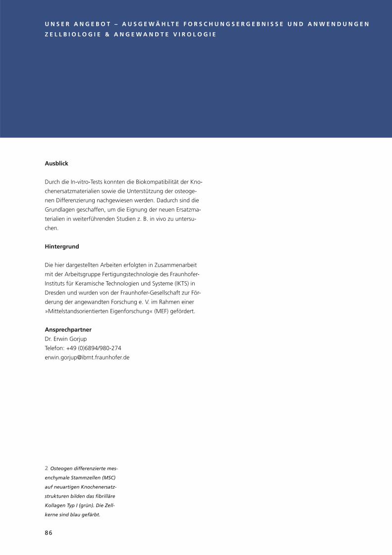
Ausblick
Durch die In-vitro-Tests konnten die Biokompatibilität der Kno-
chenersatzmaterialien sowie die Unterstützung der osteoge-
nen Differenzierung nachgewiesen werden. Dadurch sind die
Grundlagen geschaffen, um die Eignung der neuen Ersatzma-
terialien in weiterführenden Studien z. B. in vivo zu untersu-
chen.
Hintergrund
Die hier dargestellten Arbeiten erfolgten in Zusammenarbeit
mit der Arbeitsgruppe Fertigungstechnologie des Fraunhofer-
Instituts für Keramische Technologien und Systeme (IKTS) in
Dresden und wurden von der Fraunhofer-Gesellschaft zur För-
derung der angewandten Forschung e. V. im Rahmen einer
»Mittelstandsorientierten Eigenforschung« (MEF) gefördert.
Ansprechpartner
Dr. Erwin Gorjup
Telefon: +49 (0)6894/980-274
Z E L L B I O L O G I E & A N G E W A N D T E V I R O L O G I E
2 Osteogen differenzierte mes-
enchymale Stammzellen (MSC)
auf neuartigen Knochenersatz-
strukturen bilden das fibrilläre
Kollagen Typ I (grün). Die Zell-
kerne sind blau gefärbt.
U N S E R A N G E B O T – A U S G E W Ä H L T E F O R S C H U N G S E R G E B N I S S E U N D A N W E N D U N G E N
86

2
C E L L B I O L O G Y & A P P L I E D V I R O L O G Y
Outlook
With the in vitro tests it was possible to verify the biocompati-
bility of the bone substitute materials as well as the support of
the osteogenic differentiation. This means that the ground has
been laid to examine the suitability of the new substitute
materials in further-going studies, e. g. in vivo.
Background
The works represented here were carried out in collaboration
with the production technology working group of the Fraun-
hofer Institute for Ceramic Technologies and Systems (IKTS) in
Dresden and were funded by the Fraunhofer-Gesellschaft zur
Förderung der angewandten Forschung e. V. within the frame-
work of an "SME-oriented self research" project.
Contact
Dr. Erwin Gorjup
Telephone: +49 (0)6894/980-274
O U R O F F E R – S E L E C T E D R E S E A R C H R E S U L T S A N D A P P L I C A T I O N S
2 Osteogenic differentiated
mesenchymal stem cells (MSC)
on innovative bone substitute
structures form the fibril colla-
gen type I (green). The cell
nuclei are stained blue.
87

Zellbiologie & Angewandte Virologie
Labore der Sicherheitsklasse S2 und S3 mit Schleusenbereich
für mikrobiologische, molekularbiologische und zellbiologische
Arbeiten
– TER-Impedanzmesssystem (cellZscope)
– Durchflusszytometer inklusive Sortiereinheit
– Durchlicht- und Auflichtmikroskope mit Phasen- und Diffe-
renzialinterferenzkontrast, Fluoreszenzeinrichtung und
Dokumentationseinheit, mit Manipulationseinheit und Inku-
bationshaube
– Spektralphotometer für Absorptions-, Fluoreszenz- und
Lumineszenzmessungen in Mikrotiterplatten, Elispotreader
– Cobas c111 Analyzer zur photometrischen Bestimmung
klinisch-chemischer Parameter
– Mikro-, Kühl- und Ultrazentrifugen
– Gelelektrophoreseeinheiten für DNA, RNA und Proteine mit
Dokumentationseinheiten
– Gefriermikrotom
– »real time«-PCR-Cycler
– automatisierte Zellkultur- und Virusproduktionseinheit
– Leica-TCS-SP8-X-Konfokalmikroskop ausgestattet mit Weiß-
lichtlaser und Dauerstrichlaser der Wellenlänge 405 nm
– Agilent-HPLC-Anlage
Radionuklidlabor der Sicherheitsklasse S2 für den Umgang mit
offenen radioaktiven Stoffen
– Flüssigkeitsszintillationszähler Modell 2919 TR
– TER-Impedanzmesssystem (cellZscope)
Cell Biology & Applied Virology
Laboratories with safety level S2 and S3 with double-door sys-
tem for microbiological, molecular biological and cell biologi-
cal research:
– TER impedance measuring system (cellZscope)
– flow cytometer including sorting unit
– transmitted light microscopes and reflected light micro-
scopes with phase and differential interference contrast,
fluorescence unit and documentation unit, with manipula-
tion unit and incubation hood
– spectral photometer for absorption, fluorescence and lumi-
nescence measurements of microtitre plates, elispot reader
– Cobas c111 analyzer for photometric determination of clini-
cal-chemical parameters
– micro, cooling and ultra centrifuges
– gel electrophoresis units for DNA, RNA and proteins with
documentation units
– cryo microtome
– real-time PCR cycler
– automated cell culture and virus production unit
– Leica TCS SP8 X confocal microscope equipped with white
light laser and continuous wave laser with a wavelength of
405 nm
– Agilent HPLC system
Radionuclide laboratory of safety level S2 for use with open
radioactive substances:
– fluid scintillation counter model 2919 TR
– TER impedance measuring system (cellZscope)
Z E L L B I O L O G I E & A N G E W A N D T E V I R O L O G I E
AUSSTATTUNG EQUIPMENT
U N S E R A N G E B O T – A U S G E W Ä H L T E F O R S C H U N G S E R G E B N I S S E U N D A N W E N D U N G E N
88

C E L L B I O L O G Y & A P P L I E D V I R O L O G Y
O U R O F F E R – S E L E C T E D R E S E A R C H R E S U L T S A N D A P P L I C A T I O N S
Sensitive Fluoreszenz-Mikrosko-
pie und Live Cell Imaging mit
Hilfe des konfokalen Laserscan-
ning-Mikroskops Leica TCS SP8
ausgestattet mit Weißlichtlaser
und Dauerstrichlaser (405 nm) –
Sensitive fluorescence microscopy
and live cell imaging using the
confocal laser scanning micro-
scope Leica TCS SP8 equipped
with white light laser and contin-
geeignet für anspruchsvolle
Proben und sehr hohe Aufnah-
megeschwindigkeiten bei voller
konfokaler Auflösung
(Foto: Annette Maurer).
uous wave laser (405 nm) – suit-
able for difficult samples and
very high recording speeds at full
confocal resolution (Photo:
Annette Maurer).
89

16 x 16 mikrosystemtechnisch gefertigte akustische Linsen auf der
gegenüberliegenden Seite von 16 x 16 kreisförmig piezoelektrischen
Zinkoxidschichten mit Goldelektroden auf einem Silizium-Wafer.
Anwendung: Ultraschallgeber in akustischen Mikroskopen.
16 x 16 microsystem-produced acoustic lenses on the opposite side
of 16 x 16 circular piezoelectric zinc oxide layers with gold electrodes
on a silicon wafer. Application: ultrasonic transmitter in acoustic
microscopes.
U N S E R A N G E B O T – A U S G E W Ä H L T E F O R S C H U N G S E R G E B N I S S E U N D A N W E N D U N G E N
90

O U R O F F E R – S E L E C T E D R E S E A R C H R E S U L T S A N D A P P L I C A T I O N S
Angebote, Ergebnisse und Produkte der Abteilung
Medizinischer Ultraschall
Ultraschallsysteme/Klinische Anwendungen
Biomedizinische Ultraschallforschung
Hochfrequente Piezosysteme
Zulassung medizinischer Systeme
Projektbeispiel: Ultraschall-Plattform zur Präzisions-
steigerung von Diagnose- und Therapiesystemen
Angebote, Ergebnisse und Produkte der Abteilung
Technischer Ultraschall
Technische Ultraschallsysteme
Wandlerentwicklung
Fertigungstechnologie (ISO 9001 & 13485)
Mechanische Werkstätten
Projektbeispiel: »CSonic«-MBES – Kompaktes Fächer-
Echolot für den Einsatz in Tauchfahrzeugen
Ausstattung
Offers, results and products of the Medical Ultrasound
Department
Ultrasound Systems/Clinical Applications
Biomedical Ultrasound Research
High-frequency Piezosystems
Regulatory Affairs Medical Systems
Project example: Ultrasound platform to increase the
precision of diagnosis and therapy systems
Offers, results and products of the Technical Ultrasound
Department
Technical Ultrasound Systems
Transducer Development
Manufacturing Technology (ISO 9001 and 13485)
Precision Engineering
Project example: "CSonic" MBES – Compact multibeam
echo sounder for use in diving vehicles
Equipment
ULTRASCHALLULTRASOUND
91

Die Hauptabteilung Ultraschall des Fraunhofer IBMT bildet von
der Transducerentwicklung über variable Beamformer und
Software bis zur Signalverarbeitung die gesamte Kompetenz
für eigenständige Gerätesysteme zur Lösung medizinischer,
biotechnologischer und technischer Aufgabenstellungen
durchgängig ab und ist mit über 40 Mitarbeitern in zwei
Abteilungen und acht Arbeitsgruppen die größte Ultraschall-
forschungseinheit in Europa. Die Kompetenzen der Arbeits-
gruppen beruhen auf mehr als 20-jähriger Erfahrung und
erlauben die Entwicklung aller Systemkomponenten, begin-
nend bei Materialien mit speziell angepassten Eigenschaften,
anwendungsspezifischen Ultraschallwandlern, elektronischen
Systemkomponenten und Verfahren der Softwareentwicklung
bis hin zur Sensorfertigung und Prozessentwicklung. Das
Angebot reicht von Beratung und Machbarkeitsstudien über
Labormuster und Prototypentwicklung bis zur zertifizierten
Produktentwicklung, Zulassung sowie Evaluierung und wird
mit einem Umsatzanteil von ca. 70 % von der Industrie
genutzt.
Mit hochfrequentem Ultraschall können etwa Strukturen im
Submikrometerbereich, wie einzelne biologische Zellen, abge-
bildet, charakterisiert und schonend manipuliert werden. In
der Medizin bieten hochfrequente miniaturisierte Arrays neue
Möglichkeiten zur bildgebenden Darstellung von inneren und
äußeren Körperoberflächen und für die präklinische Forschung
an Kleintieren. Daneben bestätigen laufende öffentliche Pro-
jekte, etwa zur Frühdiagnostik von Mammakarzinomen und
der Arthritis, das hohe Anwendungspotenzial optoakustischer
Kombinationssysteme.
Im klassischen Frequenzbereich der medizinischen Diagnostik
etablieren sich 2D-Arrays, die mit der Echtzeiterfassung und
-verarbeitung von Volumendaten erweiterte Einsatzmöglich-
keiten, z. B. in der Therapiekontrolle, erlauben. Von der weiten
Skalierbarkeit des Ultraschalls profitierten auch in diesem Jahr
Weiterentwicklungen von bildgebenden Sonar-Arrays und
drucktoleranten Komponenten für den Einsatz in der Tiefsee.
Da Entwicklungen in der Medizintechnik vor deren Beginn und
zusätzlich weit nach Entwicklungsende eine große Anwender-
und Produktnähe erfordern, wurde zu den seit vielen Jahren
erfolgreich gelebten Zusammenarbeiten mit den Universitäts-
kliniken Saarland, Mannheim, Heidelberg, einschließlich Deut-
sches Krebsforschungszentrum DKFZ, ein Kooperationsvertrag
mit dem Klinikum Nürnberg geschlossen, einer städtischen
Einrichtung der Maximalversorgung mit dem Vorteil sehr
hoher Fallzahlen.
Ansprechpartner
Medizinischer Ultraschall
Dipl.-Ing. Steffen Tretbar
Telefon: +49 (0) 6894/980-226
Technischer Ultraschall
Dipl.-Ing. Christian Degel
Telefon: +49 (0) 6894/980-221 oder +49 (0) 6897/9071-70
Sekretariat:
Frau Nicole Dumont
Telefon: +49 (0) 6894/980-201
U L T R A S C H A L L
U N S E R A N G E B O T – A U S G E W Ä H L T E F O R S C H U N G S E R G E B N I S S E U N D A N W E N D U N G E N
92

O U R O F F E R – S E L E C T E D R E S E A R C H R E S U L T S A N D A P P L I C A T I O N S
From transducer development to variable beam formers and
software, right up to signal processing, the Ultrasound Division
of the Fraunhofer IBMT covers the whole range of compe-
tences for independent device systems for the solution of
medical, biotechnological and technical problems. With more
than 40 members of staff in two departments and eight work-
ing groups, it is the largest ultrasound research unit in Europe.
The competences of the working groups are based on more
than 20 years of experience, and allow the development of all
system components from materials with specially adapted
properties, to application-specific ultrasound transducers, elec-
tronic system components and software development proce-
dures, right up to sensor production and process development.
Services range from consulting and feasibility studies to labo-
ratory prototypes and prototype development, right up to cer-
tified product development, licensing and evaluation, and are
used by industry with a turnover share of approx. 70 %.
With the aid of high-frequency ultrasound it is possible, for
example, to visualize, characterize and carefully manipulate
structures in the submicrometre range such as individual bio-
logical cells. High-frequency, miniaturized arrays offer new
possibilities in medicine for the visual representation of inter-
nal and external body surfaces and for preclinical research on
small animals. Alongside this, current public projects, for
example on early diagnosis of breast cancer and arthritis, con-
firm the high application potential of opto-acoustic combina-
tion systems.
In the classical frequency range of medical diagnostics, 2D
arrays with real-time registration and processing of volume
data are establishing themselves which offer extended applica-
tion options, for example in therapy control. Further develop-
ments of visualizing sonar arrays and pressure-tolerant compo-
nents for use in deep sea applications are also benefitting this
year from the broad scalability of ultrasound.
As developments in medical engineering require high proxim-
ity to the user and the product before they begin and for long
after the end of development work, in addition to the many
years of successful cooperation with the university clinics of
Saarland, Mannheim, Heidelberg, including the German Can-
cer Research Centre DKFZ, a cooperation agreement has now
been concluded with the Klinikum Nürnberg, a municipal,
maximum-provision facility with the advantage of very high
case numbers.
Contact
Medical Ultrasound
Dipl.-Ing. Steffen Tretbar
Telephone: +49 (0) 6894/980-226
Technical Ultrasound
Dipl.-Ing. Christian Degel
Telephone: +49 (0) 6894/980-221 or +49 (0) 6897/9071-70
Secretary:
Ms. Nicole Dumont
Telephone: +49 (0) 6894/980-201
U L T R A S O U N D
93

Ultraschallsysteme/Klinische Anwendungen
– Skalierbare, mehrkanalige Ultraschallplattform mit offenem
Zugriff auf alle Steuerparameter und alle Ebenen der Signal-
verarbeitungskette
1. Diagnostischer Bereich (Anwendungen im Frequenzbe-
reich 1–20 MHz)
– Ultraschallsysteme kombiniert mit Lasern verschiedener
Wellenlängen zur optoakustischen multispektralen Bild-
gebung
– MR-kompatible Ultraschallsysteme zur multimodalen
Echtzeit-Bildgebung im MRT (1,5 T und 3 T)
– Systeme zur intraoperativen bildgestützten Navigation
und Therapiekontrolle
– Systeme und Verfahren zur Echtzeit-Therapiesteuerung
2. Hochfrequenter Bereich (Anwendungen im Frequenzbe-
reich 20–100 MHz)
– Ultraschallsysteme für präklinische Anwendungen
(Kleintier- und translationale Bildgebung)
– Ultraschallsysteme für die zerstörungsfreie Materialprü-
fung und Prozesskontrolle
3. Niederfrequenter Bereich (Anwendungen im Frequenzbe-
reich 100 kHz–3 MHz)
– Kompakte, hochfrequente, mehrkanalige, bildgebende
Sonarsysteme (MBES)
– Ultraschallsysteme für Luftschallanwendungen
– Therapiesysteme (im Aufbau)
– analoge und digitale Schaltungsentwicklung für kundenspe-
zifische Ultraschallanwendungen
– Software für Visualisierung und Signalverarbeitung
– moderne Bildrekonstruktions- und Abbildungsverfahren
(Plane-Wave-Imaging 2D/3D, Ultrafast-Imaging, etc.)
– Signalverarbeitungsmethoden zur Parameterextraktion (Fil-
terentwicklung) für verschiedenste Anwendungen, Materi-
alien und Objekte
– Softwareentwicklung und Softwarewerkzeuge nach ISO
62304 für die Ultraschallsignalverarbeitung und medizini-
sche Bildgebung
– Softwarelösungen für die Ultraschallsignalverarbeitung und
Bildgebung auf mobilen Endgeräten (mobile App)
Ansprechpartner
Dipl.-Ing. Steffen Tretbar
Telefon: +49 (0) 6894/980-226
Biomedizinische Ultraschallforschung
– Anwendungsspezifische Ultraschallforschung und -entwick-
lung
– Materialcharakterisierung
– akustische Mikroskopie
– optoakustische Verfahren
– Rekonstruktionsalgorithmen
– Entwicklung optoakustischer und akustischer Kontrastmittel
– Messung quantitativer Gewebeparameter
– molekulare Bildgebung
– akustische Manipulationssysteme auf zellulärer Ebene
Ansprechpartner
Dr. Marc Fournelle
Telefon: +49 (0) 6894/980-220
ANGEBOTE, ERGEBNISSE UND PRODUKTE DER ABTEILUNG MEDIZINISCHER ULTRASCHALL
U L T R A S C H A L L
U N S E R A N G E B O T – A U S G E W Ä H L T E F O R S C H U N G S E R G E B N I S S E U N D A N W E N D U N G E N
94

O U R O F F E R – S E L E C T E D R E S E A R C H R E S U L T S A N D A P P L I C A T I O N S
Ultrasound Systems/Clinical Applications
– scalable, multi-channel ultrasound platform with open
access to all control parameters and all levels of the signal
processing chain
1. Diagnostic area (applications in the frequency range
1–20 MHz)
– ultrasound systems combined with lasers of various
wavelengths for opto-acoustic multispectral imaging
– MR-compatible ultrasound systems for multimodal real-
time imaging in MRT (1.5 T and 3 T)
– systems for intra-operative, image-aided navigation and
therapy control
– systems and methods for real-time therapy control
2. High-frequency range (applications in the frequency range
20–100 MHz)
– ultrasound systems for preclinical applications (small ani-
mal and translational imaging)
– ultrasound systems for non-destructive material testing
and process control
3. Low-frequency range (applications in the frequency range
100 kHz–3 MHz)
– compact, high-frequency, multi-beam, imaging sonar
systems (MBES)
– ultrasound systems for airborne sound applications
– therapy systems (in development)
– analogue and digital circuit developments for customer-spe-
cific ultrasound applications
– software for visualization and signal processing
– modern image reconstruction and imaging methods (plane
wave imaging 2D/3D, ultrafast imaging etc.)
– signal processing methods for parameter extraction (filter
development) for a wide range of applications, materials
and objects
– software development and software tools in accordance
with ISO 62304 for ultrasound signal processing and medi-
cal imaging
– software solutions for ultrasound signal processing and
imaging on mobile terminal devices (mobile app)
Contact
Dipl.-Ing. Steffen Tretbar
Telephone: +49 (0) 6894/980-226
Biomedical Ultrasound Research
– application-specific ultrasound research and development
– material characterization
– acoustic microscopy
– opto-acoustic methods
– reconstruction algorithms
– development of opto-acoustic and acoustic contrast agents
– measurement of quantitative tissue parameters
– molecular imaging
– acoustic manipulation systems on the cellular level
Contact
Dr. Marc Fournelle
Telephone: +49 (0) 6894/980-220
OFFERS, RESULTS AND PRODUCTS OF THE MEDICAL ULTRASOUND DEPARTMENT
U L T R A S O U N D
95

Hochfrequente Piezosysteme
– Forschung und Entwicklung auf dem Gebiet materialtechni-
scher Fragestellungen in der Ultraschalltechnologie
– Entwicklung passiver und aktiver Komponenten unter Ver-
wendung existierender piezoelektrischer Materialien und
neuer Werkstoffe aus der Nanotechnologie
– Adaption von Verfahren der Mikrosystemtechnik zur Herstel-
lung hochfrequenter Ultraschalllinsen und Ultraschallarrays
– Entwicklung und Fertigung kundenspezifischer hochfrequen-
ter Ultraschalllinsen und Arrays für neue Anwendungen in
der Medizin, der Biologie und der zerstörungsfreien Werk-
stoffprüfung
– Messlabor zur Untersuchung der elektrischen, akustischen
und schwingungstechnischen Eigenschaften hochfrequenter
Ultraschallkomponenten bis 1 GHz
Ansprechpartner
Dr. Frank Tiefensee
Telefon: +49 (0) 6894/980-270
Zulassung medizinischer Systeme
– Beratung zur Umsetzung applikationsspezifischer Ultraschall-
systeme und -verfahren
– Beratung und Durchführung der Zertifizierung von Medizin-
produkten nach EU-Richtlinie 93/42 EWG
– Planung und Durchführung von Studien zur klinischen
Bewertung von Medizinprodukten
Ansprechpartner
Dipl.-Ing. Steffen Tretbar
Telefon: +49 (0) 6894/980-226
U L T R A S C H A L L
U N S E R A N G E B O T – A U S G E W Ä H L T E F O R S C H U N G S E R G E B N I S S E U N D A N W E N D U N G E N
96

O U R O F F E R – S E L E C T E D R E S E A R C H R E S U L T S A N D A P P L I C A T I O N S
High-frequency Piezosystems
– research and development in the area of material-specific
questions in ultrasound technology
– development of passive and active components using exist-
ing piezoelectric materials and new materials from nano-
technology
– adaptation of methods in microsystem technology for the
production of high-frequency ultrasound lenses and ultra-
sound arrays
– development and production of customer-specific, high-
frequency ultrasound lenses and arrays for new applications
in medicine, biology and non-destructive material testing
– measurement laboratory for the investigation of the electric,
acoustic and oscillating properties of high-frequency ultra-
sound components up to 1 GHz
Contact
Dr. Frank Tiefensee
Telephone: +49 (0) 6894/980-270
Regulatory Affairs Medical Systems
– consulting on the implementation of application-specific
ultrasound systems and processes
– consulting and execution of the certification of medical
products in accordance with EU directive 93/42 EEC
– planning and execution of studies for the clinical assessment
of medical devices
Contact
Dipl.-Ing. Steffen Tretbar
Telephone: +49 (0) 6894/980-226
U L T R A S O U N D
97

Die Ultraschallforschungsplattform »DiPhAS-MRT« wurde spe-
ziell für den Einsatz am MR-Tomographen zur simultanen Bild-
gebung entwickelt. Mit diesem System ist es möglich, die Vor-
teile der MR-Bildgebung (hoher Gewebekontrast) und der
Ultraschallbildgebung (Echtzeitfähigkeit) zu kombinieren. Ein-
gesetzt wird das DiPhAS-MRT beispielsweise für eine ultra-
schallgestützte Bewegungserfassung von inneren Organen
während der Bildgebung im MRT. Für diese Anwendung wur-
den spezielle aufklebbare Ultraschallwandler entwickelt, die in
bis zu vier Ultraschallebenen dreidimensionale Informationen
über die Organbewegung erfassen. Diese im Parallelbetrieb
erhobenen Ultraschalldaten und die daraus errechneten Bewe-
gungsvektoren werden dann zur Kompensation von Bewe-
gungsartefakten in den MRT-Aufnahmen genutzt.
Die Herausforderung bei der Realisierung dieses Systems
bestand in der Anpassung des Beamformers und der Ultra-
schallwandler an die hohen Magnetfelder des Tomographen
und in der Minimierung der elektromagnetischen Störaussen-
dung des Ultraschallsystems, um die MR-Bildgebung nicht zu
beeinflussen.
Neben der Anwendung am 1,5-T- und 3-T-Kernspintomogra-
phen wurden das System und die Verfahren auch für die Echt-
zeit-Bewegungserfassung an PET-CT-Scannern, Radio-Therapie-
Systemen und in Kombination mit Schwerionentherapie-
Systemen erprobt.
Diese Arbeit wurde im Rahmen der Fraunhofer-internen Pro-
gramme im MAVO 823 287 unterstützt.
Ansprechpartner
Dipl.-Ing. Steffen Tretbar
Telefon: +49 (0) 6894/980-226
PROJEKTBEISPIEL: ULTRASCHALL-PLATTFORM ZUR PRÄZISIONSSTEIGERUNG VON DIAGNOSE- UND THERAPIESYSTEMEN
U L T R A S C H A L L
schallgestützten Bewegungser-
fassung von inneren Organen
während der Bildgebung
im MRT.
2 Aufklebbarer MR-kompatibler
Ultraschalltransducer für
3D-Volumenmessung.
1 MR-kompatible Ultraschall-
Forschungsplattform für multi-
modale Bildgebung und Kombi-
nation der Vorteile einer
MR-Bildgebung und der Ultra-
schall-Bildgebung zur ultra-
U N S E R A N G E B O T – A U S G E W Ä H L T E F O R S C H U N G S E R G E B N I S S E U N D A N W E N D U N G E N
98

O U R O F F E R – S E L E C T E D R E S E A R C H R E S U L T S A N D A P P L I C A T I O N S
The ultrasound research platform "DiPhAS-MRT" was devel-
oped especially for use for simultaneous imaging on the MR
tomograph. With this system it is possible to combine the
advantages of MR imaging (high tissue contrast) with those of
ultrasound imaging (real-time capability). The DiPhAS-MRT is
used, for example, for the ultrasound-based motion detection
of internal organs during MRT imaging. For this application
special adhesive ultrasound transducers were developed that
can register three-dimensional information on organ move-
ment on up to four ultrasound levels. This ultrasound data
taken in parallel operation and the motion vectors calculated
from them are then used for compensation of movement arte-
facts in the MRT scans.
The challenge in the realization of this system consisted in the
adaptation of the beam former and the ultrasound transducer
to the high magnetic fi elds of the tomograph and in the mini-
mization of the electromagnetic interference of the ultrasound
system in order not to infl uence the MR imaging.
Alongside the application on the 1.5-T and 3-T magnetic reso-
nance tomographs, the system and the methods were also
tested for the real-time motion detection on PET-CT scanners,
radiotherapy systems and in combination with heavy ion ther-
apy systems.
This work was supported by the Fraunhofer Internal Programs
under Grant No. MAVO 823 287.
Contact
Dipl.-Ing. Steffen Tretbar
Telephone: +49 (0) 6894/980-226
U L T R A S O U N D
PROJECT EXAMPLE: ULTRASOUND PLATFORM TO INCREASE THE PRECISION OF DIAGNOSIS AND THERAPY SYSTEMS
1 2
tion of internal organs during
imaging in MRT.
2 Adhesive MR-compatible
ultrasound transducer for 3D
volume measurement.
1 MR-compatible ultrasound
research platform for multi-
modal imaging and combination
of the advantages of MR imag-
ing and ultrasound imaging for
ultrasound-based motion detec-
99

Technische Ultraschallsysteme
– Analoge & digitale Schaltungsentwicklung
– Embedded Systems
– Stand-alone-Systeme
– portable Systeme
– Leistungsschallsysteme
– ultraschallbasierte Transfektion
– druckneutrale Systeme
– Oberflächenfahrzeuge
– Tauchfahrzeuge und Komponenten
– druckneutrale Batterietechnik
Anwendungsbeispiele
– Flowmeter, Wärmezähler
– Abstandsmessung, Einparkhilfe
– Füllstandsmessung, Levelmeter
– Dickenmessung, Hindernisdetektion
– Transmissionsmessung, Rissdetektion
– Sonoporation
– Sonaranwendungen in der Tiefsee, z. B. Sidescanning und
Hindernisvermeidung
Ansprechpartner
Dipl.-Ing. Peter Weber
Telefon: +49 (0) 6894/980-227
Wandlerentwicklung
– Entwicklung und Optimierung von Ultraschallsensoren
– katheterbasierte Sensoren für die Medizintechnik
– bildgebende Multielementwandler (Arrays) für die Diagnos-
tik, insbesondere 2D-Matrix– und Phased-Arrays
– Sensoren für die Abstands- und Durchflussmessung in Gasen
und Flüssigkeiten
– Ultraschallsensoren für Sonderanwendungen und Umgebungen
– Sensoren für die Materialprüfung
– Sonarsensoren und Antennen
– miniaturisierte Schallwandler
– hochbreitbandige Sensoren für Spezialanwendungen
(z. B. Lackdickenmessung, Materialprüfung)
– Leistungsschallwandler
– nieder- und hochfrequente Reinigungssysteme
(z. B. Megaschallreinigung)
– Beratungsdienstleistungen im Bereich Sensorentwicklung
Ansprechpartner
Dipl.-Ing. Franz Josef Becker (komm.)
Telefon +49 (0) 6894/980-202 oder +49 (0) 6897/9071-70
Fertigungstechnologie (ISO 9001 & 13485)
– Entwicklung und Optimierung von Sensorfertigungstech-
nologie
– Entwicklung und Realisierung von Fertigungshilfsmitteln
– Herstellung von Prototyp- und Nullserien
– piezoelektrische Composites (bis 20 MHz)
– Beratungsdienstleistungen im Bereich Sensorfertigungs-
technik
Ansprechpartner
Thomas Trautmann (komm.)
Telefon: +49 (0) 6897/9071-12
ANGEBOTE, ERGEBNISSE UND PRODUKTE DER ABTEILUNG TECHNISCHER ULTRASCHALL
U L T R A S C H A L L
U N S E R A N G E B O T – A U S G E W Ä H L T E F O R S C H U N G S E R G E B N I S S E U N D A N W E N D U N G E N
100

O U R O F F E R – S E L E C T E D R E S E A R C H R E S U L T S A N D A P P L I C A T I O N S
Technical Ultrasound Systems
– analogue and digital circuit development
– embedded systems
– stand-alone systems
– portable systems
– high-power ultrasound systems
– ultrasound-based transfection
– pressure-neutral systems
– unmanned surface vehicles (USV)
– autonomous underwater vehicles (AUV) and components
– pressure balanced battery technology
Application examples
– flowmeter, heat meter
– distance measurement, parking assist system
– filling level measurement, level meter
– thickness measurement, obstacle detection
– transmission measurement, crack detection
– sonoporation
– sonar applications in the deep sea, e. g., side scanning,
obstacle avoidance
Contact
Dipl.-Ing. Peter Weber
Telephone: +49 (0) 6894/980-227
Transducer Development
– development and optimization of ultrasound sensors
– catheter-based sensors for medical engineering
– imaging multi-element transducers (arrays) for diagnostics, in
particular 2D matrix and phased arrays
– sensors for distance and flow measurement in gases and
fluids
– ultrasound sensors for special applications and environments
– sensors for material testing
– sonar sensors and antennae
– miniaturized sound transducers
– high-bandwidth sensors for special applications (e. g., paint
thickness measurement, non-destructive testing)
– high-power ultrasound transducer
– low-and high-frequency cleaning systems (e. g. megasound
cleaning)
– consulting services in the area of sensor development
Contact
Dipl.-Ing. Franz Josef Becker (comm.)
Telephone: +49 (0) 6894/980-202 or +49 (0) 6897/9071-70
Manufacturing Technology (ISO 9001 & 13485)
– development and optimization of sensor production
technology
– development and realization of production aids
– production of prototype and pre-production series
– piezoelectric composites (up to 20 MHz)
– consulting services in the area of sensor production
technology
Contact
Thomas Trautmann (comm.)
Telephone: +49 (0) 6897/9071-12
OFFERS, RESULTS AND PRODUCTS OF THE TECHNICAL ULTRASOUND DEPARTMENT
U L T R A S O U N D
101

Ausgangssituation
Bei Inspektionsaufgaben im Unterwasserbereich müssen oft
kilometerlange Installationen wie z. B. Pipelines oder Spund-
wände in Hafenanlagen mit hoher Detailauflösung vermessen
werden. Der Einsatz von Tauchern in trüben Gewässern ist
häufig zu gefährlich und aufgrund der Ausdehnung der zu
untersuchenden Bereiche viel zu teuer oder gar nicht erreich-
bar, so dass die Anwendung spezialisierter Fahrzeuge erforder-
lich ist.
Lösung
Im Projekt »CView« (Underwater inspection of ship hulls, piers
and jetties by autonomous underwater vehicles, BMWI FKZ
03SX262C) wurde gemeinsam mit den Partnern ATLAS Elekt-
ronik, DFKI, Hochschule Bremen und dem Fraunhofer IOSB
AST für die eingangs genannten Problemstellungen ein auto-
nomes Messfahrzeug modifiziert und mit Sensoren ausgestat-
tet, die Oberflächen wie z. B. Schiffsrümpfe oder Spundwände
hochauflösend abbilden können. Neben Linienlasern und
Kameras bildet ein spezielles am Fraunhofer IBMT entwickeltes
Sonar das Herzstück dieser Sensoren. Als einziger Sensor ist es
in der Lage, auch in trüben Gewässern Informationen über die
Untersuchungsobjekte zu liefern. Der Arbeitsbereich des
Fächer-Echolots beträgt 0–20 m. Hierin tastet es mit einer Fre-
quenz von 1 MHz Oberflächen ab und liefert diese Informatio-
nen an den Bordrechner des Fahrzeugs, wo z. B. Fehlstellen
analysiert und deren Position festgehalten wird. Bei der Ent-
wicklung des Fächer-Echolots wurde insbesondere eine mög-
lichst kleine Bauform angestrebt. Durch die funktionale
Zusammenführung von Sende- und Empfangsantenne und
einen hohen elektronischen Integrationsgrad wurde eine
Größe von nur 12 x 15 x 25 cm3 ermöglicht.
Potenzial
Durch die kompakte Bauform ist dieses Fächer-Echolot speziell
für den Einsatz in autonomen oder kabelgesteuerten Fahrzeu-
gen geeignet (sogenannte AUVs (Autonomous Underwater
Vehicles) oder ROVs (Remotely Operated Vehicles)). Durch den
immer größer werdenden Inspektionsbedarf bei gleichzeitig
steigendem Kostendruck wird der Markt für effiziente Mess-
systeme weiter steigen.
Ansprechpartner
Dipl.-Ing. Christian Degel
Telefon: +49 (0) 6894/980-221 oder +49 (0) 6897/9071-70
PROJEKTBEISPIEL: »CSONIC«-MBES – KOMPAKTES FÄCHER-ECHOLOT FÜR DEN EINSATZ IN TAUCHFAHRZEUGEN
U L T R A S C H A L L
1 Messobjekt »Fahrrad« im
Messbecken des Fraunhofer
IBMT mit dreidimensionaler
Rekonstruktion aus den Sonar-
daten.
U N S E R A N G E B O T – A U S G E W Ä H L T E F O R S C H U N G S E R G E B N I S S E U N D A N W E N D U N G E N
102

1
O U R O F F E R – S E L E C T E D R E S E A R C H R E S U L T S A N D A P P L I C A T I O N S
Starting situation
For submarine inspection tasks it is often necessary to survey
installations over several kilometres, for example pipelines or
sheet pile walls in harbour piers, with a high degree of detail
resolution. The use of divers in hazy waters is often too dan-
gerous and, due to the extent of the areas to be examined,
much too expensive or unfeasible, necessitating the use of
specialized vehicles.
Solution
In the project "CView" (Underwater inspection of ship hulls,
piers and jetties by autonomous underwater vehicles, BMWI
FKZ 03SX262C) in cooperation with the partners ATLAS Elek-
tronik, DFKI, Hochschule Bremen and the Fraunhofer IOSB
AST, an autonomous measurement vehicle was modified for
the aforementioned tasks and equipped with sensors that can
visualize surfaces, e. g., ships hulls or pier walls in high resolu-
tion. Alongside line lasers and cameras a sonar device espe-
cially developed at the Fraunhofer IBMT forms the heart of
these sensors. It is the only sensor that is capable of delivering
information about the inspected objects even in hazy waters.
The working range of the multibeam echo sounder is 0-20 m.
It scans surfaces within this range with a frequency of 1 MHz
and supplies the information to the on-board computer of the
vehicle, where, for example, faults can be analyzed and their
location identified. In the development of the multibeam echo
sounder, special emphasis was placed on achieving a construc-
tion that is as compact as possible. Thanks to the functional
combination of transmission and reception antennae and a
high degree of electronic integration, a size of just 12 x 15 x
25 cm3 was achieved.
Potential
Due to its compact form, this multibeam echo sounder is
especially suitable for use in autonomous or cable-controlled
vehicles (so-called AUVs (Autonomous Underwater Vehicles)
or ROVs (Remotely Operated Vehicles)). Due to the ever-
increasing need for inspections with simultaneous increases in
costs, the market for efficient measurement systems will con-
tinue to grow.
Contact
Dipl.-Ing. Christian Degel
Telephone: +49 (0) 6894/980-221 or +49 (0) 6897/9071-70
U L T R A S O U N D
1 Measurement object "bicycle"
in the measurement basin of the
Fraunhofer IBMT with three-
dimensional reconstruction from
the sonar data.
PROJECT EXAMPLE: "CSONIC" MBES – COMPACT MULTI-BEAM ECHO SOUNDER FOR USE IN DIVING VEHICLES
103
Ultraschall
– 3-Achsen-Messmikroskop inkl. Bildarchivierung und -verar-
beitung
– 5-Achs-CNC-Fräsmaschine (Primacon). Hochpräzisions-Bear-
beitungszentrum zur Mikrobearbeitung
– 5-Becken-Ultraschall-Reinigungsanlage
– 8-Kanal-Laufzeitdifferenz-Messsystem für Luftschallanwen-
dungen
– akustische Mikroskop-Systeme SASAM
– Bandsägevollautomat, Sägebereich 200 x 200 mm, Abläng-
genauigkeit + – 0,1 mm
– Bauteilvorbereitung: Innenloch-Diamantkreissäge
– Bestückungstechnik: SMD-Feinpitchbestückung
– biologisches Labor, Zellkultur
– CNC-Diamantkreissägen (Disco DAD 321)
– CNC-Drehzentrum (Weiler DZ 32 CNC); Drehdurchmesser
100 mm, Drehlänge 150 m, angetriebene Werkzeuge
– CNC-Flachbettschleifmaschine (Ziersch & Baltrusch)
– CNC-Hochpräzisions-Trenn- und Profilschleifmaschine
(Berney T 38-4 CNC), Arbeitsbereich (AB): 160 x 220 x
120 mm, NC-Rundtisch 360°, Schnittbreite min. ca. 20 μm
– CNC-Laserschneid-Schweißeinrichtung (Trumpf); YAG-Laser
mit variabler Optik, Schnittbreite 200–300 μm, Schneiden
von Keramik, Metallen, Hohlkörpern und Blechen, Material-
stärke 5 μm–2 mm
– CNC-Mikro-Bohr-Fräs-Schleifmaschine (Kern), AB: 220 x 160
x 200 mm, schwenkbarer NC-Rundtisch, fünfachsig
– CNC-Universaldrehmaschine (Rael Meka RT 5, zyklenge-
steuert); Querverstellung 200 mm, Längsverstellung
600 mm, angetriebene Werkzeuge
– CNC-Universalfräsmaschine (Mikron UM 600);
AB: 600 x 500 x 450 mm
– CNC-Werkzeugfräsmaschine (Korradi UW 10 CNC);
AB: 500 x 300 x 400 mm
– computerunterstützte Entwicklungsumgebung für Elektro-
nikboards (ORCAD)
– Doppler-Systeme
– Drehmaschine (Colchester Master VS 3250), Drehdurchmes-
ser 1–300 mm, Drehlänge 650 mm
– DSP- und Microcontroller-Entwicklungsumgebung (Mikro-
chip, Motorola)
– Durchflussmesstechnik: Labormessstände für Durchflüsse
(Speckle Tracking, Laufzeitdifferenz; flüssig: 7 m/s, DN
50/100/200; Gas: variabel bis 30 m/s, DN 200)
– Entwicklungssysteme für industrielle Bildverarbeitung (Lage,
Position, OCR, Patternmatching)
– Fertigungsanlage für Ultraschallsensoren in kleiner und mitt-
lerer Stückzahl
– Fotolithographie, Mask Aligner
– FPGA-Entwicklungsumgebung
– Gewindeschneidautomat
– Impedanzvermessungsplatz
– Insertion-Loss-Messplatz
– Klimakammermessplatz
– Kontaktwinkelmessgerät
– konventionelle Bohr-Fräs-Drehmaschinen (inkl. Rundschleif-
einrichtung)
– Kryostatmessplatz für Sensorcharakterisierung und Zero-
Flow-Messungen
– Läppmaschine
– Laserinterferometermessplatz
– Luftschall-Sensorik (3D-Oberflächen-Scanner, Volumenbe-
stimmung und Positionsdetektoren)
– Messtechnik: Pygnometer, 3D-Schallfeld-Scanner, Impedanz-
messplatz
– Motortafelschere
– optoakustisches Labor
– Phased-Array- und Linear-Array-/Ultraschall-Entwicklungssys-
teme
– Plasma-Reinigungsanlage
– Polarisator
– Präzisionsdosieranlagen
AUSSTATTUNG
U L T R A S C H A L L
U N S E R A N G E B O T – A U S G E W Ä H L T E F O R S C H U N G S E R G E B N I S S E U N D A N W E N D U N G E N
104

O U R O F F E R – S E L E C T E D R E S E A R C H R E S U L T S A N D A P P L I C A T I O N S
Ultrasound
– 3-axis measurement microscope including image archiving
and processing
– 5-axis CNC milling machine (Primacon). High-precision
machining centre for microprocessing
– 5-basin ultrasound cleaning facility
– 8-channel propagation time difference measurement system
for airborne sound applications
– acoustic microscope systems SASAM
– fully automatic band saw, sawing range 200 x 200 mm,
cut-to-length accuracy + – 0.1 mm
– component preparation: inner diameter annular diamond
saw
– surface mounting technology: SMD fine pitch surface
mounting
– biological laboratory, cell culture
– CNC diamond saws (Disco DAD 321)
– CNC drilling centre (Weiler DZ 32 CNC); drilling diameter
100 mm, drilling length 150 m, power-driven tools
– CNC flatbed grinding machine (Ziersch and Baltrusch)
– CNC high-precision separation and profile grinding machine
(Berney T 38-4 CNC), working range (WR): 160 x 220 x
120 mm, NC turntable 360°, min. cut width approx. 20 μm
– CNC laser cutting and welding centre (Trumpf); YAG laser
with variable optical system, cut width 200–300 μm, cuts
ceramics, metals, hollow bodies and sheet metal, material
thickness 5 μm–2 mm
– CNC micro drilling-milling-grinding machine (Kern),
WR: 220 x 160 x 200 mm, swivelling NC turntable, five-axis
– CNC universal lathe machine (Rael Meka RT 5, cycle-con-
trolled); vertical adjustment 200 mm, horizontal adjustment
600 mm, power-driven tools
– CNC universal milling machine (Mikron UM 600); WR: 600 x
500 x 450 mm
– CNC tool milling machine (Korradi UW 10 CNC); WR: 500 x
300 x 400 mm
– computer-aided development environment for electronic
boards (ORCAD)
– Doppler systems
– lathe machine (Colchester Master VS 3250), turning diame-
ter 1–300 mm, turning length 650 mm
– DSP and microcontroller development environment (micro-
chip, Motorola)
– flow measurement technology: laboratory measurement sta-
tions for flows (Speckle Tracking, propagation time differ-
ence; fluid: 7 m/s, DN 50/100/200; gas: variable up to
30 m/s, DN 200)
– development systems for industrial image processing (orien-
tation, position, OCR, pattern matching)
– production system for ultrasound sensors in small and
medium quantities
– photolithography, mask aligner
– FPGA development environment
– automatic thread-cutting machine
– impedance measurement station
– insertion-loss measurement station
– climate chamber measurement station
– contact angle measurement device
– conventional drilling, milling and grinding equipment
(including cylindrical grinder)
– cryostat measurement station for sensor characterization
and zero-flow measurements
– lapping machine
– laser interferometer measurement station
– airborne sound sensors (3D surface scanner, volume determi-
nation and position detectors)
– measurement technology: pygnometer, 3D sound field scan-
ner, impedance measurement station
– motor plate shear
– opto-acoustic laboratory
– phased array and linear array/ultrasound development
systems
– plasma cleaning facility
– polarizer
– precision dosing systems
EQUIPMENT
U L T R A S O U N D
105

– Präzisionsläpp- und Poliermaschinen (Wolters)
– Prüfstand für statische und dynamische Druckbelastbarkeit
– Rasterelektronenmikroskop mit EDX
– Rastersondenmikroskope (AFM, STM, MFM)
– Sandstrahlanlagen
– Schallfeldvermessungsplatz
– Single- und Multichannel-Ultraschallsysteme
– Sinteröfen
– Spezialmesssoftware für den Entwicklungsbereich, Rauheits-
messplatz
– SPS-Entwicklungsplatz (Siemens S 6)
– Sputteranlagen, PCD, PECVD, Reinraum
– Strahlungsdruckwaage
– Temperaturschock-Messplatz
– Ultraschall-Messbecken (6 m x 8 m x 6 m)
– Ultraschall-Navigationssystem-Entwicklungsplattform –
SonoPilot®
– Ultraschall-Sensorsysteme für die Therapiekontrolle (minimal-
invasive Chirurgie, laserinduzierte Thermotherapie)
– Ultraschalluniversalmessplatz für industrielle Anwendungen
(Beton, Stahl, Kunststoffe)
– Vakuumrührgerät zu Vergusszwecken
– Verbindungstechnik Elektronik: Mikrolötstation, Schwall-Löt-
anlage, Reflow-Lötanlage
– Verbindungstechnik/Sensortechnik: Lateral-Move-Klebesand-
wicher, Löt- und Bondtechnologie
– vollparametrische 3D-CAD-Systeme (Pro/Engineer)
– Zero-Flow-Messplatz
– precision lapping and polishing machines (Wolters)
– test stand for static and dynamic pressure resistance
– scanning electron microscope with EDX
– scanning probe microscopes (AFM, STM, MFM)
– sandblasting equipment
– sound field measurement station
– single and multichannel ultrasound systems
– sintering furnaces
– special measurement software for the development areas,
coarseness measurement station
– SPS development centre (Siemens S 6)
– sputter systems, PCD, PECVD, clean room
– radiation pressure gauge
– temperature shock measurement station
– ultrasound measurement basin (6 m x 8 m x 6 m)
– ultrasound navigation system development platform –
SonoPilot®
– ultrasound sensor systems for therapy control (minimally
invasive surgery, laser-induced thermotherapy)
– ultrasound universal measurement station for industrial
applications (concrete, steel, plastics)
– vacuum mixer for moulding
– joining technology in electronics: micro-soldering station,
wave soldering system, reflow soldering system
– joining technology for sensors: lateral move adhesive sand-
wicher, soldering and bonding technology
– fully parametrical 3D CAD systems (Pro/Engineer)
– zero-flow measurement station
U L T R A S C H A L L
U N S E R A N G E B O T – A U S G E W Ä H L T E F O R S C H U N G S E R G E B N I S S E U N D A N W E N D U N G E N
106

O U R O F F E R – S E L E C T E D R E S E A R C H R E S U L T S A N D A P P L I C A T I O N S
U L T R A S O U N D
Rasterelektronenmikroskop REM
(EVO MA 10, Zeiss) mit auf ener-
giedispersiver Röntgenspektro-
metrie basierendem
Mikroanalysesystem EDX (Quan-
tax 200, Bruker) zur Materialana-
lyse (Foto: Annette Maurer).
Scanning electron microscope
SEM (EVO MA 10, Zeiss) with
microanalysis system EDX based
on energy-dispersive X-ray spec-
trometry (Quantax 200, Bruker)
for material analysis
(Photo: Annette Maurer).
107

Mobiles epidemiologisches Labor während des Ersteinsatzes im
Januar 2013 am Technologiehof in Münster, Westfalen
(Foto Bernd Müller).
Mobile epidemiological laboratory during initial operation in
January 2013 at the Technologiehof in Münster, Westphalia
(Photo: Bernd Müller).
U N S E R A N G E B O T – A U S G E W Ä H L T E F O R S C H U N G S E R G E B N I S S E U N D A N W E N D U N G E N
108

O U R O F F E R – S E L E C T E D R E S E A R C H R E S U L T S A N D A P P L I C A T I O N S
Angebote, Ergebnisse und Produkte der Arbeitsgruppen
Labortechnologien
Labor- & Kryomechatronik
Medizinische Netze
Gesundheitsinformationssysteme
Projektbeispiel: Mobile Einheiten für epidemiologische
Anwendungen
Projektbeispiel: »Förderprojekt eEFA Düren« im Rahmen
der Dürener Arbeits gemeinschaft Integrierte Versorgung
(DAGIV)
Ausstattung
Offers, results and products of the working groups
Laboratory Technologies
Laboratory & Cryomechatronics
Medical Networks
Health Information Systems
Project example: Mobile units for epidemiological
applications
Project Example: "Funding Project eEFA Düren" within
the framework of the Düren Working Group "Integrated
Healthcare Provision" (DAGIV)
Equipment
LABOR- & INFORMATIONS-TECHNOLOGIELABORATORY & INFORMA-TION TECHNOLOGY
109

Die Abteilung Labor- & Informationstechnologie umfasst die
Arbeitsgruppen Labor- & Kryomechatronik, Labortechnolo-
gien, Gesundheitsinformationssysteme sowie Medizinische
Netze. In der im Jahr 2013 geschaffenen Abteilung Labor- &
Informationstechnologie bündelt das Fraunhofer-Institut für
Biomedizinische Technik IBMT seine Kompetenzen in den
Bereichen Labortechnologie für stationäre und mobile Labore,
Kryomechatronik, persönliche Gesundheits- und Assistenzsys-
teme, IKT-Infrastrukturen für die vernetzte medizinische For-
schung sowie elektronische Kommunikation im Gesundheits-
wesen.
Die Arbeitsgruppe Labortechnologien befasst sich sowohl auf
Geräte- als auch auf Systemebene mit den technologischen
Grundlagen und Workflows moderner Laboratorien. Von der
Geräteentwicklung mit Konstruktion, physikalischer Simula-
tion, 3D-Visualisierung und Rapid Prototyping bis hin zur
Geräteintegration und Automatisierung. Schwerpunkte liegen
auf dem Gebiet des konzeptionellen Designs mobiler Einheiten
sowie der Entwicklung und der Integration von Sensoren und
optischen Systemen in Hard- und Software. Hierzu gehört
unter anderem auch die Anbindung von Sensoren, Aktoren
und Laborgeräten an Gebäude- oder Fahrzeug-Bussysteme
und die Entwicklung einer übergeordneten Telemetrieplatt-
form zur Anbindung der Labore an eine Leitstelle.
Elektronische Baugruppen und Mikrosysteme für Anwendun-
gen in der Tieftemperaturelektronik bilden einen Schwerpunkt
in der Arbeitsgruppe Labor- & Kryomechatronik. Entwickelt
werden Konzepte und Systeme zur elektronischen Proben-
und Datenverwaltung, einschließlich der zugehörigen IKT-
Infrastruktur zur Identifikation und Inventarisierung biologi-
scher Proben sowie zur Automatisierung von Handhabungs-
prozessen für Kryo- und Biobanken. Weitere Arbeitsschwer-
punkte sind miniaturisierte, hochintegrierte, energieeffiziente
Sensoren, Funkschnittstellen (Bluetooth, ZigBee, WLAN),
Nahfeldkommunikation (RFID, NFC) und Energiemanagement-
systeme. Letztere werden zur Schlüsseltechnologie, wenn es
darum geht, dem stetig steigenden Energiebedarf sowie dem
Wunsch nach möglichst großer Autonomie, insbesondere in
miniaturisierten und portablen Systemen, gerecht zu werden.
Hochminiaturisierte, hocheffiziente Leistungswandler, ener-
gieautarke Systeme, kontaktlose Energieübertragung und
elektrochemische Energiespeicher bilden daher weitere
Schwerpunktthemen.
Mit PaDok/D2D wurde in der Arbeitsgruppe Medizinische
Netze eine Diensteplattform zur alltagstauglichen und sicheren
elektronischen Kooperation von Medizinern entwickelt und
geschaffen. Die Dienste von D2D werden derzeit von über
10 000 medizinischen Einrichtungen genutzt, um einrich-
tungsübergreifend Arztbriefe auszutauschen, Abrechnungen
und Verwaltungsinformationen zu transportieren und medizi-
nische Fallakten anzulegen und zu pflegen. Die Summe der
Dienste von D2D bildet eine sichere medizinische Cloud. Die
zugrundeliegenden Konzepte, aber auch die praktischen
Erfahrungen zum Aufbau sicherer medizinischer Netze werden
zurzeit auf weitere medizinische und nichtmedizinische
Anwendungen übertragen, bei denen der Schutz und die
Sicherheit personenbezogener Daten von besonderem Inter-
esse sind.
Die Arbeitsgruppe Gesundheitsinformationssysteme entwickelt
persönliche Gesundheitssysteme und intelligente Assistenten
für Senioren und chronisch Kranke, Telemedizinsysteme und
L A B O R - & I N F O R M A T I O N S T E C H N O L O G I E
U N S E R A N G E B O T – A U S G E W Ä H L T E F O R S C H U N G S E R G E B N I S S E U N D A N W E N D U N G E N
110

O U R O F F E R – S E L E C T E D R E S E A R C H R E S U L T S A N D A P P L I C A T I O N S
The Laboratory & Information Technology Department includes
the working groups Laboratory & Cryomechatronics, Labora-
tory Technologies, Health Information Systems as well as Medi-
cal Networks. In the Laboratory & Information Technology
Department created in 2013, the Fraunhofer Institute for Bio-
medical Engineering (IBMT) bundles its competences in the
areas of laboratory technology for stationary and mobile labo-
ratories, cryomechatronics, personal health and assistance sys-
tems, ICT infrastructures for networked medical research, as
well as electronic communication in the healthcare system.
The working group Laboratory Technologies is concerned with
the technological basis and workflows of modern laboratories
on the device and the system levels. This ranges from the
development of devices with design, physical simulation, 3D
visualization and rapid prototyping, right up to device integra-
tion and automation. Focal points lie in the area of conceptual
design of mobile units as well as the development and the
integration of sensors and optical systems in hardware and
software. This includes, among other things, the integration of
sensors, actuators and lab devices in building or vehicle bus
systems and the development of a superordinate telemetry
platform for linking the laboratories to a control centre.
Electronic assemblies and microsystems for applications in low-
temperature electronics are a focal point for the working
group Laboratory & Cryomechatronics. The working group
develops concepts and systems for electronic sample and data
administration including the corresponding ICT infrastructure
for identification and inventorization of biological samples as
well as the automation of handling processes for cryobanks
and biobanks. Further focal points include miniaturized, highly
integrated, energy-efficient sensors, wireless interfaces (Blue-
tooth, ZigBee, WLAN), near field communication (RFID, NFC)
and energy management systems. The latter are a key technol-
ogy when it comes to fulfilling constantly rising energy
requirements as well as the desire for the greatest possible
degree of autonomy, particularly in miniaturized and portable
systems. High-miniaturized, highly efficient power converters,
energy-autonomous systems, non-contact energy transmission
and electrochemical energy storage systems are therefore fur-
ther focus areas.
The working group Medical Networks used PaDok/D2D to
develop and create a services platform for practical and secure
electronic cooperation of doctors. The D2D services are cur-
rently used by over 10,000 medical facilities in order to
exchange cross-institutional doctor's letters, transport invoices
and administration information and to file and administrate
medical case files. The sum of the services of D2D forms a
secure medical cloud. The underlying concepts as well as the
practical experience in the development of secure medical net-
works are currently being transferred to further medical and
non-medical applications in which the protection and the
security of personal data are of particular interest.
The working group Health Information Systems develops per-
sonal healthcare systems and intelligent assistants for senior
citizens and chronic illness sufferers, telemedicine systems and
L A B O R A T O R Y & I N F O R M A T I O N T E C H N O L O G Y
111

Disease-Management-Plattformen mit integrierter Entschei-
dungsunterstützung sowie innovative IKT-Infrastrukturen für
die klinische Forschung und Big-Data-Anwendungen zur
semantischen medizinischen Informationsextraktion und
-analytik. Beispiele erfolgreicher Entwicklungen der Gruppe
bilden der »Personal Health Manager« für chronisch Kranke,
die Biobanksystemlösung »eurocryoDB« zum Management
von Biomaterialressourcen oder das ontologiebasierte Studien-
managementsystem »ObTiMA«. Ferner arbeitet die Arbeits-
gruppe an intelligenten, ad-hoc vernetzten, plug-and-play-
fähigen Medizingeräten und Sensoren und repräsentiert das
Institut in der Fraunhofer-Allianz Big Data.
Ansprechpartner
Dipl.-Ing. Peter Wambsganß
Telefon: +49 (0) 6897/9071-50
Sekretariat:
Frau Kerstin Knobe
Telefon: +49 (0) 6897/9071-40
L A B O R - & I N F O R M A T I O N S T E C H N O L O G I E
U N S E R A N G E B O T – A U S G E W Ä H L T E F O R S C H U N G S E R G E B N I S S E U N D A N W E N D U N G E N
112

O U R O F F E R – S E L E C T E D R E S E A R C H R E S U L T S A N D A P P L I C A T I O N S
disease management platforms with integrated decision sup-
port as well as innovative ICT infrastructures for clinical
research and big data applications for semantic medical infor-
mation extraction and analysis. Examples of successful devel-
opments by the group include the "Personal Health Manager"
for the chronically ill, the biobank system solution "eurocryo-
DB" for the management of biomaterial resources, or the
ontology-based study management system "ObTiMA". The
group is also working on intelligent, ad-hoc networked, plug-
and-play capable medical devices and sensors and represents
the institute in the Fraunhofer alliance "Big Data".
Contact
Dipl.-Ing. Peter Wambsganß
Telephone: +49 (0) 6897/9071-50
Secretary:
Ms. Kerstin Knobe
Telephone: +49 (0) 6897/9071-40
L A B O R A T O R Y & I N F O R M A T I O N T E C H N O L O G Y
113

Labortechnologien
Simulation, Visualisierung und Modellierung
– Computerunterstützte Entwicklung, Test und Optimierung
von (elektro-)magnetischen und piezoelektrischen Sensoren
und Systemen
– Hydrodynamik und Simulation (mikro-)fluidischer Systeme
– Festigkeitsanalysen/-berechnungen und FEM-basierte Bau-
teileoptimierung
– Feld- und Ausbreitungsberechnung in den Bereichen Akus-
tik, Elektromagnetik und Thermodynamik
– 3D-Visualisierung und Animation in Biologie, Chemie,
Physik, Medizin und Technik
– 3D-Konstruktion und Prototypendesign
Bildverarbeitung und Machine Vision
– Medizinische Bildverarbeitung und 3D-Rekonstruktion
– Implementation von automatisierter Bildverarbeitung und
»machine vision«- Software
Laborgeräteintegration
– Konzeption, Aufbau und Test von In-vitro-Zellkultur-Geräten
– fluidische CellPROM-Chipsysteme
– mehrkanalige, skalierbare CellPROM-Spritzenpumpensys-
teme
– magnetische CellPROM-Carrier mit Nanoscape-Beschichtung
– Design, Simulation, Aufbau und Test von mikrofluidischen
Chipsystemen
– Implementation von Gerätesteuerung basierend auf LabView
und C++
– Integration von Hard- und Software zur Bildakquisition
– Pilotstudien zur In-vitro-Zellkultur-Automatisierung
Magnetische und elektromagnetische Systeme
– Auftragsmessung in NMR-Bildgebung und -Spektroskopie
– Konzeption und Aufbau von magnetbasierten Manipula-
tionssystemen
– unilaterale NMR-Sensorsysteme
– Entwurf und Bau angepasster Spulen und Antennen für
NMR, RFID und drahtlose Energieübertragung
– Implementierung von Mess- und Auswertesoftware
– Beratung und Machbarkeitsstudien im Umfeld NMR, RFID
und drahtloser Energieübertragung
Ansprechpartner
Dipl.-Phys. Daniel Schmitt
Telefon: +49 (0) 6894/980-120
ANGEBOTE, ERGEBNISSE UND PRODUKTE DER ARBEITSGRUPPEN
L A B O R - & I N F O R M A T I O N S T E C H N O L O G I E
U N S E R A N G E B O T – A U S G E W Ä H L T E F O R S C H U N G S E R G E B N I S S E U N D A N W E N D U N G E N
114
O U R O F F E R – S E L E C T E D R E S E A R C H R E S U L T S A N D A P P L I C A T I O N S
Laboratory Technologies
Simulation, visualization and modelling
– computer-aided development, testing and optimization of
(electro) magnetic and piezoelectric sensors and systems
– hydrodynamics and simulation of (micro) fluidic systems
– strength analyses/calculations and FEM-based component
optimization
– field and propagation calculation in the areas of acoustic,
electromagnetic and thermodynamic systems
– 3D visualization and animation in biology, chemistry, physics,
medicine and technology
– 3D construction and prototype design
Image processing and machine vision
– medical image processing and 3D reconstruction
– implementation of automated image processing and
"machine vision" software
Lab device integration
– conception, development and testing of in vitro cell culture
devices
– fluidic CellPROM chip systems
– multi-channel, scalable CellPROM syringe pump systems
– magnetic CellPROM carrier with Nanoscape coating
– design, simulation, development and testing of microfluidic
chip systems
– implementation of device controls based on LabView and
C++
– integration of hardware and software for image acquisition
– pilot studies for in vitro cell culture automation
Magnetic and electromagnetic systems
– contract measurement in NMR imaging and spectroscopy
– conception and development of magnet-based manipulation
systems
– unilateral NMR sensor systems
– design and construction of adapted coils and antennae for
NMR, RFID and wireless energy transmission
– implementation of measurement and evaluation software
– consulting and feasibility studies in the areas of NMR, RFID
and wireless energy transmission
Contact
Dipl.-Phys. Daniel Schmitt
Telephone: +49 (0) 6894/980-120
OFFERS, RESULTS AND PRODUCTS OF THE WORKING GROUPS
L A B O R A T O R Y & I N F O R M A T I O N T E C H N O L O G Y
115

Labor- & Kryomechatronik
– Forschung und Entwicklung im Bereich (Tieftemperatur-)
Elektronik (digitale und analoge Baugruppen, Sensorik, RFID,
Optoelektronik)
– Entwicklung von drahtlosen, energiesparsamen Datenlogger-
modulen für extreme Umgebungsbedingungen (Temperatur,
Luftfeuchte, Beschleunigung, Strahlung)
– Entwicklung von Modulen für die induktive Energieübertra-
gung in Laboranwendungen
– Entwicklung von optischen Objekterkennungsmodulen für
die Laborautomatisierung
– Entwicklung von Labor-Disposables, insbesondere
Probensubstrate/-container (z. B. Röhrchen und Titerplatten)
mit integrierten elektronischen Baugruppen und Mikrosys-
temen
– Entwicklung von (Tieftemperatur-)Elektronik-Messplätzen
– Test und Charakterisierung elektronischer Bauelemente und
Baugruppen bis zu einer Temperatur von –196 °C
– Test und Charakterisierung von Materialermüdung durch
Temperaturstress
– tieftemperaturtolerante und -optimierte elektronische Infra-
struktur für Probenlagerbehälter (basierend auf einem
32-bit-Mikrocontroller)
– Konzeption und Konstruktion neuer Lagertechnologien für
die Kryokonservierung, z. B. neuartiges Rack-System für eine
elektronische Proben- und Datenarchivierung
– Fertigung mechatronischer Prototypen
– Forschung und Entwicklung im Bereich chipbasiertes, adapti-
ves Labor- und Workflowmanagement (»LabOS®«-
Technologie)
– Entwicklung spezieller Tools und Hardware (z. B. Chip-Lese-
geräte für Röhrchen und Titerplatten) für optimiertes Pro-
benhandling und -tracking
Ansprechpartner
Dr. Frank Ihmig
Telefon: +49 (0) 6897/9071-78
Medizinische Netze
– PaDok® – sichere Kommunikation und fallbasierte Netzakte
im Gesundheitssystem
– Lösungen zur Vernetzung von Dienstleistern des Gesund-
heitswesens
– elektronische patientenbegleitende Dokumentation und
elektronische Fall-(Patienten-)akte
– Einbindung des elektronischen Heilberufsausweises und der
Gesundheitskarte
– Konzepte zum Datenschutz und zur Datensicherheit in der
Medizin
– Einbindung von Praxis- und Klinikinformationssystemen,
Haus-Basisstationen und medizinischen Geräten in medizini-
sche Kommunikationsnetzwerke
– medizinische Standards (DICOM 3.0, HL7, xDT, ICD10, XML,
CDA, etc.)
– elektronisches Disease-Management
– Erstellung von Datenbanksystemen zur anonymisierten und
pseudonymisierten klinischen und Versorgungsforschung
– Vernetzung von Dienstleistern des Gesundheitswesens mit
der Gesundheitstelematiklösung PaDok® / D2D®
– Datensicherheitsgutachten
Ansprechpartner
Dipl.-Phys. Bertram Bresser
Telefon: +49 (0) 6894/980-206
L A B O R - & I N F O R M A T I O N S T E C H N O L O G I E
U N S E R A N G E B O T – A U S G E W Ä H L T E F O R S C H U N G S E R G E B N I S S E U N D A N W E N D U N G E N
116

O U R O F F E R – S E L E C T E D R E S E A R C H R E S U L T S A N D A P P L I C A T I O N S
Laboratory & Cryomechatronics
– research and development in the areas of (low-temperature)
electronics (digital and analogue assemblies, sensors, RFID,
optoelectronics)
– development of wireless, energy-saving data logger modules
for extreme environmental conditions (temperature, humid-
ity, acceleration, radiation)
– development of modules for inductive energy transmission in
laboratory applications
– development of optical object recognition modules for labo-
ratory automation
– development of laboratory disposables, in particular sample
substrates/containers (e. g., vials and titre plates) with inte-
grated electronic assemblies and microsystems
– development of (low-temperature) electronic measurement
stations
– test and characterization of electronic components and
assemblies down to a temperature of –196 °C
– test and characterization of material fatigue due to tempera-
ture stress
– low-temperature-tolerant and optimized electronic infra-
structure for sample storage containers (based on a 32-bit
microcontroller)
– conception and construction of new storage technologies
for cryopreservation, e. g., innovative rack system for elec-
tronic sample and data archiving
– construction of mechatronic prototypes
– research and development in the area of chip-based, adap-
tive laboratory and workflow management ("LabOS®" tech-
nology)
– development of special tools and hardware (e. g., chip read-
ing devices for vials and titre plates) for optimized sample
handling and tracking
Contact
Dr. Frank Ihmig
Telephone: +49 (0) 6897/9071-78
Medical Networks
– PaDok® – secure communication and case-based network
file in the healthcare system
– solutions for the networking of service providers in the
healthcare system
– electronic, patient-accompanying documentation and elec-
tronic case (patient) file
– integration of the electronic health professional’s ID and the
health insurance card
– concepts for data protection and data security in medicine
– integration of information systems for doctor’s offices and
clinics, house base stations and medical devices in medical
communication networks
– medical standards (DICOM 3.0, HL7, xDT, ICD10, XML, CDA,
etc.)
– electronic disease management
– compilation of database systems for anonymized and pseu-
donymized clinical and healthcare research
– networking of service providers in the healthcare system
with the healthcare telematic solution PaDok® / D2D®
– data security assessments
Contact
Dipl.-Phys. Bertram Bresser
Telephone: +49 (0) 6894/980-206
L A B O R A T O R Y & I N F O R M A T I O N T E C H N O L O G Y
117
Gesundheitsinformationssysteme
– Semantische Integration von heterogenen biomedizinischen
Datenquellen
– Langzeitarchivierungslösungen für die digitale Medizin
– semantische Suchmaschinen in der Medizin
– biomedizinische Big-Data-Anwendungen
– klinische Expertensysteme, Entscheidungsunterstützungssys-
teme, multivariante Datenanalyse und Biostatistik
– Telemedizinplattformen für unterversorgte, ländliche Regio-
nen und Epidemiologie
– Informationssysteme für Biobanken und deren Vernetzung
– integrierte Informationssysteme für klinische und epidemio-
logische Studien
– Disease Management-Systeme für die häusliche und mobile
Gesundheitsversorgung von Risikopatienten, älteren und
behinderten Menschen
– gesundheitliche Präventions- und Rehabilitationssysteme
– smarte, vernetzte medizinische Geräte und intelligente
Umgebungen
– medizinische Kommunikationsstandards (HL7, POCT1A,
ICD10, XML, CDISK, etc.)
– Nahfeldortungs- und Lokalisierungssysteme, Asset-Tracking
und -Management
– biomedizinische Computer-Vision-Lösungen
Lösungen:
– Semantex – Semantisches Framework zum Indizieren und
Durchsuchen großer Datenmengen
– ObTiMA – Ontologiebasiertes Studienmanagementsystem
für die klinische Forschung
– EurocryoDB – Probenverwaltung und Probenportal für Bio-
banken
– TOPCARE – Die Home-Care- und Telemedizinplattform
– d-LIVER – Prozessorientierte Disease-Management- und Ent-
scheidungsunterstützungsplattform
Ansprechpartner
Dipl.-Inform. Stephan Kiefer
Telefon: +49 (0) 6894/980-156
L A B O R - & I N F O R M A T I O N S T E C H N O L O G I E
U N S E R A N G E B O T – A U S G E W Ä H L T E F O R S C H U N G S E R G E B N I S S E U N D A N W E N D U N G E N
118

O U R O F F E R – S E L E C T E D R E S E A R C H R E S U L T S A N D A P P L I C A T I O N S
Health Information Systems
– semantic integration of heterogeneous biomedical data
sources
– long-term archiving solutions for digital medicine
– semantic search engines in medicine
– biomedical big data applications
– clinical expert systems, decision-supporting systems, multi-
variant data analysis and biostatistics
– telemedicine platforms for underserved, rural regions and
epidemiology
– information systems for biobanks and their networking
– integrated information systems for clinical and epidemiologi-
cal studies
– disease management systems for home and mobile health-
care provision for risk patients, elderly and disabled persons
– health prevention and rehabilitation systems
– smart, networked medical devices and intelligent environ-
ments
– medical communication standards (HL7, POCT1A, ICD10,
XML, CDISK, etc.)
– near field location and localization systems, asset tracking
and management
– biomedical computer vision solutions
Solutions:
– Semantex – semantic framework for the indexing and
searching of large data volumes
– ObTiMA – ontology-based study management system for
clinical research
– EurocryoDB – sample administration and sample portal for
biobanks
– TOPCARE – the home care and telemedicine platform
– d-LIVER – process-oriented disease management and deci-
sion-supporting platform
Contact
Dipl.-Inform. Stephan Kiefer
Telephone: +49 (0) 6894/980-156
L A B O R A T O R Y & I N F O R M A T I O N T E C H N O L O G Y
119

PROJEKTBEISPIEL: MOBILE EINHEITEN FÜR EPIDEMIOLOGISCHE ANWENDUNGEN
L A B O R - & I N F O R M A T I O N S T E C H N O L O G I E
Einführung
Grundlagenforschung, klinische Forschung und Epidemiologie
benötigen im zunehmenden Maße Bioproben als Basis ihrer
Arbeit. Biobanken sind ein wesentliches infrastrukturelles Inst-
rument, um die erforderlichen Materialien in ausreichender
Menge und mit hoher Qualität zur Verfügung zu stellen. Hier-
bei wurden in den vergangenen Jahren nicht zuletzt durch die
Aktivitäten des Fraunhofer IBMT technologisch große Fort-
schritte gemacht. Mit der gleichen Sorgfalt muss jedoch auch
die Probenahme selbst getätigt werden, Qualitätseinbußen an
dieser Stelle sind später nicht mehr zu korrigieren. Bei Kohor-
tenstudien über längere Zeiträume mit vielen Standorten ist
die Qualitätssicherung besonders komplex, da Probenahme
und Präanalytik unter wechselnden örtlichen Rahmenbedin-
gungen ablaufen. Nur mit neuen Ansätzen, die über das tem-
poräre Einrichten von Laboren und Behandlungsräumen hin-
ausgehen, ist man für kommende Studien mit vielen
Standorten gewappnet und nur so kann ein geschlossenes
Qualitätsmanagement von der Probenahme bis zur -lagerung
etabliert werden.
Ausgangssituation
Das Fraunhofer IBMT betreut in der Arbeitsgruppe »Umwelt-
probenbank – Humanproben« der Hauptabteilung Zellbiologie
& Angewandte Virologie seit 2012 den Humanteil der
Umweltprobenbank des Bundes. Über die direkten Umwelt-
proben wie Ackerboden, Blätter, Wasser und Tierorgane hin-
aus, werden hier die Auswirkungen von Umwelteinflüssen auf
die deutsche Bevölkerung überwacht. Dazu wird seit Mitte der
1980er Jahre jährlich an mittlerweile vier festen Standorten
Urin und Blut von jeweils 120 jungen Erwachsenen gesam-
melt, analysiert und für nachgelagerte Untersuchungen einge-
froren und gelagert.
Seit mehreren Jahren bereits entwickelt das Fraunhofer IBMT
erfolgreich mobile medizinische Einheiten, wie das weltweit
erste mobile Labor nach biologischer Sicherheitsstufe 3
(BSL-3). Diese bieten den Vorteil, identische Laborumgebung
und Geräte in kompakter Form an beliebige Orte bringen zu
können und stellen damit eine ideale Basis für Probenahmen
mit gleichbleibender Qualität dar. Basierend auf diesen Erfah-
rungen wurde im Rahmen des Projekts »Labor der Zukunft«
ein mobiles Labor für epidemiologische Anwendungen konzi-
piert, aufgebaut und Anfang des Jahres 2013 für die Probe-
nahme der Umweltprobenbank des Bundes erstmalig in Ein-
satz gebracht.
Planungsphase
Die in der Westkapregion in Südafrika laufende Kampagne zur
HIV- und Tuberkulose-Diagnostik mit dem mobilen BSL-3-La-
bor des Fraunhofer IBMT wurde vom Team des Projekts »Labor
der Zukunft« technisch begleitet. Diese Erfahrungen sind
zusammen mit dem Wissen der Fahrzeugspezialisten der
Bischoff + Scheck AG, Rheinmünster, in die Auslegung und
Konstruktion der zweiten Generation eines Mobillabors einge-
flossen. Gleichzeitig konnten während der konventionell
durchgeführten Probenahmen 2012 für die Umweltproben-
bank wesentliche Erkenntnisse für optimale Logistik, Workflow
und Geräteausstattung gewonnen werden.
Als Kern der Einheit wurde ein medizinisch-analytisches Labor
mit Schleuse, Laborraum und Kryolager (siehe Abbildung 1)
geplant. Hier musste durch eine entsprechende Aufteilung ein
vom übrigen Fahrzeug abgekapselter Arbeitsplatz für vier
Laboranten und die notwendigen Geräte geschaffen werden
(insgesamt 16 m2). Die Forderung nach einem räumlich abge-
trennten Empfangs- und Anamnesebereich sowie einem
Behandlungsbereich mit zwei Blutentnahmestellen sowie
1–12 Fotos: Bernd Müller;
Illustrationen: Hiltrud Görgen,
Fraunhofer IBMT.
U N S E R A N G E B O T – A U S G E W Ä H L T E F O R S C H U N G S E R G E B N I S S E U N D A N W E N D U N G E N
120
O U R O F F E R – S E L E C T E D R E S E A R C H R E S U L T S A N D A P P L I C A T I O N S
L A B O R A T O R Y & I N F O R M A T I O N T E C H N O L O G Y
PROJECT EXAMPLE: MOBILE UNITS FOR EPIDEMIOLOGICAL APPLICATIONS
Introduction
Basic research, clinical research and epidemiology increasingly
require biosamples as the basis for their work. Biobanks are an
important infrastructural instrument to provide the necessary
materials in sufficient quantity and at high quality. Major tech-
nological advances have been made here in recent years, not
least of all due to the efforts of the Fraunhofer IBMT. Equal
care has to be taken, however, in the actual taking of the sam-
ple as quality defects at this point cannot be corrected later. In
the case of cohort studies over long periods with multiple
locations, quality assurance is particularly complex, as the tak-
ing of the sample and the preliminary analysis take place
under different local conditions. It is only with new approaches
going beyond the temporary installation of laboratories and
treatment rooms that we can equip ourselves for future stud-
ies with multiple locations, and this is the only way to establish
a closed quality management system from the taking of the
sample right up to storage.
Starting situation
In the working group "Environmental Specimen Bank –
Human Samples" of the Cell Biology & Applied Virology
Division, the Fraunhofer IBMT takes care of the human section
of the German Environmental Specimen Bank. In addition to
direct environmental samples such as soil, leaves, water and
animal organs, the facility also monitors the effects of environ-
mental influences on the German population. For this pur-
pose, since the mid-1980s samples of urine and blood have
been taken annually at four fixed locations from 120 young
adults, analyzed and then frozen and stored for subsequent
investigations.
For several years now the Fraunhofer IBMT has been success-
fully developing mobile medical units such as the world's first
mobile laboratory in accordance with biological safety level 3
(BSL-3). These offer the advantage of putting an identical lab-
oratory environment and devices in compact form at random
locations and thus constitute the ideal basis for sample taking
with consistent quality. Based on this experience, a mobile lab-
oratory for epidemiological applications was conceived and
developed within the framework of the project "Laboratory of
the Future", and put into operation for sample taking on
behalf of the German Environmental Specimen Bank for the
first time at the start of 2013.
Planning phase
The campaign for HIV and tuberculosis diagnosis performed in
the West Cape region of South Africa with the mobile BSL-3
laboratory of the Fraunhofer IBMT was technically accompa-
nied by the team from the project "Laboratory of the Future".
This experience went into the layout and the construction of
the second generation of mobile laboratories along with the
know-how of the vehicle specialists of the company Bischoff +
Scheck AG, Rheinmünster. At the same time, during the con-
ventional sample taking in 2012 for the Environmental Speci-
men Bank, it was possible to gain important insights for opti-
mum logistics, workflow and equipment.
A medical analysis laboratory with double-door personnel lock,
lab room and cryorepository (see Fig. 1) was planned as the
core of the unit. With the corresponding subdivision, the aim
was to create a workstation isolated from the rest of the vehi-
cle to accommodate four lab technicians and the necessary
equipment (in total 16 m2). The requirement for a spatially
separate reception and medical history area and a treatment
area with two blood taking points as well as a dentist station
121

L A B O R - & I N F O R M A T I O N S T E C H N O L O G I E
1–12 Photos: Bernd Müller;
Illustrations: Hiltrud Görgen,
Fraunhofer IBMT.
1 Visualisierung der Planungsgrundlage.
3 Aufbau des Gitterrohrrahmens.
5 Innenausbau.
2 Schweißen des Chassis.
4 Beplankung.
6 Inbetriebnahme der Elektrik und IT.
Kryolager
Laborraum
Personalschleuse
Behandlungsraum
Empfang und Anamnese
LagertankSterilbankZentrifugeAnalysegerät
1 Visualization of the planning basis. 2 Welding the chassis.
3 Structure of the tubular trellis frame. 4 Panelling. 5 Interior.
6 Starting up the electric system and IT.
U N S E R A N G E B O T – A U S G E W Ä H L T E F O R S C H U N G S E R G E B N I S S E U N D A N W E N D U N G E N
122

O U R O F F E R – S E L E C T E D R E S E A R C H R E S U L T S A N D A P P L I C A T I O N S
L A B O R A T O R Y & I N F O R M A T I O N T E C H N O L O G Y
7 Devices for pre-analysis and automated analysis in the laboratory room. 8 Test drive on a race circuit in Rheinmünster.
9 Approval of the laboratory in Sulzbach at the start of November 2012. 10 Taking a blood sample (left) and laboratory opera-
tion (right) during the first operation for the German Environmental Specimen Bank. 11 First presentation at the MEDICA in
Düsseldorf in November 2012. 12 Design studies of mobile laboratories on different vehicle platforms.
7 Geräte für die Präanalytik und automatisierte Analytik im Laborraum.
9 Abnahme des Labors in Sulzbach Anfang November 2012.
11 Erstvorstellung auf der MEDICA in Düsseldorf im November 2012.
8 Testfahrt auf einer Rennstrecke in Rheinmünster.
10 Blutentnahme (links) und Laborbetrieb (rechts) während des Ersteinsatzes für die Umweltprobenbank des Bundes.
12 Designstudien mobiler Labore auf unterschiedlichen Fahrzeug-plattformen.
123

einem Zahnarztplatz zur Überprüfung des Zahnstatus´ konnte
nur durch die Konstruktion von insgesamt drei Karosserieaus-
schüben erfüllt werden. Damit kann die Grundfläche des Auf-
liegers von etwa 32 m2 auf 55 m2 erweitert werden.
Aufbau und Integration
Die durch die äußeren Abmessungen und im Gesamtgewicht
auferlegten Restriktionen können einzig durch eine zwar
modulare, aber grundlegend an den späteren Innenausbau
angepasste Grundkonstruktion erfüllt werden. Ausgehend
vom Chassis (Abbildung 2) wurde über den Gitterrohrrahmen
(Abbildung 3) und die Beplankung mit isolierten Paneelen
(Abbildung 4) der äußere Aufbau fertiggestellt.
Um autark arbeiten und redundante Energieversorgung
ermöglichen zu können, mussten ein Diesel-Generator
(36 kVA), ein Frischwassertank (400 l) und ein Abwassertank
(1 000 l) integriert werden. Die Wassertanks sind temperatur-
überwacht und mit elektrischen Zuheizern ausgestattet.
Die technischen Anlagen zur Heizung, Lüftung und Klimatisie-
rung erfordern sowohl in der Planung als auch in der Umset-
zung besondere Sorgfalt. Um auch die höchstmöglichen
Anforderungen an die Laborsicherheit erfüllen zu können,
wurde dort eine kaskadierte Unterdruckregelung (–30 und –60
Pa) mit 100 % Frischluft und einem maximal 14-fachen Luft-
wechsel pro Stunde realisiert (Reinraum Becker, Saarbrücken).
Die Frischluft wird über einen zentralen Klimaschrank vorkon-
ditioniert und dann im Fahrzeug verteilt. Über eine Diesel-
Standheizung (9 kW) und eine wassergeführte Fußbodenhei-
zung wird bei tiefen Temperaturen eine Grundheizung
realisiert, die Raumtemperatur wird über Klimaanlagen mit
Wärmepumpenfunktion eingeregelt. Die Abwärme des Gene-
rators kann in die Klimaanlage mit eingekoppelt werden.
Zulassung und Qualitätssicherung
Während der Endmontage im Innenausbau (Abbildung 5)
wurde mit der Inbetriebnahme der sicherheitstechnisch rele-
vanten Leittechnik des Fahrzeugs begonnen (Abbildung 6).
Über diese sind Betriebszustände des Labors und des Fahr-
zeugs auch über Datenfernübertragung kontrollierbar.
Wesentliche Laborkomponenten wie die Sterilbank und auch
das Kryolager wurden in dieser Phase integriert (Abbildung 7).
Das voll ausgestattete Fahrzeug wurde auf einer Teststrecke
mit speziellen Fahrdynamikflächen getestet (Abbildung 8),
bevor es dann am Standort Sulzbach der zuständigen
Behörde, dem Landesamt für Umwelt- und Arbeitsschutz (ver-
treten durch Dr. Michael Heger, Abbildung 9) vorgestellt
wurde. Basierend auf dieser technischen Überprüfung und den
technischen Unterlagen wurde schließlich eine Zulassung nach
Biostoffverordnung Schutzstufe 2 erteilt, spezifisch für die
Anforderungen der Probenahme für den Humanteil der
Umweltprobenbank des Bundes. Für die Abläufe bei der Pro-
benahme wurde für die ersten Einsätze ein Qualitätssiche-
rungssystem aufgebaut und etabliert. Damit konnte erstmalig
an allen Standorten mit der identischen Ausstattung für Probe-
nahme und Labor gearbeitet werden und damit ein einheitli-
ches Qualitätsmanagementsystem nach DIN EN ISO/IEC 17025
etabliert werden.
Einsätze
Ende Januar 2013 fand der Ersteinsatz des Fahrzeugs anläss-
lich der Probenahme in Münster statt. Bei winterlichem Wetter
mit Nachttemperaturen bis –14 °C wurde insbesondere die
Heizungsanlage unter realen und extremen Bedingungen
getestet. Die Arbeitsabläufe in der mobilen Einheit sind so
optimiert, dass auf den 55 m2 Grundfläche 12 Personen ergo-
nomisch arbeiten und im Mittel drei Probanden je 20 min
durchgeschleust werden konnten. Insgesamt wurden an Bord
die Proben von 120 Probanden an zwei Tagen genommen,
analysiert und archiviert. Nach Abschluss der Probenahme
L A B O R - & I N F O R M A T I O N S T E C H N O L O G I E
U N S E R A N G E B O T – A U S G E W Ä H L T E F O R S C H U N G S E R G E B N I S S E U N D A N W E N D U N G E N
124
O U R O F F E R – S E L E C T E D R E S E A R C H R E S U L T S A N D A P P L I C A T I O N S
for examination of the dental status could only be fulfilled by
the construction of a total of three bodywork slides. This
means that the basic area of the trailer can be extended from
around 32 m2 to 55 m2.
Construction and integration
The restrictions imposed in terms of the outer dimensions and
the overall weight can only be fulfilled by a basic construction
which is modular but already adapted for the later interior
installations. Starting from the chassis (Fig. 2) the outer struc-
ture was constructed over the tubular trellis frame (Fig. 3) and
the insulated panelling (Fig. 4).
In order to ensure autonomous operation and a redundant
energy supply, it was necessary to integrate a diesel generator
(36 kVA), a fresh water tank (400 l) and a waste water tank
(1,000 l). The water tanks are temperature-monitored and
equipped with electric heaters.
Special care is required in the planning and the implementa-
tion of the technical systems such as heating, ventilation and
air conditioning. In order to fulfil the highest possible stan-
dards for laboratory safety, a cascaded low-pressure regulation
system (–30 and –60 Pa) with 100% fresh air and up to 14 air
changes per hour was realized (Reinraum Becker, Saar-
brücken). The fresh air is preconditioned via a central air con-
ditioning cabinet and then distributed in the vehicle. Basic
heating is provided at low temperatures by an independent
diesel heating system (9 kW) and waterborne underfloor heat-
ing; the room temperature is controlled via air conditioning
systems with heat pump function. The waste heat of the gen-
erator can be coupled with the air conditioning system.
Licensing and quality assurance
During the final installation of the interior (Fig. 5), the safety-
relevant control technology of the vehicle was started up (Fig.
6). This controls the operating statuses of the laboratory and
the vehicle, also by means of remote data transmission. Essen-
tial laboratory components such as the sterile bank and the
cryorepository were integrated during this phase (Fig. 7). The
fully equipped vehicle was tested on a test route with special
drive-dynamic surfaces (Fig. 8), before being introduced in
Sulzbach to the appropriate authority, the State Office for
Environmental Protection and Occupational Safety (Landesamt
für Umwelt- und Arbeitsschutz) (represented by Dr. Michael
Heger, Fig. 9). Based on this technical inspection and the tech-
nical documents a licence was granted in accordance with the
Biological Agents Ordinance (Biostoffverordnung) Protection
Level 2 specifically for the sample taking requirements for the
human section of the German Environmental Specimen Bank.
A quality assurance system was developed and established for
the sample taking procedures in the initial operations. This
meant that, for the first time, it was possible to work at all
locations with the identical equipment for sample taking and
laboratory work, thus establishing a standardized quality man-
agement system in accordance with DIN EN ISO/IEC 17025.
Operations
The first operation of the vehicle took place at the end of Jan-
uary 2013 for sample taking in Münster. In winter weather
with night-time temperatures as low as –14°C, the heating
system in particular was tested under real, extreme conditions.
The workflow in the mobile unit is so optimized that 12 per-
sons can work ergonomically on an area of 55 m2, while an
average of three probands are processed every 20 minutes.
Samples were taken from a total of 120 probands over two
days, analyzed and archived. When sample taking was com-
pleted, the samples were brought in the laboratory vehicle to
the repository south of Münster, where they were put into
L A B O R A T O R Y & I N F O R M A T I O N T E C H N O L O G Y
125

wurden die Proben im Laborfahrzeug zum Lager südlich von
Münster gebracht und dort eingelagert. In der ersten Jahres-
hälfte konnten weiterhin die Einsätze in Halle (Saale), Greifs-
wald und Ulm erfolgreich durchgeführt werden. Die nächsten
Einsätze für die Umweltprobenbank sind turnusgemäß ab
Januar 2014 geplant.
Im November 2013 konnte ein weiteres medizinisches Einsatz-
szenario erfolgreich demonstriert werden. Gemeinsam mit der
Ärztekammer des Saarlandes und der kassenärztlichen Vereini-
gung Saarland wurde in einem Zeitraum von acht Tagen an
den drei Standorten Neunkirchen, Saarlouis und Saarbrücken
eine Kampagne zur saisonalen Grippeimpfung durchgeführt.
Erstmalig wurde hierbei aufgrund der technischen Ausstattung
der mobilen Einheit über die Beratung hinaus direkt eine Imp-
fung für Interessenten vor Ort durchgeführt.
Ausblick
Mit den beiden mobilen Laboren und den entsprechenden Ein-
sätzen in Südafrika und Deutschland wurden Machbarkeit und
Vorteile mobiler Einheiten mit Laboren der Schutzstufen 2 und
3 demonstriert. Die modulare Bauweise ermöglicht es, anwen-
dungsbezogen effizient mobile Lösungen auf unterschiedli-
chen Fahrzeugplattformen aufzubauen. Designstudien und
Anfragen für medizinisch-diagnostische Einheiten, kleinere
geländegängige Fahrzeuge für die Umweltanalytik und Flot-
tenlösungen (Abbildung 12) zeigen, dass mobile Laborlösun-
gen zukünftig eine immer stärkere Rolle spielen werden.
Projekthintergrund
Seit 2009 befasst sich eine Projektinitiative unter Führung des
Fraunhofer IBMT mit Beteiligung kleiner und mittelständischer
Industrien sowie Unterstützung des Ministeriums für Wirt-
schaft und Wissenschaft mit der Entwicklung innovativer
La bortechnologie »Made in Saarland«. Die entwickelten Ideen
für das »Labor der Zukunft« stießen bei zahlreichen Gesprä-
chen mit nationalen und internationalen Vertretern der Politik,
Anwendern aus Bereichen wie Medizin und Biologie sowie
den Marktführern für Laborprodukte durchweg auf großes
Interesse.
Projektkoordination
Prof. Dr. Heiko Zimmermann
Telefon: +49 (0) 6894/980-100
Projektleitung Labor der Zukunft
Dipl.-Phys. Daniel Schmitt
Telefon: +49 (0) 6894/980-120
Projektleitung Humanteil der Umweltprobenbank
des Bundes
Dr. Dominik Lermen
Telefon: +49 (0) 6894/980-251
L A B O R - & I N F O R M A T I O N S T E C H N O L O G I E
U N S E R A N G E B O T – A U S G E W Ä H L T E F O R S C H U N G S E R G E B N I S S E U N D A N W E N D U N G E N
126

O U R O F F E R – S E L E C T E D R E S E A R C H R E S U L T S A N D A P P L I C A T I O N S
storage. In the first half of the year, further operations were
successfully carried out in Halle (Saale), Greifswald and Ulm.
The next round of operations for the Environmental Specimen
Bank was scheduled to begin in January 2014.
In November 2013 it was possible to demonstrate another
successful medical scenario. Along with the Medical Council of
Saarland and the Saarland Association of Statutory Health
Insurance Physicians (Kassenärztliche Vereinigung (KV) Saar-
land), a seasonal flue vaccination campaign was carried out
over eight days at the three locations Neunkirchen, Saarlouis
and Saarbrücken. Thanks to the technical equipment of the
mobile unit, it was possible for the first time to go beyond a
purely advisory service and provide vaccinations for interested
persons directly onsite.
Outlook
With the two mobile laboratories and the respective opera-
tions in South Africa and Germany, the feasibility and advan-
tages of mobile units with laboratories of safety level 2 and 3
have been demonstrated. The modular construction makes it
possible to accommodate mobile solutions efficiently and on
an application-specific basis on different vehicle platforms.
Design studies and enquiries for medical-diagnostic units,
smaller off-road capable vehicles for environmental analysis
and fleet solutions (Fig. 12) show that mobile laboratory solu-
tions are going to play an increasingly important role in the
future.
Project background
Since 2009 a project initiative led by the Fraunhofer IBMT in
cooperation with small and medium-sized enterprises and sup-
ported by the Federal State Government of the Saarland has
been working on the development of innovative laboratory
technology "Made in Saarland". The ideas developed for the
"Laboratory of the Future" met with great interest in numer-
ous talks with national and international political representa-
tives, users from areas such as medicine and biology and mar-
ket leaders for laboratory products.
Project coordination
Prof. Dr. Heiko Zimmermann
Telephone: +49 (0) 6894/980-100
Project management "Laboratory of the Future"
Dipl.-Phys. Daniel Schmitt
Telephone: +49 (0) 6894/980-120
Project management of the human section of the
Federal Environmental Sample Bank
Dr. Dominik Lermen
Telephone: +49 (0) 6894/980-251
L A B O R A T O R Y & I N F O R M A T I O N T E C H N O L O G Y
127

Das »Förderprojekt eEFA Düren« ist ein von der EU und der
Landesregierung Nordrhein-Westfalens gefördertes Projekt zur
Stärkung und Verbesserung der medizinischen Versorgung der
Bevölkerung. Um dieses Ziel zu erreichen, sind zwei wesent-
liche Projektteile vorgesehen. Ein Teil wird eine einrichtungs-
übergreifende elektronische Fallakte (eEFA) im Bereich der
Region Düren-Süd (im Gebiet südlich von Aachen) zwischen
niedergelassenen Ärzten und Krankenhäusern umfassen, die
aufgebaut und getestet wird. Der andere Teil beinhaltet ein
medizinisches Controlling, in dem ausgewählte Versorgungs-
daten aus der Akte anonymisiert, zusammengefasst und nach
medizinischen und ökonomischen Aspekten ausgewertet wer-
den. An dem Projekt sind zahlreiche Partner beteiligt. Das
Fraunhofer IBMT wurde als Lieferant des »Kommunikations-
kerns« von der Kassenärztlichen Vereinigung Nordrhein im
Auftrag in das Projekt eingebunden. Es ist zusätzlich als Bera-
ter zu datenschutzrechtlichen Aspekten des Modells in das
Projekt eingebunden.
Aufgabenstellung
Die DAGIV vereint ca. 240 ärztliche Teilnehmer aus der nieder-
gelassenen Ärzteschaft und den drei Dürener Krankenhäusern.
Der ambulante Bereich ist zu über 90 % vertreten und macht
ca. 70 % der Mitgliederzahl aus. Durch diesen Behandlerkreis
wird nahezu die gesamte Bevölkerung der Region versorgt.
Die beste medizinische Versorgung von Patienten lässt sich
durch gemeinsame, koordinierte Diagnostik und Therapie der
unterschiedlichen Fachgruppen und Sektoren (niedergelassen
und stationär) erreichen. Die DAGIV hat deshalb seit 2010
intensiv daran gearbeitet, in verschiedenen Kommissionen
erarbeitete Konzepte Zug um Zug umzusetzen (Leitlinien, ver-
einheitlichte Diagnose/Behandlungspfade, Qualitätsmanage-
ment, Pflege der Schnittstelle stationär/ambulant usw.). Das
wesentliche Werkzeug zu einer derartigen »Integrierten Ver-
sorgung« ist der zügige Informationsaustausch zwischen den
beteiligten Behandlungspartnern. Um diesen zu realisieren, hat
sich die DAGIV für D2D, die vom Fraunhofer IBMT entwickelte
vertrauenswürdige Kommunikationsplattform für Health Pro-
fessionals, entschieden. Das Projekt vereint nach gewonnener
Ausschreibung bei der EU und dem Landesministerium für
Gesundheit in Nordrhein-Westfalen die KIS-Anbieter (KIS =
Krankenhausinformationssystem) der drei Dürener Kranken-
häuser, die DURIA e. G. (als ein wichtiger Anbieter eines Pra-
xissoftwaresystems (PVS) in der Region) und die KVNo als
direkte Projektteilnehmer. Zur schrittweisen Heranführung der
zukünftigen Anwender (Ärztinnen und Ärzte in Praxis und Kli-
nik) an die »Fallakte« wurde es als sinnvoll erachtet, mit der
wesentlich weniger komplexen Materie eines elektronisch ver-
sandten Arztbriefes zu beginnen. Nachdem die Beteiligten mit
den neuen elektronischen Werkzeugen vertraut geworden
sind, wird im nächsten Schritt die Notfall- oder Vertreterakte
als Sonderform der elektronischen einrichtungsübergreifenden
»Fallakte« eingeführt. Die Komponenten der Fallakte sind die
vorher eingeführten signierten Arztbriefe. Als weitere Aufgabe
wird angestrebt, anonymisierte Versorgungsdaten für das
Netz-Controlling zu gewinnen, das zur Verbesserung der
medizinischen Versorgung führen soll.
Durchführung
Im genehmigten Projektplan sind drei große Phasen vorgese-
hen. Sie können als
– Einführung des elektronischen Arztbriefs zur Etablierung
elektronischer sektorübergreifender Kommunikation,
– Realisierung einer über-sektoralen elektronischen Vertreter-
und Notfallakte und
– Etablierung eines einrichtungsübergreifenden medizinischen
Controllings beschrieben werden.
Dieser Plan tangiert zahlreiche Aspekte des Standes- und
Datenschutzrechts, technischer Problemstellungen und der
einrichtungs- und sektorübergreifenden Zusammenarbeit. Aus
diesem Grund wurden ganz am Anfang alle Aspekte des Pati-
PROJEKTBEISPIEL: »FÖRDERPROJEKT EEFA DÜREN« IM RAHMEN DER DÜRENER ARBEITSGEMEINSCHAFT INTEGRIERTE VERSORGUNG (DAGIV)
L A B O R - & I N F O R M A T I O N S T E C H N O L O G I E
U N S E R A N G E B O T – A U S G E W Ä H L T E F O R S C H U N G S E R G E B N I S S E U N D A N W E N D U N G E N
128

O U R O F F E R – S E L E C T E D R E S E A R C H R E S U L T S A N D A P P L I C A T I O N S
The "Funding Project eEFA Düren" is a project funded by the
EU and the State Government of North Rhine-Westphalia to
reinforce and improve healthcare provision to the population.
In order to achieve this aim, two essential project sections are
planned. One section involves a cross-facility, electronic case
file (eEFA) for the region of Düren-South (in the area south of
Aachen) between doctors and hospitals, which will be devel-
oped and tested. The other section is concerned with a medi-
cal controlling system in which selected healthcare provision
data from the file is anonymized, summarized and evaluated
according to medical and economic aspects. There are numer-
ous partners involved in the project. The Fraunhofer IBMT was
integrated in the project as the supplier of the "communica-
tion core" by commission of the Kassenärztliche Vereinigung
Nordrhein. It is also involved in the project as a consultant on
data protection aspects of the model.
Tasks
DAGIV brings together around 240 medical participants from
the non-hospital doctor community and the three Düren hos-
pitals. The non-hospital area is represented at a rate of over
90 % and accounts for around 70 % of the membership. This
group of healthcare practitioners takes care of almost the
whole population of the region. The best healthcare provision
for patients can be achieved by joint, coordinated diagnostics
and therapy of the various disciplines and sectors (outpatient
and inpatient). This is why DAGIV has been working inten-
sively since 2010 to gradually implement the concepts devel-
oped in various commissions (guidelines, standardized diagno-
sis/therapy paths, quality management, administration of the
interface inpatient/outpatient etc.). The main tool in this type
of "integrated healthcare provision" is the efficient informa-
tion exchange between the participating healthcare partners.
In order to realize this, DAGIV opted for D2D, the trustworthy
communication platform for health professionals developed by
the Fraunhofer IBMT. Having won the tender from the EU and
the State Ministry for Health in North Rhine-Westphalia, the
project unites the HIS providers (HIS = Hospital Information
System) of the three Düren hospitals, the DURIA e. G. (as an
important provider of a software system for doctor’s offices
(PVS) in the region) and the KVNo as direct project partici-
pants. For a gradual introduction of the future users (doctors
in surgeries and clinics) to the "case file", it was deemed rea-
sonable to start with the substantially less complex matter of
an electronically transmitted doctor's letter. After the partici-
pants have become familiar with the new electronic tools, the
next step will be to introduce the emergency or representative
file as a special form of the electronic cross-facility "case file".
The components of the case file are the previously introduced,
signed doctor's letters. Another aim is to gather anonymized
healthcare provision data for the network controlling system,
which should lead to an improvement in healthcare provision.
Execution
The approved project plan involves three main phases. They
can be described as:
– introduction of the electronic doctor's letter to establish
electronic, intersectoral communication,
– realization of a suprasectoral electronic representative and
emergency file and
– establishment of a cross-facility medical controlling system.
This plan touches on numerous aspects of professional ethics
and data protection law, technical problems and cross-facility
and intersectoral cooperation. For this reason, from the very
beginning all aspects of patient data protection were discussed
and agreed upon with the LDI (State Office for Data Protection
and Freedom of Information in North Rhine-Westphalia) so
that the rest of the project work could be carried out on a
PROJECT EXAMPLE: "FUNDING PROJECT EEFA DÜREN" WITHIN THE FRAMEWORK OF THE DÜREN WORKING GROUP "INTEGRATED HEALTHCARE PROVISION" (DAGIV)
L A B O R A T O R Y & I N F O R M A T I O N T E C H N O L O G Y
129

entendatenschutzes mit dem LDI (Landesamt für Datenschutz
und Informationsfreiheit NRW) besprochen und vereinbart, so
dass die weitere Projektarbeit auf einem rechtlich einwand-
freien Fundament durchgeführt werden konnte. Der entschei-
dende Faktor für den Projekterfolg sind motivierte Teilnehmer.
Deshalb wurden in einer sehr frühen Phase die ambulanten
Netzteilnehmer und die Ärzte der teilnehmenden Kliniken ein-
bezogen. Ihre Ideen und Bedürfnisse konnten so in der Pla-
nung weitgehend berücksichtigt werden. Dabei wurde festge-
stellt, dass im Projekt stark und weniger stark motivierte
Teilnehmer vertreten sind. Um die zwangsläufig auftretenden
technischen und organisatorischen Anlaufschwierigkeiten
nicht zu einer Belastung für das Gesamtprojekt werden zu las-
sen, wurden bei allen Schritten zuerst einige wenige der moti-
vierteren Teilnehmer einbezogen. Sobald alle Beteiligten in Ser-
vice und Support eine hinreichende Routine erreicht hatten,
wurden die übrigen Teilnehmer integriert. Aus Gründen der
Nachhaltigkeit und Rechtssicherheit wurde als Dokumentenba-
sis der VHiTG1-Arztbrief und das Dokumentenformat PDF in
elektronisch signierter Form gewählt. Die Signatur erfolgt mit
dem neuen elektronischen Arztausweis. Das Zusammenstellen
der medizinischen Informationen aus den Dokumentationssys-
temen der Ärzte und der Versand der entstandenen signierten
Dokumente war der erste Schritt zum Erlernen der neuen
Arbeitsabläufe. Nachdem hierbei eine sichere Routine eingezo-
gen war, wurde der nächste Schritt, das Einstellen der
beschriebenen Dokumente in die Akte und der Zugriff auf die
Akteninhalte, in die Fläche gebracht. Auch hier wurde mit
einer kleinen und engagierten Gruppe begonnen.
Für den zweiten Projektteil, das Controlling, sind die techni-
schen Voraussetzungen geschaffen und stehen zur Verfügung.
Zurzeit wird die Datenbank von den Netzteilnehmern mit den
anonymisierten Daten gefüllt und die Fragestellungen für die
Analysealgorithmen werden von Vertretern der DAGIV mit
Unterstützung der KV erarbeitet.
Ergebnisse
Das Projekt, das nach einer Verlängerung bis Mitte 2014 dau-
ert, hat bereits eine Reihe signifikanter Ergebnisse erreicht. Der
wesentlichste und erfreulichste Erfolg ist, dass mit dem einge-
setzten Konzept und der eingesetzten Technik (D2D) der elek-
tronische Austausch von signaturgesetzkonform mit Heilbe-
rufsausweis signierten Arztbriefen in den Alltag der Ärzte
Einzug gehalten hat. Damit ist die DAGIV das bundesweit
erste Netz, das signierte Arztbriefe in strukturierter (PDF mit
XML) und unstrukturierter (reines PDF) Form nutzt. Die
erreichte intensive Nutzung zeigt die Praxistauglichkeit des
Ansatzes. Es ist außerdem gelungen, eine gemeinsame Defini-
tion für eine Notfallakte und eine Vertreterakte sektorübergrei-
fend zu finden und den Workflow der Nutzung einvernehm-
lich festzulegen. Sowohl die technischen als auch die
organisatorischen Voraussetzungen sind realisiert, um in den
nächsten Monaten das gesamte DAGIV-Netz einzubinden.
Als weiteres Ergebnis des Projekts ist zusammen mit dem LDI
des Landes Nordrhein-Westfalen eine neue Pseudonymisie-
rungsmethode auf Basis der Diffie-Hellmann-Kryptographie
L A B O R - & I N F O R M A T I O N S T E C H N O L O G I E
1 Der Verband der Hersteller von IT-Lösungen für das Gesundheitswesen, VHitG e.V., heute Bundesverband Gesundheits-IT – bvitg e. V., organisiert politische und technische Aktivitäten der Health-IT-Anbieter besonders in Deutschland.
1 Patiententickets als Zugriffs-
schlüssel auf die Vertreterakte.
U N S E R A N G E B O T – A U S G E W Ä H L T E F O R S C H U N G S E R G E B N I S S E U N D A N W E N D U N G E N
130

O U R O F F E R – S E L E C T E D R E S E A R C H R E S U L T S A N D A P P L I C A T I O N S
sound legal foundation. Motivated participants are the deci-
sive factor for the success of the project. This is why the out-
patient network participants and the doctors of the participat-
ing clinics were involved in a very early stage of the project.
This meant that their ideas and needs could be largely taken
account of in the planning phase. It was established here that
the project has strongly and less strongly motivated partici-
pants. In order to prevent the necessarily occurring technical
and organizational teething problems becoming a burden on
the project as a whole, just a few of the more motivated par-
ticipants were included at the start of every step. As soon as
all of the participants in service and support had reached an
adequate degree of routine, the other participants were inte-
grated. For reasons of sustainability and legal certainty, the
VHiTG1 doctor's letter and the document format PDF in elec-
tronically signed form were selected as the document basis.
The signature is made with the new electronic doctor's ID. The
compilation of the medical information from the documenta-
tion systems of the doctors and the sending of the resulting
signed documents was the first step in learning the new work
processes. After a confident routine had been developed, the
next step, the entry of the written documents in the file and
access to the file contents, was completed. This also started
with a small and committed group.
For the second part of the project, the controlling system, the
technical conditions have been created and are available. At
present the database is being filled by the network partici-
pants with the anonymized data and the questions for the
analysis algorithms are being drawn up by representatives of
DAGIV with the support of the KV.
L A B O R A T O R Y & I N F O R M A T I O N T E C H N O L O G Y
1 The association of manufacturers of IT solutions for the healthcare sector (Verband der Hersteller von IT-Lösungen für das Gesundheitswesen) VHitG e.V., today Bundesverband Gesundheits-IT – bvitg e. V., organizes political and technical activities of the healthcare IT providers, particularly in Germany.
1
1 Patient tickets as access key to
the representative file.
131

entstanden, die es erlaubt, Versorgungsdaten rechtlich zu ano-
nymisieren, so dass der Patientendatenschutz gesichert ist. Die
Qualität der erzeugten Daten reicht trotz dieses hohen Daten-
schutzniveaus für ein intensives, nicht personalisiertes Control-
ling des Netzes aus. Somit ist die Basis für eine intersektorale
elektronische Kommunikation gelegt und die Supportmecha-
nismen für die weitere Verbreitung des Modells sind verfügbar
und erprobt.
Fazit
Elektronische sichere Kommunikation im deutschen Gesund-
heitswesen ist machbar. Das »Förderprojekt eEFA Düren« ist
das erste Projekt im deutschen Gesundheitswesen, in dem im
Alltagsbetrieb qualifiziert signierte Arztbriefe in erheblichem
Umfang und aus eigenem Antrieb (ohne finanzielle Förderung
der Nutzer) ausgetauscht werden. Geeignete Technik voraus-
gesetzt (PVS duria, Kommunikationsplattform D2D) kann in
einem engagierten medizinischen Netz wie der DAGIV der
häufig beklagte Technikstillstand in der Medizin überwunden
werden. Entscheidend sind der Nutzen des Anwenders und ein
Aufwand, der zum Nutzen in einem vernünftigen Verhältnis
steht.
Ansprechpartner
Dipl.-Phys. Bertram Bresser
Telefon: +49 (0) 6894/980-206
L A B O R - & I N F O R M A T I O N S T E C H N O L O G I E
U N S E R A N G E B O T – A U S G E W Ä H L T E F O R S C H U N G S E R G E B N I S S E U N D A N W E N D U N G E N
132

O U R O F F E R – S E L E C T E D R E S E A R C H R E S U L T S A N D A P P L I C A T I O N S
Results
The project, which has been extended to mid-2014, has
already achieved a series of significant results. The most
important and gratifying success is that the concept and the
technology used (D2D) have made the electronic exchange of
doctor's letters signed with professional ID and in compliance
with the signature law part of doctors' everyday routine. This
means that DAGIV is the first network in Germany using
signed doctor's letters in structured (PDF with XML) and
unstructured (pure PDF) form. The intensive usage achieved
illustrates the practical nature of the approach. It has also
been possible to find a joint definition for an emergency file
and a representative file on an intersectoral basis and to unani-
mously establish the workflow of the usage. Both the techni-
cal and the organizational conditions have been fulfilled so
that the whole DAGIV network can be integrated in the com-
ing months.
As a further result of the project, a new pseudonymization
method has been developed in cooperation with the LDI of
the Land of North Rhine-Westphalia on the basis of Diffie-Hell-
mann cryptography with which healthcare data can be legally
anonymized so that patient data protection is ensured. Despite
this high level of data protection, the quality of the data gen-
erated is sufficient for an intensive, non-personalized control-
ling of the network. Thus the basis has been laid for intersec-
toral electronic communication and the support mechanisms
for the further dissemination of the model are available, tried
and tested.
Conclusion
Secure electronic communication in the German healthcare
system is feasible. The "Funding Project eEFA Düren" is the
first project in the German healthcare system in which signed
doctor's letters qualified for everyday operation are being
exchanged to a considerable extent and on the users' own ini-
tiative (i. e. without any financial support). With suitable tech-
nology (PVS Duria, communication platform D2D) in a com-
mitted medical network such as DAGIV it is possible to
overcome the much lamented technical standstill in medicine.
The decisive factors are the benefits to the users and a reason-
able cost-benefits ratio.
Contact
Dipl.-Phys. Bertram Bresser
Telephone: +49 (0) 6894/980-206
L A B O R A T O R Y & I N F O R M A T I O N T E C H N O L O G Y
133

Labortechnologien
Simulation, Visualisierung und Modellierung
– Finite-Elemente-Software-Paket (ANSYS mit Schnittstelle zu
Parasolid)
– Software für Hydrodynamiksimulationen (ANSYS-FLOTRAN
und -CFX)
– modulare Finite-Elemente-Bibliotheken ModulEF
– CAD-Programmpaket SolidWorks
– PiezoCad zum Design von Ultraschallwandlern auf der Basis
des KLM-Modells
– Mathematica und Matlab für analytische und halbanalytische
Berechnungen
– Eigenentwicklung zur Berechnung der transienten Ausbrei-
tung akustischer oder elektromagnetischer Wellen (SCALP)
– Eigenentwicklung zur Optimierung auf der Basis genetischer
Algorithmen (Evoluti)
– 3D-S MAX zur Visualisierung und Animation komplexer phy-
sikalischer und technischer Vorgänge
Bildverarbeitung und Machine Vision
– LabView mit Vision Toolbox zur analytischen Bildverarbeitung
– 3D-Bildverarbeitung und Rekonstruktion (AMIRA)
– automatisierte Bildverarbeitung für mikroskopische Anwen-
dungen (Acapella)
– Industriekamerasysteme und Objektive für Sichtfelder von
1 mm bis 1 m
Laborgeräteintegration
– osmotisches, pulsationsfreies Pumpensystem mit 6 Kanälen
– mehrkanälige, computergesteuerte Spritzenpumpensysteme
– dielektrophoretischer Zellprozessor Elektra (vormals Evotec-
Technologies)
– Zellkultur-Plattform »MagnaLab« (Eigenentwicklung)
– offene, flexible, vollautomatisierte inverse Mikroskopplatt-
form
– Labor mit flexibler Klimatisierungs- und Reinstraumtechnik
Magnetische und elektromagnetische Systeme
– 9.4-Tesla-Hochfeld-NMR-Systeme (Avance 400, Bruker)
– portables, offenes MR-Spektroskopiesystem (KEA, Magritek)
– Hall- und Kraftsonden zur Charakterisierung von Magnetfel-
dern
– Zugang zu klinischen MRI-Systemen mit 0.5, 1.5 und 3.0
Tesla
Labor- & Kryomechatronik
– Modifizierte, programmierbare Einfrierautomaten für materi-
alwissenschaftliche und elektronische Applikationen bis
–180 °C
– Messequipment (Oszilloskope, Logic Analyzer, Pattern Gene-
rator, Spectrum Analyzer) für (Tieftemperatur-)Elektronik
– Tieftemperaturmesskammer für die Charakterisierung elekt-
ronischer Bauelemente und Baugruppen bis –196 °C (Eigen-
entwicklung)
– 20-kanaliges Messgerät für den Anschluss von Temperatur-
sensoren
– Messplatz für die Charakterisierung von RFID-Chips
– Tieftemperaturmessplatz mit Kryolagerbehälter für Langzeit-
untersuchungen an mechatronischen Komponenten unter
realen Lagerungsbedingungen (zwischen –130 °C und
–196 °C)
– Tieftemperaturmessplatz mit 6-achsigem Pick and Place-
Roboter für die beschleunigte Alterung von mechatronischen
Komponenten mittels Temperaturschocktests (z. B. zwischen
–196 °C und +85 °C)
AUSSTATTUNG
L A B O R - & I N F O R M A T I O N S T E C H N O L O G I E
U N S E R A N G E B O T – A U S G E W Ä H L T E F O R S C H U N G S E R G E B N I S S E U N D A N W E N D U N G E N
134

O U R O F F E R – S E L E C T E D R E S E A R C H R E S U L T S A N D A P P L I C A T I O N S
Laboratory Technologies
Simulation, visualization and modelling
– finite element software package (ANSYS with interface to
Parasolid)
– software for hydrodynamic simulations (ANSYS-FLOTRAN
and CFX)
– modular finite-element libraries ModulEF
– CAD program package SolidWorks
– PiezoCad for the design of ultrasound transducers on the
basis of the KLM model
– Mathematica and Matlab for analytical and semi-analytical
calculations
– own development for the calculation of the transient propa-
gation of acoustic or electromagnetic waves (SCALP)
– own development for optimization on the basis of genetic
algorithms (Evoluti)
– 3D-S MAX for the visualization and animation of complex
physical and technical processes
Image processing and machine vision
– LabView with vision toolbox for analytical image processing
– 3D image processing and reconstruction (AMIRA)
– automated image processing for microscopic applications
(Acapella)
– industrial camera systems and objectives for fields of vision
from 1 mm to 1 m
Laboratory equipment integration
– osmotic, pulsation-free pump system with 6 channels
– multi-channel, computer-controlled syringe pump systems
– dielectrophoretic cell processor Elektra (formerly Evotec Tech-
nologies)
– cell culture platform "MagnaLab" (own development)
– open, flexible, fully automated, inverse microscope platform
– laboratory with flexible air conditioning and clean room
technology
Magnetic and electromagnetic systems
– 9.4-Tesla high field NMR systems (Avance 400, Bruker)
– portable, open MR spectroscopy system (KEA, Magritek)
– Hall effect and power probes for characterization of mag-
netic fields
– access to clinical MRI systems with 0.5, 1.5 and 3.0 Tesla
Laboratory & Cryomechatronics
– modified, programmable automatic freezers for materials
science and electronic applications down to –180 °C
– measurement equipment (oscilloscopes, logic analyzers,
pattern generators, spectrum analyzers) for (low-tempera-
ture) electronics
– low-temperature measurement chamber for the characteri-
zation of electronic components and assemblies down to
–196 °C (own development)
– 20-channel measurement device for the connection of tem-
perature sensors
– measurement station for the characterization of RFID chips
– low-temperature measurement station with cryorepository
for long-term investigations on mechatronic components
under real storage conditions (between –130 °C and
–196 °C)
– low-temperature measurement station with 6-axis pick-and-
place robots for the accelerated aging of mechatronic com-
ponents with the aid of temperature shock tests (e. g.
between –196 °C and +85 °C)
EQUIPMENT
L A B O R A T O R Y & I N F O R M A T I O N T E C H N O L O G Y
135

– »LabOS®«-basiertes Labormanagement (Eigenentwicklung,
umfasst Proben-, Daten-, Geräte- und Workflowmanage-
ment); beinhaltet Arbeitsstationen mit Panel-PC, 2D-Bar-
codeleser, Barcodedrucker, RFID-Lesegerät und SmartCard-
Leser
Medizinische Netze / Gesundheitsinformationssysteme
– Mikrocontroller-Entwicklungsplätze
– Entwicklungsumgebungen für funkbasierte Ortungssysteme
– Softwareentwicklungswerkzeuge für PC- und Handy-/PDA-
Anwendungen in Java, C/C++/C#; Datenbankenentwick-
lungstools (Oracle, SQL-Server, Intersystems Caché)
– Geräte und Kommunikationseinrichtungen zum drahtlosen
kontinuierlichen Patienten-Monitoring
– Server und Datenbanken
– kryptografische Anwendungen
– "LabOS®"-based laboratory management (own develop-
ment, includes sample, data, device and workflow manage-
ment); contains workstations with panel PC, 2D barcode
reader, barcode printer, RFID reading device and SmartCard
reader.
Medical Networks / Health Information Systems
– microcontroller development stations
– development environments for wireless-based positioning
systems
– software development tools for PC and smart phones appli-
cations in Java, C/C++/C#; database development tools
(Oracle, SQL server, InterSystems Caché)
– devices and communication facilities for wireless, continuous
patient monitoring
– servers and databases
– cryptographic applications
L A B O R - & I N F O R M A T I O N S T E C H N O L O G I E
U N S E R A N G E B O T – A U S G E W Ä H L T E F O R S C H U N G S E R G E B N I S S E U N D A N W E N D U N G E N
136

O U R O F F E R – S E L E C T E D R E S E A R C H R E S U L T S A N D A P P L I C A T I O N S
L A B O R A T O R Y & I N F O R M A T I O N T E C H N O L O G Y
Ontology-based study manage-
ment application ObTiMA
(Photo: Bernd Müller).
Ontologiebasierte Studienma-
nagementanwendung ObTiMA
(Foto: Bernd Müller).
137

Pore in PDMS (Polydimethylsiloxan: ein Polymer auf Siliziumbasis)
mit Polystyrol-Nanokugeln (in Rosa dargestellt)
(Aufnahme: Katrin Bierkandt).
Pores in PDMS (polydimethylsiloxane: a silicone-based polymer)
with polystyrene nanospheres (shown in pink)
(Photo: Katrin Bierkandt).
U N S E R A N G E B O T – A U S G E W Ä H L T E F O R S C H U N G S E R G E B N I S S E U N D A N W E N D U N G E N
138

O U R O F F E R – S E L E C T E D R E S E A R C H R E S U L T S A N D A P P L I C A T I O N S
Angebote, Ergebnisse und Produkte der Arbeitsgruppen
Neuromonitoring
Neuroprothetik
Silikontechnologie
Projektbeispiel: Implantierbare, elastische nano-
funktionalisierte Polysiloxan-Strukturen für Anwendun-
gen in der Neuroprothetik (elaN)
Ausstattung
Offers, results and products of the working groups
Neuromonitoring
Neuroprosthetics
Silicone Technology
Project example: Implantable, elastic nanofunctionalized
polysiloxane structures for application in neuro-
prosthetics (elaN)
Equipment
MEDIZINTECHNIK & NEUROPROTHETIKMEDICAL ENGINEERING & NEUROPROSTHETICS
139

Der Forschungsgegenstand der Abteilung Medizintechnik &
Neuroprothetik ist die Entwicklung und Anwendung intelligen-
ter invasiver und nichtinvasiver Schnittstellen zum Nervensys-
tem und ihre Nutzung sowohl für die Stimulation als auch die
Erfassung bioelektrischer Potenziale. Die dafür benötigten
Hard- und Softwarekomponenten werden entwickelt und
gefertigt. Dabei reicht das Spektrum von miniaturisierten,
implantierbaren Elektroden über Monitoringsysteme und Sig-
nalverarbeitung bis hin zur Applikation. Die besondere Exper-
tise der Abteilung liegt im Gesamtangebot, welches Design,
Fertigung und Charakterisierung einschließlich Risikomanage-
ment einzelner Komponenten, aber auch des gesamten
implantierbaren und smarten Systems umfasst. Alle erforderli-
chen technologischen Voraussetzungen wie z. B. Reinräume,
Plasmaanlagen, Kapselungstechnologien (z. B. Parylenbe-
schichtung, Silikonverarbeitung), Elektrodencharakterisierung,
Simulationsumgebung, Referenzlabore usw. sind in der Abtei-
lung vorhanden. Darüber hinaus gibt es Labore für die Ent-
wicklung und Anwendung von Geräten und Methoden der
klinischen Neurophysiologie. Hier können unmittelbar neue
Sensoren beispielsweise in der Umgebung eines Fahrsimulators
getestet werden.
Das Neuromonitoring nutzt insbesondere elektrische Aktivitä-
ten neuronaler und myogener Strukturen für diagnostikrele-
vante Aussagen, für die Kontrolle eingeleiteter therapeutischer
Maßnahmen und zur Steuerung technischer Systeme. Der
Fokus der Arbeitsgruppe Neuromonitoring liegt in der erfor-
derlichen Gerätetechnik und Methodik der messtechnischen
Erfassung, Konditionierung und Übertragung bioelektrischer
Potenziale. Einbezogen werden auch Vitalparameter, die durch
neuronale Strukturen beeinflussbar sind (wie z. B. Temperatur,
Blutdruck, Atmung, Augenbewegungen, Hautleitwert usw.).
Damit ergeben sich Fragestellungen im Bereich der Sensorik,
Signalverarbeitung, Datenübertragung und Signalanalyse. Ein
weiterer Ansatz liegt bei Einbeziehung geeigneter Stimulato-
ren für den Aufbau von Closed-Loop-Systemen. So lassen sich
auch Netzwerke unterschiedlicher energieautarker verteilter
Aktuatoren und Sensoren aufbauen.
Neuroprothesen werden mit dem Ziel eingesetzt, eine vorhan-
dene neuronale Funktionsstörung mit einem motorischen oder
sensorischen Hintergrund möglichst zu kompensieren und die
Funktionalität weitestgehend wiederherzustellen. Dabei stimu-
lieren sie mit elektrischen Reizen myogene und neuronale
Strukturen im peripheren, spinalen und zentralen Nervensys-
tem. Bereits in der klinischen Routine eingesetzte Neuropro-
thesen sind Herzschrittmacher, Cochlea-Implantate sowie
Implantate zur Tiefenhirnstimulation, beispielsweise bei Quer-
schnittgelähmten und Patienten nach Schlaganfall.
Die Kernkompetenz auf dem Gebiet der Neuroprothetik ist die
Entwicklung und Fertigung implantierbarer Mikroelektroden,
deren Anwendung neue Felder der Neuroprothetik erschließen
hilft. So entstanden in der Arbeitsgruppe Interfaces zur Stimu-
lation der Retina, des vestibulären Systems sowie für bidirekti-
onale Anwendungen mit dem peripheren Nervensystem zur
selektiven Kontaktierung der Afferenzen und Efferenzen. Die
Anwendung der Mikrosystemtechnik ermöglicht die Herstel-
lung ultraleichter, flexibler Mikroimplantate aus biokompatib-
len Materialien zur Multikontaktierung von Nerven und zur
Ansteuerung implantierbarer Assistenzsysteme.
Der fachliche Schwerpunkt der Arbeitsgruppe Silikontechnolo-
gie liegt auf der Optimierung und Funktionalisierung von
PDMS-Strukturen. Damit werden Aktuatoren und Sensoren für
die Medizintechnik mit völlig neuen Eigenschaften und Appli-
M E D I Z I N T E C H N I K & N E U R O P R O T H E T I K
U N S E R A N G E B O T – A U S G E W Ä H L T E F O R S C H U N G S E R G E B N I S S E U N D A N W E N D U N G E N
140

O U R O F F E R – S E L E C T E D R E S E A R C H R E S U L T S A N D A P P L I C A T I O N S
The field of activity and research of the Medical Engineering &
Neuroprosthetics Department concerns the development and
application of intelligent, invasive and non-invasive interfaces
to the nervous system for the stimulation and recording of
bioelectric potentials. The necessary hardware and software
components are being developed and manufactured, ranging
from miniaturized, implantable electrodes to monitoring sys-
tems and signal processing, right up to application. The special
expertise of the department lies in its comprehensive range of
services which covers design, production and characterization
including risk management of individual components, but also
of the whole implantable and smart system. All required tech-
nological prerequisites, including clean rooms, plasma equip-
ment, encapsulation technologies (e. g., parylene coating, sili-
cone processing), electrode characterization, simulation
environment, reference laboratories etc., are available in the
department. Moreover, laboratories for the development and
application of devices and methods in clinical neurophysiology
have been established. Here, new sensors can be directly
tested, for instance in the environment of a drive simulator.
Neuromonitoring utilizes in particular the electrical activity of
neuronal and myogenic structures for diagnostic statements,
for the control of initiated therapeutic measures und for the
control of technical systems. The focus of the neuromonitoring
group is on the equipment technology and methodology
required for measurement/acquisition, conditioning and trans-
mission of bioelectric potentials. This also includes vital para-
meters that can be influenced by neuronal structures (e. g.,
temperature, blood pressure, respiration, eye movements, skin
conductance etc.). This poses challenges in the area of sensor
technology, signal processing, data transmission and signal
analysis. Another approach is the integration of suitable stimu-
lators for establishing closed-loop systems. This enables the
development of networks of different energy-autonomous,
distributed actuators and sensors.
The aim of neuroprosthetics is to compensate an existing neu-
ral dysfunction with a motor or sensory background, and to
restore functionality as far as possible. To this end, myogenic
and neuronal structures in the peripheral, spinal and central
nervous system are electrically stimulated. Cardiac pacemak-
ers, cochlear implants as well as implants for deep brain stimu-
lation, e. g., for paraplegics and stroke patients, have already
found their way into routine clinical practice.
The core competence of the neuroprosthetics group is the
development and fabrication of implantable microelectrodes
which can be applied to open up new fields in neuroprosthet-
ics. For instance, interfaces for stimulating the retina or the
vestibular system, as well as bidirectional interfaces for selec-
tively accessing the afferent and efferent fibres of the periph-
eral nervous system have been developed. The application of
microsystem technology enables the fabrication of ultra-light,
flexible microimplants made of biocompatible materials for
multi-channel contacting of nerves and the actuation of
implantable assistance systems.
The technical competence of the Silicone Technology working
group lies in the optimization and functionalization of PDMS
structures. This makes actuators and sensors for medical engi-
neering possible with completely new properties and applica-
tions. Examples of this include all-polymer electrodes which,
due to their optimized mechanical properties, are particularly
suitable for use in the musculature. With the aid of biofunc-
tionalization it is possible to achieve an interaction between
materials and their biological environment without any sacri-
M E D I C A L E N G I N E E R I N G & N E U R O P R O S T H E T I C S
141

kationen möglich. Beispiele hierfür sind All-Polymer-Elektro-
den, die sich aufgrund der optimierten mechanischen Eigen-
schaften besonders für den Einsatz in der Muskulatur eignen.
Mittels Biofunktionalisierung kann eine Interaktion von Materi-
alien mit ihrer biologischen Umgebung ohne Einbuße der phy-
sikalischen Grundeigenschaften erreicht werden. Für unter-
schiedliche Verfahren der Freisetzung von Wirkstoffen werden
die Parameter Stoffkonzentration, Abgabezeitpunkt und
Dauer der Abgabe für einzelne Anwendungen optimiert.
Ansprechpartner
Prof. Dr. Klaus-Peter Hoffmann
Telefon: +49 (0) 6894/980-401
Sekretariat:
Frau Sonja Pontius
Telefon: +49 (0) 6894/980-402
Neuroprothetik & Neuromonitoring
Technologie
– Kundenspezifisches Design, Fertigung und Charakterisierung
flexibler implantierbarer Mikroelektroden aus Polyimid unter-
schiedlichster Bauform (z. B. Cuff, Schaft, Sieb, Faden, Array)
und Anwendung (z. B. peripherer Nerv, ECoG, Retina, Vesti-
bularis, Muskulatur)
– Beschichtung der Elektrodenoberfläche zur Verringerung der
Impedanz und Erhöhung der maximal übertragbaren Ladung
– Evaluierung der Langzeitzuverlässigkeit von Implantaten
(Kapselungsmaterialien, Elektrodenschichten und Zuleitun-
gen)
– Hybridassemblierung und Kapselung biomedizinischer
Mikroimplantate
– Maskendesign für 2D- und 3D-Mikroelektroden
– Fertigung von Mikroimplantaten mit integrierter Elektronik
– Entwicklung trockener Oberflächenelektroden inklusive
Materialtests und Untersuchungen bzgl. des Übertragungs-
verhaltens
– Design und Herstellung von Ableitungs- und Stimulations-
schaltungen
– Kapselung beliebiger Strukturen und Geometrien mit Parylen
– Rapid Prototyping (3D-Drucker, 3D-Scanner)
– Entwicklung und Fertigung autarker verteilter Sensoren und
Aktuatoren
Methodik
– Aufbau kundenspezifischer Messplätze zum Nachweis der
Langzeitstabilität implantierbarer Strukturen
– Eye-Tracking für Usability-Untersuchungen und Leseverhalten
(High-speed- und helmbasiertes System)
– Erfassung und Analyse von Augenbewegungen, Blickpfad
und Fixationsdauer für unterschiedliche Anwendungen (z. B.
während des Fahrens von Fahrzeugen, Werbung, Design,
etc.)
ANGEBOTE, ERGEBNISSE UND PRODUKTE DER ARBEITSGRUPPEN
M E D I Z I N T E C H N I K & N E U R O P R O T H E T I K
U N S E R A N G E B O T – A U S G E W Ä H L T E F O R S C H U N G S E R G E B N I S S E U N D A N W E N D U N G E N
142
O U R O F F E R – S E L E C T E D R E S E A R C H R E S U L T S A N D A P P L I C A T I O N S
fices in terms of the basic physical properties. For various
methods of the release of active ingredients, the parameters
substance concentration, release time and duration of release
are optimized for individual applications.
Contact
Prof. Dr. Klaus-Peter Hoffmann
Telephone: +49 (0) 6894/980-401
Secretary:
Ms. Sonja Pontius
Telephone: +49 (0) 6894/980-402
Neuroprosthetics & Neuromonitoring
Technology
– customer-specific design, production and characterization of
flexible, implantable polyimide microelectrodes in a wide
range of construction forms (e. g., cuff, shaft, sieve, thread,
array) and application (e. g., peripheral nerve, ECoG, retina,
vestibularis, musculature)
– coating of the electrode surface to reduce impedance and
increase the reversible charge injection capacity
– evaluation of the long-term reliability of implants (encapsula-
tion materials, electrode layers and supply lines)
– hybrid assembly and encapsulation of biomedical microim-
plants
– mask design for 2D and 3D microelectrodes
– production of microimplants with integrated electronics
– development of dry surface electrodes including material
tests and investigations of the transmission behaviour
– design and production of recording and stimulation circuits
– encapsulation of random structures and geometries with
parylene
– rapid prototyping (3D printer, 3D scanner)
– development and production of autonomous, distributed
sensors and actuators
Methodology
– development of customer-specific measurement stations for
the verification of the long-term stability of implantable
structures
– eye-tracking for usability tests and reading behaviour (high-
speed and helmet-based system)
– registration and analysis of eye movements, vision path and
fixation duration for different applications (e. g., while driv-
ing vehicles, advertising, design etc.)
OFFERS, RESULTS AND PRODUCTS OF THE WORKING GROUPS
M E D I C A L E N G I N E E R I N G & N E U R O P R O S T H E T I C S
143

– Methoden- und Geräteentwicklung für die klinische Neuro-
physiologie und Psychologie einschließlich neuer Verfahren
der Signalanalyse wie z. B. Online-Beat-to-Beat-Bestimmung
der Pulswellenlaufzeit, Herzfrequenzvariabilität, usw.
– Systementwurf von Neuroprothesen
– Thermographie bei technischer und biologischer Fragestel-
lung
– Vigilanzuntersuchungen und Erfassung von Gefühlen im
Fahrsimulator
– Untersuchung von Implantatmaterialien unter physiologi-
schen Bedingungen und beschleunigter Alterung
– Entwicklung von Stimulationsmustern, Ableitsystemen,
Materialtests
– Charakterisierung von Mikroelektroden, Materialeigenschaf-
ten von Oberflächenelektroden, Langzeitverhalten von Ober-
flächenelektroden
Beratung
– Beratung bezüglich biokompatibler Beschichtungs- und Kap-
selungsmaterialien (Polyimid, Parylen, Silikon) und organi-
scher Elektrodenmaterialien
– technische Assistenz für elektrophysiologische Messungen in
vitro, in vivo und human
– Untersuchungen zur funktionellen Elektrostimulation und
Neuromodulation an peripheren Nerven
– Implantattechnologie und Erstellung von Implantationstools
für Mikroimplantate
– Risikomanagement, Qualitätssicherung sowie Nachweis der
Biokompatibilität und Langzeitstabilität
– Vorbereitung und Betreuung klinischer Studien
Ansprechpartner
Neuromonitoring
Dipl.-Ing. Roman Ruff
Telefon: +49 (0) 6894/980-176
Neuroprothetik
Dr. Wigand Poppendieck
Telefon: +49 (0) 6894/980-170
Silikontechnologie
Prof. Dr. Klaus-Peter Hoffmann
Telefon: +49 (0) 6894/980-401
M E D I Z I N T E C H N I K & N E U R O P R O T H E T I K
U N S E R A N G E B O T – A U S G E W Ä H L T E F O R S C H U N G S E R G E B N I S S E U N D A N W E N D U N G E N
144

O U R O F F E R – S E L E C T E D R E S E A R C H R E S U L T S A N D A P P L I C A T I O N S
– development of methods and equipment for clinical neuro-
physiology and psychology including new methods of signal
analysis such as online beat-to-beat determination of the
pulse transit time, heart rate variability etc.
– system design of neuroprostheses
– thermography in the case of technical and biological issues
– vigilance investigations and registration of emotions in the
drive simulator
– examination of implant materials under physiological condi-
tions and accelerated aging
– development of stimulation patterns, recording systems,
material tests
– characterization of microelectrodes, material properties of
surface electrodes, long-term behaviour of surface electrodes
Consulting
– consulting on biocompatible coating and encapsulation
materials (polyimide, parylene, silicone) and organic elec-
trode materials
– technical assistance for electrophysiological measurements in
vitro, in vivo and human
– investigations on functional electrostimulation and neuro-
modulation at peripheral nerves
– implant technology and elaboration of implantation tools for
microimplants
– risk management, quality assurance and verification of the
biocompatibility and long-term stability
– preparation and monitoring of clinical studies
Contact
Neuromonitoring
Dipl.-Ing. Roman Ruff
Telephone: +49 (0) 6894/980-176
Neuroprosthetics
Dr. Wigand Poppendieck
Telephone: +49 (0) 6894/980-170
Silicone Technology
Prof. Dr. Klaus-Peter Hoffmann
Telephone: +49 (0) 6894/980-401
M E D I C A L E N G I N E E R I N G & N E U R O P R O S T H E T I C S
145

Motivation und Zielstellung
In der Neuroprothetik kommt dem Einsatz implantierbarer
Mikrosysteme zur Interaktion mit dem menschlichen Körper
eine zentrale Rolle zu. Neben Komponenten zur Generierung,
Konditionierung, Verarbeitung und Übertragung von Signalen,
sind dabei sensorische, aktuatorische und bioaktive Kompo-
nenten von besonderer Bedeutung. Für viele Anwendungen ist
der Einsatz flexibler Strukturen wünschenswert, da diese eine
bessere mechanische Kompatibilität zum Gewebe aufweisen.
Im Rahmen des grundlagenorientierten, wissenschaftlichen
Projekts »elaN« wird durch die systemische Funktionalisierung
von Polysiloxan (z. B. PDMS) mit Nanomaterialien eine neuar-
tige Materialklasse für die Neuroprothetik wissenschaftlich
evaluiert und umgesetzt.
Das übergeordnete Ziel dieses Projekts ist die Optimierung
sensorischer und aktuatorischer Funktionalität der Schnittstelle
zwischen dem biologischen System und dem technischen Mik-
rosystem mit Hilfe nanoskaliger Effekte. Hierzu zählen die
nachhaltige Verbesserung der Schnittstellenperformance sowie
insbesondere die Erschließung von elektrischer, mechanischer
und bio-aktiver Schnittstellenfunktionalität mittels Materialien
einer Materialklasse. Ein wesentlicher Vorteil für die Entwick-
lung von Sensoren und Aktuatoren für die Neuroprothetik
besteht außerdem darin, dass durch die Verfügbarkeit eines
funktionalisierten Polysiloxans ein monolithischer Aufbau akti-
ver Systemkomponenten aus nur einem Material möglich wird.
Ergebnisse
Mechanische Aktuatoren
Es wurden Hohlkammerstrukturen aus PDMS aufgebaut, mit-
tels derer, durch Erzeugung eines Unterdrucks, gerichtete
Bewegungen ausführbar wurden. Beispielsweise konnte für
einen Finger mit einem Kammervolumen von 12,3 ml eine
66°-Flexion erzielt werden bei einer Greifkraft von 9,2 N und
einer mittleren Geschwindigkeit von 0,04 m/s. Darin wird ein
hohes Potenzial als intraoperatives Stützsystem oder zur Ver-
besserung des Kontakts einer Elektrode zum Nerv gesehen. Ein
weiterer Ansatz stellen die in Silikon eingebetteten Formge-
dächtnismaterialien dar.
Thermoelastische Silikone, die sich bei Hitzezufuhr verkürzen
und Elektroaktive Polymere (EAPs), die bei Anlegen einer Span-
nung ihre Form ohne Volumenänderung ändern, haben der-
zeit aufgrund der erforderlichen hohen Energiezufuhr nur
geringes Potenzial als Implantat. Bei Letzteren befindet sich ein
Elastomer (PDMS) zwischen zwei flexiblen leitfähigen Silikon-
Schichten (Abbildung 1).
Bioaktive Aktuatoren
Im Rahmen des Projekts wurde ein Kompositmaterial aus dem
Silikon PDMS und einem Hydrogel entwickelt (Abbildung 2).
Dieses trägt zum einen durch einen hohen Anteil des Hydro-
gels zu einer Verbesserung der Biokompatibilität als silikonba-
siertes Implantatmaterial bei. Zum anderen erlaubt das »inter-
penetrating network« dieses Komposits die Diffusion
wässriger Moleküle, wie beispielsweise hydrophiler Wirkstoffe,
durch das Silikon hindurch. Durch Integration von an Mikro-
und Nanopartikel gebundenen Stoffen, der Beeinflussung der
Porengröße innerhalb des Komposits und der Porenanzahl an
dessen Oberfläche, lässt sich der Abgabeverlauf der Substan-
zen beeinflussen. Des Weiteren wurde eine Methode entwi-
ckelt, lebende Zellen in dem PDMS-Hydrogel-Komposit zu
immobilisieren. Dies bietet die Möglichkeit, wirkstoffsekretie-
rende Zellen in die Beschichtung von Implantaten einzubrin-
gen und somit durch Biologisierung von Silikon eine langfris-
tige Verbesserung der Integration eines Implantats in den
Körper zu gewährleisten.
Elektrische Aktuatoren
Die elektrische Leitung intrinsischer leitfähiger Polymere kann
entlang der Polymerketten aber auch durch Tunnelprozesse
zwischen den Ketten erfolgen. Füllt man die Polymere mit Par-
tikeln, wie z. B. Carbon black, Carbon-Nanoröhrchen oder Sil-
ber, ist die Leitfähigkeit auf Ausbildung eines Perkolationsnetz-
werks zwischen den einzelnen Partikeln zurückzuführen.
PROJEKTBEISPIEL: IMPLANTIERBARE, ELASTISCHE NANOFUNKTIONALISIERTE POLYSILOXAN-STRUKTUREN FÜR ANWENDUNGEN IN DER NEUROPROTHETIK (ELAN)
M E D I Z I N T E C H N I K & N E U R O P R O T H E T I K
U N S E R A N G E B O T – A U S G E W Ä H L T E F O R S C H U N G S E R G E B N I S S E U N D A N W E N D U N G E N
146

O U R O F F E R – S E L E C T E D R E S E A R C H R E S U L T S A N D A P P L I C A T I O N S
Motivation and targets
The use of implantable microsystems for interaction with the
human body plays a central role in neuroprosthetics. Alongside
components for the generation, conditioning, processing and
transmission of signals, sensor, actuator and bioactive compo-
nents are also especially important. For many applications, the
use of flexible structures is desirable as these have a better
mechanical compatibility to the tissue. Within the framework
of the basic research-oriented scientific project "elaN", the
systemic functionalization of polysiloxane (e. g. PDMS) with
nanomaterials is being used to scientifically evaluate and
implement an innovative material class for neuroprosthetics.
The overriding aim of this project is the optimization of sensor
and actuator functionality of the interface between the bio-
logical system and the technical microsystem with the aid of
nano-scale effects. This includes the sustainable improvement
of the interface performance as well as, in particular, the har-
nessing of electrical, mechanical and bio-active interface func-
tionality using materials of a single material class. Another sig-
nificant advantage for the development of sensors and
actuators for neuroprosthetics lies in the fact that the availabil-
ity of a functionalized polysiloxane makes possible the mono-
lithic development of active system components from a single
material.
Results
Mechanical actuators
Hollow chamber structures of PDMS were developed with
which targeted movements can be executed by the generation
of a vacuum. For example, for a finger with a cavity volume of
12.3 ml it was possible to achieve 66° flexion at a gripping
strength of 9.2 N and an average speed of 0.04 m/s. This is
seen to have great potential as an intra-operative support sys-
tem or for the improvement of the contact of an electrode to
the nerve. Another approach involves the use of shape-mem-
ory materials embedded in silicone.
Thermoelastic silicones that contract when heated and electro-
active polymers (EAPs) that change their shape without
changing their volume when a voltage is applied currently
have only low potential for use as implants due to the high
level of energy supply required. The latter contain an elasto-
mer (PDMS) between two flexible, conductive silicone layers
(Fig. 1).
Bioactive actuators
Within the framework of the project a composite material was
developed from the silicone PDMS and a hydrogel (Fig. 2). The
high proportion of hydrogel used contributes towards an
improvement of the biocompatibility as a silicone-based
implant material. The "interpenetrating network" of this com-
posite also allows the diffusion of watery molecules, such as
hydrophilic active substances, through the silicone. Due to the
integration of materials bonded to micro and nano-particles
and the influencing of the pore size within the composite and
the number of pores on its surface, it is possible to influence
the rate of release of the substances. A method was also
developed of immobilizing living cells in the PDMS hydrogel
composite. This offers the opportunity of inserting substance-
secreting cells into the coating of implants and thus, by the
biologization of silicon, ensuring a long-term improvement of
the integration of an implant in the body.
Electrical actuators
The electrical conduction of intrinsically conductive polymers
can take place along the polymer chains or, by means of tun-
nel processes, between the chains. If the polymers are filled
with particles such as carbon black, carbon nano-tubes or sil-
ver, the conductivity is based on the formation of a percolation
network between the individual particles. Electrodes produced
in this way can be used both for stimulation and for signal
recording. Fig. 3 shows the muscle potentials registered after
supramaximal stimulation of the nervus medianus (23 mA,
500 μs) for determination of the nerve conduction velocity.
PROJECT EXAMPLE: IMPLANTABLE, ELASTIC NANO- FUNCTIONALIZED POLYSILOXANE STRUCTURES FOR APPLICATIONS IN NEUROPROSTHETICS (ELAN)
M E D I C A L E N G I N E E R I N G & N E U R O P R O S T H E T I C S
147

1
Derart hergestellte Elektroden lassen sich sowohl zur Stimula-
tion als auch Signalerfassung einsetzen. Abbildung 3 zeigt die
erfassten Muskelpotenziale nach Stimulation des nervus medi-
anus supramaximal (23 mA, 500 μs) zur Bestimmung der Ner-
venleitgeschwindigkeit.
Mechanische Sensoren
Mittels des Replica-Molding-Verfahrens wurden Teststrukturen
(Abbildung 3) aufgebaut und die mechano-elektrischen Eigen-
schaften gefüllter Polymerstrukturen erfasst. Der dehnungsab-
hängige Widerstand lässt sich in drei markante Bereiche eintei-
len: (1) proportionale Abhängigkeit und (2) reziprok-propor-
tionale Abhängigkeit von Dehnung und Widerstand sowie (3)
konstanter Widerstand bei sich verändernder Dehnung. Diese
drei Bereiche ermöglichen einen anwendungsspezifischen
Zuschnitt der mechanischen Sensoren.
Elektrische Sensoren
Der Aufbau von Elektroden zur Signalerfassung ist mittels leit-
fähiger PDMS-Strukturen möglich und wurde erfolgreich
getestet. Bioelektrische Signale unterschiedlicher Genese las-
sen sich problemlos erfassen. Die Untersuchungen der leitfähi-
gen Polymere bezüglich ihrer elektromechanischen Eigenschaf-
ten haben gezeigt, dass die Stärke der dehnungsabhängigen
Effekte von der Art der eingebrachten Partikel abhängt. Bei-
spielsweise kann der elektrische Widerstand bei geringen Deh-
nungen eines mit Carbon-Nanoröhrchen gefüllten Polymers als
dehnungsunabhängig betrachtet werden.
Wissenschaftliche und wirtschaftliche Anschlussfähigkeit
Die Erkenntnisse, die bei den Untersuchungen innerhalb des
Projekts bezüglich der Polysiloxan-basierten Aktuatoren und
Sensoren gewonnen wurden, können auf Implantate aller Art
übertragen werden. Beispielsweise können PDMS-Hydrogel-
Komposite neben ihrer Eignung als Drug-Delivery-System all-
gemein zur Kapselung von Implantaten eingesetzt werden, um
die Biokompatibilität zu erhöhen. Mit Nanopartikeln gefüllte
Polysiloxane hingegen haben das Potenzial, eine flexible und
monolithisch aufgebaute Basis für aktuatorisch und sensorisch
aktive Materialien zu schaffen. Die Erkenntnisse aus dem Pro-
jekt tragen somit insgesamt zum Know-how in der Prozess-
technik, Fertigung und Qualitätssicherung im medizintechni-
schen Bereich bei.
Eine Nutzung der Erkenntnisse und Ergebnisse für zukünftige
Anwendungen in der Neuroprothetik und Medizintechnik,
auch in Kooperation mit Unternehmen, wird daher möglich
und bietet somit gute Verwertungsmöglichkeiten außerhalb
des Vorhabens.
Projektförderung
Förderkennzeichen: 16SV5367
Projektlaufzeit: Oktober 2010 bis September 2013
Titel: Implantierbare, elastische nanofunktionalisierte Polysilo-
xan-Strukturen für Anwendungen in der Neuroprothetik (elaN)
Projektkoordinator: Prof. Dr. Klaus-Peter Hoffmann
Ansprechpartner
Prof. Dr. Klaus-Peter Hoffmann
Telefon: +49(0) 6894/980-401
Dipl.-Ing. Roman Ruff
Telefon: +49 (0) 6894/980-176
M E D I Z I N T E C H N I K & N E U R O P R O T H E T I K
1Dreischichtiger Aufbau eines
Elektroaktiven Polymers (EAP)
auf Silikonbasis.
2 Fluoreszierende L929-Zellen
immobilisiert in einem PDMS-
Hydrogel-Komposit.
3 Wafer mit Strukturen aus
Fotolack zum Abformen in
PDMS.
U N S E R A N G E B O T – A U S G E W Ä H L T E F O R S C H U N G S E R G E B N I S S E U N D A N W E N D U N G E N
148

32
O U R O F F E R – S E L E C T E D R E S E A R C H R E S U L T S A N D A P P L I C A T I O N S
Mechanical sensors
With the aid of the replica-moulding process test structures
(Fig. 3) were developed and the mechanical and electrical
properties of filled polymer structures registered. The expan-
sion-dependent resistance can be divided into three clear
areas: (1) proportional dependency and (2) reciprocal-propor-
tional dependency of expansion and resistance as well as (3)
constant resistance at varying expansion. These three areas
allow the application-specific design of the mechanical sen-
sors.
Electrical sensors
The development of electrodes for signal registration is possi-
ble with the aid of conductive PDMS structures and was suc-
cessfully tested. Bioelectric signals of different genesis can be
easily recorded. The investigations on the conductive polymers
with regard to their electromechanical properties have shown
that the strength of the expansion-dependent effects depends
on the type of particles introduced.
For example, when a polymer filled with carbon nanotubes is
slightly expanded, the electric resistance can be considered as
expansion-dependent.
Scientific and economic applicability
The findings gained in the investigations within the project
with regard to polysiloxane-based actuators and sensors can
be transferred to implants of all kinds. For example, in addition
to their suitability as drug delivery systems, PDMS-hydrogel
composites can be generally used for the encapsulation of
implants in order to increase biocompatibility. Polysiloxanes
filled with nanoparticles have the potential to create a flexible
and monolithically structured basis for active materials with
actuator and sensor function. The findings from the project
thus contribute to the know-how in process technology,
production and quality assurance in the medical engineering
sector.
The use of the findings and results for future applications in
neuroprosthetics and medical engineering, also in cooperation
with companies, is thus possible and offers commercialization
possibilities outside of the project.
Project funding
Funding code: 16SV5367
Project duration: October 2010 to September 2013
Title: Implantable, elastic nano-functionalized polysiloxane
structures for applications in neuroprosthetics (elaN)
Project coordinator: Prof. Dr. Klaus-Peter Hoffmann
Contact
Prof. Dr. Klaus-Peter Hoffmann
Telephone: +49(0) 6894/980-401
Dipl.-Ing. Roman Ruff
Telephone: +49 (0) 6894/980-176
M E D I C A L E N G I N E E R I N G & N E U R O P R O S T H E T I C S
1Three-layer structure of an
electroactive polymer (EAP) on
silicone basis.
2 Fluorescent L929 cells immobi-
lized in a PDMS hydrogel com-
posite.
3 Wafer with structures of pho-
toresist for moulding in PDMS.
149

Medizintechnik & Neuroprothetik
Technologie
– Entwurfswerkzeuge zur Entwicklung flexibler Substrate mit
integrierten Elektroden für Neuroimplantate (CAD: LASI,
elektromechanische Simulation: FlexPDE)
– Mitnutzung des institutseigenen Reinraums zur Fertigung
und Assemblierung von Neuroimplantaten mit minimaler
Strukturgröße von ca. 5 μm (Lithographie, Metallabschei-
dung, reaktives Ionenätzen, Polyimidofen, Parylene C-
Abscheidung, Bonder)
– Labor zur Assemblierung
– Kapselung (Parylene, Silikon) von Elektroden, Kabeln und
Implantaten; Herstellung von Gussformen
– messtechnisches Labor zur elektrochemischen, optischen
und mechanischen Charakterisierung von Elektroden, insbe-
sondere Untersuchung von Feldverteilungen bei Mikroelek-
troden, elektrische Impedanzspektroskopie, Stabilität unter
mechanischer Belastung und Aufnahme von Leckströmen
– elektrotechnisches Labor zur Entwicklung und Fertigung ver-
teilter Sensorik und Aktuatorik einschließlich Entwurfswerk-
zeuge zur Entwicklung analoger und digitaler Schaltungen
und Systeme für die physiologische Messtechnik und Elektro-
stimulation
– »Rapid Prototyping« mittels 3D-Drucker und 3D-Scanner
– Labor zur Be- und Verarbeitung von Silikonstrukturen ein-
schließlich der gezielten Optimierung von deren Eigenschaf-
ten durch biologische Funktionalisierung der Oberflächen
und das Einbringen spezieller Füllstoffe
Methodik
– Labor zur Entwicklung und Anwendung von Geräten und
Methoden der klinischen Neurophysiologie einschließlich
Eye-Tracking und Thermographie
– Fahrsimulator zur Beurteilung des Fahrverhaltens und des
Zustands eines Fahrzeugführers über die Dynamik erfasster
Vitalparameter
– 3D-Messplatz zur Erfassung und Beurteilung von Bewe-
gungsabläufen einschließlich 16-kanaliger Erfassung des
EMG und dessen drahtloser Übertragung
AUSSTATTUNG
M E D I Z I N T E C H N I K & N E U R O P R O T H E T I K
U N S E R A N G E B O T – A U S G E W Ä H L T E F O R S C H U N G S E R G E B N I S S E U N D A N W E N D U N G E N
150

O U R O F F E R – S E L E C T E D R E S E A R C H R E S U L T S A N D A P P L I C A T I O N S
Medical Engineering & Neuroprosthetics
Technology
– design tools for development of flexible substrates with inte-
grated electrodes for neural implants (CAD: LASI, electrome-
chanical simulation: FlexPDE)
– co-utilization of the IBMT clean room for fabrication and
assembly of neuroimplants with minimum structural size of
approx. 5 μm (lithography, metal deposition, reactive ion
etching, polyimide oven, parylene C deposition, bonder)
– laboratory for assembly
– encapsulation (parylene, silicone) of electrodes, cables and
implants; fabrication of moulds
– measurement laboratory for electrochemical, optical and
mechanical characterization of electrodes, particularly inves-
tigation of field distributions for microelectrodes, electrical
impedance spectroscopy, stability under mechanical stress
and leakage current measurement
– electrotechnical laboratory for development and fabrication
of distributed sensors and actuators, including design tools
for development of analogue and digital circuits and systems
for physiological measurement technology and electrical
stimulation
– rapid prototyping with 3D printer and 3D scanner
– laboratory for processing of silicone structures including
directed optimization of their properties by biological surface
functionalization and integration of special filling materials
Methodology
– laboratory for development and application of devices and
methods of clinical neurophysiology including eye-tracking
and thermography
– drive simulator for evaluation of driving behaviour and
condition of the driver using dynamically acquired vital
parameters
– 3D measurement station for acquisition and evaluation of
motion sequences including 16-channel recording of EMG
with wireless transmission
EQUIPMENT
M E D I C A L E N G I N E E R I N G & N E U R O P R O S T H E T I C S
151

Aktive Implantate: Blick in einen noch nicht komplett gekapselten
Hirndrucksensor (Foto: Margit Biehl).
Active implants: View of a not yet encapsulated intracranial pressure
sensor (Photo: Margit Biehl).
U N S E R A N G E B O T – A U S G E W Ä H L T E F O R S C H U N G S E R G E B N I S S E U N D A N W E N D U N G E N
152

O U R O F F E R – S E L E C T E D R E S E A R C H R E S U L T S A N D A P P L I C A T I O N S
Angebote, Ergebnisse und Produkte der Arbeitsgruppen
Mikrosensorik & Mikrofluidik
Biotelemetrie
Aktive Implantate
Projektbeispiel: Myoplant – Drahtlose Energie- und
Datenübertragung für die hochsensible Ansteuerung
einer Handprothese mittels implantierter Impuls-
ableitung an der Muskulatur
Ausstattung
Offers, results and products of the working groups
Microsensors & Microfluidics
Biotelemetry
Active Implants
Project example: Myoplant – wireless energy and data
transmission for the highly sensitive actuation of a hand
prosthesis by means of implanted impulse derivation at
the muscle
Equipment
BIOMEDIZINISCHE MIKROSYSTEMEBIOMEDICAL MICROSYSTEMS
153

Die Abteilung Biomedizinische Mikrosysteme umfasste bisher
die beiden Arbeitsgruppen Mikrosensorik & Mikrofluidik und
Biotelemetrie. Synergien zwischen Mikrosensorik und Biotele-
metrie wurden vor allem auf dem Gebiet aktiver Implantate
genutzt, welche einen Schwerpunkt der Forschungsaktivitäten
ausmachen. Die letzten Jahre waren geprägt durch Entwick-
lungsaufträge von Industrieunternehmen, die neu in das
Gebiet der aktiven Implantate eingestiegen sind und daher
einen kompetenten Entwicklungspartner benötigen. Der Fokus
lag auf aktiven Implantaten, die mit einem extrakorporalen
Gerät kommunizieren können. Meist war zudem eine draht-
lose Energieversorgung des Implantats gefordert. Mittlerweile
sind eine Vielzahl kommerzieller ASICs für die drahtlose Kom-
munikation erhältlich, was vor allem eine immer stärkere Mini-
aturisierung der Implantate ermöglicht. Auf der anderen Seite
zeigt sich immer wieder, dass trotzdem eine kundenspezifische
Entwicklung der Kommunikationselektronik und -software
sowie eine Optimierung des Antennendesigns nötig sind, um
die vom Kunden geforderte Produktspezifikation erfüllen zu
können. Trotz des gestiegenen Angebots an kommerziellen
Sende- und Empfangsmodulen ist für eine erfolgreiche Ent-
wicklungsarbeit also immer noch die Kompetenz eines erfah-
renen Entwicklers vonnöten. Weiterhin muss sichergestellt
sein, dass schon in der Entwicklungsphase gültige Normen mit
Bezug zu aktiven Implantaten eingehalten und die Entwick-
lungsarbeiten in angemessener Weise dokumentiert werden.
Um die Bedürfnisse unserer Kunden noch besser befriedigen
zu können, wurde im Jahr 2013 die Arbeitsgruppe »Aktive
Implantate« neu gegründet.
Einen weiteren Schwerpunkt der Abteilung bildet die Integra-
tion von Biochips in komplette Analysesysteme, sogenannte
Lab-On-Chip. Hier verfügt die Abteilung über ein langjähriges
Know-how sowie über zahlreiche Technologien für eine bio-
chipkompatible Aufbau- und Verbindungstechnik. Die Kompe-
tenzen umfassen das Kapseln von Biochips sowie die Integra-
tion gekapselter Biochips in Analysekartuschen. Sowohl beim
Kapseln als auch bei der Integration stellt die Realisierung
geeigneter miniaturisierter Schnittstellen für die elektrische,
fluidische und mechanische Kontaktierung sowohl des Bio-
chips als auch der Kartusche eine besondere Herausforderung
dar. Elektrische und fluidische Bereiche des Biochips müssen
dabei auf engstem Raum zuverlässig voneinander getrennt
werden. Im Bereich der Lab-On-Chip sind die Kompetenzen
des Fraunhofer IBMT jedoch nicht auf das Kapseln und die
Bereitstellung der nötigen Schnittstellen beschränkt.
Ansprechpartner
Dr. Thomas Velten
Telefon: +49 (0) 6894/980-301
Sekretariat:
Frau Sonja Pontius
Telefon: +49 (0) 6894/980-151
B I O M E D I Z I N I S C H E M I K R O S Y S T E M E
U N S E R A N G E B O T – A U S G E W Ä H L T E F O R S C H U N G S E R G E B N I S S E U N D A N W E N D U N G E N
154

O U R O F F E R – S E L E C T E D R E S E A R C H R E S U L T S A N D A P P L I C A T I O N S
The Biomedical Microsystems Department was comprised up
to now of the two working groups Microsensors & Microfluid-
ics, and Biotelemetry. It was possible to take advantage of syn-
ergies between microsensors and biotelemetry, particularly in
the field of active implants, which are a focal point of the
research activities. Recent years have been characterized by
development orders from industrial companies that are new to
the area of active implants and thus require a competent
development partner. The focus lay on active implants that can
communicate with an extracorporal device. Usually a wireless
energy supply to the implant was also required. In the mean-
time there is a wide range of ASICs available for wireless com-
munication, which has allowed, in particular, further miniaturi-
zation of implants. On the other hand, it has been repeatedly
shown that the customer-specific development of the commu-
nications electronics and software as well as the optimization
of the antenna design are nonetheless necessary in order to
fulfil the product specifications required by the customer.
Despite the increased availability of commercial transmission
and reception modules, the competence of an experienced
developer is still required for successful development work.
Moreover, it has to be ensured that the applicable standards
with reference to active implants are taken into account
already in the development stage and the development work
is appropriately documented. In order to fulfil the needs of our
customers to an even greater degree, the working group
"Active Implants" was newly founded in 2013.
Another focal point of the department is the integration of
biochips in complete analysis systems, so-called lab-on-chip.
Here the department has know-how built up over many years
as well as a wide range of techniques for a biochip-compatible
development and connection technology. Competences
include the encapsulation of biochips as well as the integration
of encapsulated biochips in analysis cartridges. In encapsula-
tion and in integration, the realization of suitable miniaturized
interfaces for the electrical, fluidic and mechanical contacting
of both the biochip and of the cartridge represents a particular
challenge. Electrical and fluidic areas of the biochip have to be
reliably separated on the minimum of space. In the lab-on-chip
area, the competences of the Fraunhofer IBMT are not, how-
ever, restricted to encapsulation and the provision of the nec-
essary interfaces.
Contact
Dr. Thomas Velten
Telephone: +49 (0) 6894/980-301
Secretary:
Ms. Sonja Pontius
Telephone: +49 (0) 6894/980-151
B I O M E D I C A L M I C R O S Y S T E M S
155

Mikrosensorik & Mikrofluidik
– Biohybridtechnik
– Bioimpedanzspektroskopie (in vitro und in vivo)
– Sensorsysteme für die medizinische In-vivo-Diagnostik
– Technologien für die schonende Charakterisierung, Bear-
beitung und Handhabung von Einzelzellen
– miniaturisierte Inkubationskammer mit integrierter Echt-
zeit-Videoüberwachung für Zellkultur
– Mikrofluidik und Biozell-Handlingsysteme
– Mikrofluidiksysteme als fluidisches Interface zu Biosensoren
und Biochips
– Mikro-Injektionschips für Zellinjektionen (Nadel und Pumpe
auf einem Mikrochip)
– Mikrosensoren und -aktoren
– Massendurchflusssensoren mit integrierter Leitfähigkeits-
messung
– Sensoren zum Messen von Filmdicken (z. B. Speichelfilm-
dicke im Mund)
– miniaturisierte Systeme, ggf. mit drahtloser Ansteuerung/
Datenakquisition
– Aufbau- und Verbindungstechnik
– Packaging von Bioanalysechips und Mikroimplantaten
– Design und Fertigung ultradünner (5–10 μm), flexibler
Printed Circuit Boards mit Leiterbahnbreite ≥ 5 μm
– hybridintegrierte Schichttechniken (Dickschicht-, Dünnfilm-
technik)
– Dünnschichttechnik
– Abscheiden stressarmer Siliziumnitrid-Schichten (PECVD)
– Abscheiden feuchteundurchlässiger Parylene-Schichten
– Abscheiden metallischer und dielektrischer Schichten
(Sputtern)
– Mikrostrukturierung
– 3D-Rapid-Prototyping von SU-8-Fotolack mittels Femto-
sekundenlaser (Strukturauflösung: 300 nm)
– Fotolithographie
– nasschemisches Ätzen
– Trockenätzen (RIE) von Parylene C und Polyimid, Silizium-
nitrid und Siliziumdioxid
– Replikationstechnologien
– Silikonabformung
– rotatives Heißprägen von (fluidischen) Mikrostrukturen in
großflächige, polymere Endlosfolien
Ansprechpartner
Dipl.-Ing. Thorsten Knoll
Telefon: +49 (0) 6894/980-350
Biotelemetrie
– Drahtlose Telemetriesysteme für
– die Erfassung physiologischer Signale/Parameter
– das Biomonitoring
– Ansteuerung medizinischer Implantate
– Verwendung verschiedener Technologien
– induktiv (RFID-Technik)
– optisch, auch transkutane IR-Übertragung
– Funk (Bluetooth)
– Entwicklung größenoptimierter Sensor-, Aktor- und Kommu-
nikationselektronik
– Entwicklung von Elektronik speziell für biomedizinische
Implantate
– Optimierung von Spulengeometrien für die drahtlose Kom-
munikation und die simultane drahtlose Energieübertragung
– Entwicklung von Antennen zur Datenübertragung (MICS-
Band)
Ansprechpartner
Dr. Carsten Müller
Telefon: +49 (0) 6894/980-139
ANGEBOTE, ERGEBNISSE UND PRODUKTE DER ARBEITSGRUPPEN
B I O M E D I Z I N I S C H E M I K R O S Y S T E M E
U N S E R A N G E B O T – A U S G E W Ä H L T E F O R S C H U N G S E R G E B N I S S E U N D A N W E N D U N G E N
156

O U R O F F E R – S E L E C T E D R E S E A R C H R E S U L T S A N D A P P L I C A T I O N S
Microsensors & Microfluidics
– biohybrid technology
– bioimpedance spectroscopy (in vitro and in vivo)
– sensor systems for medical in vivo diagnostics
– technologies for the non-destructive characterization, pro-
cessing and handling of individual cells
– miniaturized incubation chamber with integrated real-time
video monitoring for cell culture
– microfluidics and cell handling systems
– microfluidic systems as a fluidic interface to biosensors and
biochips
– micro-injection chips for cell injections (needle and pump
on a microchip)
– microsensors and microactuators
– mass flow sensors with integrated conductivity measure-
ment
– sensors for the measurement of film thicknesses
(e. g. thickness of a film of saliva in the mouth)
– miniaturized systems, as applicable with wireless actuation/
data acquisition
– construction and connection technology
– packaging of bioanalysis chips and microimplants
– design and production of ultrathin (5–10 μm), flexible
printed circuit boards with conductor width ≥ 5 μm
– hybrid-integrated film technologies (thick film, thin film
technology)
– thin film technology
– deposition of low-stress silicon-nitride films (PECVD)
– deposition of water-impermeable parylene films
– deposition of metallic and dielectric films (sputtering)
– microstructuring
– 3D rapid prototyping of SU-8 photo resist using femtosec-
ond laser (structural resolution: 300 nm)
– photolithography
– wet chemical etching
– dry etching (RIE) of parylene C and polyimide, silicon
nitride and silicon dioxide
– replication technologies
– silicone moulding
– rotative hot embossing of (fluidic) microstructures in large-
area, continuous polymer films
Contact
Dipl.-Ing. Thorsten Knoll
Telephone: +49 (0) 6894/980-350
Biotelemetry
– wireless telemetry systems for
– the assessment of physiological signals/parameters
– biomonitoring
– control of medical implants
– use of various technologies
– inductive (RFID technology)
– optical, also transcutaneous IR transmission
– wireless (Bluetooth)
– development of size-optimized sensor, actuator and commu-
nications technology
– development of electronics especially for biomedical
implants
– optimization of coil geometries for the wireless communica-
tion and the simultaneous wireless energy transmission
– development of antennae for data transmission (MICS band)
Contact
Dr. Carsten Müller
Telephone: +49 (0) 6894/980-139
OFFERS, RESULTS AND PRODUCTS OF THE WORKING GROUPS
B I O M E D I C A L M I C R O S Y S T E M S
157
Aktive Implantate
– Konzeption, Design, Entwicklung und Prototypenbau von
aktiven Implantat-Systemen
– Anwendungsbeispiele in der Dental- und Orthopädietechnik,
in Elektrostimulation und Sensorik
– Konstruktion und Elektronikentwicklung (Hardware und
Firm-/Software) in Kooperation mit den Arbeitsgruppen Bio-
telemetrie und Mikrosensorik & Mikrofluidik
– Kapselung von aktiven Implantaten (starr und flexibel)
– Anwendung ausgereifter als auch Entwicklung neuer Tech-
nologien und Methoden
– Testung in house und in Kooperation mit zertifizierten Test-
häusern
– technische Dokumentation und Risikomanagement
– Kooperation mit zertifizierten Medizintechnikherstellern und
-dienstleistern
Anwendungsbeispiele und Kompetenzen
– Dentalimplantat zur Anregung der Speichelproduktion
– Dentalimplantat zur oralen Medikamentenabgabe
– langzeitimplantierbarer Hirndrucksensor
– Shuntsystem zur Hydrozephalus-Therapie
– aktives Implantat für Therapie und Rehabilitation von Wirbel-
säulenerkrankungen
– drahtlose Ansteuerung eines Implantats zur Elektrostimula-
tion von Nerven
Ansprechpartner
Dipl.-Ing. Andreas Schneider
Telefon: +49 (0) 6894/980-125
B I O M E D I Z I N I S C H E M I K R O S Y S T E M E
U N S E R A N G E B O T – A U S G E W Ä H L T E F O R S C H U N G S E R G E B N I S S E U N D A N W E N D U N G E N
158

O U R O F F E R – S E L E C T E D R E S E A R C H R E S U L T S A N D A P P L I C A T I O N S
Active Implants
– conception, design, development and prototyping of active
implant systems
– application examples in dental and orthopaedic technology,
in electrostimulation and sensor technology
– design and electronic development (hardware and firmware/
software) in cooperation with the working groups Biotele-
metry and Microsystems & Microfluidics
– encapsulation of active implants (rigid and flexible)
– application of mature and development of new technologies
and methods
– testing in house and in cooperation with certified test insti-
tutes
– technical documentation and risk management
– cooperation with certified medical technology manufacturers
and service providers
Application examples and competences
– dental implant to stimulate saliva production
– dental implant to administer oral medication
– long-term implantable intracranial pressure sensor
– shunt system for hydrocephalus therapy
– active implant for the therapy and rehabilitation of spine
disorders
– wireless control of an implant for electrostimulation of
nerves
Contact
Dipl.-Ing. Andreas Schneider
Telephone: +49 (0) 6894/980-125
B I O M E D I C A L M I C R O S Y S T E M S
159

Ausgangssituation
Forschungs- und Entwicklungsarbeiten, besonders mit Indust-
riepartnern, sind auf die industriellen Erfordernisse und
schnelle Anwendbarkeit ausgerichtet. Dazu erfolgt neben dem
Aufbau von Prototypen eine Optimierung auf der Basis einer
Modellbildung und Simulation der zum Teil in ihrer Funktiona-
lität sehr komplexen Systeme. Diese Vorgehensweise gestattet
eine gute und fundierte Vorhersage des Systemverhaltens
z. B. bezüglich der elektrischen bzw. magnetischen und ther-
mischen Eigenschaften und reduziert die Anzahl der Entwurfs-
zyklen auf ein Minimum. Daneben besteht oft noch die Forde-
rung nach minimalem Energiebedarf. Dies ist ein bedeutsamer
Aspekt hinsichtlich der Erwärmung bei Geräten, die im oder
am menschlichen Körper betrieben werden. Das Projekt »Myo-
plant« sei hier beispielhaft genannt, da hier neben den funkti-
onellen Eigenschaften das thermische Verhalten der Elektro-
nikkomponenten ein kritisches Entwurfskriterium darstellt.
Lösung
Das Ziel des Projekts »Myoplant« besteht darin, eine durch
Muskelsignale gesteuerte, funktionale Hand- bzw. Armpro-
these zu realisieren. Dazu wird ein Implantat zur unmittelbaren
Ableitung der Muskelsignale entwickelt, das diese drahtlos an
die mit Motoren ausgestattete Kunsthand weiterleitet.
Um dies umzusetzen, werden mehrere (Funk-)Datenübertra-
gungskanäle benötigt. Das, in diesem Falle, aktive Implantat
benötigt naturgemäß eine Energieversorgung, welche über
eine induktive Anordnung mit Spulen erfolgt. Die speziellen
Bedingungen für die Optimierung dieser Übertragungsstrecke
bestehen insbesondere in der Forderung nach einem hohen
Wirkungsgrad (Batterieversorgung mit vorgegebenem
Batterie typ, möglichst keine verlustbehaftete DC-DC-
Umwandlung), bei kleinem Volumen.
Weiterhin ist im Implantat keine elektrische Energiespeiche-
rung vorgesehen. Daraus folgt, dass eine simultane Übertra-
gung von Energie und Daten erforderlich ist, wobei sich die
Module für Energieübertragung und Datenübertragung nicht
über ein gewisses Maß hinaus gegenseitig beeinflussen dür-
fen. Dies ist von besonderer Bedeutung, weil die Energieüber-
tragung die empfindliche Datenübertragung so weit stören
kann, dass die übertragenen Daten unbrauchbar werden.
Die am IBMT entwickelte Elektronik beinhaltet die Spule zur
Energieübertragung und die Antenne zur Datenübertragung.
Die Antenne ist als sogenannte magnetische Antenne ausge-
führt, da dieses Prinzip besser für die Funkübertragung im
Umfeld von Gewebe geeignet ist. Die Spule zur Energieüber-
tragung, die sich außerhalb des Körpers befindet, erwärmt
sich nach 15 Minuten Betrieb um maximal 4 °C gegenüber
einer Raumtemperatur von 24 °C, wobei sich die Sekundär-
spule im Implantat nicht messbar erwärmt.
Ansprechpartner
Dr. Carsten Müller
Telefon: +49 (0) 6894/980-139
PROJEKTBEISPIEL: MYOPLANT – DRAHTLOSE ENERGIE- UND DATENÜBERTRAGUNG FÜR DIE HOCHSENSIBLE ANSTEUERUNG EINER HANDPROTHESE MITTELS IMPLAN-TIERTER IMPULSABLEITUNG AN DER MUSKULATUR
B I O M E D I Z I N I S C H E M I K R O S Y S T E M E
Electronic circuit board of the
Myoplant implant
(Photo: Timo Koch).
Elektronikplatine des Myoplant-
Implantats (Foto: Timo Koch).
U N S E R A N G E B O T – A U S G E W Ä H L T E F O R S C H U N G S E R G E B N I S S E U N D A N W E N D U N G E N
160

O U R O F F E R – S E L E C T E D R E S E A R C H R E S U L T S A N D A P P L I C A T I O N S
Starting situation
Research and development work, in particular with industrial
partners, is aimed at industrial needs and rapid applicability.
For this purpose, alongside the construction of prototypes, an
optimization is carried out on the basis of modelling and simu-
lation of systems which are often very complex in terms of
functionality. This approach allows a good and well-founded
prediction of the system behaviour, e. g., in terms of the elec-
trical, magnetic and thermal properties, and reduces the num-
ber of design cycles to a minimum. In addition to this there is
often also the requirement of minimum energy needs. This is
an important aspect in terms of the heating of devices oper-
ated in or on the human body. The project "Myoplant" is a
good example of this because here, alongside the functional
properties, the thermal behaviour of the electronic compo-
nents is also a central design criterion.
Solution
The objective of the Myoplant project is to realize a functional
hand/arm prosthesis controlled by muscle signals. For this pur-
pose we are developing an implantable device that acquires
muscle signals directly and routes them wirelessly to the
motorized artificial hand.
In order to implement this, several (wireless) data transmission
channels are required. The in this case active implant naturally
requires a supply of energy, which is provided by means of an
inductive arrangement with coils. The special challenges for
the optimization of this transmission path consist, in particular,
in the requirement for a high degree of efficiency (battery sup-
ply with specified battery type, DC-DC conversion with mini-
mum losses) at a small volume.
Moreover, the implant does not have its own electric energy
storage. This means that a simultaneous transmission of
energy and data is necessary, whereby the modules for energy
transmission and data transmission may not influence each
other beyond a certain degree. This is particularly important
because the energy transmission can interfere with the sensi-
tive data transmission to the extent that the transmitted data
is rendered useless.
The electronic system developed at the IBMT contains the coil
for energy transmission and the antenna for data transmission.
The antenna is implemented as a so-called magnetic antenna,
as this principle is more suitable for wireless transmission in
the proximity of tissue. The coil for the energy transmission,
which is outside of the body, heats up after 15 minutes of
operation by a maximum of 4°C relative to a room tempera-
ture of 24°C, whereby the secondary coil in the implant is not
measurably heated.
Contact
Dr. Carsten Müller
Telephone: +49 (0) 6894/980-139
B I O M E D I C A L M I C R O S Y S T E M S
PROJECT EXAMPLE: MYOPLANT – WIRELESS ENERGY AND DATA TRANSMISSION FOR THE HIGHLY SENSITIVE ACTUATION OF A HAND PROSTHESIS BY MEANS OF IMPLANTED IMPULSE DERIVATION AT THE MUSCLE
161

Mikrosystemtechnik
Designwerkzeuge
– Designwerkzeuge für Maskenlayout
– 3D-Konstruktion
– Schaltungslayout für Siebdruck
Beschichtung/Mikrostrukturierung
– Vollständige Fotolithographie mit Resistprozessor und dop-
pelseitigem Mask Aligner für die Mikrostrukturierung
– Trockenätzanlage (RIE) für Siliziumwafer sowie auch für
Kunststoffsubstrate
– Prozessanlage für anisotropes Ätzen von Silizium
– Dünnfilmprozessanlagen (Sputtern, PECVD)
– Abscheideanlage für Parylene C
– Laser zum Bohren und Schneiden (z. B. von Silizium oder
Aluminiumoxid-Keramik)
– Labor für Replikationstechnologien (Silikonabformung)
– Zugriff auf 3D-Rapid-Prototyping
Aufbau- und Verbindungstechnik
– Die-Bonder
– Ball-Wedge-Bonder
– anodischer Bonder
– Hybrid-Laborlinie
Messtechnik/Analytik
– Rasterelektronenmikroskop (REM, EDX)
– Rastersondenmikroskop (SPM, AFM)
– 3D-Konfokalmikroskop
– Impedanzmessplatz mit Solartron SI 1260, SI 1287, SI 1294
Folienbasierte Mikrosystemtechnik
– Heißprägeanlage
– Anlage zum rotativen Heißprägen großflächiger Folien
(Rolle-zu-Rolle)
– Anlage zum rotativen Drucken von Proteinen (Rolle-zu-Rolle)
– Folienlaminator
Biotelemetrie/Elektronik/Soft- und Firmware
– Telemetrie-Labor mit der Ausstattung zur professionellen
Elektronikentwicklung und Vermessung, wie digitale HF-
Oszilloskope, programmierbare Netzgeräte, Spektrumanaly-
satoren, Präzisionsmultimeter, etc.
– Entwurfswerkzeuge zur Entwicklung elektronischer Schal-
tungen (Mentor Graphics, OrCAD, Solid Works)
– Entwicklungswerkzeuge zur Programmierung verschiedener
8-, 16-, und 32-bit-Microcontroller und FPGAs
– Softwarelabor
– Simulationswerkzeuge zur Simulation elektromagnetischer
Systeme
– Zugriff auf Finepitch-SMD-Bestückung und Dampfphasenlöt-
anlage
Teststände zur Charakterisierung aktiver Implantate
– Teststände zum Erfassen des Einflusses von Gewebe auf die
drahtlose Kommunikation und die drahtlose Energieüber-
tragung
– kalibrierte Messgeräte
– Kooperationen mit Testhäusern
– Aufbau spezieller Teststände
AUSSTATTUNG
B I O M E D I Z I N I S C H E M I K R O S Y S T E M E
U N S E R A N G E B O T – A U S G E W Ä H L T E F O R S C H U N G S E R G E B N I S S E U N D A N W E N D U N G E N
162

O U R O F F E R – S E L E C T E D R E S E A R C H R E S U L T S A N D A P P L I C A T I O N S
Microsystem Technology
Design tools
– design tools for mask layout
– 3D construction
– circuit layout for screen printing
Coating/microstructuring
– complete photolithography with resist processor and double-
sided mask aligner for microstructuring
– dry etching (RIE) of silicon wafers as well as for plastic sub-
strates
– process equipment for anisotropic etching of silicon
– thin film process plant (sputtering, PECVD)
– Parylene C coater
– laser for drilling and cutting (e. g. silicon or aluminium oxide
ceramics)
– laboratory for replication technologies (silicone moulding)
– access to 3D rapid prototyping
Construction and connection technologies
– die bonder
– ball-wedge bonder
– anodic bonder
– hybrid laboratory line
Measurement Technology/Analysis
– scanning electron microscope (SEM, EDX)
– scanning probe microscope (SPM, AFM)
– 3D confocal microscope
– impedance measurement station with Solartron SI 1260,
SI 1287, SI 1294
Foil-based Microsystem Technology
– hot embossing equipment
– equipment for rotative hot embossing of large area foils (reel
to reel)
– equipment for rotative printing of proteins (reel to reel)
– foil laminator
Biotelemetry/Electronics/Software and Firmware
– telemetry lab equipped with instrumentation for professional
electronics development and measurement, e. g. digital HF
oscilloscopes, programmable power supply units, spectrum
analyzers, precision multimeters, etc.
– design tools for the development of electronic circuits (Men-
tor Graphics, OrCAD, Solid Works)
– development tools for the programming of various 8, 16 and
32-bit microcontrollers and FPGAs
– software laboratory
– simulation tools for simulation of electromagnetic systems
– access to fine pitch SMT assembly and vapour phase solder-
ing system
Test Stands for Characterization of Active Implants
– test stands for detecting the effect of tissue on wireless com-
munication and wireless energy transmission
– calibrated measuring instruments
– cooperation with test institutes
– setup of special test stands
EQUIPMENT
B I O M E D I C A L M I C R O S Y S T E M S
163

LSM-Aufnahme adulter Stammzellen (MSC) auf Alginat-Mikro-
carriern mit gefärbtem Zellkern (blau) und Aktinskelett (grün).
LSM photo of adult stem cells (MSC) on alginate microcarriers with
stained nucleus (blue) and actin skeleton (green).
U N S E R A N G E B O T – A U S G E W Ä H L T E F O R S C H U N G S E R G E B N I S S E U N D A N W E N D U N G E N
164

O U R O F F E R – S E L E C T E D R E S E A R C H R E S U L T S A N D A P P L I C A T I O N S
Angebote, Ergebnisse und Produkte der Arbeitsgruppen
Kryokonservierung & Zellkultur-Automatisierung
Biomedizinische Optik
Projektbeispiel: Automatisierung der Generierung und
Expansion von humanen pluripotenten Stammzellen
Ausstattung
Offers, results and products of the working groups
Cryopreservation & Cell Culture Automation
Biomedical Optics
Project example: Automation of the generation and
expansion of human pluripotent stem cells
Equipment
MEDIZINISCHE BIOTECHNOLOGIEMEDICAL BIOTECHNOLOGY
165

Die Abteilung Medizinische Biotechnologie besteht aus zwei
angeschlossenen Arbeitsgruppen – Kryokonservierung & Zell-
kultur-Automatisierung und Biomedizinische Optik –, deren
gemeinsames Ziel die Standardisierung und Automatisierung
von Zellkulturabläufen mit Hilfe mikrofluidischer Ansätze dar-
stellt, um somit die Effizienz und Reproduzierbarkeit der ange-
wandten Protokolle zu erhöhen. Daher werden zum einen
voll- und teilautomatisierte Zellkulturabläufe im Bereich der
Kultivierung, Differenzierung und Kryokonservierung thera-
peutisch relevanter Zellsysteme entwickelt, um die permanente
Verfügbarkeit von biologischem Material mit gleichbleibend
hoher Qualität zu gewährleisten. Dazu müssen mikrofluidische
Zellkulturtechnologien an die spezifischen Bedürfnisse von
Stammzellen angepasst und existierende Protokolle adaptiert
werden. Genutzt werden hierfür sowohl Robotikplattformen
als auch verschiedene Bioreaktorsysteme in Verbindung mit
innovativen optischen Methoden zur Analyse auf Einzelzell-
ebene. Zum anderen werden die Vorteile der Automatisierung,
Miniaturisierung und Parallelisierung genutzt, die sich durch
den Einsatz von Mikrofluidiksystemen ergeben, um schnelle,
kosteneffiziente und präzise Screeningabläufe zu erreichen.
Diese neuen Systeme, z. B. basierend auf der Methode des
»Hängenden Tropfens«, ermöglichen sowohl eine vollständige
Kontrolle der Mikroumgebung als auch eine deutlich gestei-
gerte Reproduzierbarkeit der Ergebnisse. Die Etablierung hoch-
parallelisierter und automatisierter Mikrosysteme zum multipa-
rametrischen Screening von Wirkstoffen für jede individuelle
Zelllinie wäre ein Meilenstein für die klinische Anwendung
zukünftiger Stammzelltherapien und anderer zelltherapeuti-
scher Behandlungen. Dazu ist die Entwicklung neuer optischer
Methoden von entscheidender Bedeutung, die zum einen
direkt in die automatisierten Abläufe integriert werden kön-
nen, zum anderen eine hochaufgelöste Analyse der Zelleigen-
schaften auf Einzelzellebene ermöglichen. Für die Durchfüh-
rung standardisierter Screenings ist auch die effiziente
Lagerung des Zellmaterials notwendig. Daher erweitert die
Abteilung Medizinische Biotechnologie auch die einzigartige
Kompetenz des Fraunhofer IBMT im Bereich der Kryo- und
Biobanktechnologie durch die Entwicklung neuartiger Einfrier-
verfahren und -medien für die Kryokonservierung therapeu-
tisch relevanter Zellsysteme.
Ansprechpartnerin
Dr. Julia Neubauer
Telefon: +49 (0) 6894/980-258
Sekretariat:
Frau Andrea Pichler
Telefon: +49 (0) 6894/980-101
M E D I Z I N I S C H E B I O T E C H N O L O G I E
U N S E R A N G E B O T – A U S G E W Ä H L T E F O R S C H U N G S E R G E B N I S S E U N D A N W E N D U N G E N
166

O U R O F F E R – S E L E C T E D R E S E A R C H R E S U L T S A N D A P P L I C A T I O N S
The Medical Biotechnology Department is made up of two
connected working groups – Cryopreservation & Cell Culture
Automation, and Biomedical Optics – whose joint objective is
the standardization and automation of cell culture processes
by using microfluidic methods in order to increase the effi-
ciency and the reproducibility of the applied protocols. For this
purpose, fully and partially automated cell culture processes in
the area of cultivation, differentiation and cryopreservation of
therapeutically relevant cell systems are being developed in
order to ensure the permanent availability of biological mate-
rial with consistently high quality. To do this, microfluidic cell
culture technologies have to be adapted to the specific
requirements of stem cells and the existing protocols have to
be adapted accordingly. Robotic platforms as well as various
bioreactor systems are used for this in combination with inno-
vative optical methods for analysis on the individual cell level.
In addition to this, the advantages of automation, miniaturiza-
tion and parallelization resulting from microfluidic systems are
being used to achieve rapid, cost-efficient and precise screen-
ing processes. These new systems, based, for example, on the
"hanging drop" method, allow both complete control of the
microenvironment and a substantially improved reproducibility
of results. The establishment of highly parallelized and auto-
mated microsystems for the multi-parameter screening of
active substances for each individual cell line would be a mile-
stone for the clinical application of future stem cell therapies
and other cell therapies. Of decisive importance here is the
development of new optical methods which can be directly
integrated in the automated processes, allowing a high-resolu-
tion analysis of cell properties on the single cell level. For the
execution of standardized screenings, the efficient storage of
cell material is also necessary. This is why the Medical Biotech-
nology Department is extending the unique competence of
the Fraunhofer IBMT in the field of cryorepository and biobank
technology by the development of innovative freezing
methods and media for the cryopreservation of therapeutically
relevant cell systems.
Contact
Dr. Julia Neubauer
Telephone: +49 (0) 6894/980-258
Secretary:
Ms. Andrea Pichler
Telephone: +49 (0) 6894/980-101
M E D I C A L B I O T E C H N O L O G Y
167

Kryokonservierung & Zellkultur-Automatisierung
– Forschung und Entwicklung neuer zellbasierter Screening-
Methoden mit Fokus auf Miniaturisierung, Parallelisierung
und Automatisierung
– Entwicklung neuartiger Konzepte für die Automatisierung
von Zellkulturabläufen im Bereich der Stammzellforschung
– Adaption von Zellkulturprotokollen an miniaturisierte
Um gebungsbedingungen
– Untersuchung von Zellcharakteristika unter automatisierten
und miniaturisierten Kulturbedingungen
– Integration kompletter Zellkulturabläufe in Automatisie-
rungstechnologien unter Verwendung von Robotik und
Mikrofluidik
– Entwicklung neuartiger Analysemethoden für die Untersu-
chung dreidimensionaler Zellkonstrukte unter miniaturisier-
ten Bedingungen
– Forschung und Entwicklung im Bereich Kryobiologie und
Biotechnologie
– Untersuchung und Optimierung von Kryokonservierungspro-
zessen im Hinblick auf allgemeine und zellspezifische Para-
meter
– Immobilisierung und Tissue Engineering medizinisch relevan-
ter Zellsysteme unter Verwendung biokompatibler Hydrogele
– Entwicklung von Bioimaging-Methoden durch Langzeitbeob-
achtung und automatisierte Bildanalyse
Ansprechpartnerin
Dr. Julia Neubauer
Telefon: +49 (0) 6894/980-258
Biomedizinische Optik
– Konfokale und nichtlineare Laser Scanning-Mikroskopie für
biomedizinische und materialwissenschaftliche Fragestellun-
gen (Fluoreszenz und Raman, Multiphotonenanregung,
Second Harmonic Imaging)
– optische Spektroskopie (UV/Vis/NIR-Absorption, Fluoreszenz,
Raman)
– Laser Scanning-Kryomikroskopie und Tieftemperatur-Kalori-
metrie
– laserbasierte 3D-Mikro- und Nanostrukturierung von Polyme-
ren, Metallfilmen, Silizium und biologischem Material
– Fluoreszenz-Lifetime-Imaging (FLIM), spektral aufgelöstes
Fluoreszenz-Lifetime-Imaging (S-FLIM)
– Konzepte für die funktionelle optische Sensorik und Bild-
gebung
– Design miniaturisierter Scanner und Optiken
– Konzeption und Durchführung optisch-mikroskopischer Stu-
dien an Zellen, Zellverbänden, Geweben und nichtbiologi-
schen Proben (konfokal, nichtlinear, Transmission, Fluores-
zenz) für Biologie, Pharmazie und Materialwissenschaften
– Konzeption und Durchführung optisch-spektroskopischer
Studien (UV/Vis/NIR)
– Anwendung und Evaluierung molekularer Sonden zur (bild-
gebenden) Messung physikalischer, chemischer und biologi-
scher Umgebungsparameter in Biomedizin und nichtbiologi-
schen Anwendungsfeldern
– Anwendung und Evaluierung optischer Biomarker (Kontrast-
mittel, Molecular Imaging) für Diagnostik, Monitoring und
Forschung
– Entwicklung und Anpassung optischer Sensorkonzepte und
-architekturen
– Entwicklung und Anpassung bildgebender optischer Kont-
rastverfahren für Biomedizin und Materialwissenschaften
– dreidimensional ortsaufgelöste Photochemie: Photopolymeri-
sation, Uncaging, etc.
– ablative Laser-Mikrobearbeitung
– fluoreszenzspektroskopische Messungen (200–900 nm)
– absorptionsspektroskopische Messungen (200–3300 nm)
ANGEBOTE, ERGEBNISSE UND PRODUKTE DER ARBEITSGRUPPEN
M E D I Z I N I S C H E B I O T E C H N O L O G I E
U N S E R A N G E B O T – A U S G E W Ä H L T E F O R S C H U N G S E R G E B N I S S E U N D A N W E N D U N G E N
168

O U R O F F E R – S E L E C T E D R E S E A R C H R E S U L T S A N D A P P L I C A T I O N S
Cryopreservation & Cell Culture Automation
– research and development of new cell-based screening
methods with the focus on miniaturization, parallelization
and automation
– development of innovative concepts for the automation of
cell culture processes in the field of stem cell research
– adaptation of cell culture protocols to miniaturized environ-
mental conditions
– investigation of cell characteristics under automated and
miniaturized culture conditions
– integration of complete cell culture processes in automation
technologies using robotics and microfluidics
– development of innovative analytical methods for the inves-
tigation of three-dimensional cell constructs under miniatur-
ized conditions
– research and development in the fields of cryobiology and
biotechnology
– investigation and optimization of cryopreservation processes
with respect to general and cell-specific parameters
– immobilization and tissue engineering of medically relevant
cell systems using biocompatible hydrogels
– development of bioimaging methods by long-term observa-
tion and automated image analysis
Contact
Dr. Julia Neubauer
Telephone: +49 (0) 6894/980-258
Biomedical Optics
– confocal and non-linear laser scanning microscopy for tasks
in biomedical and materials science (fluorescence and
Raman, multiphoton excitation, second harmonic imaging)
– optical spectroscopy (UV/Vis/Nir absorption, fluorescence,
Raman)
– laser scanning cryomicroscopy and cryogenic calorimetry
– laser-based 3D micro and nanostructuring of polymers,
metal films, silicon and biological material
– fluorescence lifetime imaging (FLIM), spectrally resolved fluo-
rescence lifetime imaging (S-FLIM)
– concepts for functional optical sensors and imaging
– design of miniaturized scanners and optics
– conception and execution of optical-microscopic studies on
cells, cell assemblies, tissues and non-biological samples
(confocal, nonlinear, transmission, fluorescence) for biology,
pharmacy and materials science
– conception and execution of optical-spectroscopic studies
(UV/Vis/NIR)
– application and evaluation of molecular probes for imaging
of physical, chemical and biological environmental parame-
ters in biomedicine and non-biological application fields
– application and evaluation of optical biomarkers (contrast
agents, molecular imaging) for diagnostics, monitoring and
research
– development and adaptation of optical sensor concepts and
architectures
– development and adaptation of imaging optical contrast
processes for biomedicine and materials science
– 3D spatially resolved photochemistry: photopolymerization,
uncaging etc.
– ablative laser micro processing
– fluorescence-spectroscopic measurements (200-900 nm)
– absorption-spectroscopic measurements (200-3300 nm)
OFFERS, RESULTS AND PRODUCTS OF THE WORKING GROUPS
M E D I C A L B I O T E C H N O L O G Y
169

– Laser Scanning-Mikroskopie: konfokale Reflexion und Fluo-
reszenz, Multiphotonen-Mikroskopie
– SHG-Mikroskopie zur spezifischen und markerfreien Darstel-
lung von Kollagen, Stärke, Myosin, etc.
– Weitfeldmikroskopie
Ansprechpartner
Dr. Frank Stracke
Telefon: +49 (0) 6894/980-166
– laser scanning microscopy: confocal reflexion and fluores-
cence, multiphoton microscopy
– SHG microscopy for specific and marker-free representation
of collagen, starch, myosine, etc.
– wide field microscopy
Contact
Dr. Frank Stracke
Telephone: +49 (0) 6894/980-166
M E D I Z I N I S C H E B I O T E C H N O L O G I E
U N S E R A N G E B O T – A U S G E W Ä H L T E F O R S C H U N G S E R G E B N I S S E U N D A N W E N D U N G E N
170

O U R O F F E R – S E L E C T E D R E S E A R C H R E S U L T S A N D A P P L I C A T I O N S
M E D I C A L B I O T E C H N O L O G Y
Ausgangssituation
Zellbasierte Screenings sind heutzutage eine notwendige
Grundlage für alle Arten von klinischen Entwicklungen und für
die Marktzulassung neuer Medikamente und Chemikalien.
Dabei gab es im letzten Jahrzehnt eine große Verlagerung hin
zu physiologisch relevanteren, allerdings sehr komplexen und
empfindlichen Zellmodellen, wie zum Beispiel pluripotenten
Stammzellen. Humane pluripotente Stammzellen stellen auf
Grund ihres außergewöhnlichen Potenzials zur Bildung aller im
menschlichen Körper vorkommenden Zellen einen großen
Hoffnungsträger im Bereich der regenerativen Therapie dar
und sind als Modellsysteme für Zytotoxizitätstests und Medika-
mentenentwicklungen nahezu unersetzlich geworden. Durch
die Entdeckung induziert pluripotenter Stammzellen (iPS), für
die Gurdon und Yamanaka 2012 den Nobelpreis erhielten,
wurde die Verfügbarkeit humaner pluripotenter Stammzellen
deutlich verbessert. Gleichzeitig sind hier ethische Bedenken
wie bei der Verwendung embryonaler Stammzellen hinfällig.
iPS-Zellen werden generiert, indem Körperzellen, z. B. aus
einer Hautbiopsie, durch das Einbringen spezifischer Gene
künstlich in den pluripotenten Zustand zurückprogrammiert
werden. Dadurch können sich die Zellen wieder in Zelltypen
aller drei Keimblätter entwickeln. Insbesondere die Pharmain-
dustrie hat diesen Zelltyp als ultimatives Testsystem für die Ent-
wicklung neuer Wirkstoffe erkoren, da diese Zellen spezifisch
für verschiedene Krankheitsbilder mit den entsprechenden
Eigenschaften und Mutationen hergestellt werden können.
Auch ermöglichen iPS-Zellen in der Zukunft die Entwicklung
von personalisierten Behandlungsstrategien, bei denen die
effizienteste und schonendste Behandlung zunächst an den
patientenspezifischen Zellen getestet wurde.
Aufgabenstellung
Allerdings ist sowohl die Generierung als auch die Expansion
dieser Zellen sehr ineffizient, zeit- und arbeitsaufwändig und
muss momentan noch komplett manuell durchgeführt wer-
den. Für eine spätere Anwendung dieser Zellen in pharmazeu-
tischen und therapeutischen Anwendungen ist jedoch ein
ständig verfügbarer und umfangreicher Vorrat an exakt stan-
dardisiert hergestelltem Zellmaterial essenziell. Daher ist die
Entwicklung von Automatisierungsstrategien zur kontrollierten
Generierung und Expansion dieser Zellen von entscheidender
Bedeutung. Ziel hierbei muss die parallele Ausführung unter-
schiedlicher Schritte im Workflow der Generierung, Expansion,
Differenzierung und Konservierung sein. Daher soll im Rahmen
verschiedener europäischer Verbundprojekte ein System entwi-
ckelt werden, das gleichermaßen die parallele Manipulation
verschiedener patientenspezifischer iPS-Zelllinien und deren
anschließende Expansion unter vollständig kontrollierten und
standardisierten Bedingungen (GCLP) ermöglicht und damit
eine prototypische Produktionslinie für die ständige Generie-
rung höchst wertvollen Materials für die pharmazeutische und
medizinische Forschung garantiert. Insbesondere das Handling
adhärenter, multizellulärer Kolonien erfordert aber eine signifi-
kant andere Automatisierung als bisherige Ansätze.
Lösung
Eine Automatisierung dieser Prozessschritte wird zum einen
eine Hochdurchsatz-Generierung verschiedener patientenspe-
zifischer iPS-Zelllinien ermöglichen. Zum anderen wird durch
die Integration geeigneter Bilderkennungsprogramme und
Robotiksysteme eine Auswahl und Separation der iPS-Zellen
mit höchster Qualität erreicht. Damit eröffnet die Automatisie-
rung der iPS-Generierung und -Expansion die Möglichkeit, den
kompletten Arbeitsablauf unter standardisierten Bedingungen
durchzuführen und somit für spätere Studien im Bereich der
Medikamentenfindung oder für Therapieansätze ein zertifizier-
tes Zellmaterial zur Verfügung zu stellen.
Ansprechpartnerin
Dr. Julia Neubauer
Telefon: +49 (0) 6894/980-258
PROJEKTBEISPIEL: AUTOMATISIERUNG DER GENERIERUNG UND EXPANSION VON HUMANEN PLURIPOTENTEN STAMMZELLEN
171

M E D I Z I N I S C H E B I O T E C H N O L O G I E
1
3
2
4
1 Automatisierte Kultivierung
von Zellen im »Hängenden
Tropfen«.
2 Kolonie aus humanen
induziert pluripotenten Stamm-
zellen.
1 Automated cultivation of cells
in the "hanging drop".
2 Colony of human induced
pluripotent stem cells.
3 Probenvorbereitung im
Cryo-REM.
4 Humane induziert pluri-
potente Stammzellen (grün)
auf Mikrocarrier.
3 Sample preparation in
cryo-SEM.
4 Human induced pluripotent
stem cells (green) on micro-
carriers.
U N S E R A N G E B O T – A U S G E W Ä H L T E F O R S C H U N G S E R G E B N I S S E U N D A N W E N D U N G E N
172

O U R O F F E R – S E L E C T E D R E S E A R C H R E S U L T S A N D A P P L I C A T I O N S
Starting situation
Cell-based screenings are today a necessary basis of all types
of clinical developments and for the market release of new
drugs and chemicals. Over the last decade there has been a
major shift in the direction of physiologically more relevant but
also very complex and sensitive cell models such as, for exam-
ple, pluripotent stem cells. Due to their extraordinary potential
for the formation of all cells occurring in the human body,
human pluripotent stem cells represent a great bearer of hope
in the field of regenerative therapy, and have become almost
irreplaceable as model systems for cytotoxicity tests and drugs
development. With the discovery of induced pluripotent stem
cells (iPS), for which Gurdon and Yamanaka received the Nobel
Prize in 2012, the availability of human pluripotent stem cells
was considerably improved. At the same time, ethical consid-
erations, as there are with the use of embryonic stem cells, do
not exist here. iPS cells are artificially generated by reverse-
programming of somatic cells, e. g., from a skin biopsy, to the
pluripotent state by the introduction of specific genes. This
allows the cells to form cell types of all three germ layers. In
particular the pharmaceutical industry has recognized this cell
type as the ultimate test system for the development of new
drugs, because these cells can be produced specifically for var-
ious clinical pictures with the corresponding properties and
mutations. In the future, iPS cells will also allow the develop-
ment of personalized therapy strategies in which the most effi-
cient and careful treatment can be first tested on the patient-
specific cells.
Tasks
However, both the generation and the expansion of these cells
are very inefficient, time-consuming and labour-intensive, and
still have to be executed by entirely manual means. For the
future use of these cells in pharmaceutical and therapeutic
applications, however, a constantly available and extensive
stock of cell material produced in line with exact standards is
essential. This is why the development of automation strate-
gies for the controlled generation and expansion of these cells
is of decisive importance. The objective here has to be the par-
allel execution of different steps in the workflow of genera-
tion, expansion, differentiation and preservation. This is why a
system is to be developed within the framework of various
European joint projects that allows the parallel manipulation
of various patient-specific iPS cell lines and their expansion
under fully controlled and standardized conditions (GCLP) to
ensure a prototypical production line for the constant genera-
tion of highly valuable material for pharmaceutical and medi-
cal research. In particular, the handling of adherent, multicellu-
lar colonies, however, requires a significantly different form of
automation to the approaches up to now.
Solution
The automation of these process steps will allow the high-
throughput generation of various patient-specific iPS cell lines.
By the integration of suitable image recognition programs and
robotic systems, it will also allow a selection and separation of
the iPS cells in maximum quality. The automation of iPS gener-
ation and expansion thus opens up the possibility of executing
the complete process under standardized conditions and of
making available a certified cell material for later studies in the
field of drug research or for therapy approaches.
Contact
Dr. Julia Neubauer
Telephone: +49 (0) 6894/980-258
PROJECT EXAMPLE: AUTOMATION OF THE GENERATION AND EXPANSION OF HUMAN PLURIPOTENT STEM CELLS
M E D I C A L B I O T E C H N O L O G Y
173

Kryokonservierung & Zellkultur-Automatisierung
– Tieftemperatur-Lagersysteme (bis –196 °C)
– modifizierte, programmierbare Einfrierautomaten für biologi-
sche, materialwissenschaftliche und elektronische Applikatio-
nen
– zellbiologisches Labor
– modifizierte Forschungsmikroskope
– invertiertes Kryomikroskop (Eigenentwicklung, Peltier-basiert)
– kombiniertes Reflexions-/Rasterkraftmikroskop für Messun-
gen biologischer Objekte in wässriger Umgebung
– Thermographiesystem (Temperaturmessbereich –20 °C bis
+250 °C)
– Mikropipettiersystem/Automatisierungsplattform
– Hochgeschwindigkeitskamerasystem für mikrotropfenbasier-
tes Einfrieren
– Zellkulturlabor (sterile Werkbänke, CO2-Inkubatoren) und
separates Zellkulturlabor für Primärzellen
– Fluoreszenzmikroskope, CLSM, LSM, Inkubationsmikroskope
(mit und ohne Fluoreszenzbeleuchtung)
– Präparationslabor für die Elektronenmikroskopie
(REM und TEM)
– Spektrophotometer
– Bioimaging Lab mit 10 Biostations IM und einer
Biostation CT
– Kryokonservierungslabor mit PC-gesteuerten Einfriergeräten,
Flüssigstickstofflagertanks und Vitrifikationseinrichtung
– Kryomikroskop inklusive Hochgeschwindigkeitskamera
– »Freezing Spin Coater« für das Frieren ultradünner Schichten
(Eigenentwicklung)
– Reinraum zur Extraktion von hochreinen Alginaten
– Mikroverkapselungsanlage (Crystal-Gun-Prinzip) und mikro-
fluidikbasierte Verkapselungsanlage (Neuentwicklung)
– Messplatz zur Aufnahme von Deformationskurven von hoch-
viskosen Alginatlösungen
– Gussanlage zur Herstellung dünner, biokompatibler Alginat-
folien
– verschiedene Bioreaktorsysteme zur Expansion von Zellen
– Zellkulturroboter zur automatisierten Kultivierung von Zellen
Biomedizinische Optik
– Ultrakurzgepulste Ti:Saphir-Laser, verschiedene weitere
gepulste und cw-Laserquellen
– Multiphotonen-Laser-Scanning-Mikroskop mit Spectral-Ima-
ging-Modul (Zeiss LSM510-Meta-NLO)
– Epifluoreszenzmikroskop mit CCD-Einheit
– flexible Plattform zur Entwicklung und Evaluierung von Laser
Scanning-Bildgebungsverfahren (standardmäßig etabliert:
konfokale Fluoreszenz- und Ramanmikroskopie bei 375, 532
und 785 nm Anregung, Multiphotonenmikroskopie bei 710–
990 nm Anregung, Detektion per 1024-Kanal-Spektrograph,
Möglichkeit zur nachträglichen Messwertverrechnung)
– Ultrakurzpulstechnologie: Puls-Picker, Frequenzverdoppler,
Strahlanalysesysteme
– Tieftemperatur-Mikroskopieausrüstung
– Ausrüstung zur zeitkorrelierten Einzelphotonen-Zählung
(TCSPC) für Fluoreszenz-Lifetime-Imaging (FLIM) und
Spectral-FLIM
– Messelektronik (Pulsgeneratoren, Oszilloskope, Lock-In-
Verstärker, usw.)
– Fluoreszenzspektrometer (200–900 nm), UV/Vis/NIR-Absorp-
tionsspektrometer (200–3300 nm)
– Peltier-gekühltes CCD-Spektrometer (Andor Idus)
– Hardware-Korrelator ALV-5000
– Piezotische für verschiedene Stellbereiche
– Spin Coater
– Differential Scanning Calorimeter Perkin Elmer DSC 8500
(–180 °C bis +750 °C)
– Lab View-Entwicklungsumgebung
– Zemax: optische Design-Software
AUSSTATTUNG
M E D I Z I N I S C H E B I O T E C H N O L O G I E
U N S E R A N G E B O T – A U S G E W Ä H L T E F O R S C H U N G S E R G E B N I S S E U N D A N W E N D U N G E N
174
Cryopreservation & Cell Culture Automation
– cryogenic storage systems (down to –196 °C)
– modified programmable automatic freezer for applications in
biology, materials science and electronics
– cell-biological laboratory
– modified research microscopes
– inverted cryomicroscope (own development, Peltier-based)
– combined reflection/scanning force microscope for measure-
ment of biological objects in aqueous environment
– thermography system (temperature measurement range
–20 °C to +250 °C)
– micropipette system / automation platform
– high-speed camera system for microdrop-based freezing
– cell culture laboratory (sterile workbenches, CO2 incubators)
and separate cell culture laboratory for primary cells
– fluorescence microscopes, CLSM, LSM, incubation micro-
scopes (with and without fluorescent lighting)
– preparation laboratory for electron microscopy
(REM and TEM)
– spectrophotometer
– bioimaging lab with 10 biostations IM and one biostation CT
– cryopreservation laboratory with PC-controlled freezers,
liquid nitrogen storage tanks and vitrification facility
– cryomicroscope including high-speed camera
– "Freezing Spin Coater" for the freezing of ultrathin layers
(own development)
– clean room for the extraction of high-purity alginates
– microencapsulation system (Crystal Gun principle) and
microfluidics-based encapsulation system (new development)
– measurement station for the measurement of deformation
curves of highly viscous alginate solutions
– moulding system for the production of thin, biocompatible
alginate films
– different bioreactor systems for the expansion of cells
– cell culture robots for the automated cultivation of cells
Biomedical Optics
– ultra-short pulsed Ti:sapphire laser, various additional pulsed
and cw laser sources
– multiphoton laser scanning microscope with spectral imag-
ing module (Zeiss LSM510-Meta-NLO)
– epifluorescence microscope with CCD unit
– versatile platform for development and evaluation of laser
scanning imaging technology (established as standard: con-
focal fluorescence and Raman microscopy at 375, 532 and
785 nm excitation, multiphoton microscopy at 710–990 nm
excitation, detection by 1024-channel spectrograph, option
for subsequent correlation of measured values)
– ultra-short pulse technology: pulse picker, frequency doubler,
beam analysis systems
– low-temperature microscopy equipment
– equipment for time-correlated single photon counting
(TCSPC) for fluorescence lifetime imaging (FLIM) and spectral
FLIM
– measurement electronics (pulse generators, oscilloscope,
lock-in amplifier, etc.)
– fluorescence spectrometer (200–900 nm), UV/Vis/NIR
absorption spectrometer (200–3300 nm)
– Peltier-cooled CCD spectrometer (Andor Idus)
– hardware correlator ALV-5000
– piezotables for various control ranges
– spin coater
– differential scanning calorimeter Perkin Elmer DSC 8500
(–180 °C to +750 °C)
– Lab View development environment
– Zemax: optical design software
O U R O F F E R – S E L E C T E D R E S E A R C H R E S U L T S A N D A P P L I C A T I O N S
EQUIPMENT
M E D I C A L B I O T E C H N O L O G Y
175

Ultraschallfüllstandsmessung
an einer Multi-Titer-Platte.
Ultrasonic level measurement
on a micro-titer plate.
176

Messe- und Veranstaltungsspiegel
Wissenschaftliche Veröffentlichungen
Promotionen, Diplom-, Master- und Bachelorarbeiten
Personalia
Publikationen/Vorträge
Patente
Fairs and events
Scientific publications
Doctorates, diploma, master and bachelor theses
Staff
Publications/presentations
Patents
FAKTENTEILFACTS SECTION
177

MESSE- UND VERANSTALTUNGSSPIEGEL FAIRS AND EVENTS
F A K T E N T E I L
Cell-Free Protein Synthesis
3. Öffentliches Status Seminar
31.01.2013, Potsdam-Golm
MEDTEC 2013 – Messe und Konferenz
26.–28.02.2013, Stuttgart, Halle 5 Stand E20 und Halle 3
Stand B04
Koordination Gemeinschaftsstand der Fraunhofer-Gesellschaft
Fraunhofer IBMT auf dem Stand »Labor der Zukunft« – Mobi-
les epidemiologisches Labor
Medical Fair India
08.–10.03.2013, Delhi, Indien, Halle 12 Stand G20.4
Fraunhofer IBMT auf dem Stand »Labor der Zukunft« – Mobile
Labore
8. Deutsches BioSensor Symposium (DBS)
10.–13.03.2013, Wildau
64. Internationale Saarmesse
13.04.2013, Saarbrücken, Halle 1 Stand 107 der Saarländi-
schen Landesregierung
Fraunhofer IBMT auf dem Stand »Labor der Zukunft« auf dem
Technologietag
7. Senftenberger Innovationsforum Multiparameteranalytik
18.–19.04.2013, Senftenberg
LABOTEC Suisse 2013
15.–16.05.2013, Lausanne, Schweiz
Fraunhofer IBMT auf dem Stand »Labor der Zukunft« – Mobi-
les epidemiologisches Labor
HOSPITALAR 2013 – Messe
21.–24.05.2013, São Paulo, Brasilien, Grüne Halle, K93, Deut-
scher Pavillon
Fraunhofer IBMT auf dem Stand »Labor der Zukunft«
INFOPOL-XPO112 – Benelux Security & Emergency Response
Days
28.–29.05.2013, Kortrijk, Belgien
»Labor der Zukunft«
5. Berlin-Brandenburger Technologieforum »In Vitro-Diagnos-
tik und Bioanalytik«
05.–06.06.2013, Potsdam
Scienion Workshop
13.06.2013, Berlin
EU-Forschungscampus »Auf zu neuen Horizonten« – Informa-
tionsveranstaltung zum neuen EU-Rahmenprogramm für For-
schung und Innovation »Horizon 2020«
03.07.2013, Max-Planck-Institut für Softwaresysteme,
Universität des Saarlandes, Saarbrücken
Fraunhofer IBMT auf dem Stand »Labor der Zukunft«
IEEE – International Ultrasonics Symposium (IUS) 2013
21.–25.07.2013, Prag, Tschechische Republik
D2D-Integrations- und Service-Workshop
19.–20.09.2013
Alpha-Hotel, St. Ingbert
12. TechnologieTransferTag Berlin Brandenburg meets High-
tech-Transfertag Potsdam-Golm
26.09.2013, Universität Potsdam
178

F A C T S S E C T I O N
BIOTECHNICA 2013
08.–10.10.2013, Hannover, Halle 9 Stand E09
Gemeinschaftsstand des Verbunds Life Sciences
5. Potsdam Days on Bioanalysis 2013
06.–08.11.2013, Fraunhofer Forum Berlin
Impfkampagne Saarland mit der Kassenärztlichen Vereinigung
Saarland
08.–16.11.2013
Einsatz des mobilen epidemiologischen Labors in Neunkirchen,
Saarlouis, Saarbrücken
MEDICA 2013 – Weltforum der Medizin, Internationale Fach-
messe mit Kongress
20.–23.11.2013, Düsseldorf, Halle 10 Stand F05
Fachkoordination Gemeinschaftsstand der Fraunhofer-Gesell-
schaft
Fraunhofer IBMT auf dem Stand »Labor der Zukunft« im Rah-
men des Saarland-Gemeinschaftsstandes, Halle 7a Stand B09
MEDTEC 2014 – Messe und Konferenz
03.06.–05.06.2014, Stuttgart, Halle 7 Stand B04/B10 und
Halle 3 Stand 3E25
Koordination Gemeinschaftsstand der Fraunhofer-Gesellschaft
Fraunhofer IBMT auf dem Stand »Labor der Zukunft« – Mobi-
les epidemiologisches Labor
179

F A K T E N T E I L
WISSENSCHAFTLICHE VERÖFFENTLICHUNGEN SCIENTIFIC PUBLICATIONS
Promotionen, Diplom-, Master-, Bachelorarbeiten
2013/2014
Doctorates, diploma, master and bachelor theses
2013/2014
Promotionen / Doctorates
Becher, Kai
„Verteilte Sensoren für medizinische Anwendungen“
Universität des Saarlandes, Physik und Mechatronik
Frömmel, Ulrike
„Vergleichende geno- und phänotypische Charakterisierung
von Escherichia coli aus Menschen, Hausschweinen und Wild-
tieren“
Hochschule Lausitz, Senftenberg
Hoppe, Sebastian
„Identification of Novel Bacterial Antigens and Characteriza-
tion of their Linear Epitopes“
TU Berlin, Prozesswissenschaften
Kurz, Christian
„Entwurf und Aufbau eines chipbasierten Selektionssystems
für die Klonierung therapeutisch relevanter Produktionszellen“
Universität des Saarlandes, Physik und Mechatronik, Fachrich-
tung Mechatronik
Meiser, Ina
„Untersuchungen zur Präparation komplexer Zellsysteme im
Kontext neuer Therapien“
Universität des Saarlandes, FB Biotechnologie
Schmilinsky, Irene
„Entwicklung und Charakterisierung eines neuartigen biomi-
metischen, selbstorganisierenden Reaktionszentrums für den
Einsatz in farbstoffsensibilisierten Solarzellen“
TU Berlin, Mathematik und Naturwissenschaft
Diplomarbeiten / Diploma theses
Kireenko, Ludmilla
„Konzeption, Design und Entwicklung einer mikrofluidischen
Kartusche zur Identifizierung von Nanopartikeln in Flüssigkei-
ten“
Universität des Saarlandes, Physik und Mechatronik, Fachrich-
tung Mechatronik
Kleinschmidt, André
„Charakterisierung von Aptameren für die Verwendung in der
In-vitro-Diagnostik“
HTW Berlin, FB Ingenieurswissenschaften
Lang, Tobias
„Verteilte Klassifikation und Kompression zur effizienten Über-
tragung von EMG-Daten“
TU Kaiserslautern, Elektrotechnik
Sonnabend, Andrei
„Vergleich des Einflusses verschiedener Translationsbedingun-
gen auf die Effizienz der IRES-abhängigen Proteinsynthese in
zellfreien eukaryotischen Systemen“
TU Berlin, Biotechnologie
Thoring, Lena
„Etablierung der Synthese von funktionell aktiven Proteinen
des WNT Signaltransduktionswegs in eukaryotischen zellfreien
Systemen“
TU Berlin, Biotechnologie
180

F A C T S S E C T I O N
Masterarbeiten / Master theses
Bork, Tobias Benno
„Translation and Membrane Insertion of Functional Bacterior-
hodopsin in Cell-free Systems“
Universität Potsdam
Brennecke, Johannes
„A Novel Concept for Fluorescence-based Deduction Systems“
Universität Potsdam
Bruns, Annika
„Ein elektrochemischer homogener Immunoassay für MRSA“
Universität Potsdam, Biochemie
Danckert, Lena
„Charakterisierung neuer immunodominanter Proteine aus
Salmonella enterica“
Universität Potsdam, Biochemie und Molekularbiologie
Engler, Fabian
„Entwicklung und Evaluierung eines Systems zur Statusüber-
wachung einer implantierbaren Einheit zur Multikanal-EMG-
Erfassung“
TU Ilmenau
Fischer, Ben
„Untersuchungen zur Konservierung der Pluripotenz humaner
Stammzellen in Anwesenheit von adhäsiv/anti-adhäsiven Hyd-
rogelgemischen“
Universität Würzburg
Frohgrub-Kraeft, Kristina
„Die Charakterisierung von polyklonalen Antikörpern als
Grundlage für einen generischen CHO-HCP ELISA“
Universität Potsdam
Golfmann, Kristina
„Etablierung und Charakterisierung eines Kokultur-Systems als
verbessertes In-vitro-Blut-Hirnschranken-Modell“
Universität Erlangen, Department Chemie und Pharmazie
Jene, Tina
„Etablierung eines Trockenätzprozesses für Polymermateria-
lien“
Fachhochschule Kaiserslautern, Fachbereich Informatik & Mik-
rosystemtechnik, Studiengang Mikrosystem- und Nanotechno-
logie
Jetzschmann, Katharina
„Ein oberflächengeprägtes Polymer für die molekulare Erken-
nung von Acetycholinesterase – Ein möglicher Biomarker in
der Alzheimer'schen Krankheit“
Universität Potsdam, Biochemie
Klein, Jan-Christopher
„Transport von Nanopartikeln über die Dünndarmbarriere:
Vergleich eines Zell- und Primärmaterialmodells“
FH Kaiserslautern, Informatik und Mikrosystemtechnik
Kosel, Madlen
„Optimierung eines kontaktfreien und robusten Druckverfah-
rens zur Herstellung von Microarrays für Biochip-Anwendun-
gen“
Brandenburgische Technische Universität Cottbus
Motzki, Felix
„Evaluierung moderner Ultraschall-Beamforming-Verfahren“
Karlsruhe Institute of Technology
Neumann, Bettina
„Charakterisierung der peroxidatischen Aktivität von Hämin im
Vergleich mit Hämin/Peptid-Komplexen“
Universität Potsdam, Biochemie
181

F A K T E N T E I L
Schmidt, Katharina
„Vergleichende Untersuchungen und Modifikationen von algi-
natbasierten Mikrobeads für die Expansion von humanen
Stammzellen“
Fachhochschule Kaiserslautern, University of Applied Life Sci-
ences, Standort Zweibrücken
Schumacher, Sarah
„Validierung von Antikörpern für eine neue immundiagnosti-
sche Plattform (point-of-care Diagnostik)“
Humboldt-Universität zu Berlin, Organismische Biologie und
Evolution
Somogy, Georg
„Entwicklung einer Opto-Akustischen Katheter-Sonde für die
multimodale radiale Bildgebung“
Hochschule für Technik und Wirtschaft des Saarlandes
Tchouga, Yannick
„Entwicklung, Programmierung und Evaluierung eines drahtlo-
sen Sensormoduls mit Datenloggerfunktion für die Überwa-
chung von Blutproben in der Transfusionsmedizin“
HTW Saarland, Mechatronik
Toumassian, Sebou
„Design of a Real Time Communication System for Medical
Devices“
Universität Lübeck/FH Lübeck
Waldera, Elke
„Erstellung eines Kostenmodells für den wirtschaftlichen Ver-
gleich der zellbasierten und zellfreien Herstellung monoklona-
ler Antikörper“
Hochschule München
Wolf, Lisa
„In vitro Modellierung von in vivo Langzeit-Effekten implan-
tierbarer Hydrogele“
Fachhochschule Zweibrücken
Bachelorarbeiten / Bachelor theses
Bergmann, Andreas
„Etablierung eines Mehrlagenprozesses mit dem Negativ-Foto-
lack AZ 125 nXT zur Herstellung eines Mikrofluidikchips“
FH Kaiserslautern, Informatik und Mikrosystemtechnik
Diwo, Maurice
„Stabilitätsuntersuchungen Ba2+-vernetzter Alginatkapseln
unter simulierten physiologischen Bedingungen“
Technische Hochschule Mittelhessen, Fachbereich Kranken-
haus- und Medizintechnik, Umwelt- und Biotechnologie
El-Osmani, Rami
„Entwicklung eines Haut-/Gewebe-Phantoms zur Evaluierung
der Grenzflächeneigenschaften von Elektroden“
Hochschule Koblenz/Fachbereich: Mathematik und Technik,
Studiengang: Medizintechnik und Sportmedizinische Technik
Germann, Philipp
„Entwicklung und Evaluierung eines experimentellen Aufbaus
für das optische Fingerprinting von Probencontainern in bio-
medizinischen Anwendungen“
HTW Saarland, Mechatronik
Groß, Sandy
„Konzeptionierung, Aufbau und experimentelle Prüfung eines
Messplatzes zur elektrischen Vermessung von Mikroelektro-
den“
FH Kaiserslautern/Zweibrücken, Mikrosystem- und Nanotech-
nologie
182

F A C T S S E C T I O N
Hauck, Stefan
„Untersuchung leitfähiger, gefüllter Polymere für den Einsatz
als Leiterstruktur in Mikroelektrodenarrays“
TU Ilmenau, Biomedizinische Technik
König, Nicole
„Kontaktierungsmöglichkeiten von flexiblen Elektroden auf
der Basis von Liquid Crystal Polymer“
FH Jena, Medizintechnik
Kopetzky, Marcus
„Normenkonforme agile Softwareentwicklung für Medizinpro-
dukte“
Johannes Gutenberg-Universität Mainz, Fachbereich Infor-
matik
Kreiser, Marc
„Entwicklung und Evaluierung eines Elektrodenarrays zur
mehrkanaligen Ableitung eines Oberflächenelektromyo-
gramms“
FH Jena, Medizintechnik
Mast, Vitalij
„Entwicklung und Aufbau eines Prototyps für die drahtlose
Energieübertragung zu einem portablen Laborgerät“
HTW Saarland, Mechatronik
Ohlinger, Stefan
„Verifizierung der Beteiligung von LRP1 an der Bindung und
Aufnahme von ApoE-modifizierten Nanopartikeln in die Zelle“
FH Kaiserslautern, Informatik und Mikrosystemtechnik
Pirron, Bastian
„Untersuchung von humanen Stammzellen in Bioreaktorsyste-
men hinsichtlich der Korrelation von Vitalität, Proliferation und
Stoffwechselumsatz“
Fachhochschule Trier, Umwelt-Campus Birkenfeld
Scotti, Tobias
„Entwicklung und Evaluierung einer silikonbasierten Ferritum-
mantelung zur Optimierung der Spulencharakteristik telemetri-
scher Schnittstellen aktiver medizinischer Implantate“
Westfälische Hochschule, Biomedizinische Technik
Steffen, Tobias
„Messplatz zur automatisierten Charakterisierung von biotele-
metrischen Schnittstellen intelligenter Implantate“
HTW Saarland, Wirtschaftsingenieurwesen
Witt, Madlen
„Untersuchung zur Haftung von Kapselungsschichten auf fle-
xiblen, implantierbaren Polymersubstraten“
HTW Saarland, Medizintechnik
In der Summe wurden am IBMT 6 Promotionen, 5 Diplomar-
beiten, 21 Masterarbeiten sowie15 Bachelorarbeiten abge-
schlossen.
In total, 6 doctorates, 5 diploma theses, 21 master theses and
15 bachelor theses were completed at the IBMT.
183

F A K T E N T E I L
PERSONALIA STAFF
Professor Fuhr zum Ehrenbürger der Universität
zu Lübeck ernannt
Auf dem Jahresempfang der Universität zu Lübeck wurde
IBMT-Institutsleiter Prof. Dr. Günter Fuhr am 12. April 2013 vor
gut 250 geladenen Gästen aus Hochschulen, Kultur, Politik,
Wirtschaft und Gesellschaft die Ehrenbürgerwürde verliehen.
In seiner Laudatio hob der Präsident der Universität zu Lübeck,
Prof. Dr. Peter Dominiak, die Leistungen des Geehrten hervor,
die es erst ermöglichten, dass sich nach einer 5-jährigen Auf-
bauphase aus der ehemaligen Lübecker Arbeitsgruppe des
Fraunhofer IBMT im vergangenen Jahr die eigenständige
Fraunhofer-Einrichtung für Marine Biotechnologie EMB entwi-
ckeln konnte. Beginnend mit biologisch-medizinischen Arbei-
ten des Zellforschers Prof. Dr. Charli Kruse im Jahr 2004
entwickelte sich mit Unterstützung des Fraunhofer IBMT, ins-
besondere des Institutsleiters Professor Fuhr, die Keimzelle für
ein eigenständiges Institut der Marinen Biotechnologie, für
dessen Neubau am 20. Dezember 2012 der Spatenstich auf
dem Campus erfolgte.
Professor Fuhr made an honorary member of Lübeck
University
On the 12th of April 2013, IBMT Institute Director Prof. Dr.
Günter Fuhr was awarded honorary membership of Lübeck
University at the annual reception of the university attended
by more than 250 invited guests from colleges, culture, poli-
tics, business and society. In his laudatio, the President of the
University of Lübeck, Prof. Dr. Peter Dominiak, highlighted the
contributions of the new honorary member which made it
possible, after a five-year development phase, that the former
Lübeck Working Group of the Fraunhofer IBMT last year
became the independent Fraunhofer Research Establishment
for Marine Biotechnology EMB. Starting with the biological-
medical works of the cell researcher Prof. Dr. Charlie Kruse in
2004, and with the support of the Fraunhofer IBMT, in particu-
lar the head of institute Professor Fuhr, the germ cell was cre-
ated for an independent institute of marine biotechnology, for
whose new building the sod-turning ceremony took place on
the campus on the 20th of December 2012.
Prof. Dr. Peter Dominiak, Präsi-
dent der Universität zu Lübeck
(rechts) überreicht am 12. April
2013 im Audimax Prof. Dr.
Günter Fuhr, Institutsleiter
Fraunhofer IBMT (links), die
Ehrenbür ger-Urkunde (Foto:
Roeßler, Lübecker Nachrichten).
Prof. Dr. Peter Dominiak, Presi-
dent of the University of Lübeck
(right) gives Prof. Dr. Günter
Fuhr, Head of Institute of the
Fraunhofer IBMT (left), the hon-
orary membership certificate in
the main auditorium on the
12th of April 2013 (Photo:
Roeßler, Lübecker Nachrichten).
184

F A C T S S E C T I O N
Mikroelektroden auf Polyimidbasis zur Signalableitung und Stimu-
lation im Projekt LifeHand (Foto: Bernd Müller).
Polyimide-based microelectrodes for signal recording and stimula-
tion in the project life hand (Photo: Bernd Müller).
185

F A K T E N T E I L
PUBLIKATIONEN UND VORTRÄGE 2013 PUBLICATIONS AND PRESENTATIONS 2013
1. Artikel in Fachzeitschrift
(print oder online), peer-reviewed
Articles in journals (print or
online), peer-reviewed
Hauptabteilung Zellbiologie & Angewandte Virologie Division of Cell Biology & Applied Virology
GERMANN, A., OH, Y.J., SCHMIDT, T., SCHÖN, U., ZIMMERMANN, H., VON BRIESEN, H.: „Tem-perature Fluctuations During Deep Temperature Cryopreservation Reduce PBMC Recovery, Viabil-ity and T-cell Function“.Cryobiology 67,193-200 (2013)
HERRMANN, M., ABDULLAH, S., ALABI, A., ALONSO, P., FRIEDRICH, A. W., FUHR, G., GERMANN, A., KERN, W. V., KREMSER, P. G., MANDOMANDO, I., MELLMANN, A. C., PLUSCHKE, G., RIEG, S., RUFFING, U., SCHAUMBURG, F., TANNER, M., PETERS, G., VON BRIESEN, H., VON EIFF, C., VON MÜLLER, L., GROBUSCH, M. P.: „Staphylococcal Disease in Africa: Another Neglected Tropical Disease“.Future Microbiol. 8, 17-26 (2013)
KOUBA, A. J., LLOYD, R. E., HOUCK, M. L., SILLA, A. J., CALATAYUD, N., TRUDEAU, V. L., CLULOW, J., MOLINIA, F., LANGHORNE, C., VANCE, C., ARREGUI, L., GERMANO, J., LERMEN, D., TOGNA, D. G.: „Emerging Trends for Biobanking Amphibian Genetic Resources: The Hope, Reality and Challenges for the Next Decade“. Biological Conservation 164, 10–21 (2013)
MACKENZIE-DODDS, J., CLARKE, A., LERMEN, D., REY, I., ASTRIN, J. J., SEBERG, O., OSTE, C.C.: „Recent Initiatives in Biodiversity Biobanking: Summary of Presentations from the ESBB 2012 Conference“.Biopreservation and Biobanking 11(3), 182-188 (2013)
SCHULTZ, A., KOCH, S., FUSS, M., MAZZOTTA, A. S., SARZOTTI-KELSOE, M., OZAKI, D. A., MONTEFIORI, D. C., VON BRIESEN, H., ZIMMERMANN, H., MEYERHANS, A.: „An Auto-mated HIV-1 Env-Pseudotyped Virus Production
for Global HIV Vaccine Trials“.PloS One 7(12), e51715 (2012)
ZHOU, M., MEYER, T., KOCH, S., KOCH, J., VON BRIESEN, H., BENITO, J. M., SORIANO, V., HABERL, A., BICKEL, M., DÜBEL, S., HUST, M., DIETRICH, U.: „Identification of a New Epitope for HIV-neutralizing Antibodies in the gp41 Membrane Proximal External Region by an Env-tailored Phage Display Library“.Eur J Immunol. 43 (2), 499-509 (2013)
Hauptabteilung Ultraschall Division of Ultrasound
ANASTASIADIS, P., MATSUNAGA, T., BERTRAM, J., WEBER, P.-K., MOISYADI, S., ALLEN, J.: „Ultra-sound-induced Sonoporation with the Aid of Magnetic Nanoparticles”.UFFC-Symposium 2012, eingereicht
BOST, W., FOURNELLE, M., WEISS, E., LEMOR, R., KLEFFNER, B.: „Three-Dimensional combined Optoacoustic and Ultrasound Imaging using a Clinically certified Handheld Probe”.Optics Letters (2012), eingereicht
BOST, W., FOURNELLE, M.: „A Comparison of the Optoacoustic Signal Generation Efficiency of Different Nanoparticular Contrast Agents”.Applied Optics (2012), eingereicht
ROESSLER, F. C., OHLRICH, M., MARXSEN, J. H., SCHMIEGER, M., WEBER, P.-K., STELLMACHER, F., TRILLENBERG, P., EGGERS, J., SEIDEL, G.: „Introduction of a New Model for Time-continu-ous and Non-contact Investigations of In-vitro-Thrombolysis under Physiological Flow Condi-tions”. BMC Neurology 2011, 11:58http://www.biomedcentral.com/1471-2377/11/58
SHEN, Y.-T., FREIBERT, A., STERGIOPOULOS, S., PLATANIOTIS, K. N., WEBER, P.: „Computing Architecture for the Portable four Dimensional Ultrasound Diagnostic Imaging System”.Proceedings, UFFC-Symposium 2012, in Druck
ZARE, F., GHASEMI, N., DAVARI, P., WEBER, P.-K., LANGTON, C., GHOSH, A.: „Analysis and Study
of Non-linear Characteristic of High Power Ultra-sound Transducers”.Proceedings, ICEA, Juli (2012)
Abteilung Labor- & Informationstechnologie Department of Laboratory & Information Technology
BINDER, A., BRUCKNER, G., SCHOBERNIG, N., SCHMITT, D.: „Wireless Surface Acoustic Wave Pressure and Temperature Sensor with Unique Identification based on LiNbO3”. IEEE Sensors Journal 13 (5) 1801-1805 (2013)
BRAUD, M., EDELSTEIN, O., RAUCH, J.: „ENSURE: Long-term Digital Preservation of Health Care, Clinical Trial and Financial Data”. Proceedings anlässlich der 10th International Conference on Preservation of Digital Objects iPRES2013in Lissabon (Portugal) (2013)
Abteilung Medizintechnik & Neuroprothetik Department of Medical Engineering & Neuroprosthetics
BIERKANDT, K., GEPP, M., POPPENDIECK, W., RUFF, R., HOFFMANN, K.-P., ZIMMERMANN, H.: „A Novel Bioactive Implant Material Based on Aporus Silicone-Hydrogel-Composite“. Proceedings of 3-Ländertagung D-A-CH (BMT) 2013in Graz (Österreich), akzeptiert (2013)
BRENSING, A., HAUCK, S., RUFF, R., POPPENDIECK, W., HOFFMANN, K.-P.: „Comparison of Different Conductive Fillers in Silicone for the Purpose of Replacing Metallic Condutive Structures in Flexible Implants“. Proceedings of 3-Ländertagung D-A-CH (BMT) 2013in Graz (Österreich), akzeptiert (2013)
HOFFMANN, K.-P., RUFF, R., KIRSCH, M.: „SEP-Induced Activity and its Thermographic Cortical Representation in a Murine Model“. Sonderheft der Zeitschrift Biomedical Engineer-ing 58 (3), (2013)BOI 10.1515/bmt-2012-00075, 217-223
186

F A C T S S E C T I O N
KAUFF, D. W., KOCH, K. P., HOFFMANN, K.-P., LANG, H., KNEIST, W.: „Minimal Invasive Pelvic Neuromonitoring - Technical Demands and Requirements“. Proceedings of 3-Ländertagung D-A-CH (BMT) 2013in Graz (Österreich), akzeptiert (2013)
KAUFF, D. W., KOCH, K. P., SOMERLIK, K. H., HOFFMANN, K.-P., LANG, H., KNEIST, W.: „Evalu-ation of Two-Dimensional Intraoperative Neuro-monitoring for Predicting Urinary and Anorectal Function after Rectal Cancer Surgery“. Int J Colorectal Dis, akzeptiert (2013)
KNEIST, W., KAUFF, D. W., JUHRE, V., HOFFMANN, K.-P., LANG, H.: „Is Intraoperative Neuromonitoring Associated with Better Func-tional Outcome in Patients Undergoing Open TME? Results of a Case-Control Study“. The Journal of Cancer Surgery EJSO, www.sciencedirect.com, 2013: 1-6, (2013)
LEWIS, S., HAHN, M., KLEIN, C., RUSSOLD, M. F., RUFF, R., HOFFMANN, K.-P., UNGER, E., LANMÜLLER, H., ASZMANN, O., DIETL, H., KANIUSAS, E.: „Implantable Silicone Electrode for Measurement of Muscle Activity: Results of First in vivo Evaluation“.Proceedings of 3-Ländertagung D-A-CH (BMT) 2013in Graz (Österreich), akzeptiert (2013)
LEWIS, S., RUSSOLD, F. M., DIETL, H., RUFF, R., CARDONA, J., HOFFMANN, K.-P., ABU-SALEH, L., SCHRODER, D., KRAUTSCHNEIDER, W., WESTENDORFF, S., GAIL, A., MEINERS, T., KANIUSAS, E.: „Fully Implantable Multi-Channel Measurement System for Acquisition of Muscle Activity“. IEEE Transactions on Instrumentation & Measure-ment, eingereicht (2013)
MACIEJASZ, P., MARCOL, W., PASNICZEK, R., LEWIN-KOWALIK, J., HOFFMANN, K.-P.: „Auto-mated Determination of Peripheral Nerve Stimu-lation Parameters to Achieve Desired Effector Response – A Procedural Routine, Preliminary Studies and Proposal of Improvements“. BioMedical Engineering OnLine (2013); DOI:10.1186/1475-925X-12-11
MENDES, P., FIGUEIREDO, C., HOFFMANN, K.-P.: „Wireless Network of Wearable Sensors for Ubiquitous Health Monitoring on Ambient Assisted Living Applications“. IET Wireless Sensor Systems, eingereicht (2013)
NGUYEN, T. A. K., GONG, W., POPPENDIECK, W., DIGIOVANNA, J., MICERA, S.: „Investigating Vestibular Evoked Potentials as Feedback Signal in a Vestibular Neuroprosthesis: Relation to Eye Movement Velocity“. Proceedings of International Conference on Neurorehabilitationin Toledo (Spanien) (2012)
POPPENDIECK, W., FAUDE, O., WEGMANN, M., MEYER, T.: „Cooling and Recovery from Exercise - A Systematic Review with Trained Athletes“. Proceedings of 17th Annual Congress of the European College of Sport Science (ECSS)in Brügge (Belgien) (2012)
POPPENDIECK, W., FAUDE, O., WEGMANN, M., MEYER, T.: „Cooling and Recovery of Trained Athletes - A Meta-Analytical Review“.International Journal of Sports Physiology and Performance 8 (3), 227-242 (2013)
POPPENDIECK, W., KROB, M.-O., WELSCH, C., MERFELD, D., MICERA, S., HOFFMANN, K.-P.: „Development of Double-Sided Multi-Channel Microelectrodes for a Vestibular Prosthesis“.Proceedings of 4th European Conference Techni-cally Assisted rehabilitation (TAR 2013) (2013): CD
POPPENDIECK, W., KRÜGER, T., GÖTTSCHE, T., KAISER, A., LAMADÉ, W., KNEIST, W., HOFFMANN, K.-P.: „An Assistance System for Nerve Protection During Surgeries“. Proceedings of 4th European Conference Techni-cally Assisted Rehabilitation (TAR 2013) (2013): CD
POPPENDIECK, W., MUCELI, S., WELSCH, C., KROB, M.-O., SOSSALLA, A., YOSHIDA, K., FARINA, D., HOFFMANN, K.-P.: „Development of Multi-Channel Intramuscular EMG Recording Electrodes“. Proceedings of 3-Ländertagung D-A-CH (BMT) 2013in Graz (Österreich), akzeptiert (2013)
POPPENDIECK, W., SOSSALLA, A., KROB, M.-O., WELSCH, C., NGUYEN, T. A. K., GONG, W., DIGIOVANNA, J., MERFELD, D. M., MICERA, S., HOFFMANN, K.-P.: „Development, Manufactur-ing and Application of Doulbe-Sided Flexible Implantable Microelectrodes“. IEEE Transactions on Neural Systems and Rehabil-itation Engineering, eingereicht (2013)
SCOTTI, T., CARDONA AUDI, J., MÜLLER, C., RUFF, R.: „Development and Evaluation of a Sili-cone-Based Ferrite Cover to Optimize the Trans-mission-Characteristics of Telemetric Interfaces for Active Medical Implants“. Proceedings of 3-Ländertagung D-A-CH (BMT) 2013in Graz (Österreich), akzeptiert (2013)
WEGMANN, M., FAUDE, O., POPPENDIECK, W., HECKSTEDEN, A., FRÖHLICH, M., MEYER T.: „Pre-Cooling and Sports Performance: A Meta-Analytical Review“.Sports Medicine, 42 (7), 545-564 (2012)
Abteilung Biomedizinische Mikrosysteme Department of Biomedical Microsystems
KNABE, M., KURZ, C., KNOLL, T., VELTEN, T., VIETH, M., MANNER, H., ELL, C., PECH, O.: „Diagnosing Early Barrett's Neoplasia and Oesophageal Squamous Cell Neoplasia by Bioim-pedance Spectroscopy in Human Tissue”. United European Gastroenterology Journal 2013 1: 236 originally published online 24 June 2013, DOI: 10.1177/2050640613495198 (2013)
SCOTTI, T., CARDONA AUDI, J., MÜLLER, C., RUFF, R.: „Development and Evaluation of a Sili-cone-based Ferrite Cover to Optimize the Trans-mission-Characteristics of Telemetric Interfaces for Active Medical Implants“. Proceedings anlässlich der 3-Ländertagung D-A-CH (BMT) 2013, Graz (Österreich), akzeptiert (2013)
Abteilung Medizinische Biotechnologie Department of Medical Biotechnology
ANDRONIC, J., BOBAK, N., BITTNER, S., KLEIN-SCHNITZ, C., ZIMMERMANN, H., WIENDL, H., BUDDE, T., MEUTH, S. G., SUKHORUKOV, V. L.:
187

F A K T E N T E I L
„Identification of two-pore Domain Potassium Channels as Potent Modulators of Osmotic Vol-ume Regulation in Human T Lymphocytes”. Biochimica et Biophysica Acta - Biomembranes 1828(2): 699-707 (2013)
BEIER, A. F., SCHULZ, J. C., ZIMMERMANN, H.: „Cryopreservation With a Twist - Towards a Ster-ile, Serum-free Surface-Based Vitrification of hESCs“.Cryobiology 66(1): 8-16 (2013)
DÖRR, D., STRACKE, F., ZIMMERMANN, H.: „Non-invasive Quality Control of Cryopreserved Samples”.Biopreservation and Biobanking 10: 529-531 (2012)
EHRHART, F., METTLER, E., BÖSE, T., VÁSQUEZ, J. A., ZIMMERMANN, H.: „Biocompatible Coating of Encapsulated Cells Using Ionotropic Gelation”.PLOS ONE 8(9) (2013)
GERMANN, A., OH, Y.-J., SCHMIDT, T., SCHÖN, U., MA, L., YI, F., ZIMMERMANN, H., VON BRIESEN, H.: „Temperature Fluctuation during Deep Temperature Cryopreservation reduce PBMC Recovery, PBMC Viability and T-cell Dunc-tionality.”Cryobiology, 67(2):193-200 (2013)
HARTMANN, S., GÜNTHER, N., BIEHL, M., KATZER, A., KUGER, S., SUKHORUKOV, V. L., KROHNE, G., ZIMMERMANN, H., FLENTJE, M., DJUZENOVA, T. S.: „Hsp90 Inhibition by NVP-AUY922 and NVP-BEP800 decreases Migration and Invasion of Irradiated Normoxic and Hypoxic Tumor Cell Lines”.Cancer Letters 331(2): 200-210 (2013)
HÜTTEN, M., EHRHART, F., ZIMMERMANN, H., REICH, U., ESSER, K. H., LENNARZ, T., SCHEPER, V.: „UHV-Alginate as Matrix for Neurotrophic Factor Producing Cells – A Novel Biomaterial for Cochlear Implant Optimization to Preserve Inner Ear Neurons from Degeneration”.Otology & Neurotology 34(6): 1127-1133 (2013)
KOVTUN, A., NEUMANN, S., NEUMEIER, M., URCH, H., HEUMANN, R., GEPP, M. M.,
WALLAT, K., KOELLER, M., ZIMMERMANN, H., EPPLE, M.: „Nanoparticle-mediated Gene Trans-fer from Electrophoretically Coated Metal”.Journal of Physical Chemistry B 117(6): 1550-1555 (2013)
KREINER-MØLLER, A., STRACKE, F., ZIMMERMANN, H.: „Confocal Raman Microscopy as a Non-invasive Tool to Investigate the Phase Composition of Frozen Complex Cryopreservation Media”. Cryo Letters 34(3): 248-254 (2013)
LE HARZIC, R., STRACKE, F., ZIMMERMANN, H.: „Formation Mechanism of Femtosecond-Laser-Induced High Spatial-Frequency Ripples on Semi-conductors at Low Fluence and High Repetition Rate”.Journal of Applied Physics 113: 183503-1-5 (2013)
MAIRHOFER, M., SCHULZ, J. C., PARTH, M., BEER, U., ZIMMERMANN, H., KOLBUS, A.: „Eval-uation of a Xeno-free Protocol for Long-term Cryopreservation of Cord Blood Cells“. Cell Transplantation 22(6): 1087-1099 (2013)
MEISER, I., MÜLLER, S. C., EHRHART, F., SHIRLEY, S. G., ZIMMERMANN, H.: „A New Validation Method for Clinical Grade Micro-encapsulation: Quantitative High Speed Video Analysis of Algi-nate Capsule”.Microsystem Technologies, in Druck (2013)
METTLER, E., FEILEN, P. J., PETRY, I. B., ZIMMERMANN, U., EHRHART, F., ZIMMERMANN, H., WEBER, M. M.: „Poloxamer 188 as Alginate Supplement in Barium Cross-linked Ultra-high Viscosity Alginate for Immunoisolation of Trans-planted Islet Cells”. PLOS ONE, akzeptiert (2013)
METTLER, E., TRENKLER, A., FEILEN, P. J., WIEGAND, F., FOTTNER, C., EHRHART, F., ZIMMERMANN, H., HWANG, Y. H., LEE, D. Y., FISCHER, S., SCHREIBER, L. M., WEBER, M. M.: „Magnetic Separation of Encapsulated Islet Cells labeled with Superparamagnetic Iron Particles”.Xenotransplantation (2013), 20(4):219-226 (2013)
RIEMANN, I., LE HARZIC, R., MPOUKOUVALAS, K., HEIMANN, A., KEMPSKI, O., CHARALAMPAKI, P.: „Sub-cellular Tumor Identifi-cation and Markerless Differentiation in the Rat Brain in vivo by Multiphoton Microscopy”.Lasers in Surgery and Medicine 44(9): 719-725 (2012)
Abteilung Zellfreie Bioproduktion Department of Cell-Free Bioproduction
BECHLARS, S., WÜSTENHAGEN, D. A., DRÄGERT, K., DIECKMANN, R., STRAUCH, E., KUBICK, S.: „Cell-free Synthesis of Functional Thermostable Direct Hemolysins of Vibrio Para-haemolyticus”.Toxicon 76 (2013), 132-142 (2013)http://dx.doi.org/10.1016/j.toxicon.2013.09.012
BRÖDEL, A. K., RAYMOND, J. A., DUMAN, J. G., BIER, F. F., KUBICK, S.: „Functional Evaluation of Canidate Ice Structuring Proteins using Cell-free Expression Systems“.Journal of Biotechnology 163, 301-310 (2013)
BRÖDEL, A. K., SONNABEND, A., KUBICK, S.: „Cell-free Protein Expression Based on Extracts from CHO Cells”.Biotechnology and Bioengineering, 2013; 9999: 1-12. (2013)doi: 10.1002/bit.25013. [Epub ahead of print]
BRÖDEL, A. K., SONNABEND, A., ROBERTS, L. O., STECH, M., WÜSTENHAGEN, D. A., KUBICK, S.: „IRES-mediated Translation of Mem-brane Proteins and Glycoproteins in Eukaryotic Cell-free Systems”.PLOS ONE 8(12) e82234, (2013)doi: 10.1371/journal.pone.0082234
DONDAPATI, S. K., KREIR, M., QUAST, R. B., WÜSTENHAGEN, D., BRÜGGEMANN, A., FERTIG, N., KUBICK, S.: „Membrane Assembly of the Functional KcSA Potassium Channel in a Vesicle-based Eukaryotic Cell-free Translation System”.eingereicht
FENZ, S., SACHSE, R., SCHMIDT, T., KUBICK, S.: „Cell-free Synthesis of Membrane Proteins: Tailored Cell Models out of Microsomes”.BBA Biomembranes (2013), in Druck
188

F A C T S S E C T I O N
QUAST, R. B., CLAUSSNITZER, I., MERK, H., KUBICK, S., GERRITS, M.: „Synthesis and Site-directed Fluorescence Labeling of Azido Proteins Using Cell-free Orthogonal Eukaryotic Translation Systems“.Analytical Biochemistry (2013), eingereicht
QUAST, R. B., KORTT, O., HENKEL, J., DONDAPATI, S., WÜSTENHAGEN, D., SACHSE, R., STECH, M., KUBICK, S.: „Automated Produc-tion of Functional Membrane Proteins Using Eukaryotic Cell-free Translation Systems“.in Bearbeitung
SACHSE, R., WÜSTENHAGEN, D., ŠAMALÌKOVÁ, M., GERRITS, M., BIER, F. F., KUBICK, S.: „Syn-thesis of Membrane Proteins in Eukaryotic Cell-free Systems”.Eng. Life Sci. 2013, 13 No. 1, 39-48 (2013)DOI: 10.1002/elsc.201100235
STECH, M., BRÖDEL, A. K., QUAST, R. B., SACHSE, R., KUBICK, S.: „Cell-free Systems: Functional Modules for Synthetic and Chemical Biology”. Adv Biochem Engin/Biotechnol 185-221 (2013)DOI: 10.1007/10_2013_185
STECH, M., WÜSTENHAGEN, D. A., KUBICK, S.: „Continuous-exchange Cell-free Protein Synthe-sis using a Eukaryotic Translation System”.in Bearbeitung
Abteilung Zelluläre Biotechnologie Department of Cellular Biotechnology
BALABUSHEVICH, N., PECHENKIN, M. A., SHIBANOVA, E., VOLODKIN, D., MIKHALCHIK, E.: „Multifunctional Polyelectrolyte Microparti-cles for Oral Insulin Delivery”.Macromolecular Bioscience 2013, in Druck
BEHRA, M., AZZOUZ, N., SCHMIDT, S., VOLODKIN, D. V., MOSCA, S., CHANANA, M., SEEBERGER, P. H., HARTMANN, L.: „Magnetic Porous Sugar-functionalized PEG Microgels for Efficient Isolation and Removal of Bacteria from Solution”. Biomacromolecules 14, 1927-35 (2013)
MRUSEK, D., QUAST, R. B., WÜSTENHAGEN, D., KUBICK, S.: „Visualization of Membrane Proteins in a Eukaryotic Cell-free Translation System using Activated Fluorescent Dyes”.in Bearbeitung
REMIAS, D., WASTIAN, H., LÜTZ, C., LEYA, T.: „Insights into the Biology and Phylogeny of Chloromonas polyptera (Chlorophyta), an Alga Causing Orange Snow in Maritime Antarctica“.Antarct. Sci. First View Article, 1-9 (2013)
RENNER-KOTTENKAMP, A., JÄGER, M. S., LANKENAU, A., DUSCHL, C.: „Position-depen-dent Chemotactic Response of Slowly Migrating Cells in Sigmoidal Concentration Profiles”. Applied Physics A 112, 637-645 (2013)
SCHMIDT, S., BEHRA, M., UHLIG, K., MADABOOSI, N., HARTMANN, L., DUSCHL, C., VOLODKIN, D.: „Mesoporous Protein Particles Through Colloidal CaCO3 Templates”. Adv Funct Mater 23, 116-123 (2013)
SCHMIDT, S., VOLODKIN, D.: „Microparticulate Biomolecules by Mild CaCO3 Templating”. J Mater Chem B 1, 1210-1218 (2013)
STETCIURA, I. Y., MARKIN, A. V., PONOMAREV, A. N., YAKIMANSKY, A. V., DEMINA, T. S., GRANDFILS, C., VOLODKIN, D. V., GORIN, D. A.: „New Surface-enhanced Raman Scattering Plat-forms: Composite Calcium Carbonate Micro-spheres Coated with Astralen and Silver Nano-particles”. Langmuir 2013, 29, 4140-4147 (2013)Publication Date (Web): March 8, 2013 (Article)DOI: 10.1021/la305117t
Abteilung Nanobiotechnologie Department of Nanobiotechnology
BOSSERDT, M., GAJOVIC-EICHELMANN, N., SCHELLER, F. W.: „Modulation of Direct Electron Transfer of Cytochrome c by a Molecularly Imprinted Thin Film”.Anal. Bioanal. Chem. 405(20), 6437-6444 (2013)
BOSSERDT, M., GAJOVIC-EICHELMANN, N., SCHELLER, F. W.: „Modulation of Direct Electron
Transfer of Cytochrome c by a MIP Layer”. Anal. Bioanal.Chem. (2013)doi:10.1007/s00216-013-7009-8
DANCKERT, L., HOPPE, S., BIER, F. F., VON NICKISCH-ROSENEGK, M.: „Identification of Immunogenic Proteins from Salmonella enter-ica“.Microchimica Acta, akzeptiert (2013)
DECHTRIRAT, D., JETZSCHMANN, K. J., STÖCKLEIN, W. F. M., SCHELLER, F. W., GAJOVIC-EICHELMANN, N.: „Protein Rebinding to a Surface-confined Imprint”. Advanced Functional Materials 2012; 22:5231-5237 (2012)
ERMILOVA, E., BIER, F. F., HÖLZEL, R.: „Dielectric Spectroscopy of DNA up to 110 GHz”.Proceedings anlässlich der 10th International Conference on Electromagnetic Wave Interaction with Warer and Moist Substances ISEMA 2013, 358-365 (2013)
HERBEL, S., LAUZAT, B., VON NICKISCH-ROSENEGK, M., KUHN, M., MURUGAIYAN, J., WIELER, L., GÜNTHER, S.: „Species-specific Quantification of Probiotic Lactobacilli in Yoghurt by Quantitative Real Time PCR“.JAM-1092.R2, akzeptiert (2013)
HOPPE, S., BIER, F. F., VON NICKISCH-ROSENEGK: „Rapid Identification of Novel Immunodominant Proteins and Characterization of a Specific Linear Epitope of Campylobacter jejuni“.PloS One 8(5) (2013)doi: 10.1371/journal.pne.0065837
HÜTTL, C., HETTRICH, C., MILLER, R., PAULKE, B.-R., HENKLEIN, P., RAWEL, H., BIER, F. F.: „Self-assembled Peptide Amphiphiles Function as Mul-tivalent Binder with Increased Hemagglutinin Affinity“.BMC Biotechnology 13:51 (2013)doi: 10.1186/1472-6750-13-51
PRATSCH, K., LÜCK, J., WITTENBRINK, N., ORGUIL, M., SEITZ, H.: „Do Kinetic Data give New Insights into Serum Profiling?”. in Bearbeitung
189

F A K T E N T E I L
PRATSCH, K., WELLHAUSEN, R., SEITZ, H.: „Current Opinion in Chemical Biology”.Advances in the Quantification of Protein Micro-arrays (2013)in Revision
RAPSCH, K., BIER, F. F., TADROS, M., VON NICKISCH-ROSENEGK, M.: „Identification of Antimicrobial Peptides and Immobilization Strat-egy suitable for a Covalent Surface Coating with Biocompatible Properties“. Bioconjug Chem, eingereicht (2013)
SOLOMUN, T., SEITZ, H., STURM, H.: „Electron Irradiation of Immobilized DNA in Solution through a Silicon Nano-membrane”.Radiation Physics and Chemistry 2013, Vol 88, 70-73 (2013)
WELLHAUSEN, R., SEITZ, H.: „Pitfalls and Payoffs in Relative Protein Quantification using Protein Microarrays”. J Biomed Biotechnol. 2012:831347 (2012)Epub 2012 Apr 24.doi: 10.1155/2012/831347
YARMAN, A., SCHELLER, F. W.: „Coupling Bioca-talysis with Molecular Imprinting in a Novel Bio-mimetic Sensor”.Angewandte Chemie International Edition 10.1002/anie.201305368 (2013)
YARMAN, A., WOLLENBERGER, U., SCHELLER, F. W.: „Sensors based on Cytochrome P450 and CYP Mimicking Systems”. Electrochim. Acta (2013)doi: 10.1016/j.electacta.2013.03.154
Abteilung Molekulare Bioanalytik Department of Molecular Bioanalytics
HEISE, C., EHRENTREICH-FÖRSTER, E., BIER, F. F.: „Controlled Surface Immobilization of Oligonu-cleotides for Array Applications“.Manuscript ID: bc-2013-001454.Bioconjugate Chemistry, eingereicht
KOZMA, P., LEHMANN, A., WUNDERLICH, K., MICHEL, D., SCHUMACHER, S., EHRENTREICH-FÖRSTER, E., BIER, F. F.: „A Novel Handheld Fluo-rescent Microarray Reader for Point-of-Care
Diagnostic“. Biosens. Bioelectron. 45, 415-420 (2013)
LAUX, E.: „Functionality of dielectrophoretically immobilized Enzyme Molecules“. ELECTROPHORESIS (2013), akzeptiert
SCHMITZ-HERTZBERG, S.-T., LIESE, R., TELLER, C., BIER, F. F.: „Towards a Smart Encapsulation System for Small-sized Electronic Devices: A New Approach”.Adv. Healthcare Mater. (2013)10.1002/admihm.201300255, in Review
SCHMITZ-HERTZBERG, S.-T., MAK, W. C., LAI, K. K., TELLER, C., BIER, F. F.: „Multi-factorial Design of PLGA Capsules with Various Release Properties for Differently-sized Filling Agents“.Journal of Applied Polymer Science (2013)DOI: 10.1002/app.39537
TODTENBERG, N., KLATT, J., SCHMITZ- HERTZBERG, S.-T., JORDE, F., SCHMALZ, K.: „Wireless Sensor Capsule for Bioreactor”.IMWS-Bio2013 (2013), akzeptiert
2. Artikel in Fachzeitschrift (print
oder online), nicht peer-reviewed
(oder scientific papers)
Articles in journals (print or
online), not peer-reviewed
(or scientific papers)
Hauptabteilung Zellbiologie & Angewandte Virologie Division of Cell Biology & Applied Virology
BARTEL-STEINBACH, M., CONRAD, A., VON BRIESEN, H., KOLOSSA-GEHRING, M., LERMEN, D.: „Übergewicht bei jungen Erwachsenen in Deutschland: Zeitliche Trends und Relevanz für gesundheitsbezogene Umweltbeobachtung – Ergebnisse aus der Umweltprobenbank des Bun-des“.Umwelt und Mensch – Informationsdienst, UMID (3) 104-110 (2013)
Abteilung Labor- & Informationstechnologie Department of Laboratory & Information Technology
KRONSBEIN, C.-F., VOLKE, F., SCHMITT, D., BENECKE, M., SCHNIEDERBRUNS, B., BERGHAUS, A., MARTINEZ, J., HUFENDIEK, H., RUCKELSHAUSEN, A.: „Magnetische Resonanz- und Nah-Infrarot-Sensorsysteme zur Online- Messung der Feuchte bei Erntegütern“. Massendatenmanagement in der Agrar- und Ernährungswirtschaft, 175-178 (2013)Editor: M. Clasen, K. C. Kersebaum, A. Meyer-Aurich, B. Theuvsen, ISBN: 978-3-88579-605-3
Abteilung Medizinische Biotechnologie Department of Medical Biotechnology
IHMIG, F. R., SHIRLEY, S. G., KIRSCHMAN, R. K., ZIMMERMANN, H.: „Frozen Cells and Bits – Cryoelectronics advances Biopreservation”.IEEE Pulse Magazine, Sep/Okt 2013, in Druck
MEISER, I., PRIMAVESSY, D. A. A., BEIER, A. F. J., GASTROCK, G., LEMKE, K., ZIMMERMANN, H., NEUBAUER, J. C.: „Segmen-tierte, geschlossene Mikrobioreaktoren für Hoch-durchsatzapplikationen humaner embryonaler Stammzellen”.Beitrag im Tagungsband anlässlich des Mikrosys-temtechnik-Kongresses 2013 in Aachen (Nordrhein-Westfalen), 14.-16.10.2013
MEISER, I., SÉBASTIEN, I., NEUBAUER, J. C.: „Automatisierte Kultivierung von induziert pluri-potenten Stammzellen”.Biospektrum 5: 523-526 (2013)
Abteilung Zellfreie Bioproduktion Department of Cell-Free Bioproduction
BERNHARD, F., GERRITS, M., KUBICK, S., SCHÄFER, F., STIEGE, W.: „Cell-free Protein Bio-synthesis – Potential for Research and Industry“. CLIB 2021 Cluster Industrielle Biotechnologie, Edition 2012/2013, „Future Technologies“, 31-32 (2013)
190

F A C T S S E C T I O N
Abteilung Zelluläre Biotechnologie Department of Cellular Biotechnology
DUSCHL, C., JÄGER, M. S., KIRSCHBAUM, M.: „Lebende Zellen im Chip“. GIT Laborfachzeitschrift 08/2013, 511-513 (2013)
3. Weitere Publikationen (u. a. Re-
zensionen, Lexikon-, Konferenz-
beiträge, Vorträge, Abstracts, Pos-
ter), nicht peer-reviewed
Further publications (including
reviews, lexicon and conference
contributions, presentations, ab-
stracts, posters), not peer-reviewed
FUHR, G. R.: „Die Arbeitsgruppe Umweltproben-bank des Bundes – Humanproben am Fraun-hofer-Institut für Biomedizinische Technik (IBMT)“.Vortrag anlässlich der Jahrestagung der Umwelt-probenbank 2013in Berlin (Berlin), 29.01.2013
FUHR, G. R.: „Von der Zelle zum Organ – Stand der In-vitro-Kultivierung aus der Sicht der Fraun-hofer-Gesellschaft“.Vortrag anlässlich der Mitgliederversammlung der Vereinigung der Freunde der Universität des Saarlandes e. V.in Saarbrücken (Saarland), 30.01.2013
Hauptabteilung Zellbiologie & Angewandte Virologie Division of Cell Biology & Applied Virology
AHLHELM, M., GORJUP, E., VON BRIESEN, H., MORITZ, T., MICHAELIS, A.: „Freeze Foaming – Promising Approach to Manufacture Strength Enhanced Ceramic Cellular Structures Allowing the Ingrowth and Differentiation of Human Mes-enchymal Stem Cells“.Vortrag anlässlich der Materials in Medicine International Conferencein Faenza (Italy), 08.-11.10.2013
AHLHELM, M., GORJUP, E., VON BRIESEN, H., MORITZ, T., MICHAELIS, A.: „Herstellung bioak-tiver, druckkraftverstärkter zellularer Strukturen über die Gefrier-Direktschäumung“.Poster anlässlich der Jahrestagung der Deutschen Gesellschaft für Biomaterialienin Erlangen (Bayern), 26.-28.09.2013
LERMEN, D., ECKARD, R., KOLOSSA-GEHRING, M., CONRAD, A., VON BRIESEN, H., FUHR, G. R.: „Cryobanking Internal Exposure of Humans: What do Human Samples of the German Envi-ronmental Specimen Bank Tell us?“. Abstrakt anlässlich des Joint Congress of ESBB & Spanish National Biobank Networkin Granada (Spanien), 07.-09.11.2012
LERMEN, D., SCHMITT, D., VON BRIESEN, H., ZIMMERMANN, H.: „Towards the Use of Mobile Labtechnologies for the Collection of Human Samples of the German Environmental Specimen Bank“.International Conference of Environmental Spec-imen BanksAbstrakt anlässlich des Besuches der Tongji Uni-versity in Shanghai (China), 12.-15.10.2013
MEISTER, S., STAB, J., BACHES, S., ZLATEV, I., DOCTER, D., STAUBER, R., LANGER, K., WEGGEN, S., VON BRIESEN, H., WAGNER, S., PIETRZIK, C.U.: „Nanoparticulate Flurbiprofen Affects Aß1-42 Generation in an In Vitro Blood-brain Barrier Model “.Vortrag anlässlich der 15. Bad Herrenalber Trans-porter- und Barrieretagein Bad Herrenalb (Baden-Württemberg), 13.-15.05.2013
MEISTER, S., ZLATEV, I., STAB, J., DOCTER, D., BACHES, S., STAUBER, R. H., DEUTSCH, M., SCHMIDT, R., ROPELE, S., WINDISCH, M., LANGER, K., WAGNER, S., VON BRIESEN, H., WEGGEN, S., PIETRZIK, C. U.: „Nanoparticulate Flurbiprofen Affects Aß42 Generation in an In Vitro Blood-brain Barrier Model“.Vortrag anlässlich des International Symposiums on „Signalling in the Blood-Brain Barriers“in Sümeg (Ungarn), 12.-14.09.2013
MOOSMANN, A., SÖBBING, J., LANGER, K., WIEHE, A., WIELAND, G., VON BRIESEN, H., WAGNER, S.: „The Ussing Chamber for Trans-port Studies on Intestinal Tissue“.Poster anlässlich der 15. Bad Herrenalber Trans-porter- und Barrieretagein Bad Herrenalb (Baden-Württemberg), 13.-15.05.2013
ROCHLITZER, S., GOTZMANN, G., KOHL, Y., GORJUP, E., WETZEL, C., RÖGNER, F. H., MÜLLER, M.: „Development of Novel E-beam Technologies for Miniaturized Highly Efficient On-site Sterilization to Lower the Risk of Infec-tion - SteriHealth®“.Vortrag anlässlich der Nobel Conference on Bio-film Formation, its Clinical Impact and Potential Treatmentin Stockholm (Schweden), 28.-30.08.2013
SHAH, A., KOHL, Y., VON BRIESEN, H.: „Devel-opment of a New Chip-based Lensless Imaging System for Investigating Nanoparticle-cell Inter-action under Real Time Conditions“.Poster anlässlich der 2. QNano Integrating Con-ferencein Prag (Tschechische Republik), 27.02.-01.03.2013
STAB, J., ZLATEV, I., MEISTER, S., LANGER, K., WRONSKI, R., WINDISCH, M., ROPELE, S., SCHMIDT, R., DEUTSCH, M., PIETRZIK, C., VON BRIESEN, H., WAGNER, S.: „Making Transport Possible: Flurbiprofen-loaded Nanoparticles for the Treatment of Alzheimer’s Disease“.Poster anlässlich der 15. Bad Herrenalber Trans-porter- und Barrieretagein Bad Herrenalb (Baden-Württemberg), 13.-15.05.2013
STAB, J., ZLATEV, I., MEISTER, S., LANGER, K., WRONSKI, R., WINDISCH, M., ROPELE, S., SCHMIDT, R., DEUTSCH, M., PIETRZIK, C., VON BRIESEN, H., WAGNER, S.: „Nanoparticle Trans-port of Drugs over the Blood-brain Barrier: Hope for Alzheimer’s Disease “.Poster anlässlich der Barriers of CNS 2013 Con-ferencein Lanzarote (Spanien), 10.-14.04.2013
191
F A K T E N T E I L
WAGNER, S., DADPARVAR, M., VON BRIESEN, H., WOREK, F., KREUTER, J.: „Nanoparticles for the Transport of Oximes Ohe Blood-brain Bar-rier“.Poster anlässlich der C-Schutz-Tagung 2013in München (Bayern), 23.-25.05.2013
WAGNER, S., MOOSMANN, A., STAB, J.: „Fraun-hofer-Institut: Strukturen und Kompetenzen“.Vortrag anlässlich des Retreats des Instituts für Pharmazeutische Technologie und Biopharmazie der Westfälischen Wilhelms-Universität Münsterin Billerbeck (Nordrhein-Westfalen), 25.-27.03.2013
WAGNER, S., VON BRIESEN, H.: „In Vitro Blood-brain Barrier Model for Transport Studies of Drug-loaded Nanoparticles“.Vortrag anlässlich des in Vitro Barrier Workshops in St. Gallen (Schweiz), 14.-15.03.2013
WIELAND, G., LANGER, K., ANCZYKOWSKI, B., WAGNER, S., BARNEWITZ, D., KLEIN, T., KRENZLIN, S.: „Orally Administered Bioconju-gates as Carrier for BCS Class IV API’s Intended for the Treatment of Cholangiocellular Carci-noma (“BioTRAP for CCC”)“.Poster anlässlich der NanoPDT 2013 in Göteborg (Schweden), 11.-12.04.2013
WILHELM, N., OPPENBERG, J., LANGER, K., RODRIGUEZ, J. R., PROSPER, F., SERRA, M., CARRONDO, M., ALVES, P., NOSKE, N., KERN, S., HUSS, R., GÜNTHER, C., VON BRIESEN, H., GORJUP, E.: „Generation and Manipulation of Stem Cells by a Nanoparticle-mediated Gene Delivery System“.Poster anlässlich der Konferenz CLINAM 6/13in Basel (Schweiz), 23.-26.06.2013
Hauptabteilung Ultraschall Division of Ultrasound
BEHNKE, S., TRETBAR, S., KLEFFNER, B., BOST, W., FOURNELLE, M.: „Analysis of Grey Scale Dis-tribution does not disclose a Typical Pattern in Substantia Nigra Hyperechogenicity”.Poster anlässlich des 25. Euroson Kongresses der EFSUMB - 37. Dreiländertreffen DEGUM/SGUM/ÖGUMin Stuttgart (Baden-Württemberg), 09.-12.10.2013
BOST, W., FOURNELLE, M.: „Molecular Imaging of Glioblastoma Cells using Functionalized Nanorods and a High-resolution Optoacoustic Microscope”.Vortrag anlässlich des IEEE International Ultra-sonics Symposiumsin Prag (Tschechien), 21.-25.07.2013
FOURNELLE, M.:„Aktuelle Entwicklungen zu bildgebenden Ultraschallsystemen für die bio-medizinische Forschung“.Vortrag anlässlich des „Netzwerk Medizintech-nik“in Landshut (Bayern), 19.06.2013
FOURNELLE, M., BOST, W., HEWENER, H., TRETBAR, S.: „Advanced Channel-Data Signal Processing in Multimodal Ultrasound Imaging“.Vortrag anlässlich des GMDS 2013 - 58. Jahres-tagung der Deutschen Gesellschaft für Medizinische Informatik, Biometrie und Epidemi-ologie e.V. (GMDS)in Lübeck (Schleswig-Holstein), 01.-05.09.2013Düsseldorf: German Medical Science GMS Pub-lishing House (2013)DocAbstr.333
JENNE, J., SCHWAAB, J., SARTI, C., BONGERS, A., GÜNTHER, M., TRETBAR, S.: „Ultrasonogra-phy-based Motion Tracking for MRgFUS“.Vortrag anlässlich des13th International Sympo-siums on Therapeutic Ultrasoundin Shanghai (China), 12.-16.05.2013
JENNE, J., TRETBAR, S., HEWENER, H., SPEICHER, D., SCHWAAB, J., SARTI, C., GÜNTHER, M.: „An MR-Compatible Ultrasound Platform for Therapy Control and Guidance“.Vortrag anlässlich der 44. Jahrestagung der Deutschen Gesellschaft für Medizinische Physik in Köln (Nordrhein-Westfalen), 18.-21.09.2013
MOTZKI, F., HEWENER, H., TRETBAR, S.: „Com-parison and Evaluation of Classical and Modern Beamforming”.Vortrag anlässlich der 3-Länder-Tagung der DGBMT, ÖGBMT und SGBT, BMT 2013in Graz (Österreich), 19.-21.09.2013
ÖRI, M., BOST, W., ROJAN, K., FOURNELLE, M.: „Simulation-based Evaluation of Edged Ultra-
sound Transducer Shapes for Optoacoustic Tomography in Biomedical Imaging”.Vortrag anlässlich der 3-Länder-Tagung der DGBMT, ÖGBMT und SGBT, BMT 2013in Graz (Österreich), 19.-21.09.2013
TRETBAR, S.: „MR-Compatible Ultrasound Plat-form for Therapy Control and Guiding”.Vortrag anlässlich des 9. VDE Symposiums Krankenhaus der Zukunft: Vom diagnostischen zum therapeutischen Ultraschall.in München (Bayern), 13.03.2013
TRETBAR, S., HEWENER, H., SPEICHER, D., BARTSCHERER, T., JENNE, J., BONGERS, A., GÜNTHER, M.: „MR-compatible Ultrasound Research Platform for Motion Tracking to reduce Motion-induced Artifacts in MR Imaging”.Vortrag anlässlich des IEEE International Ultra-sonics Symposiumsin Prag (Tschechien), 21.-25.07.2013
Abteilung Labor- & Informationstechnologie Department of Laboratory & Information Technology
BRESSER, B., PAUL, V.: „Ein neues Pseu-donymisierungsverfahren für medizinische Ver-sorgungsdaten”. Vortrag anlässlich der Sitzung der D2D-nutzenden KVenin Düsseldorf (Nordrhein-Westfalen), 18.01.2013
GRAF, N., SCHERA, F., WEILER, G., STENZHORN, H., SCHRÖDER, C., FORGO, N., HÄNOLD, S., NWANKWO, S., KIEFER, S.: „The Need for Vir-tual Biobanks in International Clinical Trials – A Solution for SIOP – RTSG within the p-medicine Project”. Vortrag anlässlich der 8th International Confer-ence on Pediatric Renal Tumor Biology in Bethesda (USA), 08.-10.05.2013
KIEFER, S., WEILER, G., SCHRÖDER, C., SCHERA, F.: „P-BioSPRE - The p-medicine Biobank Access Framework”. Vortrag und Demonstration anlässlich der p-medicine Summer School 2013, Schloß Dag-stuhlin Wadern (Saarland), 24.-28.06.2013
192

F A C T S S E C T I O N
KRONSBEIN, C.-F., VOLKE, F., SCHMITT, D., BENECKE, M., SCHNIEDERBRUNS, B., BERGHAUS, A., MARTINEZ, J., HUFENDIEK, H., RUCKELSHAUSEN, A.: „Magnetische Resonanz- und Nah-Infrarot-Sensorsysteme zur Online- Messung der Feuchte bei Erntegütern“. Poster anlässlich der 33. GIL-Jahrestagungin Potsdam (Brandenburg), 20.-21.02.2013
PAUL, V.: „Das neue Service- und Administra-tionskonzept von D2D”. Vortrag anlässlich des Lehrgangs für KV-Mitar-beiterin St. Ingbert (Saarland), 04.09.2013
PAUL, V., BRESSER, B.: „Qualifizierte Signatur in D2D mit dem SecSigner”. Vortrag anlässlich der Sitzung der D2D-nutzenden KVenin Düsseldorf (Nordrhein-Westfalen), 18.01.2013
PAUL, V., BRESSER, B.: „Dienstemonitoring von D2D im SNK”. Vortrag anlässlich der Versammlung der KV Tele-matik ARGEin Düsseldorf (Nordrhein-Westfalen), 26.02.2013
PAUL, V., BRESSER, B.: „Erweiterung des Volu-mens der kryptographischen Algorithmen in D2D”. Vortrag anlässlich der Sitzung der D2D-nutzenden KVenin Düsseldorf (Nordrhein-Westfalen), 28.06.2013
SCHERA, F.: „p-medicine Portal”. Vortrag und Demonstration anlässlich der p-medicine Summer School 2013, Schloß Dag-stuhl in Wadern (Saarland), 24.-28.06.2013 SCHMITT, D.: „Labor der Zukunft – Tommorow’s lab technology”. Vortrag anlässlich der DELAB-Fachtagung in Mainz (Rheinland-Pfalz), 28.02.-01.03.2013
SCHRÖDER, C., HEIDTKE, K. R., DOBKOWICZ, M., WEILER, G., KIEFER, S., FORGO, N., HÄNOLD, S., NWANKWO, S., STANULLA, M., TORGE, A., GRAF, N.: „Trans-Institutional Access to Biobanks for Research in Personalized Medi-cine: The Metabiobank p-BioSPRE“. Vortrag anlässlich der 6th Mildred Scheel Cancer
Conferencein Bonn (Nordrhein-Westfalen), 05.-07.07.2013
Abteilung Medizintechnik & Neuroprothetik Department of Medical Engineering & Neuroprosthetics
BIERKANDT, K., GEPP, M., POPPENDIECK, W., RUFF, R., HOFFMANN, K.-P., ZIMMERMANN, H.: „A Novel Bioactive Implant Material Based on Aporus Silicone-hydrogel-composite“.Poster anlässlich der 3-Ländertagung D-A-CH (BMT) 2013 in Graz (Österreich), 19.-21.09.2013
BRENSING, A., HAUCK, S., RUFF, R., POPPENDIECK, W., HOFFMANN, K.-P.: „Comparison of Different Conductive Fillers in Silicone for the Purpose of Replacing Metallic Conductive Structures in Flexible Implants”.Poster anlässlich der 3-Ländertagung D-A-CH (BMT) 2013 in Graz (Österreich), 19.-21.09.2013
HOFFMANN, K.-P.: „Implantierbare Assistenzsys-teme“.Vortrag anlässlich der 1. Sitzung der VDE-über-greifenden AAL Arbeitsgruppen in Berlin (Berlin), 21.01.2013
HOFFMANN, K.-P.: „Electrodes for Nerve and Muscle Interfacing – Technologies of Manufac-turing and Application“.Vortrag im Center for Neuroprosthetics & Inter-faculty Institute of Bioengineering, Ecole Poly-technique Federale de Lausanne (EPFL) in Lausanne (Switzerland), 20.03.2013
KAUFF, D. W., KOCH, K. P., HOFFMANN, K.-P., LANG, H., KNEIST, W.: „Minimal Invasive Pelvic Neuromonitoring - Technical Demands and Requirements“.Vortrag anlässlich der 3-Ländertagung D-A-CH (BMT) 2013in Graz (Österreich), 19.-21.09.2013
LEWIS, S., HAHN, M., KLEIN, C., RUSSOLD, M. F., RUFF, R., HOFFMANN, K.-P., UNGER, E., LANMÜLLER, H., ASZMANN, O., DIETL, H., KANIUSAS, E.: „Implantable Silicone Electrode for Measurement of Muscle Activity: Results of First in vivo Evaluation“.
Vortrag anlässlich der 3-Ländertagung D-A-CH (BMT) 2013in Graz (Österreich), 19.-21.09.2013
NGUYEN, T. A. K., GONG, W., POPPENDIECK, W., DIGIOVANNA, J., MICERA, S.: „Investigating Vestibular Evoked Potentials as Feedback Signal in a Vestibular Neuroprosthesis: Relation to Eye Movement Velocity“.Vortrag anlässlich der International Conference on Neurorehabilitation in Toledo (Spanien), 14.-16.11.2012
POPPENDIECK, W., FAUDE, O., WEGMANN, M., MEYER, T.: „Cooling and Recovery from Exercise - A Systematic Review with Trained Athletes“.Vortrag anlässlich 17th Annual Congress of the European College of Sport Science (ECSS) in Brügge (Belgien), 04.-07.07.2012
POPPENDIECK, W., KROB, M.-O., WELSCH, C., MERFELD, D., MICERA, S., HOFFMANN, K.-P.: „Sandwich Electrode Technology for Precise Ves-tibula Electrodes with high Reliability“.Vortrag anlässlich der International Conference on Neurorehabilitation in Toledo (Spanien), 14.-16.11.2012
POPPENDIECK, W., KROB, M.-O., WELSCH, C., MERFELD, D., MICERA, S., HOFFMANN, K.-P.: „Development of Double-Sided Multi-Channel Microelectrodes for a Vestibular Prosthesis“.Vortrag anlässlich der 4th European Conference on Technically Assisted Rehabilitationin Berlin (Berlin), 14.-15.03.2013
POPPENDIECK, W., KRÜGER, T., GÖTTSCHE, T., KAISER, A., LAMADÉ, W., KNEIST, W., HOFFMANN, K.-P.: „An Assistance System for Nerve Protection During Surgery“.Poster anlässlich der 4th European Conference on Technically Assisted Rehabilitationin Berlin (Berlin), 14.-15.03.2013
POPPENDIECK, W., MUCELI, S., WELSCH, C., KROB, M.-O., SOSSALLA, A., YOSHIDA, K., FARINA, D., HOFFMANN, K.-P.: „Development of Multi-channel intramuscular EMG recording Elec-trodes“.Poster anlässlich der 3-Ländertagung D-A-CH (BMT) 2013 in Graz (Österreich), 19.-21.09.2013
193

F A K T E N T E I L
SCOTTI, T., CARDONA AUDI, J., MÜLLER, C., RUFF, R.: „Development and Evaluation of a Sili-cone-based Ferrite Cover to Optimize the Trans-mission-Characteristics of Telemetric Interfaces for Active Medical Implants“.Poster anlässlich der 3-Ländertagung D-A-CH (BMT) 2013 in Graz (Österreich), 19.-21.09.2013
Abteilung Biomedizinische Mikrosysteme Department of Biomedical Microsystems
MIRSKY, V. M., NIZAMOV, S., TONDER, K., KLEMM, F., ZYBIN, A., KUZMICHEV, A., KORNYSHEV, A., DEMETRIADOU, A., KNOLL, T., XU, Y., REZNICEK, M., REZNICEK, Z., REZNICEK, V., CORS, J., MATTHEY, D., MIRSKY, A., SIDORENKO, I., TUMOSA, D., VALAUSKAS, R., JOHNSON, R., RADFORD, K., COLELLA, A.: „Ultrasensitive Plasmonic Detection of Single Nanoparticles: FP7-Project „Nanodetector“.Poster anlässlich des Euro Nano Forums (ENF) 2013in Dublin (Irland), 18.06.2013
RICHTER, M., WACKERLE, M., KIBLER, S., BIEHL, M., KOCH, T., MÜLLER, C., ZEITER, O., NUFFER, J., HALTER, R.: „Miniaturized Drug Delivery Sys-tem TUDOS with Accurate Metering of Microliter Volumes“. Vortrag anlässlich der AMA Konferenz SENSOR 2013 in Nürnberg (Bayern), 14.-16.05.2013, Proceed-ings S. 420- 425
SCOTTI, T., CARDONA AUDI, J., MÜLLER, C., RUFF, R.: „Development and Evaluation of a Sili-cone-based Ferrite Cover to Optimize the Trans-mission-Characteristics of Telemetric Interfaces for Active Medical Implants“.Poster anlässlich der 3-Ländertagung D-A-CH (BMT) 2013 in Graz (Österreich), 19.-21.09.2013
Abteilung Medizinische Biotechnologie Department of Medical Biotechnology
FINKLER, B., SPIES, C., OMLOR, K., RIEMANN, I., STRACKE, F., JUNG, G.: „Highly Photostable Super-Photoacids As Versatile Probes”.Posterbeitrag anlässlich der Conference on
Methods and Applications of Fluorescence Spec-troscopy 2013 in Genua (Italien), 08.-11.09.2013
IHMIG, F. R., ZIMMERMANN H.: „Design and Evaluation of Low-frequency RFID Transponder for Cryogenic Applications”.Vortrag anlässlich des 10th International Work-shops On Low Temperature Electronicsin Paris (Frankreich), 14.-17.10.2013
KREINER-MØLLER, A., STRACKE, F., ZIMMERMANN, H.: „Raman Scattering Measure-ments of the Spatial Distribution of Hydrohalite in Slowly Frozen Mouse Fibroblast Cells“.Posterbeitrag anlässlich der Dechema-Konferenz Trends in Bioanalytocal Imaging – Bioimage 2013 in Frankfurt (Hessen), 27.-28.05.2013.
MEISER, I., PRIMAVESSY, D. A. A., BEIER, A. F. J., GASTROCK, G., LEMKE, K., ZIMMERMANN, H., NEUBAUER, J. C.: „ Segmentierte, geschlossene Mikrobioreaktoren für Hochdurchsatzapplika-tionen humaner embryonaler Stammzellen”.Posterbeitrag anlässlich des Mikrosystemtechnik Kongresses 2013 in Aachen (Nordrhein-Westfalen), 14.-16.10.2013.
MÜLLER, T., SCHMIDT, T., SCHÖN, U., VON WALCKE-WULFFEN, V.: „Relocation of Cryopre-served Therapeutic and Diagnostic Samples”.Posterbeitrag anlässlich des 46. Jahreskongresses der Deutschen Gesellschaft für Transfusions-medizin und Immunhämatologie (DGTI) e.V. in Münster (Nordrhein-Westfalen), 24.-27.09.2013
MÜLLER, T., SCHMIDT, T., SCHÖN, U., VON WALCKE-WULFFEN, V.: „Managing the Secure Transport of Cryostorage Tanks“.Posterbeitrag anlässlich der ESBB’s 2013 Annual Conference in Verona (Italien), 08.-11.10.2013, eingereicht
VON WALCKE-WULFFEN, V., SCHÄFER, B., GEPP, M., SCHMIDT, T.: „Enabling the Cryopreservation of Parathyroid Glands for Hospitals without Biobank”.Posterbeitrag anlässlich des 56. Symposiums der Deutschen Gesellschaft für Endokrinologie in
Düsseldorf (Nordrhein-Westfalen), 13.-16.03.2013
ZIMMERMANN, H.: „Encapsulation of Biological, Functional Tissue: Fundamentals, Technologies and Applications”.Vortrag anlässlich des International Workshops LASERION® 2013 on „Microfabrication, nano-structured materials and biotechnology“ in München (Bayern), 03.-06.07.2013
Abteilung Zellfreie Bioproduktion Department of Cell-Free Bioproduction
BRÖDEL, A., KUBICK, S.: „Cell-free Systems for the Production of Post-translationally Modified Proteins“.Posterpräsentation anlässlich des 3. Statussemi-nars der Arbeitsgruppe Zellfreie Proteinsynthesein Potsdam (Brandenburg), 31.01.2013
BRÖDEL, A., HOFFMEISTER, C., MRUSEK, D., QUAST, R., SACHSE, R., STECH, M., WÜSTENHAGEN, D., KUBICK, S.: „Eukaryotic in vitro Translation Systems: Cell-free Synthesis of Posttranslationally Modified Membrane Proteins“.Posterpräsentation anlässlich des Confometrx GPCR Workshopsin Maui, Hawaii (USA), 01.-05.12.2013
BRÖDEL, A., SONNABEND, A., KUBICK, S.: „Cell-free Protein Expression System Based on Extracts from CHO Cells“.Posterpräsentation anlässlich des 9. Workshops Molecular Interactions 2013 – Next Generation Biotechnologyin Berlin (Berlin), 14.-16.08.2013
BRÖDEL, A., SONNABEND, A., WÜSTENHAGEN, D., KUBICK, S.: „A Novel Protein Expression Sys-tem based on Extracts from CHO Cells“.Posterpräsentation anlässlich des 8. International Congress Forum Life Sciencein München (Bayern), 13.-14.03.2013
DONDAPATI, S. K., KREIR, M., QUAST, R. B., WÜSTENHAGEN, D., BRÜGGEMANN, A., FERTIG, N., KUBICK, S.: „Cell-free Systems for the Expression of Functional Membrane Proteins”.
194

F A C T S S E C T I O N
Posterpräsentation anlässlich des 3. Statussemi-nars der Arbeitsgruppe Zellfreie Proteinsynthese am Fraunhofer IBMT in Potsdam (Brandenburg), 31.01.2013
HOFFMEISTER, C., QUAST, R., SACHSE, R., WÜSTENHAGEN, D., KUBICK, S.:„In vitro Syn-thesis and Chemoselective Labeling of Glyco-sylated Proteins“.Posterpräsentation anlässlich des 7th Glycanforumsin Berlin (Berlin), 20.-22.03.2013
HOFFMEISTER, C., QUAST, R., SACHSE, R., WÜSTENHAGEN, D., KUBICK, S.„In vitro Synthe-sis and Chemoselective Labeling of Glycosylated Proteins“.Speed Lecture anlässlich des 7th Glycanforumsin Berlin (Berlin), 21.03.2013
HOFFMEISTER, C., WÜSTENHAGEN, D., ŠAMALÍKOVÁ, M., KUBICK, S.: „In vitro Synthe-sis and Analysis of Erythropoietin“.Posterpräsentation anlässlich der 20. Arbeitsta-gung “Micromethods in Protein Chemistry”, Ruhr-Universität Bochumin Bochum (Nordrhein-Westfalen), 24.-26.06.2013
KUBICK, S.: „Cell-free Systems for Eukaryotic Protein Production“.Vortrag anlässlich der Dechema Veranstaltung „International Workshop on New and Synthetic Bioproduction Systems“in Hamburg-Harburg (Hamburg), 06.12.2012
KUBICK, S.: „Zellfreie Proteinsynthese“.Vortrag anlässlich der Ringvorlesung „Bioche-mie“ der Freien Universität Berlinin Berlin (Berlin), 21.12.2012
KUBICK, S.: „Cell-free Protein Synthesis“.Vortrag anlässlich des 3. Statusseminars der Arbeitsgruppe Zellfreie Proteinsynthese am Fraunhofer IBMT in Potsdam (Brandenburg), 31.01.2013
KUBICK, S.: „Cell-free Systems: Production of Membrane Proteins and Glykoproteins“.Vortrag anlässlich des Besuchs der Deutsch-Südafrikanischen Delegation am Fraunhofer
IBMTin Potsdam (Brandenburg), 27.02.2013
KUBICK, S.: „Eukaryotische Lysate“.Vortrag anlässlich des Statusseminars des Fraun-hofer-Systemforschungsprojekts „Zellfreie Bio-produktion“in Berlin (Berlin), 14.03.2013
KUBICK, S.: „Expression von Membranpro-teinen“.Vortrag anlässlich des Statusseminars des Fraun-hofer-Systemforschungsprojekts „Zellfreie Bio-produktion“in Berlin (Berlin), 14.03.2013
KUBICK, S.: „The Next Generation of in vitro Translation Systems: Functional Modules for Syn-thetic and Chemical Biology“.Vortrag anlässlich des Proteomic Forums 2013in Berlin (Berlin), 20.03.2013
KUBICK, S.: „Zellfreie Proteinsynthese“.Vortrag anlässlich der Ringvorlesung „Bioche-mie“ der Universität Potsdamin Potsdam (Brandenburg), 19.04.2013
KUBICK, S.: „Cell-free Protein Production“.Vortrag an der Charité Berlin anlässlich des Semi-nars der Klinik für Anästhesiologiein Berlin (Berlin), 25.04.2013
KUBICK, S.: „Neue Produktionssysteme: Zellfreie Proteinsynthese“.Vortrag anlässlich der Deutschen Biotechnologie-tage 2013in Stuttgart (Baden-Württemberg), 14.05.2013
KUBICK, S.: „Cell-free Protein Synthesis: Produc-tion of Membrane Proteins and Antibodies“.Vortrag anlässlich der „Guided Science Tour: Sci-ence Park Potsdam-Golm“ am Fraunhofer IBMTin Potsdam (Brandenburg), 05.06.2013
KUBICK, S.: „Cell-free Protein Synthesis“.Vortrag anlässlich der „Advanced Biochemistry Lecture“ der Freien Universität Berlinin Berlin (Berlin), 14.06.2013
KUBICK, S.: „In vitro Translation of Membrane Proteins and Glykoproteins“.
Vortrag anlässlich des Kolloquiums der Konstanz Research School Chemical Biology der Universität Konstanzin Konstanz (Baden-Württemberg), 18.06.2013
KUBICK, S.: „Cell-free Protein Synthesis“.Keynote Lecture anlässlich der Vortragsreihe „ScieTalk Berlin: New Frontiers in Biomedical Research“, btS Germanyin Berlin (Berlin), 20.06.2013
KUBICK, S.: „Zellfreie Proteinsynthese: Zukünf-tige Produkte für eine individualisierte Präven-tion, Diagnose und Therapie“.Vortrag auf dem Abschlusskongress des BMBF Strategieprozesses „Nächste Generation biotech-nologischer Verfahren“, Workshop: „Auf dem Weg zu einer Biotechnologie 2020+; Produkt-visionen: Wege zur Umsetzung“in Berlin (Berlin), 27.06.2013
KUBICK, S.: „Minimal Cells or No Cells at All – Improvements in Production Processes: Cell-free Protein Synthesis“.Vortrag anlässlich des Marktplatz-Forums: Indus-trial Biotechnology auf der Messe BIOTECHNICAin Hannover (Niedersachsen), 08.10.2013
KUBICK, S.: „Zellfreie Proteinsynthese“.Vortrag anlässlich der Ringvorlesung des Fach-bereichs V „Ausgewählte Kapitel der Biotechno-logie“, Life Sciences and Technology, Beuth Hochschule für Technik Berlinin Berlin (Berlin), 24.10.2013
KUBICK, S.: „Eukaryotic in vitro Translation Sys-tems: Cell-free Synthesis of Posttranslationally Modified Membrane Proteins“.Vortrag anlässlich der Seminarreihe „Expression of G protein-coupled Receptors” in der Arbeitsgruppe Prof. B. Kobilka, Molecular and Cellular Physiology and MedicineStanford University School of Medicinein San Francisco (USA), 30.10.2013
KUBICK, S.: „Cell-free Synthesis of Membrane Proteins and Glykoproteins“.Vortrag anlässlich des „Annual Meeting“ des American Institute of Chemical Engineers (2013 AIChE)in San Francisco (USA), 08.11.2013
195

F A K T E N T E I L
MRUSEK, D.: „Cell-free Synthesis of Membrane Proteins with Incorporated Non-canonical Amino Acids.“Vortrag anlässlich des PhD Symposium on Bio-electrochemistry and Bioanalysisin Luckenwalde (Brandenburg), 28.-29.11.2013
MRUSEK, D., QUAST, R. B., WÜSTENHAGEN, D., KUBICK, S.: „Visualization of Membrane Proteins in a Eukaryotic Cell-free Translation System using Activated Fluorescent Dyes”.Abstract im Sammelband anlässlich des 20-jäh-rigen Jubiläums der Peter-und-Traudl-Engelhorn-Stiftungin Tutzing (Bayern), 30.10.-01.11.2013
QUAST, R.: „Site-directed Incorporation of Non-canonical Amino Acids in Cell-free Systems”.Vortrag anlässlich des PhD-Symposiums on Com-plex Enzymes for Biosensor Applications”in Luckenwalde (Brandenburg), 29.-30.11.2012
QUAST, R.: „A Eukaryotic Cell-free System Expanded to Incorporate a Uniquely Reactive Amino Acid into de novo Synthesized Proteins“.Vortrag anlässlich des 3. Statusseminars der Arbeitsgruppe Zellfreie Proteinsynthese am Fraunhofer IBMT in Potsdam (Brandenburg), 31.01.2013
QUAST, R., BALLION, B., MATHAIYAN, N., SCHREIBER, A., WÜSTENHAGEN, D., SCHILLER, S., KUBICK, S.: „Reconstitution of Tyrosine Kinase Signaling in Cell-free Systems: Synthetic Membrane Protein Dimerization and Lipid Modi-fication“.Poster anlässlich des 1. Berichtstreffens im SPP1623in Dortmund (Nordrhein-Westfalen), 02.-05.07.2013
QUAST, R., BALLION, B., MATHAIYAN, N., SCHREIBER, A., WÜSTENHAGEN, D., SCHILLER, S., KUBICK, S.: „Reconstitution of Tyrosine Kinase Signaling in Cell-free Systems: Synthetic Membrane Protein Dimerization and Lipid Modi-fication“.Doktorandenvortrag anlässlich des 1. Berichts-treffens im SPP1623in Dortmund (Nordrhein-Westfalen), 02.-05.07.2013
QUAST, R., BALLION, B., MATHAIYAN, N., SCHREIBER, A., WÜSTENHAGEN, D., SCHILLER, S., KUBICK, S.: „Reconstitution of Tyrosine Kinase Signaling in Cell-free Systems: Synthetic Membrane Protein Dimerization and Lipid Modi-fication“.PI-Vortrag anlässlich des 1. Berichtstreffens im SPP1623in Dortmund (Nordrhein-Westfalen), 02.-05.07.2013
QUAST, R., BRÖDEL, A., DONDAPATI, S., HOFFMEISTER, C., MRUSEK, D., SACHSE, R., STECH, M., KUBICK, S.: „Cell-free Synthesis of Posttranslationally Modified Membrane Pro-teins“.Poster anlässlich des 59. Benzon Symposiums Membrane Proteins: Structure, Function and Dynamicsin Kopenhagen (Dänemark), 19.-22.08.2013
QUAST, R., BRÖDEL, A., DONDAPATI, S., HOFFMEISTER, C., MRUSEK, D., SACHSE, R., STECH, M., KUBICK, S.: „Orthogonal Biosystems: Cell-free Synthesis of Posttranslationally Modi-fied Membrane Proteins“.Poster anlässlich der EMBO Konferenz „Protein Synthesis and Translational Control“in Heidelberg (Baden-Württemberg), 08.-12.09.2013
QUAST, R., KUBICK, S.:„Membrane Protein Expression and Modification in Eukaryotic Cell-free Systems by Cotranslational Incorporation of Non-canonical Building Blocks“.Posterpräsentation anlässlich der PEGS- Konferenz in Boston (USA), 29.04.-03.05.13
QUAST, R., KUBICK, S.:„Membrane Protein Expression and Modification in Eukaryotic Cell-free Systems by Cotranslational Incorporation of Non-canonical Building Blocks“.Vortrag anlässlich der PEGS-Konferenzin Boston (USA), 29.04.-03.05.2013
QUAST, R., KUBICK, S.: „An in vitro Expression System for the Cotranslational Insertion of Mem-brane Proteins Equipped with Non-canonical Building Blocks into Natural Membranes“.Poster anlässlich des ISACS11 „Challenges in
Chemical Biology“in Boston (USA), 23.-26.07.2013
QUAST, R., KUBICK, S.: „Site-directed Modifica-tion of Membrane Proteins in Eukaryotic Cell-free Systems“.Vortrag anlässlich des 9. Molecular Interactions Workshops Next Generation Biotechnologyin Berlin (Berlin), 14.-16.08.2013
QUAST, R., KUBICK, S.: „Modification of Mem-brane Proteins by Cotranslational Incorporation of Non-canonical Building Blocks in Eukaryotic Cell-free Systems“.Poster anlässlich des 9. Molecular Interactions Workshops Next Generation Biotechnologyin Berlin (Berlin), 14.-16.08.2013
QUAST, R., SACHSE, R., WÜSTENHAGEN, D., KUBICK, S.: „Synthesis of chemically Modified Membrane Proteins in Eukaryotic Cell-free Sys-tems“.Poster anlässlich des CPS Meetings 2013in Wien (Österreich), 03.-06.04.2013
QUAST, R., WÜSTENHAGEN, D., MERK, H., GERRITS, M., KUBICK, S.: „A Eukaryotic Cell-free System Expanded to Incorporate a Uniquely Reactive Amino Acid into de novo Synthesized Proteins“.Posterpräsentation anlässlich des 3. Statussemi-nars der Arbeitsgruppe Zellfreie Proteinsynthese am Fraunhofer IBMT in Potsdam (Brandenburg), 31.01.2013
QUAST, R., WÜSTENHAGEN, D., MERK, H., GERRITS, M., KUBICK, S.: „Cell-free Systems for the Site-directed Incorporation of Uniquely Reactive Amino Acids: In vitro Synthesis and Cotranslational Integration of Modified Mem-brane Proteins into Lipid Bilayers”.Posterpräsentation anlässlich des 64. Mosbacher Kolloquiums: „Membranes in Motion“in Mosbach (Baden-Württemberg), 04.-06.04.2013
SACHSE, R.: „Functionalized Biological Mem-branes in Eukaryotic Cell-free Systems”.Vortrag anlässlich des PhD-Symposiums on Com-plex Enzymes for Biosensor Applicationsin Luckenwalde (Brandenburg), 29.-30.11.2012
196

F A C T S S E C T I O N
SACHSE, R.: „Cell-free Expression of Membrane Proteins and Formation of Translocationally Active GUVs from Insect Microsomes“.Vortrag anlässlich des 3. Statusseminars der Arbeitsgruppe Zellfreie Proteinsynthese am Fraunhofer IBMT in Potsdam (Brandenburg), 31.01.2013
SACHSE, R., FENZ, S., WÜSTENHAGEN, D., SCHMIDT, T., KUBICK, S.: „Cell-free Expression of Membrane Proteins and Formation of Translo-cationally Active GUVs from Insect Microsomes“.Posterpräsentation anlässlich des 3. Statussemi-nars der Arbeitsgruppe Zellfreie Proteinsynthese am Fraunhofer IBMT in Potsdam (Brandenburg), 31.01.2013
SACHSE, R., FENZ, S., WÜSTENHAGEN, D., SCHMIDT, T., KUBICK, S.: „Combining Cell-free Protein Synthesis and GUV-Formation: A Perfect Tool to Analyze Membrane Proteins”.Posterpräsentation anlässlich des 64. Mosbacher Kolloquiums: Membranes in Motionin Mosbach (Baden-Württemberg), 04.-06.04.2013
SACHSE, R., FENZ, S., WÜSTENHAGEN, D., SCHMIDT, T., KUBICK, S.: „Microsomes from Cell-free Systems as Functional Modules for GUV Formation Processes”.Posterpräsentation anlässlich des Sixth Interna-tional Meetings on Synthetic Biology (SB6.0)in London (Großbritannien), 09.-11.07.2013
ŠAMALÍKOVÁ, M., WÜSTENHAGEN, D., MASCHER, C., KUBICK, S.: „Mass Spectrometry in Cell Free Protein Synthesis“.Posterpräsentation anlässlich des 3. Statussemi-nars der Arbeitsgruppe Zellfreie Proteinsynthesein Potsdam (Brandenburg), 31.01.2013
ŠAMALÍKOVÁ, M., WÜSTENHAGEN, D., MASCHER, C., KUBICK, S.: „In vitro Synthesis of Glycosylated Proteins and Analysis of their Gly-cosylation Pattern Using ESI-MS”. Posterpräsentation anlässlich des 7th Glycanforumsin Berlin (Berlin), 20.-22.03.2013
SCHULZE, C.: „Funktionalitätsstudien an zellfrei synthetisierten Antikörperfragmenten“.Vortrag im Rahmen einer Seminarreihe an der
Beuth Hochschule für Technik Berlinin Berlin (Berlin), 12.06.2013
SCHULZE, C.: „Funktionalitätsstudien an zellfrei synthetisierten Antikörperfragmenten“.Vortrag im Rahmen einer Seminarreihe an der Beuth Hochschule für Technik Berlinin Berlin (Berlin), 10.07.2013
SONNABEND, A.: „Cell-free Synthesis of μ-Opioid Receptors“. Vortrag an der Charité Berlin anlässlich des Semi-nars der Klinik für Anästhesiologiein Berlin (Berlin), 23.08.2013
SONNABEND, A.: „Cell-free Synthesis of Func-tional G-Protein-coupled Receptors“.Vortrag anlässlich des PhD Symposium on Bio-electrochemistry and Bioanalysisin Luckenwalde (Brandenburg), 28.-29.11.2013
STECH, M.: „Antibody Production in Eukaryotic Cell-free Translation Systems”.Vortrag anlässlich des 3. Statusseminars der Arbeitsgruppe Zellfreie Proteinsynthese am Fraunhofer IBMT in Potsdam (Brandenburg), 31.01.2013
STECH, M., BRÖDEL, A. K., MASCHER, C., SONNABEND, A., WÜSTENHAGEN, D. A., KUBICK, S.: „Eukaryotische Lysate für die zell-freie Bioproduktion“.Posterpräsentation anlässlich des Statusseminars des Fraunhofer-Systemforschungsprojekts „Zell-freie Bioproduktion“ in Berlin (Berlin), 14.03.2013
STECH, M., MERK, H., MICHEEL, B., SCHENK, J. A., STÖCKLEIN, W., WÜSTENHAGEN, D. A., KUBICK, S.: „Antibody Production in Eukaryotic Cell-free Translation Systems”.Posterpräsentation anlässlich des 3. Statussemi-nars der Arbeitsgruppe Zellfreie Proteinsynthese am Fraunhofer IBMT in Potsdam (Brandenburg), 31.01.2013
STECH, M., MERK, H., MICHEEL, B., SCHENK, J. A., STÖCKLEIN, W., WÜSTENHAGEN, D. A., KUBICK, S.: „Cell-free Production of Antibody Fragments in a Vesicle-based Translation Sys-tem”.
Posterpräsentation anlässlich der PEGS- Konferenz in Boston (USA), 29.04.-03.05.2013
STECH, M., MERK, H., SCHENK, J. A., STÖCKLEIN, W., WÜSTENHAGEN, D. A., MICHEEL, B., KUBICK, S.: „Antibody Production in a Vesicle-based Eukaryotic Translation System”.Posterpräsentation anlässlich des 9th Workshop Molecular Interactionsin Berlin (Berlin), 15.08.2013
WÄCHTER, A., DECKERS-HEBESTREIT, G., WÜSTENHAGEN, D., HOLLMANN, B., MASCHER, C., Kubick, S.: „Cell-free Synthesis of FOF1-ATP Synthase for Energy Recovery in Bioreactors”.Posterpräsentation anlässlich des 3. Statussemi-nars der Arbeitsgruppe Zellfreie Proteinsynthese am Fraunhofer IBMT in Potsdam (Brandenburg), 31.01.2013
WÄCHTER, A., DECKERS-HEBESTREIT, G., WÜSTENHAGEN, D., HOLLMANN, B., MASCHER, C., KUBICK, S.: „Zellfreie Synthese der Fo-F1-ATP-Synthase für die Energieregeneration in Bioreaktoren“.Posterpräsentation anlässlich des Statusseminars des Fraunhofer-Systemforschungsprojekts Zell-freie Bioproduktion in Berlin (Berlin), 14.03.2013
Abteilung Zelluläre Biotechnologie Department of Cellular Biotechnology
BEHM, L.: „Development of a Microdevice for Controlling Synapse Orientation between Pat-terned Neuronal Cells”. Poster anlässlich des Molecular Interaction Work-shops 2013in Berlin (Berlin), 14.-16.08.2013
BOTHE, A.-S., JORDE, F., SCHELLHAAS, S., LEYA, T., PAUL, D., DIETER, P.: „Aqueous Extracts of Cryophilic Microalgae Show Cytoprotective Effi-cacy by Enhancing Gap Junctional Intercellular Communication (GJIC)“.Posterpräsentation anlässlich der Konferenz 6. Bundesalgenstammtischin Hamburg (Hamburg), 13.-14.05.2013
197

F A K T E N T E I L
DE VERA, J.-P., LOREK, A., WOLTER, D., JÄNCHEN, J., LEYA, T., FEYH, N., BRAUN, B., SZEWZYK, U., SPOHN, T.: „Niches on Recent Mars could be Habitable Locations for Cyano-bacteria“.Vortrag anlässlich der Konferenz 13th European Workshop on Astrobiology EANA'13in Szczecin (Polen), 22.-25.07.2013
DUSCHL, C.: „Novel Tools for the Noninvasive Processing and Monitoring of Cells”.Vortrag anlässlich des 2nd South African-Ger-man Workshop on IT-based Technologies for Rural Health Carein Berlin (Berlin), 25.02.2013
DUSCHL, C.: „Microfluidistics and the Lab of Chip and the Application Space”.Vortrag anlässlich des Workshops BioNanoElec-tronics in iCT, Biomedicine and Development in Oulu (Finnland), 05.-06.11.2013
DUSCHL, C.: „Nano- und Mikrosysteme für die Zellforschung”.Vortrag am Weinberg-Gymnasium Kleinmach-now anlässlich des “Call-a-Scientist”-Programms in Kleinmachnow (Brandenburg), 14.11.2013
GODINO, N., BIAGI, M. C., JÄGER, M. S., DUSCHL, C.: „Low-cost, Rapid-prototyped Spiral Channel for Concentrating Particles by Inertial Microfluidics”. Posterpräsentation anlässlich der Seventeenth International Conference on Miniaturized Sys-tems for Chemistry and Life Sciences (μTAS) in Freiburg (Baden-Württemberg), 27.-31.10.2013
JÄGER, M. S.: „Sensor Technologies“. Vorlesung im Master-Studiengang „Zelluläre Biologie“ der Universität Potsdamin Potsdam (Brandenburg), 05.07.2013
JÄGER, M. S., KOCH, H., DUSCHL, C.: „Microre-actor for Continuous Cell-free Protein Synthesis using Cross-flow Filtration”. Posterpräsentation zur Seventeenth International Conference on Miniaturized Systems for Chemis-try and Life Sciences (μTAS) in Freiburg (Baden-Württemberg), 27.-31.10.2013
KIRSCHBAUM, M.: „Microstructured Thermore-sponsive Polymer Coatings as a Promising Tool for Controlling Neurite Outgrowth in Artificial Neuronal Networks”. Poster anlässlich der Seventeenth International Conference on Miniaturized Systems for Chemis-try and Life Sciences (μTAS) in Freiburg (Baden-Württemberg), 27.-31.10.2013
LEYA, T., JORDE, F.: „CCCryo - Culture Collection of Cryophilic Algae: A Novel Bioresource of Algal Production Strains and Linked-column PBR Sys-tems for High-quality Products“.Poster anlässlich der Konferenz 6. Bundesalgen-stammtischin Hamburg (Hamburg), 13.-14.05.2013
MADABOOSI, N., UHLIG, K., JAEGER, M., DUSCHL, C., VOLODKIN, D. V.: „Cellular Micro-arrays Assembled in Mulilayered Polymer Films using Microfluidic Platform”.Vortrag anlässlich der Konferenz Biotechnology: State of the Art and Prospects of Developmentin Moskau (Russland), 19.-22.03.2013
PRILL, S., BAVLI, D., EZRA, E., JÄGER, M. S., NAHMIAS, Y., DUSCHL, C.: „Applications of Electrochemical and Optical Sensors in Cell Culture”. Abstract anlässlich der Seventeenth International Conference on Miniaturized Systems for Chemis-try and Life Sciences (μTAS) in Freiburg (Baden-Württemberg), 27.-31.10.2013
SCHMIDT, S., MADABOOSI, N., UHLIG, K., MOHWALD, H., DUSCHL, C., VOLODKIN, D. V.: „Self-assembled Protein Particles by templating on Decomposable Carbonate Cores. Mechanical Properties, Stability, and Cellular Uptake”.Posterpräsentation anlässlich des Biennial Aus-tralian Colloid and Interface Symposium in Noosa (Australien), 03.-07.02.2013
VELK, N., UHLIG, K., MADABOOSI, N., VOLODKIN, D.: „Peculiarities of Adsorption and Diffusion of Lysozyme in Poly-L-lysine/Hyaluronic Acid Polyelectrolyte Multilayers”.Posterpräsentation anlässlich des 8. German Bio-sensor Symposiums TH Wildau
in Wildau (Brandenburg), 10.-13.03.2013
VOLODKIN, D. V.: „Protein Nano(micro)particles as Drug Delivery Vehicles”.Vortrag anlässlich der Konferenz Biocatalysis- 2013, Fundamentals and Applications, MSUin Moskau (Russland), 02.-05.07.2013
VOLODKIN, D. V.: „2D and 3D “Multilayer” Poly-mer Self-assemblies. Fundamental Aspects and Bio-applications”.Vortrag anlässlich des 9th Zsigmondy Colloquiumsin Essen (Nordrhein-Westfalen), 06.-08.03.2013
VOLODKIN, D. V.: „Bioapplications of soft Poly-electrolyte Layer-by-layer Films”.Vortrag anlässlich des PhD Symposium on Bio-electrochemistry and Bioanalysisin Luckenwalde (Brandenburg), 28.-29.11.2013
Abteilung Nanobiotechnologie Department of Nanobiotechnology
ARNOLD, S., BIER, F. F., VON NICKISCH-ROSENEGK, M.: „Epitope Mapping of MRSA-specific Bacterial Antigens using Pooled Human Serum“.Posterpräsentation anlässlich der FEMS 2013in Leipzig (Sachsen) 21.-25.07.2013
ARNOLD, S., VON NICKISCH-ROSENEGK, M., BIER, F. F.: „Epitope Mapping of MRSA-specific Bacterial Antigens using Pooled Human Serum“.Posterpräsentation anlässlich des 8. Deutschen BioSensor Symposiums (DBS)in Wildau (Brandenburg), 10.-13.03.2013
BADER, D., HETTRICH, C., WESSIG, P., BIER, F. F.: „Synthese und Charakterisierung molekularer Stäbe für die biosensorische Anwendung“.Posterpräsentation anlässlich des 8. Deutschen BioSensor Symposiums (DBS)in Wildau (Brandenburg), 10.-13.03.2013
BIER, F. F.: „Outsourcing von Technologieent-wicklungen - Erfolgreiche Zusammenarbeit mit Forschungseinrichtungen“.Vortrag anlässlich der 18. Handelsblatt Jahres-tagung „Pharma 2013“in Frankfurt (Hessen), 20.02.2013
198

F A C T S S E C T I O N
BIER, F. F.: „Small-Smaller-Nano: Die Vision des Lab-on-a-Chip wird immer realer“.Vortrag anlässlich der 17. Jahrestagung der IGLD Symposiums INSTADin Dresden (Sachsen), 16.03.2013
BIER, F. F.: „Development of the Integrated Diag-nostic Nano- and Microsystems“.Vortrag anlässlich der 21st International Confer-ence Biodetection Technologies 2013in Alexandria (USA), 17.-19.06.2013
BIER, F. F.: „Bioelektronik in der Anwendung“.Vortrag anlässlich der Eröffnung des gemein-samen Labors für Bioelektronik (Joint Lab Bio-electronics)in Berlin (Berlin), 20.09.2013
BIER, F. F.: „Zwischen Taschentuch und Tricorder - Bioanalytik im Alltag der Zukunft“.Vortrag anlässlich des Mini-Symposiums „Trends in Analysis and Bioanalysis“in Wildau (Brandenburg), 20.09.2013
BIER, F. F.: „Miniaturisierung der DNA-Nach-weistechniken“.Vortrag anlässlich des Life Science Day 2013 „2012 - Jahr der DNA“in Berlin (Berlin), 24.10.2013
BIER, F. F., WILLMITZER, L.: „Kooperationelle Wissenschaft/Wirtschaft in den Lebenswissen-schaften”.Vortrag anlässlich des 12. T³-Technologie-TransferTags Berlin-Brandenburgin Potsdam (Brandenburg), 26.09.2013
BOSSERDT, M.: „Surface-imprinted Polymer Films for Recognition of Proteins prepared by Electro-spotting“.Vortrag anlässlich des PhD Symposium on Bio-electrochemistry and Bioanalysisin Luckenwalde (Brandenburg), 28.-29.11.2013
BOSSERDT, M., GAJOVIC-EICHELMANN, N., SCHELLER, F. W.: „Elektropolymerisation für funktionelle »Cytochrome c MIPs«“.Posterpräsentation anlässlich des 8. Deutschen BioSensor Symposiums (DBS)in Wildau (Brandenburg), 10.-13.03.2013
DECHTRIRAT, D.: „Hybrid Material for Protein Sensing based on Electrosynthesized MIP on a Mannose Terminated SAM“.Vortrag anlässlich des PhD Symposium on Bio-electrochemistry and Bioanalysisin Luckenwalde (Brandenburg), 28.-29.11.2013
DECHTRIRAT, D., GAJOVIC-EICHELMANN, N., BIER, F. F.: „Elektrochemischer Verdrängungs-assay für saccharidbindende Proteine“.Posterpräsentation anlässlich des 8. Deutschen BioSensor Symposiums (DBS)in Wildau (Brandenburg), 10.-13.03.2013
DECHTRIRAT, D., JETZSCHMANN, K., STÖCKLEIN, W., SCHELLER, F. W., GAJOVIC-EICHELMANN, N.: „Protein Binding to an Elec-trochemical Surface-confined Epitope Imprint“.Posterpräsentation anlässlich des 8. Deutschen BioSensor Symposiums (DBS)in Wildau (Brandenburg), 10.-13.03.2013
ERMILOVA, E., BIER, F. F., HÖLZEL, R.: „Dielectric Spectroscopy of DNA up to 110 GHz“.Vortrag anlässlich der International Conference on Electromagnetic Wave Interaction with Water and Moist Substances in Weimar (Sachsen), 25.-27.09.2013
GAJOVIC-EICHELMANN, N.: „Bioanalytical Methods using Molecularly-imprinted Polymers and Rapid Homogeneous Immunoassays“.Vortrag anlässlich des Vietnamesisch-Deutschen Biotechnologieworkshopsin Hanoi (Vietnam), 14.-17.01.2013
GAJOVIC-EICHELMANN, N.: „Molekular geprägte Polymere für die Biosensorik“.Vortrag anlässlich des 8. Deutschen BioSensor Symposiums (DBS)in Wildau (Brandenburg), 10.-13.03.2013
HÖLZEL, R., WENGER, C., KNIGGE, X., LAUX, E.-M., KALETTA, U., BIER, F. F.: „Spatial Manipu-lation of Nanoparticles and Biomolecules by AC Electric Fields“.Posterpräsentation anlässlich der NanoBioEurope 2013 in Toulouse (Frankreich), 10.-12.06.2013
HÖLZEL, R., WENGER, C., KNIGGE, X., LAUX, E.-M., KALETTA, U., OTTO, S., STANKE, S., BIER, F. F.: „Dielectrophoretic Imobilisation of Antibod-ies and Enzymes on Nanoelectrodes“.Vortrag anlässlich des ITP 2013 - 20th Interna-tional Symposiums on Electro- and Liquid Phase-Separation Techniquesin Puerto de la Cruz, Teneriffa (Spanien), 06.-09.10.2013
HOPPE, S.: „Rapid Identification of Novel Immu-nodominant Proteins and Subsequent Character-ization of Linear Epitopes of Pathogenic Bacteria“.Vortrag anlässlich des World Biotechnology Con-gressin Boston (USA), 03.-06.06.2013
HOPPE, S., BIER, F. F., VON NICKISCH-ROSENEGK, M.: „Charakterisierung neuartiger immundominanter bakterieller Proteine als mögliche Zielstrukturen zur spezifischen Detek-tion“.Posterpräsentation anlässlich des 8. Deutschen BioSensor Symposiums (DBS)in Wildau (Brandenburg), 10.-13.03.2013
HÜTTL, C., HENKLEIN, P., HETTRICH, C., BIER, F. F.: „Synthetische Rezeptorstruktur für die Hämagglutinindektion auf Basis einer pepti-dischen Linkereinheit“.Posterpräsentation anlässlich des 8. Deutschen BioSensor Symposiums (DBS)in Wildau (Brandenburg), 10.-13.03.2013
JETZSCHMANN, K.: Surface-imprinted Elec-tropolymer for Molecular Recognition of Acetyl-cholinesterase“.Vortrag anlässlich des PhD Symposium on Bio-electrochemistry and Bioanalysisin Luckenwalde (Brandenburg), 28.-29.11.2013
JETZSCHMANN, K., DECHTRIRAT, D., GAJOVIC-EICHELMANN, N., SCHELLER, F. W.: „Ein vektori-ell oberflächengeprägtes Polymer für die mole-kulare Erkennung von Acetylcholinesterase - ein möglicher Biomarker in der Alzheimer’schen Krankheit“.Posterpräsentation anlässlich des 8. Deutschen BioSensor Symposiums (DBS)in Wildau (Brandenburg), 10.-13.03.2013
199

F A K T E N T E I L
KALETTA, U., WENGER, C., KNIGGE, X., HÖLZEL, R.: „Nanoelectrodes for Biomedical Applica-tions“.Posterpräsentation anlässlich des 8. Deutsches BioSensor Symposiums in Wildau (Brandenburg), 10.-13.03.2013
KERSTING, S., RAUSCH, V., VON NICKISCH-ROSENEGK, M., BIER, F. F.: „Detektion von pathogenen Mikroorganismen mit Hilfe von iso-thermischer Nukleinsäureamplifikation und Test-streifen“.Posterpräsentation anlässlich des 8. Deutschen Biosensor Symposiums (DBS)in Wildau (Brandenburg), 10.-13.03.2013
KERSTING, S., RAUSCH, V., VON NICKISCH-ROSENEGK, M., BIER, F. F.: „Isothermal Nucleic Acid Amplification as an Alternative Approach for Point-of-Care Testing“.Vortrag anlässlich des 7. Senftenberger Innova-tionsforum Multiparameteranalytik in Senftenberg (Brandenburg), 18.-19.04.2013
KNIGGE, X., LAUX, E., NICKLAS, K., KALETTA, U., BIER, F. F., WENGER, C., HÖLZEL, R.: „Dielek-trophoretische Immobilisierung von Nanopar-tikeln und Proteinen an Nanoelektroden“. Poster anlässlich der Eröffnungsveranstaltung des Gemeinsamen Labors für Bioelektronik der TU Berlin und des IHP Frankfurtin Berlin (Berlin), 20.09.2013
KNIGGE, X., LAUX, E.-M., NICKLAS, K., KALETTA, U., WENGER, C., HÖLZEL, R., BIER, F. F.: „Dielektrophoretische Immobilisierung von Nanopartikeln und Proteinen an Nano-elektroden“.Posterpräsentation anlässlich des 8. Deutschen BioSensor Symposiums in Wildau (Brandenburg), 10.-13.03.2013
KÖHLER, K.: „Improving the Biomarker Valida-tion by a Cross-platform Comparison”.Vortrag anlässlich der Konferenz Functional Genomics and Proteomics - Applications, Molec-ular Diagnostics & Personalized Medicine in Frankfurt (Hessen), 31.01.-01.02.2013
KÖHLER, K.: „Platform Technologies for a Point-of-Care Test: Protein Microarrays: Qualitative vs.
Quantitative Analysis”. Vortrag anlässlich des Scienion Workshopsin Berlin (Berlin), 13.06.2013
LAUX, E.-M., KNIGGE, X., KALETTA, U., BIER, F. F., WENGER, C., HÖLZEL, R.: „Dielectropho-retic Immobilization of Nanoparticles and Pro-teins on Nanoelectrodes“.Posterpräsentation anlässlich der NanoBioEurope 2013 in Toulouse (Frankreich), 10.-12.06.2013
LAUX, E.-M., KNIGGE, X., KALETTA, U., BIER, F. F., WENGER, C., HÖLZEL, R.: „Positioning Nanoparticles and Proteins on Nanoelectrodes Arrays by Dielectrophoresis“.Posterpräsentation anlässlich des ITP 2013 - 20th International Symposium on Electro- and Liquid Phase-Separation Techniquesin Puerto de la Cruz, Teneriffa (Spanien), 06.-09.10.2013
MÜKUSCH, S.: „Identification of Post-transla-tional Modifications on Peptide Microarrays”. Posterpräsentation anlässlich des Workshops Molecular Interactionsin Berlin (Berlin), 14.-16.08.2013
MÜKUSCH, S., SEITZ, H.: „Screening for Drug Abuse as a Model for a POC Test”.Posterpräsentation anlässlich des eBio Kickoff Meetingsin Mainz (Rheinland-Pfalz), 23.-25.09.2013
RAUSCH, V., KERSTING, S., VON NICKISCH-ROSENEGK, M., BIER, F. F.: „Detection of Plasmo-dium falciparum with Isothermal Recombinase Polymerase Amplification (RPA) and Lateral-flow (LF) Strips“.Posterpräsentation anlässlich des 7. Senften-berger Innovationsforums Multiparameter-analytik in Senftenberg (Brandenburg), 18.-19.04.2013
SCHELLER, F. W.: „Imprinted oder Nicht-imprinted - das ist die Frage“.Vortrag anlässlich des Mini-Symposiums „Trends in Analysis and Bioanalysis“in Wildau (Brandenburg), 20.09.2013
SCHUMACHER, S.: „Functional Genomics and Proteomics - Applications, Molecular Diagnostics & Personalized Medicine”.Posterpräsentation anlässlich des South African-German Workshops on IT-based Technologies for Rural Health Care „Point-of-Care-Diagnostic: First Steps towards a New Platform for Drug-screening”in Frankfurt (Hessen) 31.01.-01.02.2013
SCHUMACHER, S.: „Screening for Drug Abuse as a Model for a POC Test”. Posterpräsentation anlässlich der μ-Med-A – Microsystems for African Health - Delegations-reise mit Minister Christoffersin Pretoria (Südafrika), 11.-13.09.2013
SEITZ, H.: „MoBiMEd - IT-based Health Technolo-gies for Rural Areas”. Vortrag anlässlich der μ-Med-A – Microsystems for African Health - Delegationsreise mit Minister Christoffers Stellenbosch (Südafrika), 11.-13.09.2013
SEITZ, H.: „μ-Med-A – Microsystems for African Health”. Vortrag anlässlich der μ-Med-A – Microsystems for African Health - Delegationsreise mit Minister Christoffersin Pretoria (Südafrika), 11.-13.09.2013
TANNE, J.: „Carbon Nanotube-polymer Nanohy-brid as Potential Biosensor Material”.Vortrag anlässlich des PhD Symposium on Bio-electrochemistry and Bioanalysisin Luckenwalde (Brandenburg), 28.-29.11.2013
TANNE, J., DIETZEL, B., SCHULZ, B., BIER, F. F., TELLER, C.: „Towards a Conductive and Bio-comaptible Electrode Material for Biosensor and Biofuel Cell Applications“.Posterpräsentation anlässlich des 8. Deutschen BioSensor Symposiums (DBS)in Wildau (Brandenburg), 10.-13.03.2013
WELLHAUSEN, R.: „Pitfalls and Payoffs in Rela-tive Protein Quantification”. Vortrag anlässlich der Konferenz Functional Genomics and Proteomics - Applications, Molec-ular Diagnostics & Personalized Medicinein Frankfurt (Hessen), 31.01.-01.02.2013
200

F A C T S S E C T I O N
YARMAN, A., GAJOVIC-EICHELMANN, N., SCHELLER, F. W.: „Biomimetischer Sensor auf der Basis einer Mikroperoxidase/MIP Sandwich-Struk-tur“.Posterpräsentation anlässlich des 8. Deutschen BioSensor Symposiums (DBS)in Wildau (Brandenburg), 10.-13.03.2013
Abteilung Molekulare Bioanalytik Department of Molecular Bioanalytics
EHRENTREICH, E.: „Fraunhofer ivD-platform: Assessment of Cardiac Risk using a Credit-Card sized System for Patient-near Testing“.Posterpräsentation anlässlich der Dechema Tagungin Frankfurt (Hessen), 31.01.-01.02.2013
KOZMA, P.: „Handheld Fluorescent Microarray Reader for Biodetection“. Posterpräsentation anlässlich der Konferenz der European Materials Research Society (E-MRS)in Warschau (Polen), 17.-21.09.2012
KOZMA, P.: „Handheld Fluorescent Microarray Reader for Biodetection“. Posterpräsentation anlässlich der Konferenz der 4th Potsdam Days on Bioanalysis in Potsdam (Brandenburg), 07.-08.10.2012
KOZMA, P.: „A Novel Handheld Fluorescent Microarray Reader for Point-of-Care Diagnostic”. Vortrag anlässlich der Konferenz der Ungarischen Biophysikalischen Gesellschaftin Veszprem (Ungarn), 30.08.2013
MEMCZAK, H.: „Development of Hemaggluti-nin-binding Peptides for Influenza Virus Detec-tion and their Inhibitory Potency“.Vortrag anlässlich des PhD Symposium on Bio-electrochemistry and Bioanalysisin Luckenwalde (Brandenburg), 28.-29.11.2013
PETER, H., BERGGRAV, K., THOMAS, P., PFEIFER, Y., WITTE, W., TEMPLETON, K., BACHMANN, T. T.: „Direct Detection and Genotyping of KPC Carbapenemases from Urine using a New DNA Microarray Test“.Posterpräsentation anlässlich des 8. Deutschen BioSensor Symposiums (DBS)in Wildau (Brandenburg), 10.-13.03.2013
PETER, H., HENIHAN, G., PIERCE, K. E., TEMPLETON, K., MISTRY, R., VOLPE, C., WANGH, L. J., BACHMANN, T. T.: „Taking the Bull by the Horns - A New High Content Molecu-lar Assay addressing Complex ESBL Detection“.Posterpräsentation anlässlich des 8. Deutschen BioSensor Symposiums (DBS)in Wildau (Brandenburg), 10.-13.03.2013
PLEINER, D., ZHANG, Q., HOVESTAEDT, M., MEMCZAK, H., ZHANG, X., RAPPICH, J., EHRENTREICH-FÖRSTER, E., BIER, F. F., STÖCKLEIN, W.: „Novel functionalized Surfaces for Peptide-Protein Binding Studies using Surface Plasmon Resonance (SPR)“.Posterpräsentation anlässlich des 8. Deutschen BioSensor Symposiums (DBS)in Wildau (Brandenburg), 10.-13.03.2013
SCHMITZ-HERTZBERG, S.-T., TELLER, C., LIESE, R., BIER, F. F.: „Encapsulation of Autonomous Biosensors“. Posterpräsentation anlässlich des 7. Senften-berger Innovationsforums Multiparameter-analytik in Senftenberg (Brandenburg), 18.-19.04.2013Preis: 3. Platz
TANNE, J., DIETZEL, B., SCHULZ, B., BIER, F. F., TELLER, C.: „Towards a Conductive and Biocom-patible Electrode Material for Biosensor and Bio-fuel Cell Applications Posterpräsentation anlässlich des 8. Deutschen BioSensor Symposiums (DBS)in Wildau (Brandenburg), 10.-13.03.2013
TANNE, J., DIETZEL, B., SCHULZ, B., BIER, F. F., TELLER, C.: „Towards a Conductive and Biocom-patible Electrode Material for Biosensor and Bio-fuel Cell Applications”. Posterpräsentation anlässlich des 7th Senftenberger Innovationsforums Multi-parameteranalytikin Senftenberg (Brandenburg), 18.-19.04.2013
TANNE, J., DIETZEL, B., SCHULZ, B., BIER, F. F., TELLER, C.: „Towards a Conductive and Biocom-patible Electrode Material for Biosensor and Bio-fuel Cell Applications”.Vortrag anlässlich des ScieTalk Berlin 2013: New Frontiers in Biotechnology Researchin Berlin (Berlin) 20.06.2013
WUNDERLICH, K., MICHEL, D., EHRENTREICH-FÖRSTER, E.: „Entwicklung einer Multiparame-teranalyse als Lab-on-Chip-Messung mittels Anti-gen-Antikörper-Reaktionen in der Antidopinganalyse“.Vortrag anlässlich des 8. Deutschen Biosensor Symposiums (DBS)in Wildau (Brandenburg), 10.-13.03.2013
ZERBE, I., EHRENTREICH-FÖRSTER, E., BIER, F. F.: „Virale Infektionsarrays zur Charakterisierung molekularer Kontrastmittel“. Posterpräsentation anlässlich des 8. Deutschen BioSensor Symposiums (DBS)in Wildau (Brandenburg), 10.-13.03.2013
Arbeitsgruppe Metadatenbanken Working Group Metadata Banks
GRAF, N., SCHERA, F., WEILER, G., STENZHORN, H., SCHRÖDER, C., FORGÒ, N., HÄNOLD, S., NWANKWO, S., KIEFER, S.: „The Need for Vir-tual Biobanks in International Clinical Trials – A Solution for SIOP-RTSG within the p-medicine Project“.Vortrag anlässlich der 8th International Confer-ence on Pediatric Renal Tumor Biology in Bethesda, MD (USA), 08.–10.05.2013
SCHRÖDER, C.: „Battling against the „My-Syn-drome“: Building Metabiobanks for Translational Medical Research“. Vortrag anlässlich des 2nd South-African-Ger-man Workshop on IT-based Technologies for Rural Health Care in Berlin (Berlin), 25.–28.02.2013
SCHRÖDER, C.: „Metabiobanken – Projektpor-tale – BBMRI: Über die nationale und interna-tionale Vernetzung von Biobanken”.Vortrag anlässlich des Fraunhofer Life Sciences Kooperationsforums in Rostock (Mecklenburg-Vorpommern), 20.-21.03.2013
SCHRÖDER, C.: „Biobanks, Biobank Networks and Metabiobanks: Infrastructure, Governance, and Legal Framework“.Vortrag anlässlich der Sommerakademie „Computational Oncology”, Schloss Dagstuhlin Wadern (Saarland), 23.–28.06.2013
201

F A K T E N T E I L
SCHRÖDER, C., HEIDTKE, K. R., DOBKOWICZ, M., WEILER, G., KIEFER, S., FORGÓ, N., HÄNOLD, S., NWANKWO, S., STANULLA, M., TORGE, A., ZIMMERMANN, M., GRAF, N.: „Trans-institutional Access to Biobanks for Research in Personalized Medicine: The Meta-biobank p-BioSPRE”.Posterpräsentation anlässlich der 6th Mildred Scheel Cancer Conference in Königswinter (Nordrhein-Westfalen), 05.–07.06.2013
4. Zeitschrift (Herausgeberschaft)
Journals (Editorship)
HOFFMANN, K.-P.: „Das Neurophysiologie Labor“.Wissenschaftlicher Beirat
5. Buchbeitrag / Book contribution
Institutsleitung / Head of Institute
FUHR, G., TAUPITZ, J., ZWICK, A., SALKIC, A.: „Unterbrochenes Leben? Naturwissenschaftliche und rechtliche Betrachtung der Kryokonservier-ung von Menschen“.Hrsg.: Günter Fuhr; Fraunhofer IBMT, St. Ingbert; 2013Fraunhofer VerlagISBN 978-3-8396-0593-6
Abteilung Biomedizinische MikrosystemeDepartment of Biomedical Microsystems
NEUZIL, P., XU, Y., MANZ, A.: „Trends and Per-spectives”.In: Day, P., Manz, A., Zhang, Y.: Microdroplet Technology: Principles and Emerging Applica-tions in Biology and Chemistry. Springer, 2012, 229-240. [ISBN 978-1-4614-3264-7]
Abteilung Medizinische Biotechnologie Department of Medical Biotechnology
IHMIG, F. R.: „WHO-UNAIDS Reference Guide for HIV-1 Vaccine Trial Preparedness: Characteri-
zation of Virus and Host Immune Responses”. Kapitel 7 – Specimen storage, eingereicht
Abteilung Zellfreie Bioproduktion Department of Cell-Free Bioproduction
STECH, M., BRÖDEL, A. K., QUAST, R. B., SACHSE, R., KUBICK, S.: „Cell-free Systems: Functional Modules for Synthetic and Chemical Biology”. Advances in Biomedical Engineering/Biotechnol-ogy (2013)
SCHELLER, F. W., YARMAN, A., BACHMANN, T., HIRSCH, T., KUBICK, S., RENNEBERG, R., SCHUMACHER, S., WOLLENBERGER, U., TELLER, C., BIER, F. F.: „Future of Biosensors: A Personal View”.Advances in Biochemical Engineering/Bio-technologyVolume: Aptamer and Enzyme BiosensorsVolume Editors: Man Bock Gu and Hak-Sung Kimi, in Druck
Abteilung Zelluläre Biotechnologie Department of Cellular Biotechnology
LEYA, T.: „Snow Algae: Adaptation Strategies to Survive on Snow and Ice”. In: Seckbach, J., Oren, A. & Stan-Lotter, H., (eds.): Polyextremophiles, Vol. 27, p. 401-423, Springer Netherlands (2013)
Abteilung Nanobiotechnologie Department of Nanobiotechnology
BIER, F. F., SCHUMACHER, S.: „Integration in Bioanalysis: Technologies for Point-of-Care Test-ing“.in: Advances in Biochemical Engineering/Biotech-nology Vol. 133, 1-14Springer Berlin Heidelberg New York (2013) ISBN 978-3-642-37690-0
REGIERER, B., ZAZZU, V., SUDBRAK, R., KÜHN, A., LEHRACH, H.: „Future of Medicine: Models in Predictive Diagnostics and Personalized Medi-cine“.in: Advances in Biochemical Engineering/Biotech-nology Vol. 133, 15-33
Springer Berlin Heidelberg New York (2013) ISBN 978-3-642-37690-0
SCHELLER, F. W., YARMAN, A., BACHMANN, T., HIRSCH, T., KUBICK, S., RENNEBERG, R., SCHUMACHER, S., WOLLENBERGER, U., TELLER, C., BIER, F. F.: „Future of Biosensors: A Personal View”.Advances in Biochemical Engineering/Biotechnol-ogyVolume: Aptamer and Enzyme BiosensorsVolume Editors: Man Bock Gu and Hak-Sung Kimi, in Druck
SCHUMACHER, S., LÜDECKE, C., EHRENTREICH-FÖRSTER, E., BIER, F. F.: „Platform Technologies for Molecular Diagnostics near Patient´s Bed-site“. in: Advances in Biochemical Engineering/Biotech-nology Vol. 133, 75-87Springer Berlin Heidelberg New York (2013) ISBN 978-3-642-37690-0
SCHUMACHER, S., SEITZ, H.: „Biomarker Valida-tion”. Wiley Verlag, in Vorbereitung
SCHUMACHER, S., SEITZ, H. (Eds): „Molecular Diagnostics”. Advances in Biochemical Engineering/Biotechnol-ogy Vol. 133, Springer-Verlag Berlin Heidelberg (2013) ISBN 978-3-642-37690-0ISBN-Ebook: 978-3-642-37691-7DOI 10.1007/978-3-642-37691-7
SEITZ, H.: „Protein Microarrays – Methoden und Anwendungen”. Springer Spektrum, Springer Berlin Heidelberg New York (2013)ISBN 978-3-642-34833-4, in Druck
202

F A C T S S E C T I O N
In der Druckkammer des Fraunhofer IBMT können Komponenten
und Module für den Einsatz in der Tiefsee bei 600 bar Außendruck
getestet werden (Foto: Bernd Müller).
The pressure chamber of the Fraunhofer IBMT is used to test com-
ponents and modules for deep-sea applications at 600 bar external
pressure (Photo: Bernd Müller).
203

F A K T E N T E I L
1. Artikel in Fachzeitschrift (print
oder online), peer-reviewed
Articles in journals (print or on-
line), peer-reviewed
Hauptabteilung Zellbiologie & Angewandte Virologie Division of Cell Biology & Applied Virology
BÜLTER, A., MOOSMANN, A., SÖBBING, J., WIEHE, A., MULAC, D., WEHE, C. A., REIFSCHNEIDER, O., BLASKE, F., WAGNER, S., SPERLING, M., VON BRIESEN, H., LANGER, K., KARST, U.: „A Palladium Label to Monitor Nano-particle-Assisted Drug Delivery of a Photosensi-tizer into Tumor Spheroids by Elemental Bioimag ing“. Metallomics, 2014, 6, 77-81 (2014)
DADPARVAR, M., WAGNER, S., WIEN, S., WOREK, F., VON BRIESEN, H., KREUTER, J.: „Freeze-Drying of HI-6-Loaded Recombinant Human Serum Albumin Nanoparticles for Improved Storage Stability“.Eur. J. Pharm. Biopharm. (2014), DOI: 10.1016/j.ejpb.2014.06.008
GARMENDIA, L., IAGIRRE, U., SOTO, M., LERMEN, D., KOSCHORREK, J.: „Combining Chemical and Biological Endpoints, a Major Challenge for XXIst Centrury’s Environmental Specimen Banks“. DOI: 10.1007/s11356-014-2925-5, 04/2014
LERMEN, D., SCHMITT, D., BARTEL-STEINBACH, M., SCHRÖTER-KERMANI, C., KOLOSSA-GEHRING, M., VON BRIESEN, H., ZIMMERMANN, H.: „A New Approach to Standardize Multi-center Studies: Mobile Lab Technology for the German Environmental Specimen Bank“. PLOS ONE, Volume 9, Issue 8, August 2014
MARTINEZ VERA, N. P., SCHMIDT, R., LANGER, K., ZLATEV, I., WRONSKI, R., AUER, E., HAVAS, D., WINISCH, M., VON BRIESEN, H., WAGNER, S., STAB, J., DEUTSCH, M., PIETRZIK, C., FAZEKAS, F., ROPELE, S.: „Tracking of Magnetite Labeled Nanoparticles in the Rat Brain Using MRI”. PLOS ONE, 2014, 9(3), e92068 (2014)
MEISTER, S., ZLATEV, I., STAB, J., DOCTER, D., BACHES, S., STAUBER, R. H., DEUTSCH, M., SCHMIDT, R., ROEPELE, S., WINDISCH, M., LANGER, K., WAGNER, S., VON BRIESEN, H., WEGGEN, S., PIETRZIK, C. U.: „Nanoparticulate Flurbiprofen Reduces Amyloid-ß42 Generation in an In Vitro Blood-Brain Barrier Model“. Alzheimer’s Res Ther. 2013 Nov 27; 5(6):51
Hauptabteilung Ultraschall Division of Ultrasound
BOST, W., FOURNELLE, M., LEMOR, R.: „Optoa-coustic Imaging of Subcutaneous Microvascula-ture with a Class One Laser”.IEEE Transactions on Medical Imaging (2014)ISSN: 0278-0062DOI: 10.1109/TMI.2014.2326552
BOST, W., FOURNELLE, M., ÖRI, M.: „A Novel System Calibration Method for Photoacoustic Tomography”.IEE Transactions on Medical Imaging (2014), ein-gereicht
FOURNELLE, M.: „Ultraschallbildgebung – Neue Techniken für die Forschung und den klinischen Einsatz”.GIT Labor-Fachzeitschrift, 32-34 (Juni 2014)
Abteilung Labor- & Informationstechnologie Department of Laboratory & Information Technology
KIEFER, S., SCHÄFER, M., BRANSCH, M., BRIMMERS, P., BARTOLOMÉ, D., BAÑOS, J., ORR, J., JONES, D., JARA, M., STOCKMANN, M.: „A Novel Approach to Integrated Decision Sup-port and Guidance in Personal Health Systems for Disease Management”. Proceedings anlässlich der 25th European Medi-cal Informatics Conference MIE2014 in Istanbul (Türkei) (2014)
MARÉS, J., SHAMARDIN, L., WEILER, G., ANGUITA, A., SFAKIANAKIS, S., NERI, E., ZAS-ADA, S. J., GRAF, N., COVENEY, P. V.: „p-medi-cine: A Medical Informatics Platform for Inte-grated Large Scale Heterogeneous Patient Data”.Proceedings anlässlich der AMIA 2014 Annual
Symposium in Washington DC (USA), in Druck (2014)
SCHERA, F., WEILER, G., NERI, E., KIEFER, S., GRAF, N.: „The p-medicine Portal – A Collabora-tion Platform for Research in Personalized Medi-cine”. Ecancermedicalscience 2014; 8: 398 (2014)
WEILER, G., SCHRÖDER, C., SCHERA, F., DOBKOWICZ, M., KIEFER, S.: „p-BioSPRE - An ICT Framework for Transnational Biomaterial Sharing and Access”. Ecancermedicalscience 2014; 8: 401 (2014)
Abteilung Medizintechnik & Neuroprothetik Department of Medical Engineering & Neuroprosthetics
BIERKANDT, K., LIPPEK, J., GEPP, M. M., RUFF, R., POPPENDIECK, W., ZIMMERMANN, H., HOFFMANN, K.-P.: „Silicone-Alginate-Composite for Cell-based Therapies: A Novel Silicone-based Implant Material for Immobilization of Drug releasing Cells”.Proceedings anlässlich des 48th Annual Congress of the German Society for Biomedical Engineer-ing, akzeptiert (2014)
BIRKHOLZ, M., GLOGENER, P., BASMER, T., GLÖS, F., GENSCHOW, D., WELSCH, C., RUFF, R., HOFFMANN, K-P.: „System Integration of a Sili-cone-Encapsulated Glucose Monitor Implant”. Proceedings anlässlich des 48th Annual Congress of the German Society for Biomedical Engineer-ing, akzeptiert (2014)
ENGELBERT, S., KAUFF, D. W., HOFFMANN, K.-P., KNEIST, W.: „Sacral Neuromodulation – Evolu-tion of Techniques, Procedures and Outlook into the Future”.Proceedings anlässlich des 48th Annual Congress of the German Society for Biomedical Engineer-ing, eingereicht (2014)
LANG, T., CARDONA AUDI, J., MOSZKOWSKI, T., RUFF, R., HOFFMANN, K.-P.:„DSP-based Real-time-compression of Bio-signals for Intelligent Implants”.Proceedings anlässlich des 48th Annual Congress of the German Society for Biomedical Engineer-ing, akzeptiert (2014)
PUBLIKATIONEN UND VORTRÄGE 2014 PUBLICATIONS AND PRESENTATIONS 2014
204
F A C T S S E C T I O N
MEYER, T., WEGMANN, M., POPPENDIECK, W., FULLAGAR, H.: „Regenerative Interventions in Professional Football”.Sport-Orthopädie – Sport-Traumatologie, Vol. 30, pp. 112-118 (2014)
POPPENDIECK, W., MUCELI, S., DIDERIKSEN, J., ROCON, E., FARINA, D., PONS, J. L., HOFFMANN, K.-P.: „Multi-channel EMG Record-ing and Muscle Stimulation Electrodes for Diag-nosis and Treatment of Tremor”.Proceedings anlässlich der 19th International Functional Electrical Stimulation Society (IFESS) Annual Conference, eingereicht (2014)
POPPENDIECK, W., SOMERLIK-FUCHS, K. H., KRÜGER, T. B., KOCH, K. P., HOFFMANN, K.-P.: „Neural Stimulation and Signal Acquisition dur-ing Pelvic Intraoperative Neuromonitoring – A Technical Point of View”.Proceedings anlässlich des 48th Annual Congress of the German Society for Biomedical Engineer-ing, akzeptiert (2014)
POPPENDIECK, W., SOSSALLA, A., KROB, M.-O., WELSCH, C., NGUYEN, T. A. K., GONG, W., DIGIOVANNA, J., MICERA, S., MERFELD, D. M., HOFFMANN, K.-P.: „Development, Manufactur-ing and Application of Double-Sided Flexible Implantable Microelectrodes”.Biomedical Microdevices, in Druck (2014)
RUSSOLD, M., LEWIS, S., ABU SALEH, L., CARDONA, J., HAHN, M., SCHIESTL, M., RUFF, R., SCHROEDER, D., TAGHIZADEH, B., PLÜMER, S., HOFFMANN, K.-P., KRAUTSCHNEIDER, W., GAIL, A., MEINERS, T., LANMÜLLER, H., ASZMANN, O., DIETL, H.: „Development of a Fully Implantable EMG Measurement System: Status Report on the MyoPlant Project”.Proceedings anlässlich des 48th Annual Congress of the German Society for Biomedical Engineer-ing, akzeptiert (2014)
Abteilung Biomedizinische Mikrosysteme Department of Biomedical Microsystems
CARDOSO, V. F., KNOLL, T., VELTEN, T., REBOUTA, L., MENDES, P. M., LANCEROS- MÈNDEZ, S., MINAS, G.: „Polymer-Based Acous-tic Streaming for Improving Mixing and Reaction
Times in Microfluidic Applications”. RSC Adv. 2014, S. 4292-4300 (2014)
NEUGEBAUER, U., KURZ, C., BOCKLITZ, T., BERGER, T., VELTEN, T., CLEMENT, J. H., KRAFFT, C., POPP, J.: „Raman-Spectroscopy-Based Cell Identification on a Microhole Array Chip“.Micromachines 2014, 5(2), 204-215 (2014), DOI:10.3390/mi5020204
Abteilung Medizinische Biotechnologie Department of Medical Biotechnology
DJUZENOVA, C. S., FIEDLER, V., MEMMEL, S., KATZER, A., HARTMANN, S., KROHNE, G., ZIMMERMANN, H., SCHOLZ, C.-J., POLAT, B., FLENTJE, M., SUKHORUKOV, V. L.: „Actin Cytoskeleton Organization, Cell Surface Modifi-cation and Invasion Rate of 5 Glioblastoma Cell Lines differing in PTEN and p53 Status”. Experimental Cell Research (2014), akzeptiert
FINKLER, B., SPIES, C., VESTER, M., WALTE, F., OMLOR, K., RIEMANN, I., ZIMMER, M., STRACKE, F., GERHARDS, M., JUNG, G.:„Higly Photostable „Super“-Photoacids for Ultra-sensitive Fluorescence Spectroscopy”.Photochemical & Photobiological Sciences 13(3): 548-562 (2014), DOI: 10.1039/c3pp50404b (2014)
HERAS-BAUTISTA, C.O., KATSEN-GLOBA, A., SCHLOERER, N. E., DIELUWEIT, S., ABD EL AZIZ, O. M., PEINKOFER, G., ATTIA, W. A., KHALIL, M., BROCKMEIER, K., HESCHELER, J., PFANNKUCHE, K.: „The Influence of Physiological Matrix Condi-tions on Permanent Culture of Induced Pluripo-tent Stem Cell-derived Cardiomyocytes”.Biomaterials 35(26): 7374-7385 (2014), DOI: 10.1016/j.biomaterials.2014.05.027
KATSEN-GLOBA, A., MEISER, I., PETRENKO, Y. A., IVANOV, R. V., LOZINSKY, V. L., ZIMMERMANN, H.: „Towards Ready to Use 3-D Scaffolds for Regenerative Medicine: Adhesion-based Cryopreser vation of Human Mesenchymal Stem Cells Attached and Spread within Alginate-gelatin Cryogel Scaffolds”.
J Mater Sci: Mater Med (2014) 25:857-871, DOI: 10.1007/s10856-013-5108-x
KREINER-MØLLER, A., STRACKE, F., ZIMMERMANN, H.: „Hydrohalite Spatial Distribu-tion in Frozen Cell Cultures Measured Using Confocal Raman Microscopy”.Cryobiology (2014), published online, DOI: 10.1016/j.cryobiol.2014.04.018
LE HARZIC, R., MENZEL, R., HENNING, S., HEILMANN, A., STRACKE, F., ZIMMERMANN, H.:„Cross-sectional Study of High Spatial Frequency Ripples Performed on Siliconusing Nanojoule Femtosecond Laser Pulses at High Repetition Rate“.Appl Surf Sci (2014) 305: 670-673, DOI: 10.1016/j.apsusc.2014.03.159
LERMEN, D., SCHMITT, D., BARTEL-STEINBACH, M., SCHROTER-KERMANI, C., KOLOSSA-GEHRING, M., VON BRIESEN, H., ZIMMERMANN, H.: „A New Approach to Standardize Multi-center Studies: Mobile Lab Technology for the German Environmental Specimen Bank”.PLoS ONE, 9(8): e105401 (2014)
MEISER, I., MÜLLER, S. C., EHRHART, F., SHIRLEY, S. G., ZIMMERMANN, H.: „A New Validation Method for Clinical grade Micro-encapsulation: Quantitative high speed Video Analysis of Algi-nate Capsule”. Microsystem Technologies (2014), EPub ahead
2. Artikel in Fachzeitschrift (print
oder online), nicht peer-reviewed
(oder scientific papers)
Articles in journals (print or on-
line), not peer-reviewed
(or scientific papers)
Hauptabteilung Zellbiologie & Angewandte Virologie Division of Cell Biology & Applied Virology
SCHRÖTER-KERMANI, C., SCHMITT, D., LERMEN, D.: „Die Umweltprobenbank – Mobiles Labor füllt Probenbank des Bundes“.GIT Labor-Fachzeitschrift 7/2014
205

Abteilung Biomedizinische Mikrosysteme Department of Biomedical Microsystems
KNOLL, T., JENKE, G., BRENNER, A., VELTEN, T.: „Mikrostrukturen als Meterware“.wt-online 6-2014, 406-410 (2014)
3. Weitere Publikationen (u. a.
Rezensionen, Lexikon-, Konferenz-
beiträge, Vorträge, Abstracts,
Poster), nicht peer-reviewed
Further Publications (including
reviews, lexicon and conference
contributions, presentations,
abstracts, posters), not peer-
reviewed)
Institutsleitung / Head of Institute
KLUMP, J., LEYA, T., FUHR, G. F.: „Mapping Snowfields in the High Arctic by UAV”.Abstrakt anlässlich der European Geosciences Union General Assembly (EGU) 2014, EGU2014-9675in Wien (Österreich), 27.04.-02.05.2014
ZIMMERMANN, H.:„Automated Pluripotent Stem Cells Production and Processing”.Vortrag anlässlich der Conference Horizons in Human Cells in Edinburgh (Schottland), 26.-28.05.2014
Hauptabteilung Zellbiologie & Angewandte Virologie Division of Cell Biology & Applied Virology
CONRAD, A., LERMEN, D., WINTERMEYER, D., KOLOSSA-GEHRING, M.: „Experiences from the German Environmental Survey (GerES) and the German Environmental Specimen Bank (ESB)“.Vortrag und Poster anlässlich der ISES 2014 in Cincinnati (USA), 12.-16.10.2014
KRENZLIN, S., WIELAND, G., LANGER, K., WAGNER, S., FRANKENFELD, K., ANCZYKOWSKI, B., WELZ, R.: „Biokonjugate zur peroralen Anwendung für den Transport von
Problemarzneistoffen zur Behandlung des Chol-angiozellulären Karzinoms“.Poster und Vortrag anlässlich der MEDI-WINGin Nürnberg (Bayern), 01.-03.07.2014
LERMEN, D., CONRAD, A., BARTEL-STEINBACH, M., VON BRIESEN, H., SCHRÖTER-KERMANI, C., GÖEN, T., KOLOSSA-GEHRING, M.: „The Ger-man Environmental Specimen Bank – Long Term Monitoring of Human Exposure and Exposure Factors”.Vortrag und Poster anlässlich des Jahrestreffens der Society of Environmental Toxicology and Chemistry in Basel (Schweiz), 11.-15.05.2014
LERMEN, D., SCHMITT, D., VON BRIESEN, H., ZIMMERMANN, H.: „Towards the Use of Mobile Lab Technologies for the Collection of Human Samples of the German Environmentel Specimen Bank”.Vortrag und Poster anlässlich der ISBER 2014 in Orlando (USA), 20.-24.05.2014
MOOSMANN, A., KLEIN, J. C., KURZ, C., KNOLL, T., SÖBBING, J., WIELAND, G., LANGER, K., VELTEN, T., VON BRIESEN, H., WAGNER, S.: „Transport of Nanoparticles across the Intestinal Barrier“.Vortrag und Poster anlässlich des 9. PBP World Meetings in Lissabon (Portugal), 31.03.-03.04.2014
MOOSMANN, A., KURZ, C., KNOLL, T., VELTEN, T., VON BRIESEN, H., WAGNER, S.:„Nanoparticle Transport across the Mucus Barrier – Analysis via Microchip Array“.Vortrag und Poster anlässlich der Biological Barriers in Saarbrücken (Saarland), 17.-19.02.2014
MOOSMANN, A., KURZ, C., KNOLL, T., VELTEN, T., VON BRIESEN, H., WAGNER, S.: „The Intesti-nal Barrier – A Challenge for Drug Transport”. Vortrag und Poster anlässlich des 16. Barriere- und Transportertreffens in Bad Herrenalb (Baden-Württemberg), 26.-28.05.2014
MORHARDT, E., BENECKE, M., PETER, L., SCHMITT, D., GORJUP, E., FUHR, G.:
„The Embryogenesis of the Chicken (Gallus Gal-lus Domesticus) in the Opened Egg“.Poster anlässlich des Abschlusstreffens des Flüs-sigphasen (Eier) Projekts in St. Ingbert (Saarland), 14.05.2014
OPPENBERG, J., WILHELM, N., NOSKE, N., VON BRIESEN, H., GORJUP, E., LANGER, K.: „A Nanoparticle-Based Gene Delivery System for the Safe Virus-Free Application in Gene and Cell Therapy“.Poster anlässlich der EuroNanoMed II – 2nd Review Seminar in Düsseldorf (Nordrhein-Westfalen), 29.-30.01.2014
RIEHEMANN, K., QU, Y., SCHULZE, T., HARDES, K., ÖZTÜRK, C., KOHL, Y., WAGNER, S., SPALLEK, M., CHEN, C., FUCHS, H.: „Nanoscale Investigation of Biophysical Properties of Mac-rophages Interacted with Starch Coated Metal Oxide Nanoparticles“.Vortrag und Poster anlässlich der NANOTOX 2014 in Antalya (Türkei), 23.-26.04.2014
RUFFING, U., AKULENKO, R., KAZIMOTO, T., SCHUBERT, S., VUBIL, D., WENDE, L., ABDULLA, S., ALABI, A., GERMANN, A., GROBUSCH, M., HOFFMANN, J., ISSIFOU, S., KERN, W., MANDOMANDO, I., SCHAUMBURG, F., PETERS, G., MELLMANN, A., HELMS, V., HERRMANN, M., VON MÜLLER, L.: „DNA Microarray Based Geno-typing of 1200 Staphylococcus Aureus Isolates of the African-German Staph-Net Multicentre Study”.Poster anlässlich der ECCMID 2014 in Barcelona (Spanien), 10.-13.05.2014
SCHÖN, U.: „Mobile Open Lab Day“. Vortrag anlässlich des Open Lab Day in Kapstadt (Südafrika), 29.11.2013
STAB, J., ZLATEV, I., MEISTER, S., LANGER, K., WRONSKI, R., WINDISCH, M., ROPELE, S., SCHMIDT, R., DEUTSCH, M., PIETRIZIK, C., VON BRIESEN, H., WAGNER, S.: „Painkillers for Alzheimer’s Disease – A Nanoparticle-Based Approach to Transport Flurbiprofen to the Brain“.Poster und Vortrag anlässlich des 9. PBP World
F A K T E N T E I L
206

Meetings in Lissabon (Portugal), 31.03.-03.04.2014
STAB, J., ZLATEV, I., MEISTER, S., LANGER, K., WRONSKI, R., WINDISCH, M., ROPELE, S., SCHMIDT, R., DEUTSCH, M., PIETRZIK, C., VON BRIESEN, H., WAGNER, S.: „Trojan Horses for Alzheimer’s Disease Treatment – Flurbiprofen-Loaded Poly (Lactid Acid) Nanoparticles Reduce Aß42 Levels in a Primary Porcine In Vitro Blood-Brain Barrier Model”. Vortrag und Poster anlässlich des 16. Barriere- und Transportertreffens in Bad Herrenalb (Baden-Württemberg), 26.-28.05.2014
VON BRIESEN, H.: „Tissuebanking“.Vortrag anlässlich des MD/PhD Seminars am IBMT in Sulzbach (Saarland), 16.04.2014
Hauptabteilung Ultraschall Divison of Ultrasound
BARTSCHERER, T., DEGEL, C., FONFARA, H., GEORGII, J., GÜNTHER, M., HAHN, H., HEWENER, H., KIPSHAGEN, T., KOCEV, B., LOTZ, J., OJDANIC, D., OLESCH, J., ROTHLÜBBERS, S., SPEICHER, D., TRETBAR, S.: „Improving Breast Biopsies by Motion Tracking“.Poster anlässlich des ersten Kongresses IGIC (Image-Guided Interventions (IGIC))in Magdeburg (Sachsen-Anhalt), 13.-14.10.2014
BOST, W., FOURNELLE, M., ÖRI, M.: „A Novel System Calibration Method in Photoacoustic Tomography“.Poster anlässlich der Konferenz BMT 2014in Hannover (Niedersachsen), 08.-10.10.2014
BÜCKER, A., FRENZEL, F., GRÜNHAGE, F., HEWENER, H., KUBALE, R., TRETBAR, S.: „Analyse von Radiofrequenzdaten zur Leber-parenchymbeschreibung“.Vortrag anlässlich des 38. Dreiländertreffens Degum/SGUM/ÖGUMin Innsbruck (Österreich), 29.-31.10.2014
FOURNELLE, M.: „The Iacobus Project“.Vortrag und Poster anlässlich des European
Meeting in Molecular Imaging 2014in Antwerpen (Belgien), 04.-06.06.2014
TRETBAR, S.: „Ultraschall – automatisierte Ver-fahren und Anwendungen“.Vortrag anlässlich des 10. VDE Symposiums Krankenhaus der Zukunftin Hamburg (Hamburg), 04.04.2014
Abteilung Labor- & Informationstechnologie Department of Laboratory & Information Technology
KIEFER, S., SCHÄFER, M.: „Das EU-Projekt d-LIVER und die IT-Applikation Personal Health Manager“. Vortrag anlässlich der Digital Health - Innovation-stage Medizinin Berlin (Berlin), 01.-02.12.2014
KIEFER, S., WEILER, G.: „Technologien für die integrative Datensemantik in der personalisierten Medizin”. Poster anlässlich des Partnering Day „i:DSem - Integrative Datensemantik in der Systemmedizin“in Berlin (Berlin), 15.05.2014
WAMBSGANSS, P.: „Eigenschaften und optimale Anpassung von induktiven und kapazitiven Sys-temen zum kontaktlosen Übertragen von Ener-gie“.Vortrag anlässlich des Power Management Con-gressesin München (Bayern), 02.07.2014
WAMBSGANSS, P., KIEFER, S.: „Eine prozessori-entierte Telemedizinplattform zum Management von Patienten mit chronischen Lebererkrankun-gen“. Vortrag anlässlich des Entwicklerforums Medizinelektronik 2014in München (Bayern), 08.-09.10.2014
Abteilung Medizintechnik & Neuroprothetik Department of Medical Engineering & Neuroprosthetics
BIERKANDT, K., LIPPEK, J., GEPP, M., RUFF, R., POPPENDIECK, W., ZIMMERMANN, H., HOFFMANN, K.-P.: „Silicone-Alginate-Composite
for Cell-based Therapies: A Novel Silicone-based Implant Material for Immobilization of Drug releasing Cells”.Poster anlässlich der 48. DGBMT Jahrestagung in Hannover (Niedersachsen), 08.-10.10.2014
BIRKHOLZ, M. , GLOGENER, P., BASMER, T., GLÖS, F., GENSCHOW, D., WELSCH, C., RUFF, R., HOFFMANN. K-P.: „System Integration of a Sili-cone-Encapsulated Glucose Monitor Implant”. Vortrag anlässlich der 48. DGBMT Jahrestagung in Hannover (Niedersachsen), 08.-10.10.2014, akzeptiert
ENGELBERT, S., KAUFF, D. W., HOFFMANN, K.-P., KNEIST, W.: „Sacral Neuromodulation – Evolu-tion of Techniques, Procedures and Outlook into the Future”.Vortrag anlässlich der 48. DGBMT Jahrestagung in Hannover (Niedersachsen), 08.-10.10.2014, eingereicht
HOFFMANN, K.-P.: „Intelligente Implantate: Bio-nische Handprothesen“. Vortrag anlässlich des 2. Teils der Impulswerk-statt „Medizintechnik“in Dresden (Sachsen), 15.01.2014
HOFFMANN, K.-P.: „Neuroprothesen - biolo-gisch-technische Schnittstellen zum Nervensys-tem - Stand der Technik und Perspektiven“. Eingeladener Vortrag anlässlich der 65. Jahresta-gung der Gesellschaft für Neurochirurgie (DGNC) in Dresden (Sachsen), 11.-14.05.2014
HOFFMANN, K.-P., ABU-SALEH, L., CARDONA AUDI, J. M. , DIETL, H., FRANK, H., GAIL, A., KANIUSAS, E., KRAUTSCHNEIDER, W. H., LEWIS, S., MEINERS, T., RUFF, R., RUSSOLD, M., SCHROEDER, D., WESTENDORFF, S.: „Implantier-bares Myo-elektrisches Assistenzsystem zur intui-tiven Steuerung einer bionischen Handpro-these“.Vortrag anlässlich der OTWorld 2014 in Leipzig (Sachsen), 16.05.2014
HOFFMANN, K.-P., RUFF, R., CARDONA, J., GAIL, A., RUSSOLD, M., DIETL, H.: „Drahtlose Sig-nalübertragung zur Steuerung einer Handpro-these“.
F A C T S S E C T I O N
207

F A K T E N T E I L
Vortrag anlässlich der 48. DGBMT Jahrestagung in Hannover (Niedersachsen), 08.-10.10.2014, eingereicht
KRUEGER, T. B., SOMERLIK-FUCHS, K. H., HOFFMANN, K.-P., MATTMÜLLER, R.: „Pelvic intraoperative Neurophysiologic Monitoring – Development and Realization as Medical Device“.Vortrag anlässlich der 48. DGBMT Jahrestagung in Hannover (Niedersachsen), 08.-10.10.2014, eingereicht
LANG, T., CARDONA AUDI, J., MOSZKOWSKI, T., RUFF, R., HOFFMANN, K.-P.: „DSP-based Real-time-compression of Bio-Signals for Intelligent Implants”.Poster anlässlich der 48. DGBMT Jahrestagung in Hannover (Niedersachsen), 08.-10.10.2014
MOREL, P., FERREA, E., TAGHIZADEH- SARSHOURI, B., PLÜMER, S., CARDONA AUDÍ, J. M., LEWIS, S., RUFF, R., RUSSOLD, M., HOFFMANN, K.-P., GAIL, A.: „Long-term Contin-uous Decoding of Force Profiles for Prosthetic Control from Wireless Myoelectric Signals”.Vortrag anlässlich der Bernstein Konferenz in Göttingen (Niedersachsen), 03.-05.09.2014, akzeptiert
POPPENDIECK, W.: „Fertigung von flexiblen implantierbaren Mikrostrukturen für die Medizin-technik“.Vortrag anlässlich des Workshops „Miniatu-risierung in der Medizintechnik“ des Materials Valley e.V.in Hanau (Hessen), 20.02.2014
POPPENDIECK, W., HOFFMANN, K.-P.: „Myogen-Regluated Intelligent Implant and Interfaces to Themuskular System”. Vortrag anlässlich der MEDTEC2014in Stuttgart (Baden-Württemberg), 04.05.2014
POPPENDIECK, W., MUCELI, S., DIDERIKSEN, J., ROCON, E., FARINA, D., PONS, J. L., HOFFMANN, K.-P.: „Multi-channel EMG Record-ing and Muscle Stimulation Electrodes for Diag-nosis and Treatment of Tremor”.Vortrag anlässlich der 19th International Func-tional Electrical Stimulation Society (IFESS)
Annual Conference in Kuala Lumpur (Malaysia), 17.-19.09.2014, eingereicht
POPPENDIECK, W., SOMERLIK-FUCHS, K. H., KRÜGER, T. B., KOCH, K. P., HOFFMANN, K.-P.: „Neural Stimulation and Signal Acquisition Dur-ing Pelvic Intraoperative Neuromonitoring – A Technical Point of View”.Vortrag anlässlich der 48. DGBMT Jahrestagungin Hannover (Niedersachsen), 08.-10.10.2014
RUSSOLD, M., LEWIS, S., ABU-SALEH, L., CARDONA AUDÍ, J., RUFF, R., SCHROEDER, D., TAGHIZADEH, B., PLÜMER, S., HOFFMANN, K.-P., KRAUTSCHNEIDER, W., GAIL, A., MEINERS, T., LANMÜLLER, H., DIETL, H.: „ A fully Implanted EMG Measurement System: Compari-son of Signals from Implanted and Surface Elec-trodes in A Rhesus Macaque: First Results”. Vortrag anlässlich des MEC '14 Myoelectric Con-trols Symposiumsin Fredericton (Kanada), 18.-22.08.2014
RUSSOLD, M., LEWIS, S., ABU SALEH, L., CARDONA, J., HAHN, M., SCHIESTL, M., RUFF, R., SCHROEDER, D., TAGHIZADEH, B., PLÜMER, S., HOFFMANN, K.-P., KRAUTSCHNEIDER, W., GAIL, A., MEINERS, T., LANMÜLLER, H., ASZMANN, O., DIETL, H.: „Development of a Fully Implantable EMG Measurement System: Status Report on the MyoPlant Project”.Vortrag anlässlich der 48. DGBMT Jahrestagungin Hannover (Niedersachsen), 08.10.10.2014
Abteilung Biomedizinische Mikrosysteme Department of Biomedical Microsystems
KNOLL, T.:„Miniaturized Systems for Cell Cultur-ing and Cell Handling”.Vortrag anlässlich des Workshops "Hydrogels in Applied Science" der Research Training Group 1865 der TU Dresdenin Lübbenau (Brandenburg), 10.07.2014
MOOSMANN, A., KURZ, C., KNOLL, T., VELTEN, T., VON BRIESEN, H., WAGNER, S.:„Nanoparticle Transport across the Mucus Barrier - Analysis via Microchip Array”.Poster und Vortrag anlässlich der BioBarriers 2014 in Saarbrücken (Saarland), 17.-19.02.2014
MOOSMANN, A., KLEIN, J. C., KURZ, C., KNOLL, T., SÖBBING, J., WIELAND, G., LANGER, K., VELTEN, T., VON BRIESEN, H., WAGNER, S.: „Transport of Nanoparticles across the Intestinal Barrier”.Poster und Abstrakt anlässlich des 9th World Meeting on Pharmaceutics, Biopharmaceutics and Pharmaceutical Technologyin Lissabon (Portugal), 31.03.-03.04.2014
MOOSMANN, A., KURZ, C., KNOLL, T., VELTEN T., VON BRIESEN, H., WAGNER, S.:„The Intestinal Barrier - A Challenge for Drug Transport”.Vortrag anlässlich des 16. Barriere- und Transpor-tertreffensin Bad Herrenalb (Baden-Württemberg), 26.-28.05.2014
SCHNEIDER, A.: „Labor der Zukunft”.Vortrag anlässlich der Abschlussveranstaltung der Medizintechnikkampagne „Germany - Part-ner for Medical Technology“in Erlangen (Bayern), 19.02.2014
4. Zeitschrift (Herausgeberschaft)
Journals (Editorship)
HOFFMANN, K.-P.: „Das Neurophysiologie Labor“.Wissenschaftlicher Beirat
208
F A C T S S E C T I O N
Fuhr, Günter R.; Zimmermann, Heiko„Kryo-Lagereinrichtung und Verfahren zu deren Betrieb“Patentanmeldung: 10 2012 024 105.4Prioritätstag: 10.12.2013, 12F53785
Zimmermann, Heiko; Neubauer, Julia„Kultivierungsgefäß und Verfahren zur Kultivie-rung biologischer Zellen in hängenden Tropfen“Patentanmeldung: 10 2013 011 534.5Prioritätstag: 10.07.2013, 13F54341
Zimmermann, Heiko; Neubauer, Julia„Verkapselungseinrichtung und -verfahren zur Verkapselung einer Probe in einer Polymerkap-sel“Patentanmeldung: 10 2013 012 467.0Prioritätstag: 26.07.2013, 13F54355
Zimmermann, Heiko; Gepp, Michael„Vorrichtung und Verfahren zur Ablösung biolo-gischen Materials von einer Oberfläche eines Trägers“Patentanmeldung: 10 2013 013 003.4Prioritätstag: 02.08.2013, 13F54356
Kubick, Stefan; Wüstenhagen, Doreen; Stech; Marlitt„Entwicklung eines Verfahrens zur Steigerung der Syntheseleistung eines eukaryotischen in vitro-Translationssystems unter Anwendung eines Dialysesystems und Zusatz eines Caspase-Inhibitors“Patentanmeldung: 10 2013 015 977.6Prioritätstag: 25.09.2013, 13F54425
Fuhr, Günter R.; Zimmermann, Heiko; Tretbar, Steffen; Hewener, Holger„Mobiles Ultraschall-System mit software- basierter Ultraschall-Signalverarbeitung auf mobilen Consumer-Endgeräten“Patentanmeldung: 10 2013 019 206.4Prioritätstag: 15.11.13, 13F54450
Fuhr, Günter R.; Velten, Thomas; Knoll, Thorsten„Verfahren und Anordnung zur dreidimensio-nalen Mikroskopie von kleinen Objekten“Patentanmeldung: 10 2013 018 943.8Prioritätstag: 12.11.2013, 13F54451
Kubick, Stefan; Stech, Marlitt; Hanack, Katja; Messerschmidt, Katrin„Method for producing polyclonal antibodies using an antigenic composition comprising pro-tein-containing membrane vesicles“Patentanmeldung: EP 13004943.0Prioritätstag: 15.10.2013, 13F54593
Fournelle, Marc; Bost, Wolfgang; Kleffner, Bernhard„System zur extrakorporalen Blutbehandlung und optoakustische Messvorrichtung zur extra-korporalen Bestimmung einer Bluteigenschaft“Patentanmeldung: 10 2013 018 340.5Prioritätstag: 31.10.2013, 13F54642
Velten, Thomas; Kurz, Christian„Vorrichtung und Verfahren zum Reduzieren von Tremor“Patentanmeldung: 10 2014 002 910.7Prioritätstag: 28.02.2014, 13F54859
Fuhr, Günter R. „Verfahren und Vorrichtung zur exkorporalen Entwicklung von Organstrukturen eines nicht-humanen Säugers, insbesondere unter Verwen-dung von Embryo-Co-Kulturen“Patentanmeldung: 10 2014 004 242.1Prioritätstag: 24.03.2014, 14F55178
PATENTE PATENTS
209

F A K T E N T E I L
Am 02. Juli 2012 begann der Umbau der Rückfassade des
IBMT-Hauptgebäudes in St. Ingbert. Dieser war sowohl aus
bautechnischen als auch energietechnischen Gründen sowie
der Installation dringend erforderlicher technischer Anlagen im
Altbau des Mutterinstituts notwendig geworden. Für mehr als
1 Jahr hatten die betroffenen Mitarbeiter mit erheblichen
Lärmbelästigungen umzugehen und mussten teilweise Aus-
weichquartiere im Haus nutzen.
The reconstruction of the rear facade of the main IBMT build-
ing in St. Ingbert began on 2 July 2012. This had become nec-
essary for constructional and energy-related reasons, as well as
for the installation of urgently needed technical facilities in the
old building of the parent institute. For more than a year the
staff concerned had to live with a considerable noise burden
and, in some cases, had to use provisional quarters in the
building.
UMBAU DER RÜCKFASSADE IN ST. INGBERT RECONSTRUCTION OF THE REAR FACADE IN ST. INGBERT
210

F A C T S S E C T I O N
1 Detailansicht der Rückfassade mit Rohrleitungswildwuchs, der nun geordnet werden konnte.
3 Gerüstbau am 16.07.2012.
5 Gerüstbau und Umhüllung der Baustelle am 23.07.2012.
2 Demontage der Installationen an der Rückfassade ab 05. Juli 2012.
4 Gerüstbau am 17.07.2012.
6 Fenstereinbau auf der Rückfassade am 06.11.2012 (Fotos: Annette Maurer).
1 Detail view of the rear facade with proliferation of piping built up,
which has now been tidied up.
2 Dismantling the installation at the rear facade as of 5 July 2012.
3 Scaffolding on 16.07.2012.
4 Scaffolding on 17.07.2012.
5 Scaffolding and site hoarding on 23.07.2012.
6 Installation of windows in the rear facade on 06.11.2012
(Photos: Annette Maurer).
211

F A K T E N T E I L
7 Rückbau des Gerüsts am 17. Juni 2013.
8 Rückbau des Gerüsts am 22. Oktober 2013.
10 Verkleidungen am 11. Juni 2014.
9 Installation der Verkleidung am 15. April 2014.
11 Fertig montierte Verkleidung am 04. August 2014 (Fotos: Annette Maurer).
7 Dismantling the scaffold on 17 June 2013.
8 Dismantling the scaffold on 22 October 2013.
9 Installation of the panelling on 15 April 2014.
10 Panelling on 11 June 2014.
11 Finished panelling on 4 August 2014 (Photos: Annette Maurer).
212

F A C T S S E C T I O N
Fraunhofer-Institut
für Biomedizinische Technik (IBMT)
Fraunhofer Institute
for Biomedical Engineering (IBMT)
Ensheimer Straße 48
66386 St. Ingbert
Germany
Telefon: +49 (0) 6894/980-0
Fax: +49 (0) 6894/980-400
www.ibmt.fraunhofer.de (deutsch/englisch)
Leitung / Head of Institute
Prof. Dr. Günter R. Fuhr
Prof. Dr. Heiko Zimmermann
Presse- und Öffentlichkeitsarbeit / Redaktion
Press and Public Relations / Editing
Dipl.-Phys. Annette Eva Maurer
Telefon: +49 (0) 6894/980-102
Fax: +49 (0) 6894/980-188
Layout und Satz / Composition and layout
Stephan Scherer, Merchweiler
Lektorat / Editorial office
Gudrun M. Müller, Saarbrücken
Druck / Printing
Ottweiler Druckerei und Verlag GmbH
Johannes-Gutenberg-Straße 14
66564 Ottweiler
IMPRESSUM IMPRINT
213

F A K T E N T E I L
Mit dem Auto
Autobahn A 6/Ausfahrt St. Ingbert-West, links abbiegen in
Richtung Flughafen Saarbrücken-Ensheim, nach der Ampel
links abbiegen in Richtung St. Ingbert-Süd (Ensheimer Straße),
im Kreisverkehr geradeaus, nach ca. 1,5 km liegt das Institut
auf der linken Seite.
Autobahn A 1/bis Autobahnkreuz Saarbrücken, weiter Rich-
tung Karlsruhe/Mannheim auf der A 8 bis Autobahnkreuz
Neunkirchen, weiter in Richtung Saarbrücken auf der A 6.
Autobahn A 8/bis Autobahnkreuz Neunkirchen, weiter in Rich-
tung Saarbrücken auf der A 6.
Autobahn A 4/bis Autobahndreieck Saarbrücken, weiter in
Richtung Mannheim auf der A 6.
Mit der Bahn
Ab Saarbrücken Hauptbahnhof mit dem Taxi ca. 15 Minuten;
mit dem Bahnbus oder mit dem Zug bis Bahnhof St. Ingbert,
von dort mit dem Taxi ca. 1 Minute oder zu Fuß ca. 5 Minuten.
Mit dem Flugzeug
Ab Flughafen Saarbrücken-Ensheim mit dem Taxi
5–10 Minuten.
By car
Autobahn A 6/exit St. Ingbert-West, turn left in the direction
of Saarbrücken-Ensheim Airport, after the traffic lights left in
the direction of St. Ingbert-Süd (Ensheimer Strasse), straight
through the roundabout, the Institute is on the left after
around 1.5 km.
Autobahn A 1/drive to Autobahn junction Saarbrücken, then
continue in the direction of Karlsruhe/Mannheim on the A 8 to
Autobahn junction Neunkirchen, then in the direction of Saar-
brücken on the A 6.
Autobahn A 8/ drive to Autobahn junction Neunkirchen, then
in the direction of Saarbrücken on the A 6.
Autobahn A 4/to Autobahn junction Saarbrücken, then in the
direction of Mannheim on the A 6.
By rail
From Saarbrücken-Hauptbahnhof (main station): either 15
minutes by taxi, or first by bus or train to St. Ingbert station,
then 1 minute by taxi or approx. 5 minutes on foot.
By air
5 to 10 minutes by taxi from Saarbrücken-Ensheim Airport.
ANFAHRT HAUPTSITZ ST. INGBERT HOW TO FIND OUR HEADQUARTERS IN ST. INGBERT
214

F A C T S S E C T I O N
Mit dem Auto
Autobahn A 6 Richtung Saarbrücken, Ausfahrt St. Ingbert-
West, Hinweisschild: Richtung Sulzbach (ca. 6 km) folgen, vor
Sulzbach Abfahrt »Industriegebiet Neuweiler« nehmen, dem
Hinweisschild »Fraunhofer-Institut« folgend unter der Brücke
durchfahren, erste Möglichkeit rechts, Hinweisschild »Fraun-
hofer-Institut«, nach 10 m rechts abbiegen, rechter Hand:
Einfahrt durch blaues Doppelflügeltor.
By car
Coming from the A 6 in the direction of Saarbrücken, exit
St. Ingbert-West, follow the sign in the direction Sulzbach
(approximately 6 km); before Sulzbach take the exit "Indus-
triegebiet Neuweiler"; follow the sign "Fraunhofer-Institut",
drive under the bridge; take the first possible right, follow the
sign "Fraunhofer-Institut"; turn right after 10 metres; on the
right-hand side: entrance through the blue double gate.
ANFAHRT STANDORT SULZBACH DIRECTIONS TO THE SULZBACH LOCATION
215

216


![Biomedical Instrumentation/ - VoWi · Biomedical Instrumentation/ Biomedizinische Technik 2015, [354.042] Professors: Kaniusas, Wanzenböck, Bertagnolli, Mayr,... Basic Principles](https://static.fdocuments.us/doc/165x107/5e7697081f9ffe701a741e6f/biomedical-instrumentation-vowi-biomedical-instrumentation-biomedizinische-technik.jpg)


