
Insights into the origin of metazoan multicellularity from ...
24
RESEARCH ARTICLE Open Access Insights into the origin of metazoan multicellularity from predatory unicellular relatives of animals Denis V. Tikhonenkov 1,2* , Elisabeth Hehenberger 3 , Anton S. Esaulov 4 , Olga I. Belyakova 4 , Yuri A. Mazei 5 , Alexander P. Mylnikov 1 ˆ and Patrick J. Keeling 2* Abstract Background: The origin of animals from their unicellular ancestor was one of the most important events in evolutionary history, but the nature and the order of events leading up to the emergence of multicellular animals are still highly uncertain. The diversity and biology of unicellular relatives of animals have strongly informed our understanding of the transition from single-celled organisms to the multicellular Metazoa. Here, we analyze the cellular structures and complex life cycles of the novel unicellular holozoans Pigoraptor and Syssomonas (Opisthokonta), and their implications for the origin of animals. Results: Syssomonas and Pigoraptor are characterized by complex life cycles with a variety of cell types including flagellates, amoeboflagellates, amoeboid non-flagellar cells, and spherical cysts. The life cycles also include the formation of multicellular aggregations and syncytium-like structures, and an unusual diet for single-celled opisthokonts (partial cell fusion and joint sucking of a large eukaryotic prey), all of which provide new insights into the origin of multicellularity in Metazoa. Several existing models explaining the origin of multicellular animals have been put forward, but these data are interestingly consistent with one, the “ synzoospore hypothesis. ” Conclusions: The feeding modes of the ancestral metazoan may have been more complex than previously thought, including not only bacterial prey, but also larger eukaryotic cells and organic structures. The ability to feed on large eukaryotic prey could have been a powerful trigger in the formation and development of both aggregative (e.g., joint feeding, which also implies signaling) and clonal (e.g., hypertrophic growth followed by palintomy) multicellular stages that played important roles in the emergence of multicellular animals. Keywords: Origin of animals, Multicellularity, Protists, Holozoa, Pigoraptor , Syssomonas Background The origin of animals (Metazoa) from their unicellular ancestors is one of the most important evolutionary transitions in the history of life. Questions about the mechanisms of this transformation arose about 200 years ago, but are still far from being resolved today. Most in- vestigations on the origin of Metazoa have focused on determining the nature of the shared, multicellular an- cestor of all contemporary animals [1–3]. However, even the branching order of early, non-bilaterian lineages of animals on phylogenetic trees is still debated: some consider either sponges (Porifera) [4–6] or Ctenophora [7–9] or Placozoa [10, 11] to be the first branch of extant metazoans (although most data show the latter scenario to be the least realistic of these possibilities [2, 12]). © The Author(s). 2020 Open Access This article is licensed under a Creative Commons Attribution 4.0 International License, which permits use, sharing, adaptation, distribution and reproduction in any medium or format, as long as you give appropriate credit to the original author(s) and the source, provide a link to the Creative Commons licence, and indicate if changes were made. The images or other third party material in this article are included in the article's Creative Commons licence, unless indicated otherwise in a credit line to the material. If material is not included in the article's Creative Commons licence and your intended use is not permitted by statutory regulation or exceeds the permitted use, you will need to obtain permission directly from the copyright holder. To view a copy of this licence, visit http://creativecommons.org/licenses/by/4.0/. The Creative Commons Public Domain Dedication waiver (http://creativecommons.org/publicdomain/zero/1.0/) applies to the data made available in this article, unless otherwise stated in a credit line to the data. * Correspondence: [email protected]; [email protected] ˆ Alexander P. Mylnikov is deceased. 1 Papanin Institute for Biology of Inland Waters, Russian Academy of Sciences, Borok, Russia 152742 2 Department of Botany, University of British Columbia, Vancouver, British Columbia V6T 1Z4, Canada Full list of author information is available at the end of the article Tikhonenkov et al. BMC Biology (2020) 18:39 https://doi.org/10.1186/s12915-020-0762-1
Transcript of Insights into the origin of metazoan multicellularity from ...

Tikhonenkov et al. BMC Biology (2020) 18:39 https://doi.org/10.1186/s12915-020-0762-1
RESEARCH ARTICLE Open Access
Insights into the origin of metazoan
multicellularity from predatory unicellularrelatives of animals Denis V. Tikhonenkov1,2* , Elisabeth Hehenberger3, Anton S. Esaulov4, Olga I. Belyakova4, Yuri A. Mazei5,Alexander P. Mylnikov1ˆ and Patrick J. Keeling2*Abstract
Background: The origin of animals from their unicellular ancestor was one of the most important events in evolutionaryhistory, but the nature and the order of events leading up to the emergence of multicellular animals are still highly uncertain.The diversity and biology of unicellular relatives of animals have strongly informed our understanding of the transition fromsingle-celled organisms to the multicellular Metazoa. Here, we analyze the cellular structures and complex life cycles of thenovel unicellular holozoans Pigoraptor and Syssomonas (Opisthokonta), and their implications for the origin of animals.
Results: Syssomonas and Pigoraptor are characterized by complex life cycles with a variety of cell types including flagellates,amoeboflagellates, amoeboid non-flagellar cells, and spherical cysts. The life cycles also include the formation of multicellularaggregations and syncytium-like structures, and an unusual diet for single-celled opisthokonts (partial cell fusion and jointsucking of a large eukaryotic prey), all of which provide new insights into the origin of multicellularity in Metazoa. Severalexisting models explaining the origin of multicellular animals have been put forward, but these data are interestingly consistentwith one, the “synzoospore hypothesis.”
Conclusions: The feeding modes of the ancestral metazoan may have been more complex than previously thought, includingnot only bacterial prey, but also larger eukaryotic cells and organic structures. The ability to feed on large eukaryotic prey couldhave been a powerful trigger in the formation and development of both aggregative (e.g., joint feeding, which also impliessignaling) and clonal (e.g., hypertrophic growth followed by palintomy) multicellular stages that played important roles in theemergence of multicellular animals.
Keywords: Origin of animals, Multicellularity, Protists, Holozoa, Pigoraptor, Syssomonas
BackgroundThe origin of animals (Metazoa) from their unicellularancestors is one of the most important evolutionarytransitions in the history of life. Questions about themechanisms of this transformation arose about 200 years
© The Author(s). 2020 Open Access This articwhich permits use, sharing, adaptation, distribappropriate credit to the original author(s) andchanges were made. The images or other thirlicence, unless indicated otherwise in a creditlicence and your intended use is not permittepermission directly from the copyright holderThe Creative Commons Public Domain Dedicadata made available in this article, unless othe
* Correspondence: [email protected]; [email protected]ˆAlexander P. Mylnikov is deceased.1Papanin Institute for Biology of Inland Waters, Russian Academy of Sciences,Borok, Russia 1527422Department of Botany, University of British Columbia, Vancouver, BritishColumbia V6T 1Z4, CanadaFull list of author information is available at the end of the article
ago, but are still far from being resolved today. Most in-vestigations on the origin of Metazoa have focused ondetermining the nature of the shared, multicellular an-cestor of all contemporary animals [1–3]. However, eventhe branching order of early, non-bilaterian lineages ofanimals on phylogenetic trees is still debated: someconsider either sponges (Porifera) [4–6] or Ctenophora[7–9] or Placozoa [10, 11] to be the first branch ofextant metazoans (although most data show the latterscenario to be the least realistic of these possibilities[2, 12]).
le is licensed under a Creative Commons Attribution 4.0 International License,ution and reproduction in any medium or format, as long as you givethe source, provide a link to the Creative Commons licence, and indicate if
d party material in this article are included in the article's Creative Commonsline to the material. If material is not included in the article's Creative Commonsd by statutory regulation or exceeds the permitted use, you will need to obtain. To view a copy of this licence, visit http://creativecommons.org/licenses/by/4.0/.tion waiver (http://creativecommons.org/publicdomain/zero/1.0/) applies to therwise stated in a credit line to the data.

Tikhonenkov et al. BMC Biology (2020) 18:39 Page 2 of 24
While molecular clock-based studies and paleontologicalevidence indicate that multicellular animals arose morethan 600 million years ago [13, 14], we know very littleabout how animals arose. To establish the sequence ofevents in the origin of animals from unicellular ancestors,we also need to investigate their closest relatives, the unicel-lular opisthokont protists. Information on the diversity andbiology of the unicellular relatives of animals, their place-ment within the phylogenetic tree of opisthokonts, and theidentification of molecular and morphological traitsthought to be specific for animals within their unicellularsister lineages has all strongly informed our understandingof the transition from single-celled organisms to the multi-cellular Metazoa [15–19].Until recently, only three unicellular lineages, the choa-
noflagellates, filastereans, and ichthyosporeans, as well asCorallochytrium limacisporum, a mysterious marineosmotrophic protist described in association with corals,have been described as collectively being sisters to ani-mals. Together with animals, they form the Holozoawithin the Opisthokonta [19–21]. These unicellular organ-isms have extremely variable morphology and biology.Choanoflagellates represent a species-rich group of filter-feeding, bacterivorous, colony-forming protists, whichpossess a single flagellum surrounded by a collar of tenta-cles (microvilli). They are subdivided into two maingroups—the predominantly marine Acanthoecida and thefreshwater and marine Craspedida [22]. Filastereans areamoeboid protists producing pseudopodia. Until recently,they were represented by only two species: the endosymbi-ont of a freshwater snail, Capsaspora owczarzaki, and thefree-living marine heterotroph, Ministeria vibrans [23,24], which was recently shown to also possess a single fla-gellum [19, 25]. Ichthyosporeans are parasites or endo-commensals of vertebrates and invertebrates characterizedby a complex life cycle, reproduction through multinucle-ated coenocytic colonies, and flagellated and amoeboiddispersal stages [26, 27]. Corallochytrium is a unicellularcoccoid organism, which produces rough, raised coloniesand amoeboid limax-like (slug-shaped) spores [28]. Add-itionally, molecular data predict a cryptic flagellated stagefor Corallochytrium [19].A large number of hypotheses about the origin of multicel-
lular animals have been proposed. The most developedmodel for the origin of metazoan multicellularity is based ona common ancestor with choanoflagellates [16, 29–33]. Thisidea was initially based on the observed similarity betweenchoanoflagellates and specialized choanocyte cells in sponges.Molecular investigations also supported the idea by consist-ently indicating that choanoflagellates are the closest sistergroup to Metazoa. However, molecular phylogeny itself doesnot reveal the nature of ancestral states; it only provides ascaffolding on which they might be inferred from other data.The evolutionary positions of the other unicellular holozoans
(filastereans, ichthyosporeans, and Corallochytrium) are lessclear and sometimes controversial (e.g., [19, 24, 34–40]).As noted above, many molecular traits that were
thought to be “animal-specific” are now known to bepresent in unicellular holozoans, while conversely, theloss of other traits has been shown to correlate with theorigin of the animals. But gene content alone is not suf-ficient to provide a comprehensive understanding of thecell biology, life cycle, and regulation capabilities of theunicellular ancestor; it requires also analysis of the biol-ogy of the extant unicellular relatives of animals [41].Recently, we described phylogenomic and transcip-
tome analyses of three novel unicellular holozoans [37],which are very similar in morphology and life style butnot closely related. Pigoraptor vietnamica and Pigoraptorchileana are distantly related to filastereans, and Sysso-monas multiformis forms a new phylogenetic clade,“Pluriformea,” with Corallochytrium. The relationship of“Pluriformea” to other holozoans is controversial. Initialanalysis indicated “Pluriformea” as a sister lineage to theclade uniting Metazoa, choanoflagellates, and filastereans(Fig. 1a) [37]. Later, single-copy protein domain analysisrecovered Pluriformea as sister lineage to Ichthyosporea(Fig. 1b) with almost maximum statistical support [42],validating the Teretosporea hypothesis [19]. Both newgenera of unicellular holozoans form the shortest andmost slowly evolving branches on the tree, which im-proved support for many nodes in the phylogeny of uni-cellular holozoans. Also, comparison of gene content ofthe new taxa with the known unicellular holozoans re-vealed several new and interesting distribution patternsfor genes related to multicellularity and adhesion [37].Here, we report the detailed morphological and ultra-
structural analyses of these new species, as well as de-scribing their life cycle in culture, which have importantimplications for understanding the origin of animals asare the genetic analyses. All three species are shown tobe predatory flagellates that feed on large eukaryoticprey, which is very unusual for unicellular Holozoa.They also appear to exhibit complex life histories withseveral distinct stages, including interesting multicellularstructures that might offer important clues to precursorsof multicellularity. On the basis of these findings, we dis-cuss the current hypotheses about the origin of multicel-lular animals from their unicellular ancestors.
Results and discussionThree novel holozoan taxa were isolated from a fresh-water pool (Syssomonas multiformis) and the silty sandon the littoral of a freshwater lake (Pigoraptor vietna-mica) in tropical Vietnam, and from the sediment of afreshwater temporary water body in Tierra del Fuego(Pigoraptor chileana). The characteristics of the biotopesare specified in the “Methods” section. Samples were

Fig. 1 Schemes of the possible phylogenetic position and life cycles of Syssomonas and Pigoraptor. a Pluriformea as a sister lineage to the cladeuniting animals, choanoflagellates, and filastereans (including Pigoraptor) according to Hehenberger et al. [37]. b Pluriformea as a sister lineage toIchthyosporea within Teretosporea according to López-Escardó et al. [42]. c Life cycle of Syssomonas multiformis. d Life cycle of Pigoraptorvietnamica and P. chileana
Tikhonenkov et al. BMC Biology (2020) 18:39 Page 3 of 24
characterized by high species richness of heterotrophicflagellates including bodonids, chrysomonads (Spumellaspp., Paraphysomonas spp.), euglenids (Petalomonasspp.), cercomonads, thaumatomonads, protaspids, andloricate bicosoecids. Predatory holozoans appeared to
represent a minor fraction of the total abundance. De-tailed morphological descriptions of their cells and ag-gregates are presented below. Note that the term“arrgeration(s)” and cognate words were always used todefine a multicellular structure that formed from cells
Tikhonenkov et al. BMC Biology (2020) 18:39 Page 4 of 24
that came together as opposite to the term “clonal multi-cellularity,” which defines a multicellular structure thatformed from a single founding cell that divided re-peatedly. All stages of the life cycle (Fig. 1c, d) wereobserved at 22 °C in the clonal cultures. The main lifeform in all three studied species is the swimming fla-gellate cell, which can turn into a cyst, especially inold (~ 1 month) cultures. The amoeboid and pseudo-podial stages described below were apparent onlyafter 2 years of cultivation and even then were ex-tremely rare. The variation of temperature and pH, aswell as variation of cultivation medium and agitation,did not result in the appearance of additional mor-phological forms or increase the frequency of occur-rence of certain (e.g., amoeboid) life forms. However,increasing the temperature to 30–35 °C leads to sup-pression and immobilization of prey cells (Parabodocaudatus), which favored the feeding of opisthokontpredators on slow-moving prey, which in turn lead toan increase in the number of cell aggregations arisingfrom joint feeding.
Syssomonas multiformis morphology and life cycleThe organism is characterized by a large variety of lifeforms including flagellates, amoeboflagellates, amoeboidnon-flagellar cells, and spherical cysts (Fig. 1c). Themost common stage in the life cycle, a swimming flagel-late cell, resembles a typical opisthokont cell, reminis-cent of sperm cells of most animals and zoospores of thechytrid fungi. Cells are round to oval and propel them-selves with a single, long posterior flagellum (Fig. 2a–c,x). The flagellum is smooth and emerges from themiddle-lateral point of the cell, turns back, and alwaysdirects backward during swimming. The cell rotates dur-ing swimming (Video 1). Flagellar beating can be veryfast, which can create the appearance of two flagella.Motile flagellates can suddenly stop and change the dir-ection of movement. The flagellated cells measure 7–14 μm in diameter. The flagellum length is 10–24, rarely35 μm. Cyst diameter is 5 μm (Fig. 2d, y).Solitary cells of Syssomonas can temporarily attach to
the substrate by the anterior part of the cell body. Theyproduce water flow by rapid flagellum beating poster-iorly and in that state resemble cells of choanoflagellatesor choanocytes from sponges (Fig. 2e, Video 2). Floatingflagellated cells can also move to the bottom and trans-form to amoeboflagellates (Fig. 2j, Video 3) by producingboth wide lobopodia and thin short filopodia. Flagellarbeating becomes slower and then stops. Amoeboflagel-lates crawl along the surface using their anterior lobopo-dia and can take up clusters of bacteria. The organismcan lose the flagellum via three different modes: the fla-gellum may be abruptly discarded from its proximal partof the cell; a stretched flagellum may be retracted into
the cell; the flagellum may convolve under the cell bodyand then retract into the cell as a spiral (Video 4). As aresult Syssomonas turns into an amoeba (Fig. 2k, l,Video 4). Amoeboid cells produce thin, relatively shortfilopodia and sometimes have two contractile vacuoles.Amoeboid cells are weakly motile. The transformationof amoeboflagellates and amoebae back to flagellates wasalso observed.Amoeboid cells can also retract their filopodia, become
roundish, and transform into a cyst (Fig. 2d, Video 5).Palintomic divisions may occur inside the cyst, and upto 16 (2, 4, 8, or 16) flagellated cells are released as a re-sult (Fig. 2m, Video 6). Division into two cell structureswas also observed in culture (Video 7), but it is hard totell whether a simple binary longitudinal division of aSyssomonas cell with retracted flagellum has taken place,or the final stage of a division inside the cyst has beenobserved.Floating, flagellated cells containing vesicular struc-
tures were observed (Fig. 2n, Additional file 1: Fig. S1E,Video 8); however, the process of formation and the pur-pose of these vesicles are unknown. After some time,such cells lose their flagellum and transform into vesicu-lar cysts with a thick cover (Fig. 2o). Division inside ves-icular cysts was not observed within 10 days ofobservation. Such structures could represent restingcysts or dying cells containing autophagic vacuoles (thepartial destruction of one such cyst was observed after 4days of observation, see Video 8).The organism is a predator; it takes up other flagellates
(e.g., Parabodo caudatus and Spumella sp.) which canbe smaller, about the same size, or larger than Syssomo-nas. But in contrast to many other eukaryotrophic pro-tists, Syssomonas does not possess any extrusiveorganelles for prey hunting. After initial contact, Sysso-monas attaches to the prey cell and sucks out their cyto-plasm (without ingesting the cell membrane) (Fig. 2f–h,Additional file 1: Fig. S1A-C, Video 9). The organismfeeds better on inactive, slow-moving, or dead cells andcan also capture intact prey cells and cysts by means un-observed. After attaching to the prey, many other Sysso-monas cells become attracted to the same prey cell(likely by chemical signaling) and try to attach to it. Jointfeeding was observed: several cells of Syssomonas cansuck out the cytoplasm of the same prey cell together(Fig. 2i, Additional file 1: Fig. S1D, Video 9).In culture, Syssomonas can take up starch granules from
rice grains; the granules can be the same size as the cells(Fig. 2p, q). In the presence of Syssomonas cells, rice grainsin Petri dishes crumble into small fragments and sep-arate granules of starch (Additional file 1: Fig. S2).Cells of Syssomonas with engulfed starch granules canhide within the starch crystals druses and lose the fla-gellum (Fig. 2r, Additional file 1: Fig. S1F). Numerous

Fig. 2 External morphology and life forms of Syssomonas multiformis. a–c Swimming flagellated cells. d Cyst. e Attached flagellated cell. f–hSucking of eukaryotic prey. i Simultaneous joint feeding of three cells of Syssomonas on one prey cell with attraction of other specimens to thefeeding spot. j Amoeboflagellate. k, l Amoeboid cell. m Palintomic cell division inside the cyst: the number of observed daughter cells wasalways even, and up to 16 daughter cells have been seen. n Cell with inside vesicles. o Cyst with vesicles. p, q Cells of Syssomonas (arrows) withengulfed starch granules (bright field (p) and fluorescent microscopy, DAPI staining; arrows are pointing to Syssomonas cells). r Cells ofSyssomonas with engulfed starch granules hiding into the starch crystals druse (circles and arrows show life cells of Syssomonas floating towardsthe starch crystals druse). s–u, w Cell aggregations of Syssomonas near the bottom of Petri dish. v Floating aggregation of flagellated cells. xGeneral view of flagellated cell (scanning electron microscopy, SEM). y Cyst (SEM). ac, acroneme (pointed tip of flagellum); bc, bacterium; fl,flagellum; fp, filopodium; lb, lobopodium. Scale bars: a–p, s–w 10 μm, r 45 μm, x 3 μm, y 2 μm
Tikhonenkov et al. BMC Biology (2020) 18:39 Page 5 of 24
cysts integrated into the starch matrix were often ob-served in culture.The organism can also feed on clusters of bacteria
(Video 10) using short pseudopodia. After feeding, Sysso-monas cells become 2–3 times bigger and a large foodvacuole is formed at the posterior end of the cell body(Fig. 2c). In the absence of eukaryotic prey (cultivationon bacteria and/or rice grain/starch only), Syssomonas
either dies or forms resting cysts. Bacteria alone are notsufficient food for Syssomonas.Solitary cells of Syssomonas can partially merge and
form temporary cell aggregations. They are usuallyshapeless, observed near the bottom, and consist ofabout 3–10 flagellated or non-flagellated cells (Fig. 2s–u,Video 11). Another type of aggregation is formed byonly flagellated cells with outwards-directed flagella that

Tikhonenkov et al. BMC Biology (2020) 18:39 Page 6 of 24
can float in the water column and resemble the rosette-like colonies of choanoflagellates (Fig. 2v, Video 12).Both types of aggregations break up easily, and it seemsthat the membranes of such aggregated cells are notfused.However, in rich culture, solitary cells of Syssomo-
nas can sometimes merge completely at the bottomof the Petri dish and form syncytium-like (or pseudo-plasmodium) structures (it seems that the nuclei donot merge after cell fusion, Fig. 2w). The budding ofyoung flagellated daughter cells from such syncytiawas observed (Video 13). Such syncytial structureswith budding daughter cells have not been observedin other eukaryotes, to our knowledge, but multinu-cleated structures arising as a result of multiple cellaggregations or fusions of uninuclear cells are alsoknown in Dictyostelia (Eumycetozoa) and Copromyxa(Tubulinea) in the Amoebozoa (sister group ofOpisthokonta), as well as in other protists, such asAcrasidae in the Excavata, Sorogena in the Alveolata,Sorodiplophrys in the Stramenopiles, and Guttulinop-sis in the Rhizaria [43–47]. Within the opisthokonts,aggregation of amoeboid cells is known in the soro-carpic species Fonticula alba (Holomycota) [48].Transition from filopodial to aggregative stage wasalso observed in the filasterean Capsaspora owczar-zaki [49].We should also note that a syncytium is not an un-
usual cell structure in many fungi and animals; for ex-ample, most of the cytoplasm of glass sponges(Hexactinellida), the teguments of flatworms, and theskeletal muscles and the placenta of mammals [50, 51]have a syncytial structure.In Syssomonas, the processes of cells merging attract
(again, likely by chemical signaling) many other cells ofSyssomonas, which actively swim near aggregates orsyncytium-like structures and try to attach to them.Some of these cells succeed to merge, and the aggregatesgrow.All aggregations and syncytial-like structures appear to
form by mergers of existing cells in the culture, as op-posed to cell division (although, strictly speaking, allcells in the clonal culture are offspring of a single cell ofSyssomonas).All of the above-described life forms and cellular
changes do not represent well-defined phases of the lifecycle of Syssomonas, but rather embody temporary tran-sitions of cells in culture which are reversible.Syssomonas grows at room temperature (22 °C) and
can survive at temperatures from + 5 to 36 °C. At hightemperature (30–35 °C), the prey cells (bodonids) in cul-ture become immobile and roundish; Syssomonas ac-tively feeds on these easily accessible cells, multiplies,and produces high biomass. In the absence of live
eukaryotic prey, increasing the incubation temperaturedoes not lead to an increase in cell numbers. The cellsgrow at pH values from 6 to 11. Agitation of culturedoes not lead to the formation of cell aggregates as wasobserved in the filasterean Capsaspora [49].
Syssomonas multiformis cell ultrastructureThe cell is naked and surrounded by the plasmalemma.The naked flagellum ends in a short, narrowed tip—theacroneme (Figs. 2x and 3d). A single spiral or other add-itional elements (e.g., a central filament typical for choa-noflagellates) in the transition zone of the flagellumwere not observed (Fig. 3b, c). The flagellar axoneme(the central strand of flagellum) has an ordinary struc-ture (9 + 2) in section (not shown). The flagellum can beretracted into the cell (Fig. 3e). A cone-shaped rise atthe cell surface around the flagellum base was observed(Fig. 3b, c). The flagellar transition zone contains atransversal plate which is located above the cell surface(Fig. 3b).Two basal bodies, one flagellar and one non-flagellar
(Fig. 3a–c), lie approximately at a 45–90° angle to eachother (Fig. 3b, c). The flagellar root system consists ofseveral elements. Arc-like dense material, representingsatellites of the kinetosome, is connected with the flagel-lar basal body and initiates microtubules which run intothe cell (Fig. 3f). Radial fibrils originate from the flagellarbasal body (Fig. 4 a–c, g) and resemble transitional fi-bers. At least two fibrils connect to the basal bodies(Fig. 3b). It can be seen from serial sections that micro-tubules originate near both basal bodies (Fig. 4a–f).Dense (osmiophilic) spots are situated near the basalbodies, and some of them initiate bundles of microtu-bules (Fig. 4i, j, l). Microtubules originating from bothbasal bodies singly or in the form of a fan probably runinto the cell (Fig. 3b, Fig. 4f–k). One group of contigu-ous microtubules begins from the dense spot (Fig. 4l)and goes superficially close to the plasmalemma (Fig. 4e,f, l).The nucleus is 2.6 μm in diameter, has a central nucle-
olus, and is situated closer to the posterior part of thecell (Fig. 3a, d, Fig. 5h). The Golgi apparatus is of usualstructure and is positioned close to the nucleus (Fig. 3b,Fig. 5a). The cell contains several mitochondria with la-mellar cristae (Fig. 5b–d). Unusual reticulate or tubularcrystal-like structures of unknown nature were observedinside the mitochondria (Fig. 5c, d). A contractile vacu-ole is situated at the periphery of the cell and is usuallysurrounded by small vacuoles (Fig. 5e).A large food vacuole is usually located posteriorly or
close to the cell center and contains either remnants ofeukaryotic prey, e.g., cells (paraxial flagellar rods areseen) or cysts (fibrous cyst envelope is seen) of Parabodocaudatus, or starch granules (Fig. 3a, Fig. 5f–h).

Fig. 3 General view and flagellar structure of Syssomonas multiformis (transmission electron microscopy, TEM). a General view of the cell sectionwith flagellum and large anterior food vacuole (note that ruffled cell outline could be an artifact of fixation). b, c Arrangement of flagellum andbasal bodies and their connection. d Twisting of the acronematic flagellum around the cell with nucleus and nucleolus. e Retracted flagellumaxoneme inside the cell. f Basal body of the flagellum and nearest structures. ac, acroneme; ad, arc-like dense structure; an, flagellum axoneme;ds, dense spot; fb, fibril; fbb, flagellar basal body; fl, flagellum; fv, food vacuole; Ga, Golgi apparatus; mct, microtubule; mt, mitochondrion; n,nucleus; nfbb, non-flagellar basal body; nu, nucleolus; tp, transversal plate. Scale bars: a 2, b 0.5, c 0.5, d 2, e 0.5, f 0.5 μm
Tikhonenkov et al. BMC Biology (2020) 18:39 Page 7 of 24
Exocytosis occurs on the posterior cell end (Fig. 5i).Storage compounds are represented by roundish (pre-sumably glycolipid) granules 0.8 μm in diameter (Fig. 5a,d, j).Thin filopodia are located on some parts of cell surface
(Fig. 5d, j).A flagellum or flagellar axoneme, or two kinetosomes,
as well as an eccentric nucleus, mitochondria with la-mellate cristae and dense matrix, and a food vacuolewith remnants of the prey cells, are all visible insidecysts containing dense cytoplasm (Fig. 5k, l).Extrusive organelles for prey hunting were not ob-
served in any cell type.
Pigoraptor vietnamica morphology and life cycleThe uniflagellated, elongated-oval cells are 5–12 μm long(Fig. 6a, b, h, i). The flagellum length is 9–14 μm. Satu-rated cells with a large food vacuole become roundish.The body plan, movement, feeding, and growth condi-tions of Pigoraptor are identical to Syssomonas multifor-mis, except for the feeding on starch granules, whichwas not observed in Pigoraptor.The main stage of the life cycle is a swimming flagel-
lated cell, which can form thin long, sometimes branch-ing filopodia that can attach to the substrate (Fig. 1d,Fig. 6d). Wide lobopodia were also observed on somecells (Fig. 6e). Non-flagellate crawling amoebas were notobserved. Pigoraptor cells can retract the flagellum and
become roundish. After several hours, such sphericalcells either divide into two daughter cells or turn intocysts (Fig. 6f), which stay intact for a long period. Binarydivision was observed also inside the cyst (Fig. 6g),resulting in two daughter cells that produce flagella anddisperse.Cells of Pigoraptor vietnamica also form easily disinte-
grating aggregations (Fig. 6c) and feed jointly (Video 14).The adjacent cells can partially merge (likely withoutmembranes fusion) during feeding. These processes alsoseem to attract many other cells of Pigoraptor.
Pigoraptor vietnamica cell ultrastructureA single, naked flagellum with an acroneme originatesfrom a small lateral groove and directs backward (Fig. 6h,i). The cell is naked and surrounded by the plasma-lemma. Two basal bodies, flagellar and non-flagellar, arelocated near the nucleus, lie approximately at a 90° angleto each other, and are not connected by visible fibrils(Fig. 7a, b; Fig. 8a, b; Fig. 9a–f). The flagellum axonemehas an ordinary structure (9 + 2) in section (Fig. 7d, e). Athin central filament, which connects the central pair ofmicrotubules to the transversal plate, was observed(Fig. 7c, f). The flagellum can be retracted into the cell(Fig. 10c, see axoneme).The flagellar root system is re-duced. Radial fibrils arise from the flagellar basal body(Fig. 8c). Microtubules pass near the flagellar basal body(Fig. 8b, Fig. 9e, f). Serial sections show that the non-

Fig. 4 Arrangement of kinetosomes of Syssomonas multiformis. a–f Serial sections (50 nm thickness) of the kinetosomal area, non-flagellar andflagellar basal body with radial fibrils, and submembrane microtubules are visible. g–l Structures nearby the kinetosomes: microtubules run intothe cell, submembrane microtubules begin from the dense spot (l) and go below plasmalemma. ds, dense spot; fb, fibril; fbb, flagellar basal body;mct, microtubule; nfbb, non-flagellar basal body; rf, radial fibrils; smmt, submembrane microtubules. Scale bars: a–j, l 0.5, k 1 μm
Tikhonenkov et al. BMC Biology (2020) 18:39 Page 8 of 24
flagellar basal body does not initiate the formation ofmicrotubules (Fig. 9a, b).The roundish nucleus is about 1.5 μm in diameter,
contains a prominent nucleolus (Fig. 7a, b; Fig. 8a;Fig. 10a, d), and is situated closer to the posterior end ofthe cell. Chromatin granules (clumps) are scatteredwithin the nucleoplasm. The Golgi apparatus is adjacentto the nucleus (Fig. 10b). Cells contain several mito-chondria that possess lamellar cristae (Fig. 10a, c). Rarethin filopodia have been observed on the cell surface(Fig. 10d, e). Cells usually contain one large food vacuole(Fig. 10f), which contains remnants of eukaryotic preyand bacteria. Exocytosis takes place on the anterior cell
end (Fig. 10g). Storage compounds are represented byroundish (presumably glycolipid) granules 0.3–0.4 μm indiameter (Fig. 7a, Fig. 8a, Fig. 10c, h). Some cells containsymbiotic bacteria, which are able to divide in the hostcytoplasm (Fig. 10h, i). A single contractile vacuole is sit-uated close to the cell surface (not shown on cell sec-tions but visualized by SEM, Fig. 6h).
Pigoraptor chileana morphology and life cycleThe uniflagellated, roundish cells measure 6–14 μm indiameter. The flagellum emerges from a shallow grooveand is 8–16 μm in length (Fig. 11a, b, h, i). The flagellumends with the acroneme. This species is indistinguishable

Fig. 5 Cell structures and organelles of Syssomonas multiformis. a Golgi apparatus. b–d Mitochondria. e Contractile vacuole. f Food vacuole withremnants of eukaryotic prey (Parabodo, flagella, and paraxial rods are seen) at the beginning of exocytosis. g Food vacuole containing cyst ofParabodo at the beginning of exocytosis. h Food vacuole containing starch granule. i Exocytosis of food vacuole. j Reserve substance andfilopodia. k, l Cysts. ci, crystalloid inclusion; ct, cyst of the prey cell; cv, contractile vacuole; en, cyst envelope; fl, flagellum; fp, filopodia; fv, foodvacuole; Ga, Golgi apparatus; mt, mitochondrion; n, nucleus; nu, nucleolus; rs, reserve substance; sg, starch granule. Scale bars: a 2, b 1, c 0.5, d 2,e 2, f 2, g 2, h 2, i 2, j 2, k 2, l 1 μm
Tikhonenkov et al. BMC Biology (2020) 18:39 Page 9 of 24

Fig. 6 External morphology and life forms of Pigoraptor vietnamica. a, b, h, i General view of the cell (differential interference contrast, DIC andSEM). с Aggregation of flagellated cells. d Amoeboflagellate with filopodia. e Cell with lobodopia. f Cyst. g Binary division. ac, acroneme; cv,contractile vacuole; gr, groove. Scale bars: a–g 10, h 4, i 2 μm
Tikhonenkov et al. BMC Biology (2020) 18:39 Page 10 of 24
from Pigoraptor vietnamica in body plan, movement,feeding, growth conditions, joint feeding and aggregationbehaviors (Fig. 1d, Fig. 11c, Video 15, 16), encystation(Fig. 11d), and binary division (Fig. 11e), but additionallycharacterized by the absence of symbiotic bacteria andmuch reduced capability to produce filopodia and lobo-podia (Fig. 11f, g), which are extremely rare in Pigorap-tor chileana.
Pigoraptor chileana cell ultrastructureThe cell is naked and surrounded by the plasmalemma.The nucleus is positioned close to the posterior cell end(Fig. 12a). The flagellum is naked, and the flagellar axo-neme has an ordinary structure (9 + 2) in section(Fig. 12b–d). The flagellum can be retracted into the cellwhich is visible in some sections (Fig. 12a, c). Flagellarand non-flagellar basal bodies are located near the nu-cleus (Fig. 12a) and lie approximately at a 60–90° angleto each other (Fig. 13a–f). The flagellar basal body con-tains a wheel-shaped structure in the proximal part(Fig. 12e, f). Single microtubules and microtubule bun-dles are situated near this basal body (Fig. 12e–h). Somemicrotubules arise from dense spots close to the basalbody (Fig. 12h).Rare, thin, sometimes branching filopodia may contain
superficially microtubule-like profiles (Fig. 13g–i). Theroundish nucleus is about 1.5 μm in diameter and has acentral nucleolus (Fig. 12a). Chromatin granules arescattered within the nucleoplasm. The Golgi apparatuswas not observed. The mitochondria contain lamellar
cristae and empty space inside (Fig. 14a, b). Cells usuallycontain one large food vacuole (Fig. 12a, Fig. 14c), whichcontains remnants of eukaryotic prey and bacteria. Stor-age compounds are represented by roundish (presum-ably glycolipid) granules 0.2–0.4 μm in diameter(Fig. 14c). The single ultrathin section of the dividingcell (possible open orthomitosis) was obtained in meta-phase stage (Fig. 14d).
Key features of novel unicellular opisthokonts and originof multicellularity in MetazoaOur understanding of the origin and early evolution ofanimals has transformed as a result of the study of theirmost closely related sister groups of unicellular organ-isms: choanoflagellates, filastereans, and ichthyosporeans[16–18]. Our discovery of previously unknown unicellu-lar Holozoa from freshwater bottom sediments inVietnam and Chile provides new material for analysis.
EukaryotrophyA distinctive feature of all three new species is theirfeeding on eukaryotic prey of similar or larger size,which is unusual (if not unique) for known unicellularHolozoa. While they consume entire prey cells or onlythe cytoplasmic contents of eukaryotic cells, which re-sembles the phagocytotic uptake of the contents ofSchistosoma mansoni sporocysts by the filasterean Cap-saspora owczarzaki in laboratory conditions [52], theycan also feed on clusters of bacteria, resembling thephagocytic uptake of bacteria by choanoflagellates [53].

Fig. 7 General view and flagellum structure of Pigoraptor vietnamica, TEM. a Longitudinal cell section. b, c Longitudinal section of flagellum. d–fTransverse flagellum sections in transitional area. cf, central filament; fbb, flagellar basal body; fl, flagellum; n, nucleus; rs, reserve substance. Scalebars: a 1, b 0.5, c 0.5, d–f 0.2 μm
Tikhonenkov et al. BMC Biology (2020) 18:39 Page 11 of 24
It is particularly noteworthy that the organisms we dis-covered do not possess extrusive organelles for paralyz-ing and immobilizing the prey, which is typical foreukaryovorous protists. Our observations show thatprior to absorption, they somehow adhere to the surfaceof the prey cell. Studies on the choanoflagellate Mono-siga brevicollis have shown that cadherins, which func-tion as cell-cell adhesion proteins in animals, are locatedon the microvilli of the feeding collar and colocalize withthe actin cytoskeleton [54]. M. brevicollis is non-colonial, thus suggesting that cadherins participate inprey capture, not colony formation. In addition, studiesof the colonial choanoflagellate Salpingoeca rosetta andichthyosporean Sphaeroforma arctica did not indicate arole of the cadherins in colony or “multicellular”epithelial-like structure formation [26, 55], further sup-porting the notion that cadherins do not play a role incell-cell adhesion in choanoflagellates and ichthyospor-eans, and perhaps also did not in the unicellular ancestorof animals [41]. Also, non-cadherin-based epithelialstructure is present in slime molds [56].In the case of the unicellular predators Syssomonas
and Pigoraptor, adherence to a large and actively moving
prey seems to be crucial for feeding and important forsurvival. Interestingly, the Syssomonas transcriptomedoes not include cadherin genes, but it does express C-type lectins (carbohydrate-binding proteins performingvarious functions in animals, including intercellular inter-action, immune response, and apoptosis). The opposite isseen in Pigoraptor, where cadherin domain-containingtranscripts were found but no C-type lectins [37].The presence of eukaryotrophy as a type of feeding
within both filastereans and Pluriformea (although thesecond representative, Corallochytrium, is an osmo-troph) suggests that predation could be or have oncebeen widespread among unicellular relatives of animals,and perhaps that the ancestor of Metazoa was able tofeed on prey much larger than bacteria. The “joint feed-ing” we observed many times in cultures of Syssomonasor Pigoraptor, including the behavior where cells areattracted to the large prey by other predators alreadyfeeding on it, is probably mediated by chemical signalingof the initially attached predator cell. The newly arrivingcells also adhere to the plasmalemma of the prey, par-tially merging with each other and sucking out the con-tents of the large prey cell together (Fig. 2 i, Video 9, 14,

Fig. 9 Arrangement of two basal bodies of Pigoraptor vietnamica relative to one another. a–f Serial sections of basal bodies. fbb, flagellar basalbody; mct, microtubule; n, nucleus; nfbb, non-flagellar basal body. Scale bars: a–f 0.5 μm
Fig. 8 Nucleus and arrangement of basal bodies of Pigoraptor vietnamica. a Part of the cell containing nucleus and flagellar basal body. b Non-flagellar basal body. c Flagellar basal body and surrounding structures. fbb, flagellar basal body; mct, microtubule; n, nucleus; nu, nucleolus; rf,radial fibrils; rs, reserve substance. Scale bars: a 0.5, b 0.5, c 0.2 μm
Tikhonenkov et al. BMC Biology (2020) 18:39 Page 12 of 24
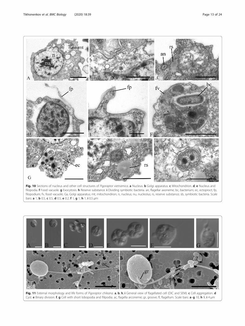
Fig. 11 External morphology and life forms of Pigoraptor chileana. a, b, h, i General view of flagellated cell (DIC and SEM). с Cell aggregation. dCyst. e Binary division. f, g Cell with short lobopodia and filipodia. ac, flagella arconeme; gr, groove; fl, flagellum. Scale bars: a–g 10, h 9, i 4 μm
Fig. 10 Sections of nucleus and other cell structures of Pigoraptor vietnamica. a Nucleus. b Golgi apparatus. c Mitochondrion. d, e Nucleus andfilopodia. f Food vacuole. g Exocytosis. h Reserve substance. i Dividing symbiotic bacteria. an, flagellar axoneme; bc, bacterium; ec, ectoproct; fp,filopodium; fv, food vacuole; Ga, Golgi apparatus; mt, mitochondrion; n, nucleus; nu, nucleolus; rs, reserve substance; sb, symbiotic bacteria. Scalebars: a 1, b 0.5, c 0.5, d 0.5, e 0.2, f 1, g 1, h 1, i 0.5 μm
Tikhonenkov et al. BMC Biology (2020) 18:39 Page 13 of 24

Fig. 12 General view, flagellum, and flagella root system of Pigoraptor chileana. a General view of the cell section. b–d Flagellum. e–h Flagellarbasal body and surrounding structures. an, axoneme; ds, dense spot; fbb, flagellar basal body; fv, food vacuole; mct, microtubule; n, nucleus; nfbb,non-flagellar basal body; nu, nucleolus. Scale bars: a 0.5, b 0.2, c 0.2, d 0.5, e 0.5, f 0.5, g 0.5, h 0.5 μm
Tikhonenkov et al. BMC Biology (2020) 18:39 Page 14 of 24
15). The merging of predator cells during feeding isquite unusual and may represent a new factor to con-sider in the emergence of aggregated multicellularity. Inaddition, putative chemical signaling to attract othercells of its species is observed during the formation ofsyncytial structures in these species. In this context,alpha- and beta-integrins and other components of theso-called integrin adhesome, which are responsible forinteraction with the extracellular matrix and the trans-mission of various intercellular signals, were found inthe transcriptomes of all three studied species [37].
Starch breakdown by SyssomonasAn interesting phenomenon was observed in clonal cul-tures of Syssomonas, where the predator can completelyengulf starch granules of the same size as the cell, also me-diating the rapid destruction of rice grains into smallerfragments and individual starch crystals (Additional file 1:Fig. S2). It is possible that Syssomonas secretes hydrolyticenzymes that provide near-membrane extracellular
digestion. The appearance of extracellular digestion isconsidered to be a major step in animal evolution [57],since it is central to the breakdown of many organic mole-cules when combined with the direct absorption of nutri-ent monomers by the gut epithelia using transmembranetransport in animals [58].This ability of Syssomonas to feed on starch is likely
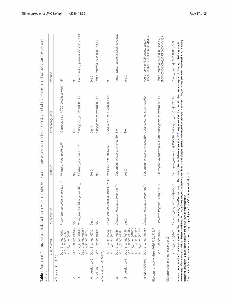
promoted by the expression of numerous enzymes thatare putatively involved in starch breakdown (several pu-tative α-amylases and α-glucosidases, a glycogen deb-ranching enzyme, and a glycogen phosphorylase)(Table 1). For example, Syssomonas has five distinct pu-tative α-amylases, one of which was not found in anyother Holozoa present in our database (Table 1). Simi-larly, one of the four putative α-glucosidases in S. multi-formis seems to be specific to this lineage, and possiblythe Filasterea, within the Holozoa. While α-amylases andα-glucosidases are able to hydrolyze α-1,4-linked glyco-sidic linkages, mobilization of the starch molecule at theα-1,6 glycosidic bonds at branch points requires the

Fig. 13 Arrangement of basal bodies and structure of filopodia of Pigoraptor chileana. a–f Serial sections of basal bodies. g–i Filipodia. fbb,flagellar basal body; fl, flagellum; fp, filopodium; mct, microtubule; n, nucleus; nfbb, non-flagellar basal body. Scale bars: a–f 0.2, g–i 0.5 μm
Tikhonenkov et al. BMC Biology (2020) 18:39 Page 15 of 24
activity of debranching enzymes. A possible candidatefor the catalysis of this reaction is a conserved glycogendebranching enzyme in S. multiformis, orthologous tothe human AGL gene (Table 1). Additionally, we identi-fied a transcript for a glycogen phosphorylase (ortholo-gous to the human PYGB, PYGL, and PYGM genes), anenzyme involved in the degradation of large branchedglycan polymers.Our observations also show that in the presence of
starch in culture, Syssomonas can form resting stages ofunidentified genesis, which tend to adhere to each otherand to starch grains.
Structural featuresSyssomonas and Pigoraptor both display a broad mor-phological plasticity: all three species have a flagellarstage, form pseudopodia and cysts, and can form aggre-gations of several cells. Syssomonas multiformis also hasan amoeboid non-flagellar stage. The dominant life formof all three species in culture is the uniflagellar
swimming cell. Interestingly, amoeboid and pseudopo-dial life forms were detected in cultures only after 2 yearsof cultivation and observation, suggesting they may beextremely rare in nature. Overall, the morphological dif-ferences between cells of the same type of the two gen-era, Syssomonas and Pigoraptor, are few and subtle.Given these genera are distantly related within the treeof Holozoa in all current phylogenomic reconstructions,it is interesting to speculate that they may be the resultof morphostasis and by extension retain features resem-bling those of an ancestral state of extant holozoan line-ages. Although other unknown lineages of unicellularholozoans undoubtly exist and their morphology re-mains to be investigated, we propose such lineages willlikely also possess a similar morphological plasticity,with flagellated and pseudopodial stages, and character-istics overall similar to Syssomonas and Pigoraptor. It hasbeen established that single cells of Syssomonas andPigoraptor can temporarily attach themselves to the sub-strate (Fig. 1c, d) and, by beating their flagellum, can

Fig. 14 Mitochondria, food vacuole, and nucleus division of Pigoraptor chileana. a, b Mitochondria. c Food vacuole and exocytose. d Nucleusdivision in metaphase stage. chr, chromosomes; fv, food vacuole; mct, microtubule; mt, mitochondrion; rs, reserve substance. Scale bars: a 0.5, b0.5, c 0.5, d 1 μm
Tikhonenkov et al. BMC Biology (2020) 18:39 Page 16 of 24
create water currents to putatively attract food particles,similar to choanoflagellates and sponge choanocytes(Fig. 2e, Video 2), although such behavior could beanalogous. Choanocytes and choanoflagellates possess,in addition to the flagellum, a collar consisting of cyto-plasmic outgrowths reinforced with actin filaments(microvilli) that serve to capture bacterial prey. The thinfilopodia that are observed on the cell surface of all threeSyssomonas and Pigoraptor species may thus be homolo-gous to collar microvilli. But this will require further evi-dence in form of homologous proteins in thesestructures or evidence of their function in Syssomonasand Pigoraptor. While the filopodia of Syssomonas haveno obvious structural contents, the outgrowths of
Pigoraptor sometimes contain microtubular-like profiles.Cross-sections of these structures were not obtained,but they may represent parallel microfilaments suchas recently found in the filopodial arms of Ministeriavibrans [25]. The organization of the Ministeria filo-podial arms resembles the microvilli of choanoflagel-lates, which have stable bundles of microfilaments attheir base. It has been proposed previously that theancestor of Filozoa (Filasterea + Choanoflagellida +Metazoa) probably had already developed filose tenta-cles, which have aggregated into a collar in the com-mon ancestor of choanoflagellates/sponges [24], andthat microvilli were present in the common ancestorof Filozoa [25].
Table
1Transcrip
tsforpu
tativestarch-deg
rading
enzymes
inS.multiformisandthepresen
ce/absen
ceof
correspo
ndingortholog
sin
othe
run
icellularho
lozoan
lineage
sand
Metazoa
S.multiformis
Ichthyospo
rea
Filasterea
Cho
anoflage
llates
Metazoa
α-Amylases
(PF00128)
1Colp1
2_sorted
@267,
Colp1
2_sorted
@268,
Colp1
2_sorted
@269
Pirum_gem
mata@
Unige
ne16826_1*
Ministeria_vibrans@37663/4*
Crasped
ida_sp_A
TCC_50635@246766*
NA
2Colp1
2_sorted
@19603
NA
NA
NA
NA
3Colp1
2_sorted
@18987,
Colp1
2_sorted
@16467,
Colp1
2_sorted
@17178
Pirum_gem
mata@
Unige
ne17488_1
Ministeria_vibrans@28157
Salpingoeca_rosetta@
00083T0
Amph
imedon
_queensla
ndica@
15720304
4=paralogto
3Colp1
2_sorted
@8715
See3
See3
See3
See3
5(SLC3A1)
Colp1
2_sorted
@10150
NA
NA
Salpingoeca_rosetta@
08827T0
Hom
o_sapiens@
ENSP00000260649
α-Glucosidases(PF01055)
1Colp1
2_sorted
@9290,
Colp1
2_sorted
@22295
Pirum_gem
mata@
Unige
ne@34248_1*
Ministeria_vibrans@34861
Salpingoeca_rosetta@
00005T0*
NA
2Colp1
2_sorted
@1339,
Colp1
2_sorted
@1341
Creolim
ax_fragran
tissim
a@8464T1
Capsaspora_owczarzaki@04827T0
NA
Amph
imedon
_queensla
ndica@
15725226
3=paralogto
2Colp1
2_sorted
@9196,
Colp1
2_sorted
@14409,
Colp1
2_sorted
@18957,
Colp1
2_sorted
@21457,
Colp1
2_sorted
@31517
See2
See2
NA
See2
4.(GAN
AB/GAN
C)Colp1
2_sorted
@13767
Creolim
ax_fragran
tissim
a@4769T1
Capsaspora_owczarzaki@00308T0
Salpingoeca_rosetta@
11360T0
Hom
o_sapiens@
ENSP00000326227/
ENSP00000349053/EN
SP00000340466
Glycoge
nph
osph
orylase(PYG
B/PYGL/PYGM)
1Colp1
2_sorted
@1564
Creolim
ax_fragran
tissim
a@2598T1
Capsaspora_owczarzaki@07395T0
Salpingoeca_rosetta@
08531T0
Hom
o_sapiens@
ENSP00000216392/
ENSP00000216962/EN
SP00000164139
Glycoge
nde
branchingen
zyme(AGL)
1Colp1
2_sorted
@14615
Creolim
ax_fragran
tissim
a@4755T1
Capsaspora_owczarzaki@06406T0
Salpingoeca_rosetta@
10237T0
Hom
o_sapiens@
ENSP00000355106
Accession
numbe
rsforS.multiformisarefrom
thecorrespo
ndingTran
sDecod
erou
tput
files
asde
scrib
edin
Heh
enbe
rger
etal.[37
];sequ
ence
iden
tifiers
forallo
ther
taxa
correspo
ndto
thede
positedalignm
ents/
phylog
enies.Pfam
domains
used
toiden
tifycand
idates
inS.multiformisan
d/or
anno
tateddirect
human
ortholog
ousge
nesareindicatedin
brackets
incolumn1.
NA,n
odirect
ortholog
srecoveredin
ourda
taset;
tran
scrip
tiden
tifiers
inita
lic,sho
rtfrag
men
t(s)of
onelin
eage
represen
tativ
e*U
nclear
whe
ther
sequ
encesaredirect
ortholog
sor
paralogs
toS.multiformis(unresolvedtree)
Tikhonenkov et al. BMC Biology (2020) 18:39 Page 17 of 24

Tikhonenkov et al. BMC Biology (2020) 18:39 Page 18 of 24
A single, posterior flagellum is the defining character-istic of opisthokonts [34]. However, the flagellum hasnot yet been found in all known Opisthokonta lineages.Torruella et al. [19] have found several proteins corre-sponding to key components of the flagellum in Corallo-chytrium and the filose amoeba Ministeria vibrans,which have been considered to lack flagella. These au-thors have shown that the stalk used by Ministeria to at-tach to the substrate is a modified flagellum. Recently,morphological observations on another strain of Minis-teria vibrans (strain L27 [25]) revealed that this strainlacks the stalk for substrate attachment, but possesses atypical flagellum that projects forward and beats at at-tached to the substrate cells (see Fig. 2h and Video S1 in[25]). The authors concluded that the filasterean ances-tor possessed a flagellum, which was subsequently lostin Capsaspora owczarzaki. In the case of Corallochy-trium limacisporum, it was suggested that it has a cryp-tic flagellate stage in its life cycle [19], as has beenproposed for other eukaryotes (Aureococcus and Ostreo-coccus, for instance) based on their genome sequences[59]. Therefore, the flagellate stage could have been theone morphological trait uniting Corallochytrium andSyssomonas within “Pluriformea.” Interestingly, the an-cestor of ichthyosporeans probably also had a flagellum,which is preserved in the Dermocystida (at the stage ofzoospores), but was again lost in the Ichthyophonida.However, some membrane-decorated vesicles with shortflagellum-like strands were visualized by TEM on cellsections in ichtyophonid Sphaeroforma sirkka and S.napiecek [60].The central filament of the flagellum, which connects
the central pair of microtubules with the transversalplate in Pigoraptor (Fig. 7c, f), is also noteworthy, sincethis character was previously known only in the choano-flagellates [61] and was considered a unique feature forthis lineage. The cone-shaped elevation of the surfacemembrane around the base of the flagellum in Syssomo-nas (Fig. 3b, c) is also typical for choanoflagellates (seeFig. 5a, b in [61]).One interesting ultrastructural peculiarity is the un-
usual reticulate or tubular structures in Syssomonasmitochondria which were not observed in other eukary-otes we are aware of.Another noteworthy feature is the presence of symbi-
otic bacteria in the cells of Pigoraptor vietnamica. Sym-biotic bacteria are common in both vertebrate andinvertebrate animals including placozoan Trichoplax[62] and fungi [63]. Bacterial endosymbionts are alsoknown in unicellular protists from all eukaryotic super-groups and perform a multitude of new biochemicalfunctions in the host [64, 65]. But as far as we know,there are only two documented cases of opisthokontprotists with prokaryotic symbionts: nucleariid amoebae
(sister lineage of fungi) with several groups of Proteobac-teria and the choanoflagellate Codosiga balthica, whichharbored two different endosymbiotic bacteria inside thecytoplasm [66, 67]. Future investigations of the intracel-lular bacteria in Pigoraptor will be essential to under-standing the role of prokaryotic symbionts in the biologyand cell functions of unicellular relatives of animals.
Origins of multicellularityAs mentioned above, numerous theories about the originof Metazoa exist. One of the first and widely acceptedevolutionary theories on the origin of animals is the Gas-trea theory of Ernst Haeckel [68]. Haeckel suggested thatthe first step in the evolution of multicellularity in ani-mals was the formation of a hollow ball, the walls ofwhich consisted of undifferentiated flagellated cells,which he called Blastea. This was followed by gastrula-tion, where the ball invaginated, leading to the primarycellular differentiation into ecto- and endoderm. Modernvariants of this, for example, the сhoanoblastea theory,which highlights the similarity between Haeckel’s Blasteaand the choanoflagellate colony [57], are the most com-mon and influential explanations for the origin of multi-cellular animals. An important assumption of thesetheories is that cell differentiation took place only aftermulticellularity arose, suggesting that animals originatedfrom a single cell type, such as choanoflagellate-likecolony-forming ancestor [41], which is supported by thepossible homology between sponge choanocytes andchoanoflagellates [69], and consistent with the Haeckel-Muller Biogenetic Law that ontogenesis recapitulatesphylogenesis [70].There are, however, some basic differences between
sponges and choanoflagellates in how their collar and fla-gella interact, suggesting that choanocyte/choanoflagellatesimilarity might be superficial and that specific homologycannot be automatically assumed (see [71] for details). In-deed, recent ultrastructural studies on sponge choanocytesand choanoflagellates show that they are fundamentallydifferent in many respects (see [72–74] for details), someof which are among the very few ultrastructural systemsin eukaryotic cells considered sufficiently conservative toindicate phylogenetic relationships [75–77]. For example,specific features of the flagellar apparatus of the spongeEphydatia fluviatilis are more similar to zoospores of chy-trids than to choanoflagellates [78]. Overall, the conclu-sion that sponges (and by extension all Metazoa) descenddirectly from a single-celled organism similar to choano-flagellates is not unambiguously supported by ultrastruc-ture, as sometimes assumed [73].Recent comparative transcriptomic analysis also argues
against a direct link between sponge choanocytes andchoanoflagellates, suggesting instead that the first animalcell had the ability to transition between multiple states

Tikhonenkov et al. BMC Biology (2020) 18:39 Page 19 of 24
in a manner similar to modern transdifferentiating stemcells [79]. This is more consistent with an alternativeclass of ideas very different from the Gastrea hypothesis,which suggest the presence of diverse life forms andcomplex life cycles were the initial step in the origin ofanimal multicellularity. Or in other words, that differen-tiation preceded multicellularity and the origin of theblastula. Unicellular relatives of animals have now longbeen known to contain a variety of genes homologous tothose involved in cell adhesion, differentiation, develop-ment, and signal transduction in Metazoa. Some of themwere once considered to be unique to animals (e.g., tran-scription factors T-box and Rel/NF-kappa B, Crumbsprotein, integrin beta) as they were absent in choanofla-gellates (the closest relatives of Metazoa), but later, theywere found to be present in other unicellular Holozoa[24, 31, 80]. Now, homologs of most genes controllingthe development of animals, their cell differentiation, ancell-cell and cell-matrix adhesion are known in variouslineages of unicellular organisms [18, 81–83], all suggest-ing that genetic programs of cellular differentiation andadhesion arose relatively early in the evolution ofopisthokonts and before the emergence of multicellular-ity [15, 24, 31, 81, 84].One specific hypothesis focusing on cell differentiation
preceding the formation of colonies is the “synzoosporehypothesis” [85, 86], and see [31] for details. In brief,three types of cell cycle are present in the ontogenesis ofmulticellular animals: monotomy (alternate phases ofcell growth and division of somatic cells), hypertrophicgrowth (in female sex cells), and palintomy (the eggundergoes a series of consecutive divisions). Zakhvatkinnoted that some protists alternate between differenttypes of life cycle, and suggested that the unicellular an-cestor of Metazoa already had differentiated cells as a re-sult of such a complex life cycle. The life cyclecomplexity, in turn, results from the fact that mono-tomic cells are usually sedentary, or at least less mobile,and can change their phenotype (from flagellated toamoeboid, etc.) depending on the environment; theprocess of palintomy is necessary for the formation ofmorphologically identical dispersal cells (spores or zoo-spores). These dispersal cells remain attached to eachother, forming a primary flagellated larva—the synzoos-pore or blastula (cited from [31]).The synzoospore hypothesis is consistent with recent
observations of complex life cycles in unicellularopisthokonts possessing cellular differentiation, the pres-ence of sedentary trophic phases, and a tendency to ag-gregation, as seen in choanoflagellates [53, 87–90],filastereans [49], Corallochytrium [28], ichthyosporeans[17, 84, 91], chytridiomycetes [92], and nucleariid amoe-bae [93]. According to this theory, multicellularity in an-imals arose through the temporal integration of various
types of cells, which were already present in differentparts of the life cycle. The hypothetical ancestor of ani-mals in this model would thus already have genetic pro-grams for cell differentiation (including cadherins,integrins, tyrosine kinases).Developing the synzoospore hypothesis further,
Mikhailov et al. [31] proposed an evolutionary mechan-ism of “transition from temporal to spatial cell differenti-ation” to explain the emergence of multicellular animals.In this model, the ancestor of Metazoa was a sedentarycolonial protist filter-feeder with colonies formed bycells of different types, which arose because filtration ef-ficiency is significantly enhanced through the cooper-ation of cells of different types. Dispersal cells producedby the sedentary stage, the zoospores, remained attachedtogether in early metazoans (which increased survivabil-ity) as a synzoospore to form a primary larva, the blas-tula. Development of a whole colony from such amulticellular larva occurred through the differentiationof genetically identical zoospore cells. This was criticalfor the maintenance of long-term cell adhesion and thusemergence of true multicellularity, as opposite to tem-porary colonies and aggregations composed of genetic-ally heterogeneous cells. The authors suggest that thedispersal stages of the sedentary trophic body—primaryblastula-like larvae—acquired adaptations to the preda-tory lifestyle, which triggered the development of pri-mary intestine, muscular, and nervous systems [31].In this view, the origin of multicellularity was a transi-
tion from temporal to spatiotemporal cell differentiation[41]. A bacteriotroph with differentiated, temporally sep-arated cell types became spatially integrated, existingsimultaneously but in different parts of a now multicel-lular conglomerate with different cell types carrying outdifferent functions. From this starting point, further di-versification could proceed straightforwardly by expan-sion of gene regulation networks and signaling pathways.Genomics alone is not sufficient to distinguish these
different models, because morphology, life cycle, andstructural features are all relevant and central. All threenovel species of unicellular Holozoa have life historiesthat are consistent with major elements of the synzoos-pore model (see Fig. 3a in [31] and Fig. 5a, b in [41]).Specifically, these organisms have complex life historiescharacterized by a variety of forms: flagellates, amoebae,amoeboflagellates, and cysts. All three species have thetendency to form aggregations. Syssomonas possessesboth clonal and aggregative multicellular stages, as pre-dicted for the ancestor of animals. Moreover, the forma-tion of aggregations can be associated with adheringcysts, but also by active feeding on large eukaryotic prey,which also leads to hypertrophic cell growth (describedas proliferative stage in [41]) with a subsequent phase ofpalintomic division (in Syssomonas). All these characters

Tikhonenkov et al. BMC Biology (2020) 18:39 Page 20 of 24
are predicted by the synzoospore model. Interestingly,feeding on large eukaryotic prey appears to be a triggerfor several key behaviors, including the formation anddevelopment of aggregates (e.g., joint feeding) and clonalmulticellularity (e.g., hypertrophic growth followed bypalintomy). This is not generally regarded as a key factorin the origin of multicellularity in ancestors of Metazoa,but we suggest this should be considered. It is an inter-esting parallel that morphological and developmentalchanges (e.g., colony formation) in choanoflagellates canbe also triggered by prey, although bacterial [94–96].The functional basis for these changes requires furtherstudy, as does the relationship between the life cycle,morphology, formation of multicellular structures, andimpacts of environmental change.
ConclusionsAs we acquire more information about the biology ofknown unicellular relatives of animals and, equally im-portantly, describe diverse new species of unicellularHolozoa, models for the evolutionary histories of specificcharacteristics that contributed to the emergence ofmulticellularity in animals can be evaluated more mean-ingfully. Syssomonas and Pigoraptor are characterized bycomplex life cycles, the formation of multicellular aggre-gations, and an unusual diet for single-celled opistho-konts (partial cell fusion and joint sucking of largeeukaryotic prey), all of these features providing new in-sights into the origin of multicellularity in Metazoa.Genome and transcriptome analyses of unicellular rel-
atives of animals have shown that genes encoding pro-teins for cellular signaling and adhesion, as well as genesfor embryonic development of multicellular organisms,arose before the emergence of multicellular animals [24,37, 81, 84]. While these genes likely have different func-tions in protists than in animals, they nevertheless prob-ably relate to the ability to recognize the cells of theirown species, prey, or organic molecules and contributeto the formation of multicellular aggregations, thus in-creasing the organism’s ability to adapt to environmentalchange. As we learn more about the natural history andbehavior of these organisms, the importance of theseprocesses becomes even more clear. The phylogeneticdistribution of unicellular holozoans with complex lifecycles suggests that the ancestor of Metazoa probablyformed cells of various types that could aggregate andhad molecular mechanisms of cell differentiation and ad-hesion related to those processes. We suggest that thissupports the conclusion that cellular differentiationarose before the emergence of multicellularity.The feeding modes of the ancestral metazoan may also
have been more complex than previously thought, in-cluding not only bacterial prey, but also larger eukaryoticcells and organic structures. Indeed, the ability to feed
on large eukaryotic prey could have been a powerfultrigger in the formation and development of both aggre-gative and clonal multicellular stages that played import-ant roles in the emergence of multicellularity in animals.Lastly, we wish to point out that other new and deep lin-eages of opisthokonts undoubtedly exist that have notyet been described, and each of these will play an im-portant role in the development of hypotheses on theorigin of multicellular animals in future.
MethodsNovel unicellular opisthokont predators were found infreshwater biotopes in Vietnam and Chile. Syssomonasmultiformis (clone Colp-12) was obtained from the sam-ple of freshwater pool (11° 23′ 08.0″ N, 107° 21′ 44.9″E; T = 39 °C; pH = 7.18; DO (ppm) = 0.64; conductivity(μS/cm) = 281; TDS (ppm) = 140), Tà Lài, Cát Tiên Na-tional Park, Dong Nai Province, Socialist Republic ofVietnam, on April 29, 2013. Pigoraptor vietnamica(clone Opistho-1) was obtained from freshwater LakeDak Minh, silty sand on the littoral (12° 54′ 50″ N, 107°48′ 26″ E; T = 27 °C; pH = 7.03; DO (ppm) = 7.43; con-ductivity (μS/cm) = 109; TDS (ppm) = 54), Dak Lak Prov-ince, Socialist Republic of Vietnam, on March 26, 2015.Pigoraptor chileana (clone Opistho-2) was obtained fromthe bottom sediments of freshwater temporary waterbody (submerged meadow, 54° 02′ 29.7″ S, 68° 55′18.3″ W; T = 16.5 °C; pH = 6.62; conductivity (μS/cm) =141; TDS (ppm) = 72) near the Lake Lago Blanca, Tierradel Fuego, Chile, on November 4, 2015.The samples were examined on the third, sixth, and
ninth days of incubation in accordance with the methodsdescribed previously [97]. Following isolation by glassmicropipette, freshwater clones Colp-12, Opistho-1, andOpistho-2 were propagated on the bodonid Parabodocaudatus (strain BAS-1, IBIW RAS) grown in Pratt’smedium or spring water (Aqua Minerale, PepsiCo,Moscow Region, Russia, or PC Natural Spring Water,President’s Choice, Toronto, Canada) by using the bac-terium Pseudomonas fluorescens as food [98]. The cloneColp-12 was perished after 5 years of cultivation. Theclones Opistho-1 and Opistho-2 are stored in the “Liveculture collection of free-living amoebae, heterotrophicflagellates and heliozoans” at the Institute for Biology ofInland Waters, Russian Academy of Science.Observations of live cells were carried out in the la-
boratory conditions at 22 °C in the clonal cultures. Stud-ied species were able to survive at 5–40 °C temperatures,pH 6–11, and tolerate salinity increasing up to 4‰. Thevariation of temperature, pH, and cultivation mediumdoes not result in appearance of additional morpho-logical forms or increasing of frequency of occurrence ofcertain (e.g., amoeboid) life forms. The agitation of thecultures (up to 1400 rpm) does not lead to the formation

Tikhonenkov et al. BMC Biology (2020) 18:39 Page 21 of 24
of cell aggregations. Clone Colp-12 was also able to growon stramenopile prey Spumella sp. (clone OF-40 from“Live culture collection of free-living amoebae, hetero-trophic flagellates and heliozoans” at the Institute forBiology of Inland Waters, Russian Academy of Science).Light microscopy observations were made by using theZeiss Axio Scope A.1 equipped with a DIC contrastwater immersion objective (× 63). The images weretaken with the AVT HORN MC-1009/S analog videocamera and directly digitized by using the Behold TV409 FM tuner. Cells with engulfed starch granules wereinspected by epifluorescence microscopy after DAPIstaining using the Zeiss Axioplan 2 Imaging microscope.For transmission electron microscopy (TEM), cells
were centrifuged, fixed at 1 °C for 15–60min in a cock-tail of 0.6% glutaraldehyde and 2% OsO4 (final concen-tration) prepared using a 0.1-M cacodylate buffer (pH7.2). Fixed cells were dehydrated in alcohol and acetoneseries (30, 50, 70, 96, and 100%, 20 min in each step).Afterward, the cells were embedded in a mixture of Ara-ldite and Epon (Luft, 1961). Ultrathin sections (50 nm)were prepared with an Leica EM UC6 ultramicrotome(Leica Microsystems, Germany) and observed by usingthe JEM 1011 transmission electron microscope (JEOL,Japan).For scanning electron microscopy (SEM), cells from
exponential growth phase were fixed as for TEM butonly for 10 min at 22 °C and gently drawn onto a poly-carbonate filter (diameter 24 mm, pores 0.8 μm). Follow-ing the filtration, the specimens were taken through agraded ethanol dehydration and acetone and finally putinto a chamber of a critical point device for drying.Then, dry filters with fixed specimens were mounted onaluminum stubs, coated with gold-palladium, and ob-served with a JSM-6510LV scanning electron micro-scope (JEOL, Japan).Analysis of enzymes involved in starch breakdown was
based on transcriptomic data obtained as described earl-ier [37]. To identify candidates putatively involved instarch breakdown, we used the results of a previoushmmscan analysis of S. multiformis [37] to search forPfam domains present in enzymes/enzyme families nat-urally involved in starch degradation (as described inhttps://doi.org/10.1016/j.sbi.2016.07.006), such as α-amylases (PF00128), glycoside hydrolase families con-taining α-glucosidases (PF02056, PF01055, PF03200,PF10566), α-glucan water dikinase 1 (GWD1, PF01326),phosphoglucan phosphatase (DSP4, PF00782, andPF16561), disproportionating enzymes (PF02446), andpullulanases (PF17967). Additionally, we submitted theS. multiformis sorted transcriptome to the KEGG Auto-matic Annotation Server (KAAS) [99] for functional an-notation and investigated the output for transcriptsinvolved in starch metabolism. All candidates were
further investigated by BLASTp search [100] againstGenBank, by domain analysis using InterProScan (doi:https://doi.org/10.1093/bioinformatics/btu031) and byphylogenetic reconstruction. For phylogenetic recon-struction, the candidates were used as queries in aBLASTp search (e value threshold 1e−5) against a com-prehensive custom database containing representativesof all major eukaryotic groups, including opisthokonts(with a focus on unicellular holozoan lineages, includingseveral choanoflagellates, filastereans, and ichthyospor-eans (including Pigoraptor sp.) as well as S. multifor-mis—see https://doi.org/10.1016/j.cub.2017.06.006 fordata sources), apusomonads, amoebozoans, discobids,archaeplastids, cryptophytes, haptophytes, dinoflagel-lates, chrompodellids, apicomplexans, ciliates, strameno-piles, and rhizarians, as well as RefSeq data from allbacterial phyla at NCBI (https://www.ncbi.nlm.nih.gov/,last accessed December 2017). The database was sub-jected to CD-HIT with a similarity threshold of 85% toreduce redundant sequences [101]. Results from blastsearches were parsed for hits with a minimum querycoverage of 50% and e values of less than 1e−5. Thenumber of bacterial hits was restrained to 20 hits perphylum (for FCB group, most classes of Proteobacteria,PVC group, Spirochaetes, Actinobacteria, Cyanobacteria(unranked), and Firmicutes) or 10 per phylum(remaining bacterial phyla) as defined by NCBI tax-onomy. Parsed hits were aligned with MAFFT v. 7.212,using the –auto option; poorly aligned regions wereeliminated using trimAl v.1.2 with a gap threshold of80% [102, 103]. Maximum likelihood tree reconstruc-tions were then performed with FastTree v. 2.1.7 usingthe default options [104]. Phylogenies with overlappingtaxa were consolidated by combining the parsed hits ofthe corresponding queries, removing duplicates and re-peating the alignment, trimming, and tree reconstructionsteps as described above. All phylogenies were manuallyinvestigated, and obvious contaminations removed fromthe underlying alignment. For the final tree reconstruc-tion, the cleaned, unaligned sequences were then sub-jected to filtering with PREQUAL using the defaultoptions (https://www.ncbi.nlm.nih.gov/pubmed/29868763) to remove non-homologous residues introducedby poor-quality sequences, followed by alignment withMAFFT G-INS-i using the VSM option (--unalignlevel0.6) (https://www.ncbi.nlm.nih.gov/pubmed/27153688)to control overalignment. After trimming ambiguouslyaligned sites with trimAl v. 1.2 (-gt 0.8), sequences withless than 50% of the alignment length were removed.Final trees were calculated with IQ-TREE v. 1.6.5(https://www.ncbi.nlm.nih.gov/pubmed/25371430), usingthe -mset option to restrict model selection to LG forModelFinder after initial model testing without any re-strictions selected the LG substitution model for all

Tikhonenkov et al. BMC Biology (2020) 18:39 Page 22 of 24
alignments (https://www.ncbi.nlm.nih.gov/pubmed/28481363), while branch support was assessed with 1000 ul-trafast bootstrap replicates (https://www.ncbi.nlm.nih.gov/pubmed/29077904). The raw tree files in newick,colored trees with taxon information (and accessionnumbers where available) in pdf format, and underlyingtrimmed alignments have been deposited to figshare re-pository [105] doi: https://doi.org/10.6084/m9.figshare.11914491.
Supplementary informationSupplementary information accompanies this paper at https://doi.org/10.1186/s12915-020-0762-1.
Additional file 1 : Fig. S1. A-C – Syssomonas multiformis sucks out thecytoplasm of the prey; D – three cells of Syssomonas (s) suck out thecytoplasm of the same prey cell together, other Syssomonas cells (arrows)become attracted and swim to the same prey cell; E – unusual flagellatedcell of Syssomonas containing vesicular structures; F – cells of Syssomonaswith engulfed starch granules swim to the starch crystals druse and hidewithin the starch crystals. Fig. S2. Rice grain destruction in Petri dish withPratt medium and presence of the cells of Parabodo caudatus (prey) only(A) and Syssomonas multiformis (B) after 9 days of incubation.
Additional file 2 Video S1. Swimming of Syssomonas multiformis cellwith rotation.
Additional file 3 Video S2. Attached cell of Syssomonas multiformisand rapid flagellum beating.
Additional file 4 Video S3. Amoeboflagellate stage of Syssomonasmultiformis. Cells of eukaryotic prey Parabodo caudatus are also visible.
Additional file 5 Video S4. Loss of flagellum in Syssomonas multiformisand transition to amoeba.
Additional file 6 Video S5. Transformation of amoeba into a cyst inSyssomonas multiformis.
Additional file 7 Video S6. Palintomic divisions inside the cyst ofSyssomonas multiformis.
Additional file 8 Video S7. Division into two cell structures inSyssomonas multiformis.
Additional file 9 Video S8. Cell and cyst of Syssomonas multiformiswith vesicular structures inside.
Additional file 10 Video S9. Feeding of Syssomonas multiformis oneukaryotic prey.
Additional file 11 Video S10. Feeding of Syssomonas multiformis onbacteria.
Additional file 12 Video S11. Temporary cell aggregations ofSyssomonas multiformis.
Additional file 13 Video S12. Floating rosette-like aggregation ofSyssomonas multiformis.
Additional file 14 Video S13. Syncytium-like structures and budding ofyoung flagellated daughter cells in Syssomonas multiformis.
Additional file 15 Video S14. Joint feeding of Pigoraptor vietnamica ondead cell of Parabodo caudatus.
Additional file 16 Video S15. Joint feeding of Pigoraptor chileana ondead cell of Parabodo caudatus.
Additional file 17 Video S16. Temporary cell aggregation of Pigoraptorchileana.
AcknowledgementsWe thank Kristina I. Prokina for sample collection in Chile, Dr. Hoan Q. Tranand Tran Duc Dien for assistance with sample collection and tripmanagement in Vietnam, Sergei A. Karpov for help with interpretationof transmission electron microscopy images, Jürgen F.H. Strassert for help
with DAPI staining, and Vladimir V. Aleshin and Kirill V. Mikhailov for fruitfuldiscussion on different aspects of the origin of Metazoa. Field work inVietnam is part of the project “Ecolan 3.2” of the Russian-Vietnam TropicalCentre.
Authors’ contributionsDVT, APM, EH, and PJK designed the study. DVT and APM isolated andcultured the cells and performed the electron microscopy analysis. DVT, ASE,OIB, and YAM performed the light microscopy analysis. EH performed thestarch-degrading enzyme analysis. DVT, EH, and PJK wrote the manuscriptwith input from all authors. All authors read and approved the finalmanuscript.
FundingThis work was supported by the Russian Science Foundation grant no. 18-14-00239 (cell isolation and culturing, light and electron microscopy, andanalyses) and by the Natural Sciences and Engineering Research Council ofCanada (grant no. 227301).
Availability of data and materialsVideos of morphology, life cycle, and feeding of novel predatory unicellularrelatives of animals are available on figshare [105].The raw tree files in newick, colored trees with taxon information (andaccession numbers where available) in pdf format, and trimmed alignmentshave been deposited to the same figshare deposition [105] doi: https://doi.org/10.6084/m9.figshare.11914491.
Ethics approval and consent to participateNot applicable.
Consent for publicationNot applicable.
Competing interestsThe authors declare that they have no competing interest.
Author details1Papanin Institute for Biology of Inland Waters, Russian Academy of Sciences,Borok, Russia 152742. 2Department of Botany, University of British Columbia,Vancouver, British Columbia V6T 1Z4, Canada. 3Ocean EcoSystems BiologyUnit, RD3, GEOMAR Helmholtz Centre for Ocean Research Kiel,Duesternbrookerweg 20, 24105 Kiel, Germany. 4Penza State University, Penza,Russia 440026. 5Moscow State University, Moscow, Russia 119991.
Received: 9 November 2019 Accepted: 2 March 2020
References1. Moroz LL, Kocot KM, Citarella MR, Dosung S, Norekian TP, Povolotskaya IS,
et al. The ctenophore genome and the evolutionary origins of neuralsystems. Nature. 2014;510:109–14.
2. Srivastava M, Begovic E, Chapman J, Putnam NH, Hellsten U, Kawashima T,et al. The Trichoplax genome and the nature of placozoans. Nature. 2008;454:955–60.
3. Srivastava M, Simakov O, Chapman J, Fahey B, Gauthier ME, Mitros T, et al.The Amphimedon queenslandica genome and the evolution of animalcomplexity. Nature. 2010;466:720–6.
4. Feuda R, Dohrmann M, Pett W, Philippe H, Rota-Stabelli O, Lartillot N, et al.Improved modeling of compositional heterogeneity supports sponges assister to all other animals. Curr Biol. 2017;27:3864–3870.e4.
5. Philippe H, Derelle R, Lopez P, Pick K, Borchiellini C, Boury-Esnault N, et al.Phylogenomics revives traditional views on deep animal relationships. CurrBiol. 2009;19:706–12.
6. Simion P, Philippe H, Baurain D, Jager M, Richter DJ, Di Franco A, et al. Alarge and consistent phylogenomic dataset supports sponges as the sistergroup to all other animals. Curr Biol. 2017;27:958–67.
7. Dunn CW, Hejnol A, Matus DQ, Pang K, Browne WE, Smith SA, et al. Broadphylogenomic sampling improves resolution of the animal tree of life.Nature. 2008;452:745–9.

Tikhonenkov et al. BMC Biology (2020) 18:39 Page 23 of 24
8. Ryan JF, Pang K, Schnitzler CE, Nguyen AD, Moreland RT, Simmons DK, et al.The genome of the ctenophore Mnemiopsis leidyi and its implications forcell type evolution. Science. 2013;342:1242592.
9. Whelan NV, Kocot KM, Moroz LL, Halanych KM. Error, signal, and theplacement of Ctenophora sister to all other animals. Proc Natl Acad Sci U SA. 2015;112:5773–8.
10. Schierwater B, Eitel M, Jakob W, Osigus H-J, Hadrys H, Dellaporta SL, et al.Concatenated analysis sheds light on early metazoan evolution and fuels amodern “Urmetazoon” hypothesis. PLoS Biol. 2009;7:e1000020.
11. Signorovitch AY, Buss LW, Dellaporta SL. Comparative genomics of largemitochondria in Placozoans. PLoS Genet. 2007;3:e13.
12. Laumer CE, Gruber-Vodicka H, Hadfield MG, Pearse VB, Riesgo A, Marioni JC,et al. Support for a clade of Placozoa and Cnidaria in genes with minimalcompositional bias. eLife. 2018;7:e36278.
13. Maloof AC, Rose CV, Beach R, Samuels BM, Calmet CC, Erwin DH, et al.Possible animal-body fossils in pre-Marinoan limestones from SouthAustralia. Nat Geosci. 2010;3:653–9.
14. Sharpe SC, Eme L, Brown MW, Roger AJ. 2015. Timing the origins ofmulticellular eukaryotes through phylogenomics and relaxed molecularclock analyses. In: Ruiz-Trillo I, Nedelcu AM, editors. Evolutionary transitionsto multicellular life. Advances in marine genomics. Netherlands: Springer;2015. p. 3–29.
15. Brunet T, King N. The origin of animal multicellularity and celldifferentiation. Dev Cell. 2017;43:124–40.
16. King N, Westbrook MJ, Young SL, Kuo A, Abedin M, Chapman J, et al. Thegenome of the choanoflagellate Monosiga brevicollis and the origin ofmetazoans. Nature. 2008;451:783–8.
17. Suga H, Ruiz-Trillo I. Development of ichthyosporeans sheds light on theorigin of metazoan multicellularity. Dev Biol. 2013;377:284–92.
18. Suga H, Chen Z, de Mendoza A, Sebé-Pedrós A, Brown MW, Kramer E, et al.The Capsaspora genome reveals a complex unicellular prehistory of animals.Nat Commun. 2013;4:2325.
19. Torruella G, de Mendoza A, Grau-Bové X, Antó M, Chaplin MA, del Campo J,et al. Phylogenomics reveals convergent evolution of lifestyles in closerelatives of animals and fungi. Curr Biol. 2015;25:2404–10.
20. Aleshin VV, Konstantinova AV, Mikhailov KV, Nikitin MA, Petrov NB. Do weneed many genes for phylogenetic inference? Biochemistry Mosc. 2007;72:1313–23.
21. Lang BF, O’Kelly C, Nerad T, Gray MW, Burger G. The closest unicellularrelatives of animals. Curr Biol. 2002;12:1773–8.
22. Carr M, Richter DJ, Fozouni P, Smith YJ, Jeuck A, Leadbeater BSC, et al. A six-gene phylogeny provides new insights into choanoflagellate evolution. MolPhylogenet Evol. 2017;107:166–78.
23. Hertel LA, Bayne CJ, Loker ES. The symbiont Capsaspora owczarzaki, nov.gen. Nov. sp., isolated from three strains of the pulmonate snailBiomphalaria glabrata is related to members of the Mesomycetozoea. Int JParasitol. 2002;32:1183–91.
24. Shalchian-Tabrizi K, Minge MA, Espelund M, Orr R, Ruden T, Jakobsen KS,et al. Multigene phylogeny of choanozoa and the origin of animals. PLoSOne. 2008;3:e2098.
25. Mylnikov AP, Tikhonenkov DV, Karpov SA, Wylezich C. Microscopicalstudies on picoplanctonic Ministeria vibrans Tong, 1997 (Filasterea)highlights the cytoskeletal structure of the common ancestor ofFilasterea, Metazoa and Choanoflagellata. Protist. 2019. https://doi.org/10.1016/j.protis.2019.07.001.
26. Dudin O, Ondracka A, Grau-Bové X, Haraldsen AAB, Toyoda A, Suga H, et al.A unicellular relative of animals generates a layer of polarized cells byactomyosin-dependent cellularization. eLife. 2019;8:e49801.
27. Glockling SL, Maeshall WL, Gleason FG. Phylogenetic interpretations andecological potentials of the Mesomycetozoea (Ichthyosporea). Fungal Ecol.2013;6:237.
28. Raghukumar S. Occurrence of the Thraustochytrid, Corallochytriumlimacisporum gen. et sp. nov. in the coral reef lagoons of the LakshadweepIslands in the Arabian Sea. Bot Mar. 1987;30:83–9.
29. James-Clark H. Note on the Infusoria flagellata and the Spongiae ciliatae. AmJ Sci. 1866;1:113–4.
30. Ivanov AV. Origin of multicellular animals. Phylogenetic essays. Leningrad:Nauka; 1968. p. 287. (in Russian).
31. Mikhailov KV, Konstantinova AV, Nikitin MA, Troshin PV, Rusin LY, LyubetskyVA, et al. The origin of Metazoa: a transition from temporal to spatial celldifferentiation. Bio Essays. 2009;31:758–68.
32. Nielsen C. Structure and function of metazoan ciliary bands and theirphylogenetic significance. Acta Zoolog. 1987;68:205–62.
33. Ratcliff WC, Denison RF, Borrello M, Travisano M. Experimental evolution ofmulticellularity. Proc Natl Acad Sci U S A. 2012;109:1595–600.
34. Cavalier-Smith T, Chao EE. Phylogeny of Choanozoa, Apusozoa, and otherProtozoa and early eukaryote megaevolution. J Mol Evol. 2003;56:540–63.
35. del Campo J, Ruiz-Trillo I. Environmental survey meta-analysis revealshidden diversity among unicellular opisthokonts. Mol Biol Evol. 2013;30:802–5.
36. Grau-Bové X, Torruella G, Donachie S, Suga H, Leonard G, Richards TA, et al.Dynamics of genomic innovation in the unicellular ancestry of animals.eLife. 2017;6:e26036.
37. Hehenberger E, Tikhonenkov DV, Kolisko M, del Campo J, Esaulov AS,Mylnikov AP, et al. Novel freshwater predators reshape holozoan phylogenyand reveal the presence of a two-component signalling system in theancestor of animals. Curr Biol. 2017;27:2043–50.
38. Medina M, Collins AG, Taylor JW, Valentine JW, Lipps JH, Amaral-Zettler L,et al. Phylogeny of Opisthokonta and the evolution of multicellularity andcomplexity in Fungi and Metazoa. J Astrobiol. 2003;2:203–11.
39. Ruiz-Trillo I, Roger AJ, Burger G, Gray MW, Lang BF. A phylogenomicinvestigation into the origin of metazoa. Mol Biol Evol. 2008;25:664–72.
40. Torruella G, Derelle R, Paps J, Lang BF, Roger AJ, Shalchian-Tabrizi K, et al.Phylogenetic relationships within the Opisthokonta based on phylogenomicanalyses of conserved single-copy protein domains. Mol Biol Evol. 2012;29:531–44.
41. Sebé-Pedrós A, Degnan BM, Ruiz-Trillo I. The origin of Metazoa: a unicellularperspective. Nat Rev Genet. 2017;18:498–512.
42. López-Escardó D, Grau-Bové X, Guillaumet-Adkins A, Gut M, Sieracki ME, Ruiz-Trillo I. Reconstruction of protein domain evolution using single-cell amplifiedgenomes of uncultured choanoflagellates sheds light on the origin of animals.Phil Trans R Soc B. 2019;374. https://doi.org/10.1098/rstb.2019.0088.
43. Brown MW, Silberman JD, Spiegel FW. “Slime molds” among the Tubulinea(Amoebozoa): molecular systematics and taxonomy of Copromyxa. Protist.2011;162:277–87.
44. Brown MW, Kolisko M, Silberman JD, Roger AJ. Aggregative multicellularityevolved independently in the eukaryotic supergroup Rhizaria. Curr Biol.2012;22:1123–7.
45. Brown MW, Silberman JD, Spiegel FW. A contemporary evaluation of theacrasids (Acrasidae, Heterolobosea, Excavata). Eur J Protistol. 2012;48:103–23.
46. Lasek-Nesselquist E, Katz LA. Phylogenetic position of Sorogenastoianovitchae and relationships within the class Colpodea (Ciliophora)based on SSU rDNA sequences. J Eukaryot Microbiol. 2001;48:604–7.
47. Schaap P, Winckler T, Nelson M, Alvarez-Curto E, Elgie B, Hagiwara H, et al.Molecular phylogeny and evolution of morphology in the social amoebas.Science. 2006;314:661–3.
48. Brown MW, Spiegel FW, Silberman JD. Phylogeny of the “forgotten” cellularslime mold, Fonticula alba, reveals a key evolutionary branch withinOpisthokonta. Mol Biol Evol. 2009;26:2699–709.
49. Sebé-Pedrós A, Irimia M, Del Campo J, Parra-Acero H, Russ C, Nusbaum C,et al. Regulated aggregative multicellularity in a close unicellular relative ofmetazoan. eLife. 2013;2:e01287.
50. Gobert GN, Stenzel DJ, McManus DP, Jones MK. The ultrastructuralarchitecture of the adult Schistosoma japonicum tegument. Int J Parasitol.2003;33:1561–75.
51. Leys SP, Cheung E, Boury-Esnaulty N. Embryogenesis in the glass spongeOopsacas minuta: formation of syncytia by fusion of blastomeres. IntegrComp Biol. 2006;46:104–17.
52. Owczarzak A, Stibbs HH, Bayne CJ. The destruction of Schistosoma mansonimother sporocysts in vitro by amoebae isolated from Biomphalaria glabrata:an ultrastructural study. J Invertebr Pathol. 1980;35:26–33.
53. Dayel MJ, King N. Prey capture and phagocytosis in the choanoflagellateSalpingoeca rosetta. PLoS One. 2014;9:e95577.
54. Abedin M, King N. The premetazoan ancestry of cadherins. Science. 2008;319:946–8.
55. Fairclough SR, Chen Z, Kramer E, et al. Premetazoan genome evolution andthe regulation of cell differentiation in the choanoflagellate Salpingoecarosetta. Genome Biol. 2013;14:R15.
56. Dickinson D, Nelson WJ, Weis WI. A polarized epithelium organized by β-and α-catenin predates cadherin and metazoan origins. Science. 2011;331:1336–9.
57. Nielsen C. Six major steps in animal evolution: are we derived spongelarvae? Evol Dev. 2008;10:241–57.
Tikhonenkov et al. BMC Biology (2020) 18:39 Page 24 of 24
58. Steinmetz PRH. A non-bilaterian perspective on the development andevolution of animal digestive systems. Cell Tissue Res. 2019;377:321–39.
59. Wickstead B, Gull K. Evolutionary biology of dyneins. In: King S, editor.Dyneins. San Diego: Elsevier; 2012. p. 89–121.
60. Hassett BT, Lópes JA, Gradinger R. Two new species of marine saprotrophicsphaeroformids in the Mesomycetozoea isolated from the sub-Arctic BeringSea. Protist. 2015;166:310–22.
61. Karpov SA. Flagellar apparatus structure of choanoflagellates. Cilia. 2016;5:11.62. Gruber-Vodicka HR, Leisch N, Kleiner M, Hinzke T, Liebeke M, McFall-Nhai M,
et al. Two intracellular and cell type-specific bacterial symbionts in theplacozoan Trichoplax H2. Nat Microbiol. 2019;4:1465–74.
63. Pawlowska TE, Gaspar ML, Latovetsky O, Mondo SJ, Real-Ramirez I, Shahya E,et al. Biology of fungi and their bacterial endosymbionts. Ann RevPhytopatol. 2018;56:289–309.
64. Husnik F, Keeling P. The fate of obligate endosymbionts: reduction,integration, or extinction. Curr Opin Genet Dev. 2019;58–59:1–8.
65. Nowack EC, Melkonian M. Endosymbiotic associations within protists. PhilosTrans R Soc Lond Ser B Biol Sci. 2010;365(1541):699–712.
66. Dirren S, Posch T. Promiscuous and specific bacterial symbiont acquisitionin the amoeboid genus Nuclearia (Opisthokonta). FEMS Microbiol Ecol. 2016;92. https://doi.org/10.1093/femsec/fiw105.
67. Wylezich C, Karpov SA, Mylnikov AP, Anderson R, Jürgens K. Ecologicallyrelevant choanoflagellates collected from hypoxic water masses of theBaltic Sea have untypical mitochondrial cristae. BMC Microbiol. 2012;12:271.
68. Haeckel E. Die Gastraea-Theorie, die phylogenetische Klassifikation desThierreichs und die Homologie der Keimblatter. Jenaische Z Naturwiss.1874;8:1–55 (in German).
69. Adamska M. Sponges as models to study emergence of complex animals.Curr Opin Genet Dev. 2016;39:21–8.
70. Hashimshony T, Feder M, Levin M, Hall BK, Yanai I. Spatiotemporaltranscriptomics reveals the evolutionary history of the endoderm germlayer. Nature. 2015;519:219–22.
71. Mah JL, Christensen-Dalsgaard KK, Leys SP. Choanoflagellate andchoanocyte collar-flagellar systems and the assumption of homology. EvolDev. 2014;16:25–37.
72. Pozdnyakov IR, Karpov SA. Kinetid structure in choanocytes of sponges(Heteroscleromorpha): toward the ancestral kinetid of Demospongiae. JMorph. 2016;277:925–34.
73. Pozdnyakov IR, Sokolova AM, Ereskovsky AV, Karpov SA. Kinetid structure ofchoanoflagellates and choanocytes of sponges does not support their closerelationship. Protistology. 2017;11:248–64.
74. Pozdnyakov IR, Sokolova AM, Ereskovsky AV, Karpov SA. Kinetid structure insponge choanocytes of Spongillida and Scopalinida in the light ofevolutionary relationships within Demospongiae. Zool J Linnean Soc. 2018;184:255–72.
75. Lynn DH, Small EB. Protist kinetids: structural conservatism, kinetid structure,and ancestral states. Biosystems. 1981;14:377–85.
76. Moestrup Ø. The flagellate cytoskeleton: introduction of a generalterminology for microtubular roots in protists. In: Leadbeater BS, Green JC,editors. The flagellates: unity, diversity and evolution. London: Taylor &Francis; 2000. p. 69–94.
77. Yubuki N, Leander BS. Evolution of microtubule organizing centers acrossthe tree of eukaryotes. Plant J. 2013;75:230–44.
78. Karpov SA, Efremova SM. Ultrathin structure of the flagellar apparatus in thechoanocyte of the sponge Ephydatia fluviatilis. Tsitologia. 1994;36:403–8 (in Russian).
79. Sogabe S, Hatleberg WL, Kocot KM, Say TE, Stoupin D, Roper KE, et al.Pluripotency and the origin of animal multicellularity. Nature. 2019;570:519–22.
80. Sebé-Pedrós A, Ariza-Cosano A, Weirauch MT, Leininger S, Yang A, TorruellaG, et al. Early evolution of the T-box transcription factor family. Proc NatlAcad Sci U S A. 2013;110:16050–5.
81. King N, Hittinger CT, Carroll SB. Evolution of key cell signaling and adhesionprotein families predates animal origins. Science. 2003;301:361–3.
82. Sebé-Pedrós A, Peña MI, Capella-Gutiérrez S, Antó M, Gabaldón T, Ruiz-TrilloI, et al. High-throughput proteomics reveals the unicellular roots of animalphosphosignaling and cell differentiation. Dev Cell. 2016;39:186–97.
83. Williams F, Tew HA, Paul CE, Adams JC. The predicted secretomes ofMonosiga brevicollis and Capsaspora owczarzaki, close unicellular relatives ofmetazoans, reveal new insights into the evolution of the metazoanextracellular matrix. Matrix Biol. 2014;37:60–8.
84. Ruiz-Trillo I, Burger G, Holland PW, King N, Lang BF, Roger AJ, et al. Theorigins of multicellularity: a multi-taxon genome initiative. Trends Genet.2007;23:113–8.
85. Sachwatkin AA. Vergleichende embryologie der niederen wirbellosen:ursprung und gestaltungswege der individuellen entwicklung der vielzeller.Berlin: VEB Deutscher Verlag der Wissenschaften; 1956.
86. Zakhvatkin AA. The comparative embryology of the low invertebrates.Sources and method of the origin of metazoan development. Moscow:Soviet Science; 1949. (in Russian).
87. Cavalier-Smith T. Origin of animal multicellularity: precursors, causes,consequences – the choanoflagellate/sponge transition, neurogenesis andthe Cambrian explosion. Philos Trans R Soc Lond Ser B Biol Sci. 2017;372:20150476.
88. Dayel MJ, Agelado RA, Fairclought SR, Levin TC, Nichols SA, McDonald K.Cell differentiation and morphogenesis in the colony-formingchoanoflagellate Salpingoeca rosetta. Dev Biol. 2011;357:73–82.
89. Leadbeater BSC. Life-history and ultrastructure of a new marine species ofProterospongia (Choanoflagellida). J Mar Biol Assoc UK. 1983;63:135–60.
90. Maldonado M. Choanoflagellates, choanocytes, and animal multicellularity.Invertebr Biol. 2004;123:1–22.
91. Arkush KD, Mendoza L, Adkison MA, Hedrick RP. Observations on the lifestages of Sphaerothecum destruens n. g., n. sp., a mesomycetozoean fishpathogen formally referred to as the rosette agent. J Eukaryot Microbiol.2003;50:430–8.
92. Money NP. Fungal diversity. In: Watkinson SC, Boddy L, Money NP, editors.The fungi. 3rd ed. San Diego: Elsevier; 2016. p. 1–36.
93. Smirnov AV. Subclass Aconchulina. In: Alimov AF, editor. Protista. Pt. 1.Handbook on zoology. St-Petersburg: Nauka; 2000. p. 485–90.
94. Alegado RA, Brown LW, Cao S, Dermenjian RK, Zuzow R, Fairclough SR, et al.A bacterial sulfonolipid triggers multicellular development in the closestliving relatives of animals. eLife. 2012;1:e00013.
95. Brunet T, Larson B, Linden T, Vermeij MJA, McDonald K, King N. Light-regulated collective contractility in a multicellular choanoflagellate. Science.2019;366:326–34.
96. Woznica A, King N. Lessons from simple marine models on the bacterialregulation of eukaryotic development. Curr Opin Microbiol. 2018;43:108–16.
97. Tikhonenkov DV, Mazei YA, Embulaeva EA. Degradational succession of acommunity of heterotrophic flagellates in microcosm experiments. ZhObshch Biol. 2008;69:57–64.
98. Tikhonenkov DV, Janouškovec J, Mylnikov AP, Mikhailov KV, Simdyanov TG,Aleoshin VV, et al. Description of Colponema vietnamica sp.n. andAcavomonas peruviana n. gen. n. sp., two new alveolate phyla(Colponemidia nom. nov. and Acavomonidia nom. nov.) and theircontributions to reconstructing the ancestral state of alveolates andeukaryotes. PLoS One. 2014;9:e95467.
99. Kanehisa M, Goto S, Sato Y, Kawashima M, Furumichi M, Tanabe M. Data,information, knowledge and principle: back to metabolism in KEGG. NucleicAcids Res. 2014;42:D199–205.
100. Altschul SF, Gish W, Miller W, Myers EW, Lipman DJ. Basic local alignmentsearch tool. J Mol Biol. 1990;215:403–10.
101. Li W, Godzik A. Cd-hit: a fast program for clustering and comparing largesets of protein or nucleotide sequences. Bioinformatics. 2006;22:1658–9.
102. Katoh K, Standley DM. MAFFT multiple sequence alignment softwareversion 7: improvements in performance and usability. Mol Biol Evol. 2013;30:772–80.
103. Capella-Gutiérrez S, Silla-Martínez JM, Gabaldón T. trimAl: a tool forautomated alignment trimming in large-scale phylogenetic analyses.Bioinformatics. 2009;25:1972–3.
104. Price MN, Dehal PS, Arkin AP. FastTree 2–approximately maximum-likelihood trees for large alignments. PLoS One. 2010;5:e9490.
105. Tikhonenkov DV, Hehenberger E, Esaulov AS, Belyakova OI, Mazei YA,Mylnikov AP, Keeling PJ. Insights into the origin of metazoan multicellularityfrom predatory unicellular relatives of animals. Support Datasets. 2020.figshare https://doi.org/10.6084/m9.figshare.11914491.
Publisher’s NoteSpringer Nature remains neutral with regard to jurisdictional claims inpublished maps and institutional affiliations.


![[BIO] 02 - Origin of Multicellularity (Calsado)](https://static.fdocuments.us/doc/165x107/54686e2bb4af9f3a3f8b5cfa/bio-02-origin-of-multicellularity-calsado.jpg)


![[BIO] 02 - Origin of Multicellularity Crisologo](https://static.fdocuments.us/doc/165x107/577dabac1a28ab223f8ccae7/bio-02-origin-of-multicellularity-crisologo.jpg)