Pediatric Radiation Dose Reduction during Direct Radiography Exams

Innovations in Digital Radiography and Dose Reduction
FUJIFILM Medical Systems U.S.A., Inc.

Innovations in Digital Radiography and Dose Reduction2
Table of Contents
Introduction.........................................................................................................................................3
Innovations in Dose Reduction and Exposure Surveillance............................................................4
Fujifilm’s Innovative Irradiated Side Sampling (ISS) Method ..........................................................4
Fujifilm’s Dose Reduction Initiative ...................................................................................................5
Radiation Dose Reduction and Monitoring at White Memorial Medical Center..........................6
Fujifilm’s ISS Method Improves Cesium Iodide (Csl) .......................................................................8
Continuing the Dose Reduction Initiative, CsI-ISS ..........................................................................9
Reducing Patient Exposure at Via Christi Health Hospital............................................................10
Fujifilm’s 24x30cm ISS-CsI ................................................................................................................13
Imaging Neonates in the Isolette at Children’s Hospital & Medical Center ...............................14
References.........................................................................................................................................16
About FUJIFILM Medical Systems U.S.A., Inc................................................................................16
Innovations in Digital Radiography and Dose Reduction 3
Introduction
As a pioneer in digital x-ray, Fujifilm offers innovative digital imaging technologies which maximize workflow efficiency and provide exceptional image quality and renowned reliability.
Healthcare professionals looking for the latest advancements in dose reduction can count on Fujifilm, the company that developed the first Computed Radiography (CR) system. Today, Fujifilm detectors are engineered with patented Irradiated Side Sampling (ISS) technology, designed to significantly reduce dose as much as 10-20% compared to conventional detectors and 30-75% compared to CR.
With over 110,000 CR and 10,000 Digital Radiography (DR) systems installed worldwide, Fujifilm continues to lead the way in new product development and innovations in digital radiography technology.
Innovations in Digital Radiography and Dose Reduction4
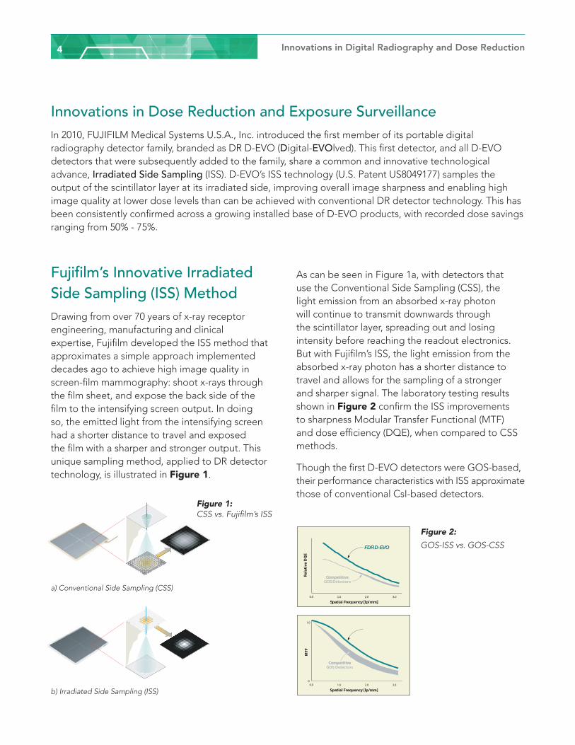
Innovations in Dose Reduction and Exposure Surveillance In 2010, FUJIFILM Medical Systems U.S.A., Inc. introduced the first member of its portable digital radiography detector family, branded as DR D-EVO (Digital-EVOlved). This first detector, and all D-EVO detectors that were subsequently added to the family, share a common and innovative technological advance, Irradiated Side Sampling (ISS). D-EVO’s ISS technology (U.S. Patent US8049177) samples the output of the scintillator layer at its irradiated side, improving overall image sharpness and enabling high image quality at lower dose levels than can be achieved with conventional DR detector technology. This has been consistently confirmed across a growing installed base of D-EVO products, with recorded dose savings ranging from 50% - 75%.
Fujifilm’s Innovative Irradiated Side Sampling (ISS) Method Drawing from over 70 years of x-ray receptor engineering, manufacturing and clinical expertise, Fujifilm developed the ISS method that approximates a simple approach implemented decades ago to achieve high image quality in screen-film mammography: shoot x-rays through the film sheet, and expose the back side of the film to the intensifying screen output. In doing so, the emitted light from the intensifying screen had a shorter distance to travel and exposed the film with a sharper and stronger output. This unique sampling method, applied to DR detector technology, is illustrated in Figure 1.
As can be seen in Figure 1a, with detectors that use the Conventional Side Sampling (CSS), the light emission from an absorbed x-ray photon will continue to transmit downwards through the scintillator layer, spreading out and losing intensity before reaching the readout electronics. But with Fujifilm’s ISS, the light emission from the absorbed x-ray photon has a shorter distance to travel and allows for the sampling of a stronger and sharper signal. The laboratory testing results shown in Figure 2 confirm the ISS improvements to sharpness Modular Transfer Functional (MTF) and dose efficiency (DQE), when compared to CSS methods.
Though the first D-EVO detectors were GOS-based, their performance characteristics with ISS approximate those of conventional CsI-based detectors.
a) Conventional Side Sampling (CSS)
Figure 1: CSS vs. Fujifilm’s ISS
b) Irradiated Side Sampling (ISS)
0.0 1.0 2.0 3.0
Spatial Frequency [lp/mm]
Rela
tive
DQ
E
FDR D-EVO
CompetitiveCompetitiveGOS Detectors
1.0
00.0 1.0 2.0 3.0
Spatial Frequency [lp/mm]
MTF
FDR D-EVO
CompetitiveCompetitiveGOS Detectors
Figure 2: GOS-ISS vs. GOS-CSS
Innovations in Digital Radiography and Dose Reduction 5
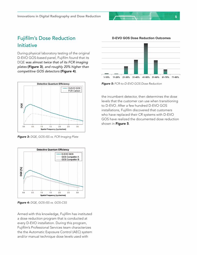
Fujifilm’s Dose Reduction Initiative During physical laboratory testing of the original D-EVO GOS-based panel, Fujifilm found that its DQE was almost twice that of its FCR imaging plates (Figure 3), and roughly 20% higher than competitive GOS detectors (Figure 4).
Armed with this knowledge, Fujifilm has instituted a dose reduction program that is conducted at every D-EVO installation. During this program, Fujifilm’s Professional Services team characterizes the the Automatic Exposure Control (AEC) system and/or manual technique dose levels used with
the incumbent detector, then determines the dose levels that the customer can use when transitioning to D-EVO. After a few hundred D-EVO GOS installations, Fujifilm discovered that customers who have replaced their CR systems with D-EVO GOS have realized the documented dose reduction shown in Figure 5.
Figure 3: DQE, GOS-ISS vs. FCR Imaging Plate
Figure 4: DQE, GOS-ISS vs. GOS-CSS
Figure 5: FCR-to-D-EVO GOS Dose Reduction
D-EVO GOS Dose Reduction Outcomes
1-10% 11-20% 21-30% 31-40% 41-50% 51-60% 61-70% 71-80%

Innovations in Digital Radiography and Dose Reduction6
Radiation Dose Reduction and Monitoring at White Memorial Medical CenterAt White Memorial Medical Center, the development and implementation of a dose reduction and monitoring program had been a priority since 2005. The goals of the program were threefold: to leverage the organization’s position as a teaching hospital to impact the culture of patient safety; to develop principles that would
guide current and future technology purchasing, making dose reduction a central priority; and to combine the recommendations of the Image Gently and Image Wisely campaigns with local quality control and quality assurance.
Like most other hospitals, White Memorial has undergone a vast transformation since 2005, transitioning its imaging technology offerings from analog to digital. Recent advancements in digital radiography, in particular, represent a significant re-birth of x-ray as a diagnostic
modality. For instance, the cesium detectors used in Fujifilm’s FDR Go platform have helped the hospital achieve dose reduction of more than 50% over computed radiography techniques with equivalent or improved image quality.
These improvements were made possible by the new technology combined with additional technologist and physician training. A growing reliance on automation has meant a gradual abandonment of the basics of patient measurement and utilization of ideal exposure combinations. Turning back to these basics, White Memorial found that technologists have the tools they need to reduce dose exposure effectively while maintaining image quality. They just need to be trained, supported, and inculcated within a new culture of patient safety.
Accordingly, staff were informed that going forward, all departments utilizing ionizing radiation for diagnostic studies would be required to monitor and track patients’ radiation exposure. Standardized protocols were developed and customized to each device, and technologists underwent an educational program to help them better understand the principles of dose-limiting imaging.
These principles include: the comparatively new concept of exposure index (EI), a function of body part selected, body part thickness, kVp, added
White Memorial Medical Center is a 353-bed not-for-profit, faith-based, teaching hospital, which provides a full range of inpatient, outpatient, emergency and diagnostic services to communities in and near downtown Los Angeles.
The cesium detectors used in Fujifilm’s FDR Go platform have helped the hospital achieve dose reduction of more than 50% over computed radiography techniques with equivalent or improved image quality.
Innovations in Digital Radiography and Dose Reduction 7
filtration in the x-ray beam and detector type; the importance of proper positioning, elimination of patient motion, collimation, and appropriate use of grids; the appropriateness of using automatic exposure control sensors, which are often not calibrated for pediatrics; and manual techniques for exposure control for patients with whom automatic exposure control is not an option.
Acceptable dose ranges for imaging equipment were posted in the technologist workroom as well as on the equipment itself. As a matter of policy, dose reduction protocol changes, determined by the medical director of radiology services at White Memorial, were implemented across the board. For instance, in some cases DR can be used for chest imaging in place of Computed Tomography (CT), because the higher image resolution it offers enables clinical determination regarding chest opacities.
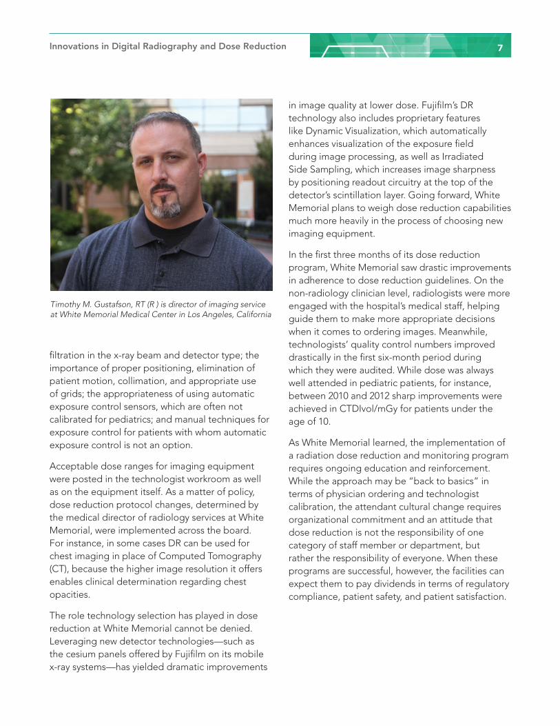
The role technology selection has played in dose reduction at White Memorial cannot be denied. Leveraging new detector technologies—such as the cesium panels offered by Fujifilm on its mobile x-ray systems—has yielded dramatic improvements
in image quality at lower dose. Fujifilm’s DR technology also includes proprietary features like Dynamic Visualization, which automatically enhances visualization of the exposure field during image processing, as well as Irradiated Side Sampling, which increases image sharpness by positioning readout circuitry at the top of the detector’s scintillation layer. Going forward, White Memorial plans to weigh dose reduction capabilities much more heavily in the process of choosing new imaging equipment.
In the first three months of its dose reduction program, White Memorial saw drastic improvements in adherence to dose reduction guidelines. On the non-radiology clinician level, radiologists were more engaged with the hospital’s medical staff, helping guide them to make more appropriate decisions when it comes to ordering images. Meanwhile, technologists’ quality control numbers improved drastically in the first six-month period during which they were audited. While dose was always well attended in pediatric patients, for instance, between 2010 and 2012 sharp improvements were achieved in CTDIvol/mGy for patients under the age of 10.
As White Memorial learned, the implementation of a radiation dose reduction and monitoring program requires ongoing education and reinforcement. While the approach may be “back to basics” in terms of physician ordering and technologist calibration, the attendant cultural change requires organizational commitment and an attitude that dose reduction is not the responsibility of one category of staff member or department, but rather the responsibility of everyone. When these programs are successful, however, the facilities can expect them to pay dividends in terms of regulatory compliance, patient safety, and patient satisfaction.
Timothy M. Gustafson, RT (R ) is director of imaging service at White Memorial Medical Center in Los Angeles, California
Innovations in Digital Radiography and Dose Reduction8
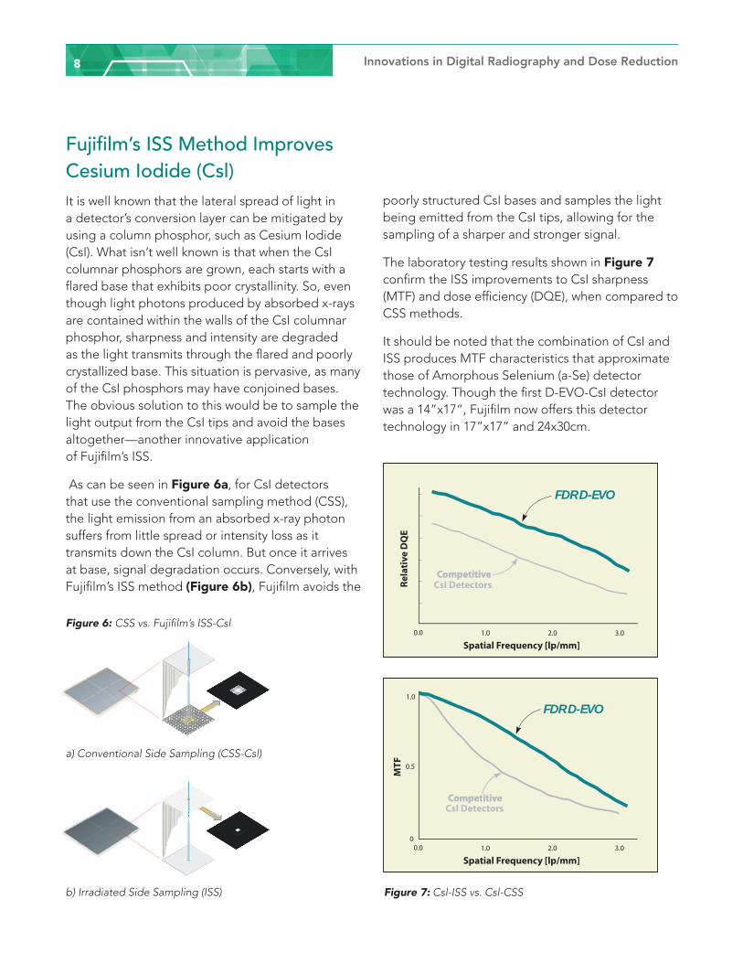
Fujifilm’s ISS Method Improves Cesium Iodide (Csl)It is well known that the lateral spread of light in a detector’s conversion layer can be mitigated by using a column phosphor, such as Cesium Iodide (CsI). What isn’t well known is that when the CsI columnar phosphors are grown, each starts with a flared base that exhibits poor crystallinity. So, even though light photons produced by absorbed x-rays are contained within the walls of the CsI columnar phosphor, sharpness and intensity are degraded as the light transmits through the flared and poorly crystallized base. This situation is pervasive, as many of the CsI phosphors may have conjoined bases. The obvious solution to this would be to sample the light output from the CsI tips and avoid the bases altogether—another innovative application of Fujifilm’s ISS.
As can be seen in Figure 6a, for CsI detectors that use the conventional sampling method (CSS), the light emission from an absorbed x-ray photon suffers from little spread or intensity loss as it transmits down the CsI column. But once it arrives at base, signal degradation occurs. Conversely, with Fujifilm’s ISS method (Figure 6b), Fujifilm avoids the
poorly structured CsI bases and samples the light being emitted from the CsI tips, allowing for the sampling of a sharper and stronger signal.
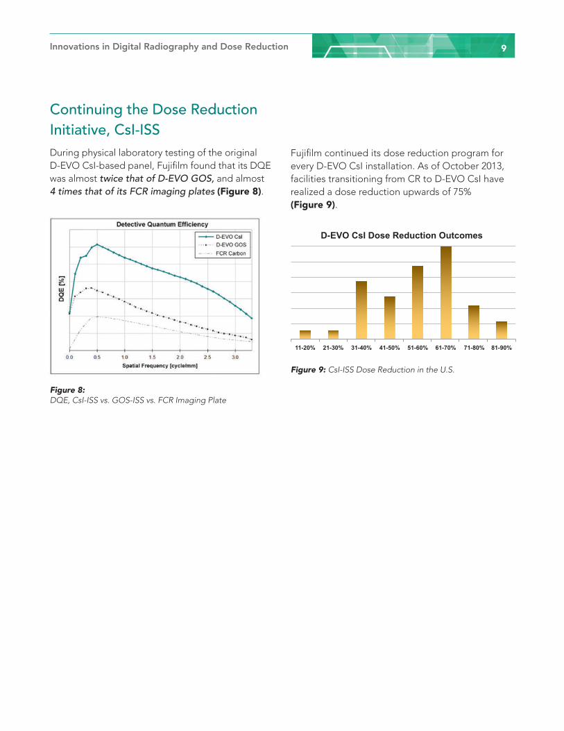
The laboratory testing results shown in Figure 7 confirm the ISS improvements to CsI sharpness (MTF) and dose efficiency (DQE), when compared to CSS methods.
It should be noted that the combination of CsI and ISS produces MTF characteristics that approximate those of Amorphous Selenium (a-Se) detector technology. Though the first D-EVO-CsI detector was a 14”x17”, Fujifilm now offers this detector technology in 17”x17” and 24x30cm.
a) Conventional Side Sampling (CSS-Csl)
Figure 6: CSS vs. Fujifilm’s ISS-Csl
b) Irradiated Side Sampling (ISS) Figure 7: Csl-ISS vs. Csl-CSS
0.0 1.0 2.0 3.0
Spatial Frequency [lp/mm]
Rel
ativ
e D
QE
CompetitiveCompetitiveCsI Detectors
FDR D-EVO
1.0
0.5
00.0 1.0 2.0 3.0
Spatial Frequency [lp/mm]
MTF
CompetitiveCompetitiveCsI Detectors
FDR D-EVO
Innovations in Digital Radiography and Dose Reduction 9
Continuing the Dose Reduction Initiative, CsI-ISSDuring physical laboratory testing of the original D-EVO CsI-based panel, Fujifilm found that its DQE was almost twice that of D-EVO GOS, and almost 4 times that of its FCR imaging plates (Figure 8).
Fujifilm continued its dose reduction program for every D-EVO CsI installation. As of October 2013, facilities transitioning from CR to D-EVO CsI have realized a dose reduction upwards of 75% (Figure 9).
Figure 8: DQE, CsI-ISS vs. GOS-ISS vs. FCR Imaging Plate
Figure 9: CsI-ISS Dose Reduction in the U.S.
D-EVO CsI Dose Reduction Outcomes
11-20% 21-30% 31-40% 41-50% 51-60% 61-70% 71-80% 81-90%

Innovations in Digital Radiography and Dose Reduction10
Reducing Patient Exposure at Via Christi Health Hospital Reducing patient exposure to medical ionizing radiation in diagnostic procedures is a public health issue that has gained increased public awareness over the last several years. As a result, many hospitals are initiating dose management programs to address this important issue.
At Via Christi Health’s hospitals in Wichita, Kansas, CAPT (ret) Jerry Thomas, MS, DABR, CHP, DABSNM has been spearheading a comprehensive dose
management program that encompasses all ionizing radiation modalities—including digital radiography (DR). While much of the focus on reducing dose has historically been on computed tomography (CT), CAPT Thomas and Alan Cebula, MS, diagnostic medical physicist, believe it’s
just as important to manage dose for all diagnostic imaging modalities that use ionizing radiation.
“We have an obligation to our patients to obtain the highest quality diagnostic image at the lowest possible exposure,” CAPT Thomas says. “Additionally, we need to determine an acceptable noise level so that noise in an image doesn’t interfere with the diagnostic process. This is important in all areas of medical imaging, not just in CT.”
Reducing dose starts with the detector
As part of the dose reduction initiative, in addition to the installed diagnostic x-ray DR imaging systems, Via Christi has replaced all chest and portable x-ray imaging systems with FDR D-EVO®, Fujifilm’s family of cesium iodide flat panel detectors. Fujifilm’s patented Irradiated
Side Sampling (ISS) technology is engineered to improve detective quantum efficiency (DQE) for clearer images at lower doses when compared to traditional design detectors. ISS positions its capture electronics (TFTs) at the irradiation side, in contrast to traditional detectors. This design suppresses scattering and attenuation of the captured x-ray beam, thereby producing sharper images at lower doses. ISS capture circuitry is designed to reduce dose and noise by as much as 10-20% when compared with other DR detectors and 30-75% when compared to CR.
Other built-in features, such as Dynamic Visualization™, help Via Christi’s technologists further clarify imaging detail for higher diagnostic content. Plus, Fujifilm was one of the first companies to implement the IHE standard for EI/DI (Exposure Index/Deviation Index) dose tracking functionality.
“Fujifilm has been very helpful in supporting our software development and in configuring the devices,” says CAPT Thomas. “The image quality and efficiency of the detector, (measured by DQE), is one of the primary tools for reducing dose to our patients.”
With the high DQE, there is real potential for
Via Christi Health is the largest provider of health care services in Kansas. Based in Wichita, it serves Kansas and northeast Oklahoma through its doctors, hospitals, senior villages and health services.
”With the high DQE, there is real potential for dose reduction in all our imaging studies.”
Innovations in Digital Radiography and Dose Reduction 11
dose reduction in all our imaging studies,” CAPT Thomas adds. By moving from CR to DR with the FDR D-EVO detectors Via Christi now provides clearer x-ray images at lower doses. In fact, Cebula notes that Via Christi has reduced dose across DR studies by at least 50% compared to CR. However, the facility is going one step further with the development of a dose management quality control (QC) program.
“The tools we are developing are really the core of the QC program,” CAPT Thomas explains. “We want to establish our own internal dose and deviation index for every exam, as well as conform to manufacturer recommended values. With Fujifilm’s assistance, we are developing a software program that allows us to capture user-specified DICOM data elements from the header data of acquired images.” These tools will be used to monitor and evaluate exposures from each of the different manufacturers’ DR products currently in use at Via Christi. “Our ultimate goal is to track and compare all diagnostic radiology exposures taken at each of our Wichita hospitals, clinics and free standing imaging centers, independently of the x-ray machine or DR vendor,” says CAPT Thomas. “This program can also be extended to older
CR technology, with the limitation of not having automated mining of techniques used for the examination.”
According to Cebula, the software enables the user to examine exposure values for each FPD/detector across ten different DICOM tags, including exam type, date, patient name, technologist name, S value, exposure and deviation indices, DAP, kVp and mAs. “We are looking for trends in techniques and ensuring that the images taken are of diagnostic quality and consistent across technologists.”
This is accomplished through the continuous monitoring of “S” values and/or deviation index values from all fixed units and the exposure value of each portable detector. All analyses are performed automatically through the use of MATLAB, an image processing analysis tool. CAPT Thomas feels that the tool can be extended for use over the Via Christi Health enterprise throughout the State of Kansas.
“One of my long-term interests is to automatically analyze daily QC images from each DR x-ray unit for artifacts. This test should provide an ‘early warning’ of spot damage to the DR detector – whether fixed or portable,” CAPT Thomas says. “I want to identify damage to a detector prior to a
Alan Cebula, MS, diagnostic medical physicist at Via Christi Health’s hospitals in Wichita, Kansas
CAPT (ret) Jerry Thomas, MS, DABR, CHP, DABSNM at Via Christi Health’s hospitals in Wichita, Kansas
Innovations in Digital Radiography and Dose Reduction12
radiologist calling me to review clinical images with artifacts. Additionally, variations in QC image Signal to Noise Ratio (SNR) may be a useful indicator of an electronic malfunction in a detector or x-ray machine. We want to be proactive with equipment maintenance.”
Evaluating protocols and workflow
In addition to evaluating the detector performance, Via Christi’s dose management program also takes into account the imaging protocols and the technologists’ workflow. In fact, Via Christi is one of the few hospitals in the country utilizing all the dose management capabilities that Fujifilm provides, including detector efficiency, monitoring and adapting protocols, and taking a proactive approach to maintenance.
In addition to refining the automated protocols provided by Fujifilm to fit the facility’s preferences,
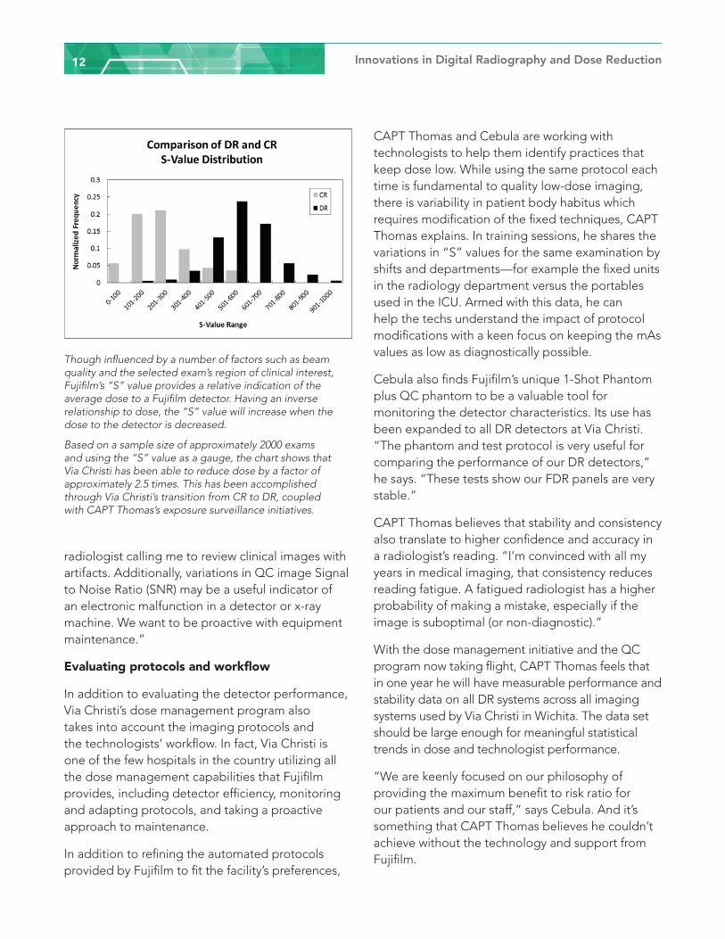
Though influenced by a number of factors such as beam quality and the selected exam’s region of clinical interest, Fujifilm’s “S” value provides a relative indication of the average dose to a Fujifilm detector. Having an inverse relationship to dose, the “S” value will increase when the dose to the detector is decreased.
Based on a sample size of approximately 2000 exams and using the “S” value as a gauge, the chart shows that Via Christi has been able to reduce dose by a factor of approximately 2.5 times. This has been accomplished through Via Christi’s transition from CR to DR, coupled with CAPT Thomas’s exposure surveillance initiatives.
CAPT Thomas and Cebula are working with technologists to help them identify practices that keep dose low. While using the same protocol each time is fundamental to quality low-dose imaging, there is variability in patient body habitus which requires modification of the fixed techniques, CAPT Thomas explains. In training sessions, he shares the variations in “S” values for the same examination by shifts and departments—for example the fixed units in the radiology department versus the portables used in the ICU. Armed with this data, he can help the techs understand the impact of protocol modifications with a keen focus on keeping the mAs values as low as diagnostically possible.
Cebula also finds Fujifilm’s unique 1-Shot Phantom plus QC phantom to be a valuable tool for monitoring the detector characteristics. Its use has been expanded to all DR detectors at Via Christi. “The phantom and test protocol is very useful for comparing the performance of our DR detectors,” he says. “These tests show our FDR panels are very stable.”
CAPT Thomas believes that stability and consistency also translate to higher confidence and accuracy in a radiologist’s reading. “I’m convinced with all my years in medical imaging, that consistency reduces reading fatigue. A fatigued radiologist has a higher probability of making a mistake, especially if the image is suboptimal (or non-diagnostic).”
With the dose management initiative and the QC program now taking flight, CAPT Thomas feels that in one year he will have measurable performance and stability data on all DR systems across all imaging systems used by Via Christi in Wichita. The data set should be large enough for meaningful statistical trends in dose and technologist performance.
“We are keenly focused on our philosophy of providing the maximum benefit to risk ratio for our patients and our staff,” says Cebula. And it’s something that CAPT Thomas believes he couldn’t achieve without the technology and support from Fujifilm.
Innovations in Digital Radiography and Dose Reduction 13
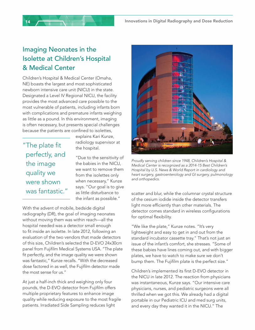
Fujifilm’s 24x30cm ISS-CsI Furthering its dominant role in innovative technology, Fujifilm introduced its D-EVO plus C24i in 2012. This was the world’s first 24x30cm wireless cesium detector. It was designed to offer advanced imaging capabilities, high DQE and low dose x-ray exams. Its size is perfect for small anatomy, orthopedics and the smallest of patients allowing for efficient workflow and increased patient comfort. The small format size of the detector provides the unique ability to easily fit into every neonatal isolette tray in use. Isolettes and incubators were traditionally designed with 24x30cm cassette trays. Fujifilm’s FDR D-EVO plus C24i is the same size as the 24x30cm cassette, therefore it fits perfectly, ensuring that the baby can be left at rest, safe and protected from contaminations and disruptions.
Comparative laboratory testing of D-EVO plus C35 and D-EVO plus C24i are shown in Figure 10. As can be seen, though both detectors have similar DQE performance at the standard measurement dose level (1mR), D-EVO plus C24i has almost twice the DQE when measured at 0.01mR.
This significant improvement to DQE at lower doses ushers in a new era of the highest image quality at the lowest possible dose.
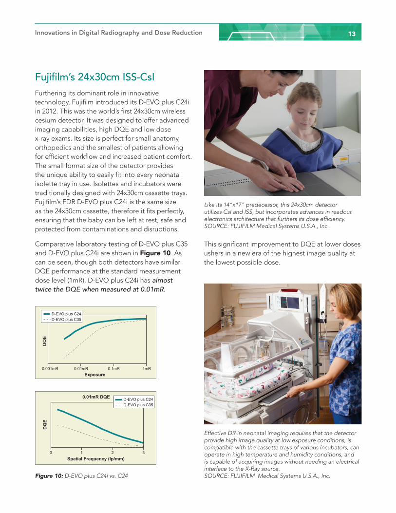
Like its 14”x17” predecessor, this 24x30cm detector utilizes CsI and ISS, but incorporates advances in readout electronics architecture that furthers its dose efficiency. SOURCE: FUJIFILM Medical Systems U.S.A., Inc.
Effective DR in neonatal imaging requires that the detector provide high image quality at low exposure conditions, is compatible with the cassette trays of various incubators, can operate in high temperature and humidity conditions, and is capable of acquiring images without needing an electrical interface to the X-Ray source. SOURCE: FUJIFILM Medical Systems U.S.A., Inc. Figure 10: D-EVO plus C24i vs. C24
0.001mR 0.01mR 0.1mR 1mRExposure
DQ
E
D-EVO plus C24D-EVO plus C35
DQ
E
0 1 2 3
0.01mR DQE
Spatial Frequency (lp/mm)
D-EVO plus C24D-EVO plus C35
Innovations in Digital Radiography and Dose Reduction14
Imaging Neonates in the Isolette at Children’s Hospital & Medical CenterChildren’s Hospital & Medical Center (Omaha, NE) boasts the largest and most sophisticated newborn intensive care unit (NICU) in the state. Designated a Level IV Regional NICU, the facility provides the most advanced care possible to the most vulnerable of patients, including infants born with complications and premature infants weighing as little as a pound. In this environment, imaging is often necessary, but presents special challenges because the patients are confined to isolettes,
explains Kari Kunze, radiology supervisor at the hospital.
“Due to the sensitivity of the babies in the NICU, we want to remove them from the isolettes only when necessary,” Kunze says. “Our goal is to give as little disturbance to the infant as possible.”
With the advent of mobile, bedside digital radiography (DR), the goal of imaging neonates without moving them was within reach—all the hospital needed was a detector small enough to fit inside an isolette. In late 2012, following an evaluation of the two vendors that made detectors of this size, Children’s selected the D-EVO 24x30cm panel from Fujifilm Medical Systems USA. “The plate fit perfectly, and the image quality we were shown was fantastic,” Kunze recalls. “With the decreased dose factored in as well, the Fujifilm detector made the most sense for us.”
At just a half-inch thick and weighing only four pounds, the D-EVO detector from Fujifilm offers multiple proprietary features to enhance image quality while reducing exposure to the most fragile patients. Irradiated Side Sampling reduces light
scatter and blur, while the columnar crystal structure of the cesium iodide inside the detector transfers light more efficiently than other materials. The detector comes standard in wireless configurations for optimal flexibility.
“We like the plate,” Kunze notes. “It’s very lightweight and easy to get in and out from the standard incubator cassette tray.” That’s not just an issue of the infant’s comfort, she stresses. “Some of these babies have lines coming out, and with bigger plates, we have to watch to make sure we don’t bump them. The Fujifilm plate is the perfect size.”
Children’s implemented its first D-EVO detector in the NICU in late 2012. The reaction from physicians was instantaneous, Kunze says. “Our intensive care physicians, nurses, and pediatric surgeons were all thrilled when we got this. We already had a digital portable in our Pediatric ICU and med surg units, and every day they wanted it in the NICU.” The
Proudly serving children since 1948, Children’s Hospital & Medical Center is recognized as a 2014-15 Best Children’s Hospital by U.S. News & World Report in cardiology and heart surgery, gastroenterology and GI surgery, pulmonology and orthopedics.
”The plate fit perfectly, and the image quality we were shown was fantastic.”
Innovations in Digital Radiography and Dose Reduction 15
detector immediately distinguished itself in terms of image quality, she adds, thanks to the two-story set-up of the hospital’s NICU.
“When we got our first Fujifilm detector we kept it on one floor, but sometimes a patient would have to be moved to another floor, meaning we’d get an x-ray one day on the Fujifilm portable and the next day on a different system,” Kunze says. “The image quality on the other system wasn’t poor, but it also wasn’t as sharp.”
As a result, the hospital eventually decided to invest in a second D-EVO detector for the other floor of the NICU, ensuring consistency across the board. “People like to interface with the Fujifilm,” Kunze says. “It’s easy to annotate, easy to rotate your image to collimate it if necessary—it’s very user-friendly. On the rare occasion where we have to repeat an x-ray, the process is very simple.” Ease of annotation is critical because it plays a key role in the hospital’s tracking and management of dose, Kunze notes. “We always annotate our distance, whether we imaged through Plexiglass, our mAs output and our kV technique,” she says.
Perhaps most importantly, the use of the small detectors protects the infants from exposure to infection, a critical concern given the fragility of their immune systems. The D-EVO detectors never leave the NICU and are cleaned after each exam using a CaviCide solution. “The cleaning agent is effective but also very harsh, and the equipment is holding up extremely well,” Kunze says, noting that the detectors could be used anywhere between fifteen and thirty times a day. “The detectors get wiped down a lot, so durability is extremely important.”
With the ability to safely and effectively image the infants within their isolettes, Children’s can elect to decrease risks associated with transferring them to the OR for routine procedures like umbilical line placement, ET tube placement or catheterization for extracorporeal membrane oxygenation. “Fujifilm’s detectors are fantastic at decreasing the dose to the patient while getting excellent image quality,” Kunze concludes. “It’s a huge advantage to be able to bring that to the bedside—we disturb the babies so much less than we would otherwise.”
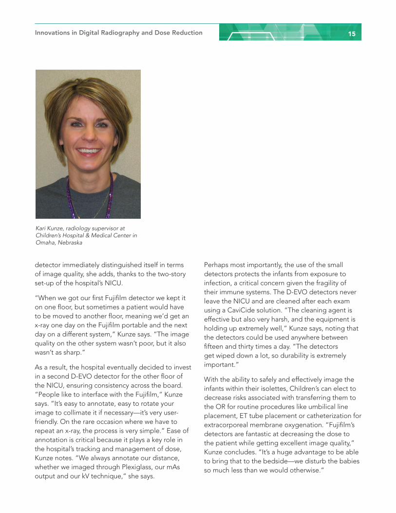
Kari Kunze, radiology supervisor at Children’s Hospital & Medical Center in Omaha, Nebraska
Innovations in Digital Radiography and Dose Reduction16
References1. The Combination of Reduced Patient Dose and Optimized Image Quality. FUJIFILM Medical System R&D Center,
R&D Management Headquarters, FUJIFILM Corporation. References; K. Sato, et al. Effect of X-ray incident direction and scintillator layer design on image quality of indirect-conversion flat –panel detector with GOS phosphor, SPIE Vol.7961 796141 (2011)
2. FUJIFILM Medical System R&D Center, R&D Management Headquarters, FUJIFILM Corporation. References;a. Robert M., et al. Med. Phys. 16, 773 (1989)b. Sato K., et al. Development of “CALNEO”, an Indirect-conversion Digital Radiography System with High- conversion Efficiency, FUJIFILM Research & Development, 55, 10 (2010)c. IEC62220-1-1: Medical electrical equipment-Characteristics of digital X-ray imaging devices- Part 1: Determination of the detective quantum efficiency Ed. 1.0 (2003)
3. Proceedings of SPIE Reprint SPIE manuscript 7961-162. Effect of x-ray incident direction and scintillator layer design on image quality of indirect-conversion flat panel detector with GOS phosphor. K. Sato, F. Nariyuki, H. Nomura, S. Fukui, M. Nakatsu, Y. Okada, T. Nabeta, Y. Hosoi. Medical Imaging 2011: Physics of Medical Imaging 2011
4. High Conversion Efficiency Flat Panel Detector with ISS Technology. FUJIFILM Medical System R&D Center, R&D Management Headquarters, FUJIFILM Corporation
5. Development of FUJIFILM-DR-D-EVO plus C24i/s for X-ray Imaging of Neonatal Care. FUJIFILM Medical System R&D Center, R&D Management Headquarters, FUJIFILM Corporation. Keita Watanabe, Ryo Imamura, Tomonari Sendai
6. FUJIFILM-DR D-EVO plus C24i/s. FUJIFILM Corporation, Medical Systems R&D Center. Kouichi Kitano, Keita Watanabe and Akihito Bettouyashiki
About FUJIFILM Medical Systems U.S.A., Inc.FUJIFILM Medical Systems U.S.A., Inc. is a leading provider of diagnostic imaging products and medical informatics solutions to meet the needs of healthcare facilities today and well into the future. From an unrivaled selection of digital x-ray systems, to the Synapse® brand of PACS, RIS and cardiovascular products, to advanced women’s health imaging systems, Fujifilm has products that are ideal for any size imaging environment. The Endoscopy Division of FUJIFILM Medical Systems U.S.A., Inc. supplies high quality, technologically advanced FUJINON brand endoscopes to the medical market. FUJIFILM Medical Systems U.S.A., Inc. is headquartered in Stamford, CT. For more information please visit www.fujimed.com and www.fujifilmendoscopy.com.
FUJIFILM Holdings Corporation, Tokyo, Japan brings continuous innovation and leading-edge products to a broad spectrum of industries, including: healthcare, with medical systems, pharmaceuticals and cosmetics; graphic systems; highly functional materials, such as flat panel display materials; optical devices, such as broadcast and cinema lenses; digital imaging; and document products. These are based on a vast portfolio of chemical, mechanical, optical, electronic, software and production technologies. In the year ended March 31, 2014, the company had global revenues of $23.9 billion, at an exchange rate of 102 yen to the dollar. Fujifilm is committed to environmental stewardship and good corporate citizenship. For more information, please visit: www.fujifilmholdings.com.
FUJIFILM Medical Systems U.S.A., Inc.419 West Avenue, Stamford, CT 06902-6348www.fujimed.com © 2015 FUJIFILM Medical Systems U.S.A., Inc. All product and company names herein may be trademarks of their registered owners. XBUSDR097