Innovation and Development of Study Field Nanomaterials at ... · Nanomaterials at the Technical...
27
These materials have been developed within the ESF project: Innovation and development of study field Nanomaterials at the Technical University of Liberec Innovation and Development of Study Field Nanomaterials at the Technical University of Liberec nano.tul.cz
Transcript of Innovation and Development of Study Field Nanomaterials at ... · Nanomaterials at the Technical...

These materials have been developed within the ESF project: Innovation and development of study field Nanomaterials at the Technical University of Liberec
Innovation and Development of Study Field Nanomaterials at the Technical University of Liberec
nano.tul.cz

NANODIAMONDS
Applications in Biology and
Nanoscale Medicine
Technical University of Liberec
2

Outline
3
Alotropic forms of carbon
Diamond powders
Synthesis
Single – Nano Buckydiamond Particles SNBD
The Fundamental Properties and Characteristics
of Nanodiamonds
- Modification
- Bioapplications

4
Carbon is unique in the number and the variety of its allotropes due to its
valency. Well known forms of carbon include diamond and graphite. In
recent decades many more allotropes and forms of carbon have been
discovered such as fullerene graphene of nanotubes. By mixing
diamond and graphite phases by a nanoscale level of structure,
diamond-like carbon material may be created that at the same time is
amorphous, flexible, and yet purely sp3 bonded "diamond".
Alotropic forms and other
types of carbon
H. O. Pierson, Handbook of carbon, graphite, diamond and fullerens, Properties, processing and application, Noyes
Publications, New Jersey 1993

5
mixture
Allotropic forms and other
types of carbon
● Structure of diamond and graphite
● Amorphous carbon
● Fullerene
● Carbon nanotube
http://en.wikipedia.org/wiki/Diamond-like_carbon
C. Soldano, A. Mahmood, E. Dujardin, Carbon 48 (2010) 2127-2150

6
There are several methods to produce nanoscale diamond particles. The
simplest one is the milling of larger synthetic or natural microdiamonds
and sorting the smaller fraction out by sieving and fractionated
centrifugation.
Synthesis of diamond powders
● Vertical-type beads-milling machine
● Diamond micropowder
A. Krueger, Journal of Materials Chemistry, 18(2008) 1485 – 1792
B. E. Osawa, Nanodiamonds, Applications in Biology and Nanoscale Medicine, Ed. D. Ho, Springer, 2009

7
High density material like zirconia is used to give beads
higher momentum,
Beads are suspended in liquid medium to remove heat,
suspension rotation increase the v term of momentum (not
more than 4000 rpm to prevent the graphitization)
Beads are prepared to precisely spherical shape to reduce
the area of collision,
Axial rotation is adopted to obtain shearing collision,
After the process the centrifugal separation of milled
material is used.
Synthesis of diamond powders
- milling
Nanodiamonds, Applications in Biology and Nanoscale Medicine, Ed. D. Ho, Springer, 2009

8
In recent years, we are often shown in TV on how
accurate are the missile attacks, which usually end
up with black smoke coming from the
exact target position.
How many people watching TV realize that about half of
smoke is the DIAMOND.
Synthesis of diamond powders
- detonation
Shenderova, O. A. & Gruen, D. M. Ultrananocrystalline Diamond: Synthesis, Properties, and Applications (William
Andrew, 2006).
Nanodiamonds Applications in Biology and Nanoscale Medicine, Ed. D. Ho, Springer, 2009

9
In 1963 Ukrainian physicist named Vladimir Danilenko found
that soot from the explosion of well known military explosive
1:1 mixture of TNT – Trinitrotoluene (C7H5N3O6) and
Hexogen (C6H6N6O6) contain nanodiamond in high
concentration.
The greatest importance among the industrial methods of
synthesis of diamond powders have definitely the
detonation technologies
On the history of the discovery of
nanodiamond synthesis
V. Mochalin, O. Shenderova, D. Ho, Y. Gogotsi, Nature Nanotechnology 7, (2012) 11–23
Danilenko, V. V. On the history of the discovery of nanodiamond synthesis. Phys. Solid State 46, 595–599 (2004)

10
● Detonation synthesis of
nanodiamonds
Detonation nanodiamonds
● Industrial reactor for the
detonation synthesis of
diamond powders
● Detonation nanodiamonds
http://what-is-nanotechnology.com/co.htm
V. V. Danilenko, Synthesizing and sintering of diamond by explosion, Energomizdat, 2003
Nanodiamonds Applications in Biology and Nanoscale Medicine, Ed. D. Ho, Springer, 2009

11
The most critical step in production of single-nano buckydiamond SNBD is
the detonation process. After the detonation of explosives in inert medium,
invisibly small diamond crystals will start to grow from deposition of
unoxidized carbon atoms from the explosive molecules in the thin high-
pressure high-temperature zone, formed behind the rapidly propagating
front of shock wave. It seems that a very large number of crystallization
nuclei simultaneously start the diamond growth process under abundant
supply of carbon atoms generated from incomplete combustion of the
explosives
The crystal growth is suddenly and all at once suspended as the shock
wave passes at supersonic speed from the diamond growth area.
Detonation nanodiamonds

12
Structure of detonation nanodiamonds
● A possible structure model for diamond
agglomerates in detonation diamond
● Structure of a single nanodiamond particle
In the certain stage of the detonation synthesis, further drop of the pressure
and temperature drives the P-T parameters to the region where the diamond
is thermodynamically unstable, and this is the reason that detonation carbon
is a mixture of sp2 (graphite) and sp3 (diamond) hybridized carbon atoms.
V. Mochalin, O. Shenderova, D. Ho, Y. Gogotsi, Nature Nanotechnology 7, (2012) 11–23
A. Krueger, Journal of Materials Chemistry, 18(2008) 1485 – 1792

13
oxygen termination hydrogen termination
Surface of nanodiamonds
P. Niedzielski, Wytwarzanie i zastosowanie proszków diamentowych, Wydawnictwo Politechniki Łódzkiej, Łódź, 2011
The surface of nanodiamonds is full of free (dangling) bonds, that
chemically bind to the atoms of the environment (usually oxygen and
hydrogen). This phenomenon called „termination” strongly affects the
physicochemical properties of nanodiamond particles.
● Bonds on the surface of diamond
Terminated surface of
nanodiamonds should be
considered as perfect
starting material for further
chemical of biological
modifications.

14 Nanodiamonds Applications in Biology and Nanoscale Medicine, Ed. D. Ho, Springer, 2009
It has been proposed that DND may
be applied for medical purposes,
either alone or biofunctionalized, in
diverse medical fields, including
oncology, cardiology, gastroenterology
and dermatology. DND conjugates are
chemically stable and do not alter the
activity of bound biomolecules.
After the synthesis and purification the surface of nanodiamond particles
contains a complex array of surface groups, including carboxylic acids,
esters, ethers, lactones, amines, etc.
Surface of nanodiamonds
● Schematic of possible surface
functionalization of nanodiamonds

15
The suspension of nanodiamond particles introduced into a tumor cell
culture led to their aggregation and in a consequence by damaging the cell
membrane resulted in their death.
A.P.Puzyr, D.A.Neshumayev, V.S.Bondar V.Yu.Dolmatov, I.V.Shugalei, N.P.Dubyago, S.V.Tarskikh, G.V.Makarskaya: the
influence of detonation nanodiamond powder on blood cells: . Lee and N. Novikov (eds.), Innovative Superhard Materials
and Sustainable Coatings for Advanced Manufacturing, 155–167. 2005 Springer. Printed in the Netherlands
● Aggregation of tumour cells
● Destroyed tumour cells
Properties of nanodiamonds

16
Depending on size of nanodiamond particles and aggregates
the possible areas of their application differ from each other.
● Schematics of the size ranges of DNDD primary particles and aggregates in
relation to different possible areas of biomedical and healthcare application
Size vs. destiny
Nanodiamonds Applications in Biology and Nanoscale Medicine, Ed. D. Ho, Springer, 2009

17
Applications of nanodiamonds
- Seeding of CVD Diamond Films
As a seed material for CVD diamond
growth, nanodiamond particles play an
important role for the development of
medical implants. Seeding with DND
allows obtaining coatings with small grain
size and, therefore smooth surface that are
important for medical implant applications.
Nanodiamonds Applications in Biology and Nanoscale Medicine, Ed. D. Ho, Springer, 2009

18
0
2
4
6
8
10
12
14
16
Day 0 Day 1 Day 2 Day 3
Days
Ear
Sw
elling
(mm
x 1
0 -
2)
DNFB negative control
DNFB positive control
DETONATION Ear only
DETONATION Ear +Abdomen
The application of diamond powders in pharmaceutical – cosmetological
industry is related to their properties such as: bactericidal activity, inhibition
of the oxidative stress, anti-allegric and anti-inflamatory activity.
M. Batory, D. Batory, J. Grabarczyk, W. Kaczorowski, B. Kupcewicz, K. Mitura, T. H. Nasti, N. Yusuf, P. Niedzielski, Journal of
Nanoscience and Nanotechnology Vol. 12, 1–10, 2012
● Allergy tests results of nanodiamond powder
based cosmetic formula
Applications of nanodiamonds
– health care products
The study shows that diamond
powder particles do not cause
allergies. All obtained results of
ears swelling are below 4 ×10⁻²
mm, which is the limit value
assumed in the clinical
examinations.
19
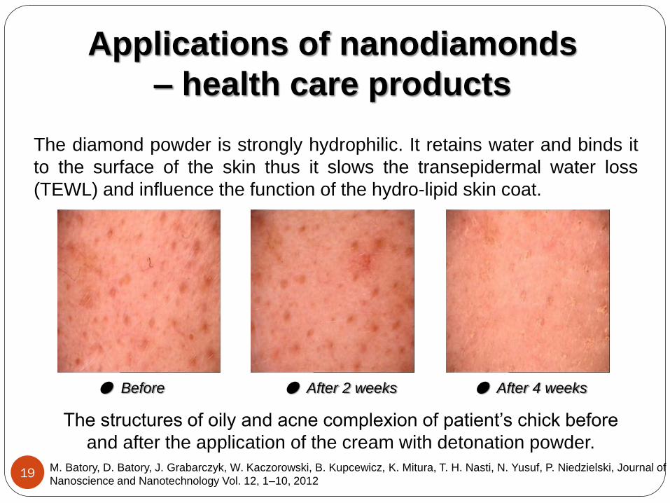
The diamond powder is strongly hydrophilic. It retains water and binds it
to the surface of the skin thus it slows the transepidermal water loss
(TEWL) and influence the function of the hydro-lipid skin coat.
Applications of nanodiamonds
– health care products
The structures of oily and acne complexion of patient’s chick before
and after the application of the cream with detonation powder.
● Before
● After 2 weeks
● After 4 weeks
M. Batory, D. Batory, J. Grabarczyk, W. Kaczorowski, B. Kupcewicz, K. Mitura, T. H. Nasti, N. Yusuf, P. Niedzielski, Journal of
Nanoscience and Nanotechnology Vol. 12, 1–10, 2012

20
Nanodiamonds are also considered for cosmetics and health care
applications due to their ability to bond with biological materials, improve
durability and robustness of a composition. Provide protection against
harmful UV light.
P. Niedzielski, Wytwarzanie i zastosowanie proszków diamentowych, Wydawnictwo Politechniki Łódzkiej, Łódź, 2011
● Examples of cream bases
prepared using nanodiamonds
● Example of the pattern for the
commercial product
Applications of nanodiamonds
– health care products

21 R.A.Shimkunas, E.Robinson, R.Lam, S.Lu, X.Xu, X.Q.Zhang, H.Huang, E.Osawa, D.Ho: Nanodiamond–insulin
complexes as pH-dependent protein delivery vehicles, Biomaterials 30 (2009) 5720–5728
Nanodiamond Insulin
Applications of nanodiamonds
– insulin carriers
According to research carried out by a North Western University team in the
US, nanodiamonds were found to have a very important medical use when
they loaded with insulin and placed around open wounds.
Insulin encourages skin cells to
proliferate and divide, restores
blood flow to the wound and
suppresses inflammation and fights
infection.

22
Applications of nanodiamonds
– films in drug delivery
Zhu Y, Li J, Li W, Zhang Y, Yang X, Chen N, Sun Y, Zhao Y, Fan C, Huang Q. Theranostics 2012; 2(3):302-312
A ND-drug film can be implanted
immediately after surgical removal of a
tumour to target residual cancerous cells
so as to effectively prevent the tumour
from recurring. In addition, a ND-drug
film can be more applicable to the
treatment of superficial tumours such as
breast cancer, head and neck cancer,
and skin cancer, or superficial skin
inflammations, wherein the drug is
delivered transdermally to tumours or
inflammation sites, reducing the toxicity
on normal tissues. ● Nanodiamond drug-funtionalized film

23
Applications of nanodiamonds
– clusters in drug delivery
Zhu Y, Li J, Li W, Zhang Y, Yang X, Chen N, Sun Y, Zhao Y, Fan C, Huang Q. Theranostics 2012; 2(3):302-312
The investigation of the assembly
principles and characteristics of
different functional molecules on NDs
would help to establish the models and
theories for building ND-based
versatile drug delivery systems, which
would serve as the basis for
developing a variety of ND-based drug
delivery systems with high efficiency
and low toxicity to prevent and/or treat
various cancers.
● Schematic illustration showing the different
loading of different functional molecules on NDs

24
Applications of nanodiamonds –
gene delivery
● Example of the attachment of specific DNA
to the nanodiamond surface for gene delivery
to cells / tissues using DNA plasmids.
● Example how nanodiamonds carrying a toxic
drug or gene-regulating microRNA are taken up by
a cell and how the attached “drug” may be released
for a specific treatment.
X. Zhang, M. Chen, R. Lam, X. Xu, E. Osawa, D. Ho, ACS. Nano, 2009, 3, 26092616
http://boards.medscape.com/forums/?128@@.2a36aa4d!comment=1&cat=All

25 V. Mochalin, Y. Gogotsi, J. Am. Chem. Soc., 2009, 131 (13), pp 4594–4595
The ODA-modified (octadecylamine) nanodiamond is highly fluorescent.
The intensity is so high that a bright blue fluorescence can be easily
detected with the bare eye at diamond concentrations as low as 0.004 %
by weight. Though the fluorescence mechanism requires further studies,
the ODA-functionalized nanodiamond can now be used in many
applications where visual detection of nanoparticles is required, such as
biomedical imaging and drug delivery systems.
● Bright blue fluorescence of the octadecylamine-modified nanodiamond
Fluorescent nanodiamonds as
cellular biomarkers

26
Nanodiamonds emit bright fluorescence at 550-800 nm from nitrogen-
vacancy-centres produced by high-energy ion beam irradiation and
subsequent thermal annealing. The emission, together with
noncytotoxicity and easiness of surface functionalization, makes nano-
sized diamonds a promising fluorescent probe for single-particle tracking
in heterogeneous environments.
Fluorescent nanodiamonds as
cellular biomarkers
Proc Natl Acad Sci U S A. 2007 January 16; 104(3): 727–732
http://awsch-web.physics.ucsb.edu/research/solid_state/nitrogen_vacancy/index.php
● Observation of single FNDs in a HeLa cell.
● Schematic of Nitrogen Vacancy Centre

27
Utilizing the inherent surface chemistry of
detonation nanodiamonds, an amine-
functionalized Gd(III) complex was
covalently bound allowing visualization of
nanodiamond particles by MR imaging. The
relaxivity of the Gd(III) contrast agent
increased nearly 10-fold in comparison to
the free agent upon conjugation to the
nanodiamond platform. Numbers 4 and 5
denote samples with the highest Gd(III)
concentration
Applications of nanodiamonds
– Magnetic Resonance imaging
● MR images of
nanodiamond samples.
L. Manus, D. Mastarone, E. Waters, X. Zhang, E. Schult-Sikma, K. MacRenaris, D. Ho, T. Meade, Nano Lett. 10 (2010)
484–489.