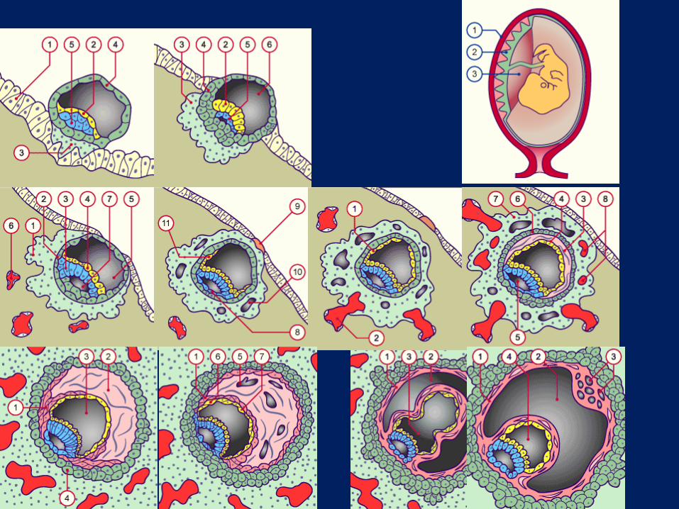
Inner Cell Mass (ICM) delaminates to form hypoblast and epiblast Occurs just prior to implantation &...
47
• Inner Cell Mass (ICM) delaminates to form hypoblast and epiblast • Occurs just prior to implantation & gastrulation • Epiblast (green cells) is 2-layered (i.e., it is bilaminate) disc of approximately cuboidal cells & will form the embryo proper • Flatter hypoblast cells lie on top of epiblast and will form yolk sac Formation of the epiblast & hypoblast(review)
-
Upload
kory-dorsey -
Category
Documents
-
view
214 -
download
0
Transcript of Inner Cell Mass (ICM) delaminates to form hypoblast and epiblast Occurs just prior to implantation &...
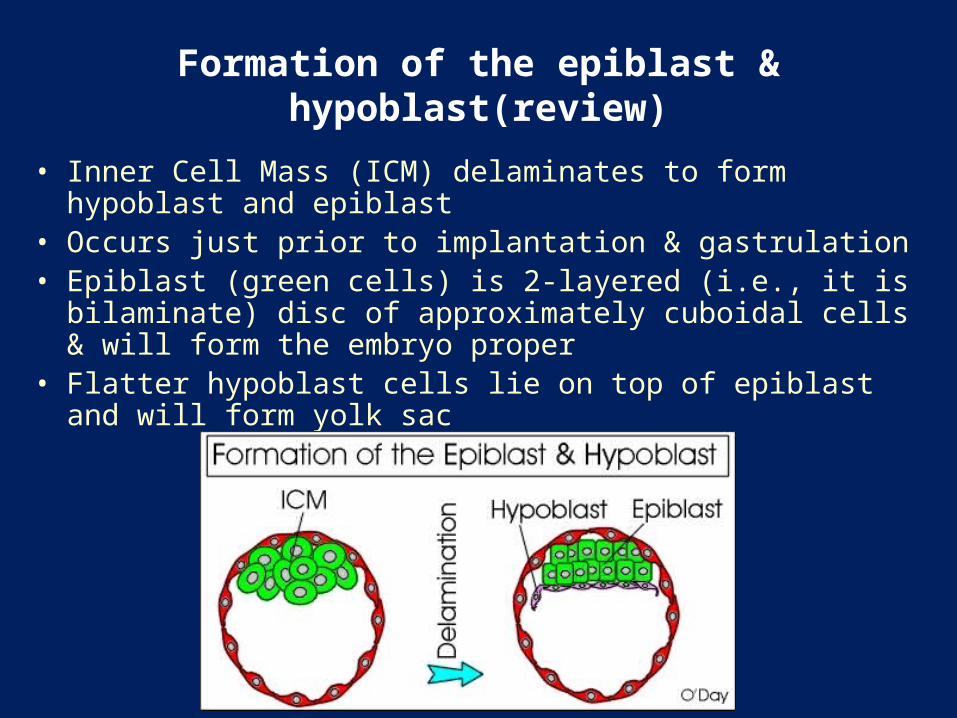
• Inner Cell Mass (ICM) delaminates to form hypoblast and epiblast • Occurs just prior to implantation & gastrulation • Epiblast (green cells) is 2-layered (i.e., it is bilaminate) disc of
approximately cuboidal cells & will form the embryo proper • Flatter hypoblast cells lie on top of epiblast and will form yolk sac
Formation of the epiblast & hypoblast(review)

morphogenetic movements and their significance to human
embryogenesis
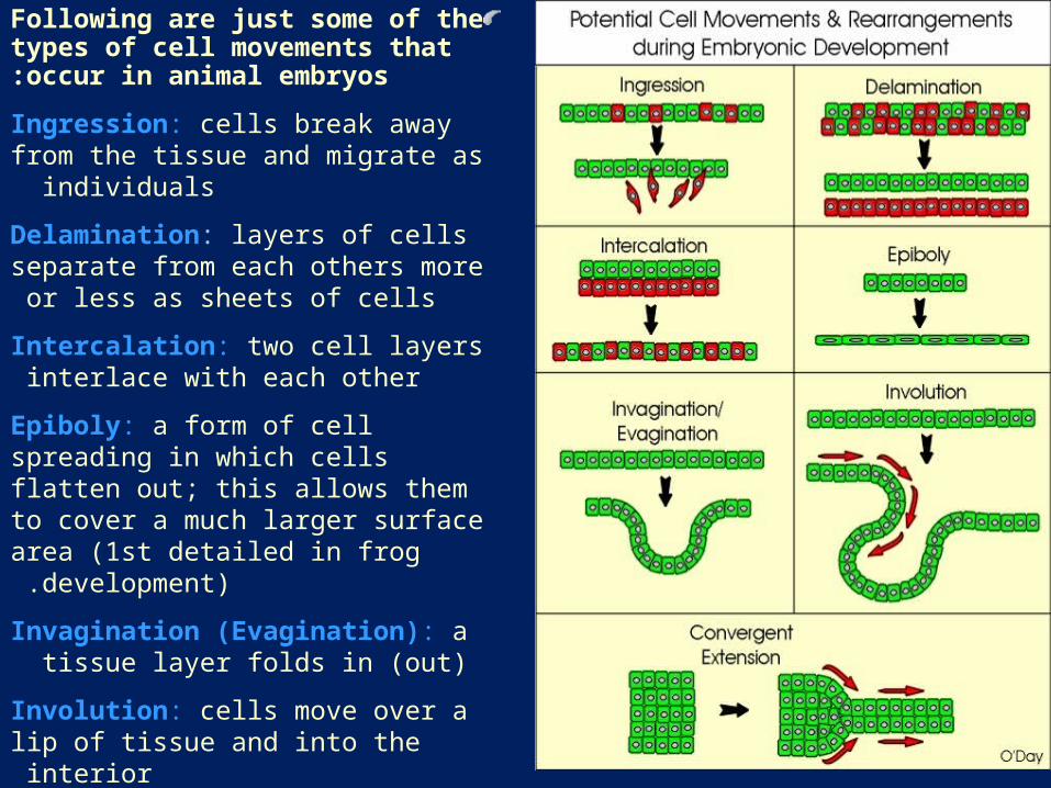
Following are just some of the types of cell movements that occur in animal embryos:
Ingression: cells break away from the tissue and migrate as individuals
Delamination: layers of cells separate from each others more or less as sheets of cells
Intercalation: two cell layers interlace with each other
Epiboly: a form of cell spreading in which cells flatten out; this allows them to cover a much larger surface area (1st detailed in frog
development) .
Invagination (Evagination): a tissue layer folds in (out)
Involution: cells move over a lip of tissue and into the interior
Convergent Extension: cells reorganize to form less layers allowing the cells to extend
out from a point .

04/20/234
Gastrulation
sohrabi

Role of Gastrulation
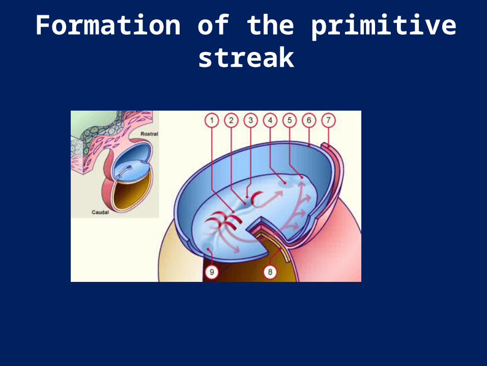
Formation of the primitive streak
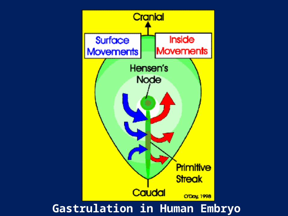
Gastrulation in Human Embryo

Cross Section of Human Gastrula


Bottle Cells • Bottle cells were first observed in amphibian
gastrulation • Occur during gastrulation in humans and many
other species • Also seen during neurulation and during other
types of cellular rearrangements • Involves a loss of cell-cell adhesion so cells can
move as individuals; recent work has shown that this is due to the loss of E-cadherin
• Change in shape of epithelial to bottle cell is believed to be one of driving forces for event – Cytoskeletal changes lead to bottle cell shape
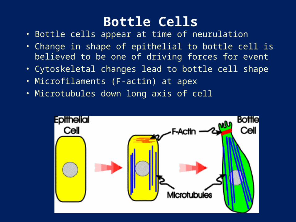
Bottle Cells • Bottle cells appear at time of neurulation • Change in shape of epithelial to bottle cell is believed to be
one of driving forces for event • Cytoskeletal changes lead to bottle cell shape • Microfilaments (F-actin) at apex • Microtubules down long axis of cell
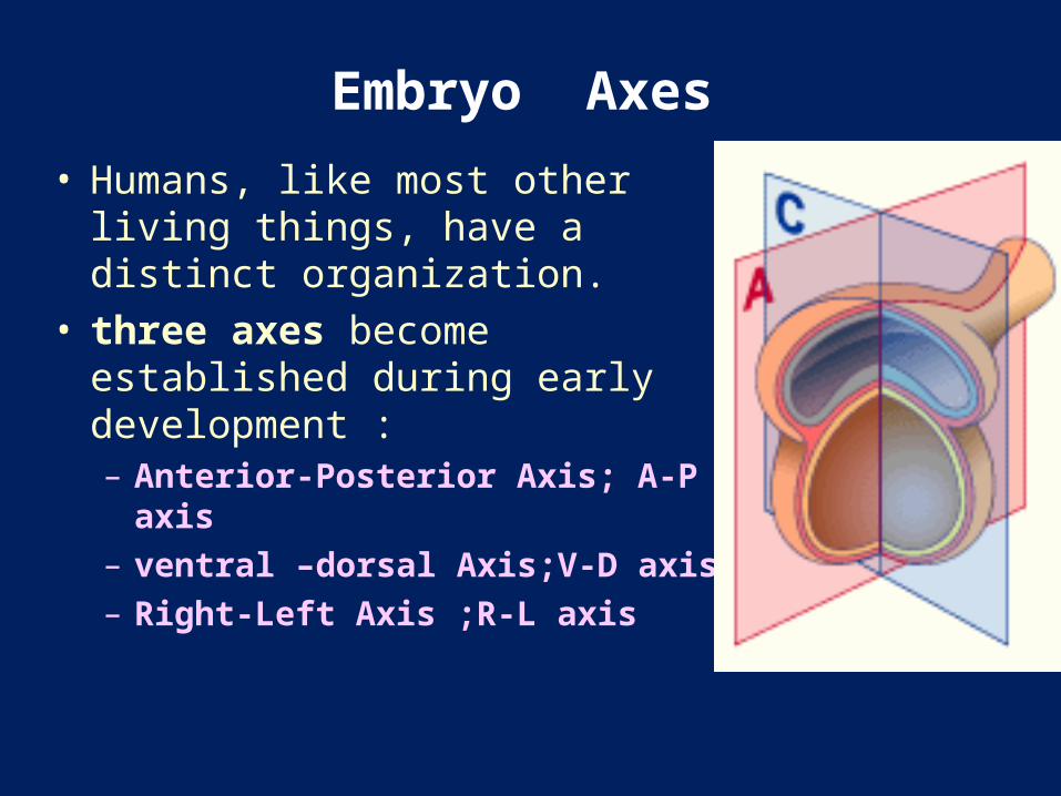
Embryo Axes
• Humans, like most other living things, have a distinct organization.
• three axes become established during early development :– Anterior-Posterior Axis; A-P axis – ventral –dorsal Axis;V-D axis– Right-Left Axis ;R-L axis

Factors involved in genetic control of :early embryonic development
• 1 -Transcription factors:• Homeodomain - Hox gene family• Zinc finger – a family of steroid-binding transcrition factors• Basic helix-loop-helix protein – myogenic regulatory factors• Winged helix – hepatocyte nuclear factor-3
2- Signalling molecules• Transforming growth factor-B (TGF-B) – activate posterior Hox genes• Fibroblast growth factor (FGF) – activate anrterior Hox genes• Nerve growth factor (NGF) – stimulate growth of axons• Hedgehog proteins – mediate early inductive interactions
3-Cell adhesion molecules• Some are calcium-dependent (Cadherins)• Some are calcium-independent (CAM)

HOX and the A-P Axis
• The A-P axis of all animals appears to be specified by the expression of Hox genes
• expressed sequentially in the same order that they are arranged in the chromosomes– The Human Hox complex (HOXA-HOXB-HOXC-HOXD)
is present in four sets on four separate chromosomes in each haploid set of chromosomes.
– Retinoic acid affects A-P axis formation and HOX gene expression.
Differentiation Factors:• Fibroblast Growth Factors (FGF) :
–Over 12 genes encode the FGF family of proteins
–FGFs function in axon extension, angiogenesis, and mesoderm formation
Differentiation Factors:• Hedgehog Proteins:
– Vertebrates have at least three genes for hedgehog proteins:
• sonic hedgehog (shh)– Sonic hedgehog--has the most diverse roles of all three
especially in the patterning of the embryo; functions in the patterning of the neural tube and somites, mediates formation of L-R axis of the embryo and of limbs, induction of differentiation of gut regions
• indian hedgehog (ihh) – Indian hedgehog--operates in gut and cartilage development
• desert hedgehog (dhh) – Desert hedgehog--functions in the Sertoli cells

Differentiation Factors:• Wnt Family
– consists of of at least 15 different cysteine-rich glycoproteins
– They are involved in inducing dorsal cells of somite to form muscle; in establishing polarity of limb and in development of the urogenital system, etc.
• TGFB family, activin family, bone morphogenetic proteins (BMPs), VG1 family and more – TGF-B proteins regulate matrix formation and cell division – BMPs were 1st discovered as bone morphogens also
regulate cell division, apoptosis, cell migration and differentiation &in the control of limb development

Signaling Centers in embryo development
• There are two signaling centres regulate gene expression in the different regions of the embryo to ensure that it develops with the relevant components in the right places at the right time: – After the endoderm has migrated internally during gastrulation, the
"anterior visceral endoderm" instructs the formation of head components.
• the anterior visceral endoderm expresses genes called Lim-1, Hesx-1 and Otx-2 which are essential for head
development – “Hensen's node” that appears at gastrulation at the anterior end of
the primitive streak contains information that oversees the construction of the whole body form.
• The node expresses genes called Noggin and Chordin that are not expressed by the anterior visceral endoderm.

cranio-caudal axis
• The primitive node is the first and central structure.
• primitive node determines the growth of the notochord cranially (by factor HNF-3) and of the primitive streak caudally (by the factor nodal) and the body axis (by the factor goosecoid).
• primitive node is also responsible for inducing the cells to become motile and migrate to pre-determined sites (by the factor T-gene).
• Note also how the prochordal plate determines the cranial end of the embryo (by factor Lim-1).
Right-Left asymmetry
• This right left differentiation is determined by • several laterality genes.• The signalling molecule:-sonic hedgehog (Shh) is expressed bilaterally
and symmetrically .-Activin is expressed only on the left, inhibits the
further action of Shh, and causes a differential expression of the growth factor nodal.

Right-Left asymmetry
• genes that specifically determine development of structures only on the left side.
• These have been called Inversus viscerum and left-right dynein.


Induction of Neural Tissue by Chordamesoderm
• During gastrulation special region of mesoderm underlies overlying ectoderm
• Chordamesoderm is future notochord
• Signals emitted by chordamesoderm have not been identified

•After gastrulation, three germ layers are evident: Ectoderm, mesoderm, endoderm:
Late Gastrula

Notochord Formation • the presumptive notochordal
tissue will separate from the endoderm and re-organize as the notochord
• As notochord formation is occuring, neurulation will separate the ectoderm into the epithelial ectoderm and the neural ectoderm
• the neural ectoderm begins folding to form the neural tube – The future neural ectoderm
overlies notochordal process (chordamesoderm)
– Folding of the neural ectoderm results in the neural tube
• The neural tube is the precursor of the brain & spinal cord
04/20/2331
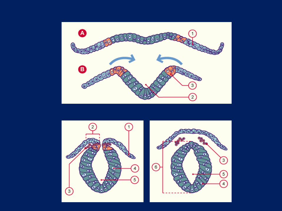
Neurulation

Hensen’s Node Induces Neural Axis
• this special region of tissue has been shown to establish the vertebrate body plan in everything from frogs to zebra fish to mice
• the node is an embryonic organizer in mammals– the mammalian node is not a classic organizer
because it can't act alone to induce the embryonic axis but requires other anterior germ tissues to be fully effective
• Thus progressive cellular interactions are more important rather than a single inductive event


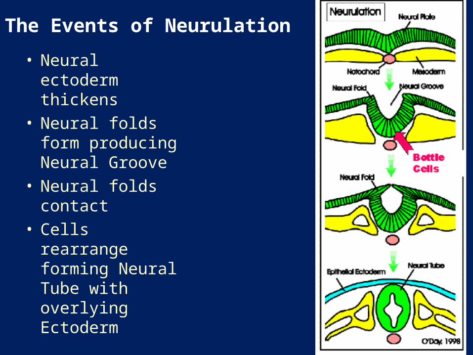
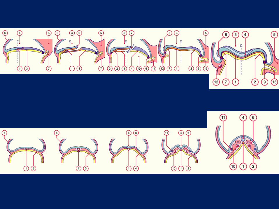
The Events of Neurulation • Neural ectoderm
thickens • Neural folds form
producing Neural Groove
• Neural folds contact
• Cells rearrange forming Neural Tube with overlying Ectoderm

Hensen’s Node Induces Neural Axis
• These classic experiments were done in chicken embryos
• Remove HN: No neural tissue development
• Transplant HN to host embryo: 2nd Neural Axis induced

Cell Lineages At Neurula Stage


Embryo consists of all primary germ layers Development continues to form tissues & organs As the neural tube is forming,the somites are forming & the embryo is elongating & flexing
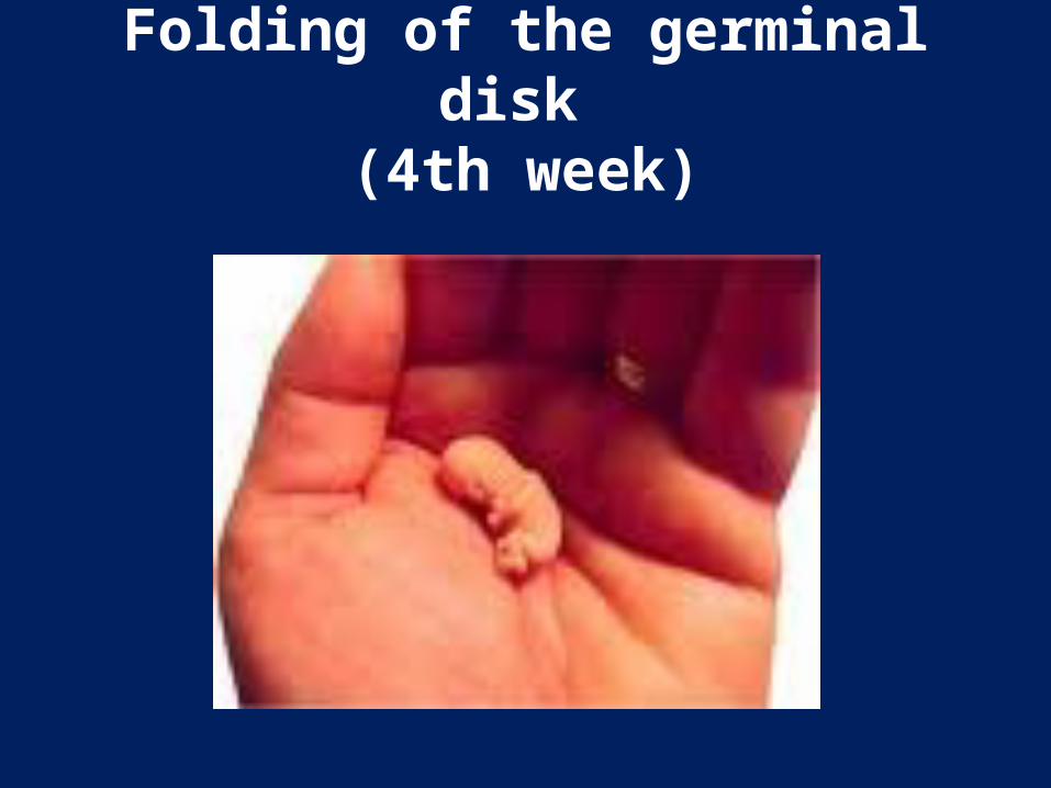
Folding of the germinal disk (4th week)


The End