Collision tumour involving a rectal gastrointestinal stromal tumour
Upload
mariaastrianiCategory
view
114download
1
AJR:200, January 2013 3
nosed lesions are best divided into biologically aggressive or nonaggressive categories. Biopsy is indicated if the lesion is aggressive. Other-wise, watchful waiting with follow-up imag-ing can be pursued. Aggressive features are detected on radiographs by evaluation of im-aging characteristics, such as the appearance of the margin, cortical expansion, and perios-teal reaction. Clinical examination findings, such as pain, are also contributory.
MarginsAfter assessment of features such as patient
age, the identity of the affected bone, and the location of the tumor in the bone, the tumor it-self must be closely scrutinized. The imaging characteristic that is most reflective of the ag-gressive (typically malignant) or nonaggres-sive (typically benign) nature of primary bone tumors is the appearance of the margin, which is an indicator of the growth rate of the lesion [3, 4]. A radiographic classification of bone tu-mor margins has been developed [5] that identi-fies three main types (Fig. 1). The least-aggres-sive margins (type I) correspond to tumors that are round or ovoid in shape (geographic). Geo-graphic margins have been divided into three subcategories. Type IA margins are the least aggressive, exhibiting a narrow zone of transi-tion from the tumor to the normal surrounding bone and a sclerotic rim (e.g., fibrous cortical defect, fibrous dysplasia, nonossifying fibroma/fibroxanthoma (Fig. 2). Lesions with type IB margins have no sclerotic rim, while remain-ing well defined with a narrow zone of transi-tion. These margins indicate indeterminate bi-ologic potential and may be seen with benign
Radiography in the Initial Diagnosis of Primary Bone Tumors
Colleen M. Costelloe1
John E. Madewell
Costelloe CM, Madewell JE
1Both authors: Department of Diagnostic Radiology, The University of Texas M. D. Anderson Cancer Center, Unit 1273, 1515 Holcombe Blvd, Houston, TX 77030. Address correspondence to C. M. Costelloe ([email protected]).
AJR 2013; 200:3–7
0361–803X/13/2001–3
© American Roentgen Ray Society
1. To understand how the radiographic characteristics of the margins of primary bone tumors reflect the biologic activity/growth rate of the lesions
2. To learn the radiographic principles of bone tumor margin classification
3. To identify the secondary role of corti-cal expansion and periosteal reaction in the characterization of primary bone tumors
4. To correlate the radiographic appearance of mineralized matrix with histologic type
5. To understand the advantages and limi-tations of radiography in the initial evalua-tion of primary bone tumors
Imaging TechniqueRadiographs are recommended as the initial
imaging modality for the evaluation of bone pain [1, 2], and standard radiographic tech-niques are typically adequate for tumor im-aging. The long bones should be imaged with frontal and lateral views, whereas joint imag-ing benefits from the addition of oblique views. Rib imaging should also include oblique views. Radiography is limited in areas that are suscep-tible to large amounts of anatomic overlap and complex bony structures, such as the posterior iliac bones, acetabula, and the spine. CT with multiplanar reformations can be considered in those areas, and MRI is appropriate for deter-mining the local extent of disease.
Imaging FeaturesImaging plays a key role in establishing the
initial diagnosis of bone tumors and subse-quently affects initial management. Undiag-
Keywords: bone tumors, diagnosis, imaging, radiography
DOI:10.2214/AJR.12.8488
Received December 30, 2011; accepted after revision June 17, 2012.
Radiography is the optimal initial imaging modality for evaluating undiagnosed primary bone tumors. The advantage of radiographic technique is to collapse the density of all points in the imaging plane into a 2D image. The resulting unique anatomic information allows the efficient evaluation of characteristics that reflect
the biologic activity or growth rate of primary bone tumors, such as lesion margins, perios-teal reaction, cortical expansion, thinning, and destruction.
Costelloe and MadewellRadiography of Primary Bone Tumors
Residents’ SectionStructured Review
Dow
nloa
ded
from
ww
w.a
jronl
ine.
org
by 1
25.1
61.1
78.1
64 o
n 05
/28/
16 fr
om IP
add
ress
125
.161
.178
.164
. Cop
yrig
ht A
RR
S. F
or p
erso
nal u
se o
nly;
all
right
s res
erve
d
4 AJR:200, January 2013
or malignant lesions (e.g., giant cell tumor Fig. 3), aneurysmal bone cyst, aggressive osteoblas-toma, and low-grade chondrosarcoma). Type IC margins are ill defined and indistinct, with
a wide zone of transition, corresponding to ag-gressive bone tumors. Most tumors with type IC margins are malignant, such as small/early chondrosarcomas or osteosarcomas (Fig. 4).
Fig. 1—Drawings show margin classification system of primary bone tumors. Type I margins are round or oval and typically correspond to less-aggressive (benign) or less-advanced malignancies than moth-eaten (type II) or permeative (type III) fields of osteolysis. Margin classification system provides general guidelines for determining aggressive from nonaggressive lesions. Information such as patient age, bone affected, and location of tumor in bone are also critical for assessing identity of primary bone tumors.
Fig. 2—Fibroxanthoma (nonossifying fibroma) of distal tibial metadiaphysis in 23-year-old man. Radiograph shows shape of lesion is well-defined oval (geographic) indicating that margin is type I. Narrow zone of transition (arrowheads) between tumor and normal bone indicates least aggressive margin (IA). Slowly expansile nature of tumor has resulted in mild bowing of adjacent distal fibula.
Fig. 3—Giant cell tumor of bone of proximal tibial epiphysis in 28-year-old woman. Radiograph shows round/oval lesion is geographic (type I) with narrow zone of transition (arrowheads) but no sclerotic rim (type IB). Lack of sclerotic rim indicates that lesion is of indeterminate biologic potential and could be benign or malignant. Lesions with IB margins can be subtle in appearance on radiographs. Biopsy indicated giant cell tumor of bone, which is locally aggressive benign lesion.
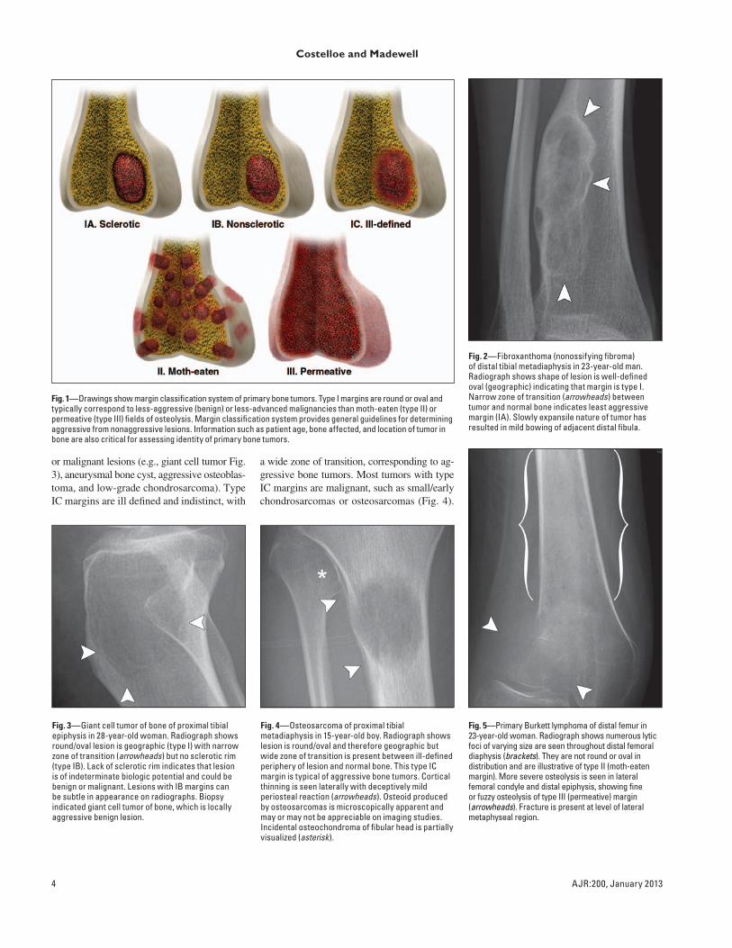
Fig. 4—Osteosarcoma of proximal tibial metadiaphysis in 15-year-old boy. Radiograph shows lesion is round/oval and therefore geographic but wide zone of transition is present between ill-defined periphery of lesion and normal bone. This type IC margin is typical of aggressive bone tumors. Cortical thinning is seen laterally with deceptively mild periosteal reaction (arrowheads). Osteoid produced by osteosarcomas is microscopically apparent and may or may not be appreciable on imaging studies. Incidental osteochondroma of fibular head is partially visualized (asterisk).
Fig. 5—Primary Burkett lymphoma of distal femur in 23-year-old woman. Radiograph shows numerous lytic foci of varying size are seen throughout distal femoral diaphysis (brackets). They are not round or oval in distribution and are illustrative of type II (moth-eaten margin). More severe osteolysis is seen in lateral femoral condyle and distal epiphysis, showing fine or fuzzy osteolysis of type III (permeative) margin (arrowheads). Fracture is present at level of lateral metaphyseal region.
Dow
nloa
ded
from
ww
w.a
jronl
ine.
org
by 1
25.1
61.1
78.1
64 o
n 05
/28/
16 fr
om IP
add
ress
125
.161
.178
.164
. Cop
yrig
ht A
RR
S. F
or p
erso
nal u
se o
nly;
all
right
s res
erve
d

AJR:200, January 2013 5
Radiography of Primary Bone Tumors
Aggressive benign lesions, such as giant cell tumors, can also show this appearance.
Type II and III margins are nongeographic and consist of ill-defined “fields” of bone de-struction. The type II margin is described as moth-eaten and is composed of numerous foci of osteolysis that vary in size and shape on a background of relatively intact cortex. The type III margin corresponds to the most highly ag-gressive appearance and is described as perme-
ative. The margin types can be intermingled. Margins are the interface of the tumor with the bone and therefore are typically the most sensi-tive radiographic indicator of lesion behavior. The osteolysis represented by these non-geo-graphic lucent areas is fine or fuzzy in ap-pearance (Fig. 5). Analysis of the margins is readily performed on radiographs, which are inexpensive, easily accessible, and provide a concise assessment of lesion behavior on a limited number of images.
Cortical ExpansionCortical expansion is most commonly seen
with benign tumors that grow slowly enough to allow the cortex to remain completely or partially intact. Although not all bone tumors expand the cortex, the degree of cortical ex-pansion, when present, is reflective of the growth rate of the lesion. With a number of notable exceptions, such as low-grade chon-drosarcoma (Fig. 6) and some metastases (e.g., renal cell and thyroid carcinoma), ma-lignancies are more likely to progress rapid-ly and destroy rather than expand the cortex. Lesions that produce mild cortical expansion are typically well marginated and show IA margins (e.g., fibrous cortical defect, fibrous dysplasia). Lesions that produce a larger de-gree of cortical expansion are more likely to predispose to pathologic fracture (e.g., non-
ossifying fibromas/fibroxanthomas, unicam-eral bone cyst; (Fig. 7). Lesions that exhib-it a marked degree of cortical expansion can produce severe local bone deformity or de-struction, even if benign. For example, an an-eurysmal bone cyst may grow across open physes (Fig. 8), predisposing the patient to limb length discrepancy, and giant cell tumor may destroy the articular surface of bone, leading to the need for arthroplasty.
Periosteal ReactionPeriosteal reaction can reflect the biologic
potential of tumors [6], but it is less specific than other radiographic signs. Highly aggres-sive tumors often result in interrupted or mul-tilaminar periosteal reactions, and the mass effect of the tumor can produce a triangular interface with the periosteum that is termed a “Codman’s triangle” (Fig. 9). Less-aggres-sive processes typically produce a unilaminar periosteal reaction, although high-grade sar-comas may produce unilaminar or no detect-able periosteal reaction.
Matrix and Tumor MineralizationOsteoid, chondroid, and fibrous lesions often
produce characteristic mineralization of ma-trix, which is a substance that is located in the extracellular space between tumor cells. Ma-trix is found in benign and malignant tumors
Fig. 6—Low-grade chondrosarcoma of proximal femur in 84-year-old woman. Low-grade sarcomas can mimic benign lesions and low-grade chondrosarcomas are among most potentially confusing bone tumors. Thickened cortex does not always equate with sclerotic rim. Unlike benign fibroxanthoma in Fig. 1, radiograph shows that entire circumference of this bone is enlarged by low-grade chondrosarcoma. Although large fibrous dysplasias can also enlarge entire circumference of bone, fibrous dysplasia will typically exhibit sclerotic rim with possible fibrous matrix mineralization. This chondrosarcoma has no sclerotic rim and exhibits cartilaginous matrix mineralization (see Figs. 11 and 12 regarding mineralized matrix). In addition to thickening of cortex (arrow), endosteal scalloping of cortex by tumor also thins areas of cortex (arrowhead). Thinning of large sections of cortex by large cartilaginous bone tumors can be indicative of malignancy.
Fig. 8—Aneurysmal bone cyst of proximal humerus in 17-year-old girl. Radiograph shows this large expansile lesion crosses physis into humeral head and also thin cortex, resulting in pathologic fracture (arrow). These features produced bowing deformity of bone. Physeal involvement can predispose to limb length discrepancy.
Fig. 7—Unicameral bone cyst of proximal humeral metaphysis in 16-year-old boy. Large lesions that expand bone without adequate cortical reinforcement can result in pathologic fracture. Radiograph shows this unicameral bone cyst is slightly expansile, thins cortex, and has resulted in fracture with subsequent callous formation (arrow).
Dow
nloa
ded
from
ww
w.a
jronl
ine.
org
by 1
25.1
61.1
78.1
64 o
n 05
/28/
16 fr
om IP
add
ress
125
.161
.178
.164
. Cop
yrig
ht A
RR
S. F
or p
erso
nal u
se o
nly;
all
right
s res
erve
d

6 AJR:200, January 2013
Fig. 12—Fibrous dysplasia of tibia in 30-year-old woman. Lesion shows spectrum of opacities. Radiograph shows mineralized fibrous matrix is dense and mature proximally (arrow), whereas it has ground-glass appearance distally (arrowheads).
A
Fig. 13—Fibrous cortical defect of proximal tibial metaphysis with fibrous dysplasia in distal diaphysis of 15-year-old girl.A, Radiograph shows small fibrous cortical defect is markedly eccentric because it is located in lateral cortex (arrowheads). Nonaggressive lesion shows sclerotic rim (IA margin). Fibrous dysplasia in distal diaphysis is centered in medullary cavity and expands entire circumference of bone. In some respects, this lesion resembles low-grade chondrosarcoma in Figure 6. Nevertheless, it shows ground-glass fibrous matrix and sclerotic distal rim (arrow).B, Radiograph obtained 3 years later shows fibrous cortical defect (arrowheads) has matured and become dense. Because of risk of pathologic fracture caused by cortical thinning, fibrous dysplasia was curetted and packed with dense bone graft, which has incorporated into surgical defect (arrow).
B
Fig. 11—This chondrosarcoma of calcaneus in 26-year-old male originated from sessile osteochondroma (not readily identified on radiograph). Radiograph shows arcs and rings of mineralized chondroid matrix (arrowhead) throughout lesion. Mineralized matrix is stippled posteriorly at periphery of lesion (arrow).Fig. 9—Osteosarcoma of distal femoral
metadiaphysis in 14-year-old boy. Radiograph shows multilaminar onionskin periosteal reaction anteriorly (white arrow). Interrupted periosteal reaction and Codman’s triangle (black arrow) are seen posteriorly and proximally, and hair-on-end or sunburst periosteal reaction (arrowheads) is seen posteriorly and distally. Although multilaminar periosteal reactions are commonly seen with Ewing sarcoma, each type of reaction can be seen in each type of tumor. Cortical and trabecular margins of this aggressive tumor are permeative (type III).
Fig. 10—Dedifferentiated parosteal osteosarcoma of posterior metaphysis of distal femur in 33-year-old man. Radiograph shows proximal aspect of lesion is dedifferentiated and shows fluffy or cloudlike osteoid matrix (arrowhead) within soft-tissue mass. Distal aspect of lesion is more typical of parosteal osteosarcoma and shows dense ivory osteoid (arrow).
Dow
nloa
ded
from
ww
w.a
jronl
ine.
org
by 1
25.1
61.1
78.1
64 o
n 05
/28/
16 fr
om IP
add
ress
125
.161
.178
.164
. Cop
yrig
ht A
RR
S. F
or p
erso
nal u
se o
nly;
all
right
s res
erve
d

AJR:200, January 2013 7
Radiography of Primary Bone Tumors
and therefore does not closely correlate to malignant potential but may be useful in iden-tifying the histologic type of the tumor [7].
Each type of matrix mineralization shows a spectrum of radiographic appearances. Oste-oid matrix may be fluffy and cloudlike or ivo-ry and dense (Fig. 10). Cartilaginous matrix may produce arcs and rings that form around globular cartilage lobules, or it may be stippled in appearance (Fig. 11). Fibrous lesions may show a slightly increased radiographic density, moderate ground-glass appearance, or dense mature mineralization that can be uniform or heterogeneous (Fig. 12). The gradual increase in mineralization with time is termed “matu-ration” and can be seen in tumors such as fi-brous dysplasia, nonossifying fibroma, fibrous cortical defect (Fig. 13), osteoid osteoma, and bone island [8]. Maturation is an indolent pro-cess and should not be confused with the rapid posttherapeutic response of aggressive lesions that have responded well to systemic or radi-ation therapy. Posttherapeutic mineralization may be most evident peripherally in lesions
Fig. 14—Osteosarcoma of proximal tibial metaphysis in 31-year-old woman after chemotherapy. Radiograph shows periphery of lesion has mineralized after therapy and could be confused with type IA margin (arrowheads). Nevertheless, focal areas of moth-eaten osteolysis remain visible centrally (arrow). Review of medical history is essential for proper interpretation of primary bone tumors.
such as plasmacytomas or unresectable giant cell tumors of bone. The rim may become so solidly mineralized that it can be confused with a IA margin. The margin classification system discussed in this article applies only to untreat-ed primary bone tumors (Fig. 14).
AdvantagesRadiography and CT are the two major
imaging modalities used for evaluating min-eralized structures. The main advantages of radiography over CT include affordability, accessibility, and a concise method by which to assess the lesion on a limited number of images. It can be argued that cross-sectional imaging, even with reformations, cannot re-produce the perceived quality of depth that results from collapsing a 3D structure onto a 2D image. The margin classification system described in this article is designed primar-ily for use with radiography.
PitfallsDeficits of radiography include anatomic
overlap that can obscure abnormalities and a limited capacity to evaluate soft tissue. Al-though mass effect may be detected on radio-graphs, they are limited for determining the degree of extraosseous tumor volume, rela-tionship of extraosseous tumor to surrounding structures, and extent of disease in the intact marrow cavity. MRI is the modality of choice for simultaneously evaluating these relation-ships [9–11]. Other considerations include lower sensitivity for the detection of mineral-ized matrix when compared with CT or for the detection of undisplaced fracture when com-pared with CT or MRI.
ConclusionRadiography is the single most helpful im-
aging modality when establishing the initial differential diagnosis of primary bone tumors. Evaluation of the margins is the greatest con-tributing factor to radiographic assessment of the biologic potential of the lesions. Other fea-tures, such as cortical expansion and periosteal reaction, provide additional clues as to whether immediate biopsy is indicated. Identification of mineralized matrix may help to noninvasively
identify histologic type. Although MRI and CT provide superior soft-tissue assessment and are free from structural overlap, the unique infor-mation afforded by radiography is optimal for the efficient formation of an initial differential diagnosis of primary bone tumors.
References 1. Berquist TH, Dalinka MK, Alazraki N, et al.
Bone tumors: American College of Radiology—ACR appropriateness criteria. Radiology 2000; 215(suppl):261–264
2. Costelloe CM, Rohren EM, Madewell JE, et al. Imaging bone metastases in breast cancer: tech-niques and recommendations for diagnosis. Lan-
cet Oncol 2009; 10:606–614 3. Lodwick GS, Wilson AJ, Farrell C, Virtama P,
Dittrich F. Determining growth rates of focal le-sions of bone from radiographs. Radiology 1980; 134:577–583
4. Oudenhoven LF, Dhondt E, Kahn S, et al. Accu-racy of radiography in grading and tissue-specific diagnosis: a study of 200 consecutive bone tumors of the hand. Skeletal Radiol 2006; 35:78–87
5. Madewell JE, Ragsdale BD, Sweet DE. Radio-logic and pathologic analysis of solitary bone le-sions. Part I. Internal margins. Radiol Clin North
Am 1981; 19:715–748 6. Ragsdale BD, Madewell JE, Sweet DE. Radio-
logic and pathologic analysis of solitary bone le-sions. Part II. Periosteal reactions. Radiol Clin
North Am 1981; 19:749–783 7. Sweet DE, Madewell JE, Ragsdale BD. Radio-
logic and pathologic analysis of solitary bone le-sions. Part III. Matrix patterns. Radiol Clin North
Am 1981; 19:785–814 8. Yanagawa T, Watanabe H, Shinozaki T, Ahmed
AR, Shirakura K, Takagishi K. The natural his-tory of disappearing bone tumours and tumour-like conditions. Clin Radiol 2001; 56:877–886
9. Aisen AM, Martel W, Braunstein EM, McMillin KI, Phillips WA, Kling TF. MRI and CT evalua-tion of primary bone and soft-tissue tumors. AJR 1986; 146:749–756
10. Tehranzadeh J, Mnaymneh W, Ghavam C, Moril-lo G, Murphy BJ. Comparison of CT and MR im-aging in musculoskeletal neoplasms. J Comput
Assist Tomogr 1989; 13:466–472 11. Zimmer WD, Berquist TH, McLeod RA, et al. Bone
tumors: magnetic resonance imaging versus com-puted tomography. Radiology 1985; 155:709–718
Dow
nloa
ded
from
ww
w.a
jronl
ine.
org
by 1
25.1
61.1
78.1
64 o
n 05
/28/
16 fr
om IP
add
ress
125
.161
.178
.164
. Cop
yrig
ht A
RR
S. F
or p
erso
nal u
se o
nly;
all
right
s res
erve
d