Initial experience of complete laparoscopic radical ...
11
TECHNICAL INNOVATIONS Open Access Initial experience of complete laparoscopic radical nephroureterectomy combined with transvesical laparoscopic excision of distal ureter in patients with upper urinary tract cancer Makito Miyake * , Nobutaka Nishimura, Katsuya Aoki, Chihiro Ohmori, Takuto Shimizu, Takuya Owari, Shunta Hori, Yosuke Morizawa, Daisuke Gotoh, Yasushi Nakai, Satoshi Anai, Kazumasa Torimoto, Nobumichi Tanaka and Kiyohide Fujimoto Abstract Background: Selecting the treatment procedure for cancer patients is a challenging task. We report our initial experience of complete laparoscopic radical nephroureterectomy (RNU) for patients with upper urinary tract urothelial cancer (UTUC). Methods: A total of four patients with UTUC underwent complete laparoscopic RNU combined with transvesical laparoscopic excision of the distal ureter using three 5-mm ports. Transvaginal specimen extraction was applied in female patients to reduce incisional pain and improve cosmesis. Peri-operative complications were evaluated using the Clavien-Dindo classification system. Postoperative pain was evaluated during hospitalization using a numeric pain rating scale (scales of 1 to 10). Patients who underwent retroperitoneal laparoscopic surgery combined with open excision of the distal ureter during the same period were included as a control group (conventional RNU, consisting of laparoscopic nephrectomy combined with open bladder cuff excision) for pain scale evaluation. Results: The novel surgery was successfully completed for all four patients (two males and two females). The mean pneumoperitoneum time for retroperitoneoscopic nephroureterectomy and specimen extraction was 174 min, while the mean pneumovesicum time for the ureteral orifice excision was 88 min. One male patient had bladder leakage at the suture site of the bladder wall, which lasted for 2 weeks. No patient experienced recurrent disease during the follow-up period (median, 10 months). Mild to moderate pain lasted for 5 or 6 days after RNU. A couple of days after surgery, the numeric pain rating scale of complete laparoscopic RNU and conventional RNU group reached its peak level at 3.0 ± 1.8 and 5.3 ± 2.8, respectively. There was no statistical difference in the degree of postoperative pain (P = 0.31). (Continued on next page) © The Author(s). 2020 Open Access This article is licensed under a Creative Commons Attribution 4.0 International License, which permits use, sharing, adaptation, distribution and reproduction in any medium or format, as long as you give appropriate credit to the original author(s) and the source, provide a link to the Creative Commons licence, and indicate if changes were made. The images or other third party material in this article are included in the article's Creative Commons licence, unless indicated otherwise in a credit line to the material. If material is not included in the article's Creative Commons licence and your intended use is not permitted by statutory regulation or exceeds the permitted use, you will need to obtain permission directly from the copyright holder. To view a copy of this licence, visit http://creativecommons.org/licenses/by/4.0/. The Creative Commons Public Domain Dedication waiver (http://creativecommons.org/publicdomain/zero/1.0/) applies to the data made available in this article, unless otherwise stated in a credit line to the data. * Correspondence: [email protected] Department of Urology, Nara Medical University, 840 Shijo-cho, Kashihara, Nara 634-8522, Japan Miyake et al. World Journal of Surgical Oncology (2020) 18:104 https://doi.org/10.1186/s12957-020-01872-1
Transcript of Initial experience of complete laparoscopic radical ...
TECHNICAL INNOVATIONS Open Access
Initial experience of complete laparoscopicradical nephroureterectomy combined withtransvesical laparoscopic excision of distalureter in patients with upper urinary tractcancerMakito Miyake* , Nobutaka Nishimura, Katsuya Aoki, Chihiro Ohmori, Takuto Shimizu, Takuya Owari, Shunta Hori,Yosuke Morizawa, Daisuke Gotoh, Yasushi Nakai, Satoshi Anai, Kazumasa Torimoto, Nobumichi Tanaka andKiyohide Fujimoto
Abstract
Background: Selecting the treatment procedure for cancer patients is a challenging task. We report our initialexperience of complete laparoscopic radical nephroureterectomy (RNU) for patients with upper urinary tracturothelial cancer (UTUC).
Methods: A total of four patients with UTUC underwent complete laparoscopic RNU combined with transvesicallaparoscopic excision of the distal ureter using three 5-mm ports. Transvaginal specimen extraction was applied infemale patients to reduce incisional pain and improve cosmesis. Peri-operative complications were evaluated usingthe Clavien-Dindo classification system. Postoperative pain was evaluated during hospitalization using a numericpain rating scale (scales of 1 to 10). Patients who underwent retroperitoneal laparoscopic surgery combined withopen excision of the distal ureter during the same period were included as a control group (conventional RNU,consisting of laparoscopic nephrectomy combined with open bladder cuff excision) for pain scale evaluation.
Results: The novel surgery was successfully completed for all four patients (two males and two females). The meanpneumoperitoneum time for retroperitoneoscopic nephroureterectomy and specimen extraction was 174 min,while the mean pneumovesicum time for the ureteral orifice excision was 88 min. One male patient had bladderleakage at the suture site of the bladder wall, which lasted for 2 weeks. No patient experienced recurrent diseaseduring the follow-up period (median, 10 months). Mild to moderate pain lasted for 5 or 6 days after RNU. A coupleof days after surgery, the numeric pain rating scale of complete laparoscopic RNU and conventional RNU groupreached its peak level at 3.0 ± 1.8 and 5.3 ± 2.8, respectively. There was no statistical difference in the degree ofpostoperative pain (P = 0.31).
(Continued on next page)
© The Author(s). 2020 Open Access This article is licensed under a Creative Commons Attribution 4.0 International License,which permits use, sharing, adaptation, distribution and reproduction in any medium or format, as long as you giveappropriate credit to the original author(s) and the source, provide a link to the Creative Commons licence, and indicate ifchanges were made. The images or other third party material in this article are included in the article's Creative Commonslicence, unless indicated otherwise in a credit line to the material. If material is not included in the article's Creative Commonslicence and your intended use is not permitted by statutory regulation or exceeds the permitted use, you will need to obtainpermission directly from the copyright holder. To view a copy of this licence, visit http://creativecommons.org/licenses/by/4.0/.The Creative Commons Public Domain Dedication waiver (http://creativecommons.org/publicdomain/zero/1.0/) applies to thedata made available in this article, unless otherwise stated in a credit line to the data.
* Correspondence: [email protected] of Urology, Nara Medical University, 840 Shijo-cho, Kashihara,Nara 634-8522, Japan
Miyake et al. World Journal of Surgical Oncology (2020) 18:104 https://doi.org/10.1186/s12957-020-01872-1

(Continued from previous page)
Conclusions: We described our initial experience and outcome of complete laparoscopic RNU for UTUC. Furtherexperience and research are required to determine whether this advanced laparoscopic technique yields betteroutcomes and has true clinical value.
Keywords: Upper urinary tract urothelial cancer, Transvesical laparoscopy, Complete laparoscopy, Pneumovesicum,Numeric pain rating scale
IntroductionSelecting the best treatment procedure for cancer patientsin the clinical practice is a challenging task. Radicalnephroureterectomy (RNU) with complete removal of thedistal ureter, including ureteral orifice (bladder cuff exci-sion), is the standard surgical treatment for upper urinarytract urothelial cancer (UTUC) [1, 2]. Although openRNU was commonly performed before 1990, minimallyinvasive laparoscopic surgery has rapidly evolved since1991, with the first reports of laparoscopic RNU being re-ported by Clayman et al. [3]. Despite the lack of well-designed prospective randomized control trials, meta-analysis-based comparisons between laparoscopic RNUand its open counterpart demonstrated that both offerequivalent outcomes in terms of oncological efficacy, peri-operative safety, and mortality [4, 5].To date, laparoscopic RNU has been broadly accepted
by urologists mainly due to reduced postoperative painand improved cosmetic results. Many urologists select acombination of excision of the kidney and upper ureter byretroperitoneoscopic approach and bladder cuff excisionby lower abdominal incision-open surgery. This combin-ation is not a complete laparoscopic surgery and thus isnot a minimally invasive method. The best method forcomplete laparoscopic RNU for patients with UTUC hasnot yet been established. The approaches for bladder cuffexcision are still controversial and include intravesical,extravesical, and transurethral incisions [6]. The extravesi-cal technique carries a potential risk of residual tumor atthe distal intramural part of the ureter, while the intravesi-cal technique carries the risk of tumor cell disseminationto the outside of the bladder.Here, we report the initial experience of complete laparo-
scopic nephroureterectomy with transvesical laparoscopicexcision of the distal ureter using three ports in patientswith UTUC. The transvesical laparoscopic approach is analternative to open surgery in children with vesicouret-eral reflux and bladder diverticulum [7, 8]. Transvaginalspecimen extraction was applied in the case of female pa-tients to reduce incisional pain and improve cosmesis.
Patients and methodsClinical dataThis research was approved by the ethics committee ofthe Nara Medical University, and all participants
provided informed consent (reference ID: 1256 and1719). The date of ethical approval was May 30, 2017,which is prior to the first surgery performed. We pro-spectively selected the patients and gave full informationregarding the novel surgical method. All patients under-went dynamic computed tomography (CT) or CT urog-raphy before surgery to determine tumor location andsize. The main exclusion criteria were (1) tumor of thedistal ureter, (2) advanced tumor detected by CT (sus-pected T3/T4 disease or node-positive disease), (3) con-traindications to laparoscopic surgery, or (4) concurrentbladder tumor.We conducted a review of four patients with UTUC
who underwent complete laparoscopic nephroureterect-omy with transvesical laparoscopic bladder cuff excisionbetween January 2018 and December 2019. All surgerieswere performed by a single laparoscopically experiencedsurgeon (M. Miyake). Peri-operative complications wereobjectively evaluated using the Clavien-Dindo classifica-tion system [9]. This system includes seven grades (I, II,IIIa, IIIb, IVa, IVb, and V). The degree of postoperativepain was evaluated every day during hospitalization andon the first outpatient visit day using a numeric pain rat-ing scale (NPRS), where 0 indicates no pain and 10 indi-cates the worst imaginable pain [10]. Four patients whounderwent conventional retroperitoneal laparoscopicsurgery combined with an open bladder cuff using alower abdominal midline incision, by the same surgeon(M. Miyake), during the same period, were included as acontrol group for pain scale evaluation.
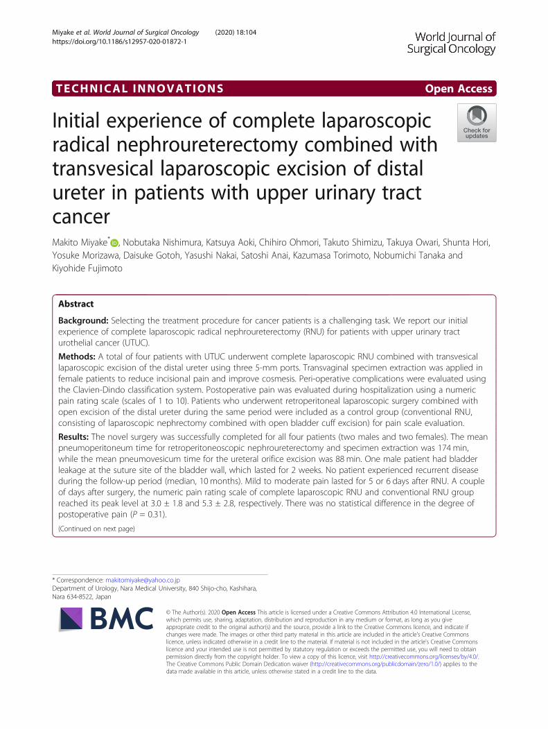
Surgical procedure for complete laparoscopic RNUAn operation video demonstrating the surgical proce-dures is given in Supplementary Video S1. A diagram ofeach step of the surgical procedure is depicted in Fig. 1.Under general anesthesia, each patient was placed in alateral decubitus position with the cancerous side up fora retroperitoneoscopic nephrectomy. The retroperitonealcavity was dilated with a retroperitoneal balloon andmaintained with 8 mmHg CO2 of insufflation pressure.We carried out conventional retroperitoneal approachwith four ports as shown in Fig. 2a: a flexible endoscope(camera) trocar, 12-mm trocar, 5-mm trocar, andauxiliary 5-mm trocar. The procedure includes thestandard nephrectomy using laparoscopic monopolar
Miyake et al. World Journal of Surgical Oncology (2020) 18:104 Page 2 of 11
scissors (e.g., AESCULAP® laparoscopic instruments), Liga-Sure™ Maryland jaw sealer (Covidien Japan, Tokyo, Japan),and Hem-o-lok® clips [11]. The renal artery was securedwith a size L clip, followed by clamping of the ureter at thedistal area with a size L clip or ML clip after stopping urineproduction in the kidney (Fig. 2b). Then, the renal vein wassecured with a size XL clip, and the kidney was completelyfreed. The adrenal gland was retained in all cases.
A 5-mm trocar was added to the ipsilateral pelvic area tofacilitate a wide operation space in the phase of dissection ofthe distal ureter (Fig. 2a, red triangle). The ureter was dis-sected under the common iliac artery to the bladder. Thedistal ureter was dissected maximally, and the urinary blad-der remained unclosed during this phase (Fig. 2c). Duringthe retroperitoneoscopic procedure, the ureter was not cutto enable complete en bloc removal of the kidney and ureter.
Fig. 1 Diagram of the surgical procedure
Miyake et al. World Journal of Surgical Oncology (2020) 18:104 Page 3 of 11

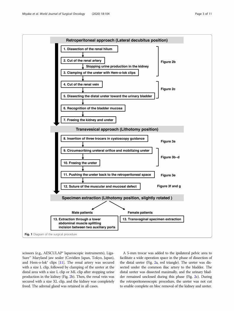
Next, the patient was changed to the lithotomy pos-ition for transvesical laparoscopic bladder cuff excision.The operation was restarted with cystoscopy and per-formed according to the procedure reported by Yeunget al. [12]. The surgeon stands on the patient’s left side.Under cystoscopic guidance, three 5-mm trocars (KiiAdvanced Fixation Sleeve; Applied Medical, RanchoSanta Margarita, CA, USA) were placed from the supra-pubic region into the bladder (Fig. 3a). Under 8 mmHgCO2 of pneumovesicum pressure, the ureterovesicaljunction and Waldeyer’s sheath were excised with a 3-mm laparoscopic monopolar scissors until the paravesi-cal adipose tissue was visible (Fig. 3b–d). After com-pletely mobilizing the ureter, the ureter was pushed backto the retroperitoneal space (Fig. 3e). Then, the musculardefect and mucosal defect in the ureteral hiatus were su-tured intravesically (Fig. 3f, g). Intravesical trocars were
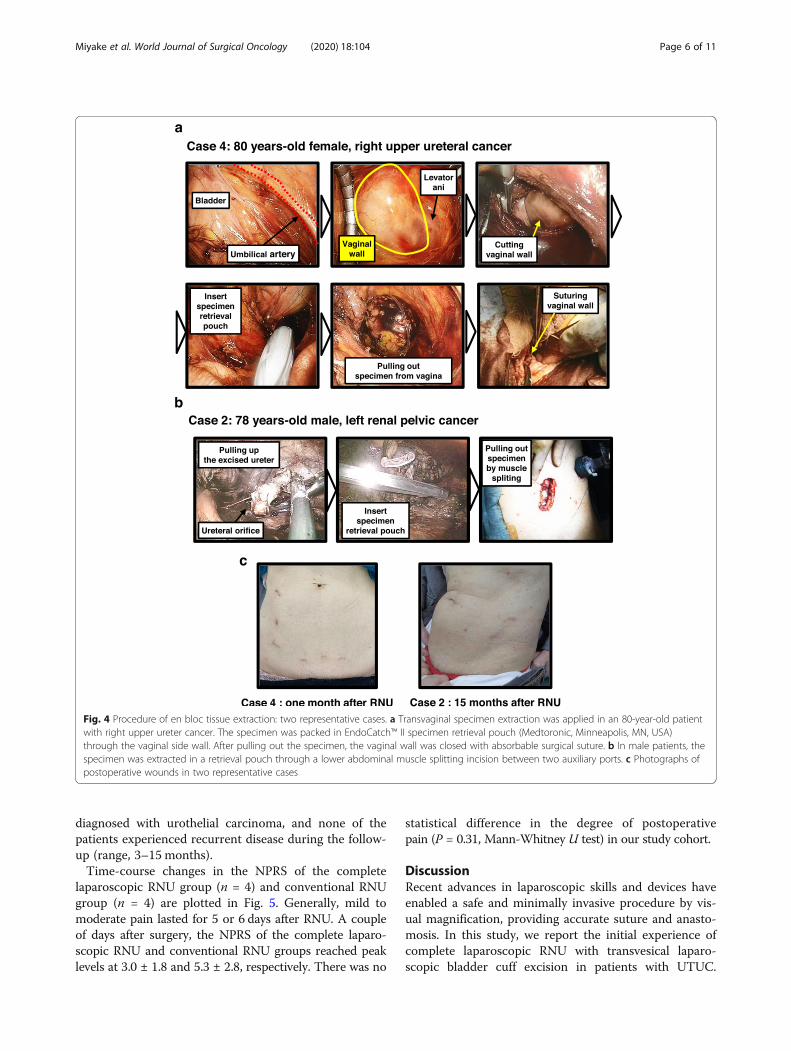
removed under endoscopic vision without suturing thebladder wall. Each port site entry wound was closed witha 4-0 PDSII monocryl suture (Ethicon, NJ, USA).In female patients, the specimen was extracted trans-
vaginally in a bag (Fig. 4a). In male patients, the speci-men was extracted in a bag through a lower abdominalmuscle splitting incision between the two auxiliary ports.A pelvic drain tube was placed through the pelvic auxil-iary port.
Control group undergoing conventional RNUConventional surgery consists of laparoscopic nephrec-tomy with open bladder cuff excision. In the retroperito-neoscopic phase, the ureter was freed to the bifurcationlevel of iliac vessels along the level of the lower pole ofthe kidney. Next, the patient was placed in the supineposition for open bladder cuff excision. An 8–10-cm
Fig. 2 Representative image (case 4 in Table 1) of a patient undergoing complete laparoscopic nephroureterectomy. a Trocar positions fornephroureterectomy of a right-side UTUC. The lower-level auxiliary port (red open triangle) is added to provide a wide surgical view of the pelvicarea. Suprapubic ports (black triangles) are used for transvesical laparoscopic bladder cuff excision. b The cut of the renal artery, clamping of theureter, and cut of the renal vein were performed with Hem-o-lok® clips.c Pulling up the proximal ureter to assist in dissecting the distal uretertoward the urinary bladder. When the junction of the ureter and bladder is exposed, the muscle layer is cut off, followed by recognition of thebladder mucosa
Miyake et al. World Journal of Surgical Oncology (2020) 18:104 Page 4 of 11
midline incision was made on the lower abdomen. Afterextracting the kidney from the body, pulling the ureterexposed the bladder, thus facilitating excision of thebladder cuff. The remaining junction of the ureter andbladder was cut off, and the bladder wall was suturedwith a 3-0 Vicryl suture. A pelvic drain tube was placed,and the incision was closed.
Follow-up after the RNUA urinary catheter was left for approximately a week andremoved after cystography. Cystoscopy and chest/abdo-men/pelvis CT scans were performed approximatelyevery 3 months for 2 years after the RNU, every 6months from year 2 to 5, and annually thereafter.
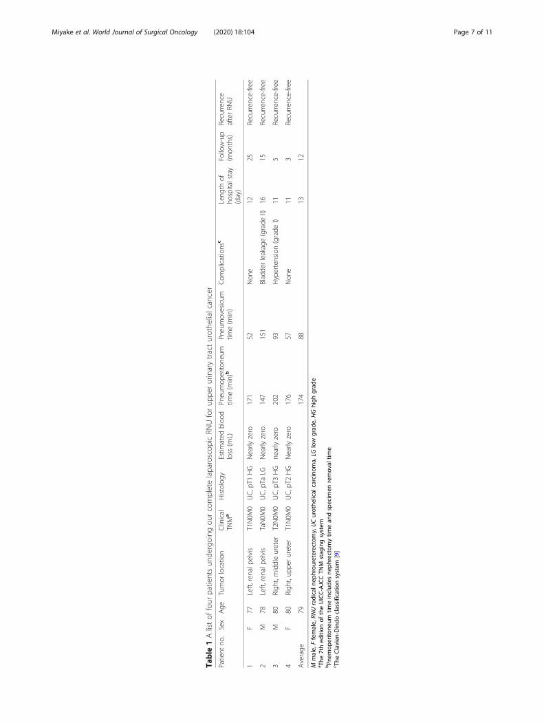
ResultsFour patients undergoing complete laparoscopic RNUand four patients undergoing conventional RNU duringthe same period were included in this study. All four pa-tients successfully underwent complete laparoscopic
RNU. Clinicopathological background and surgical infor-mation are shown in Tables 1 and 2, respectively. Therewere no statistically significant differences in age, lengthof hospital stay, and follow-up duration between the twogroups.Pneumoperitoneum time was longer in the complete
laparoscopic RNU group compared to the conventionalRNU group (174min vs 99 min; P = 0.029). The pneu-movesicum time of the second patient was longer thanthat of the other three patients because he had a historyof radiotherapy for localized prostate cancer. His bladderwall did not have elasticity, and so it took longer and itwas more difficult to suture the bladder wall. Cystora-diography was performed on postoperative day 6 or 7,and the urethral catheters were removed. No leakage atthe transvesical port was observed. However, in the sec-ond patient, bladder leakage at the suture site of thebladder wall lasted for 2 weeks. Repeat cystoradiographydemonstrated resolution of the leak, followed by removalof the urethral catheter. All patients were pathologically
Fig. 3 Procedure of transvesical laparoscopic bladder cuff excision. a Postoperative wound for the suprapubic three ports is shown (black arrows).The bladder was distended with 400–500mL of saline. A total of three 5-mm trocars were placed at the bladder dome and on both sides of thelateral wall of the distended bladder under cystoscopy guidance. A 3-0 monofilament traction suture is passed percutaneously through thebladder walls to prevent the bladder wall from falling away from the abdominal wall. b A 4-cm-long segment of an 8Fr pediatric feeding tube isinserted into the ipsilateral ureter to facilitate ureteral mobilization and dissection and secured by a 5-zero monofilament suture. c Circumscribingureteral orifice and mobilizing ureter using fine 3-mm endoscopic scissors. d Traction on the ureteric catheter and cut of fibrovascular tissuesurrounding the ureter to free it. e The ureter is pushed back to the retroperitoneal space. f The muscular defect and mucosal defect in theureteral hiatus are sutured intravesically using 5-zero absorbable monofilament sutures, usually with an extracorporeal knot-tying technique. gComplete suturing of the bladder wall defect
Miyake et al. World Journal of Surgical Oncology (2020) 18:104 Page 5 of 11
diagnosed with urothelial carcinoma, and none of thepatients experienced recurrent disease during the follow-up (range, 3–15 months).Time-course changes in the NPRS of the complete
laparoscopic RNU group (n = 4) and conventional RNUgroup (n = 4) are plotted in Fig. 5. Generally, mild tomoderate pain lasted for 5 or 6 days after RNU. A coupleof days after surgery, the NPRS of the complete laparo-scopic RNU and conventional RNU groups reached peaklevels at 3.0 ± 1.8 and 5.3 ± 2.8, respectively. There was no
statistical difference in the degree of postoperativepain (P = 0.31, Mann-Whitney U test) in our study cohort.
DiscussionRecent advances in laparoscopic skills and devices haveenabled a safe and minimally invasive procedure by vis-ual magnification, providing accurate suture and anasto-mosis. In this study, we report the initial experience ofcomplete laparoscopic RNU with transvesical laparo-scopic bladder cuff excision in patients with UTUC.
Fig. 4 Procedure of en bloc tissue extraction: two representative cases. a Transvaginal specimen extraction was applied in an 80-year-old patientwith right upper ureter cancer. The specimen was packed in EndoCatch™ II specimen retrieval pouch (Medtoronic, Minneapolis, MN, USA)through the vaginal side wall. After pulling out the specimen, the vaginal wall was closed with absorbable surgical suture. b In male patients, thespecimen was extracted in a retrieval pouch through a lower abdominal muscle splitting incision between two auxiliary ports. c Photographs ofpostoperative wounds in two representative cases
Miyake et al. World Journal of Surgical Oncology (2020) 18:104 Page 6 of 11
Table
1Alistof
four
patientsun
dergoing
ourcompletelaparoscop
icRN
Uforup
perurinarytracturothe
lialcancer
Patient
no.
Sex
Age
Tumor
locatio
nClinical
TNM
aHistology
Estim
ated
bloo
dloss
(mL)
Pneumop
erito
neum
time(m
in)b
Pneumovesicum
time(m
in)
Com
plications
cLeng
thof
hospitalstay
(day)
Follow-up
(mon
ths)
Recurren
ceafterRN
U
1F
77Left,
renalp
elvis
T1N0M
0UC,p
T1HG
Nearly
zero
171
52Non
e12
25Recurren
ce-free
2M
78Left,
renalp
elvis
TaN0M
0UC,p
TaLG
Nearly
zero
147
151
Bladde
rleakage(grade
II)16
15Recurren
ce-free
3M
80Righ
t,middleureter
T2N0M
0UC,p
T3HG
nearlyzero
202
93Hypertension(grade
I)11
5Recurren
ce-free
4F
80Righ
t,up
perureter
T1N0M
0UC,p
T2HG
Nearly
zero
176
57Non
e11
3Recurren
ce-free
Average
79174
8813
12
Mmale,
Ffemale,
RNUradicaln
ephrou
reterectom
y,UCurothe
lical
carcinom
a,LG
low
grad
e,HGhigh
grad
ea The
7thed
ition
oftheUICC-AJCCTN
Mstag
ingsystem
bPn
emop
erito
neum
timeinclud
esne
phrectom
ytim
ean
dspecim
enremov
altim
ec The
Clavien
-Dindo
classificationsystem
[9]
Miyake et al. World Journal of Surgical Oncology (2020) 18:104 Page 7 of 11

Table
2Alistof
controlp
atientsun
dergoing
conven
tionalR
NUforup
perurinarytracturothe
lialcancer
Patient
no.Sex
Age
Tumor
locatio
nClinical
TNM
aHistology
Estim
ated
bloo
dloss
(mL)
Pneumop
erito
neum
time(m
in)
Ope
nsurgerytim
e(m
in)
Com
plications
bLeng
thof
hospitalstay
(day)
Follow-up
(mon
ths)
Recurren
ceafterRN
U
1F
76Righ
t,renalp
elvis
T2N0M
0UC,p
T3HG
150
98112
Non
e8
24Intravesicalrecurren
ce(5mon
ths,Ta
LG)
2M
72Left,m
iddleureter
T1N0M
0UC,p
T2HG
Nearly
zero
72117
Non
e12
12Recurren
ce-free
3M
84Left,ren
alpe
lvis
T2N0M
0UC,p
T3HG
Nearly
zero
7887
Hypertension(grade
I)9
3Recurren
ce-free
4F
81Righ
t,renalp
elvis
T1N0M
0UC,p
T2HG
185
146
85Non
e11
18Recurren
ce-free
Average
7899
100
1014
Mmale,
Ffemale,
RNUradicaln
ephrou
reterectom
y,UCurothe
lical
carcinom
a,LG
low
grad
e,HGhigh
grad
ea The
7thed
ition
oftheUICC-AJCCTN
Mstag
ingsystem
bTh
eClavien
-Dindo
classificationsystem
[9]
Miyake et al. World Journal of Surgical Oncology (2020) 18:104 Page 8 of 11

Several techniques, especially in excision of the distal ur-eter, have been reported to date [13–16]. However, weshould carefully consider the application of laparoscopicsurgery in patients with UTUC, which is known as oneof the most aggressive malignancies. During the follow-up, no patients experienced any recurrent disease in-cluding intravesical, extravesical, distant, and vaginal
recurrence. As to the learning curve, apparent progresswas not observed in terms of operation time. Longerfollow-up and larger sample size are mandatory to clarifythe real benefit of this surgical method.Maximal effort should be made to ameliorate postop-
erative pain, shorten hospitalization stay, prevent peri-operative complications, and maintain cosmesis. Based
Fig. 5 Time-course change of pain scale of complete laparoscopic nephroureterectomy and conventional nephroureterectomy. Postoperativepain was assessed using a numerical pain rating scale (NPRS) every day during the hospitalization and at the first outpatient visit day (indicatedwith “day 30”). The data included four patients undergoing complete laparoscopic radical nephroureterectomy (RNU), as shown in Table 1, andfour patients undergoing the conventional retroperitoneal laparoscopic nephrectomy combined with an open bladder cuff by the same surgeon,during the same period, as shown in Table 2
Miyake et al. World Journal of Surgical Oncology (2020) 18:104 Page 9 of 11

on the initial experience of our four patients, we consid-ered our novel trial of complete laparoscopic RNU as anacceptable method (Table 1). The intravesical space islimited and can be continually obscured by effluxingurine. To counter this problem, the retroperitoneal pro-cedure, including ligation of the renal artery and ureter,proceeds the transvesical laparoscopic procedure in ouroperation strategy. This can provide an additional pos-sible advantage that occluding the ureter results in de-creased risk of dissemination of tumor cells from theupper urinary tract to the intravesical cavity and parave-sical space. The biggest concern with transurethral exci-sion of the distal ureter, the so-called pluck technique, isthe risk of spillage of urine containing tumor cells [17].In a case report by Sotelo et al. [16], they performed a
transvesical single-site surgery enabling full-thickness in-cision of the bladder around the ureteral orifice,followed by a water-tight suture of the bladder wall de-fect. Transvesical single-site surgery requires insertion ofa special device into the bladder and subsequent sutureof the bladder anterior wall. In contrast to single-sitesurgery, the out method does not require suture of thebladder port site because intravesical trocars are thin(only 5 mm thickness). The technique for dissection ofthe intramural ureter and intracorporeal suturing seemsto be challenging and time-consuming, even for a lap-aroscopically experienced surgeon. In our second patient(Table 1), the intravesical laparoscopic procedure tookno less than 2.5 h, mainly due to a history of radiother-apy for prostate cancer, which could induce ischemicconditions around the bladder trigon and decreased elas-ticity of the bladder wall. Eventually, he suffered fromprolonged urethral catheterization. Based on this experi-ence, a history of pelvic radiotherapy may be a potentialcontraindication for transvesical laparoscopic bladdercuff excision.We applied three 5-mm trocars to the intravesical
space, which may lead to a risk of port-site recurrence.No patients experienced port-site recurrence during thefollow-up period in our cohort. Other options for laparo-scopic excision of the distal ureter and bladder cuff are asfollows: thermo-sealing system [18], cold excision of thebladder cuff with intracorporeal suturing [14, 15], modifi-cation with a bulldog clamp [19], purse-string technique[20], and robot-assisted surgery [21]. As pointed out previ-ously, some extravesical approaches have the potential riskof failing to resect the intramural ureter. Our peumovesi-coscopic bladder cuff excision ensures complete resectionof the ureteral orifice, intramural ureter, and ureterovesi-cal junction. This advantage strengthens our novelmethod. The transvesical laparoscopic bladder cuff tech-nique has not yet been standardized.We applied transvaginal specimen extraction in two
female patients (cases. 1 and 4 in Table 1). We believe
that the potential benefits of our method over conven-tional RNU could include decreased incisional pain, re-duction in postoperative pain, and improved cosmesis.Since the first preclinical model of natural orifice trans-luminal endoscopic surgery (NOTES) nephrectomy inthe urologic field [22], this approach has attracted muchinterest from urologists. Transvaginal NOTES has beenintroduced into donor nephrectomy and nephrectomyfor renal infection, renal calculus, and renal malignancies[23–26]. In the literature, the multiaccess alternative ofpure NOTES is often mentioned as “Hybrid NOTES,” inwhich standard laparoscopic nephrectomy is performedand the vagina is only utilized for specimen extraction.This study has several limitations. Given that the deci-
sion between complete laparoscopic RNU or conven-tional RNU was made by physicians rather than throughrandomization, there is a potential selection bias. Sec-ond, the sample size was small, with only four patientsin the complete laparoscopic RNU group and four pa-tients in the conventional RNU group, and the follow-uptime was short. We could not make conclusion regard-ing oncological outcome and benefit. However, we be-lieve the potential of functional outcomes includingdecreased postoperative pain and improved cosmesis.This report can emphasize on the safety and feasibilityof the new surgical method, which lead to our prospect-ive multi-institutional trial in the near future. Third, noassessment was conducted regarding patient-reportedoutcomes such as health-related quality of life and sex-ual satisfaction questionnaire pre- and post-operatively,especially in female patients.
ConclusionWe reviewed our experience with complete laparoscopicRNU for patients with UTUC. Although there have beenmany reports on the feasibility of complete laparoscopicsurgery, few studies have reported on transvesical three-port bladder cuff excision. Further experience and re-search are mandatory to determine whether this ad-vanced laparoscopic technique yields better outcomesand has true clinical value.
Supplementary informationSupplementary information accompanies this paper at https://doi.org/10.1186/s12957-020-01872-1.
Additional file 1: Supplementary Video S1: Operation videodemonstrating the surgical procedures.
AbbreviationsCT: Computed tomography; NOTES: Natural orifice translumenal endoscopicsurgery; NPRS: Numeric pain rating scale; RNU: Radical nephroureterectomy;UTUC: Upper urinary tract urothelial cancer
Miyake et al. World Journal of Surgical Oncology (2020) 18:104 Page 10 of 11

AcknowledgementsWe would like to thank Editage (www.editage.jp) for English languageediting.
Authors’ contributionsConceptualization, M.M. and K.A. Methodology, N.N., C.O., T.S., and T.O.Investigation, S.H., Y.M., and D.G. Data curation, M.M. and S.A.Writing—original draft preparation, M.M. Writing—review and editing, K.A.,K.T., and K.F. Visualization, M.M. Supervision, N.T. The author(s) read andapproved the final manuscript.
FundingNo specific funding in this study
Availability of data and materialsAll data generated or analyzed during this study are included in thispublished article and reference articles.
Ethics approval and consent to participateThis study was approved by the ethics committee of the Nara MedicalUniversity (reference ID: 1256 and 1966). Informed consent to participate inthe study was obtained from all participants. The study was conducted incompliance with the study’s protocol and in accordance with the provisionsof the Declaration of Helsinki (2013).
Consent for publicationConsent for publication of individual patients was obtained from theparticipants.
Competing interestsThe authors declare that they have no competing interests.
Received: 16 March 2020 Accepted: 6 May 2020
References1. Miyake M, Tatsumi Y, Fujimoto K, et al. Changes in oncological
outcomes after radical nephroureterectomy in patients with upperurinary tract urothelial carcinoma treated in the last two decades: aretrospective analysis based on a multicenter collaborative study. Jpn JClin Oncol. 2016;46:1148–55.
2. Roupret M, Babjuk M, Comperat E, et al. European Association of UrologyGuidelines on upper urinary tract urothelial cell carcinoma: 2015 update. EurUrol. 2015;68:868–79.
3. Clayman RV, Kavoussi LR, Figenshau RS, et al. Laparoscopicnephroureterectomy: initial clinical case report. J Laparoendosc Surg. 1991;1:343–9.
4. Ni S, Tao W, Chen Q, et al. Laparoscopic versus open nephroureterectomyfor the treatment of upper urinary tract urothelial carcinoma: a systematicreview and cumulative analysis of comparative studies. Eur Urol. 2012;61:1142–53.
5. Zhang X, Wang K, Ma J, et al. Total laparoscopic nephroureterectomy forupper urinary tract urothelial carcinoma under a single surgical position.World J Surg Oncol. 2019;17:65.
6. Li WM, Shen JT, Li CC, et al. Oncologic outcomes following three differentapproaches to the distal ureter and bladder cuff in nephroureterectomy forprimary upper urinary tract urothelial carcinoma. Eur Urol. 2010;57:963–9.
7. Casale P, Kojima Y. Robotic-assisted laparoscopic surgery in pediatricurology: an update. Scand J Surg. 2009;98:110–9.
8. Badawy H, Eid A, Hassouna M, et al. Pneumovesicoscopic diverticulectomyin children and adolescents: is open surgery still indicated? J Pediatr Urol.2008;4:146–9.
9. Dindo D, Demartines N, Clavien PA. Classification of surgical complications:a new proposal with evaluation in a cohort of 6336 patients and results of asurvey. Ann Surg. 2004;240:205–13.
10. Shigematsu-Locatelli M, Kawano T, Kitamura S, et al. Does preoperativepatient's estimated acceptable pain affect the satisfaction withpostoperative pain management? JA Clin Rep. 2017;3:5.
11. Casale P, Pomara G, Simone M, et al. Hem-o-lok clips to control both theartery and the vein during laparoscopic nephrectomy: personal experienceand review of the literature. J Endourol. 2007;21:915–8.
12. Yeung CK, Sihoe JD, Borzi PA. Endoscopic cross-trigonal ureteralreimplantation under carbon dioxide bladder insufflation: a noveltechnique. J Endourol. 2005;19:295–9.
13. Hora M, Eret V, Urge T, et al. Complete laparoscopic nephroureterectomywith intravesical lockable clip. Cent European J Urol. 2012;65:75–9.
14. Hemal AK, Kumar A, Gupta NP, et al. Retroperitoneal nephroureterectomywith excision of cuff of the bladder for upper urinary tract transitional cellcarcinoma: comparison of laparoscopic and open surgery with long-termfollow-up. World J Urol. 2008;26:381–6.
15. Hattori R, Yoshino Y, Komatsu T, et al. Pure laparoscopic complete excisionof distal ureter with a bladder cuff for upper tract urothelial carcinoma.World J Urol. 2009;27:253–8.
16. Sotelo R, Ramírez D, Carmona O, et al. A novel technique for distalureterectomy and bladder cuff excision. Actas Urol Esp. 2011;35:168–74.
17. Srirangam SJ, van Cleynenbreugel B, van Poppel H. Laparoscopicnephroureterectomy: the distal ureteral dilemma. Adv Urol. 2009, 2019;316807.
18. Hora M, Stránský P, Eret V, et al. Complete laparoscopicnephroureterectomy with thermo-sealing system. CEJU. 2010;63:77–81.
19. Cho HJ, Kim SJ, Yoon BI, et al. A novel bulldog clamp technique formanagement of a distal ureter and bladder cuff during laparoscopicnephroureterectomy. J Endourol. 2010;24:1719–20.
20. Shoma AM. Purse-string technique for laparoscopic excision of a bladdermucosal cuff in patients with transitional cell carcinoma of the upperurinary tract: initial report with intermediate follow-up. BJU Int. 2009;104:1505–9.
21. Park SY, Neony W, Ham WS, et al. Initial experience of roboticnephroureterectomy: a hybrid-port technique. BJU Int. 2009;104:1718–21.
22. Gettman MT, Lotan Y, Napper CA, et al. Transvaginal laparoscopicnephrectomy: development and feasibility in the porcine model. Urology.2002;59:446–50.
23. Georgiopoulos I, Kallidonis P, Kyriazis I, et al. Hybrid transvaginalnephrectomy: development of our technique. Urology. 2014;84:99–104.
24. Guner Can M, Ozcan P, Hatipoglu S, et al. Laparoscopic-assisted live donornephrectomy: a comparison of conventional and transvaginal routes forkidney extraction. Ann Transplant. 2015;20:634–8.
25. Xue Y, Zou X, Zhang G, et al. Transvaginal natural orifice transluminalendoscopic nephrectomy in a series of 63 cases: stepwise transition fromhybrid to pure NOTES. Eur Urol. 2015;68:302–10.
26. Sener TE, Sahin B, Fichera M, et al. Does vaginal wall surgical trauma duringhybrid transvaginal NOTES nephrectomy have traumatic effects on sexualfunctions? A prospective study. J Invest Surg. 2020;6:1–8. https://doi.org/10.1080/08941939.2019.1710627 [Epub ahead of print].
Publisher’s NoteSpringer Nature remains neutral with regard to jurisdictional claims inpublished maps and institutional affiliations.
Miyake et al. World Journal of Surgical Oncology (2020) 18:104 Page 11 of 11