Inhibition of the Na /dicarboxylate cotransporter by anthranilic acid...
35
MOL #35352 1 Inhibition of the Na + /dicarboxylate cotransporter by anthranilic acid derivatives. Ana M. Pajor and Kathleen M. Randolph Department of Biochemistry and Molecular Biology, University of Texas Medical Branch, Galveston, TX 77555-0645 Molecular Pharmacology Fast Forward. Published on August 22, 2007 as doi:10.1124/mol.107.035352 Copyright 2007 by the American Society for Pharmacology and Experimental Therapeutics. This article has not been copyedited and formatted. The final version may differ from this version. Molecular Pharmacology Fast Forward. Published on August 22, 2007 as DOI: 10.1124/mol.107.035352 at ASPET Journals on March 13, 2020 molpharm.aspetjournals.org Downloaded from
Transcript of Inhibition of the Na /dicarboxylate cotransporter by anthranilic acid...

MOL #35352
11
Inhibition of the Na+/dicarboxylate cotransporter
by anthranilic acid derivatives.
Ana M. Pajor and Kathleen M. Randolph
Department of Biochemistry and Molecular Biology,
University of Texas Medical Branch, Galveston, TX 77555-0645
Molecular Pharmacology Fast Forward. Published on August 22, 2007 as doi:10.1124/mol.107.035352
Copyright 2007 by the American Society for Pharmacology and Experimental Therapeutics.
This article has not been copyedited and formatted. The final version may differ from this version.Molecular Pharmacology Fast Forward. Published on August 22, 2007 as DOI: 10.1124/mol.107.035352
at ASPE
T Journals on M
arch 13, 2020m
olpharm.aspetjournals.org
Dow
nloaded from

MOL #35352
22
Running title: Inhibitor binding in NaDC1
Corresponding author: Ana M. Pajor, Ph.D., Department of Biochemistry and Molecular Biology, University of Texas Medical Branch, Galveston, TX 77555-0645. tel. 409-772-3434, fax 409-772-5102, email: [email protected]
Document statistics:
Number of: text pages 12
tables 1
figures 11
references 26
Number of words in:
Abstract 216
Introduction 474
Discussion 976
Nonstandard abbreviations: AACOCF3, arachidonyl trifluoromethyl ketone;
ACA, N-(p-amylcinnamoyl) anthranilic acid; BPB, p-bromophenacyl bromide; CUBS cell line,
HRPE cells stably transfected with hNaDC1; DMSO, dimethylsulfoxide; Fmoc-anthranilic acid,
N-(9-fluorenylmethoxycarbonyl)-anthranilic acid; HRPE, cell line derived from human
embryonic retinal pigment epithelium; NaDC1, Na+/dicarboxylate cotransporter 1; ONO-RS-
082, 2-(p-amylcinnamoyl)amino-4-chloro benzoic acid; Tranilast, N-(3',4'-
dimethoxycinnamoyl) anthranilic acid
This article has not been copyedited and formatted. The final version may differ from this version.Molecular Pharmacology Fast Forward. Published on August 22, 2007 as DOI: 10.1124/mol.107.035352
at ASPE
T Journals on M
arch 13, 2020m
olpharm.aspetjournals.org
Dow
nloaded from

MOL #35352
33
ABSTRACT
The Na+/dicarboxylate cotransporter NaDC1 absorbs citric acid cycle intermediates from the
lumen of the small intestine and kidney proximal tubule. No effective inhibitor has been
identified yet, although previous studies showed that the non-steroidal anti-inflammatory drug,
flufenamate, inhibits the human (h) NaDC1 with an IC50 of 2 mM. In the present study, we have
tested compounds related in structure to flufenamate, all anthranilic acid derivatives, as potential
inhibitors of hNaDC1. We find that N-(p-amylcinnamoyl) anthranilic acid (ACA) and 2-(p-
amylcinnamoyl)amino-4-chloro benzoic acid (ONO-RS-082) are the most potent inhibitors with
IC50 values below 15 µM, followed by N-(9-fluorenylmethoxycarbonyl)-anthranilic acid (Fmoc-
anthranilic acid) with an IC50 ~80 µM. The effects of ACA on NaDC1 are not mediated through
a change in transporter protein abundance on the plasma membrane and appear to be independent
of its effect on phospholipase A2 activity. ACA acts as a slow inhibitor of NaDC1, with slow
onset and slow reversibility. Both uptake activity and efflux are inhibited by ACA. Other
Na+/dicarboxylate transporters from the SLC13 family, including hNaDC3 and rbNaDC1, were
also inhibited by ACA, ONO-RS-082 and Fmoc-anthranilic acid, whereas the Na+/citrate
transporter (hNaCT) is much less sensitive to these compounds. The endogenous sodium-
dependent succinate transport in Caco-2 cells is also inhibited by ACA. In conclusion, ACA
and ONO-RS-082 represent promising lead compounds for the development of specific
inhibitors of the Na+/dicarboxylate cotransporters.
Introduction
The metabolic intermediates of the citric acid cycle, including succinate, citrate and α-
ketoglutarate, are transported across the plasma membranes of epithelial cells by the
This article has not been copyedited and formatted. The final version may differ from this version.Molecular Pharmacology Fast Forward. Published on August 22, 2007 as DOI: 10.1124/mol.107.035352
at ASPE
T Journals on M
arch 13, 2020m
olpharm.aspetjournals.org
Dow
nloaded from

MOL #35352
44
Na+/dicarboxylate cotransporters from the SLC13 family (Pajor, 2006). SLC13 family members
include the low affinity dicarboxylate transporter, NaDC1, the high affinity Na+/dicarboxylate
cotransporter, NaDC3, and the Na+/citrate transporter, NaCT. NaDC1 is found on the apical
membranes of renal proximal tubule and small intestinal cells. In the kidney, NaDC1 helps to
regulate the concentration of citrate in the urine, which is an important determinant in the
development of kidney stones, as ~ 50% of patients with kidney stones have hypocitraturia (Pak,
1991). NaDC1 also participates in organic anion secretion in the kidney by providing
dicarboxylate substrates, particularly α-ketoglutarate, to the organic anion transporter of the
basolateral membrane (Dantzler and Evans, 1996). NaDC1 may play an indirect role in
regulating blood pressure, as succinate and α-ketoglutarate activate specific G-protein-coupled
receptors in the renal proximal tubule (He et al., 2004). Finally, mutations in NaDC1 homologs
found in the digestive tract of Drosophila and C. elegans lead to an extension of lifespan (Rogina
et al., 2000;Fei et al., 2004) suggesting a possible role for NaDC1 in metabolic control or aging.
To date, no high-affinity inhibitors have been identified for any member of the SLC13
family. Our previous studies identified the non-steroidal anti-inflammatory drug flufenamate as
an inhibitor of hNaDC1. The IC50 was 2 mM using succinate as a substrate and 0.5 mM using
methylsuccinate (Pajor and Sun, 1996;Smith et al., 2003). A high-affinity inhibitor would be
very useful for in vitro studies of NaDC1, to quantitate the transporter in the membrane and to
characterize mutant transporters with alterations in substrate translocation but not binding. An
effective and selective inhibitor could help to identify physiological roles of NaDC1.
Furthermore, an inhibitor may lead to a potential drug if inhibition of NaDC1 activity in humans
is linked to aging or metabolism.
This article has not been copyedited and formatted. The final version may differ from this version.Molecular Pharmacology Fast Forward. Published on August 22, 2007 as DOI: 10.1124/mol.107.035352
at ASPE
T Journals on M
arch 13, 2020m
olpharm.aspetjournals.org
Dow
nloaded from
MOL #35352
55
In the present study, we have tested anthranilic acid derivatives related in structure to
flufenamate as potential inhibitors of hNaDC1. We find that N-(p-amylcinnamoyl) anthranilic
acid (ACA) and 2-(p-amylcinnamoyl)amino-4-chloro benzoic acid (ONO-RS-082) are the most
potent inhibitors with IC50 values below 15 µM, followed by N-(9-fluorenylmethoxycarbonyl)-
anthranilic acid (Fmoc-anthranilic acid) with an IC50 ~80 µM. The effects of ACA on hNaDC1
are not mediated through a change in transporter protein abundance on the plasma membrane and
appear to be independent of its effect on phospholipase A2 (PLA2) activity. ACA acts as a slow
inhibitor of NaDC1, with slow onset and slow reversibility. Both transport uptake and efflux are
inhibited by ACA. Other Na+/dicarboxylate transporters from the SLC13 family, including
hNaDC3 and rbNaDC1, were also inhibited by ACA, ONO-RS-082 and Fmoc-anthranilic acid,
whereas the Na+/citrate transporter hNaCT is much less sensitive to these compounds. The
endogenous sodium-dependent succinate transport activity in the human colon carcinoma cell
line, Caco-2, is also sensitive to inhibition by ACA. In conclusion, ACA and ONO-RS-082 are
promising lead compounds for the development of specific inhibitors of the Na+/dicarboxylate
cotransporters.
This article has not been copyedited and formatted. The final version may differ from this version.Molecular Pharmacology Fast Forward. Published on August 22, 2007 as DOI: 10.1124/mol.107.035352
at ASPE
T Journals on M
arch 13, 2020m
olpharm.aspetjournals.org
Dow
nloaded from
MOL #35352
66
Materials and Methods
Chemicals. The structures of the compounds used in this study are shown in Fig. 1 and the
names are listed in Table 1. ACA, ONO-RS-082 and Tranilast were purchased from Biomol
Research Laboratories, all other chemicals were from Sigma-Aldrich.
Cell culture. The CUBS cell line consisting of HRPE cells (derived from human retinal
pigment epithelium) stably transfected with the human NaDC1 cDNA (Smith et al., 2003) was a
generous gift of Dr. Chari Smith (GlaxoSmithKline). Control cells were non-transfected HRPE
cells. Cells were cultured in Modified Eagle’s Medium containing Earle’s salts, Glutamax I and
25 mM HEPES (Gibco-BRL), supplemented with 10% heat-inactivated fetal calf serum
(Hyclone), 100 units/ml penicillin and 100 µg/ml streptomycin and 700 µg/ml Geneticin (G418;
Gibco-BRL) at 37oC in 5% CO2. For uptake experiments, cells were plated on white-sided 24-
well plastic Visiplates (PerkinElmer/Wallac) at a density of 2.5 x 10 5 cells per well (24-well
plate) in the same medium with 2.5 mM buytrate added. Control HRPE cells were plated at the
same density but without G418 or butyrate in the medium. Uptake assays were done in confluent
monolayers approximately 36 hours after plating.
For transient transfections with hNaDC3 and hNaCT, HRPE cells were plated at 1.2 x 10⁵
cells per well and each well of cells was transfected with 1.8 µl Fugene 6 (Roche) and 0.6 µg
plasmid DNA (9:3 ratio) (Pajor and Randolph, 2005). Transport assays were done 48 hours after
transfections.
Succinate uptake assay. Transport assays at room temperature were as described
previously (Pajor and Randolph, 2005). The sodium buffer contained in mM: 120 NaCl, 5 KCl,
1.2 MgCl2, 1.2 CaCl2, 5 D-glucose, 25 HEPES, pH adjusted to 7.4 with 1 M Tris. Choline
This article has not been copyedited and formatted. The final version may differ from this version.Molecular Pharmacology Fast Forward. Published on August 22, 2007 as DOI: 10.1124/mol.107.035352
at ASPE
T Journals on M
arch 13, 2020m
olpharm.aspetjournals.org
Dow
nloaded from

MOL #35352
77
buffer contained 120 mM choline chloride in place of NaCl. For the assays, each well was
washed twice with 1 ml sodium buffer, then pre-incubated with inhibitor in sodium buffer for 15
min, followed by two washes with 1 ml each sodium buffer. Inhibitors were prepared fresh each
day as DMSO stocks, with a final volume of DMSO of less than 1%. Cells were preincubated
with inhibitors 15 min (unless otherwise specified in the figure legends). Transport solution
(0.25 ml/well) consisting of [14C]succinate (10 or 100 µM final concentration) in sodium buffer,
with or without added inhibitor, was then added to the cells. After 30 minutes, the uptake
measurements were stopped and radioactivity removed with four 1 ml washes of choline buffer.
After the last wash was removed, each well of cells was dissolved in 0.5 ml scintillation cocktail,
OptiPhase Supermix (Wallac/Perkin Elmer). The plates were sealed with plastic plate covers
and counted directly in a Microbeta Trilux 1450 plate scintillation counter (Wallac/Perkin
Elmer). For experiments involving CUBS cells, background corrections were made based on
uptake activities in control HRPE cells. For experiments involving transfected cells, uptake
activities in vector-transfected cells were subtracted from activities in NaDC plasmid-transfected
cells. There was no difference in the background counts obtained with vector-transfected or non-
transfected cells (results not shown).
For calculation of Ki values from Dixon plots, inhibition of transport activity was measured
at multiple inhibitor and substrate concentrations. The data were analyzed by linear regression
and the x-axis value corresponding to the intersection of the lines was taken as -Ki (Segel, 1975).
To determine IC50 values (the inhibitor concentration resulting in 50% inhibition), the transport
of 10 µM [14C]succinate was measured in the presence of increasing concentrations of inhibitor
and the resulting activity (v%, as a percentage of control) was analyzed by non-linear regression
using v%= IC50/([I] + IC50) * 100, where [I] represents the inhibitor concentration. Statistical
This article has not been copyedited and formatted. The final version may differ from this version.Molecular Pharmacology Fast Forward. Published on August 22, 2007 as DOI: 10.1124/mol.107.035352
at ASPE
T Journals on M
arch 13, 2020m
olpharm.aspetjournals.org
Dow
nloaded from
MOL #35352
88
analysis with One-way Anova or Student’s t-test was done using the Sigma Stat program
(Jandel). Each experimental point was the mean of four replicate wells, and each experiment
was repeated at least two or three times (different plating or transfection experiments).
Efflux assays. Efflux assays in CUBS cells (Smith et al., 2003) were done using the non-
metabolizable substrate, [3H]methylsuccinate (a gift of Dr. Chari Smith, GlaxoSmithKline). In
preliminary experiments (not shown) we found that the cells required at least 30 minutes of
incubation with substrate for preloading, after which the intracellular radioactivity reached a
plateau. Therefore, the cells were preloaded by incubating with 10 or 100 µM
[3H]methylsuccinate in Na+ buffer for 30 minutes. The cells were then washed four times with
choline buffer to remove extracellular radioactivity, followed by incubation in sodium buffer for
one minute to allow efflux of radiolabeled substrate. To stop the efflux, each well was washed
four times with choline buffer and 1% SDS was added to solubilize the cells. The samples were
transferred to scintillation vials (since the Wallac Plate counter does not measure 3H accurately),
combined with scintillation cocktail and counted on a Packard Tri-Carb 2100TR scintillation
counter.
Caco-2 cells. The Caco-2 cell line (human colon adenocarcinoma) was obtained from the
American Type Culture Collection (Rockville, MD). The cells were cultured in DMEM with 4.5
g/l glucose (GIBCO-Invitrogen) supplemented with 10% heat-inactivated fetal calf serum
(Hyclone), 100 units/ml penicillin G, 100 ug/ml streptomycin, 1% (v/v) sodium pyruvate and 1%
(v/v) nonessential amino acids (Sigma). Cells were seeded in 24 well collagen-coated plates,
maintained with medium changes every other day and then used for transport assays 21 days
after reaching confluency. Sodium-dependent transport activity was the difference between
transport measured in sodium and choline-containing buffers.
This article has not been copyedited and formatted. The final version may differ from this version.Molecular Pharmacology Fast Forward. Published on August 22, 2007 as DOI: 10.1124/mol.107.035352
at ASPE
T Journals on M
arch 13, 2020m
olpharm.aspetjournals.org
Dow
nloaded from
MOL #35352
99
Cell surface biotinylation. Cell surface biotinylations of NaDC1 were done using a
membrane impermeant reagent, Sulfo-NHS-LC-biotin, as described previously (Pajor and
Randolph, 2005). Biotinylated proteins were separated by SDS-PAGE in 7.5% acrylamide and
NaDC1 was identified using antibodies (1:1000 dilution).
Results
Initial inhibitor screen. The time course of [14C]succinate uptake into the CUBS cell line
(stably transfected HRPE cells expressing the human Na+/dicarboxylate cotransporter, hNaDC1)
is linear through 45 min (Fig. 2). Therefore, 30 min uptake activities were used in subsequent
experiments as estimates of initial rates. Succinate uptake activity in control non-transfected
HRPE cells was very low (Fig. 2). The transport of succinate in choline buffer in the CUBS cell
line was the same as in non-transfected HRPE cells (results not shown).
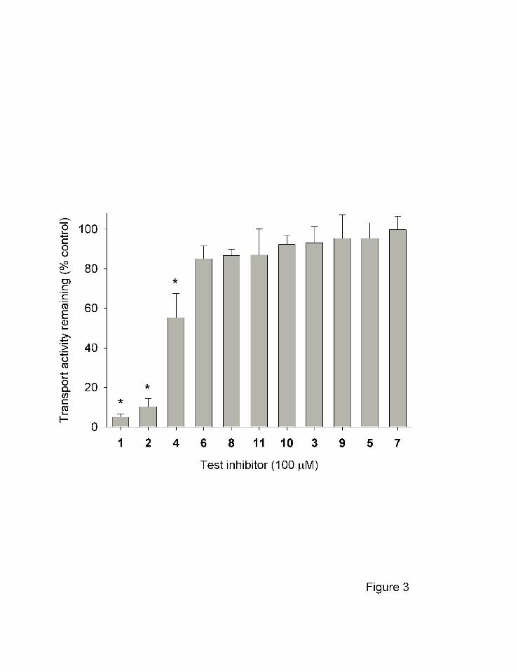
Several commercially-available anthranilic acid derivatives were tested as potential
inhibitors of NaDC1 (Fig. 3). CUBS cells expressing hNaDC1 were incubated with test
inhibitors, each at 100 µM concentration, and the effect on succinate transport was measured.
We found that ACA (compound 1) and the related compound ONO-RS-082 (compound 2)
produced greater than 80% inhibition of succinate transport. Incubation with N-Fmoc-
anthranilic acid (compound 4) resulted in approximately 45% inhibition. None of the other
compounds produced significant inhibition at 100 µM concentration. There was also no
inhibition of succinate transport by anthranilic acid, the parent compound (not shown). The lack
This article has not been copyedited and formatted. The final version may differ from this version.Molecular Pharmacology Fast Forward. Published on August 22, 2007 as DOI: 10.1124/mol.107.035352
at ASPE
T Journals on M
arch 13, 2020m
olpharm.aspetjournals.org
Dow
nloaded from
MOL #35352
1010
of inhibition by 100 µM flufenamate was consistent with the apparent IC50 value of 2 mM in
hNaDC1 expressed in Xenopus oocytes (Pajor and Sun, 1996).
Kinetics of inhibition. The mechanism of inhibition of NaDC1 by ACA was examined
using Dixon plots (Fig. 4). The lines intersected at the x-axis, suggesting that the mechanism of
inhibition is non-competitive (Segel, 1975). In the experiment shown in Fig. 4, the Ki was 12.5
µM, and in four experiments the Ki was 15.3 ± 3.3 µM (mean +/- SEM). We also compared the
IC50 values for the three inhibitory compounds, ACA, ONO-RS-082 and Fmoc-anthranilic acid,
measured at a single substrate concentration (10 µM succinate). The IC50 values were 10.6 ± 2.4
µM (ACA), 9.2 ± 0.3 µM (ONO-RS-082) and 85.3 ± 13.3 µM (Fmoc-anthranilic acid), n=3
(results not shown).
Cell surface biotinylation. One possible explanation for the decrease in transport activity
after ACA treatment is a decrease in the amount of transporter protein at the plasma membrane.
This was tested by cell surface biotinylation using the membrane impermeant reagent, Sulfo-
NHS-LC-biotin. Cells treated with ACA did not show any evidence of internalization of
hNaDC1 protein compared with controls incubated with buffer for the same incubation period
(Fig. 5). Therefore, the decreased activity seen after incubation with ACA is not due to
internalization of the transporter.
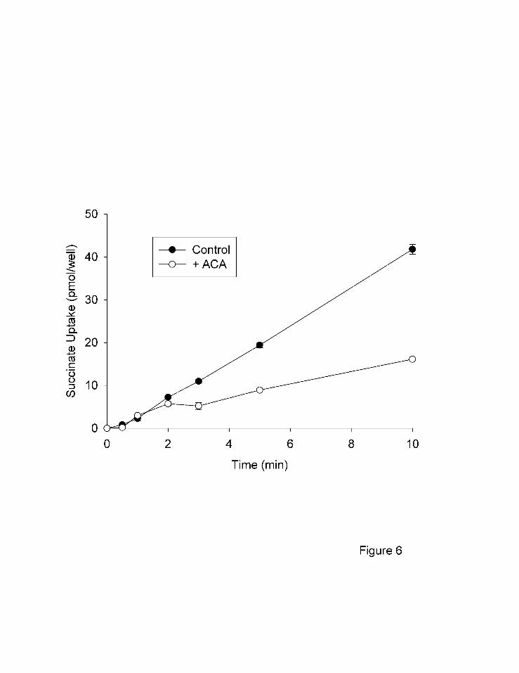
Time courses of ACA inhibition. Several alternative explanations could account for the
apparent non-competitive inhibition of hNaDC1 by ACA. For example, ACA could act as an
irreversible inhibitor or a slow, reversible inhibitor, both of which would produce apparent non-
competitive inhibition (Segel, 1975; Morrison, 1982). In time course experiments, the inhibition
by ACA did not occur immediately and the inhibition was not evident until after 2 minutes of
incubation (Fig. 6). A longer time course (Fig. 7A) shows that the inhibition by ACA requires
This article has not been copyedited and formatted. The final version may differ from this version.Molecular Pharmacology Fast Forward. Published on August 22, 2007 as DOI: 10.1124/mol.107.035352
at ASPE
T Journals on M
arch 13, 2020m
olpharm.aspetjournals.org
Dow
nloaded from
MOL #35352
1111
at least 15 to 30 minutes of preincubation. The inhibition also appeared to be reversible,
although very slow. In one hour, the activity of hNaDC1 recovered by approximately 20% (Fig.
7B). The inhibition by ACA was not dependent on cations, since the extent of inhibition was
the same in sodium or choline buffer. Transport activity remaining after pretreatment with 30
µM ACA was 66 ± 5% in sodium buffer compared with 71 ± 3.5% in choline buffer (n=3
experiments).
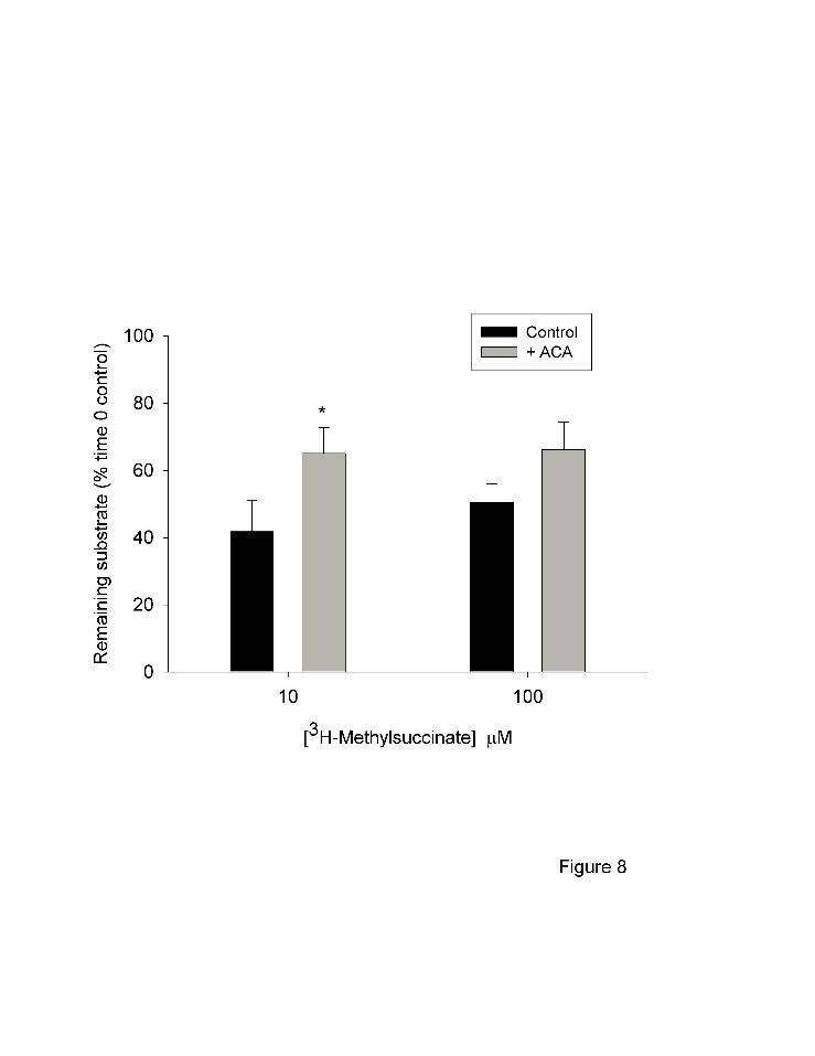
Efflux experiments. We next tested the efflux of the non-metabolizable substrate,
[3H]methylsuccinate, in the CUBS cell line. The cells were pre-loaded with radiolabled substrate
in sodium buffer for 30 minutes. The extracellular radioactivity was washed away and the efflux
was measured for 1 minute in sodium buffer with or without 100 µM ACA. As shown in Fig. 8,
approximately 40-50% of the [3H]methylsuccinate remained in the cells after 1 min compared to
controls at time 0. The addition of 100 µM ACA resulted in an increased retention of
[3H]methylsuccinate during the efflux period (Fig. 8), indicating that efflux as well as influx is
inhibited by ACA.
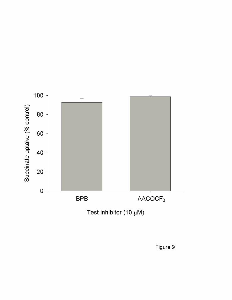
Phospholipase A2 inhibitors. Since ACA is known to be a non-specific inhibitor of
phospholipase A2 (PLA2) (Konrad et al., 1992), we wanted to rule out the possibility that the
effects of ACA on NaDC1 are mediated indirectly by inhibition of PLA2. Two other inhibitors
of PLA2, structurally unrelated to ACA, were tested but they did not inhibit succinate transport
by hNaDC1 (Fig. 9). p-Bromophenacyl bromide (BPB) inhibits secreted PLA2 by binding
covalently to a histidine residue (Mayer and Marshall, 1993). A previous study showed that
BPB at 5 or 10 µM inhibits PLA2 activity in cultured cells without producing toxicity (Liu and
Levy, 1997). AACOCF3 (arachidonyl trifluoromethyl ketone) is a slow-onset inhibitor of the
calcium-dependent cytosolic PLA2 (Street et al., 1993). Both compounds were tested after 15
This article has not been copyedited and formatted. The final version may differ from this version.Molecular Pharmacology Fast Forward. Published on August 22, 2007 as DOI: 10.1124/mol.107.035352
at ASPE
T Journals on M
arch 13, 2020m
olpharm.aspetjournals.org
Dow
nloaded from
MOL #35352
1212
min preincubation time. The results suggest that the effects of ACA on hNaDC1 are distinct from
its inhibition of PLA2.
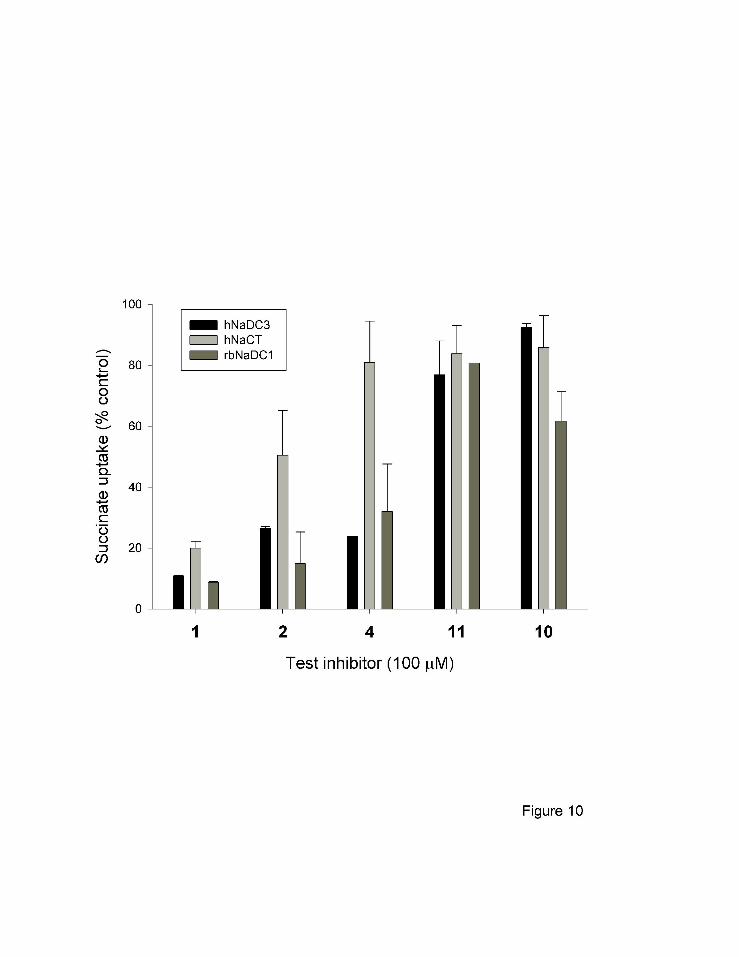
Species and isoform differences in specificity. We also examined the effects of the test
inhibitors on transport activity of other SLC13 members, the high affinity Na+/dicarboxylate
cotransporter, hNaDC3 (Wang et al., 2000), the Na+/citrate cotransporter, hNaCT (Inoue et al.,
2002), and the rabbit (rb) NaDC1 (Pajor and Sun, 1996). The activity of hNaCT was tested
using citrate as a substrate, whereas the substrate for hNaDC3 and rbNaDC1 was succinate. As
shown in Fig. 10, the three transporters were sensitive to inhibition by some of the anthranilic
acid derivatives. All three transporters were very sensitive to inhibition by ACA (compound 1),
but in general, hNaCT was less sensitive to ONO-RS-082 (compound 2) and Fmoc-anthranilic
acid (compound 4) compared with the other transporters. The rbNaDC1 was inhibited by
flufenamate by ~40% (compound 10), whereas hNaDC3 and hNaCT were not affected by
flufenamate. There was no inhibition of the three transporters by the other compounds
(compound 11 shown in Fig. 10, other compounds not shown).
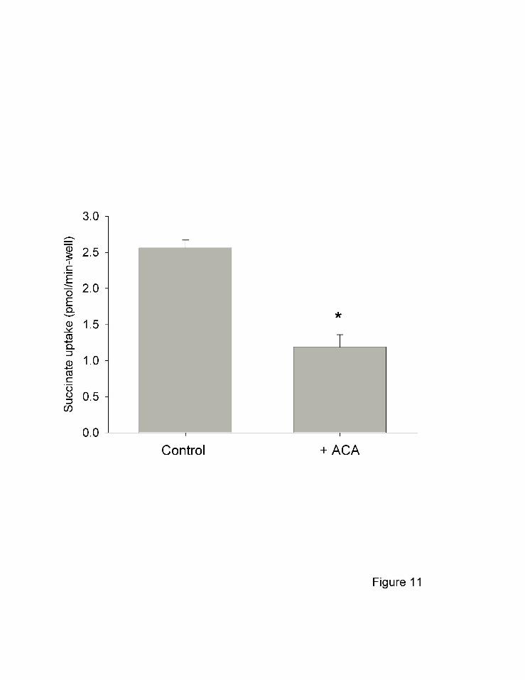
Inhibition of endogenous transporter. The human colon carcinoma cell line, Caco-2, is
often used as a model of the small intestine. This cell line expresses sodium-dependent transport
of succinate that is predominantly mediated by NaDC1, with some contribution by NaCT
(Weerachayaphorn and Pajor, 2005). As shown in Fig. 11, the endogenous succinate transport
activity in Caco-2 cells was inhibited more than 50% by treatment with ACA. Therefore, ACA
inhibits both transfected recombinant NaDC1 and the endogenous NaDC1 found in Caco-2 cells.
This article has not been copyedited and formatted. The final version may differ from this version.Molecular Pharmacology Fast Forward. Published on August 22, 2007 as DOI: 10.1124/mol.107.035352
at ASPE
T Journals on M
arch 13, 2020m
olpharm.aspetjournals.org
Dow
nloaded from
MOL #35352
1313
Discussion
The main finding of this study is that anthranilic acid derivatives, including ACA, ONO-
RS-082, and Fmoc-anthranilic acid, are inhibitors of the Na+/dicarboxylate cotransporters from
the SLC13 family. ACA and ONO-RS-082 inhibit transport with IC50 values less than 15 µM
and thus represent the highest affinity inhibitors of hNaDC1 to date. The effects of ACA are not
mediated through internalization of the transporter and appear to be distinct from the effects of
ACA on phospholipase A2. The preferred structure for inhibition of hNaDC1 appears to require
a lipophilic moiety attached to the anthranilic acid moiety of the compound, since Fmoc-
anthranilic acid (compound 4) was a more effective inhibitor compared with Tranilast
(compound 3) or the compounds with smaller or more polar substituents.
Flufenamate was identified in previous studies as a low affinity inhibitor of both low affinity
(NaDC1) and high affinity (NaDC3) Na+/dicarboxylate cotransporters. There are species
differences in sensitivity to flufenamate, with rbNaDC1 being most sensitive, IC50 250 µM
(Pajor and Sun, 1996), followed by flounder NaDC3 with an IC50 ~1mM (Burckhardt et al.,
2004), and lastly hNaDC1, IC50 2 mM (Pajor and Sun, 1996). All of the experiments were done
using Xenopus oocytes as an expression system and the cells were not preincubated with
inhibitor. In the present study, the IC50 for flufenamate in rbNaDC1 was ~ 100 µM, and the cells
were preincubated with inhibitor before the assay, which may account for the apparent difference
in affinity. Inhibition of the flounder NaDC3 by flufenamate appears to be mediated, at least in
part, by an increased K+ conductance that is associated with the transporter, since it was not seen
in control oocytes (Burckhardt et al., 2004). It is possible that flufenamate activates a channel
activity of the fNaDC3 protein, and several members of the SLC13 family have both channel and
transport activity (Oshiro and Pajor, 2005).
This article has not been copyedited and formatted. The final version may differ from this version.Molecular Pharmacology Fast Forward. Published on August 22, 2007 as DOI: 10.1124/mol.107.035352
at ASPE
T Journals on M
arch 13, 2020m
olpharm.aspetjournals.org
Dow
nloaded from
MOL #35352
14
The onset of inhibition of hNaDC1 by ACA was not evident until after at least 2 min
incubation time, consistent with the properties of a slow-binding inhibitor. This is the first report
that ACA behaves as a slow-binding inhibitor. Slow-binding inhibitors are reversible,
competitive inhibitors but the rate of reaching equilibrium is slow relative to a classical inhibitor
(Morrison, 1982). Slow inhibition may occur if the formation of the enzyme-inhibitor complex
occurs more slowly than the production of reaction product (in the case of a transporter, the
“product” of the reaction is the appearance of substrate on the inside of the cell). Alternatively,
the enzyme and inhibitor complex forms quickly but there is a slow isomerization step to an
inactive complex. Slow inhibitors tend to be useful drugs because of their slow reversibility and
the gradual accumulation of the inactive enzyme-inhibitor complex. Examples of drugs that are
slow inhibitors include non-steroidal anti-inflammatory drugs (NSAIDS), such as indomethacin,
that inhibit prostaglandin H synthase I by slow, reversible isomerization to a second enzyme-
inhibitor complex (Callan et al., 1996). The arachidonic acid analog, AACOCF3, is a slow and
tight-binding inhibitor of the cytosolic phospholipase A2 (Street et al., 1993).
Our current working hypothesis for the effects of the inhibitors identified in the present
study is that they act directly on hNaDC1 to inhibit transport. Other hypotheses could also
explain the results but they can be ruled out. For example, ACA could inhibit transport by
affecting protein targeting. Both ACA (IC50 8 µM) and ONO-RS-082 (IC50 6 µM) have been
shown to disrupt Golgi trafficking (de Figueiredo et al., 1999). However, the cell surface
abundance of hNaDC1 was not affected by incubation with ACA. A second possible
explanation is that ACA inhibits transport indirectly by affecting the membrane potential or Na+
chemical gradient, which would affect hNaDC1 since it is electrogenic (Yao and Pajor, 2000).
This article has not been copyedited and formatted. The final version may differ from this version.Molecular Pharmacology Fast Forward. Published on August 22, 2007 as DOI: 10.1124/mol.107.035352
at ASPE
T Journals on M
arch 13, 2020m
olpharm.aspetjournals.org
Dow
nloaded from
MOL #35352
15
ONO-RS-082 has been shown to inhibit the H+/K+-ATPase in stomach with an IC50 of 3.5 µM,
and the same study also reported that ONO-RS-082 has a protonophore effect (Sugita et al.,
2005). ACA is known to directly inhibit transient receptor potential (TRP) cation channels with
an IC50 of less than 4 µM (Kraft et al., 2006). However, changes in membrane potential or
cation concentrations would not explain the time delay in the inhibition of hNaDC1 by ACA, nor
would it explain the different sensitivities of NaDC1 homologs, all of which are electrogenic.
The hypothesis that ACA inhibition is mediated indirectly through its inhibition of PLA2 is
less likely than a direct effect of ACA on hNaDC1. There was no inhibition of hNaDC1 activity
by two structurally unrelated PLA2 inhibitors, BPB and AACOCF3. A compound that has not
been reported to have PLA2 inhibitory activity, Fmoc-anthranilic acid (compound 4), also
inhibited transport with an IC50 of about 80 µM. Although flufenamate has been shown to
inhibit PLA2 (Franson et al., 1980), only the rabbit and not the human NaDC1 was inhibited by
flufenamate. Furthermore, meclofenamate (compound 11) is also a PLA2 inhibitor with an IC50
~ 1 µM at 1 mM Ca2+ (Franson et al., 1980), yet it had no effect on any of the transporters in this
study.
In conclusion, we have identified several inhibitors of the Na+/dicarboxylate cotransporters,
with micromolar affinities, that represent the highest affinity inhibitors of the SLC13 family to
date. The most potent inhibitors of hNaDC1 were ACA, ONO-RS-082 and Fmoc-anthranilic
acid. There were some differences in sensitivity to inhibition by anthranilic acid derivatives
among the NaDC1 homologs tested (hNaDC3, rbNaDC1 and hNaCT), although the selectivity of
the compounds was relatively low. The endogenous sodium-dependent succinate transport
activity in the human colon carcinoma cell line, Caco-2, is also sensitive to inhibition by ACA.
This article has not been copyedited and formatted. The final version may differ from this version.Molecular Pharmacology Fast Forward. Published on August 22, 2007 as DOI: 10.1124/mol.107.035352
at ASPE
T Journals on M
arch 13, 2020m
olpharm.aspetjournals.org
Dow
nloaded from

MOL #35352
16
The kinetics of ACA inhibition of hNaDC1 were consistent with slow inhibition, and both
uptake and efflux were affected. Our data indicate that ACA inhibits the activity of hNaDC1
independently of PLA2 inhibition. However, the lack of specificity of ACA and ONO-RS-082
with NaDC orthologs and the effects on other targets including PLA2, TRP cation channels
(Kraft et al., 2006) and the stomach H+/K+-ATPase (Sugita et al., 2005) would limit the use of
these compounds as NaDC1 inhibitors. Therefore, the structures of the anthranilic acid
derivatives will serve as a starting point in drug design targeting hNaDC1.
Acknowledgements. We thank Dr. Chari D. Smith, GlaxoSmithKline, for the generous
donation of the CUBS cell line and [3H]methylsuccinate, and for many helpful discussions. We
also thank Jittima Weerachayaphorn for advice on Caco-2 cell cultures and transport assays.
Footnote. This work was supported by National Institutes of Health grant DK46269.
This article has not been copyedited and formatted. The final version may differ from this version.Molecular Pharmacology Fast Forward. Published on August 22, 2007 as DOI: 10.1124/mol.107.035352
at ASPE
T Journals on M
arch 13, 2020m
olpharm.aspetjournals.org
Dow
nloaded from

MOL #35352
17
References
Burckhardt BC, Lorenz J, Burckhardt G and Steffgen J (2004) Interactions of benzylpenicillin
and non-steroidal anti-inflammatory drugs with the sodium-dependent dicarboxylate
transporter NaDC-3. Cell Physiol Biochem 14:415-424.
Callan OH, So O Y and Swinney D C (1996) The kinetic factors that determine the affinity and
selectivity for slow binding inhibition of human prostaglandin H synthase 1 and 2 by
indomethacin and flurbiprofen. J Biol Chem 271:3548-3554.
Dantzler WH and Evans K K (1996) Effect of αKG in lumen on PAH Transport by isolated
perfused proximal tubules. Am J Physiol 271:F521-F526.
de Figueiredo P, Polizotto R S, Drecktrah D and Brown W J (1999) Membrane tubule-mediated
reassembly and maintenance of the golgi complex is disrupted by phospholipase A2
antagonists. Mol Biol Cell 10:1763-1782.
Fei YJ, Liu J C, Inoue K, Zhuang L, Miyake K, Miyauchi S and Ganapathy V (2004) Relevance
of NAC-2, an Na+-coupled citrate transporter, to life span, body size and fat content in
Caenorhabditis elegans. Biochem J 379:191-198.
Franson RC, Eisen D, Jesse R and Lanni C (1980) Inhibition of Highly purified mammalian
phospholipases A2 by non-steroidal anti-inflammatory agents. Modulation by calcium ions.
Biochem J 186:633-636.
He W, Miao F J P, Lin D C H, Schwander R T, Wang Z, Gao J, Chen J.L., Tian H and Ling L
(2004) Citric acid cycle intermediates as ligands for orphan G-protein-coupled receptors.
Nature 429:188-193.
This article has not been copyedited and formatted. The final version may differ from this version.Molecular Pharmacology Fast Forward. Published on August 22, 2007 as DOI: 10.1124/mol.107.035352
at ASPE
T Journals on M
arch 13, 2020m
olpharm.aspetjournals.org
Dow
nloaded from

MOL #35352
18
Inoue K, Zhuang L and Ganapathy V (2002) Human Na+ -coupled citrate transporter: primary
structure, genomic organization, and transport function. Biochem Biophys Res Commun
299:465-471.
Konrad RJ, Jolly Y C, Major C and Wolf B A (1992) Inhibition of phospholipase A2 and insulin
secretion in pancreatic islets. Biochim Biophys Acta 1135:215-220.
Kraft R, Grimm C, Frenzel H and Harteneck C (2006) Inhibition of TRPM2 cation channels by
N-(p-amylcinnamoyl)anthranilic Acid. Br J Pharmacol 148:264-273.
Liu Y and Levy R (1997) Phospholipase A2 has a role in proliferation but not in differentiation
of HL-60 cells. Biochim Biophys Acta 1355:270-280.
Mayer RJ and Marshall L A (1993) New insights on mammalian phospholipase A2(s);
comparison of arachidonoyl-selective and -nonselective enzymes. FASEB J 7:339-348.
Morrison, J. F. The slow-binding and slow, tight-binding inhibition of enzyme-catalysed
reactions. TIBS 7[3], 102-105. 1982.
Oshiro N and Pajor A M (2005) Functional characterization of high-affinity Na+/dicarboxylate
cotransporter found in Xenopus laevis kidney and heart. Am J Physiol Cell Physiol
289:C1159-C1168.
Pajor AM (2006) Molecular properties of the SLC13 family of dicarboxylate and sulfate
transporters. Pflugers Arch 451:597-605.
Pajor AM and Randolph K M (2005) Conformationally sensitive residues in extracellular loop 5
of the Na+/dicarboxylate co-transporter. J Biol Chem 280:18728-18735.
This article has not been copyedited and formatted. The final version may differ from this version.Molecular Pharmacology Fast Forward. Published on August 22, 2007 as DOI: 10.1124/mol.107.035352
at ASPE
T Journals on M
arch 13, 2020m
olpharm.aspetjournals.org
Dow
nloaded from

MOL #35352
19
Pajor AM and Sun N (1996) Functional differences between rabbit and human Na+-dicarboxylate
cotransporters, NaDC-1 and hNaDC-1. Am J Physiol Renal Fluid Electrolyte Physiol
271:F1093-F1099.
Pak CYC (1991) Etiology and treatment of urolithiasis. Am J Kidney Diseases 18:624-637.
Rogina B, Reenan R A, Nilsen S P and Helfand S L (2000) Extended life-span conferred by
cotransporter gene mutations in Drosophila. Science 290:2137-2140.
Segel IH (1975) Enzyme Kinetics. John Wiley and Sons, NY.
Smith, C., McCoy, D. Vaughan M., Pajor, A. M., Kerner, S., and Witherspoon, S. (2003)
Functional expression and molecular pharmacology of hNaDC1. FASEB J. 17 (Abstract
331.13).
Street IP, Lin H K, Laliberte F, Ghomashchi F, Wang Z, Perrier H, Tremblay N M, Huang Z,
Weech P K and Gelb M H (1993) Slow- and tight-binding inhibitors of the 85-KDa human
phospholipase A2. Biochemistry 32:5935-5940.
Sugita Y, Nagao T and Urushidani T (2005) Nonspecific effects of the pharmacological probes
commonly used to analyze signal transduction in rabbit parietal cells. Eur J Pharmacol
365:77-89.
Wang H, Fei Y J, Kekuda R, Yang-Feng T L, Devoe L D, Leibach F H, Prasad P D and
Ganapathy M E (2000) Structure, function and genomic organization of human Na+-
dependent high-affinity dicarboxylate transporter. Am J Physiol Cell Physiol 278:C1019-
C1030.
Weerachayaphorn, J., and Pajor, A.M. (2005) Na+-dependent and –independent transport of
succinate and citrate in the Caco-2 cell line. FASEB J 19 (4): A748 (Abstract )
This article has not been copyedited and formatted. The final version may differ from this version.Molecular Pharmacology Fast Forward. Published on August 22, 2007 as DOI: 10.1124/mol.107.035352
at ASPE
T Journals on M
arch 13, 2020m
olpharm.aspetjournals.org
Dow
nloaded from

MOL #35352
20
Yao X. and Pajor A.M. (2000) The transport properties of the human renal Na+-dicarboxylate
cotransporter under voltage-clamp conditions. Am. J. Physiol. Renal Physiol. 279:F54-F64.
This article has not been copyedited and formatted. The final version may differ from this version.Molecular Pharmacology Fast Forward. Published on August 22, 2007 as DOI: 10.1124/mol.107.035352
at ASPE
T Journals on M
arch 13, 2020m
olpharm.aspetjournals.org
Dow
nloaded from

MOL #35352
21
Figure Legends
Fig. 1 Structures of the compounds used in this study. The names of the compounds are listed
in Table 1.
Fig. 2 Time course of [14C]succinate transport (10 µM) in the CUBS cell line (HRPE cells
stably expressing hNaDC1) compared with control HRPE cells. Each point represents
the mean ± range of two wells from a single representative experiment.
Fig. 3 Inhibition of 10 µM [14C]succinate transport by anthranilic acid derivatives in hNaDC1
expressed in CUBS cells. The numbers refer to different compounds as described in
Table 1 and Fig.1. The test inhibitor concentrations were 100 µM. Thirty minute uptake
measurements were done and the data are expressed as a percentage of control in the
absence of inhibitor. The data points represent mean ± range or SEM, n=2-3
experiments. The * denotes significant difference from control, p<0.05.
Fig. 4 Dixon plots of ACA inhibition of hNaDC1 in the CUBS cell line. Transport assays were
done in 24 well plates at two different [14C]succinate concentrations (10 and 100 µM)
and increasing amounts of ACA. The data points represent mean ± SEM, n=4 wells,
from a single representative experiment. The Ki in this experiment is 12.5 µM.
Fig. 5 Western blot of cell surface biotinylated hNaDC1 in the CUBS cell line. Cells were
incubated with sodium buffer with or without 25 µM ACA for 20 min. The cell surface
proteins were labeled with the membrane impermeant reagent, Sulfo-NHS-LC-biotin, as
described in Methods. Western blots of biotinylated proteins were probed with anti-
This article has not been copyedited and formatted. The final version may differ from this version.Molecular Pharmacology Fast Forward. Published on August 22, 2007 as DOI: 10.1124/mol.107.035352
at ASPE
T Journals on M
arch 13, 2020m
olpharm.aspetjournals.org
Dow
nloaded from
MOL #35352
22
NaDC1 antibodies (1:1000 dilution) followed by horseradish peroxidase linked anti-
rabbit immunoglobulin (1:5000 dilution). The chemiluminescent size standards (Magic
Mark) are shown in lane 1.
Fig. 6 Time course of inhibition by ACA. Transport of 10 µM [14C]succinate by hNaDC1 in
the CUBS cell line was measured in the absence (control) or presence (+ACA) of 50
µM ACA. Datapoints represent means ± range of duplicate measurements, some error
bars are smaller than the points.
Fig. 7 Time courses of ACA preincubation and postincubation with hNaDC1 in the CUBS cell
line. A. Onset. The cells were preincubated with or without 50 µM ACA in sodium
buffer for up to 30 minutes, then transport of [14C]succinate was assayed for 30 minutes.
Data are expressed as a percentage of control at each time point (parallel incubation in
the absence of ACA). B. Reversibility. The cells were incubated with 30 µM ACA for
15 minutes, the ACA was washed away and the cells were then incubated in sodium
buffer for up to 60 minutes. Control cells were incubated with sodium buffer only.
Transport of 10 µM [14C]succinate was measured for 30 min. Data shown are means ±
SEM, n=4 wells.
Fig. 8 Effect of ACA on efflux. CUBS cells expressing hNaDC1 were preloaded by
incubating with [3H]methylsuccinate (10 or 100 µM) for 30 minutes. The extracellular
radioactivity was washed away and the cells were then incubated with sodium buffer
alone (control) or with 100 µM ACA (+ ACA) for 1 minute. The data shown are the
means ± SEM, n=4 experiments, * significantly different from control group, p < 0.05.
This article has not been copyedited and formatted. The final version may differ from this version.Molecular Pharmacology Fast Forward. Published on August 22, 2007 as DOI: 10.1124/mol.107.035352
at ASPE
T Journals on M
arch 13, 2020m
olpharm.aspetjournals.org
Dow
nloaded from

MOL #35352
23
Fig. 9 Effect of phospholipase A2 inhibitors on [14C]succinate transport by hNaDC1 in CUBS
cells. The inhibitors, p-bromophenacyl bromide (BPB) and arachidonyl trifluoromethyl
ketone (AACOCF3), were applied at 10 µM for a 15 min preincubation followed by co-
incubation with [14C]succinate during the 30 min uptake period. Control cells were
incubated in sodium buffer without inhibitor. Data are means ± range of duplicate
experiments.
Fig. 10 Sensitivity of the high affinity Na+/dicarboxylate cotransporter, hNaDC3, the Na+/citrate
cotransporter, hNaCT, and the low-affinity Na+/dicarboxylate cotransporter, rbNaDC1 to
inhibition by anthranilic acid derivatives. Compound names are listed in Table 1,
structures are shown in Fig. 1. HRPE cells were transiently transfected with plasmids
containing transporter cDNA, control cells were transfected with the pcDNA3.1 vector
plasmid. Transport of 10 µM [14C]succinate (hNaDC3, rbNaDC1) or [14C]citrate
(hNaCT) was measured for 30 min in the presence or absence of 100 µM inhibitor. Data
shown are means ± range or SEM, n=2-3 experiments, except for rbNaDC1 with
compound 11 (n=1).
Fig. 11 Endogenous sodium-dependent succinate transport in Caco-2 cells treated with or
without 50 µM ACA. Caco2 cell monolayers were grown 21 days after reaching
confluence. The cells were pre-incubated 15 min with ACA or sodium buffer alone and
then transport of 100 µM [14C]succinate was measured in sodium or choline buffer for 30
min. Data shown are means ± SEM, n=3 experiments. The * denotes significant
difference from control, p<0.05.
This article has not been copyedited and formatted. The final version may differ from this version.Molecular Pharmacology Fast Forward. Published on August 22, 2007 as DOI: 10.1124/mol.107.035352
at ASPE
T Journals on M
arch 13, 2020m
olpharm.aspetjournals.org
Dow
nloaded from
MOL #35352
24
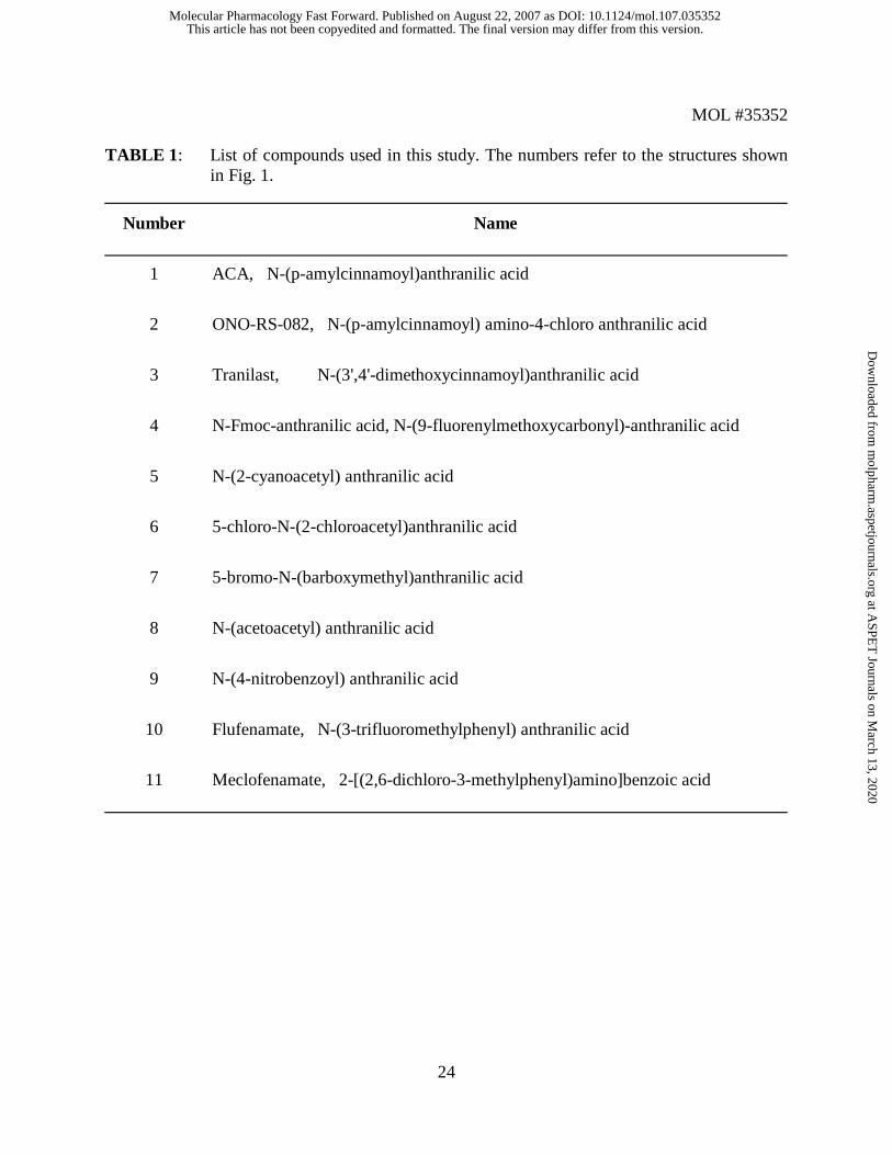
TABLE 1: List of compounds used in this study. The numbers refer to the structures shown in Fig. 1.
Number Name
1 ACA, N-(p-amylcinnamoyl)anthranilic acid
2 ONO-RS-082, N-(p-amylcinnamoyl) amino-4-chloro anthranilic acid
3 Tranilast, N-(3',4'-dimethoxycinnamoyl)anthranilic acid
4 N-Fmoc-anthranilic acid, N-(9-fluorenylmethoxycarbonyl)-anthranilic acid
5 N-(2-cyanoacetyl) anthranilic acid
6 5-chloro-N-(2-chloroacetyl)anthranilic acid
7 5-bromo-N-(barboxymethyl)anthranilic acid
8 N-(acetoacetyl) anthranilic acid
9 N-(4-nitrobenzoyl) anthranilic acid
10 Flufenamate, N-(3-trifluoromethylphenyl) anthranilic acid
11 Meclofenamate, 2-[(2,6-dichloro-3-methylphenyl)amino]benzoic acid
This article has not been copyedited and formatted. The final version may differ from this version.Molecular Pharmacology Fast Forward. Published on August 22, 2007 as DOI: 10.1124/mol.107.035352
at ASPE
T Journals on M
arch 13, 2020m
olpharm.aspetjournals.org
Dow
nloaded from

This article has not been copyedited and formatted. The final version may differ from this version.Molecular Pharmacology Fast Forward. Published on August 22, 2007 as DOI: 10.1124/mol.107.035352
at ASPE
T Journals on M
arch 13, 2020m
olpharm.aspetjournals.org
Dow
nloaded from

This article has not been copyedited and formatted. The final version may differ from this version.Molecular Pharmacology Fast Forward. Published on August 22, 2007 as DOI: 10.1124/mol.107.035352
at ASPE
T Journals on M
arch 13, 2020m
olpharm.aspetjournals.org
Dow
nloaded from
This article has not been copyedited and formatted. The final version may differ from this version.Molecular Pharmacology Fast Forward. Published on August 22, 2007 as DOI: 10.1124/mol.107.035352
at ASPE
T Journals on M
arch 13, 2020m
olpharm.aspetjournals.org
Dow
nloaded from

This article has not been copyedited and formatted. The final version may differ from this version.Molecular Pharmacology Fast Forward. Published on August 22, 2007 as DOI: 10.1124/mol.107.035352
at ASPE
T Journals on M
arch 13, 2020m
olpharm.aspetjournals.org
Dow
nloaded from

This article has not been copyedited and formatted. The final version may differ from this version.Molecular Pharmacology Fast Forward. Published on August 22, 2007 as DOI: 10.1124/mol.107.035352
at ASPE
T Journals on M
arch 13, 2020m
olpharm.aspetjournals.org
Dow
nloaded from
This article has not been copyedited and formatted. The final version may differ from this version.Molecular Pharmacology Fast Forward. Published on August 22, 2007 as DOI: 10.1124/mol.107.035352
at ASPE
T Journals on M
arch 13, 2020m
olpharm.aspetjournals.org
Dow
nloaded from

This article has not been copyedited and formatted. The final version may differ from this version.Molecular Pharmacology Fast Forward. Published on August 22, 2007 as DOI: 10.1124/mol.107.035352
at ASPE
T Journals on M
arch 13, 2020m
olpharm.aspetjournals.org
Dow
nloaded from
This article has not been copyedited and formatted. The final version may differ from this version.Molecular Pharmacology Fast Forward. Published on August 22, 2007 as DOI: 10.1124/mol.107.035352
at ASPE
T Journals on M
arch 13, 2020m
olpharm.aspetjournals.org
Dow
nloaded from
This article has not been copyedited and formatted. The final version may differ from this version.Molecular Pharmacology Fast Forward. Published on August 22, 2007 as DOI: 10.1124/mol.107.035352
at ASPE
T Journals on M
arch 13, 2020m
olpharm.aspetjournals.org
Dow
nloaded from
This article has not been copyedited and formatted. The final version may differ from this version.Molecular Pharmacology Fast Forward. Published on August 22, 2007 as DOI: 10.1124/mol.107.035352
at ASPE
T Journals on M
arch 13, 2020m
olpharm.aspetjournals.org
Dow
nloaded from
This article has not been copyedited and formatted. The final version may differ from this version.Molecular Pharmacology Fast Forward. Published on August 22, 2007 as DOI: 10.1124/mol.107.035352
at ASPE
T Journals on M
arch 13, 2020m
olpharm.aspetjournals.org
Dow
nloaded from
![Thermochemistry of organic molecules: The way to ...iupac.org/publications/pac/pdf/2009/pdf/8110x1857.pdfcarboxylate) and dimethyl cuneane-2,6-dicarboxylate (dimethyl pentacyclo[3.3.0.02,4.03,7.06,8]octane-2,6-dicarboxylate),](https://static.fdocuments.us/doc/165x107/5abcf19d7f8b9ad1768e8e2b/thermochemistry-of-organic-molecules-the-way-to-iupacorgpublicationspacpdf2009pdf.jpg)


![Herbivore-Induced SABATH Methyltransferases of …Herbivore-Induced SABATH Methyltransferases of Maize That Methylate Anthranilic Acid Using S-Adenosyl-L-Methionine1[W] Tobias G. Ko](https://static.fdocuments.us/doc/165x107/5a86a1367f8b9a14748ceb84/herbivore-induced-sabath-methyltransferases-of-herbivore-induced-sabath-methyltransferases.jpg)