Inhibition of HIV-1 transcription and replication by a ... · Inhibition of HIV-1 transcription and...
34
1 Inhibition of HIV-1 transcription and replication by a newly identified cyclin T1 splice variant Guozhen Gao 1,2 , Xiaoyun Wu 1 , Jieqiong Zhou 1 , Mingfeng He 2 , Johnny J. He 2, 3, 4, * and Deyin Guo 1,5, * 1 State Key Laboratory of Virology and Modern Virology Research Center, Wuhan University College of Life Sciences, Wuhan, P.R. China; 2 Department of Microbiology and Immunology, Indiana University School of Medicine, Indianapolis, IN 46202; 3 Center for AIDS Research, Indiana University School of Medicine, Indianapolis, IN 46202; 4 University of North Texas Health Science Center, 3500 Camp Bowie Blvd., Fort Worth, TX 76107; 5 Institute of Medical Virology, Wuhan University School of Medicine, Wuhan, P. R. China *To whom correspondence should be addressed: DG- State Key Laboratory of Virology and Modern Virology Research Center, College of Life Sciences, Wuhan University, Wuhan 430072, P.R. China. • Tel 011-86-27-68752506 • Fax 011-86-27-68752897 • E-mail: [email protected]; JJH- University of North Texas Health Science Center, 3500 Camp Bowie Blvd., Fort Worth, TX 76107. • Tel (817) 735-2642 • Fax (817) 735-0181 • E-mail: [email protected] Running title: A cyclin T1 splice variant inhibits HIV-1 transcription BACKGROUND: Production of full-length HIV-1 mRNA requires interaction of cyclin T1 and cyclin-dependent kinase 9 (CDK9) with HIV-1 Tat protein. RESULTS: An inhibitory cyclin T splice variant was identified. CONCLUSION: The newly identified cyclin T1 splice variant inhibits HIV-1 gene transcription. SIGNIFICANCE: These new findings may provide new clues for the development of anti-HIV strategies. ABSTRACT A variety of cellular factors participate in the HIV-1 life cycle and transcription. Among them is the well-characterized cyclin T1 (CycT1). CycT1 binds to cyclin-dependent kinase 9 (CDK9) and forms the positive transcription elongation factor-b (P-TEFb), which is then recruited by HIV-1 Tat to the HIV-1 long terminal repeat (LTR) promoter to phosphorylate the C-terminal domain (CTD) of RNA polymerase II (RNAP II), leading to enhanced processivity of RNAP II and transcription of a full-length HIV-1 RNA. In this study, we report the identification of a new CycT1 splice variant, designated as CycT1b and accordingly renamed CycT1 as CycT1a. CycT1b was detected in several cell lines, including primary human CD4 T cells, and its expression was subject to cell cycle regulation. Similar to CycT1a, CycT1b was primarily localized in the nucleus. CycT1b expression was found to be inversely correlated with HIV-1 gene expression and replication. This inverse correlation appeared to involve Tat transactivation, CDK9 binding, and subsequent recruitment of p-TEFb to the HIV-1 LTR promoter and RNAP II CTD phosphorylation. In agreement with these findings, CycT1b expression led to direct inhibition of Tat-transactivated transcription of the HIV-1 http://www.jbc.org/cgi/doi/10.1074/jbc.M112.438465 The latest version is at JBC Papers in Press. Published on April 8, 2013 as Manuscript M112.438465 Copyright 2013 by The American Society for Biochemistry and Molecular Biology, Inc. by guest on August 9, 2018 http://www.jbc.org/ Downloaded from by guest on August 9, 2018 http://www.jbc.org/ Downloaded from by guest on August 9, 2018 http://www.jbc.org/ Downloaded from by guest on August 9, 2018 http://www.jbc.org/ Downloaded from by guest on August 9, 2018 http://www.jbc.org/ Downloaded from by guest on August 9, 2018 http://www.jbc.org/ Downloaded from by guest on August 9, 2018 http://www.jbc.org/ Downloaded from by guest on August 9, 2018 http://www.jbc.org/ Downloaded from by guest on August 9, 2018 http://www.jbc.org/ Downloaded from by guest on August 9, 2018 http://www.jbc.org/ Downloaded from
Transcript of Inhibition of HIV-1 transcription and replication by a ... · Inhibition of HIV-1 transcription and...

1
Inhibition of HIV-1 transcription and replication by a newly identified cyclin T1 splice variant Guozhen Gao1,2, Xiaoyun Wu1, Jieqiong Zhou1, Mingfeng He2, Johnny J. He2, 3, 4,* and Deyin Guo1,5,* 1State Key Laboratory of Virology and Modern Virology Research Center, Wuhan University College of Life Sciences, Wuhan, P.R. China; 2Department of Microbiology and Immunology, Indiana University School of Medicine, Indianapolis, IN 46202; 3Center for AIDS Research, Indiana University School of Medicine, Indianapolis, IN 46202; 4University of North Texas Health Science Center, 3500 Camp Bowie Blvd., Fort Worth, TX 76107; 5Institute of Medical Virology, Wuhan University School of Medicine, Wuhan, P. R. China *To whom correspondence should be addressed: DG- State Key Laboratory of Virology and Modern Virology Research Center, College of Life Sciences, Wuhan University, Wuhan 430072, P.R. China. • Tel 011-86-27-68752506 • Fax 011-86-27-68752897 • E-mail: [email protected]; JJH- University of North Texas Health Science Center, 3500 Camp Bowie Blvd., Fort Worth, TX 76107. • Tel (817) 735-2642 • Fax (817) 735-0181 • E-mail: [email protected] Running title: A cyclin T1 splice variant inhibits HIV-1 transcription BACKGROUND: Production of full-length HIV-1 mRNA requires interaction of cyclin T1 and cyclin-dependent kinase 9 (CDK9) with HIV-1 Tat protein. RESULTS: An inhibitory cyclin T splice variant was identified. CONCLUSION: The newly identified cyclin T1 splice variant inhibits HIV-1 gene transcription. SIGNIFICANCE: These new findings may provide new clues for the development of anti-HIV strategies. ABSTRACT A variety of cellular factors participate in the HIV-1 life cycle and transcription. Among them is the well-characterized cyclin T1 (CycT1). CycT1 binds to cyclin-dependent kinase 9 (CDK9) and forms the positive transcription elongation factor-b (P-TEFb), which is then recruited by HIV-1 Tat to the
HIV-1 long terminal repeat (LTR) promoter to phosphorylate the C-terminal domain (CTD) of RNA polymerase II (RNAP II), leading to enhanced processivity of RNAP II and transcription of a full-length HIV-1 RNA. In this study, we report the identification of a new CycT1 splice variant, designated as CycT1b and accordingly renamed CycT1 as CycT1a. CycT1b was detected in several cell lines, including primary human CD4 T cells, and its expression was subject to cell cycle regulation. Similar to CycT1a, CycT1b was primarily localized in the nucleus. CycT1b expression was found to be inversely correlated with HIV-1 gene expression and replication. This inverse correlation appeared to involve Tat transactivation, CDK9 binding, and subsequent recruitment of p-TEFb to the HIV-1 LTR promoter and RNAP II CTD phosphorylation. In agreement with these findings, CycT1b expression led to direct inhibition of Tat-transactivated transcription of the HIV-1
http://www.jbc.org/cgi/doi/10.1074/jbc.M112.438465The latest version is at JBC Papers in Press. Published on April 8, 2013 as Manuscript M112.438465
Copyright 2013 by The American Society for Biochemistry and Molecular Biology, Inc.
by guest on August 9, 2018
http://ww
w.jbc.org/
Dow
nloaded from
by guest on August 9, 2018
http://ww
w.jbc.org/
Dow
nloaded from
by guest on August 9, 2018
http://ww
w.jbc.org/
Dow
nloaded from
by guest on August 9, 2018
http://ww
w.jbc.org/
Dow
nloaded from
by guest on August 9, 2018
http://ww
w.jbc.org/
Dow
nloaded from
by guest on August 9, 2018
http://ww
w.jbc.org/
Dow
nloaded from
by guest on August 9, 2018
http://ww
w.jbc.org/
Dow
nloaded from
by guest on August 9, 2018
http://ww
w.jbc.org/
Dow
nloaded from
by guest on August 9, 2018
http://ww
w.jbc.org/
Dow
nloaded from
by guest on August 9, 2018
http://ww
w.jbc.org/
Dow
nloaded from

2
LTR promoter. Taken together, these results show that the newly identified CycT1b splice variant inhibits HIV-1 transcription and may provide new clues for the development of anti-HIV strategies. INTRODUCTION Human immunodeficiency virus type 1 (HIV-1) is the causative agent of acquired immunodeficiency disease (AIDS). Currently there are an estimated 33.3 million people around the world under the threat of this virus, according to the World Health Organization annual reports. Currently, there is no practical vaccine available for HIV-1 and treatment of its infection is primarily based on antiretroviral drugs that include nucleoside/nucleotide reverse transcriptase inhibitors, non-nucleoside reverse transcriptase inhibitors, protease inhibitors, fusion or entry inhibitors and integrase inhibitors. These drugs are designed to interfere with multiple steps during the HIV-1 life cycle by targeting the viral components of HIV-1. Alternatively, in the past 20 years a large number of host cellular factors were discovered to be involved in the regulation of its life cycle, thereby becoming potential targets for HIV-1 therapeutics. Subsequently, interactions between host cellular factors and viral proteins or DNA/RNA elements have recently been considered progressively more important in the regulation of HIV-1 replication. In latent HIV-1 infection, the genomic RNA is reverse-transcribed and integrated into the host genome. Upon activation by Tat/TAR/P-TEFb, the HIV-1 RNA is transcribed and introduced into the general RNA metabolism. However, this viral RNA is distinguishable from host RNA due to the presence of introns and other cis-elements,
leading to occasional specific regulation by host factors. During the activation of HIV-1 transcription from the genome, several transcription factors are recruited to the LTR promoter to initiate basic transcription, including NFκB, often considered the most important factor (see reviews (1) and (2)). Following sufficient induction of protein expression, Tat binds to the trans-activating response element (TAR) RNA and recruits P-TEFb to stimulate viral transcription (3). HIV-1 RNA is alternatively spliced into 3 groups: 2 kb fully spliced mRNAs, 4 kb incompletely spliced mRNAs and the 9 kb unspliced genomic RNA (4). Cellular splicing factors, including SR proteins and hnRNP family members, are involved in this process and play alternative roles in the splicing of HIV-1 RNA (5). Export of unspliced or incompletely spliced mRNAs are usually blocked by cellular factors encoded by eukaryotic cells. The HIV-1 Rev protein may recognize the Rev response element (RRE) on these “immature” mRNAs, allowing them to be exported to the cytoplasm through the CRM-1 pathway (6). Cellular factors, such as eIF5A, Sam68, DDX1 and DDX3, are implicated in the mRNA export process mediated by Rev (7-10). When the mature and immature mRNAs are exported into the cytoplasm, cellular translation initiation factors and ribosomes bind to the m7-pppG structure resulting in ribosome scanning. A few cellular factors specifically regulate HIV-1 translation. For example, DDX3 has been reported to stimulate general translation through the interaction with the translation initiation factor eIF3, and may also enhance HIV-1 IRES mediated translation to a significantly higher level (11,12). Our previous work on Sam68 has shown that it specifically enhances the translation of Nef mRNA, and is involved in the genome-wide translation control
by guest on August 9, 2018
http://ww
w.jbc.org/
Dow
nloaded from

3
of HIV-1 genes (12,13). Positive transcription elongation factor b (P-TEFb) is the key factor regulating eukaryotic mRNA transcription at the level of elongation (14,15). It is composed of CDK9 and one of the three cyclins: T1, T2 (T2a and T2b) or K (16), of which only the primary cyclin partner of CDK9, cyclin T1, is able to support HIV-1 Tat mediated transactivation (3). Once Tat protein is expressed through basic transcription and translation, it binds to the HIV-1 TAR structure and recruits P-TEFb to the viral promoter for transcriptional activation (17-19). The activated cyclin dependent kinase complex, P-TEFb, then phosphorylates negative elongation factors, i.e. DSIF and NELF, and serine 2 on the C-terminal domain (CTD) of the RNA polymerase II (Pol II) large subunit (20,21). P-TEFb (CycT1/CDK9) is incorporated into two super complex forms: 1) the catalytically inactive form, 7SK snRNP, which also contains 7SK snRNA, HEXIM1/2, LARP7 and MePCE (22-25), and 2) the active form, called the super elongation complex, which includes ELL2 and the transcription factors/activators ENL, AF9, AFF4, AFF1 and MLLT1/3 (26-31). A number of other P-TEFb binding proteins were identified (30,32,33). HIV-1 Tat triggers the release of P-TEFb from the 7SK snRNP in vitro and in vivo, resulting in the activation of P-TEFb by Tat (34,35). Other than HIV-1 Tat, several other sequence-specific activators, such as Brd4, NFκB, c-Myc and CIITA, have been reported to interact with P-TEFb and contribute to the activation of transcription elongation (36-39). Currently, three cyclin T isoforms (CycT1, CycT2a and 2b) have been identified to be associated with CDK9 to constitute the P-TEFb
complex (16). CycT1 is an approximate 86 kD protein, found in 80% of human P-TEFb heterodimers, and is the only cyclin that can bind to Tat to support HIV-1 transcription (3,20). P-TEFb is believed to play a general role in transcription, since its inhibition affects a vast majority of cellular genes in uninfected cells (40). P-TEFb, containing CycT1 or CycT2, is recruited by different transcriptional activators to stimulate the expression of various genes (38,41-43). It was revealed by microarray analysis that there was limited redundancy in the genes regulated by either CycT1 or CycT2 (44). The human CycT1 gene has been mapped to human chromosome 12, with four transcripts of different sizes detected by Northern blot analysis, indicating that CycT1 mRNA may be alternatively transcribed or spliced (3,16). However, there is no data yet available on the alternative splicing product of CycT1 or the post-transcriptional regulation of CycT1 variants. In this study, we identified a novel transcript of CycT1, which we named CycT1b. This novel transcript is an antagonist of CycT1a (formerly CycT1), resulting in inhibition of Tat mediated LTR transactivation. Mechanistically, we demonstrate that CycT1b competes with CycT1a for binding to CDK9, thereby interfering with the hyperphosphorylation of RNA polymerase II, resulting in the suppression of transcriptional elongation. MATERIAL AND METHODS Cell lines and transfection 293T, Hela and Jurkat cells were obtained from American Tissue Culture Collection (ATCC, Manassas, VA) and maintained in Dulbecco ′s modified Eagle’s medium (DMEM) or Roswell Park
by guest on August 9, 2018
http://ww
w.jbc.org/
Dow
nloaded from

4
Memorial Institute (RPMI) 1640 medium supplemented with 10% fetal bovine serum (Hyclone, Logan, UT) and 1% (v/v) penicillin and streptomycin (CellGro, Manassas, VA) at 37ºC with 5% CO2. For transfection with DNA only, 293T cells were transfected by the standard calcium phosphate method as previously described (45); for transfection with RNA only or together with DNA, 293T cells were transfected with Lipofectamine 2000, according to the manufacturer’s instructions (Invitrogen, Carlsbad, CA). Transfection medium was replaced with fresh medium containing the same supplements 6 h post-transfection and the cells were harvested for: protein analysis by western blot or immunoprecipitation followed by western blot; for analysis of luciferase reporter gene activity; or for isolation of total RNA and RNA analysis by reverse transcription-PCR (RT-PCR). In all transfections, pcDNA3 was used to equalize the amount of total transfected DNA. Plasmids Plasmids pNL4-3, pLTR.luc, pMyc.Tat, HIVdGless have been described elsewhere (46,47). pMyc.CycT1b was constructed using standard cloning methods with the primers (Myc.CycT1b5: 5’-GCG AAT TCT GGA GGG AGA GAG GAA GAA C-3’, EcoRI site underlined; Myc.CycT1b3: 5’-GCG GTA CCT CAT GTG TCA GTT CTG TTG G-3’, KpnI site underlined) to amplify the CycT1b coding sequence to insert into pCMV-myc (Clontech, Mountain View, CA). pGFP.CycT1b was constructed by creating a single mutated site in the CycT1b coding sequence, by deletion of the tenth base (guanine in italics) in the primer Myc.CycT1b5, and inserting it into the EcoRI and KpnI sites of peGFP-C1 (Clontech). pET.CycT1b was constructed by inserting CycT1b coding sequence into EcoRI and SalI
sites of pET28a (Clontech) vector using primers 5’-GCG AAT TCG AGG GAG AGA GGA AGA AC-3’ and 5’-GCG TCG ACT CAT GTG TCA GTT CTG TTG G-3’. To construct pLL.GFP-CycT1b, overlapping PCR was performed to obtain GFP.CycT1b fusion coding sequence with primers 5’-CTA GCT AGC GCT ACC GGT CG-3’, 5’-GTT CTT CCT CTC TCC CTC CTT GTA CAG CTC GTC CAT GC-3’, 5’-GCA TGG ACG AGC TGT ACA AGG AGG GAG AGA GGA AGA AC-3’ and 5’-GCG AAT TCT CAT GTG TCA GTT CTG TTG G-3’; then the coding sequence was inserted into the NheI and EcoRI sites of pLentilox3.7 (Invitrogen). pLL.GFP was similarly constructed and included as the control. Virus packaging and infection GFP-CycT1b-expressing lentivurses were packaged in 293T cells by co-transfection with pLL.GFP-CycT1b (4 µg) / pLentilox3.7 (4 µg) and pLP1 (2 µg), pLP2 (2 µg), pVSV-G (2 µg) helper plasmids. Forty-eight hours post medium change, cell culture supernatants were collected, filtered and saved as lentivirus stock. GFP-expressing lentiviruses were similarly prepared using pLL.GFP plasmid. HIV-1 NL4-3 viruses were prepared in 293T cells as previously described (48). Viruses were titred by infecting 293T or Ghost.CXCR4 cell lines. Jurkat cells were first transduced with lentiviruses (MOI = 0.5) for 30 hr and then infected with HIV-1 NL4-3 virus (MOI =1). The cultures were cultured for 7 more days and then harvested for cell lysates and Western blotting. Cell culture supernatants were collected for p24 ELISA. All transductions and infections were performed in the presence of 8 µg/ml polybrene.
by guest on August 9, 2018
http://ww
w.jbc.org/
Dow
nloaded from

5
RNA analysis by RT-PCR Total RNA was extracted from 293T cells or Hela cells using the TRIzol reagent (Invitrogen), according to the manufacturer’s instructions. For CycT1 transcript amplification, 200 ng total mRNA was used as a template for RT-PCR with primers 5’-GCG AAT TCA TGG AGG GAG AGA GGA AGA ACA A-3’ and 5’-GCG TCG ACT TAC TTA GGA AGG GGT GGA AT-3’. For simultaneous detection of CycT1a/CycT1b transcripts, 50 ng of total RNA was reverse transcribed and PCR-amplified with the CycT1 primers: 5’-CTT CAC ACA GTT CCC TGG AAA TTC TG and 5’-AGG CAC TGC ACT TGT GGT AG-3’) using the Titan One Tube RT-PCR system (Roche Diagnostics, Indianapolis, IN). For CycT1a detection, Primers were: 5’-CCT GCA TTT GAC CAC ATT TAG C-3’ and 5’-CAT CTA AAA GTT CCA AGG TCA CA-3’. For CycT1b detection, primers were 5’-CTT ACT TCA TGG CAA CCA ACA GAA C-3’ and 5’-CTT GTG GTA GAA GTT GAC ATG CTC-3’. For analysis with the HIV-1 LTR-driven G-less transcription cassette, 200 ng of total RNA was used as the original template for RT-PCR, with the corresponding PCR primers: 5’-CCC GAA TTC GGG TCT CTC TGG TTA GAC CAG ATC TGA GCC TGG GAG CTC-3’ and, 5’-AAA ACC AAA CCC TGC GCT CCA TCG CCA-3’; 5’-GCG AGG CAT AAA GTT GCG TGT-3’ and 5’-AGG AGG GAG TGA GGA GAG GAT-3’. The GAPDH mRNA level was detected by RT-PCR in each experiment to normalize the total mRNA input, with the primers: 5’-GAA AGG TGA AGG TCG GAG T-3’ and 5’-GAA GAT GGT GAT GGG ATT TC-3’. The expression level of CycT1-dependent genes was detected by SYBR green (Roche) qRT-PCR with primers listed in Table 1.
Preparation of whole cell lysates, immunoprecipitation, and western blotting analysis Briefly, cells were washed twice with ice-cold phosphate-buffered saline (PBS) and cell pellets were suspended in two volumes of RIPA buffer containing 25 mM Tris-HCl, pH 7.5, 150 mM NaCl, 1% NP-40, 0.1% SDS, 1% sodium deoxycholate, or IP lysis buffer containing 0.5% NP40, 500 mM Tris-Cl, 120 mM NaCl, 25 mM NaF, 1 mM EDTA, 5 mM EGTA, 1 mM sodium orthovanadate for immunoprecipitation; both supplemented with the proteinase inhibitor cocktail (Calbiochem, La Jolla, CA), and incubated on ice for 20 min. Whole cell lysates were obtained by centrifugation and removal of the cell debris. For western blotting, cell lysates (60-100 µg of total protein) or immunoprecipitates (800 µg of original total protein) were electrophoretically separated on 10% SDS-PAGE and analyzed by immunoblotting using antibodies against: Myc (Santa Cruz Biotechnology, Santa Cruz, CA), p24 (mouse anti p24 serum), CycT1 (Santa Cruz Biotechnology) CDK9 (Santa Cruz Biotechnology), GFP (Clontech) or β-actin (Sigma), at dilutions recommended by the manufacturers, followed by the appropriate horseradish peroxidase-conjugated secondary antibodies, and visualization with the ECL system (GE Healthcare, Piscataway, NJ). For CycT1b far-Western blotting, protein blot was incubated with 0.2 ng/mL CDK9 recombinant protein (Abnova, Taipei) overnight, followed by incubation with anti-CDK9 antibody and anti-rabbit secondary antibody, and visualized by a standard ECL colorimetric system. Immunofluoresence staining for endogenous cyclin T1 293T cells were plated onto polylysine coated coverslips and cultured for 48 h. Cells were washed, fixed with 4%
by guest on August 9, 2018
http://ww
w.jbc.org/
Dow
nloaded from

6
paraformaldehyde (PFA) for 10 min at room temperature, permeabilized with PBS containing 0.2% Triton and 0.8% fetal bovine serum (FBS) and washed three times with PBS. The cells were blocked with PBS containing 1% FBS for 1 h, and α-HA antibody was added to the coverslips (1:500) and incubated for 1 h. The cells were stained with Alexa Fluor 488-conjugated α-mouse antibody (1:500 diluted) followed by staining with 0.25 µg/mL DAPI. HIV-1 reverse transcriptase (RT) activity assay Cell culture supernatants were collected 48 h after medium change, filtered, and assayed for RT activity, as previously described (49). Briefly, viral pellets were recovered by centrifugation of the cell culture supernatants at 18,000 g and 4°C for 1 h, and solubilized in the dissociation buffer (0.25% Triton X-100, 20% glycerol, 50 mM Tris-HCl, pH 7.5, 1 mM dithiothreitol, 0.25 M KCl). Following three cycles of freeze/thaw, the virus suspension was mixed with poly(A)-dT15 (Roche Molecular Biochemicals) and 3H-labeled thymidine 5’-triphosphate (GE Healthcare) in a buffer (0.05% Triton X-100, 50 mM Tris-HCl, pH 7.5, 0.5 mM dithiothreitol, 7.5 mM MgCl2) and incubated at 37°C for 1 h. The reaction mixture was spotted onto DE81 membrane disks (Whatman), dried, and washed extensively with 2×SSC (0.3 M NaCl, 30 mM sodium citrate). The membrane disks were counted on a scintillation counter to determine RTase activity. Yeast two-hybrid assays Coding sequences of CycT1a, CycT1b, CDK9 and Tat were individually inserted into the EcoRI and SalI sites of the pGBKT7 or pGADT7 vectors (Clontech) to create pBD-CycT1a, pBD-CycT1b, pAD-CDK9 and pAD-Tat constructs for the yeast two-hybrid assays. Briefly, transformed
yeast (strain AH109) was grown in SD liquid medium (SD/Leu-/Trp-) to saturation overnight and diluted the following morning. Diluted yeast media was grown to OD600=0.8 and pelleted. The yeast were suspended in 1 ml of Z buffer (60 mM Na2HPO4, 40 mM NaH2PO4, 10 mM KCl, 1 mM MgSO4 and 50 mM β-mercaptoethanol, pH adjusted to 7.0) and diluted in Z buffer at 1:10. The yeast was lysed with chloroform and SDS and incubated with 4 mg/ml O-nitrophenyl-β-D-galactoside at 30 ºC until they turned yellow. The reaction was stopped with 1 M Na2CO3, and the OD420 was measured. β-galactosidase activity was computed by the formula: β-galactosidase activity =(1000 × OD420) / time [min] × Volume [mL] × OD600). Chromatin immunoprecipitation (ChIP) assay Cells were cross-linked, 48 h post-transfection, by adding formaldehyde to a final concentration of 1% for 10 min, followed by the addition of glycine to a final concentration of 125 mM to terminate cross-linking. Cells were then collected and lysed with cell lysis buffer (50 mM Tris-HCl, pH 8.0, 85 mM KCl, 0.5% NP-40, supplemented with proteinase inhibitors) on ice. The nuclei portion was pelleted and suspended in nuclear lysis buffer (50 mM Tris-HCl, pH 8.0, 10 mM EDTA, 1% SDS, supplemented with proteinase inhibitors). The two lysates were combined and sonicated, 10 times for 15 seconds each. The debris was removed and the samples were diluted 5-fold in the ChIP dilution buffer (16.7 mM Tris-HCl, pH 8.0, 167 mM NaCl, 1.2 mM EDTA, 1.1% Triton X-100, 0.01% SDS). The samples were pre-cleared with ssDNA/protein A agarose slurry for 30 min at 4℃ . The corresponding antibodies (mouse normal IgG, CycT1a, serine 5- or serine 2-phosphorylated RNA Pol II C-terminal
by guest on August 9, 2018
http://ww
w.jbc.org/
Dow
nloaded from
7
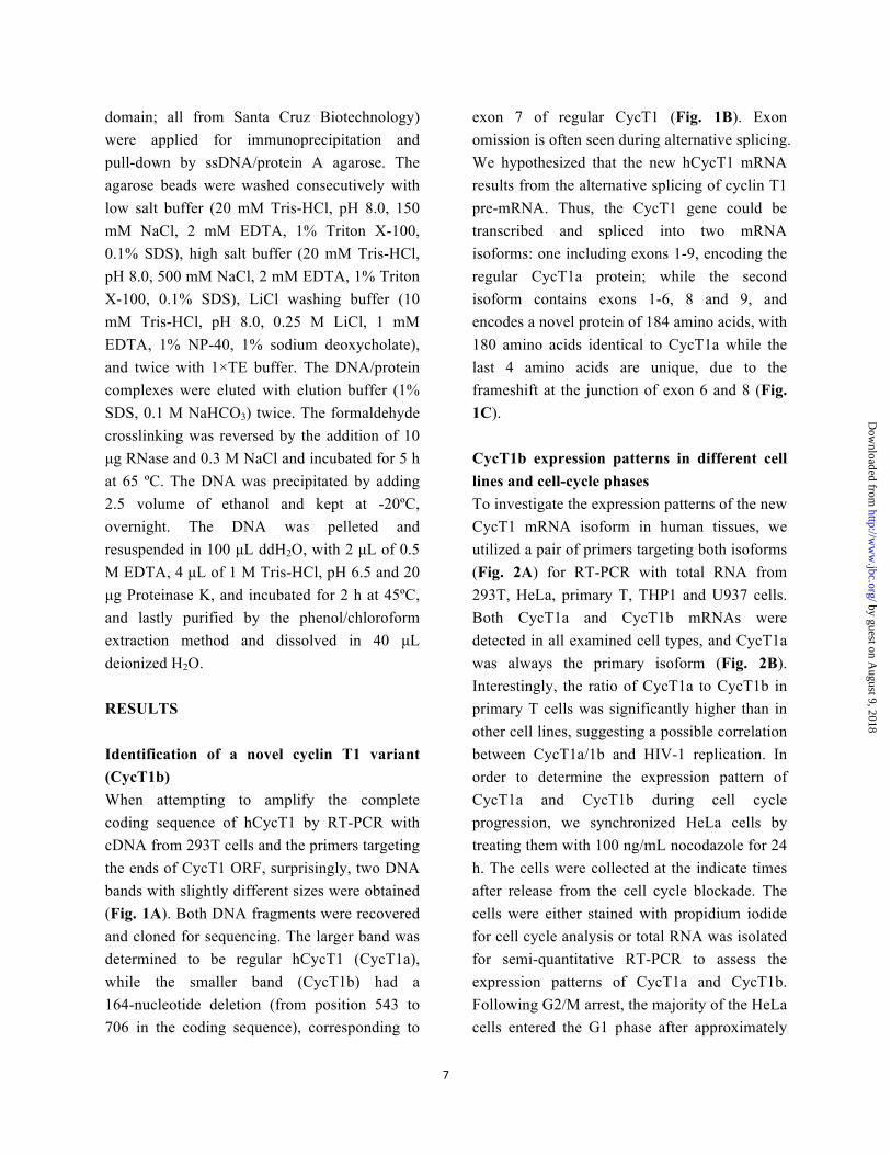
domain; all from Santa Cruz Biotechnology) were applied for immunoprecipitation and pull-down by ssDNA/protein A agarose. The agarose beads were washed consecutively with low salt buffer (20 mM Tris-HCl, pH 8.0, 150 mM NaCl, 2 mM EDTA, 1% Triton X-100, 0.1% SDS), high salt buffer (20 mM Tris-HCl, pH 8.0, 500 mM NaCl, 2 mM EDTA, 1% Triton X-100, 0.1% SDS), LiCl washing buffer (10 mM Tris-HCl, pH 8.0, 0.25 M LiCl, 1 mM EDTA, 1% NP-40, 1% sodium deoxycholate), and twice with 1×TE buffer. The DNA/protein complexes were eluted with elution buffer (1% SDS, 0.1 M NaHCO3) twice. The formaldehyde crosslinking was reversed by the addition of 10 µg RNase and 0.3 M NaCl and incubated for 5 h at 65 ºC. The DNA was precipitated by adding 2.5 volume of ethanol and kept at -20ºC, overnight. The DNA was pelleted and resuspended in 100 µL ddH2O, with 2 µL of 0.5 M EDTA, 4 µL of 1 M Tris-HCl, pH 6.5 and 20 µg Proteinase K, and incubated for 2 h at 45ºC, and lastly purified by the phenol/chloroform extraction method and dissolved in 40 µL deionized H2O. RESULTS Identification of a novel cyclin T1 variant (CycT1b) When attempting to amplify the complete coding sequence of hCycT1 by RT-PCR with cDNA from 293T cells and the primers targeting the ends of CycT1 ORF, surprisingly, two DNA bands with slightly different sizes were obtained (Fig. 1A). Both DNA fragments were recovered and cloned for sequencing. The larger band was determined to be regular hCycT1 (CycT1a), while the smaller band (CycT1b) had a 164-nucleotide deletion (from position 543 to 706 in the coding sequence), corresponding to
exon 7 of regular CycT1 (Fig. 1B). Exon omission is often seen during alternative splicing. We hypothesized that the new hCycT1 mRNA results from the alternative splicing of cyclin T1 pre-mRNA. Thus, the CycT1 gene could be transcribed and spliced into two mRNA isoforms: one including exons 1-9, encoding the regular CycT1a protein; while the second isoform contains exons 1-6, 8 and 9, and encodes a novel protein of 184 amino acids, with 180 amino acids identical to CycT1a while the last 4 amino acids are unique, due to the frameshift at the junction of exon 6 and 8 (Fig. 1C).
CycT1b expression patterns in different cell lines and cell-cycle phases To investigate the expression patterns of the new CycT1 mRNA isoform in human tissues, we utilized a pair of primers targeting both isoforms (Fig. 2A) for RT-PCR with total RNA from 293T, HeLa, primary T, THP1 and U937 cells. Both CycT1a and CycT1b mRNAs were detected in all examined cell types, and CycT1a was always the primary isoform (Fig. 2B). Interestingly, the ratio of CycT1a to CycT1b in primary T cells was significantly higher than in other cell lines, suggesting a possible correlation between CycT1a/1b and HIV-1 replication. In order to determine the expression pattern of CycT1a and CycT1b during cell cycle progression, we synchronized HeLa cells by treating them with 100 ng/mL nocodazole for 24 h. The cells were collected at the indicate times after release from the cell cycle blockade. The cells were either stained with propidium iodide for cell cycle analysis or total RNA was isolated for semi-quantitative RT-PCR to assess the expression patterns of CycT1a and CycT1b. Following G2/M arrest, the majority of the HeLa cells entered the G1 phase after approximately
by guest on August 9, 2018
http://ww
w.jbc.org/
Dow
nloaded from
8
15 h, and gradually entered S phase in the proceeding 6 h, finally reentering G2 after 27 h (Fig. 2C). During the entire cell cycle, the expression of CycT1a mRNA was dominant compared to CycT1b, and the ratio of CycT1a to CycT1b mRNA oscillated, following cell cycle progression (Fig. 2D). Subcellular localization of CycT1 isoforms It is known that P-TEFb functions as an important regulatory factor during transcriptional elongation for many genes. Correspondingly, CycT1a has been reported to be localized in the nucleus (50,51). In order to characterize the subcellular distribution of the novel CycT1 isoform, we constructed the pGFP.CycT1b plasmid and investigated its expression in HeLa cells. Unlike CycT1a, CycT1b was located both in the nucleus and the cytoplasm (Fig. 3A). However, the majority was present in the nucleus. We confirmed the nuclear localization pattern of CycT1a by transfecting HeLa cells with HA.CycT1a and incubating the cells with α-HA antibodies followed by FITC-conjugated secondary antibody staining (Fig. 3B).
CycT1b inhibit HIV-1 replication in a dose-dependent manner CycT1 is the primary cyclin involved in P-TEFb regulation. Despite the fact that CycT2a and CycT2b have similar domains as CycT1a, they are not associated with HIV-1 Tat mediated transactivation. In order to determine the potential role of CycT1b in the HIV-1 life cycle, we co-transfected 293T cells with HIV-1 pNL4-3 with increasing amounts of myc.CycT1b. The cell culture media and cell lysates were collected 48 hours post-transfection and analyzed for RTase activity (Fig. 4A) or western blotting (Fig. 4B), respectively. As the
exogenous expression level of CycT1b increased, viral reverse transcriptase (RT) activity in the media and Gag protein expression in the cell lysates were downregulated. The extracellular and intracellular virus replication levels were inhibited by overexpression of CycT1b in a dose-dependent manner. To further confirm the inhibitory effect and eliminate the possible artificial effect caused by overexpression, we took advantage of an siRNA which is specific to CycT1b (targeting sequence: 5’-CCA ACA GAA CUG ACA CAU G-3’) to knock-down endogenous CycT1b expression. The efficiency and specificity of the siRNA was certified by detecting the endogenous mRNA level of CycT1a and CycT1b at 24 or 42 h post-transfection. The ratio of CycT1b mRNA level to CycT1a mRNA level was decreased at 24 h post-transfection and significantly decreased at 42 h post-transfection (Fig. 4C). Following siRNA confirmation, 293T cells were transfected with the specific siRNA together with the pNL4-3 plasmid, and viral RT activity in the culture media and Gag protein expression levels in the cell lysates were measured as described above (Fig. 4D and 4E, respectively. In concordance with the overexpression results, knock-down of CycT1b up-regulated both the extracellular RT activity and the intracellular Gag expression levels of HIV-1. To further ascertain the roles of CycT1b in the context of HIV infection, CD4+ lymphocytes Jurkat cells were first transduced with GFP-CycT1b- or GFP-expressing lentiviruses and then infected with HIV-1 NL4-3 viruses. Transduction efficiency was determined to be comparable by Western blotting for using an anti-GFP antibody (left panel, Fig. 4F). Compared to the GFP control, GFP-CycT1b expression resulted in a significant decrease in the intracellular level of Gag and p24 expression determined by Western
by guest on August 9, 2018
http://ww
w.jbc.org/
Dow
nloaded from

9
blotting (right panel, Fig. 4F), as well as the level of p24 in the cell culture supernatants (Fig. 4G). Taken together, these results showed that CycT1b expression suppressed HIV-1 gene expression and replication of HIV-1 and suggest that CycT1b acts as a negative regulator of HIV-1 replication.
CycT1b inhibits HIV-1 replication through suppression of Tat mediated transactivation The mechanism of how P-TEFb stimulates Tat mediated transactivation was well-studied during the late 1990s (17-20). Briefly, Tat protein binds to the HIV-1 TAR structure and recruits the CycT1a/CDK9 complex. The active P-TEFb is then able to hyperphosphorylate the CTD domain of RNA polymerase II as well as two negative transcription elongation factors, NELF and DSIF. This allows the dissociation of the negative factors from RNA polymerase II, and the hyperphosphorylated polymerase is then able to break the transcriptional pause and continue transcriptional elongation. To determine whether the inhibitory effect of CycT1b on HIV-1 replication occurs on this complex, we transfected 293T cells with an HIV-1 LTR-driven firefly luciferase reporter plasmid, pLTR-luc, alone or together with either CycT1a or CycT1b expression plasmids. Overexpression of CycT1a or CycT1b did not significantly alter the expression level of firefly luciferase, indicating that CycT1a or CycT1b does not affect the LTR basic transcription level (Fig. 5A, column 1-3). Transfection of 293T cells with the reporter gene, Tat and additional CycT1a enhanced Tat transactivation by 5-6 fold. However, the addition of CycT1b alleviated Tat mediated transactivation (Fig. 5A, column 4-6). Expression of CycT1a and CycT1b was confirmed by western blotting, as shown in Fig. 5B. This suggests that CycT1a and CycT1b
inversely regulate HIV-1 replication at the level of transcriptional elongation. To confirm this, we introduced Tat, pLTR-luc and increasing amounts of CycT1b into cells. CycT1b dose-dependently inhibited Tat mediated transactivation, similar to the effect observed on HIV-1 replication (Fig. 5C). The dose-dependent increase in CycT1b protein levels was confirmed by western blotting (Fig. 5D).
CycT1b retains the ability to interact with CDK9 but not Tat Early studies revealed that the N-terminal region (amino acids 1–290) of CycT1a was sufficient for binding to both Tat and CDK9, and was capable of supporting Tat mediated transactivation. Further truncation to amino acid 188 completely eliminated the ability to bind to Tat, but this region still retained the ability to associate with CDK9 (52-54). Moreover, the inability of CycT1 region 1–189 aa to bind to Tat, is associated with its inability to enhance Tat mediated transactivation in murine cells (52) Due to the loss of exon 7, which is present in CycT1a mRNA, the first 180 amino acids of CycT1b are identical to CycT1a, while the remaining 4 amino acids on the C-terminus of CycT1b are unique. Therefore, given its amino acid structure, CycT1b may only interact with CDK9 and not with Tat. To experimentally test this possibility, we performed yeast two-hybrid assays to investigate possible protein-protein interactions. Coding sequences of CycT1a, CycT1b, CDK9 and Tat were separately inserted into pGBKT7 or pGADT7 vectors to obtain pBD-CycT1a, pBD-CycT1b, pAD-CDK9 and pAD-Tat constructs for yeast two-hybrid assays. The indicated combinations of BD-fused and AD-fused plasmids were co-transformed into AH109 yeast cells, and the endogenous lacZ
by guest on August 9, 2018
http://ww
w.jbc.org/
Dow
nloaded from

10
expression level was assessed by measuring β-galactosidase (β-Gal) activity. High β-Gal activity was observed when AD-CDK9 was co-expressed with either BD-CycT1a or BD-CycT1b, indicating the positive interactions of CDK9 with either CycT1a or CycT1b (Fig. 6A, column 3 and 5). However, high β-Gal activity was only observed when AD-Tat was co-expressed with BD-CycT1a, but not with BD-CycT1b, indicating that CycT1b lost the ability to associate with Tat (Fig. 6A, column 4 and 6). To confirm the interaction between CycT1b and CDK9, we transiently expressed myc.CycT1b in 293T cells and immunoprecipitated the CDK9 bound complex from cell lysates prepared 48 h post-transfection. Myc.CycT1b was detected in the CDK9 bound complex by Western blot (Fig. 6B and 6C), indicating the association of CycT1b with CDK9. To further confirm that CycT1b does not interact with HIV-1 Tat, 293T cells were transfected with myc.Tat and GFP.Tip110 as a positive control (Tip110, Tat interacting protein of 110 kD, (45)), or GFP.CycT1b. GFP.Tip110 was detected in the myc.Tat bound complex but GFP.CycT1b was not, suggesting that CycT1b does not bind to HIV-1 Tat (Fig. 6D and 6E).
CycT1b interferes with CycT1a/CDK9 heterodimer formation and inhibits RNA Polymerase II hyperphosphorylation on the HIV-1 LTR promoter Association of corresponding cyclins is required to activate a CDK (55). Binding of CycT1a, CycT2a or CycT2b to CDK9 is a prerequisite for its kinase activity (16). Since CycT1b is able to bind to CDK9, it may be involved in the regulation of transcriptional elongation. However, the heterodimer consisting of CDK9 and CycT1b could not be recruited to the Tat/TAR/LTR complex due to its inability with
Tat. Therefore, we propose that CycT1b may compete with CycT1a for the binding of CDK9 and thereby interfere with Tat/CycT1a/CDK9 complex formation. To test this hypothesis, we expressed CycT1b in 293T cells and immunoprecipitated the CDK9 bound complex from cell lysates with an α-CDK9 antibody, to examine whether the amount of CycT1b incorporated in the complex interferes with CycT1a binding. While CycT1b does not affect the endogenous CycT1a expression level (Fig. 5D), the amount of CycT1a binding to CDK9 was significantly reduced (Fig. 7A and 7B). This result suggests that CycT1b is able to compete with CycT1a for binding to CDK9, leading to less CycT1a/CDK9 complex formation. As a direct result of the competition between CycT1b and CycT1a, the amount of P-TEFb (CycT1a/CDK9) recruited to the HIV-1 LTR-1 would be decreased, and the subsequent hyperphosphorylation of the RNA polymerase II CTD domain would also be inhibited. To experimentally address this possibility, we transfected 293T cells with pLTR.luc, with or without CycT1b and Tat or combinations thereof, followed by chromatin immunoprecipitation (ChIP) assays with antibodies against CycT1a or hyperphosphorylated Pol II to analyze the levels of the corresponding bound protein. The relative levels of protein binding to the HIV-1 LTR promoter were measured by semi-quantitative RCR. The binding of CycT1a to the HIV-1 LTR promoter was decreased when CycT1b was overexpressed, either in the presence or absence of Tat. The levels of bound hyperphosporylated Pol II were increased after Tat mediated transactivation, but significantly decreased when CycT1b was overexpressed (Fig. 7C). These results suggest that overexpression of CycT1b inhibits the recruitment of P-TEFb to the HIV-1 LTR and thus inhibits the hyperphosphorylation
by guest on August 9, 2018
http://ww
w.jbc.org/
Dow
nloaded from

11
of Pol II on the LTR promoter. To further confirm these observations, we introduced siRNA specific to CycT1b into 293T cells to knock-down the endogenous CycT1b levels and performed ChIP assays after a secondary transfection with both pLTR.luc and Tat. Downregulation of CycT1b expression enhanced the hyperphosphorylation of Pol II, and correspondingly, decreased the hypophosphorylated form (Fig. 7D).
CycT1b inhibits HIV-1 LTR-driven transcriptional elongation Hyperphosphorylation of RNA polymerase II is a prerequisite of productive transcriptional elongation. We have demonstrated that CycT1b inhibits the phosphorylation of the C-terminal domain of Pol II. This may result in the direct inhibition of transcriptional elongation. We utilized a reporter gene, HIVdGless, whose transcription is driven by the HIV-1 promoter, to analyze the regulation of transcription initiation and elongation (Fig. 8A). The full-length transcript contains two G-less boxes. After transcription initiation, the first G-less box will be transcribed, followed by a pause, which is only overcome by the complete activation of Pol II; i.e., the dissociation of the negative regulatory transcription factors and hyperphosphorylation of the Pol II CTD domain. The productive expression of the second G-less box is due to the activation of transcriptional elongation. Therefore, the mRNA levels of the first and second G-less boxes represent the levels of transcription initiation and transcription elongation, respectively. 293T cells were transfected with the HIVdGless reporter gene alone, or together with CycT1b or Tat, or combinations thereof. Two pairs of primers targeting each G-less box were employed to detect the relative level of each transcript by
RT-PCR. Increased expression of CycT1b reduced the transcription elongation level of the reporter gene to approximately 40% compared with control, in the absence of Tat; and the inhibitory effect was even more pronounced when transcriptional elongation was activated by Tat (approximately 90% reduction, Fig. 8B). To our surprise, Tat significantly increased the transcription initiation level of the reporter gene, while CycT1b counteracted this effect. There are reports that Tat not only stimulates transcription elongation, but may also stimulate translation initiation (see review (56)); however, the mechanisms by which CycT1b counteracts this stimulation are yet to be determined. Endogenous CycT1b protein expression Despite that CycT1b mRNA species was detected in various cell lines derived from different human tissues, suggesting its universal expression (Fig. 2B), it is imperative to determine whether the CycT1b mRNA is translated to the protein. However, we were not successful in probing CycT1b protein expression by Western blotting using a variety of commercially available anti-CycT1a antibodies including one from LifeSpan Bioscience, one from ProSci, Inc., four from Santa Cruz, two from Bethyl Laboratories, and one from Thermo Scientific. We even attempted to make the anti-CycT1b antibody ourselves but without much success. Thus, we took advantage of our finding that CycT1b binding to CDK9 (Fig. 6) and adapted an alternative indirect detection strategy, i.e., Far-Western blotting. Cell lysates were prepared from 293T cells and blotted onto the membrane. The membrane was first incubated with recombinant CDK9 protein, then with an anti-CDK9 antibody and an appropriate
by guest on August 9, 2018
http://ww
w.jbc.org/
Dow
nloaded from

12
secondary antibody and then visualized the CDK9-binding cellular proteins by a standard ECL system. To ensure the detection specificity, recombinant His-tagged CycT1b protein (rT1b) was expressed and purified (Fig. 9A). Recombinant CycT1b was included as a reference in the Far-Western blotting assay and was successfully detected (lane 1, Fig. 9B). 293T cells were transfected with pcDNA3 cloning backbone vector, CycT1b-expressing plasmids, or CycT1b shRNA-expressing plasmid, the lysates from these transfected cells were also subjected to Far-Western blotting. A protein of about 20 kD was detected in 293T cells transfected with pcDNA3 only (lane 2, Fig. 9B). Importantly, an increased level of the protein expression was detected in 293T cells transfected with CycT1b expressing plasmid (lane 3, Fig. 9B), while a decreased level of the protein expression was detected in 293T cells transfected with CycT1b shRNA-expressing plasmids (lane 4 & 5, Fig. 9B). The His tag in rT1b likely accounts for the difference in the molecular weight between rT1b and endogenous T1b. These results suggest that CycT1b protein is expressed from the alternative splice mRNA variant. Effects of CycT1b expression on the expression of CycT1-dependent genes CycT1a/CDK9 complex is involved in regulation of the transcriptional elongation of a number of cellular genes (36-38,41,43,44,57). Since CycT1b counteracts with CycT1a in regulating the transcription of HIV-1 genes, it would be interesting to determine whether CycT1b regulates the transcription of those genes. To this end, we knocked-down the expression level of CycT1a or CycT1b in 293T cells with specific shRNAs (targeting sequence: CycT1a-1, 5’-GAA GCA CTG GTG GGA GTA
T-3’; CycT1b, 5’-CCA ACA GAA CUG ACA CAU G-3’), and detected the mRNA level of several selected target genes by real-time RT-PCR. The knockdown of CycT1a and CycT1b was determined to be efficient and specific by RT-PCR (Fig. 10A). Consistent with previous reports (36,43), CycT1a knockdown led to decreased mRNA expression of IL-8 and Cyp1a1 (Fig. 10B). To the contrary, CycT1b knockdown showed increased mRNA expression of these two genes, suggesting that CycT1b may also counteract with CycT1a by suppressing the transcription elongation of these two CycT1a-dependent genes. However, knockdown of CycT1a or CycT1b had little effects on mRNA expression of other four tested genes CAD, PSA, eIF4EBP2 and BAMB1 in this experimental setting. DISSCUSSION P-TEFb is one of the most well-investigated host factors that HIV-1 manipulates to facilitate its own replication. This heterodimer consists of one molecule of a cyclin and one molecule of its dependent CDK (CDK9). Previous reports showed that there are two T-type cyclins, CycT1 and CycT2; the latter is spliced into two variants, CycT2a and CycT2b (16). Two isoforms of CDK9 were also reported, which differ in size (58). Another study revealed that cyclin K is also associated with the P-TEFb complex, regulating the kinase activity of CDK9 in Pol II transcription (59). However until now, only the CycT1/CDK9 heterodimer has been shown to regulate HIV-1 transcription (60). Other cyclins are able to activate CDK9, but are unable to enhance HIV-1 transcription, most likely because they cannot be recruited by the Tat/TAR machinery.
by guest on August 9, 2018
http://ww
w.jbc.org/
Dow
nloaded from

13
In this study, we report the identification of a novel transcript spliced from CycT1 pre-mRNA, leading to production of a new variant of CycT1, namely, CycT1b, which shares the N-terminal 180 amino acids with CycT1a. We failed to detect the endogenous CycT1b protein with all the available antibodies for CycT1a by direct Western blotting. However, we could easily show the existence of endogenous CycT1b protein by probing with its interacting partner CDK9 and then the CDK9 antibody (Fig. 9). These observations may suggest that the CycT1a antibody may not be suitable for detection the CycT1b. The expression of CycT1b was verified in various cell lines derived from different human tissues, suggesting its universal expression. Comparisons between CycT1a and CycT1b mRNA expression patterns revealed that CycT1a mRNA was always detected as the dominant variant, especially in primary T cells (Fig. 2B). Similar to CycT1a, CycT1b was primarily localized in the nucleus, despite the fact that it does not contain any potential nuclear localization signal. CycT1b was found to have the opposite effect of CycT1a in the regulation of HIV-1 replication at the Tat mediated transactivation level. Mechanistically, we confirmed that CycT1b could interact with CDK9 but not Tat, thereby leading to competition between CycT1a and 1b for binding to CDK9. Consequentially, the CycT1b/CDK9 complex cannot be utilized by Tat/TAR for the activation of transcription elongation. Therefore, as the expression level of CycT1b increases, HIV-1 replication becomes less efficient. CycT1a was known to regulate the transcription elongation of a number of cellular genes; and our findings suggested that CycT1b may also counteract with CycT1a on this regulation (Fig. 10).
During the differentiation of monocytes to macrophages, CycT1a was transiently induced at the protein level, but not at the mRNA level (61), where RNA analysis was performed with primers targeting exon 9 of CycT1a and therefore detected both CycT1a and CycT1b mRNA. The change of CycT1 in protein but not in mRNA level could be likely explained by the alternative splicing of CycT1 pre-mRNA during the differentiation of monocytes. In our research, we noticed that the ratio of CycT1a to CycT1b mRNA levels is significantly higher in primary T cells than in monocytic cell lines (Fig. 2B). As HIV-1 replication is much more active in T cells than in monocytes, it would be advantageous to evaluate the significance of alternative splicing of the CycT1 gene in the context of HIV-1 infection. CycT1b mRNA is expressed as an approximate 20 kD protein, composed of 180 amino acids, including the N-terminal cyclin box of CycT1 and four additional amino acids. Even though CycT1b retains the ability to interact with CDK9, whether it could activate this kinase is not yet known. In the case of CycT1a, a truncation mutant including amino acids 1-298 and the entire cyclin box domain, interacts with CDK9 and activates its kinase activity to an extent comparable with the full-length CycT1a. However, a truncation mutant including amino acids 1-206, which contains the N-terminal 2/3 of the cyclin box, activated CDK9 kinase activity to a much lower extent, although the binding to CDK9 was not affected (62). In this study, we focused on the role of CycT1b in HIV-1 replication; however, we also observed that knockdown of CycT1b in 293T cells increased the mRNA level of 2 CycT1- dependent genes (Fig. 10), and overexpression of CycT1b in 293T cells decreased the
by guest on August 9, 2018
http://ww
w.jbc.org/
Dow
nloaded from

14
hyperphosphorylation of RNA polymerase II (data not shown). It is possible that CycT1b/CDK9 could negatively regulate the expression of certain genes in the cell, but the significance of this regulation remains elusive. HIV-1 is one of the major health concerns in most countries around the world. While current antiretroviral treatments are generally effective, particularly in combination therapy, there are still limitations due to drug resistance. The targets of traditional HIV-1 drugs are viral proteins, which are highly mutable. Host cellular proteins, if applied as targets for HIV-1 therapy, may prove to be more durable than viral proteins. There are dozens of identified, and over 1000 candidate cellular factors involved in multiple steps during the HIV-1 life cycle (63-67). However, developed anti-HIV-1 drugs seldom target these host proteins. The first FDA-approved antiretroviral drug to target a cellular factor was Maraviroc, a negative allosteric modulator of the CCR5 receptor, which serves to interfere with the entry of HIV-1 (see review (68)). Similar to CCR5, CycT1 is also a critical cellular factor for the robust production of HIV-1. The increase in CycT1a expression levels is associated with the differentiation from monocytes to macrophages (69-71). Previous studies have also shown that CycT1a mRNA and protein levels increased when quiescent primary T cells were activated by either PMA or antibodies to CD3 and CD28 (72,73). Therefore, the up-regulation of CycT1a levels is a prerequisite for HIV-1 infection via both of its major targets, raising the possibility of using CycT1 a potential target for HIV-1 therapy. In our previous study, knockdown of CycT1 in the host cells dramatically reduced HIV-1
replication, without any associated increase in cell death (74). Taken together, CycT1 would be a viable target for HIV-1 gene therapy in two ways: 1) by knockdown of endogenous CycT1a levels by RNAi with siRNAs targeting the 7th exon, which is specific to CycT1a, preserving the inhibitory effect of CycT1b; or 2) to search for a molecule which specifically regulates the alternative splicing of CycT1 pre-mRNA. Nevertheless, any CycT1b-based anti-HIV therapeutic strategy should be dealt with caution as CycT1b could be potentially involved in and important for regulation of host gene expression.
by guest on August 9, 2018
http://ww
w.jbc.org/
Dow
nloaded from

15
REFERENCES 1. Rohr, O., Marban, C., Aunis, D., and Schaeffer, E. (2003) Regulation of HIV-‐1 gene transcription: from
lymphocytes to microglial cells. Journal of leukocyte biology 74, 736-‐749
2. Hiscott, J., Kwon, H., and Genin, P. (2001) Hostile takeovers: viral appropriation of the NF-‐kappaB pathway.
The Journal of clinical investigation 107, 143-‐151
3. Wei, P., Garber, M. E., Fang, S. M., Fischer, W. H., and Jones, K. A. (1998) A novel CDK9-‐associated C-‐type
cyclin interacts directly with HIV-‐1 Tat and mediates its high-‐affinity, loop-‐specific binding to TAR RNA. Cell
92, 451-‐462
4. Purcell, D. F., and Martin, M. A. (1993) Alternative splicing of human immunodeficiency virus type 1 mRNA
modulates viral protein expression, replication, and infectivity. Journal of virology 67, 6365-‐6378
5. Jablonski, J. A., and Caputi, M. (2009) Role of cellular RNA processing factors in human immunodeficiency
virus type 1 mRNA metabolism, replication, and infectivity. Journal of virology 83, 981-‐992
6. Bogerd, H. P., Echarri, A., Ross, T. M., and Cullen, B. R. (1998) Inhibition of human immunodeficiency virus
Rev and human T-‐cell leukemia virus Rex function, but not Mason-‐Pfizer monkey virus constitutive
transport element activity, by a mutant human nucleoporin targeted to Crm1. Journal of virology 72,
8627-‐8635
7. Bevec, D., Jaksche, H., Oft, M., Wohl, T., Himmelspach, M., Pacher, A., Schebesta, M., Koettnitz, K.,
Dobrovnik, M., Csonga, R., Lottspeich, F., and Hauber, J. (1996) Inhibition of HIV-‐1 replication in
lymphocytes by mutants of the Rev cofactor eIF-‐5A. Science (New York, N.Y 271, 1858-‐1860
8. Fang, J., Kubota, S., Yang, B., Zhou, N., Zhang, H., Godbout, R., and Pomerantz, R. J. (2004) A DEAD box
protein facilitates HIV-‐1 replication as a cellular co-‐factor of Rev. Virology 330, 471-‐480
9. Li, J., Liu, Y., Kim, B. O., and He, J. J. (2002) Direct participation of Sam68, the 68-‐kilodalton Src-‐associated
protein in mitosis, in the CRM1-‐mediated Rev nuclear export pathway. Journal of virology 76, 8374-‐8382
10. Yedavalli, V. S., Neuveut, C., Chi, Y. H., Kleiman, L., and Jeang, K. T. (2004) Requirement of DDX3 DEAD box
RNA helicase for HIV-‐1 Rev-‐RRE export function. Cell 119, 381-‐392
11. Lee, C. S., Dias, A. P., Jedrychowski, M., Patel, A. H., Hsu, J. L., and Reed, R. (2008) Human DDX3 functions
in translation and interacts with the translation initiation factor eIF3. Nucleic acids research 36, 4708-‐4718
12. Liu, J., Henao-‐Mejia, J., Liu, H., Zhao, Y., and He, J. J. (2011) Translational regulation of HIV-‐1 replication by
HIV-‐1 Rev cellular cofactors Sam68, eIF5A, hRIP, and DDX3. J Neuroimmune Pharmacol 6, 308-‐321
13. Henao-‐Mejia, J., Liu, Y., Park, I. W., Zhang, J., Sanford, J., and He, J. J. (2009) Suppression of HIV-‐1 Nef
translation by Sam68 mutant-‐induced stress granules and nef mRNA sequestration. Molecular cell 33,
87-‐96
14. Marshall, N. F., and Price, D. H. (1995) Purification of P-‐TEFb, a transcription factor required for the
transition into productive elongation. The Journal of biological chemistry 270, 12335-‐12338
15. Marshall, N. F., Peng, J., Xie, Z., and Price, D. H. (1996) Control of RNA polymerase II elongation potential
by a novel carboxyl-‐terminal domain kinase. The Journal of biological chemistry 271, 27176-‐27183
16. Peng, J., Zhu, Y., Milton, J. T., and Price, D. H. (1998) Identification of multiple cyclin subunits of human
P-‐TEFb. Genes & development 12, 755-‐762
17. Mancebo, H. S., Lee, G., Flygare, J., Tomassini, J., Luu, P., Zhu, Y., Peng, J., Blau, C., Hazuda, D., Price, D.,
and Flores, O. (1997) P-‐TEFb kinase is required for HIV Tat transcriptional activation in vivo and in vitro.
by guest on August 9, 2018
http://ww
w.jbc.org/
Dow
nloaded from

16
Genes & development 11, 2633-‐2644
18. Zhu, Y., Pe'ery, T., Peng, J., Ramanathan, Y., Marshall, N., Marshall, T., Amendt, B., Mathews, M. B., and
Price, D. H. (1997) Transcription elongation factor P-‐TEFb is required for HIV-‐1 tat transactivation in vitro.
Genes & development 11, 2622-‐2632
19. Gold, M. O., Yang, X., Herrmann, C. H., and Rice, A. P. (1998) PITALRE, the catalytic subunit of TAK, is
required for human immunodeficiency virus Tat transactivation in vivo. Journal of virology 72, 4448-‐4453
20. Price, D. H. (2000) P-‐TEFb, a cyclin-‐dependent kinase controlling elongation by RNA polymerase II.
Molecular and cellular biology 20, 2629-‐2634
21. Kim, J. B., and Sharp, P. A. (2001) Positive transcription elongation factor B phosphorylates hSPT5 and RNA
polymerase II carboxyl-‐terminal domain independently of cyclin-‐dependent kinase-‐activating kinase. The
Journal of biological chemistry 276, 12317-‐12323
22. Nguyen, V. T., Kiss, T., Michels, A. A., and Bensaude, O. (2001) 7SK small nuclear RNA binds to and inhibits
the activity of CDK9/cyclin T complexes. Nature 414, 322-‐325
23. Yik, J. H., Chen, R., Nishimura, R., Jennings, J. L., Link, A. J., and Zhou, Q. (2003) Inhibition of P-‐TEFb
(CDK9/Cyclin T) kinase and RNA polymerase II transcription by the coordinated actions of HEXIM1 and 7SK
snRNA. Molecular cell 12, 971-‐982
24. Jeronimo, C., Forget, D., Bouchard, A., Li, Q., Chua, G., Poitras, C., Therien, C., Bergeron, D., Bourassa, S.,
Greenblatt, J., Chabot, B., Poirier, G. G., Hughes, T. R., Blanchette, M., Price, D. H., and Coulombe, B. (2007)
Systematic analysis of the protein interaction network for the human transcription machinery reveals the
identity of the 7SK capping enzyme. Molecular cell 27, 262-‐274
25. He, N., Jahchan, N. S., Hong, E., Li, Q., Bayfield, M. A., Maraia, R. J., Luo, K., and Zhou, Q. (2008) A
La-‐related protein modulates 7SK snRNP integrity to suppress P-‐TEFb-‐dependent transcriptional
elongation and tumorigenesis. Molecular cell 29, 588-‐599
26. Dhillon, S., Witteveldt, J., Gatherer, D., Owsianka, A. M., Zeisel, M. B., Zahid, M. N., Rychlowska, M., Foung,
S. K., Baumert, T. F., Angus, A. G., and Patel, A. H. (2010) Mutations within a conserved region of the
hepatitis C virus E2 glycoprotein that influence virus-‐receptor interactions and sensitivity to neutralizing
antibodies. J Virol 84, 5494-‐5507
27. Lin, C., Smith, E. R., Takahashi, H., Lai, K. C., Martin-‐Brown, S., Florens, L., Washburn, M. P., Conaway, J. W.,
Conaway, R. C., and Shilatifard, A. (2010) AFF4, a component of the ELL/P-‐TEFb elongation complex and a
shared subunit of MLL chimeras, can link transcription elongation to leukemia. Molecular cell 37, 429-‐437
28. Mueller, D., Bach, C., Zeisig, D., Garcia-‐Cuellar, M. P., Monroe, S., Sreekumar, A., Zhou, R., Nesvizhskii, A.,
Chinnaiyan, A., Hess, J. L., and Slany, R. K. (2007) A role for the MLL fusion partner ENL in transcriptional
elongation and chromatin modification. Blood 110, 4445-‐4454
29. Shilatifard, A., Duan, D. R., Haque, D., Florence, C., Schubach, W. H., Conaway, J. W., and Conaway, R. C.
(1997) ELL2, a new member of an ELL family of RNA polymerase II elongation factors. Proceedings of the
National Academy of Sciences of the United States of America 94, 3639-‐3643
30. Sobhian, B., Laguette, N., Yatim, A., Nakamura, M., Levy, Y., Kiernan, R., and Benkirane, M. (2010) HIV-‐1
Tat assembles a multifunctional transcription elongation complex and stably associates with the 7SK
snRNP. Molecular cell 38, 439-‐451
31. Benedikt, A., Baltruschat, S., Scholz, B., Bursen, A., Arrey, T. N., Meyer, B., Varagnolo, L., Muller, A. M.,
Karas, M., Dingermann, T., and Marschalek, R. (2011) The leukemogenic AF4-‐MLL fusion protein causes
by guest on August 9, 2018
http://ww
w.jbc.org/
Dow
nloaded from

17
P-‐TEFb kinase activation and altered epigenetic signatures. Leukemia : official journal of the Leukemia
Society of America, Leukemia Research Fund, U.K 25, 135-‐144
32. Ramakrishnan, R., Liu, H., Donahue, H., Malovannaya, A., Qin, J., and Rice, A. P. (2012) Identification of
novel CDK9 and Cyclin T1-‐associated protein complexes (CCAPs) whose siRNA depletion enhances HIV-‐1
Tat function. Retrovirology 9, 90
33. Chou, S., Upton, H., Bao, K., Schulze-‐Gahmen, U., Samelson, A. J., He, N., Nowak, A., Lu, H., Krogan, N. J.,
Zhou, Q., and Alber, T. (2013) HIV-‐1 Tat recruits transcription elongation factors dispersed along a flexible
AFF4 scaffold. Proceedings of the National Academy of Sciences of the United States of America 110,
E123-‐131
34. Schulte, A., Czudnochowski, N., Barboric, M., Schonichen, A., Blazek, D., Peterlin, B. M., and Geyer, M.
(2005) Identification of a cyclin T-‐binding domain in Hexim1 and biochemical analysis of its binding
competition with HIV-‐1 Tat. The Journal of biological chemistry 280, 24968-‐24977
35. Sedore, S. C., Byers, S. A., Biglione, S., Price, J. P., Maury, W. J., and Price, D. H. (2007) Manipulation of
P-‐TEFb control machinery by HIV: recruitment of P-‐TEFb from the large form by Tat and binding of
HEXIM1 to TAR. Nucleic acids research 35, 4347-‐4358
36. Barboric, M., Nissen, R. M., Kanazawa, S., Jabrane-‐Ferrat, N., and Peterlin, B. M. (2001) NF-‐kappaB binds
P-‐TEFb to stimulate transcriptional elongation by RNA polymerase II. Molecular cell 8, 327-‐337
37. Eberhardy, S. R., and Farnham, P. J. (2001) c-‐Myc mediates activation of the cad promoter via a post-‐RNA
polymerase II recruitment mechanism. The Journal of biological chemistry 276, 48562-‐48571
38. Kanazawa, S., Okamoto, T., and Peterlin, B. M. (2000) Tat competes with CIITA for the binding to P-‐TEFb
and blocks the expression of MHC class II genes in HIV infection. Immunity 12, 61-‐70
39. Jang, M. K., Mochizuki, K., Zhou, M., Jeong, H. S., Brady, J. N., and Ozato, K. (2005) The bromodomain
protein Brd4 is a positive regulatory component of P-‐TEFb and stimulates RNA polymerase II-‐dependent
transcription. Molecular cell 19, 523-‐534
40. Chao, S. H., and Price, D. H. (2001) Flavopiridol inactivates P-‐TEFb and blocks most RNA polymerase II
transcription in vivo. The Journal of biological chemistry 276, 31793-‐31799
41. Lee, D. K., Duan, H. O., and Chang, C. (2001) Androgen receptor interacts with the positive elongation
factor P-‐TEFb and enhances the efficiency of transcriptional elongation. The Journal of biological
chemistry 276, 9978-‐9984
42. Simone, C., Stiegler, P., Bagella, L., Pucci, B., Bellan, C., De Falco, G., De Luca, A., Guanti, G., Puri, P. L., and
Giordano, A. (2002) Activation of MyoD-‐dependent transcription by cdk9/cyclin T2. Oncogene 21,
4137-‐4148
43. Tian, Y., Ke, S., Chen, M., and Sheng, T. (2003) Interactions between the aryl hydrocarbon receptor and
P-‐TEFb. Sequential recruitment of transcription factors and differential phosphorylation of C-‐terminal
domain of RNA polymerase II at cyp1a1 promoter. The Journal of biological chemistry 278, 44041-‐44048
44. Ramakrishnan, R., Yu, W., and Rice, A. P. (2011) Limited redundancy in genes regulated by Cyclin T2 and
Cyclin T1. BMC research notes 4, 260
45. Liu, Y., Li, J., Kim, B. O., Pace, B. S., and He, J. J. (2002) HIV-‐1 Tat protein-‐mediated transactivation of the
HIV-‐1 long terminal repeat promoter is potentiated by a novel nuclear Tat-‐interacting protein of 110 kDa,
Tip110. The Journal of biological chemistry 277, 23854-‐23863
46. Liu, Y., Jones, M., Hingtgen, C. M., Bu, G., Laribee, N., Tanzi, R. E., Moir, R. D., Nath, A., and He, J. J. (2000)
by guest on August 9, 2018
http://ww
w.jbc.org/
Dow
nloaded from
18
Uptake of HIV-‐1 tat protein mediated by low-‐density lipoprotein receptor-‐related protein disrupts the
neuronal metabolic balance of the receptor ligands. Nature medicine 6, 1380-‐1387
47. Montanuy, I., Torremocha, R., Hernandez-‐Munain, C., and Sune, C. (2008) Promoter influences
transcription elongation: TATA-‐box element mediates the assembly of processive transcription complexes
responsive to cyclin-‐dependent kinase 9. The Journal of biological chemistry 283, 7368-‐7378
48. He, J., Chen, Y., Farzan, M., Choe, H., Ohagen, A., Gartner, S., Busciglio, J., Yang, X., Hofmann, W., Newman,
W., Mackay, C. R., Sodroski, J., and Gabuzda, D. (1997) CCR3 and CCR5 are co-‐receptors for HIV-‐1 infection
of microglia. Nature 385, 645-‐649
49. He, J., deCastro, C. M., Vandenbark, G. R., Busciglio, J., and Gabuzda, D. (1997) Astrocyte apoptosis
induced by HIV-‐1 transactivation of the c-‐kit protooncogene. Proceedings of the National Academy of
Sciences of the United States of America 94, 3954-‐3959
50. Marcello, A., Cinelli, R. A., Ferrari, A., Signorelli, A., Tyagi, M., Pellegrini, V., Beltram, F., and Giacca, M.
(2001) Visualization of in vivo direct interaction between HIV-‐1 TAT and human cyclin T1 in specific
subcellular compartments by fluorescence resonance energy transfer. The Journal of biological chemistry
276, 39220-‐39225
51. Yedavalli, V. S., Benkirane, M., and Jeang, K. T. (2003) Tat and trans-‐activation-‐responsive (TAR)
RNA-‐independent induction of HIV-‐1 long terminal repeat by human and murine cyclin T1 requires Sp1.
The Journal of biological chemistry 278, 6404-‐6410
52. Napolitano, G., Licciardo, P., Gallo, P., Majello, B., Giordano, A., and Lania, L. (1999) The CDK9-‐associated
cyclins T1 and T2 exert opposite effects on HIV-‐1 Tat activity. AIDS (London, England) 13, 1453-‐1459
53. Garber, M. E., Wei, P., KewalRamani, V. N., Mayall, T. P., Herrmann, C. H., Rice, A. P., Littman, D. R., and
Jones, K. A. (1998) The interaction between HIV-‐1 Tat and human cyclin T1 requires zinc and a critical
cysteine residue that is not conserved in the murine CycT1 protein. Genes & development 12, 3512-‐3527
54. Fraldi, A., Licciardo, P., Majello, B., Giordano, A., and Lania, L. (2001) Distinct regions of cyclinT1 are
required for binding to CDK9 and for recruitment to the HIV-‐1 Tat/TAR complex. Journal of cellular
biochemistry Suppl 36, 247-‐253
55. Morgan, D. O. (1995) Principles of CDK regulation. Nature 374, 131-‐134
56. Brady, J., and Kashanchi, F. (2005) Tat gets the "green" light on transcription initiation. Retrovirology 2, 69
57. Garriga, J., Xie, H., Obradovic, Z., and Grana, X. (2010) Selective control of gene expression by CDK9 in
human cells. Journal of cellular physiology 222, 200-‐208
58. Shore, S. M., Byers, S. A., Maury, W., and Price, D. H. (2003) Identification of a novel isoform of Cdk9.
Gene 307, 175-‐182
59. Fu, T. J., Peng, J., Lee, G., Price, D. H., and Flores, O. (1999) Cyclin K functions as a CDK9 regulatory subunit
and participates in RNA polymerase II transcription. The Journal of biological chemistry 274, 34527-‐34530
60. Wimmer, J., Fujinaga, K., Taube, R., Cujec, T. P., Zhu, Y., Peng, J., Price, D. H., and Peterlin, B. M. (1999)
Interactions between Tat and TAR and human immunodeficiency virus replication are facilitated by
human cyclin T1 but not cyclins T2a or T2b. Virology 255, 182-‐189
61. Liou, L. Y., Herrmann, C. H., and Rice, A. P. (2002) Transient induction of cyclin T1 during human
macrophage differentiation regulates human immunodeficiency virus type 1 Tat transactivation function.
Journal of virology 76, 10579-‐10587
62. Ivanov, D., Kwak, Y. T., Nee, E., Guo, J., Garcia-‐Martinez, L. F., and Gaynor, R. B. (1999) Cyclin T1 domains
by guest on August 9, 2018
http://ww
w.jbc.org/
Dow
nloaded from

19
involved in complex formation with Tat and TAR RNA are critical for tat-‐activation. Journal of molecular
biology 288, 41-‐56
63. Brass, A. L., Dykxhoorn, D. M., Benita, Y., Yan, N., Engelman, A., Xavier, R. J., Lieberman, J., and Elledge, S. J.
(2008) Identification of host proteins required for HIV infection through a functional genomic screen.
Science (New York, N.Y 319, 921-‐926
64. Bushman, F. D., Malani, N., Fernandes, J., D'Orso, I., Cagney, G., Diamond, T. L., Zhou, H., Hazuda, D. J.,
Espeseth, A. S., Konig, R., Bandyopadhyay, S., Ideker, T., Goff, S. P., Krogan, N. J., Frankel, A. D., Young, J.
A., and Chanda, S. K. (2009) Host cell factors in HIV replication: meta-‐analysis of genome-‐wide studies.
PLoS pathogens 5, e1000437
65. Konig, R., Zhou, Y., Elleder, D., Diamond, T. L., Bonamy, G. M., Irelan, J. T., Chiang, C. Y., Tu, B. P., De Jesus,
P. D., Lilley, C. E., Seidel, S., Opaluch, A. M., Caldwell, J. S., Weitzman, M. D., Kuhen, K. L., Bandyopadhyay,
S., Ideker, T., Orth, A. P., Miraglia, L. J., Bushman, F. D., Young, J. A., and Chanda, S. K. (2008) Global
analysis of host-‐pathogen interactions that regulate early-‐stage HIV-‐1 replication. Cell 135, 49-‐60
66. Rato, S., Maia, S., Brito, P. M., Resende, L., Pereira, C. F., Moita, C., Freitas, R. P., Moniz-‐Pereira, J.,
Hacohen, N., Moita, L. F., and Goncalves, J. (2010) Novel HIV-‐1 knockdown targets identified by an
enriched kinases/phosphatases shRNA library using a long-‐term iterative screen in Jurkat T-‐cells. PloS one
5, e9276
67. Zhou, H., Xu, M., Huang, Q., Gates, A. T., Zhang, X. D., Castle, J. C., Stec, E., Ferrer, M., Strulovici, B.,
Hazuda, D. J., and Espeseth, A. S. (2008) Genome-‐scale RNAi screen for host factors required for HIV
replication. Cell host & microbe 4, 495-‐504
68. Friedrich, B. M., Dziuba, N., Li, G., Endsley, M. A., Murray, J. L., and Ferguson, M. R. (2011) Host factors
mediating HIV-‐1 replication. Virus research
69. Dong, C., Kwas, C., and Wu, L. (2009) Transcriptional restriction of human immunodeficiency virus type 1
gene expression in undifferentiated primary monocytes. Journal of virology 83, 3518-‐3527
70. Liou, L. Y., Haaland, R. E., Herrmann, C. H., and Rice, A. P. (2006) Cyclin T1 but not cyclin T2a is induced by
a post-‐transcriptional mechanism in PAMP-‐activated monocyte-‐derived macrophages. Journal of
leukocyte biology 79, 388-‐396
71. Sung, T. L., and Rice, A. P. (2009) miR-‐198 inhibits HIV-‐1 gene expression and replication in monocytes and
its mechanism of action appears to involve repression of cyclin T1. PLoS pathogens 5, e1000263
72. Garriga, J., Peng, J., Parreno, M., Price, D. H., Henderson, E. E., and Grana, X. (1998) Upregulation of cyclin
T1/CDK9 complexes during T cell activation. Oncogene 17, 3093-‐3102
73. Herrmann, C. H., Carroll, R. G., Wei, P., Jones, K. A., and Rice, A. P. (1998) Tat-‐associated kinase, TAK,
activity is regulated by distinct mechanisms in peripheral blood lymphocytes and promonocytic cell lines.
Journal of virology 72, 9881-‐9888
74. Li, Z., Xiong, Y., Peng, Y., Pan, J., Chen, Y., Wu, X., Hussain, S., Tien, P., and Guo, D. (2005) Specific
inhibition of HIV-‐1 replication by short hairpin RNAs targeting human cyclin T1 without inducing apoptosis.
FEBS letters 579, 3100-‐3106
by guest on August 9, 2018
http://ww
w.jbc.org/
Dow
nloaded from

20
FIGURE LEGENDS Figure 1. Identification of a novel CycT1 splice variant. A. Total RNA was isolated from 293T cells and subjected to RT-PCR using primers spanning the full-length CycT1 cDNA. The RT-PCR products were analyzed on a 0.8% agarose gel. M: DNA size markers. The arrowheads indicate the amplified cDNA bands for CycT1a (upper) and CycT1b (lower). B. The full-length RT-PCR products were cloned and sequenced. 1a: cyclin T1a; 1b: cyclin T1b; dashed line in CycT1b: 164 nucleotide deletion of CycT1a ORF between nucleotide 543 and 706. C. Inferred alternative splicing schematic of CycT1 pre-mRNA to generate a CycT1a RNA species containing all 9 exons (E) and a CycT1b RNA species containing E1-6, 8 and 9 without E7. Omission of E7 leads to a shift in the open reading frame after amino acid 180 and translation of a truncated CycT1b protein with four new amino acid residues (RTDT) at its C terminus. Figure 2. The expression of CycT1 isoforms. A & B. Total RNA was isolated from 293T, HeLa, primary (10) T cells, THP-1 and U937 cells and subjected to RT-PCR using a pair of primers spanning E7 (A). The RT-PCR products were analyzed on a 1% agarose gel (B). C & D. HeLa cells were treated with 100 ng/mL nocodazole for 20 h to synchronize cells at G2/M. The cells were cultured for the indicated lengths of time and either stained with propidium iodide, followed by FACS for cell cycle distribution in G2/M, G1 and S phases (C), or processed for total RNA isolation and RT-PCR (D). RT-PCR using primers for GAPDH was also performed and used as a reference to calculate the relative level (Rel) CycT1a to CycT1b. Figure 3. The subcellular localization of the CycT1b isoform protein. A. 293T cells were transfected with pcDNA3 (c3) or pGFP.CycT1b (1b). The cells were fixed 24 h post-transfection, stained with 0.25 ng/mL DAPI, and processed for confocal imaging. B. 293T cells were transfected with pHA.CycT1a plasmid. The cells were fixed 24 hr post-transfection, permeabilized, and immunostained with an α-HA antibody followed by an Alexa Fluor 488-conjugated α-mouse secondary antibody and DAPI. An isotype IgG control (IgG) stain was also performed. Figure 4. The effects of CycT1b expression on HIV-1 replication. A & B. 293T cells were seeded in a 24-well plate at a density of 193T5/well and transfected with 250 ng pNL4-3 and 0, 50, 100, 200, or 500 ng pMyc.CycT1b. Forty-eight hours post-transfection, the culture media were collected for the RT activity assay (A), while the cells were lysed for Western blot analysis using an anti-Myc, anti-HIV-1 p24, or anti-β-actin antibody (B). C-E. 293T cells were seeded as above. The cells were transfected with 150 nM siRNA specific to CycT1b, or control siRNA (Ctrl), cultured for 24 h, and transfected with 250 ng pNL4-3. The cells and the culture media were collected at 24 or 42 h post-transfection for RT-PCR (C), RT activity assay (D), or Western blotting (E), as above. The protein level at 42 hr post-transfection is shown by Western blot analysis (E). F&G. Jurkat cells were infected with CycT1b-expressing pseudovirus (pLL.1b) or control pseudovirus (pLL) at the MOI of 0.5 for 30 hr, and sequentially infected with HIV-1 NL4-3 (MOI=0.5) for 7 days. Cell lysates as well as culture medium
by guest on August 9, 2018
http://ww
w.jbc.org/
Dow
nloaded from

21
were collected for either anti-p24 Western blotting (F) or p24 ELISA (G). Figure 5. The effect of CycT1b on Tat transactivation of the HIV-1 LTR promoter. A & B. 293T cells were seeded as described above and transfected with reporter construct pLTR.luc (10 ng) alone or co-transfected with pHA.CycT1a (50 ng), pMyc.CycT1b (50 ng), +/- pMyc.Tat (10 ng). Forty-eight hours post-transfection, the cell culture supernatants were collected for the luciferase reporter gene assay (A), and the cells were harvested for Western blotting (B). C & D. 293T cells were transfected with pLTR.luc (10 ng), pMyc.Tat (10 ng) and an increasing amount of pMyc.CycT1b. The luciferase reporter gene assay (C) and western blotting (D) were performed, as described above. Figure 6. The CycT1b interaction with CDK9 and Tat. A. The yeast strain AH109 was transformed with 0.1 µg each of activation domain (AD) and DNA binding domian (BD) fusion constructs, as indicated. The transformants were grown in SD medium, lacking tryptophan and leucine, and the expression of the β-gal reporter gene was determined by using the liquid β-gal assay. SV40 Large T antigen (T-ag) and p53 were included as a positive control, while T-ag and Laminin B1 (Lam) were used as a negative control. B & C. 293T cells were transfected with pcDNA3 (c3) or pMyc.CycT1b. Cell lysates were prepared 48 hr post-transfection and subjected to Western blotting (B), or immunoprecipitation (IP) followed by Western blotting (C). D & E. 293T cells were transfected with pMyc.Tat pGFP.CycT1b. Cell lysates were prepared 48 h post-transfection and subjected to Western blotting (D), or immunoprecipitation (IP) followed by Western blotting (E). Figure 7. The effects of CycT1b on the interaction of CycT1a with CDK9 and recruitment to the HIV-1 LTR promoter. A & B. 293T cells were transfected with pcDNA3 (c3) or pMyc.CycT1b. Cell lysates were prepared 48 h post-transfection and subjected to Western blotting (A), or immunoprecipitation (IP) followed by Western blotting (B). The relative binding (Rel) of CycT1a to CDK9 in pcDNA3-transfected cells was set at 1 and used to calculate the binding in pMyc.CycT1b-transfected cells. C. 293T cells were transfected with pLTR.luc alone or together with pMyc.CycT1b, pMyc.Tat or both. The cells were harvested 48 h post-transfection and subjected to chromatin immunoprecipitation using antibodies against CycT1a or the serine 2-phosphorylated C-terminal domain of RNA polymerase II (CTDo2). Anti-mouse IgG was included as a control. The total input genomic DNA and immunoprecipitated genomic DNA were quantitated by semi-quantitative PCR. The input genomic DNA level was set at 1 and used to calculate the relative level of immunoprecipitated genomic DNA. D. 293T cells were transfected with 150 nM control siRNA (Ctrl) or CycT1b siRNA, followed by transfection with pLTR.luc and pMyc.Tat. The cells were harvested and subjected to chromatin immunoprecipitation, as above, using antibodies against CycT1a or the serine 2- or serine 5-phosphorylated C-terminal domain of RNA polymerase II (CTDo2 or CTDo5). Anti-mouse IgG was included as a control. Figure 8. The effects of CycT1b on transcription of the HIV-1 LTR promoter. A. The HIV-1 LTR-driven G-less transcription cassette transcribes two G-less RNA transcripts: a short proximal G-less RNA, representative of transcription initiation, and a long distal G-less RNA, representative of
by guest on August 9, 2018
http://ww
w.jbc.org/
Dow
nloaded from

22
transcription elongation. These two RNA transcripts were quantitated by RT-PCR using primers spanning each of these two regions, as indicated. B. 293T cells were transfected with the HIV-1 LTR-driven G-less transcription cassette (200 ng) alone, or together with pMyc.CycT1b (500 ng), pMyc.Tat (100 ng) or both. Total RNA was isolated from the cells 48 h post-transfection and used for RT-PCR with the indicated primers. RT-PCR using primers specific for GAPDH was also performed and used to calculate the relative level (Rel) of the long and short transcripts, with the value from the transfection with the HIV-1 LTR-driven G-less transcription cassette alone, as 1. The overall effect (Total) on transcription was the product of the Rellong and Relshort. Figure 9. Endogenous CycT1b expression by Far-Western blotting. A. Recombinant 6xhistidine-tagged CycT1b (rT1b) was expressed in and purified from BL21 E. coli transformed with plasmid pET.CycT1b. The recombinant protein was purified using Ni-beads and analyzed on SDS-PAGE followed by Coomassie staining. Protein molecular weight marker and bovine serum albumin (BSA) were included. B. 293T cells were transfected with pcDNA3, CycT1b-expressing and CycT1b shRNA-expressing plasmids. Cell lysates were separated on SDS-PAGE, the proteins were transferred to nitrocellulose membrane, followed by incubation with recombinant CDK9 protein, anti-CDK9 antibody and anti-rabbit secondary antibody, then visualized with a standard ECL system. Recombinant His.CycT1b (rT1b, 10 ng) was loaded to the gel as a reference in the Far-Western blotting analysis. T1b: endogenous cyclin T1b. Figure 10. The effects of CycT1b on the expression of CycT1a-dependent cellular genes. 293T cells were transfected with 3.5 µg of control shRNA (ctrl), CycT1a shRNA-expressing (T1a), or CycT1b shRNA-expressing (T1b). Total RNA was isolated from the transfected cells and reverse-transcribed. The mRNA levels of CycT1a and CycT1b were detected by semi-quantitative RT-PCR, GAPDH was included as an internal control (A); the mRNA levels of CycT1-dependent genes were determined by real-time RT-PCR (B).
by guest on August 9, 2018
http://ww
w.jbc.org/
Dow
nloaded from

Primers Sequence (5’-3’) Cyp1a1-RT-F GTCATCTGTGCCATTTGCTTTG Cyp1a1-RT-R CAACCACCTCCCCGAAATTATT BAMB1-RT-F CGC CAC TCC AGC TAC ATC TT BAMB1-RT-R CAG ATG TCT GTC GTG CTT GC IL-8 -RT-F GCCATAAAGTCAAATTTAGCTGGAA IL-8 -RT-R GTGCTTCCACATGTCCTCACA EIF4EBP2-RT-F CGCAGCTACCTCATGACTAT EIF4EBP2-RT-R TGTCATAAATGATTCGAGTTCC CAD-RT-F TAGTCCTTGGCTCTGGCGTCTA CAD-RT-R TAGTCGGTGCTGACTGTCTCTG PSA-RT-F ACTTCCTGTCCAAGCCAGTGGA PSA-RT-R CTGCACCTTGTATTCCAGGACC
Table 1
Table 1. Sequences of primers used for qRT-‐PCR analysis by guest on A
ugust 9, 2018http://w
ww
.jbc.org/D
ownloaded from

E1 E7 E8 E9 E6
E1-‐9 E1-‐6, 8, 9 1 2181 nt 1 2017 nt
1a 1 726 aa
1b 1 180 aa
RTDT
CycT gene
Splicing
TranslaFon
RT-‐PCR M (kb) A
B
C
Figure 1
1a
1b 1a
1b 1a
1b 1a
1b 1a
1b

Figure 2
0
20
40
60
80
100 G2/M G1 S
1a 1b
1a 1b
Cells (%
)
hr 0 3 6 9 12 15 18 21 24 27
GAPDH
Rel:
B
C
D
E7 A
nt 1237-‐1256
nt 545-‐570

DAPI EGFP
c3
1b
Figure 3
α-‐HA
IgG
DAPI AF 488
A
B

α-‐β-‐acFn
α-‐Myc
0
6000
12000
18000
24000
30000
Myc.1b (ng) 0
α-‐p24
RT acFvity (cpm
)
A
B
Figure 4
0
7000
14000
21000
28000
35000 Ctrl siRNA 1b siRNA
RT acFvity (cpm
)
siRNA Ctrl Ctrl 1b 1b
Rel:
1a 1b
GAPDH
hr 24 42
hr 24 42
α-‐β-‐acFn
α-‐p24
C
D
E
50 100 200 500
p55
p24 α-‐β-‐acFn
GFP
GFP.1b
F
0
15
30
45
60
75 pg p24/m
L (×100)
G pLL.GFP pLL.GFP-‐CycT1b

Figure 5
0
50
100
150
200
250
Myc.1b (ng)
α-‐1a
α-‐β-‐acFn
α-‐Myc (1b)
C
D
Luc acFvity
(rlu)
0 50 100 200 500
α-‐β-‐acFn
α-‐Myc(1b)
(Tat)
α-‐1a
0
100
200
300
400
500
600
Luc acFvity
(rlu)
Myc.Tat - + + + - -
HA.1a - - + - + -
Myc.1b - - - + - +
A
B

0
5
10
15
20
25
30
BD-‐ AD-‐
T-‐ag p53
T-‐ag Lam
1a CDK9
1a Tat
1b CDK9
1b Tat
c3 Myc.1b c3 Myc.1b
IgG α-‐CDK9 IgG α-‐CDK9 IP α-‐CDK9
α-‐Myc
α-‐Myc
α-‐β-‐acFn
DNA
DNA
α-‐Myc
α-‐β-‐acFn
α-‐GFP
α-‐GFP (Tip110)
(1b)
GFP.Tip110 GFP.1b
IgG α-‐Myc IgG α-‐Myc IP α-‐GFP
α-‐Myc
A B
D
C
E
Β-‐Ga
l acFvity (A
U)
Figure 6

c3 Myc.1b
c3 Myc.1b
IgG α-‐CDK9 IgG α-‐CDK9 IP
α-‐CDK9
α-‐1a α-‐Myc
α-‐CDK9
DNA
Rel: 1.00 0.26
0
0.2
0.4
0.6
0.8
1
1.2 None 1b Tat 1b+Tat
0
0.2
0.4
0.6
0.8
1
1.2 Ctrl siRNA 1b siRNA
RelaFve DN
A level
RelaFve DN
A level
IgG α-‐1a α-‐CTDo2 Input
IgG α-‐1a α-‐CTDo2 Input α-‐CTDo5
C
B
D
A
Figure 7

Figure 8
Long
Short
RelLong: 1.00 1.27
GAPDH
0.42 0.10
Myc.Tat Myc.1b
-‐ + + -‐
-‐ -‐ + +
85 nt 377 nt
HIV-‐1 LTR
+183 +1960 Short G-‐less
Long G-‐less
299 bp 518 bp
B
A
RelShort: 1.00 6.41 1.47 1.41 Total: 1.00 8.14 0.62 0.14

Figure 9
90
25
55
M BSA r1b
rT1b
T1b
sh.T1b 1 2
B
CDK9
A

Figure 10
shRNA ctrl T1a-‐1 T1a-‐2 T1b
1a
1b
GAPDH
A
0
0.5
1
1.5
2
Rel. mRN
A Level
ctrl T1a-‐1 T1b B

Guozhen Gao, Xiaoyun Wu, Jieqiong Zhou, Mingfeng He, Johnny J. He and Deyin Guovariant
Inhibition of HIV-1 transcription and replication by a newly identified cyclin T1 splice
published online April 8, 2013J. Biol. Chem.
10.1074/jbc.M112.438465Access the most updated version of this article at doi:
Alerts:
When a correction for this article is posted•
When this article is cited•
to choose from all of JBC's e-mail alertsClick here
by guest on August 9, 2018
http://ww
w.jbc.org/
Dow
nloaded from