Inhibition of Eg5 Acts Synergistically with Checkpoint...
11
DNA Damage and Cellular Stress Responses Inhibition of Eg5 Acts Synergistically with Checkpoint Abrogation in Promoting Mitotic Catastrophe Yue Chen, Jeremy P.H. Chow, and Randy Y.C. Poon Abstract The G 2 DNA damage checkpoint is activated by genotoxic agents and is particularly important for cancer therapies. Overriding the checkpoint can trigger precocious entry into mitosis, causing cells to undergo mitotic catastrophe. But some checkpoint-abrogated cells can remain viable and progress into G 1 phase, which may contribute to further genome instability. Our previous studies reveal that the effectiveness of the spindle assembly checkpoint and the duration of mitosis are pivotal determinants of mitotic catastrophe after checkpoint abrogation. In this study, we tested the hypothesis whether mitotic catastrophe could be enhanced by combining genotoxic stress, checkpoint abrogation, and the inhibition of the mitotic kinesin protein Eg5. We found that mitotic catastrophe induced by ionizing radiation and a CHK1 inhibitor (UCN-01) was exacerbated after Eg5 was inhibited with either siRNAs or monastrol. The combination of DNA damage, UCN-01, and monastrol sensitized cancer cells that were normally resistant to checkpoint abrogation. Importantly, a relatively low concentration of monastrol, alone not sufficient in causing mitotic arrest, was already effective in promoting mitotic catastrophe. These experiments suggest that it is possible to use sublethal concentrations of Eg5 inhibitors in combination with G 2 DNA damage checkpoint abrogation as an effective therapeutic approach. Mol Cancer Res; 10(5); 626–35. Ó2012 AACR. Introduction DNA checkpoints are essential mechanisms that halt cell- cycle progression after DNA damage. Genetic defects of checkpoint components or agents that abrogate the check- points can promote genome instability and cell death. The G 2 DNA damage checkpoint, which is particularly impor- tant for radiotherapies, prevents damaged cells from entering into mitosis. CDK1 (cyclin-dependent kinase 1) is one of the major protein kinases for promoting mitosis (1). The check- point involves the activation of ATM (ataxia telangiectasia mutated) and ATR (ATM- and Rad3-related) kinases, which then activate CHK1 and CHK2. These in turn alter the balance of control between CDC25 and WEE1, thereby increasing the inhibitory phosphorylation of CDK1 (2). Uncoupling of the G 2 DNA damage checkpoint can promote unscheduled activation of CDK1, thereby trigger- ing premature mitosis. The checkpoint-abrogated cells then undergo a process termed mitotic catastrophe (3). The cells often undergo apoptosis directly during the aberrant mitosis. Alternatively, checkpoint-abrogated cells can exit mitosis and progress into the next interphase. Importantly, cells that exit mitosis and survive can contribute to further genome instability, yielding more aggressive tumors that are more resistant to subsequent therapies. Hence, a salient factor for therapies based on the approach of checkpoint abrogation is how to eliminate cancer cells during the first mitosis. Previously, we have used long-term time-lapse microscopy to track the fates of individual cells after the ionizing radiation (IR)-mediated G 2 DNA damage checkpoint is abrogated with the CHK1 inhibitor UCN-01 (4). We found that the extent of mitotic cell death is closely related to the duration of mitosis. In support of this, mitotic cell death was suppressed by weakening of the spindle assembly check- point, either through depletion of MAD2 or overexpression of the MAD2-binding protein p31 comet . Conversely, delay- ing of mitotic exit by depletion of either p31 comet or CDC20 tipped the balance toward mitotic cell death. Significantly, these results indicate that extending mitosis by activating the spindle assembly checkpoint has the same effect on mitotic cell death as using a higher dose of IR. An implication is that lower concentrations of DNA-damaging agents can be used (thus causing less general toxicity) if UCN-01 is adminis- tered together with a drug that can procrastinate mitotic exit. On the basis of these premises, we investigated whether drugs that extend mitosis can be used in conjunction with IR and UCN-01 to promote more mitotic cell death. Eg5 [kinesin spindle protein (KSP), HKSP, KNSL1, TRIP5, or kinesin family member 11 (KIF11)] is a plus-end directed Authors' Affiliation: Division of Life Science and Center for Cancer Research, Hong Kong University of Science and Technology, Clear Water Bay, Kowloon, Hong Kong Note: Supplementary data for this article are available at Molecular Cancer Research Online (http://mcr.aacrjournals.org/). Corresponding Author: Randy Y.C. Poon, Division of Life Science, Hong Kong University of Science and Technology, Clear Water Bay, Kowloon, Hong Kong. Phone: 852-23588703; Fax: 852-23581552; E-mail: [email protected] doi: 10.1158/1541-7786.MCR-11-0491 Ó2012 American Association for Cancer Research. Molecular Cancer Research Mol Cancer Res; 10(5) May 2012 626 on July 2, 2018. © 2012 American Association for Cancer Research. mcr.aacrjournals.org Downloaded from Published OnlineFirst April 20, 2012; DOI: 10.1158/1541-7786.MCR-11-0491
Transcript of Inhibition of Eg5 Acts Synergistically with Checkpoint...

DNA Damage and Cellular Stress Responses
Inhibition of Eg5 Acts Synergistically with CheckpointAbrogation in Promoting Mitotic Catastrophe
Yue Chen, Jeremy P.H. Chow, and Randy Y.C. Poon
AbstractThe G2 DNA damage checkpoint is activated by genotoxic agents and is particularly important for cancer
therapies. Overriding the checkpoint can trigger precocious entry into mitosis, causing cells to undergo mitoticcatastrophe. But some checkpoint-abrogated cells can remain viable and progress into G1 phase, which maycontribute to further genome instability. Our previous studies reveal that the effectiveness of the spindle assemblycheckpoint and the duration of mitosis are pivotal determinants of mitotic catastrophe after checkpoint abrogation.In this study, we tested the hypothesis whether mitotic catastrophe could be enhanced by combining genotoxicstress, checkpoint abrogation, and the inhibition of the mitotic kinesin protein Eg5. We found that mitoticcatastrophe induced by ionizing radiation and a CHK1 inhibitor (UCN-01) was exacerbated after Eg5 wasinhibited with either siRNAs or monastrol. The combination of DNA damage, UCN-01, andmonastrol sensitizedcancer cells that were normally resistant to checkpoint abrogation. Importantly, a relatively low concentration ofmonastrol, alone not sufficient in causing mitotic arrest, was already effective in promoting mitotic catastrophe.These experiments suggest that it is possible to use sublethal concentrations of Eg5 inhibitors in combinationwith G2 DNA damage checkpoint abrogation as an effective therapeutic approach. Mol Cancer Res; 10(5);626–35. �2012 AACR.
IntroductionDNA checkpoints are essential mechanisms that halt cell-
cycle progression after DNA damage. Genetic defects ofcheckpoint components or agents that abrogate the check-points can promote genome instability and cell death. TheG2 DNA damage checkpoint, which is particularly impor-tant for radiotherapies, prevents damaged cells from enteringintomitosis. CDK1 (cyclin-dependent kinase 1) is one of themajor protein kinases for promoting mitosis (1). The check-point involves the activation of ATM (ataxia telangiectasiamutated) andATR (ATM- andRad3-related) kinases, whichthen activate CHK1 and CHK2. These in turn alter thebalance of control between CDC25 and WEE1, therebyincreasing the inhibitory phosphorylation of CDK1 (2).Uncoupling of the G2 DNA damage checkpoint can
promote unscheduled activation of CDK1, thereby trigger-ing premature mitosis. The checkpoint-abrogated cells thenundergo a process termed mitotic catastrophe (3). The cells
often undergo apoptosis directly during the aberrant mitosis.Alternatively, checkpoint-abrogated cells can exit mitosisand progress into the next interphase. Importantly, cells thatexit mitosis and survive can contribute to further genomeinstability, yielding more aggressive tumors that are moreresistant to subsequent therapies. Hence, a salient factor fortherapies based on the approach of checkpoint abrogation ishow to eliminate cancer cells during the first mitosis.Previously, we have used long-term time-lapsemicroscopy
to track the fates of individual cells after the ionizingradiation (IR)-mediated G2 DNA damage checkpoint isabrogated with the CHK1 inhibitor UCN-01 (4).We foundthat the extent of mitotic cell death is closely related to theduration of mitosis. In support of this, mitotic cell death wassuppressed by weakening of the spindle assembly check-point, either through depletion of MAD2 or overexpressionof the MAD2-binding protein p31comet. Conversely, delay-ing of mitotic exit by depletion of either p31comet or CDC20tipped the balance toward mitotic cell death. Significantly,these results indicate that extending mitosis by activating thespindle assembly checkpoint has the same effect on mitoticcell death as using a higher dose of IR. An implication is thatlower concentrations of DNA-damaging agents can be used(thus causing less general toxicity) if UCN-01 is adminis-tered together with a drug that can procrastinatemitotic exit.On the basis of these premises, we investigated whether
drugs that extendmitosis can be used in conjunction with IRand UCN-01 to promote more mitotic cell death. Eg5[kinesin spindle protein (KSP), HKSP, KNSL1, TRIP5, orkinesin family member 11 (KIF11)] is a plus-end directed
Authors' Affiliation: Division of Life Science and Center for CancerResearch, Hong Kong University of Science and Technology, Clear WaterBay, Kowloon, Hong Kong
Note: Supplementary data for this article are available at Molecular CancerResearch Online (http://mcr.aacrjournals.org/).
Corresponding Author: Randy Y.C. Poon, Division of Life Science,Hong Kong University of Science and Technology, Clear Water Bay,Kowloon, Hong Kong. Phone: 852-23588703; Fax: 852-23581552;E-mail: [email protected]
doi: 10.1158/1541-7786.MCR-11-0491
�2012 American Association for Cancer Research.
MolecularCancer
Research
Mol Cancer Res; 10(5) May 2012626
on July 2, 2018. © 2012 American Association for Cancer Research. mcr.aacrjournals.org Downloaded from
Published OnlineFirst April 20, 2012; DOI: 10.1158/1541-7786.MCR-11-0491

microtubule motor of the kinesin-5 family (5) which loca-lizes along the interpolar spindle microtubules and spindlepoles (6). Eg5 is implicated in various mitotic microtubulefunctions including microtubule cross-linking, antiparallelmicrotubule sliding, and bipolar spindle formation, ensuringthe fidelity of chromosome segregation.The expression of Eg5 is closely related to cell proliferation
and cancer. For example, overexpression of Eg5 is found inbladder cancer (7) and pancreatic cancer (8). Furthermore,transgenic mice overexpressing Eg5 are prone to develop avariety of tumors (9). These and other observations favorEg5 as an attractive target for chemotherapy. Using aphenotypic screen that was designed to identify antimitoticcompounds that do not directly interfere with microtubuledynamics, monastrol was identified as the first small-mol-ecule inhibitor of Eg5 (10).Monastrol induces the formationof monoastral spindle, resulting in an activation of thespindle assembly checkpoint followed by apoptosis (11).Nevertheless, the clinical potential of monastrol is limitedbecause of its relatively weak Eg5 inhibitory activity and thevariety of side effects associated with high dosages.Here, we showed that mitotic catastrophe induced by IR
and UCN-01 was enhanced after Eg5 was inhibited witheither siRNAs or monastrol. Importantly, a relatively lowconcentration of monastrol, alone not sufficient to causemitotic arrest, was already effective in promoting mitoticcatastrophe after checkpoint abrogation. These proof of prin-ciple experiments underscore the possibility of using sublethaldoses of existing drugs includingmonastrol, IR, andUCN-01as effective combinatorial therapeutic approaches.
Materials and MethodsMaterialsAll reagents were obtained from Sigma-Aldrich unless
stated otherwise.
Cell culture and cell growth analysisHeLa (cervical carcinoma), H1299 (non–small cell lung
carcinoma), and HCT116 (colorectal carcinoma) wereobtained from American Type Culture Collection. U2OS(osteosarcoma) Tet-On cell line was obtained from Clon-tech. No authentication was done by the authors. The HeLacell line used in this study was a clone that expressed the Tetrepressor (12).HeLa cells expressing histoneH2B-GFPweregenerated as described previously (13). Cells were propa-gated in Dulbecco's Modified Eagle's Medium supplemen-ted with 10% (v/v) calf serum (for HeLa) or fetal bovineserum (for other cells; Invitrogen) and 50 U/mL penicillin-streptomycin (Invitrogen) in a humidified incubator at 37�Cin 5% CO2. Unless stated otherwise, cells were treated withthe following reagents at the indicated final concentration:monastrol (Enzo Life Sciences;100 mmol/L) and UCN-01(100 nmol/L). Trypan blue analyses were conducted asdescribed (14). For clonogenic survival assays, 10,000 cellswere irradiated with indicated doses of IR and seeded onto60-mm dishes. After 16 hours, the cells were either mock-treated or exposed to the indicated concentrations of UCN-
01 and/ormonastrol for another 16 hours. Both floating andattached cells were collected by centrifugation. The cells(100) were seeded onto 12-well plates. Fresh medium werereplenished every 3 days. After 2 weeks, colonies were fixedwith methanol/acetic acid (2:1 v/v) and visualized by stain-ing with 2% (w/v) crystal violet in 20% methanol.
RNA interferenceStealth siRNAs targeting Eg5 (CCGAAGUGUUGUUU-
GUCCAAUUCUA and GAGAGAUUCUGUGCUUUG-GAGGAAA), CHK1 (GGCUUGGCAACAGUAUUUC-GGUAUA), and control siRNA were obtained fromInvitrogen. Cells were transfected with siRNA using Lipo-fectamine RNAiMAX (Invitrogen) according to the manu-facturer's instructions.
Ionizing radiationIR was delivered with a caesium137 source from a MDS
Nordion Gammacell 1000 Elite Irradiator. Unless statedotherwise, cells were irradiated with a dose of 15 Gy.
Live cell imagingCells were seeded onto poly-lysine–coated glass plates and
imaged with a TE2000E-PFS inverted fluorescent micro-scope (Nikon) equipped with a SPOT BOOST EMCCDcamera (Diagnostic Instrument) and a INU-NI-F1 temper-ature, humidity, and CO2 control system (Tokai Hit). Dataacquisition were carried out at 3 min/frame.
Flow cytometryFlow cytometric analysis after propidium iodide staining
was conducted as described previously (14).
Antibodies and immunologic methodsMonoclonal antibodies againstb-actin (15), cyclinA2 (16),
and cyclin B1 (13) were obtained from sources as describ-ed previously. Polyclonal antibodies against phospho-CDK1Tyr15 (Cell Signaling Technology), phospho-histoneH3Ser10 (Santa Cruz Biotechnology), PLK1 (Santa CruzBiotechnology) and monoclonal antibodies against cleavedPARP (Asp214; BD Biosciences) and Eg5 (BD Biosciences)were obtained from the indicated suppliers. Immunoblottingand immunoprecipitation were carried out as described (17).
ResultsExpression of Eg5 during checkpoint abrogationBefore investigating the contribution of Eg5 on mitotic
catastrophe, we first examined the expression of Eg5 aftercheckpoint abrogation. IR mainly activated the G2 DNAdamage checkpoint in HeLa cells (in part due to the fact thatp53 is inactivated by human papilloma virus E6), whichcould be effectively overridden with the CHK1 inhibitorUCN-01 (4). Figure 1A shows that Eg5 accumulated duringthe IR-induced G2 phase arrest. In accordance with theactivation of the checkpoint, proteins that typically accu-mulate in G2 phase such as PLK1 as well as Tyr15-phos-phorylated CDK1 also increased after irradiation. As
Eg5 and Checkpoint Abrogation
www.aacrjournals.org Mol Cancer Res; 10(5) May 2012 627
on July 2, 2018. © 2012 American Association for Cancer Research. mcr.aacrjournals.org Downloaded from
Published OnlineFirst April 20, 2012; DOI: 10.1158/1541-7786.MCR-11-0491

expected, abrogation of the checkpoint with UCN-01 leadto premature mitosis, as revealed by an increase in histoneH3Ser10 phosphorylation (lane 6). UCN-01–treated cellswere only in mitosis transiently before they entered the nextG1 phase (4). This correlated with the decrease in PLK1 andphospho-CDK1Tyr15 at later time points (lanes 8 and 10).Likewise, Eg5 expression also decreased as the cells exitedmitosis. The persistent of histone H3Ser10 phosphorylationat later time points was probably due to a portion of cellsundergoing apoptosis. This is also corroborated by theincrease in the cleavage of the caspase substrate PARP. Inagreement with this, live cell imaging revealed that UCN-01induced a transient increase in mitosis in IR-treated cells(Fig. 1B). These data verified the presence of Eg5 duringcheckpoint abrogation.
Downregulation of Eg5 promotes mitotic catastrophe incheckpoint-abrogated cellsThe identity of the Eg5 band as detected by immuno-
blotting was further verified by siRNA-mediated down-regulation (Fig. 1C). Histone H3Ser10 was highly phos-phorylated after siRNA transfection (in particular at48 hours), indicating that cells were enriched in mitosisin the absence of Eg5. This mitotic delay was also verifiedby the accumulation of cells with G2–M DNA contents(Fig. 1D) and the rounding up of cells under microscopy(data not shown).We next studied the effects of Eg5 depletion on UCN-
01–induced checkpoint abrogation. Cells were transfectedwith Eg5 siRNA before irradiation. After 16 hours, theG2-arrested cells were treated with UCN-01. Figure 2A
CEg5 siRNA:
Time (h):
-24
+24
Actin
1 2 3 4
Eg5
H3(S10-p)
-48
+48
DControl
2N 4N
DNA
Cell
24 h
48 h
Eg5 siRNA
2N 4N
A
PLK1
PARP(cleaved)
IR:
UCN-01:
Time (h):
--3
-+3
-+6
-+9
+-3
++3
++6
++9
+-6
+-9
Actin
1 2 3 4
Eg5
5 6 7 8 9 10
CDK1(Y15-p)
H3(S10-p)
0
10
20
30
40
50
0 2 4 6 8 10 12
Mitosis
Apoptosis
Time (h)
Cells
(%
)
B
Figure1. Eg5 is expressedduring checkpoint abrogation andcanbedownregulatedwith siRNA.A, expressionof Eg5during checkpoint abrogation.HeLa cellswere either mock treated or irradiated as indicated. After incubation for 16 hours, the cells were treated with either buffer or UCN-01. At the indicated timepoints after UCN-01addition, the cellswere harvested andanalyzedwith immunoblotting. Equal loadingof lysateswas confirmedby immunoblotting for actin.B, precocious mitosis after checkpoint abrogation. HeLa cells expressing histone H2B-GFP were irradiated, cultured for 16 hours, followed by UCN-01treatment. The cells were then subjected to time-lapse microscopy to track individual cells for 12 hours (n¼ 50). The percentage of cells undergoing mitosis(solid line) and apoptosis (dotted line) was quantified. C, depletion of Eg5 promotes histone H3Ser10 phosphorylation. HeLa cells were transfected with eithercontrol or Eg5 siRNA and harvested at the indicated time points. Lysates were prepared and analyzed with immunoblotting. Actin analysis wasincluded to assess protein loading and transfer. D, depletion of Eg5 induces an accumulation of G2–M cells. Cells were transfected with either controlor Eg5 siRNA and harvested at the indicated time points. The cells were fixed, stained with propidium iodide, and analyzed with flow cytometry. The positionsof 2N and 4N DNA contents are indicated.
Chen et al.
Mol Cancer Res; 10(5) May 2012 Molecular Cancer Research628
on July 2, 2018. © 2012 American Association for Cancer Research. mcr.aacrjournals.org Downloaded from
Published OnlineFirst April 20, 2012; DOI: 10.1158/1541-7786.MCR-11-0491
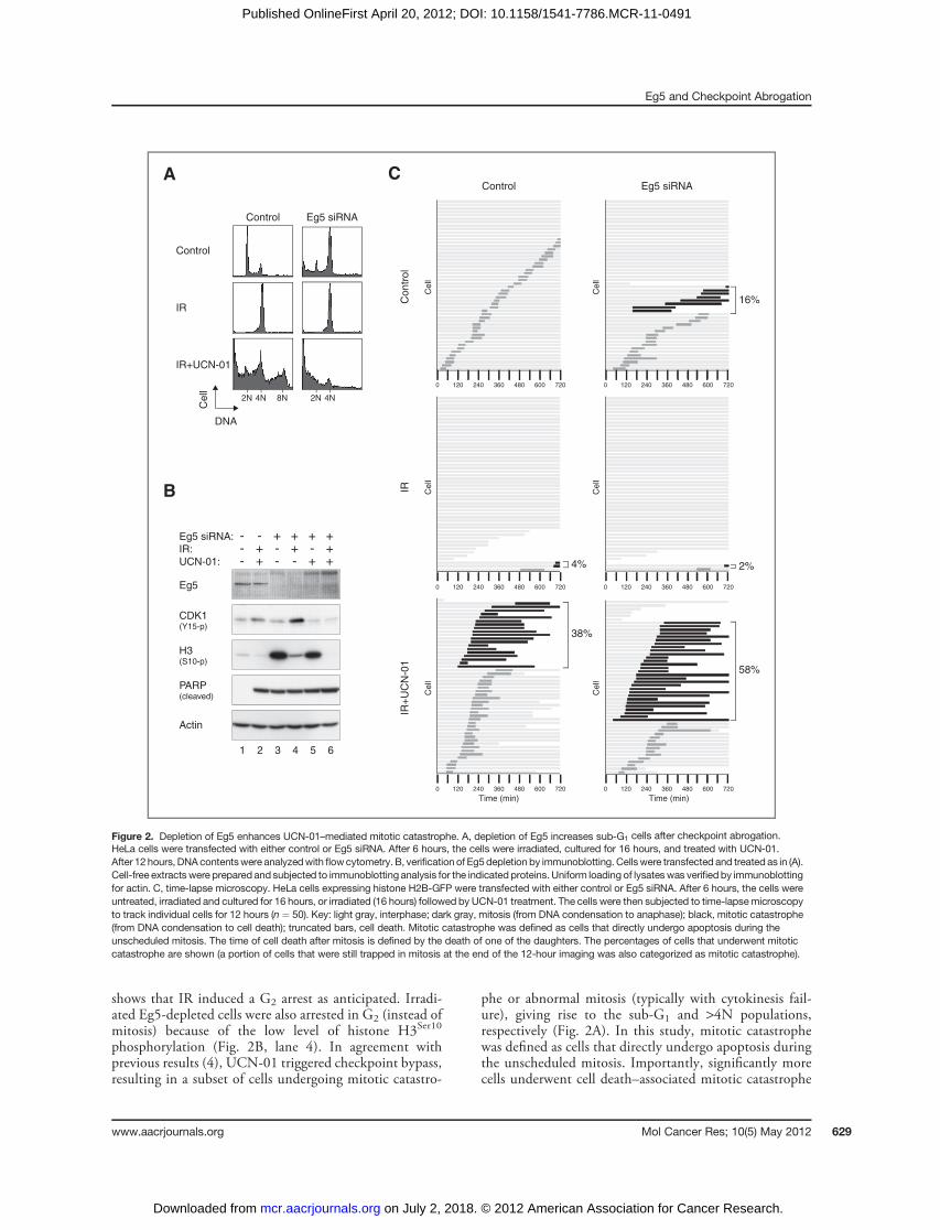
shows that IR induced a G2 arrest as anticipated. Irradi-ated Eg5-depleted cells were also arrested in G2 (instead ofmitosis) because of the low level of histone H3Ser10
phosphorylation (Fig. 2B, lane 4). In agreement withprevious results (4), UCN-01 triggered checkpoint bypass,resulting in a subset of cells undergoing mitotic catastro-
phe or abnormal mitosis (typically with cytokinesis fail-ure), giving rise to the sub-G1 and >4N populations,respectively (Fig. 2A). In this study, mitotic catastrophewas defined as cells that directly undergo apoptosis duringthe unscheduled mitosis. Importantly, significantly morecells underwent cell death–associated mitotic catastrophe
A
B
Control
2N 4N
DNA
Cell
IR
Eg5 siRNA
IR+UCN-01
Control
8N 2N 4N
Eg5 siRNA:
IR:
UCN-01:
Actin
1 2 3 4
Eg5
5 6
CDK1(Y15-p)
H3(S10-p)
---
-++
+--
++-
+-+
+++
PARP(cleaved)
C
0 120 240 360 480 600 720
0 120 240 360 480 600 720
0 120 240 360 480 600 720
0 120 240 360 480 600 720
0 120 240 360 480 600 720
0 120 240 360 480 600 720
Time (min) Time (min)
Cell
Cell
Cell
Cell
Cell
Cell
Control Eg5 siRNA
IRIR
+U
CN
-01
Contr
ol
16%
2%
58%
38%
4%
Figure 2. Depletion of Eg5 enhances UCN-01–mediated mitotic catastrophe. A, depletion of Eg5 increases sub-G1 cells after checkpoint abrogation.HeLa cells were transfected with either control or Eg5 siRNA. After 6 hours, the cells were irradiated, cultured for 16 hours, and treated with UCN-01.After 12 hours, DNA contentswere analyzedwith flowcytometry. B, verification of Eg5depletion by immunoblotting. Cellswere transfected and treated as in (A).Cell-free extracts were prepared and subjected to immunoblotting analysis for the indicated proteins. Uniform loading of lysateswas verifiedby immunoblottingfor actin. C, time-lapse microscopy. HeLa cells expressing histone H2B-GFP were transfected with either control or Eg5 siRNA. After 6 hours, the cells wereuntreated, irradiated and cultured for 16 hours, or irradiated (16 hours) followed by UCN-01 treatment. The cells were then subjected to time-lapsemicroscopyto track individual cells for 12 hours (n ¼ 50). Key: light gray, interphase; dark gray, mitosis (from DNA condensation to anaphase); black, mitotic catastrophe(from DNA condensation to cell death); truncated bars, cell death. Mitotic catastrophe was defined as cells that directly undergo apoptosis during theunscheduled mitosis. The time of cell death after mitosis is defined by the death of one of the daughters. The percentages of cells that underwent mitoticcatastrophe are shown (a portion of cells that were still trapped in mitosis at the end of the 12-hour imaging was also categorized as mitotic catastrophe).
Eg5 and Checkpoint Abrogation
www.aacrjournals.org Mol Cancer Res; 10(5) May 2012 629
on July 2, 2018. © 2012 American Association for Cancer Research. mcr.aacrjournals.org Downloaded from
Published OnlineFirst April 20, 2012; DOI: 10.1158/1541-7786.MCR-11-0491

after Eg5 was downregulated. The same results wereobtained using another siRNA targeting a different regionof Eg5, indicating that the specificity of the effects (datanot shown).
Because of the various cell fates after checkpoint abro-gation, the extent of cell death could not be accuratelyrevealed when the entire cell population was analyzed. Forexample, although the increase in sub-G1 population was
C
1 2 3 4 5 6
Monastrol (µmol/L): 0
Eg5
Cyclin B1
Cyclin A2
50
75
100
150
200
Actin
H3(S10-p)
PARP(cleaved)
Control
2N 4N
DNA
Cell
50 µmol/L
75 µmol/L
100 µmol/L
150 µmol/L
200 µmol/L
Monastrol:A B
0 120 240 360 480 600 720
Time (min)0 120 240 360 480 600 720
Time (min)0 120 240 360 480 600 720
Time (min)0 120 240 360 480 600 720
Time (min)0 120 240 360 480 600 720
Time (min)
0 120 240 360 480 600 720
Time (min)0 120 240 360 480 600 720
Time (min)0 120 240 360 480 600 720
Time (min)0 120 240 360 480 600 720
Time (min)0 120 240 360 480 600 720
Time (min)0 120 240 360 480 600 720
Time (min)
Ce
ll
Ce
ll
Ce
ll
Ce
ll
Ce
ll
Ce
ll
Ce
ll
Ce
ll
Ce
ll
Ce
ll
Ce
ll
50 µmol/L Monastrol 150 µmol/L Monastrol 200 µmol/L Monastrol100 µmol/L Monastrol75 µmol/L Monastrol
50 µmol/L Monastrol 150 µmol/L Monastrol 200 µmol/L Monastrol100 µmol/L Monastrol75 µmol/L MonastrolControl
Contr
ol
44%
48%
6%
2% 12%
24%
62%
68%
86%90% 92%
IR +
UC
N-0
1
E
IR + UCN-01 IR + UCN-01
Monastrol
D
Monastrol (µmol/L): 0 50 75 100 150 200
0
10
20
30
40
Cell
death
(%
)
0
50
100
150
200
250
300
Length
of m
itosis
(m
in)
Figure 3. Monastrol promotes the mitotic catastrophe triggered by IR and UCN-01. A, mitotic arrest and cell death are triggered by monastrol in aconcentration-dependent manner. HeLa cells were treated with the indicated concentrations of monastrol. After incubation for 24 hours, lysates wereprepared and the expression of the indicated proteins was detected by immunoblotting. Equal loading of lysates was confirmed by immunoblotting foractin. B, concentration-dependent accumulation of G2–M and sub-G1 populations by monastrol. Cells were treated exactly as in (A). After incubation for24 hours, the cells were fixed, stained with propidium iodide, and analyzed with flow cytometry. The positions of 2N and 4N DNA contents are indicated.C, mitotic catastrophe is induced by high concentrations of monastrol. HeLa cells expressing histone H2B-GFP were treated with differentconcentrations of monastrol. The cells were then subjected to time-lapse microscopy to track individual cells for 12 hours (n ¼ 50). The duration ofmitosis (from DNA condensation to anaphase; only counting the cells that successfully finished mitosis) was quantified (average �90% confidencecoefficient; top). The percentage of cells undergoing cell death during the imaging period was also quantified (bottom). D, monastrol promotes mitosiscontaining monoastral spindles after IR and UCN-01 treatments. HeLa cells expressing histone H2B-GFP were irradiated. After 16 hours, the cells weretreated with UCN-01 either in the absence or presence of 100 mmol/L of monastrol. The cells were then tracked with time-lapse microscopy to detect thehistone H2B-GFP. Representative mitotic cells are shown (arrows). The complete videos are shown in Supplementary Video S1 and S2. E, monastrolpromotes mitotic catastrophe triggered by IR and UCN-01. HeLa cells expressing histone H2B-GFP were either untreated or irradiated (16 hours)followed by treatments with UCN-01. The cells were incubated with different concentrations of monastrol. The cells were then subjected to time-lapsemicroscopy to track individual cells for 12 hours (n ¼ 50). Key: light gray, interphase; dark gray, mitosis (from DNA condensation to anaphase); black,mitotic catastrophe (from DNA condensation to cell death); truncated bars, cell death. The percentages of cells that underwent mitotic catastrophe areshown (a portion of cells that were still trapped in mitosis at the end of the 12-hour imaging was also categorized as mitotic catastrophe). Quantificationof the data can be found in (C).
Chen et al.
Mol Cancer Res; 10(5) May 2012 Molecular Cancer Research630
on July 2, 2018. © 2012 American Association for Cancer Research. mcr.aacrjournals.org Downloaded from
Published OnlineFirst April 20, 2012; DOI: 10.1158/1541-7786.MCR-11-0491
conspicuous in the absence of Eg5 (Fig. 2A), the increase incleaved PARP signal was only marginal (Fig. 2B). To obtainmore quantitative data on different cell fates, time-lapsemicroscopy was used to track individual cells after check-point abrogation. HeLa cells expressing histone H2B-GFPwere used, allowing us to simultaneous image both cellmorphology and DNA. Figure 2C shows that most controlcells entered mitosis once over the 12-hour imaging period.In contrast, mitosis was attenuated after irradiation. Thiscell-cycle arrest was overcome after the addition of UCN-01,with approximately 80% of the cells entering mitosis within4 hours. As expected, a subset of the checkpoint-abrogatedcells underwent cell death during mitosis. Knockdown ofEg5 caused a slight increase in mitotic catastrophe inunperturbed cell cycle. Furthermore, neither the IR-medi-ated cell-cycle arrest nor the extent of UCN-01–mediatedcheckpoint bypass was affected in Eg5-depleted cells. Nev-ertheless, markedly more Eg5-depleted cells underwentmitotic catastrophe after checkpoint abrogation than controlcells. Taken together, these observations revealed that down-regulation of Eg5 sensitizes cells to mitotic catastrophecaused by IR and UCN-01.
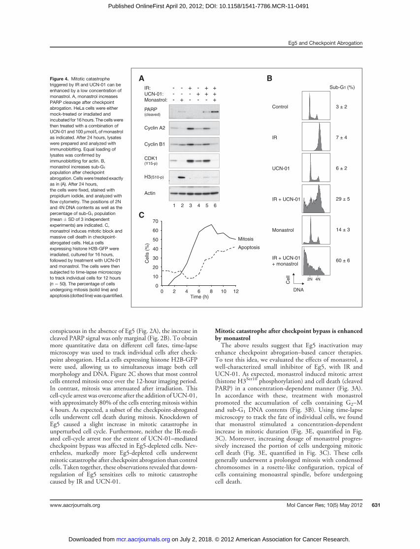
Mitotic catastrophe after checkpoint bypass is enhancedby monastrolThe above results suggest that Eg5 inactivation may
enhance checkpoint abrogation–based cancer therapies.To test this idea, we evaluated the effects of monastrol, awell-characterized small inhibitor of Eg5, with IR andUCN-01. As expected, monastrol induced mitotic arrest(histone H3Ser10 phosphorylation) and cell death (cleavedPARP) in a concentration-dependent manner (Fig. 3A).In accordance with these, treatment with monastrolpromoted the accumulation of cells containing G2–Mand sub-G1 DNA contents (Fig. 3B). Using time-lapsemicroscopy to track the fate of individual cells, we foundthat monastrol stimulated a concentration-dependentincrease in mitotic duration (Fig. 3E, quantified in Fig.3C). Moreover, increasing dosage of monastrol progres-sively increased the portion of cells undergoing mitoticcell death (Fig. 3E, quantified in Fig. 3C). These cellsgenerally underwent a prolonged mitosis with condensedchromosomes in a rosette-like configuration, typical ofcells containing monoastral spindle, before undergoingcell death.
Figure 4. Mitotic catastrophetriggered by IR and UCN-01 can beenhanced by a low concentration ofmonastrol. A, monastrol increasesPARP cleavage after checkpointabrogation. HeLa cells were eithermock-treated or irradiated andincubated for 16 hours. The cells werethen treated with a combination ofUCN-01 and 100 mmol/L ofmonastrolas indicated. After 24 hours, lysateswere prepared and analyzed withimmunoblotting. Equal loading oflysates was confirmed byimmunoblotting for actin. B,monastrol increases sub-G1
population after checkpointabrogation. Cells were treated exactlyas in (A). After 24 hours,the cells were fixed, stained withpropidium iodide, and analyzed withflow cytometry. The positions of 2Nand 4N DNA contents as well as thepercentage of sub-G1 population(mean � SD of 3 independentexperiments) are indicated. C,monastrol induces mitotic block andmassive cell death in checkpoint-abrogated cells. HeLa cellsexpressing histone H2B-GFP wereirradiated, cultured for 16 hours,followed by treatment with UCN-01and monastrol. The cells were thensubjected to time-lapse microscopyto track individual cells for 12 hours(n ¼ 50). The percentage of cellsundergoing mitosis (solid line) andapoptosis (dotted line)wasquantified.
A
UCN-01
2N 4N
DNAC
ell
Control
IR
Monastrol
IR + UCN-01
IR + UCN-01
+ monastrol
Sub-G1 (%)
3 ± 2
14 ± 3
7 ± 4
6 ± 2
29 ± 5
60 ± 6
B
PARP(cleaved)
IR:
UCN-01:
Monastrol:
---
Actin
1 2 3 4 5 6
CDK1(Y15-p)
H3(S10-p)
--+
-+-
+--
++-
+++
Cyclin A2
Cyclin B1
0
10
20
30
40
50
60
70
0 2 4 6 8 10 12
Mitosis
Apoptosis
Time (h)
Ce
lls (
%)
C
Eg5 and Checkpoint Abrogation
www.aacrjournals.org Mol Cancer Res; 10(5) May 2012 631
on July 2, 2018. © 2012 American Association for Cancer Research. mcr.aacrjournals.org Downloaded from
Published OnlineFirst April 20, 2012; DOI: 10.1158/1541-7786.MCR-11-0491

We next combined different concentrations of monastrolwith IR and UCN-01 treatments and monitored the cellswith time-lapse microscopy (Fig. 3E). Challenging cells with100 mmol/L of monastrol alone did not induce substantialcell death. This was evident even when the cells werefollowed for more than a period of 24 hours (SupplementaryFig. S1). Compared with cells treated with IR and UCN-01only, addition of 100 mmol/L of monastrol substantiallyincreased the percentage of cells undergoing mitotic catas-trophe (from �40%–90%). Although higher concentra-tions of monastrol also induced similar increase in mitoticcatastrophe, they already triggered significant level ofmitoticcatastrophe even in the absence of IR andUCN-01 (Fig. 3E).As expected, while IR- and UCN-01–treated cells couldundergo a relatively normal mitosis, no metaphase plate wasformed in the presence of monastrol (Fig. 3D).The above results were further confirmed by immuno-
blotting and flow cytometry. The level of cleaved PARP incells treated with IR, UCN-01, and 100 mmol/L of mon-astrol was higher than in cells receiving the individualtreatments separately (Fig. 4A). Likewise, the sub-G1 pop-ulation was significantly higher in the presence of monastrol
than in cells challenged with IR andUCN-01 only (Fig. 4B).These data are consistent with the time-dependent accumu-lation of mitotic cells and massive apoptotic cell death afterthe irradiated cells were treated withUCN-01 andmonastrol(Fig. 4C; compared with cells treated with IR and UCN-01only in Fig. 1B).To ensure that the effects of UCN-01 was specific to
inhibition of CHK1, we also depleted CHK1 using siRNA.Similar to after UCN-01 treatment, depletion of CHK1abolished the IR-mediated cell-cycle arrest (SupplementaryFig. S2). Mitotic catastrophe was significantly increased inthe presence of monastrol.To verify that themitotic catastrophe caused bymonastrol
was not limited to HeLa cells, we next carried out the sametreatments on another cell line. The presence of Eg5 proteinin H1299, HCT116, and U2OS was confirmed by immu-noblotting (Fig. 5A). H1299 cells were selected because theyexpressed high level of Eg5. Furthermore, unlike HeLa cells,H1299 cells were relatively resistant to the mitotic catas-trophe mediated by IR and UCN-01. Live cell imagingrevealed that 80% of cells survived mitosis induced after IRand UCN-01 treatments (4). Consistent with this, IR
Actin
1 2 3 4
Eg5
-Eg5 siRNA: +
5
- - -HeL
a
HeL
a
H12
99
HCT11
6
U2O
S
UCN-01
2N 4N
DNA
Ce
ll
Control
IR
Monastrol
IR + UCN-01
IR + UCN-01
+ monastrol
Sub-G1 (%)
1 ± 0.1
6 ± 1
5 ± 2
2 ± 2
9 ± 1
20 ± 2
B
C
A
0
5
10
15
20
25
30
Ce
ll d
ea
th (
%)
IR:
UCN-01:
Monastrol:
---
--+
-+-
+--
++-
+++
Figure 5. Monastrol enhancesmitotic catastrophe in H1299 cells.A, expressionof Eg5 indifferent celllines. Lysates fromasynchronouslygrowing H1299, HCT116, andU2OS were subjected toimmunoblotting for Eg5. HeLa cellstransfected with control or Eg5siRNA served as controls. Actinanalysis was included to assessprotein loading and transfer. B,monastrol increases sub-G1
population after checkpointabrogation.H1299cellswere eithermock-treated or irradiated andincubated for 16 hours. The cellswere treated with a combination ofUCN-01 and 100 mmol/L ofmonastrol as indicated. After 48hours, the cells were fixed, stainedwith propidium iodide, andanalyzed with flow cytometry. Thepositions of 2N and 4N DNAcontents as well as the percentageof sub-G1 population (mean � SDof 3 independent experiments) areindicated. C, monastrol increasescell death after checkpointabrogation. H1299 cells weretreated exactly as in (B). Viabilitywas determined with Trypan blueexclusion assay. Mean � SD of 3independent experiments areshown.
Chen et al.
Mol Cancer Res; 10(5) May 2012 Molecular Cancer Research632
on July 2, 2018. © 2012 American Association for Cancer Research. mcr.aacrjournals.org Downloaded from
Published OnlineFirst April 20, 2012; DOI: 10.1158/1541-7786.MCR-11-0491

followed by UCN-01 treatments induced only a low per-centage of sub-G1 cells (Fig. 5B).Nevertheless, apoptotic celldeath was increased in the presence of 100 mmol/L ofmonastrol (from 9% to 22%). Similar conclusions wereobtained by measuring cell death by Trypan blue exclusionassays (Fig. 5C).Collectively, these results indicate that a relatively low
concentration of monastrol, itself not causing significant celldeath, can promote the cell death induced with IR andUCN-01.
Monastrol and checkpoint abrogation reduce long-termcell survivalWe next examined the effects of checkpoint abrogation
and monastrol on long-term survival of cancer cells byclonogenic survival assays. Results using IR alone indicatedthat approximately 75% and 50% survival after cells wereirradiated with approximately 1 and 2 Gy of IR, respectively(Fig. 6A). Likewise, titration with different concentrations ofmonastrol alone indicated that more than 80% clonogenicsurvival after cells were treatedwith 100mmol/L ofmonastrol(Fig. 6B). Cells were next treated with different doses of IR,together with UCN-01 and monastrol (100 mmol/L). After12 hours incubation, the UCN-01 and monastrol werewashed away; and the cells were incubated further forclonogenic survival assays. Figure 6C shows that althoughtreatment with IR and UCN-01 caused some degree of cellgrowth inhibition, addition of monastrol reduced the clono-genic survival further. Collectively, our data indicate that cellproliferation could be effectively attenuated using a DNAdamage and checkpoint abrogation strategy in conjunctionwith the inhibition of Eg5 with monastrol.
DiscussionIn this study, we tested the idea of whether the combi-
nation of the G2 DNA damage checkpoint, checkpointabrogation, and Eg5 inhibition could enhance the popula-tion of cells undergoingmitotic catastrophe. The central ideais that cells exhibit different fates after checkpoint abro-gation. They either undergo apoptosis during mitosis orprogress into the following interphase. It is likely that thesurvived cells are more prone to further genome instabilityand may develop into more aggressive tumors. Thereforefrom the view point of cancer therapies, it is desirable toeliminate as many cancer cells as possible during the firstmitosis.For HeLa cells treated with IR followed by UCN-01,
more than 90% of the cells were forced into mitosis pre-maturely (Figs. 2C and 3E). While approximately 40% cellsunderwent mitotic catastrophe, the rest of the cells were ableto survive and enter G1 phase. We found that mitoticcatastrophe was enhanced after Eg5 was inactivated, eitherwith siRNAs (Fig. 2) or monastrol (Figs. 3E and 4). This wasconfirmed by assays including measurement of PARP cleav-age (Figs. 2B and 4A), sub-G1 cells (Figs. 2A and 4B), andlive cell imaging (Figs. 2C and 3E).In agreement with the increase in checkpoint abrogation–
mediated mitotic catastrophe by Eg5 inhibition, we found
B
A
0
25
50
75
100
Nu
mb
er
of
co
lon
ies
(% m
ax)
IR (Gy): 0 0.05 0.1 0.2 0.4 0.9 1.8 2.5 5 10 15
0
20
40
60
80
100
120
0 50 75 100 150 200Monastrol (µmol/L):N
um
be
r o
f co
lon
ies
(% m
ax)
C
0
20
40
60
80
100
120
Nu
mb
er
of
co
lon
ies
(% m
ax)
IR (Gy):
UCN-01:
Monastrol:--
+-
-+
++
0--
+-
-+
++
0.5--
+-
-+
++
1--
+-
-+
++
2
Figure 6. Monastrol and checkpoint abrogation reduce long-term cellsurvival. A, dose-dependent inhibition of clonogenic survival by IR. HeLacells were irradiatedwith different doses of IR as indicated. After 2weeks,the colonies were fixed and visualized by staining with crystal violet. Thenumber of colonies was quantified. B, concentration-dependentinhibition of clonogenic survival by monastrol. HeLa cells were treatedwith different concentrations of monastrol. After 16 hours of incubation,the cells were washed and reseeded. After 2 weeks, the colonies werefixed, visualized by stainingwith crystal violet, and quantified.Mean�SDof 3 independent experiments are shown. C, monastrol further reducesthe clonogenic survival caused by checkpoint abrogation. HeLa cellsweremock-irradiated or irradiatedwith 0.5, 1, or 2Gyof IR. The cells weretreated with UCN-01 in the absence or presence of monastrol asindicated. After 16 hours of incubation, the cells were washed andreseeded. After 2 weeks, the colonies were fixed, visualized by stainingwith crystal violet, and quantified. Mean � SD of 3 independentexperiments are shown.
Eg5 and Checkpoint Abrogation
www.aacrjournals.org Mol Cancer Res; 10(5) May 2012 633
on July 2, 2018. © 2012 American Association for Cancer Research. mcr.aacrjournals.org Downloaded from
Published OnlineFirst April 20, 2012; DOI: 10.1158/1541-7786.MCR-11-0491
that monastrol reduced the clonogenic survival after IR andUCN-01 treatment (Fig. 6C). A caveat is that UCN-01alone also reduced cell survival. This DNA damage-inde-pendent cell death was presumably due to the functions ofCHK1 in normal DNA replicative folk progression. The celldeath induced by UCN-01 alone cannot be simply additivewith other treatments when used in combination. This isbecause in our study, UCN-01 was applied to IR-treatedcells only after they have already passed S-phase and arrestedin G2 phase. This again highlights the importance of theorder of the treatments: checkpoint bypass and Eg5 inhibi-tion should be applied only after the DNA damage check-point is activated.Corroboration with checkpoint abrogation probably did
not require the complete inactivation of Eg5 functions, asresidual Eg5 was presumably still present after siRNA-mediated depletion (Fig. 2B). Furthermore, a relatively lowconcentration of monastrol (100 mmol/L) was sufficient topromote mitotic catastrophe (Fig. 3E). Although treatmentwith this concentration of monastrol alone increased theduration of mitosis [as indicated by both histone H3Ser10
phosphorylation (Fig. 3A) and direct measurement of mitot-ic time (Fig. 3C)], it neither blocked cells in mitosis norinduced cell death [as indicated by flow cytometry (Fig. 3B)and live cell imaging (Fig. 3E and Supplementary Fig. S1)].We believe that the extension of mitosis caused by Eg5siRNA or 100 mmol/L of monastrol was sufficient inpromoting mitotic catastrophe. This is in agreement withthe conclusion of our previous studies, in which mitoticcatastrophe was increased when mitosis was extended bydepletion of p31comet or CDC20 (4). Interestingly, not allthe drugs that delayed mitosis we tested were able to increasecell death associated with IR and UCN-01 treatments. Forexample, inhibitors of PLK1 or Aurora A were less effectivein this regard (our unpublished data). The underlyingreasons and which other types of drugs are effective inpromoting mitotic catastrophe awaits further investigation.Other factors that govern the extent of mitotic catastrophe
after checkpoint abrogation are the dose of IR and the natureof the cell line (4). IR treatment followed by checkpointabrogation in other cell lines, including H1299, HCT116,U2OS, induced significantly less mitotic catastrophe thanHeLa cells (4). However, we found that monastrol was alsoable to sensitize H1299 cells after checkpoint abrogation(Fig. 5B and C). Hence, it is likely that the combinedtreatment with monastrol can be beneficial for treating
cancer cells that exhibit resistance to IR and UCN-01. Forfuture clinical use, the order of the treatments is likely to beof crucial importance. Our studies indicated that the Eg5inhibitor and checkpoint abrogator should be introducedonly after the DNA damage checkpoint is activated.The clinical use of monastrol is limited by the high
dosages needed to achieve complete Eg5 inhibition (18).Moreover, such high dosages of monastrol are associatedwith a variety of side effects, including neurotoxicity.Sensory neurons appear to be particularly sensitive toprolonged exposure of monastrol (18). Monastrol was alsoshown to affect growth of dendrites and axons in primarycortical neuron cultures (19). By combining checkpointabrogation and Eg5 inhibition, a relatively low concentra-tion of monastrol was already highly effective in enhancingmitotic catastrophe. This should reduce mitotic arrest andsubsequent apoptosis for normal mitotic cells and mini-mize the neurotoxicity. Moreover, the principle of thecurrent study should be applicable to other newer gener-ation of Eg5 inhibitors at various stages of developmentand clinical trials (20). Whether newer generation of Eg5inhibitors can promote mitotic catastrophe more effectivelythan monastrol should warrant further cell line and animalstudies.In conclusion, we found that although not all cancer
cells were eliminated by mitotic catastrophe after check-point abrogation, cell death could be enhanced by inhibitionof Eg5 with either siRNAs or monastrol. A relatively lowconcentration of monastrol, alone not sufficient in causingmitotic arrest, was effective in promotingmitotic catastrophewhen combined with checkpoint abrogation.
Disclosure of Potential Conflicts of InterestNo potential conflicts of interest were disclosed.
AcknowledgmentsThe authors thankmembers of the Poon laboratory for constructive criticism on the
manuscript.
Grant SupportThis work was supported in part by the Research Grants Council grants 662208
and AOE-MG/M-08/06 to R.Y.C. Poon.The costs of publication of this article were defrayed in part by the payment of page
charges. This article must therefore be herebymarked advertisement in accordance with18 U.S.C. Section 1734 solely to indicate this fact.
Received October 12, 2011; revised January 23, 2012; accepted March 14, 2012;published OnlineFirst April 20, 2012.
References1. Ma HT, Poon RYC. How protein kinases co-ordinate mitosis in animal
cells. Biochem J 2011;435:17–31.2. Smith J, Tho LM, Xu N, Gillespie DA. The ATM-Chk2 and ATR-Chk1
pathways in DNA damage signaling and cancer. Adv Cancer Res2010;108:73–112.
3. Vitale I, Galluzzi L, Castedo M, Kroemer G. Mitotic catastrophe: amechanism for avoiding genomic instability. Nat Rev Mol Cell Biol2011;12:385–92.
4. On KF, Chen Y, Ma HT, Chow JP, Poon RYC. Determinantsof mitotic catastrophe on abrogation of the G2 DNA
damage checkpoint by UCN-01. Mol Cancer Ther 2011;10:784–94.
5. Lawrence CJ, Dawe RK, Christie KR, Cleveland DW, Dawson SC,Endow SA, et al. A standardized kinesin nomenclature. J Cell Biol2004;167:19–22.
6. Cochran JC, Sontag CA, Maliga Z, Kapoor TM, Correia JJ, Gilbert SP.Mechanistic analysis of the mitotic kinesin Eg5. J Biol Chem2004;279:38861–70.
7. Ding S, Xing N, Lu J, Zhang H, Nishizawa K, Liu S, et al. Over-expression of Eg5 predicts unfavorable prognosis in non-
Chen et al.
Mol Cancer Res; 10(5) May 2012 Molecular Cancer Research634
on July 2, 2018. © 2012 American Association for Cancer Research. mcr.aacrjournals.org Downloaded from
Published OnlineFirst April 20, 2012; DOI: 10.1158/1541-7786.MCR-11-0491
muscle invasive bladder urothelial carcinoma. Int J Urol 2011;18:432–8.
8. LiuM,Wang X, Yang Y, Li D, Ren H, ZhuQ, et al. Ectopic expression ofthe microtubule-dependent motor protein Eg5 promotes pancreatictumourigenesis. J Pathol 2010;221:221–8.
9. Castillo A, Morse HCr, Godfrey VL, NaeemR, JusticeMJ. Overexpres-sion of Eg5 causes genomic instability and tumor formation in mice.Cancer Res 2007;67:10138–47.
10. Mayer TU, Kapoor TM,Haggarty SJ, KingRW,Schreiber SL,MitchisonTJ. Small molecule inhibitor of mitotic spindle bipolarity identified in aphenotype-based screen. Science 1999;286:971–4.
11. Huszar D, Theoclitou ME, Skolnik J, Herbst R. Kinesin motor proteinsas targets for cancer therapy. Cancer Metastasis Rev 2009;28:197–208.
12. Yam CH, Siu WY, Lau A, Poon RYC. Degradation of cyclin A does notrequire its phosphorylation by CDC2 and cyclin-dependent kinase 2. JBiol Chem 2000;275:3158–67.
13. ChanYW,MaHT,WongW,HoCC,OnKF, PoonRYC.CDK1 inhibitorsantagonize the immediate apoptosis triggered by spindle disruptionbut promote apoptosis following the subsequent rereplication andabnormal mitosis. Cell Cycle 2008;7:1449–61.
14. Siu WY, Arooz T, Poon RYC. Differential responses of proliferatingversus quiescent cells to adriamycin. Exp Cell Res 1999;250:131–41.
15. Chan YW, On KF, Chan WM, Wong W, Siu HO, Hau PM, et al.The kinetics of p53 activation versus cyclin E accumulationunderlies the relationship between the spindle-assembly check-point and the postmitotic checkpoint. J Biol Chem 2008;283:15716–23.
16. Yam CH, Siu WY, Kaganovich D, Ruderman JV, Poon RYC. Cleavageof cyclin A at R70/R71 by the bacterial protease OmpT. Proc Natl AcadSci U S A 2001;98:497–501.
17. Poon RYC, Toyoshima H, Hunter T. Redistribution of the CDK inhibitorp27 between different cyclin.CDK complexes in the mouse fibroblastcell cycle and in cells arrested with lovastatin or ultraviolet irradiation.Mol Biol Cell 1995;6:1197–213.
18. Haque SA, Hasaka TP, Brooks AD, Lobanov PV, Baas PW. Monastrol,a prototype anti-cancer drug that inhibits a mitotic kinesin, inducesrapid bursts of axonal outgrowth from cultured postmitotic neurons.Cell Motil Cytoskeleton 2004;58:10–6.
19. Yoon SY, Choi JE, Huh JW, Hwang O, Lee HS, Hong HN, et al.Monastrol, a selective inhibitor of the mitotic kinesin Eg5, induces adistinctive growth profile of dendrites and axons in primary corticalneuron cultures. Cell Motil Cytoskeleton 2005;60:181–90.
20. Knight SD,ParrishCA.Recent progress in the identification andclinicalevaluation of inhibitors of themitotic kinesin KSP. Curr TopMedChem2008;8:888–904.
Eg5 and Checkpoint Abrogation
www.aacrjournals.org Mol Cancer Res; 10(5) May 2012 635
on July 2, 2018. © 2012 American Association for Cancer Research. mcr.aacrjournals.org Downloaded from
Published OnlineFirst April 20, 2012; DOI: 10.1158/1541-7786.MCR-11-0491

2012;10:626-635. Published OnlineFirst April 20, 2012.Mol Cancer Res Yue Chen, Jeremy P.H. Chow and Randy Y.C. Poon in Promoting Mitotic CatastropheInhibition of Eg5 Acts Synergistically with Checkpoint Abrogation
Updated version
10.1158/1541-7786.MCR-11-0491doi:
Access the most recent version of this article at:
Material
Supplementary
http://mcr.aacrjournals.org/content/suppl/2012/03/20/1541-7786.MCR-11-0491.DC1
Access the most recent supplemental material at:
Cited articles
http://mcr.aacrjournals.org/content/10/5/626.full#ref-list-1
This article cites 20 articles, 10 of which you can access for free at:
Citing articles
http://mcr.aacrjournals.org/content/10/5/626.full#related-urls
This article has been cited by 2 HighWire-hosted articles. Access the articles at:
E-mail alerts related to this article or journal.Sign up to receive free email-alerts
Subscriptions
Reprints and
To order reprints of this article or to subscribe to the journal, contact the AACR Publications Department at
Permissions
Rightslink site. Click on "Request Permissions" which will take you to the Copyright Clearance Center's (CCC)
.http://mcr.aacrjournals.org/content/10/5/626To request permission to re-use all or part of this article, use this link
on July 2, 2018. © 2012 American Association for Cancer Research. mcr.aacrjournals.org Downloaded from
Published OnlineFirst April 20, 2012; DOI: 10.1158/1541-7786.MCR-11-0491


![Inhibition of Eg5 Acts Synergistically with Checkpoint ...Eg5 [kinesin spindle protein (KSP), HKSP, KNSL1, TRIP5, or kinesin family member 11 (KIF11)] is a plus-end directed Authors'](https://static.fdocuments.us/doc/165x107/606c1b0397e9f26c261c3873/inhibition-of-eg5-acts-synergistically-with-checkpoint-eg5-kinesin-spindle.jpg)