
Inhaled Antibiotics for Gram-Negative Respiratory Infections · resistance protein 1 and breast...
52
Inhaled Antibiotics for Gram-Negative Respiratory Infections Eric Wenzler, a Dustin R. Fraidenburg, b Tonya Scardina, c Larry H. Danziger a,d University of Illinois at Chicago, College of Pharmacy, Chicago, Illinois, USA a ; Department of Medicine, Division of Pulmonary, Critical Care, Sleep and Allergy Medicine, University of Illinois at Chicago, Chicago, Illinois, USA b ; Loyola University Medical Center, Chicago, Illinois, USA c ; University of Illinois at Chicago, College of Medicine, Chicago, Illinois, USA d SUMMARY ..................................................................................................................................................582 INTRODUCTION ............................................................................................................................................582 FORMULATION AND DRUG DELIVERY .....................................................................................................................584 Pulmonary Physiology ....................................................................................................................................584 Drug Deposition Considerations .........................................................................................................................584 Particle Engineering ......................................................................................................................................585 Delivery Devices ..........................................................................................................................................586 Nebulizers .............................................................................................................................................586 Inhalers ................................................................................................................................................587 Administration Technique................................................................................................................................588 IN VITRO, PK/PD, AND MICROBIOLOGICAL CONSIDERATIONS ...........................................................................................588 Inhaled Antibiotic Admixtures............................................................................................................................588 Pharmacokinetics and Pharmacodynamics of Inhaled Antibiotic Compounds ..........................................................................589 Microbiological Considerations ..........................................................................................................................590 PULMONARY PHARMACOKINETICS .......................................................................................................................591 Animals ..................................................................................................................................................591 Tobramycin ............................................................................................................................................591 Gentamicin ............................................................................................................................................592 Amikacin ...............................................................................................................................................592 Imipenem-cilastatin....................................................................................................................................593 Ceftazidime ............................................................................................................................................593 Fluoroquinolones ......................................................................................................................................594 Colistin .................................................................................................................................................594 Humans ..................................................................................................................................................595 Tobramycin ............................................................................................................................................595 Gentamicin ............................................................................................................................................596 Amikacin ...............................................................................................................................................596 Ciprofloxacin...........................................................................................................................................597 Colistin .................................................................................................................................................598 CLINICAL OUTCOMES ......................................................................................................................................599 Non-CF Bronchiectasis ...................................................................................................................................600 Tobramycin ............................................................................................................................................601 Gentamicin ............................................................................................................................................601 Ciprofloxacin...........................................................................................................................................601 Colistin .................................................................................................................................................601 Aztreonam .............................................................................................................................................602 Summary of inhaled antibiotics for non-CF bronchiectasis ............................................................................................602 Ventilator-Associated Tracheobronchitis .................................................................................................................603 Gentamicin ............................................................................................................................................604 Polymyxins .............................................................................................................................................605 Summary of inhaled antibiotics for VAT................................................................................................................605 (continued) Published 25 May 2016 Citation Wenzler E, Fraidenburg DR, Scardina T, Danziger LH. 2016. Inhaled antibiotics for Gram-negative respiratory infections. Clin Microbiol Rev 29:581– 632. doi:10.1128/CMR.00101-15. Address correspondence to Larry H. Danziger, [email protected]. Copyright © 2016, American Society for Microbiology. All Rights Reserved. crossmark July 2016 Volume 29 Number 3 cmr.asm.org 581 Clinical Microbiology Reviews on March 17, 2019 by guest http://cmr.asm.org/ Downloaded from
-
Upload
vuongkhanh -
Category
Documents
-
view
217 -
download
0
Transcript of Inhaled Antibiotics for Gram-Negative Respiratory Infections · resistance protein 1 and breast...

Inhaled Antibiotics for Gram-Negative Respiratory Infections
Eric Wenzler,a Dustin R. Fraidenburg,b Tonya Scardina,c Larry H. Danzigera,d
University of Illinois at Chicago, College of Pharmacy, Chicago, Illinois, USAa; Department of Medicine, Division of Pulmonary, Critical Care, Sleep and Allergy Medicine,University of Illinois at Chicago, Chicago, Illinois, USAb; Loyola University Medical Center, Chicago, Illinois, USAc; University of Illinois at Chicago, College of Medicine,Chicago, Illinois, USAd
SUMMARY . . . . . . . . . . . . . . . . . . . . . . . . . . . . . . . . . . . . . . . . . . . . . . . . . . . . . . . . . . . . . . . . . . . . . . . . . . . . . . . . . . . . . . . . . . . . . . . . . . . . . . . . . . . . . . . . . . . . . . . . . . . . . . . . . . . . . . . . . . . . . . . . . .582INTRODUCTION . . . . . . . . . . . . . . . . . . . . . . . . . . . . . . . . . . . . . . . . . . . . . . . . . . . . . . . . . . . . . . . . . . . . . . . . . . . . . . . . . . . . . . . . . . . . . . . . . . . . . . . . . . . . . . . . . . . . . . . . . . . . . . . . . . . . . . . . . . . .582FORMULATION AND DRUG DELIVERY . . . . . . . . . . . . . . . . . . . . . . . . . . . . . . . . . . . . . . . . . . . . . . . . . . . . . . . . . . . . . . . . . . . . . . . . . . . . . . . . . . . . . . . . . . . . . . . . . . . . . . . . . . . . . . . . . . . . .584
Pulmonary Physiology. . . . . . . . . . . . . . . . . . . . . . . . . . . . . . . . . . . . . . . . . . . . . . . . . . . . . . . . . . . . . . . . . . . . . . . . . . . . . . . . . . . . . . . . . . . . . . . . . . . . . . . . . . . . . . . . . . . . . . . . . . . . . . . . . . . .584Drug Deposition Considerations . . . . . . . . . . . . . . . . . . . . . . . . . . . . . . . . . . . . . . . . . . . . . . . . . . . . . . . . . . . . . . . . . . . . . . . . . . . . . . . . . . . . . . . . . . . . . . . . . . . . . . . . . . . . . . . . . . . . . . . . .584Particle Engineering . . . . . . . . . . . . . . . . . . . . . . . . . . . . . . . . . . . . . . . . . . . . . . . . . . . . . . . . . . . . . . . . . . . . . . . . . . . . . . . . . . . . . . . . . . . . . . . . . . . . . . . . . . . . . . . . . . . . . . . . . . . . . . . . . . . . . .585Delivery Devices . . . . . . . . . . . . . . . . . . . . . . . . . . . . . . . . . . . . . . . . . . . . . . . . . . . . . . . . . . . . . . . . . . . . . . . . . . . . . . . . . . . . . . . . . . . . . . . . . . . . . . . . . . . . . . . . . . . . . . . . . . . . . . . . . . . . . . . . . .586
Nebulizers . . . . . . . . . . . . . . . . . . . . . . . . . . . . . . . . . . . . . . . . . . . . . . . . . . . . . . . . . . . . . . . . . . . . . . . . . . . . . . . . . . . . . . . . . . . . . . . . . . . . . . . . . . . . . . . . . . . . . . . . . . . . . . . . . . . . . . . . . . . . .586Inhalers . . . . . . . . . . . . . . . . . . . . . . . . . . . . . . . . . . . . . . . . . . . . . . . . . . . . . . . . . . . . . . . . . . . . . . . . . . . . . . . . . . . . . . . . . . . . . . . . . . . . . . . . . . . . . . . . . . . . . . . . . . . . . . . . . . . . . . . . . . . . . . . .587
Administration Technique. . . . . . . . . . . . . . . . . . . . . . . . . . . . . . . . . . . . . . . . . . . . . . . . . . . . . . . . . . . . . . . . . . . . . . . . . . . . . . . . . . . . . . . . . . . . . . . . . . . . . . . . . . . . . . . . . . . . . . . . . . . . . . . .588IN VITRO, PK/PD, AND MICROBIOLOGICAL CONSIDERATIONS . . . . . . . . . . . . . . . . . . . . . . . . . . . . . . . . . . . . . . . . . . . . . . . . . . . . . . . . . . . . . . . . . . . . . . . . . . . . . . . . . . . . . . . . . . .588
Inhaled Antibiotic Admixtures. . . . . . . . . . . . . . . . . . . . . . . . . . . . . . . . . . . . . . . . . . . . . . . . . . . . . . . . . . . . . . . . . . . . . . . . . . . . . . . . . . . . . . . . . . . . . . . . . . . . . . . . . . . . . . . . . . . . . . . . . . . .588Pharmacokinetics and Pharmacodynamics of Inhaled Antibiotic Compounds . . . . . . . . . . . . . . . . . . . . . . . . . . . . . . . . . . . . . . . . . . . . . . . . . . . . . . . . . . . . . . . . . . . . . . . . . .589Microbiological Considerations . . . . . . . . . . . . . . . . . . . . . . . . . . . . . . . . . . . . . . . . . . . . . . . . . . . . . . . . . . . . . . . . . . . . . . . . . . . . . . . . . . . . . . . . . . . . . . . . . . . . . . . . . . . . . . . . . . . . . . . . . .590
PULMONARY PHARMACOKINETICS . . . . . . . . . . . . . . . . . . . . . . . . . . . . . . . . . . . . . . . . . . . . . . . . . . . . . . . . . . . . . . . . . . . . . . . . . . . . . . . . . . . . . . . . . . . . . . . . . . . . . . . . . . . . . . . . . . . . . . .591Animals . . . . . . . . . . . . . . . . . . . . . . . . . . . . . . . . . . . . . . . . . . . . . . . . . . . . . . . . . . . . . . . . . . . . . . . . . . . . . . . . . . . . . . . . . . . . . . . . . . . . . . . . . . . . . . . . . . . . . . . . . . . . . . . . . . . . . . . . . . . . . . . . . .591
Tobramycin . . . . . . . . . . . . . . . . . . . . . . . . . . . . . . . . . . . . . . . . . . . . . . . . . . . . . . . . . . . . . . . . . . . . . . . . . . . . . . . . . . . . . . . . . . . . . . . . . . . . . . . . . . . . . . . . . . . . . . . . . . . . . . . . . . . . . . . . . . . .591Gentamicin . . . . . . . . . . . . . . . . . . . . . . . . . . . . . . . . . . . . . . . . . . . . . . . . . . . . . . . . . . . . . . . . . . . . . . . . . . . . . . . . . . . . . . . . . . . . . . . . . . . . . . . . . . . . . . . . . . . . . . . . . . . . . . . . . . . . . . . . . . . .592Amikacin . . . . . . . . . . . . . . . . . . . . . . . . . . . . . . . . . . . . . . . . . . . . . . . . . . . . . . . . . . . . . . . . . . . . . . . . . . . . . . . . . . . . . . . . . . . . . . . . . . . . . . . . . . . . . . . . . . . . . . . . . . . . . . . . . . . . . . . . . . . . . . .592Imipenem-cilastatin. . . . . . . . . . . . . . . . . . . . . . . . . . . . . . . . . . . . . . . . . . . . . . . . . . . . . . . . . . . . . . . . . . . . . . . . . . . . . . . . . . . . . . . . . . . . . . . . . . . . . . . . . . . . . . . . . . . . . . . . . . . . . . . . . . . .593Ceftazidime . . . . . . . . . . . . . . . . . . . . . . . . . . . . . . . . . . . . . . . . . . . . . . . . . . . . . . . . . . . . . . . . . . . . . . . . . . . . . . . . . . . . . . . . . . . . . . . . . . . . . . . . . . . . . . . . . . . . . . . . . . . . . . . . . . . . . . . . . . . .593Fluoroquinolones . . . . . . . . . . . . . . . . . . . . . . . . . . . . . . . . . . . . . . . . . . . . . . . . . . . . . . . . . . . . . . . . . . . . . . . . . . . . . . . . . . . . . . . . . . . . . . . . . . . . . . . . . . . . . . . . . . . . . . . . . . . . . . . . . . . . . .594Colistin . . . . . . . . . . . . . . . . . . . . . . . . . . . . . . . . . . . . . . . . . . . . . . . . . . . . . . . . . . . . . . . . . . . . . . . . . . . . . . . . . . . . . . . . . . . . . . . . . . . . . . . . . . . . . . . . . . . . . . . . . . . . . . . . . . . . . . . . . . . . . . . . .594
Humans . . . . . . . . . . . . . . . . . . . . . . . . . . . . . . . . . . . . . . . . . . . . . . . . . . . . . . . . . . . . . . . . . . . . . . . . . . . . . . . . . . . . . . . . . . . . . . . . . . . . . . . . . . . . . . . . . . . . . . . . . . . . . . . . . . . . . . . . . . . . . . . . . .595Tobramycin . . . . . . . . . . . . . . . . . . . . . . . . . . . . . . . . . . . . . . . . . . . . . . . . . . . . . . . . . . . . . . . . . . . . . . . . . . . . . . . . . . . . . . . . . . . . . . . . . . . . . . . . . . . . . . . . . . . . . . . . . . . . . . . . . . . . . . . . . . . .595Gentamicin . . . . . . . . . . . . . . . . . . . . . . . . . . . . . . . . . . . . . . . . . . . . . . . . . . . . . . . . . . . . . . . . . . . . . . . . . . . . . . . . . . . . . . . . . . . . . . . . . . . . . . . . . . . . . . . . . . . . . . . . . . . . . . . . . . . . . . . . . . . .596Amikacin . . . . . . . . . . . . . . . . . . . . . . . . . . . . . . . . . . . . . . . . . . . . . . . . . . . . . . . . . . . . . . . . . . . . . . . . . . . . . . . . . . . . . . . . . . . . . . . . . . . . . . . . . . . . . . . . . . . . . . . . . . . . . . . . . . . . . . . . . . . . . . .596Ciprofloxacin. . . . . . . . . . . . . . . . . . . . . . . . . . . . . . . . . . . . . . . . . . . . . . . . . . . . . . . . . . . . . . . . . . . . . . . . . . . . . . . . . . . . . . . . . . . . . . . . . . . . . . . . . . . . . . . . . . . . . . . . . . . . . . . . . . . . . . . . . . .597Colistin . . . . . . . . . . . . . . . . . . . . . . . . . . . . . . . . . . . . . . . . . . . . . . . . . . . . . . . . . . . . . . . . . . . . . . . . . . . . . . . . . . . . . . . . . . . . . . . . . . . . . . . . . . . . . . . . . . . . . . . . . . . . . . . . . . . . . . . . . . . . . . . . .598
CLINICAL OUTCOMES . . . . . . . . . . . . . . . . . . . . . . . . . . . . . . . . . . . . . . . . . . . . . . . . . . . . . . . . . . . . . . . . . . . . . . . . . . . . . . . . . . . . . . . . . . . . . . . . . . . . . . . . . . . . . . . . . . . . . . . . . . . . . . . . . . . . . .599Non-CF Bronchiectasis . . . . . . . . . . . . . . . . . . . . . . . . . . . . . . . . . . . . . . . . . . . . . . . . . . . . . . . . . . . . . . . . . . . . . . . . . . . . . . . . . . . . . . . . . . . . . . . . . . . . . . . . . . . . . . . . . . . . . . . . . . . . . . . . . . .600
Tobramycin . . . . . . . . . . . . . . . . . . . . . . . . . . . . . . . . . . . . . . . . . . . . . . . . . . . . . . . . . . . . . . . . . . . . . . . . . . . . . . . . . . . . . . . . . . . . . . . . . . . . . . . . . . . . . . . . . . . . . . . . . . . . . . . . . . . . . . . . . . . .601Gentamicin . . . . . . . . . . . . . . . . . . . . . . . . . . . . . . . . . . . . . . . . . . . . . . . . . . . . . . . . . . . . . . . . . . . . . . . . . . . . . . . . . . . . . . . . . . . . . . . . . . . . . . . . . . . . . . . . . . . . . . . . . . . . . . . . . . . . . . . . . . . .601Ciprofloxacin. . . . . . . . . . . . . . . . . . . . . . . . . . . . . . . . . . . . . . . . . . . . . . . . . . . . . . . . . . . . . . . . . . . . . . . . . . . . . . . . . . . . . . . . . . . . . . . . . . . . . . . . . . . . . . . . . . . . . . . . . . . . . . . . . . . . . . . . . . .601Colistin . . . . . . . . . . . . . . . . . . . . . . . . . . . . . . . . . . . . . . . . . . . . . . . . . . . . . . . . . . . . . . . . . . . . . . . . . . . . . . . . . . . . . . . . . . . . . . . . . . . . . . . . . . . . . . . . . . . . . . . . . . . . . . . . . . . . . . . . . . . . . . . . .601Aztreonam . . . . . . . . . . . . . . . . . . . . . . . . . . . . . . . . . . . . . . . . . . . . . . . . . . . . . . . . . . . . . . . . . . . . . . . . . . . . . . . . . . . . . . . . . . . . . . . . . . . . . . . . . . . . . . . . . . . . . . . . . . . . . . . . . . . . . . . . . . . . .602Summary of inhaled antibiotics for non-CF bronchiectasis . . . . . . . . . . . . . . . . . . . . . . . . . . . . . . . . . . . . . . . . . . . . . . . . . . . . . . . . . . . . . . . . . . . . . . . . . . . . . . . . . . . . . . . . . . . .602
Ventilator-Associated Tracheobronchitis . . . . . . . . . . . . . . . . . . . . . . . . . . . . . . . . . . . . . . . . . . . . . . . . . . . . . . . . . . . . . . . . . . . . . . . . . . . . . . . . . . . . . . . . . . . . . . . . . . . . . . . . . . . . . . . . .603Gentamicin . . . . . . . . . . . . . . . . . . . . . . . . . . . . . . . . . . . . . . . . . . . . . . . . . . . . . . . . . . . . . . . . . . . . . . . . . . . . . . . . . . . . . . . . . . . . . . . . . . . . . . . . . . . . . . . . . . . . . . . . . . . . . . . . . . . . . . . . . . . .604Polymyxins. . . . . . . . . . . . . . . . . . . . . . . . . . . . . . . . . . . . . . . . . . . . . . . . . . . . . . . . . . . . . . . . . . . . . . . . . . . . . . . . . . . . . . . . . . . . . . . . . . . . . . . . . . . . . . . . . . . . . . . . . . . . . . . . . . . . . . . . . . . . .605Summary of inhaled antibiotics for VAT. . . . . . . . . . . . . . . . . . . . . . . . . . . . . . . . . . . . . . . . . . . . . . . . . . . . . . . . . . . . . . . . . . . . . . . . . . . . . . . . . . . . . . . . . . . . . . . . . . . . . . . . . . . . . . . .605
(continued)
Published 25 May 2016
Citation Wenzler E, Fraidenburg DR, Scardina T, Danziger LH. 2016. Inhaledantibiotics for Gram-negative respiratory infections. Clin Microbiol Rev29:581– 632. doi:10.1128/CMR.00101-15.
Address correspondence to Larry H. Danziger, [email protected].
Copyright © 2016, American Society for Microbiology. All Rights Reserved.
crossmark
July 2016 Volume 29 Number 3 cmr.asm.org 581Clinical Microbiology Reviews
on March 17, 2019 by guest
http://cmr.asm
.org/D
ownloaded from
Nosocomial Pneumonia . . . . . . . . . . . . . . . . . . . . . . . . . . . . . . . . . . . . . . . . . . . . . . . . . . . . . . . . . . . . . . . . . . . . . . . . . . . . . . . . . . . . . . . . . . . . . . . . . . . . . . . . . . . . . . . . . . . . . . . . . . . . . . . . . .606Ventilator-associated pneumonia . . . . . . . . . . . . . . . . . . . . . . . . . . . . . . . . . . . . . . . . . . . . . . . . . . . . . . . . . . . . . . . . . . . . . . . . . . . . . . . . . . . . . . . . . . . . . . . . . . . . . . . . . . . . . . . . . . . . .606Administration of inhaled antimicrobials in mechanically ventilated patients with nosocomial pneumonia . . . . . . . . . . . . . . . . . . . . . . . . . . . . . . . . . . . . . . . . .607Treatment with inhaled colistin monotherapy . . . . . . . . . . . . . . . . . . . . . . . . . . . . . . . . . . . . . . . . . . . . . . . . . . . . . . . . . . . . . . . . . . . . . . . . . . . . . . . . . . . . . . . . . . . . . . . . . . . . . . . .608Treatment with inhaled adjunctive therapy . . . . . . . . . . . . . . . . . . . . . . . . . . . . . . . . . . . . . . . . . . . . . . . . . . . . . . . . . . . . . . . . . . . . . . . . . . . . . . . . . . . . . . . . . . . . . . . . . . . . . . . . . . .609
(i) Aminoglycosides . . . . . . . . . . . . . . . . . . . . . . . . . . . . . . . . . . . . . . . . . . . . . . . . . . . . . . . . . . . . . . . . . . . . . . . . . . . . . . . . . . . . . . . . . . . . . . . . . . . . . . . . . . . . . . . . . . . . . . . . . . . . . . . . .609(ii) Polymyxins . . . . . . . . . . . . . . . . . . . . . . . . . . . . . . . . . . . . . . . . . . . . . . . . . . . . . . . . . . . . . . . . . . . . . . . . . . . . . . . . . . . . . . . . . . . . . . . . . . . . . . . . . . . . . . . . . . . . . . . . . . . . . . . . . . . . . . .611(iii) β-Lactams . . . . . . . . . . . . . . . . . . . . . . . . . . . . . . . . . . . . . . . . . . . . . . . . . . . . . . . . . . . . . . . . . . . . . . . . . . . . . . . . . . . . . . . . . . . . . . . . . . . . . . . . . . . . . . . . . . . . . . . . . . . . . . . . . . . . . . .616(iv) Summary of inhaled antibiotics for treatment of VAP . . . . . . . . . . . . . . . . . . . . . . . . . . . . . . . . . . . . . . . . . . . . . . . . . . . . . . . . . . . . . . . . . . . . . . . . . . . . . . . . . . . . . . . . . . .616
Inhaled antibiotics for prevention Gram-negative LRTIs . . . . . . . . . . . . . . . . . . . . . . . . . . . . . . . . . . . . . . . . . . . . . . . . . . . . . . . . . . . . . . . . . . . . . . . . . . . . . . . . . . . . . . . . . . . . . . .617(i) Polymyxins . . . . . . . . . . . . . . . . . . . . . . . . . . . . . . . . . . . . . . . . . . . . . . . . . . . . . . . . . . . . . . . . . . . . . . . . . . . . . . . . . . . . . . . . . . . . . . . . . . . . . . . . . . . . . . . . . . . . . . . . . . . . . . . . . . . . . . .617(ii) β-Lactams . . . . . . . . . . . . . . . . . . . . . . . . . . . . . . . . . . . . . . . . . . . . . . . . . . . . . . . . . . . . . . . . . . . . . . . . . . . . . . . . . . . . . . . . . . . . . . . . . . . . . . . . . . . . . . . . . . . . . . . . . . . . . . . . . . . . . . . .618(iii) Summary of inhaled antibiotics for prevention of VAP . . . . . . . . . . . . . . . . . . . . . . . . . . . . . . . . . . . . . . . . . . . . . . . . . . . . . . . . . . . . . . . . . . . . . . . . . . . . . . . . . . . . . . . . . .618
SAFETY. . . . . . . . . . . . . . . . . . . . . . . . . . . . . . . . . . . . . . . . . . . . . . . . . . . . . . . . . . . . . . . . . . . . . . . . . . . . . . . . . . . . . . . . . . . . . . . . . . . . . . . . . . . . . . . . . . . . . . . . . . . . . . . . . . . . . . . . . . . . . . . . . . . . . .618LIMITATIONS OF INHALED ANTIMICROBIALS . . . . . . . . . . . . . . . . . . . . . . . . . . . . . . . . . . . . . . . . . . . . . . . . . . . . . . . . . . . . . . . . . . . . . . . . . . . . . . . . . . . . . . . . . . . . . . . . . . . . . . . . . . . . .620DISCUSSION . . . . . . . . . . . . . . . . . . . . . . . . . . . . . . . . . . . . . . . . . . . . . . . . . . . . . . . . . . . . . . . . . . . . . . . . . . . . . . . . . . . . . . . . . . . . . . . . . . . . . . . . . . . . . . . . . . . . . . . . . . . . . . . . . . . . . . . . . . . . . . . .620FUTURE DIRECTIONS . . . . . . . . . . . . . . . . . . . . . . . . . . . . . . . . . . . . . . . . . . . . . . . . . . . . . . . . . . . . . . . . . . . . . . . . . . . . . . . . . . . . . . . . . . . . . . . . . . . . . . . . . . . . . . . . . . . . . . . . . . . . . . . . . . . . . . .621ACKNOWLEDGMENTS. . . . . . . . . . . . . . . . . . . . . . . . . . . . . . . . . . . . . . . . . . . . . . . . . . . . . . . . . . . . . . . . . . . . . . . . . . . . . . . . . . . . . . . . . . . . . . . . . . . . . . . . . . . . . . . . . . . . . . . . . . . . . . . . . . . . . .622REFERENCES . . . . . . . . . . . . . . . . . . . . . . . . . . . . . . . . . . . . . . . . . . . . . . . . . . . . . . . . . . . . . . . . . . . . . . . . . . . . . . . . . . . . . . . . . . . . . . . . . . . . . . . . . . . . . . . . . . . . . . . . . . . . . . . . . . . . . . . . . . . . . . . .622AUTHOR BIOS . . . . . . . . . . . . . . . . . . . . . . . . . . . . . . . . . . . . . . . . . . . . . . . . . . . . . . . . . . . . . . . . . . . . . . . . . . . . . . . . . . . . . . . . . . . . . . . . . . . . . . . . . . . . . . . . . . . . . . . . . . . . . . . . . . . . . . . . . . . . . .632
SUMMARY
Gram-negative organisms comprise a large portion of the patho-gens responsible for lower respiratory tract infections, especiallythose that are nosocomially acquired, and the rate of antibioticresistance among these organisms continues to rise. Systemicallyadministered antibiotics used to treat these infections often havepoor penetration into the lung parenchyma and narrow therapeu-tic windows between efficacy and toxicity. The use of inhaled an-tibiotics allows for maximization of target site concentrations andoptimization of pharmacokinetic/pharmacodynamic indiceswhile minimizing systemic exposure and toxicity. This review is acomprehensive discussion of formulation and drug delivery as-pects, in vitro and microbiological considerations, pharmacoki-netics, and clinical outcomes with inhaled antibiotics as they applyto disease states other than cystic fibrosis. In reviewing the litera-ture surrounding the use of inhaled antibiotics, we also highlightthe complexities related to this route of administration and theshortcomings in the available evidence. The lack of novel anti-Gram-negative antibiotics in the developmental pipeline will en-courage the innovative use of our existing agents, and the inhaledroute is one that deserves to be further studied and adopted in theclinical arena.
INTRODUCTION
If I had asked people what they wanted, they would havesaid faster horses.—Henry Ford
The concept of delivering therapeutic compounds directly tothe respiratory tract has been around for thousands of years. In
early mythology, Pythia, the Oracle of Delphi, inhaled emanationsfrom the temple of Apollo. Claudius Galen, physician to the glad-iators in Pergamon, instructed his patients to breathe fumes car-rying sulfuric vapors from Mount Vesuvius. In history and med-icine, Pedanius Dioscorides, the father of the science of pharmacy,initially prescribed inhaled sulfur vapors in the first century (1). Itwas not until 1932 that the word aerosol was coined by Whitlawand Patterson based on “aer,” meaning air, and “sol,” meaningsolution (2). Long before the adoption of this word, the process of
delivering medicinal agents to the lungs via inhalation had been inuse for decades in one form or another. From Native Americanshamans harnessing the psychotropic effects of Datura to physi-cians attempting to treat tuberculosis with inhaled iodine and sul-furic acid, aerosolized therapy has played a role in the treatment ofpulmonary ailments long before the advent of the antimicrobial.Nebulized therapeutics have fallen in and out of favor throughoutthe decades, gaining a rebirth at the time of the First World Warwith the commercialization of ephedrine, which was nebulizedthrough primitive devices akin to perfume vaporizers. World WarII brought about major advancements in aerosol expertise owingto technological improvements related to chemical warfare. Theseevolutions improved the understanding of the qualities necessaryfor an inhaled agent to reach the lower airway, the site of thera-peutic efficacy, and in 1947, Tiffeneau and Brun first reported thediameter of droplets necessary to avoid pharyngeal impaction (3).This progression paved the way for the use of inhaled antibiotics totreat acute and chronic bacterial infections of the airway. The1940s and the late 1950s gave rise to attempts to aerosolize neo-mycin, polymyxin, and even penicillin G for patients with pneu-mococcal pneumonia (4, 5). The use of aerosolized antimicrobialseventually stagnated due to the lack of reliable delivery devices (6)and the conclusion from the 1975 article by Feeley et al. that “con-tinuous use of polymyxin B aerosol appears to be a dangerousform of therapy” (7), a conclusion based on results observed withsuboptimal methodologies and techniques for the administrationof inhaled antibiotics. More recent technological advances occur-ring in the late 1990s and 2000s led to the introduction of tobra-mycin specifically manufactured for inhalation in patients withcystic fibrosis (CF) and chronic Pseudomonas aeruginosa coloni-zation. Despite the revival of interest in aerosolized drug delivery,there remains a paucity of data on the appropriate formulation,optimal delivery device, pharmacokinetics (PK), pharmacody-namics (PD), safety, and clinical efficacy of the inhalation routefor the treatment of non-CF patients.
Infections of the respiratory tract are some of the most com-mon causes of human illness and the leading cause of death frominfectious diseases worldwide (8). The lung is constantly exposedto the mixture of gases, particulate matter, and microbes that con-
Wenzler et al.
582 cmr.asm.org July 2016 Volume 29 Number 3Clinical Microbiology Reviews
on March 17, 2019 by guest
http://cmr.asm
.org/D
ownloaded from
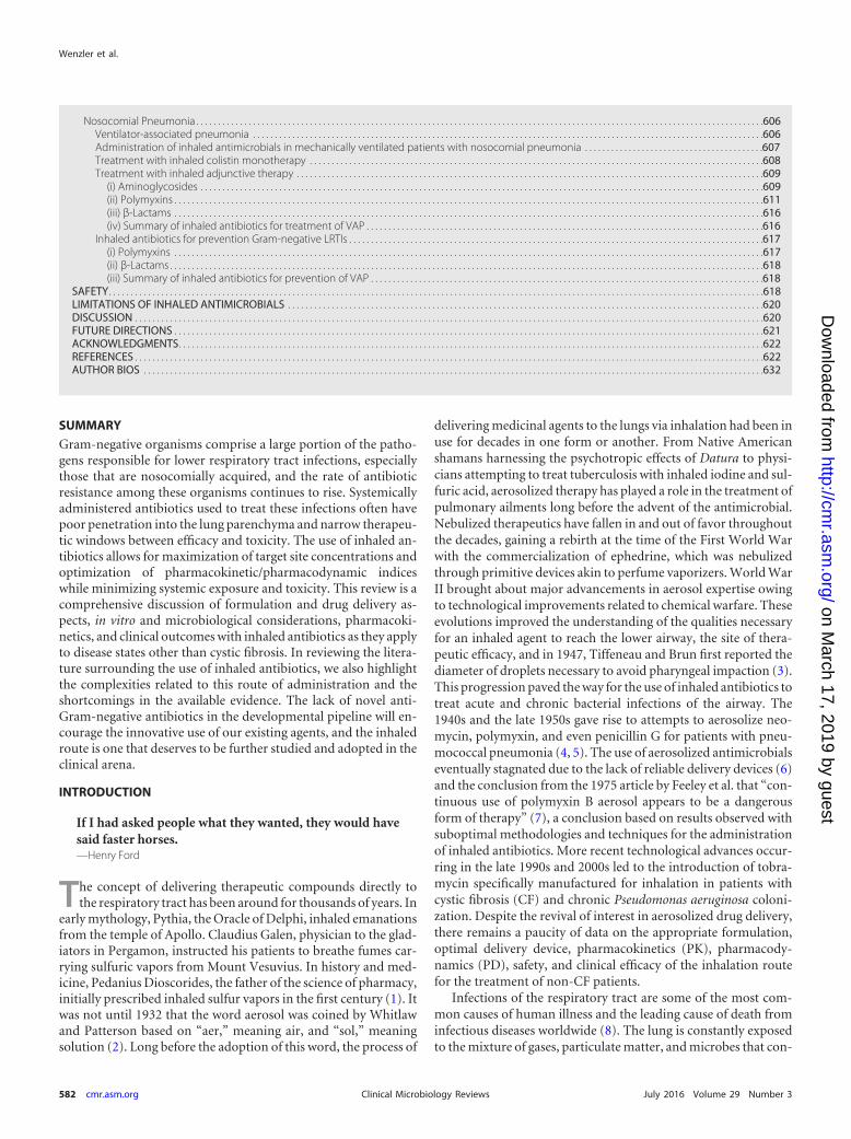
stitute inspired air. Pneumonia is most commonly defined as aninfection of the lung parenchyma, or the portion of the lung in-volved in gas transfer (the respiratory bronchioles, alveolar ducts,and alveoli) (9). Pneumonia results from the proliferation of mi-crobial pathogens at the alveolar level and the concomitant hostresponse to these pathogens. In general, lower respiratory tractinfections (LRTIs) are difficult to treat due to the sequestration ofmicroorganisms deep within the airways, where only limited por-tions of drug gain access after traditional systemic therapy. Sys-temically delivered antimicrobials, in particular those used to treatLRTIs due to Gram-negative organisms (Gram-negative LRTIs),such as aminoglycosides and �-lactams, often have poor pulmo-nary penetration into the lung parenchyma (10). When hypoxiaoccurs in response to pneumonia, the pulmonary vasculature va-soconstricts in order to shunt blood away from areas of low oxy-gen toward areas maintaining adequate ventilation gas exchange(11). This shunting, as well as chemokine-induced inflammation,can further reduce the amount of drug that is systemically deliv-ered to the lung parenchyma. In addition to the poor penetrationof systemic antimicrobials, commonly encountered respiratorypathogens can escape the innate pulmonary defenses and avoidphagocytosis by alveolar macrophages (AMs) while surviving andproliferating in the epithelial lining fluid (ELF) (12–14). In thecase of systemically administered antimicrobials, they must dis-tribute to the alveoli and ELF from the blood, requiring them topass through the alveolar barrier of the capillary lumen, connec-tive tissue, and alveolar epithelial cells. Alveolar epithelial cells area particular challenge given their connection via zonula oc-cludens, which provide a barrier between plasma and ELF (Fig. 1).This barrier is also fortified by efflux pumps, including multidrugresistance protein 1 and breast cancer-resistant protein (15–18).Delivering antibiotics directly to the site of infection, the lungparenchyma, via inhalation could overcome the obstacles to pul-monary drug deposition faced by systemically administered anti-microbials.
Over 1 million people are admitted to hospitals in the UnitedStates each year for pneumonia (19). Gram-negative organismsaccount for about 11% of community-acquired bacterial pneu-monia isolates and 33% of isolates in cases of nosocomial pneu-monia (20, 21). Five of the top six bacteria that cause nosocomial
infections are Gram-negative bacteria (22, 23), while P. aeruginosarepresented nearly 25% of ventilator-associated pneumonia (VAP)isolates in one study, more than any other single bacterium (24).In the intensive-care setting, Gram-negative pathogens accountfor �65% of pneumonia cases (25). Nosocomially acquired pneu-monias due to Gram-negative pathogens, particularly P. aerugi-nosa, are difficult to treat and exceptionally problematic to eradi-cate, leading to high rates of recurrent infection despite adequatesystemic antimicrobial therapy (26, 27). The consistent andalarming rise in the rate of antimicrobial resistance, particularlyamong these Gram-negative pathogens, represents a formidablethreat to public health, and the demand for new antimicrobials isever intensifying (28, 29). Although we have made tremendousstrides in our knowledge of infectious diseases and our under-standing of optimal antimicrobial therapy, the number of deathsdue to drug-resistant bacteria continues to rise (8). Despite this,there has been a scarcity of novel antimicrobials available to fightthis growing epidemic (30). The advent of life-saving technologiessuch as the mechanical ventilator has extended our ability to treatcritically ill patients while also effectively inventing VAP, an infec-tion associated with a 2- to 10-fold increase in mortality rates,affecting up to 20% of critically ill patients (24, 31). The reductionin the efficacy of our existing antimicrobials due to increasingbacterial resistance and the lack of new therapeutic agents haveencouraged the innovative use of existing antibiotics.
It is well understood that effective antimicrobial therapy re-quires adequate drug concentrations at the target site of infection(32). To reach the deep airways in sufficient concentrations, high,often toxic doses of drugs would need to be given systemically. Theinhalation of antibacterial agents allows higher concentrations tobe deposited directly in the lungs so that pathogens are exposed tosupralethal concentrations while minimizing potential systemictoxicity by limiting absorption and avoiding unfavorable PK/PDconsequences (33–35). The large alveolar surface area (100 m2)and thin epithelial layer (0.2 to 0.7 �m) of the lungs provide anadvantageous environment for pulmonary deposition of inhaledcompounds for which lung penetration after systemic administra-tion is problematic (36, 37). Inhalation therapy has the capabilityof directly targeting the airways, creating increased and more sus-tained local concentrations and thereby increasing the therapeuticindex, improving efficacy, minimizing toxicities, and decreasingthe time of onset for the administered drug. Despite these theo-retical advantages, practical issues concerning the use of nebulizeddrugs, in particular the ideal method of delivery, and an overalllack of robust clinical data have become hurdles to their wide-spread adoption (38). In addition, many clinicians rarely appreci-ate the complexities associated with inhaled therapy due in part tomisconceptions based on inadequate techniques performed in thepast, including instilling antibiotic solutions through the endotra-cheal (ET) tube (39, 40) and using existing parenteral formula-tions for inhalation (41–43). Finally, developing new inhaledproducts remains tremendously challenging due to the intricaciesof particle engineering and the necessity for an effective drug-device combination.
This review provides an in-depth discussion of inhaled anti-bacterials in the treatment of non-CF patients with Gram-nega-tive LRTIs. The use of inhaled antibiotics in CF patients is ex-cluded from this work, as this topic was reviewed in depth recently(44). Formulation considerations, including particle characteris-tics and drug delivery systems, along with microbiological and in
FIG 1 Representation of the alveolar capillary barrier. The barrier consists ofthree layers, of which the epithelium constitutes the least permeable layerbecause of the presence of numerous zonula occludens. Epithelial lining fluidlies in pools on the inside surface of the alveolus. 1,000 Å equals 100 nm.(Republished from reference 15 with permission.)
Inhaled Antibiotics for Pulmonary Infections
July 2016 Volume 29 Number 3 cmr.asm.org 583Clinical Microbiology Reviews
on March 17, 2019 by guest
http://cmr.asm
.org/D
ownloaded from

vitro concerns, PK/PD parameters, and a review of safety and clin-ical outcomes are discussed. Throughout this review, the word“inhaled” is used to describe the pulmonary delivery of antibiot-ics, as it most accurately represents the physiological actionrequired to deposit drugs into the lungs. Other words, such as“nebulized” and “aerosolized,” are often used interchangeably,although they more accurately describe the characteristics anddelivery mechanisms for inhaled antimicrobials. In addition,wherever possible, we avoid discussing studies utilizing intratra-cheal (i.t.) instillation of antibacterial solutions as a means of in-trapulmonary delivery, as this method has shown nonuniformdeposition in both animals and humans and is no longer com-monly employed. Intratracheal delivery is mentioned as it appliesto animal and human studies in which antibiotics are adminis-tered to the lungs without a delivery device but are sprayed oraerosolized in the lungs.
FORMULATION AND DRUG DELIVERY
Barriers to the delivery of inhaled antibiotics include the naturalpulmonary physiology, administration techniques, tolerability,physical characteristics of the aerosolized particles, and specifica-tions of the delivery device, among many others. Considerationsfor optimal pulmonary drug delivery include overcoming the in-hibitory effect of sputum, rapid delivery to reduce treatment bur-den and increase patient convenience, and effective particle distri-bution to critical areas of the lungs.
Inhaled drug formulation considerations are extremely impor-tant when considering administering inhaled antimicrobial ther-apy to a patient with a Gram-negative LRTI. Even differences inchemical entities of the same compound can have important im-plications when utilized as inhaled agents, as has been shown withcolistin (45), highlighting the importance of understanding thenuances of inhaled antimicrobials. In addition to the detailed con-siderations discussed below, drugs prepared and manufacturedspecifically for inhalation should be pyrogen free, isotonic, sterile,pH balanced to the airway epithelium (pH 6), and dispensed inunit-dose, single-use containers. Importantly, preservatives shouldbe avoided if possible, as they have been specifically associatedwith adverse effects when inhaled.
This section focuses on the local delivery of inhaled antibioticsto the surface of the lungs and does not discuss issues related toachieving adequate systemic absorption via the inhalation route.(For a detailed review of this concept, see reference 46.)
Pulmonary Physiology
The natural physiology of the human pulmonary system makesefficient delivery of inhaled antibacterials to the target site of ac-tion extremely challenging. As air is inhaled through the mouthand nose, it passes through the larynx and the trachea and even-tually passes through 16 bifurcations of rapidly dividing bronchiand bronchioles (Fig. 2). The alveoli begin at the 17th generationof bronchioles and end at the 23rd generation, transitioning fromrespiratory bronchioles to alveolar ducts and finally to alveolarsacs. Inhaled particles that are able to traverse the labyrinth of theupper airway are then deposited and efficiently transported out ofthe lungs by active mucociliary clearance prior to reaching therespiratory bronchiole and subsequent alveoli (46). The mucocili-ary elevator carries mucus covering the airways toward the mouth,where �500 ml of airway fluid can be swallowed daily. In addition,the air-facing sides of the lungs’ �500 million alveoli are each
policed by macrophages designed to phagocytize and digest anyinsoluble particles deposited there (47). The pattern of depositionof drugs in the lungs also changes as the internal landscapechanges from thick-walled ciliated central airways to bronchiolesand alveolar sacs. This armamentarium of innate pulmonary de-fenses designed to clear exogenous debris and bacterial microbescan also effectively eliminate antibiotic particles delivered via in-halation, particularly if these particles are not optimally formu-lated for this environment.
While the natural, healthy physiology of the lungs presents dif-ficulties for developers of inhaled antibacterials, the pathophysio-logical changes in diseased lungs can be dramatic. These changesalso make it challenging to extrapolate data from healthy volun-teers or from CF patients to other disease states, as the distributionof inhaled antibiotics is uneven due to numerous factors, includ-ing areas of airway contraction secondary to edema, increasedsecretions, or smooth muscle constriction. This could in turn im-pact clinical outcomes by reducing the amount of drug availablefor distribution to the distal airway and lead to differences in out-comes observed in clinical evaluations (48). Future studies evalu-ating both the deposition and the safety and efficacy of inhaledantimicrobials are needed for patients suffering from Gram-neg-ative LRTIs in order to properly formulate inhaled agents for thepulmonary physiological changes occurring in these disease states.
Drug Deposition Considerations
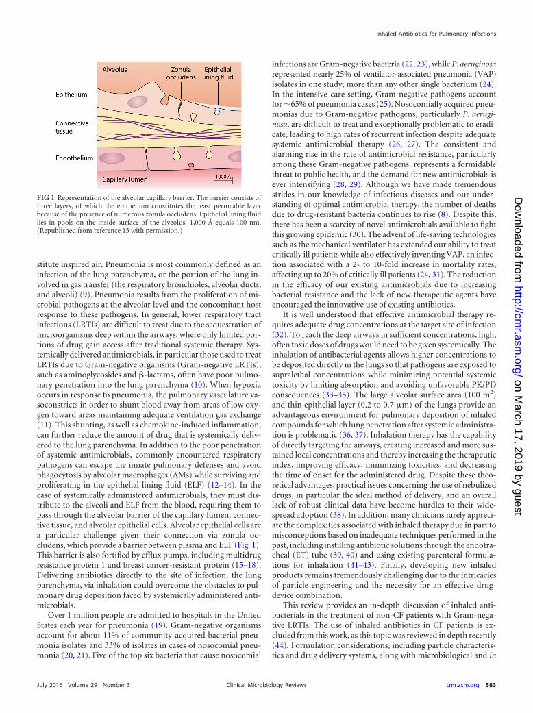
The efficacy of inhaled antibiotics correlates with the amount ofdrug deposited into the patient’s lungs, which in turn depends onthree main parameters: airway anatomy, patient ventilation, and
FIG 2 The airways branch roughly 16 to 17 times before alveolar sacs areencountered. The surface area of the human airways averages �2 to 3 m2,compared with roughly 100 m2 for the alveolar surface. In the upper airways,the inertia of the larger particles causes them to break free of the streamlines ofthe flow and collide with a wall to be deposited. As impaction clears these largerparticles in the upper airways, slightly smaller particles are filtered out of theairstream in the middle airways by gravitational sedimentation. Finally, forvery small particles, particle motion is determined by Brownian diffusion,which accounts for the dominant mechanism of deposition in the alveolarregion. (Reprinted from reference 46 by permission from Macmillan Publish-ers Ltd.)
Wenzler et al.
584 cmr.asm.org July 2016 Volume 29 Number 3Clinical Microbiology Reviews
on March 17, 2019 by guest
http://cmr.asm
.org/D
ownloaded from

aerosol characteristics. The aerosol characteristics of the inhaledparticle are the most modifiable factor and have the largest impacton satisfactory drug deposition. An inhaled medicine designed topenetrate deeply into peripheral lung regions rich in alveoli with�90% efficiency should be manufactured to deliver aerodynamicparticles between 1 and 5 �m in size, known as the mean massaerodynamic diameter (MMAD). The MMAD is the mean parti-cle size produced by the combination of the medication and neb-ulizer (34). Particles that are �1 �m may be removed duringexhalation, and particles that are �5 �m are deposited into theoropharynx (49). Particles of �5 �m may also rain out in thenebulizer circuit before reaching the upper airway. Therefore, de-position of particles into the lung parenchyma and alveolar spacein humans occurs ideally when particles are between 1 �m and 5�m (50–52). These optimally sized particles should be combinedwith low tidal breathing rates of at least 6 liters/min in order toachieve adequate delivery to the deep lower airway.
In order to utilize the potential of inhaled agents for their quickonset and high pulmonary concentrations, they must reach theappropriate target site and remain there in therapeutically signif-icant concentrations for a sufficient time period. This concept isbeginning to be appreciated and studied as it pertains to systemi-cally administered antibiotics, but there are very limited dataabout how this process occurs with regard to inhaled antimicro-bials.
Optimal properties of a drug that is designed to be systemicallyabsorbed and active when administered via the inhaled route arelow molecular mass, hydrophilicity, and a net negative charge(46). For antibacterial agents, the goal is to maintain drug concen-trations within the lungs and on the appropriate pulmonary sur-face in order to combat bacterial pathogens that reside there dur-ing infection. This means that these drugs need to have theopposite properties of those of compounds developed specificallyfor systemic activity after inhalation (i.e., inhaled insulin). Theideal inhaled antimicrobial would therefore be manufactured tobe lipophilic and positively charged and to have a high molecularmass. In addition, inhaled compounds intended for local treat-ment should also have high first-pass hepatic elimination to cir-cumvent systemic effects after oral uptake of the inhaled dosedeposited in the mucosal cavity and swallowed. These properties,balanced against an appropriate particle size, would allow opti-mum antibacterial efficacy of an inhaled antibiotic within the lungparenchyma and maximization of the agent-specific PK/PD indexby both providing high local concentrations and avoiding imme-diate diffusion into the bloodstream and removal by lung de-fenses.
Particle Engineering
There are several methods available to manufacture inhalable par-ticles, including micronization, precipitation, freeze-drying, andspray-drying. To obtain fine drug powders in the appropriate sizerange for optimal lung deposition, jet milling has been tradition-ally used by the pharmaceutical industry. However, jet millingoften produces cohesive particles due to the high surface energiesof these milled particles. A carrier powder can be added to im-prove flow, although this increases the overall powder volume, thenumber of inhalations, and the time needed to receive a dose.Spray-drying has become the state-of-the-art method for engi-neering aerosolized particles, as it is a one-step, high-throughputprocess able to engineer particles in a much more tightly con-
trolled manner. The process of spray-drying allows optimizationof particle size, size distribution, and surface morphology (53).This advanced technique has allowed consistent aerosol perfor-mances across temperatures, humidities, and airflow rates (54).
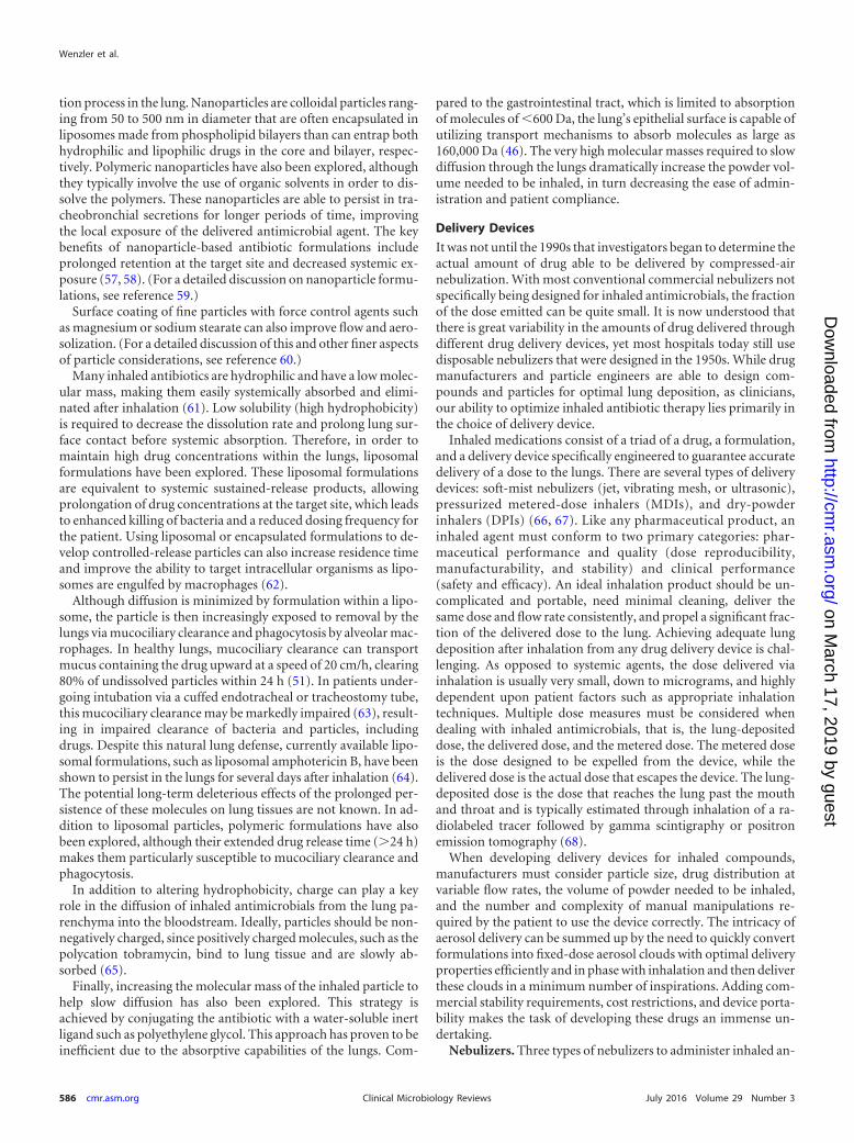
A recent example of the advancements made in particle engi-neering comes in the form of the Pulmosphere technology devel-oped for tobramycin inhalation powder (55). Briefly, Pulmo-sphere particles are manufactured by rapidly evaporating anatomized liquid stock with a heated gas to form a spray-driedpowder. The key to this process is the formation of the emulsion-based feedstock, which is an oil-in-water emulsion stabilized by asurfactant. High-pressure homogenization is then used to formsubmicrometer emission droplets, which are then mixed with adrug annex solution comprised of an aqueous solution of free-base tobramycin sulfate. This same process has also been com-pleted by using organic alcohols such as methanol in order todecrease surface tension, minimize residual water content, andfurther decrease particle size (53). The culmination of mixing thisfeedstock with hot air and atomizing leaves small porous particleswith tobramycin concentrated at the core of the particle and theexcipients within the outer walls. The result of this highly ad-vanced particle engineering process also allows improved controlover the particle surface properties. By intentionally manufactur-ing the morphology of these particles, the outer hydrophobic sur-faces can be used to decrease cohesive forces between the individ-ual particles. This improves aerosol performance and eliminatesthe need for lactose carriers. A high drug load can be achieved withthese specifically designed particles, enabling higher doses of drugper inhalation and better lung deposition. Additionally, this for-mation of tobramycin inhalation powder into an amorphous solidimproves the physiochemical stability of the powder over thoseof other crystalline powder formulations (Fig. 3). (For a morein-depth discussion, including the methods used to character-ize the tobramycin inhalation powder on the particle level, seereference 56.)
Although small aerosol particles (�1 �m) are usually exhaledduring breathing, nanoparticles (�100 nm) are able to be depos-ited in the alveolar space by sedimentation due to an accumula-
FIG 3 Scanning electron microscope image of tobramycin inhalation powder.(Reprinted from reference 56 with permission of the publisher. Copyright2015 American Chemical Society.)
Inhaled Antibiotics for Pulmonary Infections
July 2016 Volume 29 Number 3 cmr.asm.org 585Clinical Microbiology Reviews
on March 17, 2019 by guest
http://cmr.asm
.org/D
ownloaded from
tion process in the lung. Nanoparticles are colloidal particles rang-ing from 50 to 500 nm in diameter that are often encapsulated inliposomes made from phospholipid bilayers than can entrap bothhydrophilic and lipophilic drugs in the core and bilayer, respec-tively. Polymeric nanoparticles have also been explored, althoughthey typically involve the use of organic solvents in order to dis-solve the polymers. These nanoparticles are able to persist in tra-cheobronchial secretions for longer periods of time, improvingthe local exposure of the delivered antimicrobial agent. The keybenefits of nanoparticle-based antibiotic formulations includeprolonged retention at the target site and decreased systemic ex-posure (57, 58). (For a detailed discussion on nanoparticle formu-lations, see reference 59.)
Surface coating of fine particles with force control agents suchas magnesium or sodium stearate can also improve flow and aero-solization. (For a detailed discussion of this and other finer aspectsof particle considerations, see reference 60.)
Many inhaled antibiotics are hydrophilic and have a low molec-ular mass, making them easily systemically absorbed and elimi-nated after inhalation (61). Low solubility (high hydrophobicity)is required to decrease the dissolution rate and prolong lung sur-face contact before systemic absorption. Therefore, in order tomaintain high drug concentrations within the lungs, liposomalformulations have been explored. These liposomal formulationsare equivalent to systemic sustained-release products, allowingprolongation of drug concentrations at the target site, which leadsto enhanced killing of bacteria and a reduced dosing frequency forthe patient. Using liposomal or encapsulated formulations to de-velop controlled-release particles can also increase residence timeand improve the ability to target intracellular organisms as lipo-somes are engulfed by macrophages (62).
Although diffusion is minimized by formulation within a lipo-some, the particle is then increasingly exposed to removal by thelungs via mucociliary clearance and phagocytosis by alveolar mac-rophages. In healthy lungs, mucociliary clearance can transportmucus containing the drug upward at a speed of 20 cm/h, clearing80% of undissolved particles within 24 h (51). In patients under-going intubation via a cuffed endotracheal or tracheostomy tube,this mucociliary clearance may be markedly impaired (63), result-ing in impaired clearance of bacteria and particles, includingdrugs. Despite this natural lung defense, currently available lipo-somal formulations, such as liposomal amphotericin B, have beenshown to persist in the lungs for several days after inhalation (64).The potential long-term deleterious effects of the prolonged per-sistence of these molecules on lung tissues are not known. In ad-dition to liposomal particles, polymeric formulations have alsobeen explored, although their extended drug release time (�24 h)makes them particularly susceptible to mucociliary clearance andphagocytosis.
In addition to altering hydrophobicity, charge can play a keyrole in the diffusion of inhaled antimicrobials from the lung pa-renchyma into the bloodstream. Ideally, particles should be non-negatively charged, since positively charged molecules, such as thepolycation tobramycin, bind to lung tissue and are slowly ab-sorbed (65).
Finally, increasing the molecular mass of the inhaled particle tohelp slow diffusion has also been explored. This strategy isachieved by conjugating the antibiotic with a water-soluble inertligand such as polyethylene glycol. This approach has proven to beinefficient due to the absorptive capabilities of the lungs. Com-
pared to the gastrointestinal tract, which is limited to absorptionof molecules of �600 Da, the lung’s epithelial surface is capable ofutilizing transport mechanisms to absorb molecules as large as160,000 Da (46). The very high molecular masses required to slowdiffusion through the lungs dramatically increase the powder vol-ume needed to be inhaled, in turn decreasing the ease of admin-istration and patient compliance.
Delivery Devices
It was not until the 1990s that investigators began to determine theactual amount of drug able to be delivered by compressed-airnebulization. With most conventional commercial nebulizers notspecifically being designed for inhaled antimicrobials, the fractionof the dose emitted can be quite small. It is now understood thatthere is great variability in the amounts of drug delivered throughdifferent drug delivery devices, yet most hospitals today still usedisposable nebulizers that were designed in the 1950s. While drugmanufacturers and particle engineers are able to design com-pounds and particles for optimal lung deposition, as clinicians,our ability to optimize inhaled antibiotic therapy lies primarily inthe choice of delivery device.
Inhaled medications consist of a triad of a drug, a formulation,and a delivery device specifically engineered to guarantee accuratedelivery of a dose to the lungs. There are several types of deliverydevices: soft-mist nebulizers (jet, vibrating mesh, or ultrasonic),pressurized metered-dose inhalers (MDIs), and dry-powderinhalers (DPIs) (66, 67). Like any pharmaceutical product, aninhaled agent must conform to two primary categories: phar-maceutical performance and quality (dose reproducibility,manufacturability, and stability) and clinical performance(safety and efficacy). An ideal inhalation product should be un-complicated and portable, need minimal cleaning, deliver thesame dose and flow rate consistently, and propel a significant frac-tion of the delivered dose to the lung. Achieving adequate lungdeposition after inhalation from any drug delivery device is chal-lenging. As opposed to systemic agents, the dose delivered viainhalation is usually very small, down to micrograms, and highlydependent upon patient factors such as appropriate inhalationtechniques. Multiple dose measures must be considered whendealing with inhaled antimicrobials, that is, the lung-depositeddose, the delivered dose, and the metered dose. The metered doseis the dose designed to be expelled from the device, while thedelivered dose is the actual dose that escapes the device. The lung-deposited dose is the dose that reaches the lung past the mouthand throat and is typically estimated through inhalation of a ra-diolabeled tracer followed by gamma scintigraphy or positronemission tomography (68).
When developing delivery devices for inhaled compounds,manufacturers must consider particle size, drug distribution atvariable flow rates, the volume of powder needed to be inhaled,and the number and complexity of manual manipulations re-quired by the patient to use the device correctly. The intricacy ofaerosol delivery can be summed up by the need to quickly convertformulations into fixed-dose aerosol clouds with optimal deliveryproperties efficiently and in phase with inhalation and then deliverthese clouds in a minimum number of inspirations. Adding com-mercial stability requirements, cost restrictions, and device porta-bility makes the task of developing these drugs an immense un-dertaking.
Nebulizers. Three types of nebulizers to administer inhaled an-
Wenzler et al.
586 cmr.asm.org July 2016 Volume 29 Number 3Clinical Microbiology Reviews
on March 17, 2019 by guest
http://cmr.asm
.org/D
ownloaded from
tibiotics are currently available. Jet nebulizers produce an aerosolwhen compressed gas is forced through a small hole into an adja-cent reservoir containing medication in solution. Advantages tojet nebulizers are their low cost, efficiency, and disposability. Inaddition, large particles within the reservoir condense on the topof the chamber and drip back into the reservoir, minimizing med-ication waste. Disadvantages include wide variability in perfor-mance between manufacturers and lengthy administration time.
Fill volume, airflow and pressure, the choice of continuous orintermittent use, placement within a ventilation circuit, solutionproperties, and the use of a spacer can all affect output from a jetnebulizer (69, 70). The Pari LC Plus jet nebulizer is currently theonly device recommended in the European CF Society’s consen-sus document for the administration of inhaled tobramycin, as itis the nebulizer that was used in pivotal trials. Pari LC Plus is areusable jet nebulizer, which means that it requires cleaning, ne-cessitating disassembly and reassembly. Single-use, disposable jetnebulizers have been developed to eliminate the need for cleaningand to reduce the risk of bacterial contamination. When dispos-able and reusable nebulizers were compared in both in vitro and invivo models, the Pari LC Plus jet nebulizer demonstrated the high-est mass of tobramycin deposited in the lungs, performing betterthan both of the disposable nebulizers (71).
The need for a power source and long treatment times, therequired setup and cleaning, significant variations in performancebetween manufacturers, and the loss of expensive drug in consid-erably high residual volumes are significant drawbacks to jet neb-ulizers (72–75). In vitro data have demonstrated that drug deliveryfrom a jet nebulizer can be optimized via operation with a nitro-gen-oxygen mixture with particles entrained in a helium-oxygenoperating circuit (76).
Ultrasonic nebulizers use a piezoelectric crystal that vibrates athigh frequency to break the medication into a microscopic fog,resulting in aerosolization. Medication output is therefore directlyproportional to the crystal’s vibration amplitude, while dropletsize is inversely proportional to vibration frequency. The output isadditionally affected by the source of flow gas used to carry theaerosol. Advantages of ultrasonic nebulizers include consistency,efficiency, and short administration time. Disadvantages includecost, routine cleaning, and potential deleterious effects on the sta-bility of medications due to the heat generated by vibration. Incomparison to jet nebulizers, ultrasonic nebulizers utilize aslightly larger particle size but have a high rate of nebulization anda short operating time.
Finally, vibrating-mesh or -plate nebulizers use a vibratingmesh or plate to pump liquid droplets through multiple taperedapertures to produce aerosol particles. In this machine, the size ofthe particles is determined by the diameter of the holes in the meshor plate. Modern vibrating-mesh nebulizers have a higher rate ofnebulization than, and 2 to 3 times the drug output of, jet nebu-lizers (77, 78). Advantages of this device include less heat andconsistency in particle size. Disadvantages include the potentialfor obstruction and damage to the nebulizer through the use of ahighly concentrated or viscous solution. Vibrating-mesh nebuliz-ers also do not alter the temperature of the solution like jet nebu-lizers, which can adversely affect the stability of the inhaled drug(79).
All nebulizers suffer from the contribution of humidity and itsability to increase the hygroscopic growth of aerosolized particles,potentially causing them to impact or rain out of the circuit (80).
Renewed attention to the scientific foundation of aerosol therapyand the increased understanding of the relationship between theamount of drug deposited in the lower airways and the corre-sponding therapeutic response have led to improvements in drugdelivery devices. Modern delivery devices are able to achieve lungdeposition fractions of up to 50%, compared to the 10 to 15%attained previously (81). Nebulizers typically generate aerosolduring the entire respiratory cycle of the patient, leading to signif-icant drug loss during exhalation. Newer breath-actuated jet neb-ulizers counteract this issue. Even more novel nebulizers utilizeelectronic control systems to personalize aerosolizations to theindividual patient’s breathing pattern. These nebulizers areknown as adaptive aerosol delivery (AAD) systems, such as theActivareo Akita Jet system and the Philips Respironics iNeb sys-tem. Although these systems have not yet been used for antimi-crobials, they have been shown to shorten treatment time andimprove lung deposition of therapeutic pharmaceutical agents.
Furthermore, nebulization devices are available for the sinona-sal inhalation of antibiotics in the treatment of upper airway col-onization. The Pari Sinus vibrating-mesh nebulizer has success-fully been used in this fashion to effectively reduce the quantity ofP. aeruginosa bacteria and increase quality of life while maintain-ing undetectable serum concentrations in patients with CF (82).
Inhalers. The use of nebulizers requires disassembly and clean-ing of after each dose and extended administration times, causinga significant treatment burden to those patients using this methodlong term. MDIs are small, portable devices typically used to de-liver aerosolized formulation of �-agonists, anticholinergics, andcorticosteroids (83). Limitations of MDIs include difficult admin-istration techniques and high particle exit velocity (83). Otherfactors to consider are the small quantity of medication delivered(�1 mg per puff), the need for the medication to be stable in amultidose canister, and compatibility with the propellant (83).Because of these issues, MDIs are not typically manufactured toadminister antibiotics (84, 85). Similar to MDIs, DPIs are smalland portable, do not require extensive cleaning after use, and aredisposable (83, 84). In DPIs, the dry formulation of the medica-tion is enclosed in a capsule, which is punctured and then inhaledinto the lungs (84). DPIs are smaller and portable, take less time touse, and do not require special cleaning. The decreased adminis-tration time and lack of a need for a power supply improve thefreedom and portability for patients using this delivery device.However, this DPI method requires good inspiratory effort, whichmay not always be possible for patients with advanced lung dis-eases (51). In addition, patients need to be adequately educated onthe appropriate use of these devices, as lung deposition is highlydependent on the inhalation technique (84). Finally, for antibiot-ics, the loading dose within the capsule ranges from 20 to 150 mg,and multiple capsules are often required for inhalation of a suffi-cient amount of antibiotics into the lungs (84).
The introduction of the Tobi Podhaler provides an example ofan appropriately designed and manufactured DPI. The Tobi Pod-haler combines three key elements of an efficient DPI: a powderspecifically engineered for use in a DPI, a hard-capsule packagecontaining the drug, and a drug-specific inhalation device (56).In this system, the tobramycin inhalation powder is packedinto hard-capsule shells that are individually packaged intoblister packs. A single dose is delivered via puncturing andinhaling four capsules of tobramycin inhalation powder, and
Inhaled Antibiotics for Pulmonary Infections
July 2016 Volume 29 Number 3 cmr.asm.org 587Clinical Microbiology Reviews
on March 17, 2019 by guest
http://cmr.asm
.org/D
ownloaded from

efficiency is maintained through proprietary technology usedto engineer the powder.
In addition to the ease of use compared to nebulizers, inhaledantibiotic powders have demonstrated better outcomes than in-halation solutions. For example, inhaled tobramycin powder hasbeen shown to improve adherence, decrease the need for adjunctintravenous (i.v.) antibiotic courses, and improve patient prefer-ence compared to a tobramycin inhalation solution in adult pa-tients with CF (86). In contrast, an older study showed that tobra-mycin inhalation powder achieved similar reductions in P.aeruginosa density and increases in lung function (percent forcedexpiratory volume in 1 s [FEV1]) compared to the solution butcaused more coughing and had a higher discontinuation rate. De-spite this, subjective treatment satisfaction was higher, likely ow-ing to the almost 4-fold-shorter administration time for tobramy-cin inhalation powder (87). Although the administration timemay be reduced, more adverse events have been reported with theuse of DPIs than with nebulizers due to the need for rapid inhala-tion of highly concentrated solutions or powders that may causecoughing due to the sheer volume of inhalation or changes in theosmotic environment of the airway. Safety challenges with smallerdoses should be considered first before administering the fulldose, and pretreatment with a bronchodilator has been shown toreduce the incidence of bronchospasm (88).
DPIs can represent an option for transition of care for patientsbeing treated using a nebulizer while undergoing mechanical ven-tilation in a hospital. Much like transitioning from i.v. to oralsystemic antibiotics, patients can be switched to a DPI for admin-istration in the hospital or as an outpatient. Unfortunately, not allinhaled antibiotics are available as a DPI. Tobramycin and colistinare available as both inhaled solutions and powders, while aztreo-nam is available only as a solution. As mentioned above, the for-mulation used for inhalation administration is extremely impor-tant, and different formulations of the same drug are notinterchangeable. For example, aztreonam lysine is designed forinhalation, whereas the i.v. form of aztreonam contains arginine,which has been associated with pulmonary inflammation afterlong-term inhalation (89). Table 1 shows a summary of the majoradvantages and disadvantages of commonly used inhaled deliverydevices. (For a comprehensive review, see reference 66.)
Administration Technique
An appropriate administration technique for inhaled antibiotics isessential for achieving therapeutic concentrations within the re-
spiratory tract. Optimally, an inhaled antibiotic should be admin-istered during a slow and deep inhalation (84). This increases theprobability that larger particles containing more drug will bypassthe upper airways and be distributed into the smaller airways (84).To decrease the risk of cough or bronchoconstriction, the compo-sition of inhaled antibiotics should include an osmolarity of be-tween 150 and 1,200 mosmol/liter (90–93), and normal salineshould be used as the diluent (90–92). The drug should be dilutedin a volume that fills the nebulizer (94–96). Patients and theirfamily/caregivers should be adequately trained on how to properlyadminister antibiotics via the specified delivery device and how toclean it properly. As only �10% of the nominal dose is actuallydelivered to the lungs by any of the delivery devices discussed, theroom for technique error is uncomfortably low.
IN VITRO, PK/PD, AND MICROBIOLOGICALCONSIDERATIONS
Inhaled Antibiotic Admixtures
Multidrug-resistant (MDR) Gram-negative pathogens often de-velop resistance via a variety of different resistance mechanisms.As such, it is rare for a single antimicrobial agent to have the abilityto neutralize or avoid all of these resistance mechanisms. Combi-nation antimicrobial therapy offers a powerful means of combat-ing these MDR pathogens by decreasing the probability that agiven pathogen may develop resistance to all the antibiotics in agiven combination (97). In addition, antimicrobial combinationsmay provide synergistic activity to further enhance the bacteri-cidal activity and improve the rate of killing over those attainablewith individual agents. Utilizing antimicrobial combinations tocombat MDR Gram-negative pathogens has become common-place with systemic agents (98) and is beginning to be explored asit applies to inhaled antibiotics.
Several combinations have been explored, including combiningmultiple antimicrobial agents and combining antibiotics withagents that reduce mucus viscosity, such as dornase alfa. Mixing ofdrugs for simultaneous nebulization is also commonly done bypatients with CF to limit the frequency and time required fortreatment (99). The compatibility of these admixtures is mostlyunknown with respect to antimicrobials. In some studies, combi-nations of antibiotics such as tobramycin have been shown to beincompatible with dornase alfa due to bisulfite excipients used inspecific products. This incompatibility often results in the loss ofactivity of one of the two compounds, which could obviously have
TABLE 1 Brief overview of the properties of inhalation devices
Device Advantages Disadvantages
Jet nebulizer Delivery independent of inspiratory effort, able to deliver highdoses without reloading
Large device, disassembly and cleaning required after eachdose, complicated to use, long delivery times,low-efficiency delivery
Ultrasonic nebulizer Consistent dose delivery, faster delivery time than jetnebulizers, more efficient dose delivery than jet nebulizers
Expensive, disassembly and cleaning required after each dose,heat generated may damage medication
Vibrating nebulizer Smaller than jet nebulizers, faster delivery time than jetnebulizers, more efficient dose delivery than jet nebulizers
Large size, expensive, disassembly and cleaning required aftereach dose, some suspensions may clog mesh holes
MDI Handheld, multidose, activity independent of inspiratory effort Difficulty coordinating actuation and inhalation, typicallyrequires shaking and priming actuations, ill suited forhigh-dose delivery
DPI Handheld, most are multidose, no disassembly required forcleaning, dose delivered upon inspiration
Dose delivery dependent on good inspiratory effort, requiresmanual refill prior to each dose
Wenzler et al.
588 cmr.asm.org July 2016 Volume 29 Number 3Clinical Microbiology Reviews
on March 17, 2019 by guest
http://cmr.asm
.org/D
ownloaded from

deleterious effects on treatment outcomes (100). More recently,the stability of neither dornase alfa nor tobramycin (Bramitob orTobi) was affected by admixing them for up to 24 h, potentiallyallowing simultaneous nebulization. Although dornase alfa hasbeen used primarily in the treatment of CF, it has been exploredfor other disease states with impaired mucus clearance in whichantimicrobials could also be used (101).
The majority of work examining drug admixtures has focusedon nebulizer solutions. Several studies have attempted to combineantimicrobial agents in DPI formulations but have failed to dem-onstrate synergy in vitro (102, 103). A recent study examined thecombination of ciprofloxacin and gatifloxacin and the combina-tion of ciprofloxacin, gatifloxacin, and the naturally occurringmucolytic lysozyme against Gram-negative respiratory tractpathogens in vitro (104). The combination of ciprofloxacin andgatifloxacin demonstrated synergy against P. aeruginosa but onlyindifferent activity against Klebsiella pneumoniae and Acinetobac-ter baumannii. The combination of colistin and rifampin has beenshown to be synergistic in vitro and has been explored systemicallyin vivo in patients with serious infections due to A. baumannii(105). When combined in a dry-powder formulation via spray-drying of rifapentine particles suspended in an aqueous colistinsolution, the combination produced enhanced antimicrobial ac-tivity against both planktonic and biofilm cultures of P. aeruginosain vitro. The combination showed high aerosol performance, andthe addition of rifampin to the surface coating contributed to themoisture protection of colistin by minimizing contact betweenhygroscopic colistin particles (106). This same co-spray-driedcombination of colistin and rifampin has also shown in vitro syn-ergy against A. baumannii (107). The combination product offosfomycin and tobramycin has been explored clinically in aphase II trial in CF patients with chronic P. aeruginosa airwayinfections (108). A phase II study evaluating the efficacy andsafety of the combination of inhaled amikacin and fosfomycinin mechanically ventilated patients with Gram-negative pneu-monia is currently recruiting patients (ClinicalTrials registra-tion number NCT01969799). Several other combinations tar-geted at patients with CF are being explored in vitro (109).
Nebulized combinations such as the combination of tobramy-cin and clarithromycin have been considered for use in CF pa-tients (110). Tobramycin has long been the inhaled antibiotic ofchoice for treating lung infections due to P. aeruginosa in patientswith CF, while macrolides such as clarithromycin have beenshown to be effective immunomodulators, bactericidal enhanc-ers, and suppressors of virulence factors of P. aeruginosa (111–113). Given these factors, deposition of both of these agents simul-taneously in the lungs of patients with infections due to P.aeruginosa could be an optimal therapeutic strategy. In a studyexamining the deposition of spray-dried tobramycin inhalationpowder with amorphous clarithromycin present in the particlecoating, identical depositions of the two drugs were achieved,lending credence to this combination idea (110). In addition, thecoformulation of azithromycin and colistin into liposomes allowscolistin to permeate the liposome and accelerate the rate of azi-thromycin release from the liposome (114). This research mayeventually lead to the ability to utilize colistin to tailor the rate ofrelease of other agents from liposomes after inhalation, potentiallyallowing less frequent inhalation dosing.
Other unique combinations have also been explored. A novelsteroid-antibiotic dry-powder formulation consisting of cipro-
floxacin and beclomethasone was developed via spray-drying(115). The combination of ciprofloxacin and beclomethasoneshowed increased drug release and a high fine-particle fractioncompared to those of the individual agents. The combination alsohad good activity against P. aeruginosa and K. pneumoniae in vitro.Combining inhaled anti-infective and anti-inflammatory agentscould hold promise for future formulations, as corticosteroidshave been shown to benefit patients with pneumonia and exacer-bations of obstructive airway diseases when used in combinationwith systemic antibiotics (116, 117). Iron has been shown to playan essential role in the formation of biofilms by Gram-negativepathogens (118). A recent in vitro investigation examined theeffect of combining an iron-binding glycoprotein and the bac-tericidal agent hypothiocyanite (ALX-109) with tobramycin oraztreonam on biofilm production by P. aeruginosa. The combi-nation of ALX-109 and tobramycin or aztreonam reducedbiofilm formation and disrupted established biofilms on CFairway epithelial cells. Importantly, ALX-109 reduced the con-centration of tobramycin required to eradicate P. aeruginosabiofilms by 5-fold (119).
A novel combination of an antibiotic and compounds exhibit-ing mucolytic properties and the ability to suppress quorum sens-ing has been investigated. Lee et al. combined ciprofloxacin andgatifloxacin with ambroxol hydrochloride to investigate antimi-crobial synergy along with quorum quenching, mucoactive prop-erties, and pulmonary protective effects of ambroxol (120). Thiscombination showed adequate lung deposition that was higher inthe triple combination than with any agent alone while also sig-nificantly increasing microbiological activity against P. aeruginosain artificial sputum medium when ambroxol was added to theantibiotic combination.
Despite the lure of inhaled combination therapy, it is importantto note that not all antibiotics are compatible and stable in thepresence of an admixture (121). Antimicrobial agents withoutreported data confirming their stability and compatibilityshould not be mixed in a nebulizer solution for use as combi-nation therapy.
Pharmacokinetics and Pharmacodynamics of InhaledAntibiotic Compounds
The efficacy of an inhaled antibiotic compound is difficult to as-sess in an in vitro or PD system. Unlike systemic antimicrobials, invitro and/or PK/PD data are less easily attainable and translatableas they pertain to inhaled antibiotics. Serum concentrations can-not be linked directly to target concentrations on the surface of thelung or in the ELF, and it is nearly impossible to accurately esti-mate the concentration-time profile in the lungs, as the concen-trations cannot be directly or practically measured. Therefore, es-tablishing the link between different pharmaceutical propertiesand clinical performance is extremely complex compared to sys-temically delivered agents. Although multicompartment modelshave been developed to model the variability and mucociliaryclearance of inhaled corticosteroids (122), similar models as theyrelate to inhaled antimicrobials are scarce. These difficulties cul-minate in a lack of reliable and accurate preclinical data withwhich to move forward to animal and/or human studies. From adrug approval process standpoint, this creates tremendous prob-lems for pharmaceutical companies attempting to navigate theregulatory system and move to phase II and III trials without thenecessary in vitro, PK/PD, and animal data that agencies such as
Inhaled Antibiotics for Pulmonary Infections
July 2016 Volume 29 Number 3 cmr.asm.org 589Clinical Microbiology Reviews
on March 17, 2019 by guest
http://cmr.asm
.org/D
ownloaded from

the Food and Drug Administration (FDA) and the EuropeanMedicines Agency (EMA) are accustomed to seeing with systemicantimicrobials.
Correlation of the in vitro and in vivo activities of a drug iscrucial if in vitro methods are to be used to predict performance.In addition, realistic testing conditions that mimic different pa-tient scenarios are also necessary if these models are to be used toforecast clinical outcomes. Unlike models evaluating systemicallyadministered agents, this testing environment involves simulatingthe oropharyngeal geometry, matching inspiratory flow rates indifferent patient populations and disease states, and consideringdifferent delivery devices and individual patient inhalation tech-niques, among many other factors. In addition, the concentra-tion-time profile that has become so familiar within PK modelingof systemic antimicrobials is complicated as it applies to inhaledantibiotics by aerosol deposition, particle dissolution, permeationinto lung tissue, binding, and transfer to the systemic circulation.Each component within this equation is further complicated byspecific variables. For example, aerosol deposition of the inhaledproduct depends on the patient-specific respiratory tract physiol-ogy, product-dependent variables such as the emitted dose, parti-cle size distribution, and device resistance.
In simple cases, solutes administered to the lungs follow whatwould be similar to a one-compartment plasma model. Absorp-tion occurs in a dose-dependent, first-order fashion, with no evi-dence of binding or metabolism. In the case of nonmetabolizeddrugs that are absorbed following first-order kinetics, sequestra-tion to lung tissue occurs and can be modeled similarly to a two-compartment model where the second compartment is of “bounddrug” and association and dissociation constants are calculatedalong with rates of transfer to the systemic circulation. Variablesthat must also be considered in the inhalation PK model includethe difference between the dose administered and the absorbableamount, i.e., subtracting the dose lost in the inhalation process,and the amount of drug that is systemically absorbed versusbound to tissue. Advanced modeling techniques such as the one-dimensional algebraic approach, International Commission onRadiological Protection (ICRP)-96, and computational flow dy-namic methods have been developed to improve predictions ofaerosol deposition on lung surfaces (123, 124). Several studieshave also attempted to create in vitro models, not specifically withantibiotics, to simulate the clinical use of inhaled antimicrobialsalong with a mass-balance-type technique in patients receivingmechanical ventilation (95, 125–128).
In addition to the lack of adequate in vitro approaches, there isno clear consensus on the site of action within the lungs that ismost predictive of an optimal PK/PD response. Reported studieshave evaluated PK/PD parameters in respiratory sputum, tracheo-bronchial secretions, and ELF and have often arrived at differentconclusions. Moreover, the residence time in and rate of clearanceof these compounds from the lungs are difficult to assess in hu-mans but are essential to our understanding of how these agentsshould be dosed. For time-dependent drugs such as �-lactams, itis imperative to know the rate of clearance of the agent from thelungs in order to maximize the known PK/PD index of antibacte-rial efficacy for these agents. This rate is often not known, anddosing regimens are based arbitrarily on systemic doses or thoseused in animal studies without proper allometric scaling. Further-more, different PK/PD targets may exist for the same agent whenused for inhalation compared to systemic administration. For ex-
ample, maximal tobramycin concentrations need to exceed 25times the pathogen MIC to overcome the inhibitory effect of spu-tum in CF patients (129), compared to 10 times the MIC in hu-man plasma.
Microbiological Considerations
As discussed above, antibiotics developed for inhalation need tobe designed and formulated specifically for this purpose. Thismanufacturing process may lead to differences in the preparationsof the drug, and these alterations may have an impact on theintrinsic antibacterial efficacy of the compound and thereforeshould be explicitly tested in vitro for their microbiological effi-cacy. In the only head-to-head comparison of different inhalationformulations, the in vitro efficacies of Bramitob versus Tobi inha-lation solutions were tested against P. aeruginosa isolates collectedfrom patients with CF. The mean MIC values of the two formula-tions were similar, at 0.42 and 0.45 mg/liter (130). The above-discussed combination of amikacin and fosfomycin designed forinhalation in patients with Gram-negative pneumonia has beentested extensively in vitro in order to solidify the bactericidal ac-tivity of this fixed-dose combination prior to entry into clinicaltrials. The amikacin-fosfomycin inhalation system (AFIS) is afixed-dose combination of 300 mg of amikacin and 120 mg offosfomycin administered via the Pari eFlow Inline nebulizationsystem. This combination of amikacin and fosfomycin in a 5:2ratio has been shown to significantly increase the potency of ami-kacin against amikacin-nonsusceptible Gram-negative isolates(131). In vitro checkerboard synergy assays demonstrated no an-tagonism between the amikacin-fosfomycin combination andother Gram-negative antibacterials commonly used to treat pneu-monia, including aztreonam, cefepime, and meropenem (132). A4:1 fixed-dose combination of fosfomycin and tobramycin hasalso been explored in in vitro synergy assays and an in vivo murinepneumonia model (133).
These combination agents allow increases in the antibacterialspectrum, a reduction in the required dose of aminoglycoside andtherefore an improved safety profile, synergistic and mutant pre-vention activities, and finally an additive effect of combiningagents with different PK/PD indices.
Finally, the currently approved and accepted MIC breakpointsfor antibiotics used to treat Gram-negative LRTIs are developed inpart from achievable plasma concentrations after systemic admin-istration. These concentrations are not representative of the con-centrations achieved at the target site of infection, the lung paren-chyma, after either systemic or inhaled administration. Inhaledadministration of antimicrobials achieves exponentially higherlung concentrations than administration of the same agent via thesystemic route. This means that antibiotics administered via theinhalation route may still achieve the required PK/PD index ofantibacterial efficacy even if the target pathogen is reported to beresistant by using conventional interpretations. For example, lungconcentrations of gentamicin after systemic and inhaled adminis-trations were �1.0 mg/liter and 400 mg/liter, respectively. A max-imal effect of aminoglycosides is demonstrated when the achievedmaximum concentration of drug in serum (Cmax) is at least 10times the MIC of the infecting pathogen. Therefore, in this exam-ple, an isolate of P. aeruginosa with an MIC of up to 40 mg/litercould be treated via inhalation, but only an isolate with an MIC ofup to 0.1 mg/liter could be treated via systemic therapy. The vastmajority of qualitative interpretations used by clinical microbiol-
Wenzler et al.
590 cmr.asm.org July 2016 Volume 29 Number 3Clinical Microbiology Reviews
on March 17, 2019 by guest
http://cmr.asm
.org/D
ownloaded from

ogy laboratories for susceptibility reporting are developed in rela-tion to systemically achievable concentrations after parenteraland/or oral administration of an antibiotic and do not considerthe high local concentrations obtained when inhaled therapy isused. The Spanish Antibiogram Committee has recommendedspecific breakpoints for inhaled tobramycin against P. aeruginosathat differ from breakpoints for systemic therapy (134). Thisgroup recommends that the resistant breakpoint be set at �128mg/liter, compared to the Clinical and Laboratory StandardsInstitute (CLSI) resistance breakpoint of �16 mg/liter. In onestudy comparing the susceptibilities of P. aeruginosa isolatesobtained from CF patients to tobramycin based on these dif-ferent breakpoints, an additional 14% of isolates would havebeen deemed susceptible by using the Spanish AntibiogramCommittee breakpoints (135). Reporting of these isolates asbeing resistant to inhaled tobramycin based on plasma-derivedbreakpoints may force clinicians to avoid inhaled administra-tion unnecessarily when it is likely to still be effective and whenfew other options exist. In the future, organizations like theCLSI should adjust breakpoint interpretations to be specific forthe site and route of administration in order to assist cliniciansin making decisions about the utility and effectiveness of alter-native routes of administration.
A recent study examined the pharmacodynamic profile of ami-kacin as the inhalation product BAY41-6551 in concentrationsrepresentative of those achieved in two phase II trials (INHALE 1and 2) against organisms commonly encountered in mechanicallyventilated patients with pneumonia (136). This study examinedthe simulated human ELF concentrations of amikacin after deliv-ery via the pulmonary drug delivery system (PDDS) against sus-ceptible and resistant K. pneumoniae and P. aeruginosa isolates.Mean steady-state concentrations of amikacin in ELF after admin-istration of 400 mg every 12 h delivered via the PDDS were simu-lated by using a previously unreported pharmacokinetic model ina virtual population of 1,000 patients built from the INHALE tri-als. Resulting peak and trough ELF concentrations used were5,252 and 507 mg/liter, respectively, with an area under the con-centration-time curve from 0 to 12 h (AUC0 –12) of 17,940 mg ·h/liter. Amikacin achieved maximal killing effect within 6 to 12 hand sustained bactericidal activity over 24 h for all the isolatestested with an amikacin of MIC �256 mg/liter. Importantly, whenMICs were retested over the 72-h experiment, amikacin MICs didnot change across all models. The isolates for which amikacinactivity was limited in this study had MICs of up to 32,768 mg/literand possessed dozens of resistance mechanisms. These isolates areextremely rare in clinical practice outside areas of endemicity, andsurveillance studies examining amikacin MICs have consistentlyreported a maximal MIC90 of 32 mg/liter against Enterobacteria-ceae, even against carbapenem-resistant strains, and an MIC90 of 8mg/liter against P. aeruginosa. Given the current CLSI resistancebreakpoint for amikacin against Enterobacteriaceae and P. aerugi-nosa of �64 mg/liter, the rare occurrence of isolates with MICs of�256 mg/liter, and the fact that commonly utilized automatedbroth microdilution systems can often report MICs of only up to256 mg/liter, in vitro susceptibility testing by clinical microbiologylaboratories for inhaled amikacin currently provides very littleclinically relevant information. This same situation is likely appli-cable to all inhaled antibiotics given the extremely high intrapul-monary concentrations.
PULMONARY PHARMACOKINETICS
Aminoglycosides have several characteristics that make them at-tractive options for aerosol delivery. First, their antibacterial effi-cacy is concentration dependent, meaning that the high concen-trations of drug achieved in the lungs after inhalation could lead toincreased killing of bacteria and therefore improved clinical effi-cacy. Second, they have dose-dependent systemic toxicities suchthat the highest doses that can be tolerated when given intrave-nously lead to low pulmonary penetration ratios. Finally, amin-oglycosides often retain activity against MDR Gram-negativepathogens and therefore are viable therapeutic options from amicrobiological susceptibility standpoint. For these reasons, themajority of the literature concerning antimicrobial delivery via theinhalation route has focused on the aminoglycosides.
The erratic and often low-level penetration of systemically ad-ministered aminoglycosides, such as gentamicin, into pulmonarysecretions and subsequent treatment failures have been recog-nized since the late 1960s and early 1970s (137, 138). These find-ings prompted clinicians and investigators to explore the directadministration of gentamicin into the lungs to help improve clin-ical responses. Indeed, more recent investigations have supportedthe findings of poor penetration of aminoglycosides into the lungs(139, 140). In contrast to intravenously administered aminogly-cosides, which often achieve maximal plasma concentration val-ues of 20 to 40 mg/liter, delivery of inhaled aminoglycosides to thelungs has consistently demonstrated lung concentrations of �200mg/liter, although these studies were performed primarily withpatients with CF. More advanced bioanalytical assays and PK pro-cedures have allowed researchers to better characterize the degreesof exposure, distribution, and elimination of inhaled antimicro-bials in both animal and human models.
Animals
Tobramycin. Ex vivo animal models have been used to examinethe physiochemical properties of antimicrobials that lead to sus-tained effects between doses. In one such model utilizing tobra-mycin administered to an ex vivo rat lung, the magnitude of ab-sorption of tobramycin was clearly dose dependent, while the rateconstant for absorption was dose independent (141).
As mentioned above, liposomes can be used to encapsulatehydrophilic drugs in the core and to form bilayers with lipophilicdrugs in order to produce sustained release within the lungs andreduce system absorption, thereby reducing the adverse-effectprofile. In rats infected with P. aeruginosa, liposomal concentra-tions of tobramycin in the lungs were consistently �5 times thoseof free tobramycin after intratracheal (i.t.) instillation. Systemicconcentrations remained undetectable for both the free and lipo-somal formulations. Surprisingly, despite the increased concen-trations of liposomal tobramycin over the sampling period, nodifferences in bacterial colony count reductions between lipo-somal and free tobramycin were seen. This may have been due toa lack of liberation of tobramycin from liposome encapsulation.
In a similar study examining single and multiple doses of con-ventional and liposomal tobramycin in rats with pulmonary in-fections due to P. aeruginosa, the liposomal formulation showed asignificantly longer residence time and a significantly higher de-gree of exposure. Total pulmonary exposure as measured by theAUC was significantly higher for liposomal tobramycin than forconventional tobramycin (3,890 mg · h/liter versus 663 mg ·h/liter). Despite this difference, the overall numbers of CFU of P.
Inhaled Antibiotics for Pulmonary Infections
July 2016 Volume 29 Number 3 cmr.asm.org 591Clinical Microbiology Reviews
on March 17, 2019 by guest
http://cmr.asm
.org/D
ownloaded from

aeruginosa were similar after single or multiple doses of eitherformulation, although a higher percentage of observations fell be-low 103 CFU for the liposomal formulation after multiple doses(142).
In contrast to these results, i.t. administration of both liquidand dry-powder formulations of liposomal tobramycin demon-strated improved antibacterial efficacy in rats with P. aeruginosalung infections compared to the administration of free tobramy-cin (143, 144). In addition, liposome-encapsulated tobramycinhas been shown to have improved in vitro activity over free tobra-mycin, even at sub-MICs, which is not entirely explained by anincreased residence time (145). The lipid composition of the car-rier vesicle is an important consideration and may have manyvariable downstream effects, such as lung tissue binding and en-gulfment by alveolar macrophages.
Similar to the nebulized admixtures discussed above, combi-nations of drugs within a single liposome have been explored.Bismuth is a compound that has been used for years to treat gas-trointestinal ulcers due to Helicobacter pylori, although it has alsodemonstrated activity against Gram-negative organisms, reducedP. aeruginosa biofilms, and acted synergistically with tobramycinagainst P. aeruginosa (146–150). Despite these advantageousproperties, low concentrations of bismuth have been shown to betoxic to human lung cells (151). One study combined bismuthethanedithiol (BiEDT) and tobramycin coencapsulated in a lipo-some in order to decrease the toxic effects of BiEDT while preserv-ing the antimicrobial activity (152). This liposomal combinationof BiEDT and tobramycin was able to reduce the virulence factorsand quorum-sensing molecules of P. aeruginosa and enhance theantimicrobial activity and anti-inflammatory properties whengiven to rats with simulated chronic P. aeruginosa lung infections.The combination successfully reduced the number of CFU pergram of P. aeruginosa in the lungs of rats, without evidence ofsignificant systemic absorption or toxicity. In contrast, concentra-tions of free tobramycin were found only in the kidneys and not inthe lung, indicating a higher potential for systemic toxicity, withminimal to no sustained lung deposition.
Gentamicin. Liquid perfluorocarbons (PFCs) have demon-strated therapeutic promise in their ability to support gas ex-change and have been used experimentally as a protective strategyfor patients with acute respiratory distress syndrome (153, 154).PFCs have also demonstrated effectiveness as a vehicle for distrib-uting drugs throughout the lungs as a mixture of aqueous drugsolutions. PFC vehicles serve as an advantageous medium for pul-monary drug delivery due to their biochemical inertness, low sur-face tension, and high respiratory gas solubility. In addition, theirestablished benefit in improving ventilation-perfusion mismatchin human lungs provides an additional impetus to evaluate themfor pulmonary drug delivery. In healthy rabbits receiving genta-micin in a PFC vehicle, the Cmax and AUC0 – 8 in the lung after i.t.administration were 1,928 mg/liter and 680,540.2 mg · liter/h,respectively (155). i.t. administration resulted in a plasma Cmax of5 mg/liter, with an absolute bioavailability over 4 h of 57%. Thebioavailability of gentamicin in the lung following intramuscular(i.m.) administration compared to i.t. administration was only0.73%. At 1 week, there were still detectable lung concentrationsafter i.t. administration, whereas concentrations were undetect-able 4 h after i.m. administration. This study again reveals the lowlevel of lung penetration after systemic administration and dem-onstrates the high concentrations of gentamicin in lung tissue
achieved by instillation of gentamicin in a PFC vehicle, which mayallow increased local pulmonary delivery of antibiotics while alsosimultaneously improving ventilation and minimizing systemictoxicity.
As discussed above, lung injury in response to infection cancreate hypoperfusion and shunting away from the infected area, inturn affecting antibiotic supply to the infected area when givensystemically. A model was created in order to simulate this byutilizing newborn lambs with induced acid lung injury undergo-ing partial liquid ventilation. The induced lung injury created pul-monary hypoperfusion secondary to hypoxia and impaired thedistribution of oxygen resulting from consolidation and pulmo-nary edema, causing a ventilation-perfusion mismatch, as is com-monly seen in pneumonia. Injury was induced until the animals’lung function was �50% of the baseline value. Gentamicin wasgiven both i.v. and i.t. at 5 mg/kg of body weight, and lambs werestudied for 4 h postadministration, after which lungs were har-vested and homogenized. In this study, serum concentrations ofgentamicin were almost identical between i.t. and i.v. administra-tions, while mean tissue lung concentrations for the entire lungwere significantly higher after i.t. administration. In contrast tothe above-described study utilizing i.m. administration, lung con-centrations of i.v. gentamicin were adequate throughout the lung,ranging from 14.6 to 21.8 �g/g (156). Interestingly, the concen-trations were consistent and ubiquitous throughout the lungtissues sampled despite differences in ventilation-perfusionmismatches. These differences are likely due to the use of aninjured-lung model, allowing increased drug permeation fromplasma to the lung parenchyma and vice versa. Although thismodel attempts to more accurately reflect the ventilation-per-fusion mismatch occurring in human lungs during infection,the use of homogenized lung tissue and collection of only asingle concentration in pulmonary tissue inhibit the ability tothoroughly extrapolate these results to patients.
A similar study performed on newborn lambs undergoing liq-uid ventilation without acute lung injury given i.t. or i.v. gentami-cin in the same dose showed that lung tissue concentrations wereagain significantly higher after i.t. administration than after i.v.administration (157). These findings confirm the differences indiffusion occurring between diseased and healthy mammalianlungs.
Like tobramycin, liposomal encapsulation of gentamicin hasbeen shown to prolong the residence time, increase concentra-tions within the lungs, and minimize systemic absorption com-pared to conventional formulations (158).
Amikacin. The most modern animal model for studying thedeposition, PK, and clinical outcomes of inhaled antimicrobialtherapy is the ventilated-piglet model. In mechanically ventilatedpiglets with induced Escherichia coli pneumonia, lung depositionand efficacy of inhaled amikacin were studied. An ultrasonic neb-ulizer was positioned in the inspiratory line and filled with 45mg/kg of amikacin in 12 ml of saline, and the drug was nebulizedfor �20 min in 10 piglets. Eight piglets received a 15-mg/kg i.v.dose of amikacin. Amikacin concentrations were measured inlung tissue, plasma, and urine. Of the initial amikacin nebulizercharge, only 38% of the initial dose reached the respiratory tract,equivalent to a mean dose of 17 mg/kg. The remainder of the dosewas either retained in the chamber and reservoir or fixed in theventilator circuit, endotracheal tube, or expiratory filter. Lung tis-sue concentrations determined 1 h after the second amikacin dose
Wenzler et al.
592 cmr.asm.org July 2016 Volume 29 Number 3Clinical Microbiology Reviews
on March 17, 2019 by guest
http://cmr.asm
.org/D
ownloaded from

were significantly higher for inhaled amikacin than for i.v. amika-cin in all sections of lung tested. Aerosol concentrations werehighest in the lower lobe, with a maximal concentration of �100�g/g. Lung samples from the aerosol treatment group alsohad lower bacterial burdens than did those from the i.v. treat-ment group, and 71% of the tissues cultured in the aerosoltreatment group were sterile, compared to only 16% in the i.v.treatment group. Surprisingly, the mean peak plasma concen-trations were similar between the aerosol and i.v. treatmentgroups (22.5 mg/liter versus 36.4 mg/liter), with trough concen-trations falling below 5 mg/liter after 6 h in both groups. Theseconcentrations equated to a plasma AUC of 87.9 � 42 mg · h/literfor inhaled amikacin, versus 72 � 19 mg · h/liter for i.v. amikacin,and half-lives of 3.37 � 0.93 and 4.7 � 0.3 h, respectively. Basedon the amount eliminated in the urine, the systemic bioavailabilityof inhaled amikacin was 70% � 11%. This study clearly demon-strates improved microbiological eradication and lung concentra-tions after inhaled administration of amikacin compared to i.v.delivery. This is true even though �50% of the actuated dosereached the respiratory tract and despite the severe pulmonaryconsolidation and lack of aeration induced by inoculation (159).Interestingly, inhaled amikacin achieved a higher plasma AUCthan did i.v. amikacin, and concentrations remained above the4-mg/liter MIC for E. coli through the 6-h sampling window, in-dicating the potential lack of a need for both modes of adminis-tration when treating a systemic infection. This efficient systemicabsorption can likely be attributed to the fact that these animalshad diseased lungs. Conversely, the same extent of systemic ab-sorption has not been seen in patients with pneumonia given in-haled amikacin.
The same group of investigators attempted to specificallyexamine the influence of lung aeration on the pulmonary con-centrations of inhaled and i.v. amikacin, at 40 and 15 mg/kg,respectively, by histologically studying areas of induced bron-chopneumonia. In this study, amikacin concentrations in lungtissue were 197 � 165 �g/g for the aerosol treatment group and6 � 5 �g/g for the i.v. treatment group in lung areas with focalbronchopneumonia, compared to 40 � 62 �g/g and 5 � 3�g/g, respectively, in areas of severe bronchopneumonia. Impor-tantly, pulmonary concentrations of inhaled amikacin increasedsignificantly in areas of adequate lung aeration. In contrast, pul-monary concentrations of i.v. amikacin decreased in areas of im-proved aeration. This demonstrates that the severity of lung dis-ease is inversely proportional to the concentration of inhaledamikacin achieved in these lung tissues. This is likely due to theability of particles to penetrate the alveoli via open distal bronchi-oles and the lack of penetration through purulent bronchial plugsinduced by bacterial inoculation. Additionally, in nondiseasedlungs, drug penetration is prevented by a healthy alveolar epithe-lium and vascular endothelium, explaining why more aerated ar-eas had decreased i.v. amikacin concentrations. Despite this, lungconcentrations achieved via the inhalation route were �20-foldhigher than those achieved by the i.v. route (160).
Imipenem-cilastatin. The concentrations of imipenem-cilasta-tin in the bronchoalveolar lavage (BAL) fluid of rats after admin-istration via continuous i.v. infusion and inhalation were com-pared. In this study, imipenem-cilastatin was also examined for itsability to affect bacterium-induced lung injury due to P. aerugi-nosa when given preinoculation. Imipenem-cilastatin was admin-istered via aerosol at 200 mg over 1 h or by continuous i.v. infusion
at 25 mg/kg/h over 5 h before pneumonia was induced via instil-lation of P. aeruginosa. The i.v. dose was designed to mimic humanplasma concentrations, while the aerosol dose was based on datafrom studies utilizing tobramycin. Of note, rodents possess dehy-dropeptidase, the enzyme responsible for the degradation of imi-penem, in their lungs, whereas primates primarily produce it al-most exclusively in the kidneys. After an hour of administration,the mean concentration of imipenem-cilastatin in BAL fluid was�4-fold higher with inhaled imipenem than with i.v. imipenem(107.9 �g versus 25.8 �g), while the plasma concentrations were200-fold lower (0.3 mg/liter versus 56 mg/liter). Inhaled imi-penem-cilastatin demonstrated significantly higher concentra-tions in rat BAL fluid than did continuous systemic infusion inhealthy rats. Four hours after inoculation of bacteria, both the i.v.and aerosol pretreatments reduced the inoculum to �0.1 � 108
CFU, compared to the control arm of 6.9 � 108 CFU (161). Thesedata suggest that inhalation of imipenem-cilastatin might proveuseful for prophylactic treatment of patients at risk for coloniza-tion and subsequent infection by Gram-negative pathogens suchas P. aeruginosa. This study again demonstrates that intrapulmo-nary concentrations are exceedingly high after inhaled adminis-tration, while systemic concentrations remain almost undetect-able, thereby optimizing efficacy and minimizing toxicity.
Ceftazidime. In both humans and animals undergoing me-chanical ventilation, vibrating-plate and ultrasonic nebulizer de-livery demonstrated better lung deposition than did jet nebulizers.In animals, the efficiency of a vibrating-plate nebulizer was com-pared to that of an ultrasonic nebulizer for the ability to aerosolizeand deposit ceftazidime into healthy mechanically ventilated pig-lets (162). One gram of ceftazidime was nebulized continuouslyinto ventilator circuits by the respective nebulizers without hu-midification. Of note, a 65% helium–35% oxygen mixture (he-liox) was used to enhance lung deposition, with ventilator settingsoptimized for nebulization. The respiratory fractions of ceftazi-dime available after administration via two different nebulizertypes were similar, at 62% and 66% for the ultrasonic and vibrat-ing-plate nebulizers, respectively. Mean lung tissue concentra-tions were also similar, at 452 �g/g and 553 �g/g, respectively,which equates to �100 mg (10% of the nebulized dose) of cefta-zidime reaching the lungs given the weight of the piglets included.Given their above-mentioned simplicity of use, vibrating-platenebulizers may be the optimal selection for patients who are un-dergoing mechanical ventilation. Considerations for inhaled-drug delivery in patients undergoing mechanical ventilation arediscussed in detail below.
In a similar study by the same group, i.v. and inhaled ceftazi-dime were compared in piglets with and those without P. aerugi-nosa pneumonia undergoing mechanical ventilation (163). Thisstudy utilized an ultrasonic nebulizer and heliox to improve lungdeposition. In noninfected animals, mean lung concentrations ofceftazidime after i.v. and inhaled administrations were 17 �g/gand 576 �g/g, respectively. In piglets with induced pneumonia,concentrations were significantly lower after i.v. and inhaled ad-ministrations, at 10 �g/g and 111 �g/g, respectively. The helioxmixture also increased lung deposition of inhaled ceftazidime by33% in noninfected animals but had no effect in those with pneu-monia, likely due to the loss of alveolar aeration. The etiology andseverity of lung disease are important considerations when deter-mining the appropriate dose of inhaled antibiotics, as evidenced
Inhaled Antibiotics for Pulmonary Infections
July 2016 Volume 29 Number 3 cmr.asm.org 593Clinical Microbiology Reviews
on March 17, 2019 by guest
http://cmr.asm
.org/D
ownloaded from

by the dramatic difference in lung tissue concentrations of cefta-zidime between healthy an infected animals in this study.
Fluoroquinolones. In one of the only studies evaluating boththe PK and PD of an inhaled antimicrobial in an in vivo model,ciprofloxacin was administered to healthy rats as a dose of 200�g/kg via the nasal cavity by using a liquid microsprayer, similarto an atomizer, and as an oral dose of 10 mg/kg. Plasma and BALfluid concentrations were determined at specified time pointsafter drug administration. Concentrations in the ELF and alveolarmacrophages (AMs) were markedly higher after inhalation thanafter oral administration, while plasma concentrations werelower. The AUCs of ciprofloxacin in the ELF and AM followinginhalation were 103 mg · h/liter and 244 mg · h/liter, respectively,compared to 0.74 mg · h/liter and 14.1 mg · h/liter after oral ad-ministration, respectively. Given the PK/PD index associated withefficacy in the treatment of Streptococcus pneumoniae and P.aeruginosa infections, intrapulmonary administration wouldachieve optimal activity, with MICs of up to and equal to 0.5 and0.25 mg/liter, respectively, if the currently approved breakpointsare applied to the inhalation route.
In rats given ciprofloxacin, moxifloxacin, and grepafloxacin,the mean ELF concentration of ciprofloxacin 30 min after nebu-lization was 10-fold higher than that after i.v. administration (10.5mg/liter versus 1.03 mg/liter), although there were no significantdifferences in ELF concentrations between inhaled and i.v. admin-istrations at 2, 4, or 6 h postdose. Compared to ciprofloxacin,moxifloxacin and grepafloxacin showed no significant differencesat any time point. This is true even when considering the potentialoverestimation of ELF concentrations due to the possibilities oflysis of macrophages and leakage of intracellular drug, given thatthe fluoroquinolones have been shown to accumulate intracellu-larly within macrophages (10). In this study, no obvious benefit ofadministering these fluoroquinolone agents via nebulization wasseen. Although extremely high intrapulmonary concentrations ofciprofloxacin were initially achieved, which may be advantageousdue to its primarily concentration-dependent activity, these con-centrations eventually showed a time course and magnitude sim-ilar to those of i.v. administration. Of note, this study was con-ducted in healthy animals, which could have impacted thediffusion of drug both into and out of the lung (164).
In contrast to ciprofloxacin, aerosol administration of levo-floxacin achieved an AUC that was �9-fold higher than thoseachieved by intraperitoneal administration in healthy mice given asingle dose of levofloxacin at 60 mg/kg via a microspray device oran intraperitoneal dose of 20 mg/kg. Neutropenia was then in-duced, and mice were infected with P. aeruginosa 24 to 48 h priorto the administration of an inhaled antibiotic twice daily for 24 or48 h. The reduction in numbers of bacteria with inhaled levofloxa-cin was greater than that with intraperitoneally administered levo-floxacin on a per-dose basis. Finally, compared to aztreonam andtobramycin, in a model of acute lethal lung infection, levofloxacinproduced the greatest decrease in the amount of CFU per lung.Surprisingly, levofloxacin also provided the highest survival rate(100%) compared to tobramycin (60%) and aztreonam (20%)(165). The reason for this dramatic difference in survival is un-clear, and few animal studies have evaluated both PK and survivalas they relate to the treatment of induced infections with inhaledantibiotics, especially in neutropenic animals.
Colistin. Throughout this review, the doses of colistin and poly-myxin B are reported as they were in the studies being discussed.
The confusion surrounding the decision to dose these agents inunits of milligrams or international units was detailed previouslyelsewhere (166–168). The currently recommended conversionfactors between units and milligrams are 12,500 U colistin metha-nosulfate (CMS)/mg or 30,000 U colistin/mg for colistin and10,000 U/mg for polymyxin B.
Although CMS (supplied commercially as either Coly-MycinM or Colomycin) has long been administered to CF patients forairway colonization due to P. aeruginosa, it was only recently in-troduced as an adjunct for patients with Gram-negative LRTIs.After initial studies, a more comprehensive population PK studywas designed in order to develop a model to better characterizeCMS-to-colistin conversion and to understand its disposition inplasma and ELF in rats. This model would ultimately assist inidentifying the targeted advantage achieved by delivering CMSdirectly to the lungs. In this study, CMS and colistin sulfate wereadministered both intravenously and via i.t. instillation. Rats wereadministered either 14 or 28 mg/kg of CMS or 0.41 mg/kg, 0.62mg/kg, 0.99 mg/kg, or 1.49 mg/kg of colistin intratracheally (3 ratsper dosing level), and blood samples were obtained serially. BALfluid was collected only for the 14-mg/kg and 0.62-mg/kg doses ofCMS and colistin, respectively. A Cmax of 21,391 mg/liter was ob-served 5 min after pulmonary administration of CMS, with a max-imum concentration of formed colistin occurring 4 h after CMSdosing of �1,000 mg/liter. Concentrations of colistin were main-tained above 200 mg/liter throughout the 12-h sampling period.Based on the results of the established three-compartment PKmodel for plasma data, the respiratory fractions of CMS and colis-tin were estimated to be 40.9% and 48.5%, respectively. Impor-tantly, the fraction of colistin converted from CMS in ELF was0.226%, which was �9-fold higher than the conversion rate of0.0255% in plasma after i.v. administration. Maximum concen-trations of CMS and colistin achieved in plasma after i.t. admin-istration were roughly 2 mg/liter (AUC, 3.01 � 0.476 mg · h/liter).The AUC of formed colistin in ELF was 11,245 mg · h/liter, ofwhich only 0.03% was absorbed into plasma. This study demon-strates that ELF concentrations of active formed colistin were ex-tremely high and maintained above the MIC90 of 1 mg/liter for P.aeruginosa and A. baumannii (169) for the entire 12-h dosing in-terval, while concentrations in plasma remained low and belowthe toxic limit. Importantly, more of the CMS was converted tocolistin in the lungs than in plasma, likely due to the longer resi-dence time. Of note, at the highest i.t. dose of CMS (14 mg/kg),systemic exposure of formed colistin was 2-fold higher than thatafter the same dose was given i.v., likely due to the extensive con-version in the lung prior to passive diffusion into the systemiccirculation. These data suggest that a lower dose could be givenintratracheally to achieve adequate lung exposure while reducingsystemic exposure; however, this needs to be confirmed with in-haled delivery of CMS (170). Data from this study must also beinterpreted in light of the method of pulmonary delivery. Previousstudies in rats and hamsters utilizing technetium-labeled particlesdemonstrated nonuniform distribution patterns after i.t. admin-istration (171). As mentioned above, this delivery strategy can nolonger be recommended in humans, and future animal studiesshould be performed only with true antibiotic inhalation in orderto provide data that are the most accurate and translatable tohumans.
A similar study evaluated i.v. and inhaled colistin sulfate at adose of 0.35 mg/kg with serial blood samples and BAL fluid
Wenzler et al.
594 cmr.asm.org July 2016 Volume 29 Number 3Clinical Microbiology Reviews
on March 17, 2019 by guest
http://cmr.asm
.org/D
ownloaded from

collections at 0.5, 2, and 4 h postdose. The plasma concentra-tion-time profiles were similar between i.v. and inhaled admin-istrations, with estimated unbound concentrations at 2 h of0.05 mg/liter and 0.06 mg/liter, respectively. Conversely, ELFconcentrations of colistin after i.v. administration were unde-tectable, while mean concentrations after nebulization were153 mg/liter, 112 mg/liter, and 20 mg/liter at 0.5, 2, and 4 h post-dose, respectively. In this analysis, inhalation of formed colistinequated to lower plasma concentrations than with CMS after in-halation, which may limit toxicity. Then again, the i.v. dose givenin this study may have been too low at 0.35 mg/kg, as doses of atleast 1 mg/kg were used previously (172). Given the higher rate ofadverse effects with nebulization of formed colistin, as discussedbelow in this review, and the high concentrations of formed colis-tin in the lungs after inhalation of CMS, CMS should almost cer-tainly be used in clinical practice.
In a model of lung deposition and the bactericidal effect ofinhaled versus i.v. colistin, VAP was induced in 12 piglets by in-stillation of P. aeruginosa into both lungs. Twenty-four hours afterinoculation, half of the piglets received i.v. colistin at 40,000 Uevery 8 h, while the other half received 100,000 U via inhalationevery 12 h by using a vibrating-plate nebulizer over 30 min. Therespiratory fraction of colistin in this study was 60% of the initialnebulized dose, equaling �9.6 mg/kg. The median peak lung tis-sue concentration in the inhalation treatment group was 2.8 �g/g,while significantly lower concentrations were observed in lungareas with severe pneumonia than in those with mild pneumonia.Lung concentrations of colistin after i.v. administration were un-detectable even at steady state and using homogenized lung tissue.When bacterial counts in homogenized lungs were compared, in-haled colistin effectively sterilized 67% of piglet lungs at 24 h post-dose, versus only 28% in the i.v. treatment group. Serum concen-trations were also significantly lower after aerosol administration,with a mean Cmax of 1.6 mg/liter, compared to 6.0 mg/liter afteri.v. administration. Trough concentrations were virtually unde-tectable after inhalation, and the serum AUC was approximatelyhalf that with i.v. administration (7.5 mg · h/liter versus 13.5 mg ·h/liter) (173). High concentrations of colistin were observed in thelungs after aerosol administration, along with low serum concen-trations, and the antibacterial effect was significantly improved.Given that colistin is negatively charged at human physiologicalpH, the negative charge of the alveolar basement membrane likelycontributes to the slow systemic passage of colistin into the sys-temic circulation (174). The decreased lung concentrations in ar-eas of severe pneumonia may require increased dosages of inhaledcolistin in these situations to achieve the required AUC/MIC ratio,which may be possible while avoiding toxicity given the low sys-temic concentrations. Given that lung concentrations were mini-mal after i.v. administration and serum concentrations after aero-sol administration were too low to treat a systemic infection, itmay be prudent to combine these two routes when treating themost severely ill patients with Gram-negative LRTIs. This combi-nation may also allow for lower i.v. doses, thereby decreasing therate of toxicities. Further data are needed regarding the treatmentof Gram-negative LRTIs with inhaled antibiotics alone as mono-therapy, especially in critically ill patients, before systemic therapyshould be limited or abandoned completely.
Finally, a study examining the performance of jet and vibrat-ing-mesh nebulizers for the pulmonary deposition and PK ofCMS and colistin sulfate was performed in baboons, whose airway
structures most closely resemble that of a human child. In thisstudy, CMS and colistin sulfate were radiolabeled and given vianebulization at a dose of 26.6 mg. Plasma samples were obtained,and the lungs were scanned by using a gamma camera to deter-mine lung deposition of the radiolabeled nebulization. Greateraerosol deposition was achieved via the Pari LC jet nebulizer, witha mean aerosol fraction deposited in the lung of 3.5%, versus 1.3%with the vibrating-mesh nebulizer. Plasma concentrations weredetectable after nebulization but were never above 0.3 mg/literand were roughly equivalent between CMS and formed colistin.The low lung deposition fractions in this study were attributed tothe use of a conical mask over the mouth of the baboons, allowingescape of the delivered dose prior to lung deposition (175).
The observed efficacy of several different inhaled antibiotics indiverse animal models demonstrates the ability of locally admin-istered agents to achieve extremely high concentrations within thelungs while maintaining low systemic exposure. Differences indiffusion to and from plasma and alveoli have been demonstratedin animals with healthy and diseased lungs, while the inhalationroute has consistently achieved improved microbiological eradi-cation in infection models. These studies demonstrate the abilityof this route to bypass problematic physiological barriers and ac-cess deep-seated sites of infection in the lung parenchyma. Theseanimal models help provide valuable insight into the potentialclinical utility of inhaled antibiotics in humans with Gram-nega-tive LRTIs.
Humans
As discussed above, the difficulty in constructing a PK model ofinhaled antibiotics makes allometric scaling and extrapolationfrom animal to human studies challenging. Fortunately, manystudies that observed the PK of inhaled antibiotics in both healthyand infected humans have been completed.
Tobramycin. In an open-label crossover study of 12 healthyvolunteers, subjects inhaled a radiolabeled tobramycin inhalationsolution via a jet nebulizer, while whole-lung deposition viagamma scintigraphy and serum concentrations were measured.After inhalation of 25 mg of radiolabeled tobramycin via a dry-powder inhaler, the average lung deposition among all subjectswas 34.3%. Approximately 22% of the dose was retained withinthe inhaler, while 44% was deposited in the oropharynx. In con-trast, after inhalation of 300 mg of a commercial tobramycin in-halation solution via a jet nebulizer, only 5% was deposited in thelung, and 8% was deposited in the oropharynx; �50% of the dosewas retained within the nebulizer cup. For both formulations,�60% of the total dose that reached the lungs was deposited in thealveoli. Finally, after inhalation of six 25-mg capsules via a DPIevery 3 min over 15 min and one nebulization of unlabeled tobra-mycin over 15 min, serial blood samples were obtained for deter-mination of tobramycin concentrations. The Cmax of tobramycinwas almost 2-fold higher with the DPI than with nebulized tobra-mycin, although the concentrations were negligible (0.60 mg/literversus 0.28 mg/liter). Serum concentrations were undetectable innearly all subjects (85%) by 18 h with either device. These healthysubjects were trained in the appropriate use of both the DPI andthe nebulizer, although the nebulizer was not run to dryness (30min), which likely contributed to the low lung deposition. Therewere no adverse events reported in this study. The improved effi-ciency and ease of use of the DPI make it an advantageous selec-tion for patients requiring inhaled tobramycin, while the advent of
Inhaled Antibiotics for Pulmonary Infections
July 2016 Volume 29 Number 3 cmr.asm.org 595Clinical Microbiology Reviews
on March 17, 2019 by guest
http://cmr.asm
.org/D
ownloaded from
tobramycin inhalation powder allows larger doses to be deliveredin a lower powder volume, helping to decrease adverse effectsfrom use (176).
The lung, plasma, and urine PK of tobramycin have been in-vestigated for both spontaneously breathing healthy volunteersand mechanically ventilated patients without pneumonia. Onegroup studied included 10 patients who inhaled tobramycin 4 to12 h before a planned pneumonectomy for lung cancer, afterwhich a sample of lung was removed and homogenized for deter-mination of drug concentrations. A commercially purchasedpneumatic nebulizer was utilized to nebulize 300 mg of a techne-tium-labeled tobramycin inhalation solution. The amount of to-bramycin exiting the nebulizer device in this study was 49% of theoriginal dose, and maximum plasma concentrations were unde-tectable in 6/10 subjects in this study (3 in each group). Averagelung concentrations 4 and 12 h after nebulization were 5.57 �g/gand 3.61 �g/g, respectively. Based on urine concentrations, thebioavailabilities were estimated to be �4.3% and 6.6% for healthvolunteers and patients, respectively (177). Older studies of pa-tients with lung disorders demonstrated mean pulmonary con-centrations of tobramycin after inhalation of 2 � 2.26 mg/liter, asassessed by using BAL fluid (178), although these studies wereperformed early in the history of ELF studies, and tobramycinconcentrations were obtained from the first BAL fluid aliquot, atechnique that is no longer used.
The negligible systemic absorption following inhalation of in-haled tobramycin has also been proven in a prospective study ofhospitalized patients with an endotracheal tube or tracheotomywho received 300 mg of inhaled tobramycin twice daily. Of the 9patients included, 92% of trough concentrations were below 0.5mg/liter, and only one patient experienced a decline in renal func-tion. This study suggests a lack of need for serum therapeutic drugmonitoring when administering inhaled tobramycin and demon-strates a lack of systemic toxicity when monotherapy is used, aswas observed in animal and preclinical studies (179).
Finally, a prospective, randomized trial was designed to assessthe difference in lower respiratory tract concentrations of tobra-mycin and imipenem-cilastatin after nebulization or instillationin patients with suspected or proven respiratory infections whowere undergoing mechanical ventilation. Imipenem-cilastatinwas delivered at 1,500 mg every 8 h for two doses, and tobramycinwas given at 200 mg every 12 h for two doses. Patients were ran-domized to receive either instilled or inhaled drug via injectioninto the tracheal tube or inhalation with an ultrasonic nebulizer,respectively. BAL fluid concentrations of imipenem-cilastatin af-ter 1 h were remarkably higher after instillation than after nebuli-zation (4,695 mg/liter versus 72 mg/liter), although inhaled con-centrations still exceeded 20 times the MIC for most respiratorypathogens. There were no marked differences in tobramycin con-centrations by either route after 2 h (102 mg/liter after nebuliza-tion versus 142 mg/liter after instillation). The majority of patientshad undetectable plasma concentrations of both drugs at all timepoints, although 3 patients given instilled tobramycin had detect-able concentrations at 12 h, compared to none who received in-haled tobramycin. These three patients all had renal failure atbaseline in this study. There were no adverse events recorded inthis study. Both serum and pulmonary concentrations of imi-penem-cilastatin were significantly higher after instillation thanafter nebulization, although inhaled concentrations were morethan adequate to treat a Gram-negative LRTI. Despite the higher
concentrations of imipenem-cilastatin after instillation, nebuliza-tion likely achieves a more homogenous and uniform distributionthroughout the lungs than instillation (180).
Gentamicin. Five patients with healthy lungs and a recent tra-cheostomy received 2 mg/kg of gentamicin either i.m. or via in-stillation through a polyethylene catheter introduced into thedeep trachea. Four hospitalized patients without tracheostomytubes were also given 40 mg of gentamicin via nebulization, andconcentrations were evaluated in the same fashion. Bronchial se-cretion concentrations in four of the five patients after i.m. injec-tion of gentamicin were �1.0 mg/liter, while the mean peakplasma concentration after endotracheal administration was 1.04mg/liter. Despite low serum concentrations, the mean urine con-centration was 19.3 mg/liter after endotracheal administration,indicating some systemic absorption. In contrast, peak concentra-tions in bronchial secretions exceeded 400 mg/liter and remainedabove 10 mg/liter 6 h after endotracheal administration. In thefour patients administered gentamicin via nebulization, mean tra-cheal aspirate and induced sputum concentrations were 22.2 mg/liter and 17.8 mg/liter, respectively. Peak concentrations in bothmatrices were also significantly lower than those after endotra-cheal administration, while plasma concentrations were virtuallyundetectable (181). As demonstrated previously, local adminis-tration achieved supratherapeutic concentrations of antibiotic atthe target site while maintaining negligible plasma concentra-tions. Given the substantial recovery of gentamicin in the urine ofpatients after endotracheal instillation, inhaled delivery is likely asafer and equally effective method. Compared to the typical 96%urinary excretion observed after i.v. dosing, only 11% of the dosewas recovered in the urine after nebulization in healthy subjects(182).
Amikacin. In order to reduce the treatment burden of tradi-tionally marketed inhaled products such as tobramycin inhalationpowder, which require up to 4 administrations per day, amikacinwas developed into a liposomal carrier form in order to create amore-sustained-release product. Not only does this liposome-en-capsulated formulation have the ability to improve treatment ad-herence, but the extended lung residence time may also improvethe antibacterial efficacy. As discussed previously, liposomal for-mulations have shown improved activity over free inhaled drug inanimal models (142, 183). These liposomal formulations may playa particularly advantageous role in critically ill patients with accel-erated antimicrobial elimination and impairment in passive dif-fusion borders (183). Remarkably, the virulence factors present inbiofilms of P. aeruginosa have been shown to promote the releaseof amikacin from the liposome in vitro, allowing improved site-specific delivery of the antimicrobial (184). In order to evaluatethe lung disposition and elimination of liposome-encapsulatedinhaled amikacin, 3 healthy male volunteers inhaled a single ra-diolabeled dose of 120 mg of amikacin via a Pari LC Plus nebulizerover a 20-min period. In this study, �30% of the emitted dose wasdeposited in the lungs, while �50% was trapped in the exhalationfilter as a result of continuous output from the jet nebulizer.Gamma scintigraphy images at 24 h postdose showed significantpulmonary radioactivity in the lung periphery, with 75% and 38%of the initially deposited dose remaining after 3 and 48 h, respec-tively. There were no adverse events reported in this trial. Ex-trapulmonary deposition was high, as expected with the Pari LCPlus device, as outflow is continuous, even during exhalation bysubjects. The majority of the liposomes that were inhaled were
Wenzler et al.
596 cmr.asm.org July 2016 Volume 29 Number 3Clinical Microbiology Reviews
on March 17, 2019 by guest
http://cmr.asm
.org/D
ownloaded from

deposited in the lungs, the bulk of which made it to the lungperiphery. The use of liposome-encapsulated amikacin in thisstudy allowed a sustained residence time of up to 48 h (184).
In an important proof-of-concept study, six healthy volunteersunderwent noninvasive pressure-support ventilation and admin-istration of i.v. and inhaled amikacin. A vibrating-mesh nebulizerwas placed on the inspiratory limb with a standardized ventilationpattern. Four dosage sequences were used, with a 7-day washoutperiod in between. The first sequence consisted of a 1-h infusionof 15 mg/kg of i.v. amikacin, while the following three sequenceswere nebulization of 40 mg/kg, 50 mg/kg, and 60 mg/kg of ami-kacin. Serial blood samples were measured over 24 h postdose.There were no adverse events reported, and no changes in renal,audiological, or respiratory function were observed. At all inhaleddoses and time points assessed, serum concentrations were lessthan or equal to those observed after i.v. administration. Allplasma concentrations at 24 h were below 2.5 mg/liter after inha-lation, while Cmax and AUC values were higher after i.v. infusion.In this study, median doses as high as 3,510 mg were inhaled, witha median maximal serum concentration of 9.2 mg/liter. Theplasma AUC after inhalation was roughly half of that achievedafter systemic administration (66 mg · h/liter versus 138 mg · h/li-ter). The maximum median bioavailability reported was 13% forinhaled amikacin. Given that toxicity associated with aminoglyco-sides is most closely associated with the plasma AUC value, in-haled amikacin may provide a less toxic option for treatment ofGram-negative LRTIs. The mean absorption time in this study(time needed for an inhaled molecule to reach the systemic circu-lation) was �2 h for all subjects. Given the concentration-depen-dent activity of amikacin, this residence time within the lung pa-renchyma may allow exponentially better antibacterial efficacyafter inhaled administration than after parenteral administration.These results may also be due to the extremely high inhaled dos-ages given in this study, which may be prudent to maximize thePK/PD index of efficacy. The subjects in this study were not me-chanically ventilated, making it impossible to control specific fac-tors known to influence lung deposition, such as tidal volume andinspiratory flow (185).
BAY41-6551 is a drug-device combination of amikacin specif-ically formulated for inhalation and a gasless piezoelectric vibrat-ing-mesh nebulizer. This nebulizer is also designed to be inte-grated with standard mechanical ventilation equipment so thatdrug release occurs only during the first 75% of inspiration andhas been shown to deliver up to 70% of the nominal dose in lab-oratory settings. A multicenter study evaluated the penetration ofamikacin into the ELF of patients with Gram-negative nosocomialpneumonia who were mechanically ventilated using the BAY41-6551 system. Patients received 400 mg of inhaled amikacin over 45to 60 min twice daily for 7 to 14 days. Patients had to receivepositive-pressure ventilation during nebulization, and a heatedhumidifier was permitted. If patients were extubated during thestudy, they completed therapy with a handheld version of theBAY41-6551 system. All patients underwent BAL after the end ofthe first inhaled dose on day 3, along with serial plasma and urinesampling. The median plasma PK parameters for the 28 patientsincluded in the study were a Cmax of 0.85 mg/liter, a time to max-imum concentration of drug in serum (Tmax) of 1 h, and anAUC0 –12 of 6.15 mg · h/liter. The median amount secreted in theurine over 12 h was 19 mg/liter. In contrast to these low plasmaconcentrations, the median concentration of amikacin in ELF was
976.07 mg/liter, with no correlation between ELF concentrationsand ventilator settings. Tracheal secretions were also collected atrandom and showed amikacin concentrations ranging from1,517.5 mg/liter to 472 mg/liter at various time points from 1 to 24h postdose. Two adverse events of nephrotoxicity and broncho-spasm were considered possibly related to the study drug. In thisstudy, pulmonary concentrations of amikacin were �10-foldhigher than the CLSI breakpoint for P. aeruginosa, while serumconcentrations were trivial. There were very wide variations inELF amikacin concentrations in this study due to the small num-bers and interpatient variability of critically ill patients, althoughthe minimum concentrations were always maintained above theMIC for common VAP pathogens (186).
The above-discussed AFIS combination was explored in aphase I, placebo-controlled, dose escalation study in patients un-dergoing mechanical ventilation (187). An in-line vibrating-membrane nebulizer placed on the inspiratory arm was used todeliver the AFIS in this study. The nebulizer was placed 15 cmupstream of the Y piece, and the ventilator humidifier was left onduring study drug administration. Patients with either VAP orventilator-associated tracheobronchitis (VAT) were enrolledfrom a single intensive care unit (ICU) in Australia. Each patientreceived three ascending doses with one dose administered daily.On day 3, patients randomly received either placebo or the studydrug. Three cohorts of patients were enrolled, with three patientsper cohort. Doses of AFIS ranged from 100 and 40 mg to 500 and200 mg of amikacin-fosfomycin, respectively. Systemic and tra-cheal concentrations of amikacin and fosfomycin were measuredserially up to 24 h after dose 3. Eight adult male patients completedthe study, seven of whom had VAP and one of whom had VAT.Five patients had monomicrobial Gram-negative infections, andall 8 were receiving concomitant systemic antibiotic therapy. Pa-tient demographics were well balanced between the dosing co-horts, and the majority of patients were trauma victims requiringmechanical ventilator support. There were no adverse events ob-served throughout the study, and three patients, one from eachdosing cohort, demonstrated clinical improvement and were ex-tubated prior to the third dose of the study drug. Importantly,mean amikacin plasma Cmax values never exceeded 1 mg/liter,even at the highest administered dose. Amikacin tracheal concen-trations increased proportionally with a dose up to 300 mg, rang-ing from 6,103.33 to 11,617.50 �g/g from 100 to 400 mg, respec-tively. In contrast, the fosfomycin tracheal concentrations showedno relationship to dose. The mean plasma Cmax did not exceed 0.5mg/liter. The mean tracheal Cmax reached an asymptote of �6,000�g/g after the 80-, 120-, and 160-mg doses. Based on the results ofthis study, the 300-mg–20-mg dose was selected for the above-mentioned phase II study.
Ciprofloxacin. The Pulmosphere DPI technology has also beenapplied to a zwitterionic betaine salt form of ciprofloxacin in aphase I study of healthy volunteers (188). Six male subjects weregiven a single dose of 32.5 mg of ciprofloxacin or placebo via aportable passive DPI. After inhalation, subjects were instructed todrink 240 ml of water in order to wash any powder remaining inthe oral cavity into the gastrointestinal tract. Plethysmographywas used to measure lung function, and serial blood and urinesamples were collected up to 48 h postdose. Unique to the litera-ture on inhaled antibiotics, this study utilized a physiologicallybased pharmacokinetic model in order to estimate the total andregional depositions of the ciprofloxacin DPI after inhaled admin-
Inhaled Antibiotics for Pulmonary Infections
July 2016 Volume 29 Number 3 cmr.asm.org 597Clinical Microbiology Reviews
on March 17, 2019 by guest
http://cmr.asm
.org/D
ownloaded from

istration. This model included the fraction of the dose depositedin the oral cavity and ingested as an oral dose, the fraction depos-ited in the trachea and bronchi, the fraction of the dose depositedin the deep alveolar space, and the half-life of mucociliary clear-ance. Previously simulated concentration-time profiles after in-travenous and oral dosings of ciprofloxacin were also incorpo-rated and normalized to the dose of inhaled ciprofloxacin used inthis study. The model was used to fit the parameters of the threedeposition fractions to the observed concentration-time profilesfor each subject after inhalation. Maximum plasma concentra-tions of ciprofloxacin occurred 15 min after inhalation but neverexceeded 0.065 mg/liter. The terminal systemic half-life after in-halation was 9.5 h. The geometric mean AUC in plasma after asingle inhaled dose was 0.35 mg · h/liter. Complete urine collec-tion data were available for only two subjects, for whom theamounts excreted in the urine over 48 h were 21.4 and 26.9% ofthe dose. Based on the pharmacokinetic model, the half-life formucociliary clearance was �10.8 h, and an average of 17.2% of thedose was deposited within the alveolar space. Overall, the cipro-floxacin DPI was well tolerated. Five of the six subjects com-plained of mild, transient dysgeusia after inhalation of the studydrug, which resolved spontaneously. One subject experienced amarked reduction in FEV1 values after inhalation of ciprofloxacinbut did not report any subjective pulmonary complaints. Therewere no abnormal laboratory values observed in this study. Theoverall systemic absorption of inhaled ciprofloxacin in this studywas substantially lower than the exposure seen after oral or intra-venous dosing, despite the longer half-life observed in this study.This increased half-life can be attributed to the mucociliary clear-ance of inhaled ciprofloxacin and subsequent delayed absorptionvia the gastrointestinal tract.
Colistin. As discussed above, colistin is delivered as the prodrugformulation CMS, which requires in vivo conversion to the activecompound colistin sulfate. This requirement for conversionmakes achieving adequate drug concentrations challenging, espe-cially within the pulmonary space. Studies on the intrapulmonaryconcentrations of colistin after i.v. administration in humans havereported conflicting results (189–191), while the animal studiesdiscussed above, in which CMS or colistin was given via inhala-tion, showed higher pulmonary concentrations with lower sys-
temic concentrations. In a human study, the pulmonary PK ofCMS in patients with VAT in an ICU were assessed after a singleinhalation. Patients received monotherapy with 80 mg inhaledCMS every 8 h via a vibrating-mesh nebulizer over 30 min for 7days. The nebulizer was placed 15 cm from the Y piece in a non-humidified ventilator circuit, and all patients had fixed ventilatorsettings in the assist control/volume control mode with a set re-spiratory rate, tidal volume, and peak-end expiratory pressure(PEEP). Twenty patients completed the study, 16 of whomachieved clinical cure. There were no significant changes in renalfunction in any patient. Median colistin concentrations in the ELFat 1, 4, and 8 h determined by BAL were 6.73 mg/liter, 3.9 mg/liter,and 2.0 mg/liter, respectively. The majority of the included pa-tients (11 patients) had Acinetobacter baumannii cultured, withMICs ranging from 0.5 to 1 mg/liter, indicating that ELF colistinconcentrations were maintained at or above the MIC up to 8 hafter a single dose. As with previous studies including small co-horts of patients, ELF concentrations varied widely between pa-tients, with coefficients of variation ranging from 56.4 to 89.4%.At the same time intervals, median colistin concentrations in se-rum were 1.2 mg/liter, 0.75 mg/liter, and 0.31 mg/liter, respec-tively. These concentrations translated into AUC0 – 8 values in ELFand serum of 29.8 mg · h/liter and 6.8 mg · h/liter, respectively(192). Figures 4 and 5 detail median and individual colistin con-centrations in ELF and serum over time in relation to MIC break-points. Given the substantial proportion of patients (8/20) withELF concentrations below 2 mg/liter at 8 h postadministration,higher initial doses may be needed for patients with suspectedinfections due to Gram-negative bacteria with higher MIC break-points, such as P. aeruginosa (breakpoint MIC of 4 mg/liter). Ahigher dose would also increase the AUC, as the PD index associ-ated with efficacy for the polymyxins seems to be dependent onthe AUC/MIC ratio (193). Given the low serum concentrationsachieved in this study, a higher dose may still avoid systemic tox-icity. Of note, this study used nonbroncoschopic BAL techniques,so different portions of the lungs were likely sampled in differentpatients. Also, these patients were critically ill and had VAT asopposed to bronchopneumonia. Finally, given the requirementfor conversion from CMS to formed colistin, inhaled colistin sul-
FIG 4 Colistin concentrations in ELF 1, 4, and 8 h after treatment with 80 mg of inhaled CMS. (Left) Median and interquartile range values; (right) individualconcentrations. The dashed line represents the MIC of colistin for A. baumannii and K. pneumoniae. (Republished from reference 192 with kind permission fromSpringer ScienceBusiness Media. © Copyright jointly held by Springer and ESICM 2012.)
Wenzler et al.
598 cmr.asm.org July 2016 Volume 29 Number 3Clinical Microbiology Reviews
on March 17, 2019 by guest
http://cmr.asm
.org/D
ownloaded from

fate may also be used initially to achieve higher concentrations atthe expense of increased adverse reactions, as discussed above.
Two studies have evaluated the safety and efficacy of CMS in aDPI in healthy volunteers and patients. It was well tolerated byhealthy volunteers but achieved suboptimal pulmonary deposi-tion compared to liquid nebulization. In patients, decreases inpulmonary function and severe coughing after inhalation wereexperienced. As discussed above, DPIs require good inspiratoryeffort from the patient in order to administer a sufficient dose. Inthese studies, a Twincer inhaler was used, which combines highdispersion effectiveness with high internal resistance. This gener-ates a maximal fine-particle fraction with low inspiratory effort,therefore reducing the dependency for effectiveness on inspira-tory flow rate and patient effort. In the first study, eight healthyvolunteers inhaled 25 mg of dry-powder CMS in equal divideddoses (194). Participants were instructed on the proper use of theinhaler and inhaled the 25-mg dose of CMS on day 1. On day 2,each subject swallowed 80 mg of CMS dissolved in 3 ml of normalsaline on an empty stomach to evaluate the gastrointestinal ab-sorption of colistin. Pulmonary function tests were performed oneach subject at baseline and 5 and 30 min after inhalation. Serialblood sampling was performed after inhalation to 24 h postdose.After ingestion of CMS, blood sampling was performed at 1 and 3h postdose. None of the subjects experienced any adverse events,and there were no clinically significant changes in lung functiontest results throughout the study. The mean plasma colistin Cmax
observed was 0.089 mg/liter, with a half-life of 2.75 h, equating toan AUC0 – 4 of 0.275 mg · h/liter. Interestingly, serum concentra-tions of colistin after oral ingestion remained below the limit ofquantification (0.01 mg/liter), indicating that there is no mean-ingful contribution of gastrointestinally absorbed colistin after in-halation to total systemic exposure. These results confirm theminimal systemic absorption of colistin after inhalation of CMSand also reinforce the ability to inhale higher doses, in milligrams,via a DPI with the use of a Twincer inhaler.
A second study examined the use of a colistin DPI in bothhealthy volunteers and patients with CF (195). Importantly, thisstudy utilized a dry-powder formulation of colistin sulfate and notCMS as in the above-described study. Six healthy volunteers andfive patients with stable CF who were already being treated with
daily nebulized colistin for at least 6 months were included. Pa-tients were asked to stop taking nebulized colistin at least 3 daysprior to the day of study drug administration. After the 3-dayhiatus, patients were given 160 mg of colistin via a jet nebulizer onday 4, followed by serial blood sampling. On a second visit, afteranother 3-day hiatus from nebulized colistin, patients were askedto inhale 25 mg of colistin sulfate via a DPI, followed by serialblood sampling. Healthy volunteers were given only 25 mg ofcolistin via a DPI. The DPI used in this study was developed in-house by these authors in a previous study (196). Blood sampleswere drawn prior to and up to 4 h postdose. Pulmonary functiontests were performed before and 3 h after inhalation.
CLINICAL OUTCOMES
The suboptimal outcomes achieved in treating Gram-negativeLRTIs, despite maximal parenteral antibiotic therapy, combinedwith the rapid and unrelenting increase in bacterial resistanceamong these pathogens have fueled the demand for new antimi-crobials and spurred the use of existing antibiotics in innovativeways, including alternate routes of administration. Although in-haled antibiotics have been used to treat patients with respiratoryinfections for decades, reports on efficacy and safety have beenalmost primarily restricted to patients with CF. Table 2 displaysthe currently available antibiotics specifically manufactured forinhaled use and their corresponding FDA-approved indications.The more pervasive use of inhaled antimicrobials in recent yearshas led to a promising increase in the amount of available dataassociated with this mode of administration in a myriad of differ-ent pulmonary pathophysiological conditions. Unfortunately, al-though the overall volume of data has increased, the quality of theclinical outcome studies evaluating inhaled antibiotics has notimproved proportionally. The vast majority of clinical outcomestudies that examine the use of inhaled antibiotics are retrospec-tive and observational in nature, suffering from significant heter-ogeneity and a lack of rigor for patient selection, definition ofclinical endpoints, and methodology for the evaluation of pa-tients. The level of detail in which the following studies are dis-cussed is relative to both the number and quality of studies in thatspecific area, and discussions are included as felt appropriate bythe authors. Throughout this section, the microbiological terms
FIG 5 Colistin concentrations in serum 0.16, 0.5, 1, 2, 4, and 8 h after administration of 80 mg of inhaled CMS. (Left) Median and interquartile range values;(right) individual concentrations. The dashed line represents the MIC of colistin for A. baumannii and K. pneumoniae. (Republished from reference 192 with kindpermission from Springer ScienceBusiness Media. © Copyright jointly held by Springer and ESICM 2012.)
Inhaled Antibiotics for Pulmonary Infections
July 2016 Volume 29 Number 3 cmr.asm.org 599Clinical Microbiology Reviews
on March 17, 2019 by guest
http://cmr.asm
.org/D
ownloaded from

bacterial colonization and infection are used to convey the pres-ence of bacteria with no clinical signs or symptoms of infectionand clinical signs or symptoms of infection due to the invasion ofbacteria, respectively.
Non-CF Bronchiectasis
In contrast to pneumonia, the pathogens involved in bronchiec-tasis are found primarily in the sputum and bronchial mucosa.These pathogens release toxins that damage the mucosa and re-cruit neutrophils, leading to further insult. This injury preventsnormal mucociliary clearance and exposes receptor moleculeson the mucosal surface, allowing bacterial adherence and even-tually allowing bronchial epithelial cell invasion by the respec-tive infecting pathogen. These alterations also lead to increasesin antimicrobial penetration, as the barriers to antibacterialdrug movement are relatively permeable in the bronchial cap-illary endothelium, as it is devoid of zonula occludens.
While early reports of suppurative lung disease date back to the19th century, primarily credited to Laennec (197), the contempo-rary defining characteristics of this lung disease are found uponcomputed tomography imaging. A bronchus with an internal di-ameter larger than the accompanying vessel or a bronchus thatfails to taper at the periphery of the lung identifies subjects withbronchiectasis (198). Both genetic, particularly CF transmem-brane conductance regulator gene mutations in CF patients, andenvironmental factors have been linked to bronchiectasis.
Bronchiectasis not owing to CF, is a “catchall” classification fora number of similar and possibly overlapping conditions that re-sult in airway distortion and destruction. It is a condition that hasincreasing prevalence with age, and recognition of this disease hasbeen steadily increasing (199). The pathogenesis is thought toarise as an exaggerated inflammatory response to lung tissue in-jury and/or infection (200). In this vicious-cycle hypothesis, a trig-gering event leads to an inflammatory response, which leads tostructural damage to the airways and impaired mucociliary clear-ance. This milieu favors retained mucous and creates an environ-ment that predisposes to bacterial colonization. This in turn pro-motes sustained inflammation, thus creating a vicious cycleleading to the disease state. Non-CF bronchiectasis represents acomplex disease in which a multimodal approach to managementtargets the various factors responsible for the vicious cycle of dis-ease.
Bronchiectasis is characterized by inflammation, dilation, in-fection, and sputum expectoration along with cartilage and epi-thelial destruction of the airway lining. While toxic exposures andpostinfectious bronchiectasis are most common in developingcountries, there are many etiologies of bronchiectasis in devel-oped countries, including congenital diseases, bronchomalacia,and abnormalities resulting in impaired mucociliary clearance. Inmany cases, the true underlying cause is undetermined (201). Thediagnosis of bronchiectasis is made largely based on radiographicdata from computed tomography scans, along with the patient’sbackground and clinical signs and symptoms. Patients with bron-chiectasis have chronic low-level inflammation and low-grade in-fection, which can be exacerbated by superimposed viral or bac-terial infections as well as other airway irritants. These patientsoften suffer from persistent cough, sputum expectoration, recur-rent infections, and a large reliance on medical care, with a poorquality of life. In this way, management of these patients is similarto that of CF patients, consisting of antibiotics for exacerbations,immunizations, proper airway hygiene, and treatment of the un-derlying etiology if possible (202, 203). Patients with non-CFbronchiectasis have been shown to be infected predominantlywith P. aeruginosa, Haemophilus influenzae, Prevotella spp., andVeillonella spp. (204). Chronic infection with P. aeruginosa is seenin up to 33% of patients with bronchiectasis and is associated withdecreased lung function, poor quality of life, and more frequenthospitalizations. Additionally, organisms such as Staphylococcusaureus and nontuberculous mycobacteria have been isolated (205,206). Given the many etiologies responsible for non-CF bronchi-ectasis along with the extensive flora of potential pathogens, treat-ment guidelines and algorithms should be adapted to the individ-ual patient scenario. Additionally, antibiotics may be used aslong-term therapy or for acute treatment of exacerbations. Alongwith the economic burden, the incidence of non-CF bronchiecta-sis continues to rise in the United States, with annual percentagesincreasing almost 9% every year from 2000 to 2007 (199, 207).Although non-CF bronchiectasis is seen across the whole popula-tion, the prevalence appears to be highest in individuals of ad-vanced age and females.
Although CF is pathophysiologically responsible for the major-ity of cases of bronchiectasis, there are several other medical con-ditions that can result in this altered lung pathology (202). Theencouraging results of treating CF bronchiectasis with inhaledaminoglycosides have led to their extrapolation to other similardisease states. To date, there have been four reported studies eval-uating the effectiveness of inhaled tobramycin for non-CF bron-chiectasis and chronic P. aeruginosa infection (41, 208–210).These studies have consistently demonstrated a significantly de-creased respiratory burden of P. aeruginosa, similar to that in CFpatients. Unlike CF patients, however, the use of inhaled tobra-mycin in cases of non-CF bronchiectasis has not led to sustainedimprovement of objective clinical measures such as pulmonaryfunction test results.
As opposed to acute pulmonary infections, where the goal ofantimicrobial therapy is to eradicate the infecting pathogen, theaim of treatment of chronic bronchial infections is to reduce thebacterial load and the coinciding inflammatory response. Addi-tionally, chronic bronchial infections represent a different envi-ronment of infection, as bacteria are often growing on the surfaceof the airway epithelium rather than invading in a more virulentmanner as in true pneumonia. This chronic airway colonization
TABLE 2 Current commercially available inhaled antibioticformulations
Antibiotic (trade name) FDA indication
Aztreonam solution(Cayston)
Improvement of respiratory symptoms inpatients �6 yr of age with CF, FEV1 of �25%and �75%, and P. aeruginosa infection
Colistin dry powder(Colobreathe)
Not FDA approved; approved by the EMA formanagement of chronic pulmonary infectionsdue to P. aeruginosa in patients �6 yr of agewith CF
Tobramycin inhalationsolution (Tobi)
Management of CF patients �6 yr of age with P.aeruginosa infection with FEV1 of �25% and�75% on a 28-day alternating on/off schedule
Tobramycin inhalationpowder(Tobi Podhaler)
Management of CF patients �6 yr of age with P.aeruginosa infection with FEV1 of �25% and�80% on a 28-day alternating on/off schedule
Wenzler et al.
600 cmr.asm.org July 2016 Volume 29 Number 3Clinical Microbiology Reviews
on March 17, 2019 by guest
http://cmr.asm
.org/D
ownloaded from
often leads to the formation of biofilms, particularly in the case ofP. aeruginosa. The high doses, prolonged penetration, and in-creased residence time of inhaled antimicrobials make them amore suitable agent for this particular type of infection. Treatmentof CF bronchiectasis with inhaled antibiotics, especially tobramy-cin, has been shown to improve pulmonary function, decrease thesputum density of bacteria, and reduce the risk of exacerbation(211). Given the similarities in pathophysiologies of cases of CFand non-CF bronchiectasis, it stands to reason that similar resultsshould be observed in this patient population. Chronic infectionwith P. aeruginosa has been associated with a deterioration of lungfunction, and inhaled antibiotic treatments of both acute andchronic infections have proven beneficial in this population,much like the CF counterpart (212–214). Inhaled antibiotics havebeen used in patients with bronchiectasis since the 1940s (215)and are currently recommended as treatment strategies in Britishand Spanish bronchiectasis guidelines (216, 217). A recent reviewdetailed the utility of inhaled tobramycin in the treatment of pa-tients specifically with non-CF bronchiectasis (218).
Tobramycin. Orriols et al. evaluated 17 patients who had failedoral antibiotic therapy for non-CF bronchiectasis at least once inthe previous 90 days in a prospective pilot trial. All patients re-ceived either i.v. ceftazidime or tobramycin for 2 weeks, afterwhich they were randomized to receive either the same drug givenvia inhalation for 12 months or symptomatic therapy only. Cefta-zidime was given via inhalation through a jet nebulizer at a doseof 1 g every 12 h, and tobramycin was given as 100 mg every 12h. The frequency and duration of hospitalization were lower inthe inhalation treatment group, while bacterial eradication wasnot achieved in any patient (219).
In a second placebo-controlled crossover trial by the samegroup, 30 patients received 300 mg of inhaled tobramycin every 12h in two 6-month cycles separated by a 1-month washout period.Again, fewer and shorter hospitalizations were observed in thetreatment periods, along with a decrease in the sputum density ofP. aeruginosa. Despite these outcomes, no differences were seenwith regard to quality of life or systemic inflammatory markers(41).
In a blind, active-control study, the effect of the addition of atwice-daily tobramycin inhalation solution to a 14-day outpatientcourse of oral ciprofloxacin was examined in patients withnon-CF bronchiectasis who were chronically infected with P.aeruginosa and suffering from an acute exacerbation. A jet nebu-lizer was utilized to administer 300 mg of tobramycin twice daily,and oral ciprofloxacin was given at a dose of 750 mg twice daily. Atotal of 43 subjects completed the study, and there was no statis-tically significant difference in cure rates at the day 21 test-of-cureassessment, despite a 20.4% numerically higher cure rate in theciprofloxacin-only group. Clinical success was defined as a reduc-tion in sputum volume, improvement in purulence, and de-creased cough and/or dyspnea, as assessed by the investigator. Anonsignificant difference in P. aeruginosa eradication from spu-tum was seen in subjects receiving tobramycin and ciprofloxacin(34.6% versus 18.5%). There was no difference in the emergenceof resistance, while microbiological and clinical outcomes showedgreater concordance in the tobramycin-plus-ciprofloxacin group(80% versus 40.9%). Wheezing was reported more commonly inthe tobramycin arm, as expected, while more patients in the cip-rofloxacin-only group withdrew from the study due to adverseevents (18.5% versus 11.5%). There was no observed difference in
lung function between the groups, as measured by FEV1, whiletwice as many subjects in the ciprofloxacin-only group requiredhospitalization for worsening symptoms. This study follows suitwith previous studies indicating that inhaled antibiotic therapydecreases bacterial density and improves eradication but fails toprovide improved clinical outcomes. While evidence for patientswith CF has shown that clinical success is often associated with areduction in bacterial density, the studies of non-CF bronchiecta-sis to date have not shown this same finding. Of note, this studydid not assess time to exacerbation or time to next hospitalization,which could potentially coincide with improved bacterial loadreduction (208).
An additional phase II, multicenter, placebo-controlled, ran-domized, double-blind trial was conducted by Barker et al. (209).As opposed to the two studies mentioned above, which used an i.v.formulation of tobramycin in a nebulizer, this study utilized 300mg of a tobramycin inhalation solution every 12 h. The tobramy-cin inhalation solution was given daily for 28 days along withplacebo. Compared to placebo, tobramycin decreased the bacte-rial density of P. aeruginosa in the sputum and increased clinicalimprovement, as assessed by the investigators.
In a similar open-label trial, 41 patients received three treat-ment cycles with a tobramycin inhalation solution for 14 days onfollowed by 14 days off. After the three cycles, significant improve-ments in the subjective symptom severity score and quality of lifewere seen (210).
Gentamicin. In 65 patients who received either 80 mg of in-haled gentamicin or placebo over a year-long period, decreasedexacerbations, improved quality of life, and no severe adverseevents were observed for the gentamicin group (220). In a separatestudy, 28 patients with bronchiectasis and mucus hypersecretionreceived 40 mg of gentamicin inhaled twice daily for 3 days com-pared to placebo. Sputum myeloperoxidase (MPO) levels wereassessed to determine whether gentamicin prevents MPO-in-duced airway injury. Compared to placebo, MPO levels along withthe daily volume of sputum production were significantly reducedin patients receiving gentamicin. Additionally, improvements inthe 6-min walking test, the subjective Borg scale, and self-sputumassessment were seen only in the gentamicin group (221).
Ciprofloxacin. In a phase II trial that enrolled 124 patients,inhalation of 32.5 mg dry-power ciprofloxacin twice daily for 28days via a DPI achieved significant reductions in sputum bacterialloads and was well tolerated. Pathogen eradication was achievedmore often in the ciprofloxacin group than in the placebo group,and reports of bronchospasm were minimal (222). Additionally,treatment with 150 mg of inhaled dual-release liposomal cipro-floxacin for 28 days followed by 28 antibiotic-free days showedsignificant antimicrobial efficacy against P. aeruginosa in 42 pa-tients infected with this pathogen at baseline and delayed the timeto first pulmonary exacerbation by a median of 76 days. Fewerpulmonary adverse events were observed with inhaled liposomalciprofloxacin than with placebo (223).
Colistin. In the first controlled clinical trial to use an adaptiveaerosol delivery device, patients with non-CF bronchiectasis andchronic P. aeruginosa infection were randomized to receive eitherinhaled colistin or placebo after receiving systemic antibiotics foran acute exacerbation. Of the patients with documented adher-ence to treatment, the time to next exacerbation significantly im-proved (224).
Studies have shown that up to half of patients with chronic
Inhaled Antibiotics for Pulmonary Infections
July 2016 Volume 29 Number 3 cmr.asm.org 601Clinical Microbiology Reviews
on March 17, 2019 by guest
http://cmr.asm
.org/D
ownloaded from

obstructive pulmonary disease (COPD) have associated lowerlobe bronchiectasis, and this leads to higher rates of microbialcolonization and severe exacerbations (225). As such, Steinfortand Steinfort examined the effect of long-term inhaled colistinadded to standard treatment regimens in patients with COPD andbronchiectasis. Colistin was given via inhalation as 30 mg daily to18 patients over a mean treatment duration of 41 months. All ofthe patients had P. aeruginosa or Stenotrophomonas maltophiliaisolated from their sputum. Only 3 patients (16.6%) showed im-provement in FEV1 over the course of the study, although quality-of-life measures were significantly better after the addition of in-haled colistin to the standard-of-care regimen. No emergence ofresistance to colistin was observed, although only 3 patientsshowed eradication of bacteria. No adverse effects were reporteddespite the lengthy duration of treatment (226).
COPD often leads to chronic bronchial bacterial infection andcolonization, and up to 33% of patients with bronchiectasis arechronically colonized with P. aeruginosa (227). The goals for thesepatients are much like those for CF patients: to prevent exacerba-tions and reduce the number and frequency of hospitalizations. Astudy examined the clinical and microbiological outcomes inadult patients with non-CF bronchiectasis or COPD and chronicP. aeruginosa colonization who received inhaled tobramycin orcolistin alone or in combination via a jet nebulizer as outpatients.Both drugs were given twice daily, and patients received inhaledantibiotics for a minimum of 12 weeks. The doses of inhaled to-bramycin and colistin used were either 100 or 200 mg and either 1million or 2 million IU, respectively. In total, 81 patients wereevaluated, the majority of whom received inhaled tobramycin fol-lowed by colistin followed by the combination. The primary find-ing in this study was that therapy with colistin showed better effi-cacy in eradicating P. aeruginosa and less development ofresistance than did tobramycin, although there were no differ-ences in clinical outcomes, including the number of hospitaliza-tions. This study suffered from several limitations, including am-biguous inclusion criteria, observational design, differences indosages and adjunctive antibiotic use that were not accounted for,and results that conflict with many previously reported results,including a treatment-emergent resistance rate of 48% for tobra-mycin. Most importantly, the outcomes for patients with bron-chiectasis and those with COPD were reported together (228).
Aztreonam. Two large, phase III clinical trials evaluating theefficacy of inhaled aztreonam in this patient population have beenconducted. Unfortunately, both of these trials failed to show aclinical benefit, and the rates of adverse events were higher in theaztreonam group than in the placebo group (229).
Summary of inhaled antibiotics for non-CF bronchiectasis.Taking the results of these studies together, a meta-analysis eval-uated randomized controlled trials of adults with stable non-CFbronchiectasis given inhaled antimicrobials for at least 4 weeks.Eight studies encompassing 590 patients and utilizing amikacin,aztreonam, ciprofloxacin, colistin, gentamicin, or tobramycinwere included. All studies administered the agent via a jet nebu-lizer, with the exception of ciprofloxacin, for which a DPI wasused. In terms of sputum load of P. aeruginosa, inhaled antibioticsproduced a significantly greater reduction in bacterial loads thanin control groups and were associated with a 4-fold-higher chanceof achieving complete bacterial eradication. Inhaled antibioticswere also shown to significantly reduce the risk of acute exacerba-tions compared to controls, although the meta-analysis failed to
show a benefit in the incidence of hospitalizations. There was alsono statistically significant difference in quality-of-life scores be-tween the two groups. The rate of bronchospasm was 10% in theinhaled-antibiotic group, compared to 2.3% in the controls, andwas higher with aminoglycosides than with colistin or ciprofloxa-cin. Despite heterogeneity in the testing methodology, the meta-analysis did not show a statistically significant difference in theemergence of bacterial resistance in the inhalation treatmentgroup compared to the controls. This meta-analysis lends somesupport to the use of inhaled antibiotics for the treatment ofchronic bacterial infections in the airways of patients with stablenon-CF bronchiectasis (230). There was significant heterogeneityin this analysis, especially surrounding the duration of inhaledtherapy. Additional studies are needed to delineate the ideal drug,dose, frequency, and duration for these patients.
A more recent meta-analysis specifically examined the clinicalutility of prolonged inhaled antibiotics for patients with non-CFbronchiectasis (231). The promise of long-term oral antibiotictherapy with macrolides such as azithromycin in this population(212) has led to the discussion of longer courses of inhaled thera-pies, using either a continuous or rotating schedule. This analysisincluded all of the same studies as those in the analysis describedabove and found similar results, i.e., reductions in the bacterialdensity of P. aeruginosa in the sputum and increased eradicationwith treatment. Again, no difference in the emergence of resis-tance was observed. In contrast to the above-described analysis,this group determined that patients in the inhaled-antibioticgroups required additional antibiotic therapy less often. Therewere also fewer exacerbations in the inhaled-antibiotic group. Theincidences of wheezing and bronchospasm were higher in thetreatment groups. This group did not perform an analysis of hos-pitalizations due to the modest sample size and heterogeneity.Again, inhaled antibiotics did not improve lung function or qual-ity of life.
Two Cochrane reviews of the use of prolonged antibiotics fornon-CF bronchiectasis in children and adults have been com-pleted, with the most recent update being in August 2015 (232,233). The more current review included 18 randomized trials, 6 ofwhich reported the use of inhaled antibiotics, examining the use ofprolonged antibiotic therapy (�4 weeks) in 1,157 patients. Unfor-tunately, the outcomes associated with inhaled antibiotics werenot analyzed separately. The overall quality of evidence was mod-erate, although the analysis was limited due to the heterogeneity ofoutcomes reported in the trials. Overall, significant effects in favorof prolonged antibiotics were observed for the treatment of bron-chiectasis. A nonsignificant reduction in hospitalizations was alsoseen, while a significantly higher rate of drug resistance was ob-served in the intervention arm. There was no difference in adverseevents. The authors of this study stress the need for appropriatepatient selection for prolonged antibiotic therapy and call for im-provements of the shortcomings of current trials in future studies,including study duration, bacterial colonization versus infection,and drug resistance at the individual and community levels.
The vast majority of data presented in this section for the treat-ment of non-CF bronchiectasis with inhaled antibiotics lies withtobramycin. These studies most often administered tobramycin at300 mg every 12 h via a jet nebulizer as adjunct therapy or mono-therapy either long term or in an on/off cycle, similar to the way inwhich it is utilized in CF patients. Other inhaled agents are begin-ning to be explored in this population, including ciprofloxacin via
Wenzler et al.
602 cmr.asm.org July 2016 Volume 29 Number 3Clinical Microbiology Reviews
on March 17, 2019 by guest
http://cmr.asm
.org/D
ownloaded from

a DPI. The theoretical benefit of inhaled antibiotics in this popu-lation is largely extrapolated from data for CF patients, and theiruse is recommended in at least two national guidelines. The resultsof the included studies show consistently reduced bacterial densi-ties in the sputum of patients with non-CF bronchiectasis and apositive effect on subjective quality-of-life measurements. Withthe exception of aztreonam, pulmonary adverse events were rarelyreported, even after prolonged use (�3 years).
Unfortunately, unlike for patients with CF, few of the trialsdiscussed above showed an improvement in objective lung func-tion as measured by FEV1. Patients with non-CF bronchiectasismay be more difficult to treat than CF bronchiectasis patients, asother studies evaluating inhaled antibiotics have also failed toshow an improvement in FEV1, which may be due in part tonon-CF bronchiectasis being precipitated by a fixed airflow ob-struction in many cases (222–224). A limitation to the studiesexamining inhaled antimicrobials for non-CF bronchiectasis is alack of information regarding the pathophysiology of the disease.The vast majority of the patients in the trials conducted in theUnited States were smokers. Patients with non-CF bronchiectasisare often older and a much more heterogeneous population, sodifferent dosages and dosing strategies may also need to be con-sidered for these patients. Additionally, the heterogeneity in dos-ing regimens and clinical endpoints makes interpretation andextrapolation of these results challenging. Despite encouragingoutcomes regarding reductions in sputum bacterial density andmicrobial eradication, inconsistent and/or a lack of results withregard to quality-of-life improvement, a reduced need for sys-temic antibiotics, and amelioration of objective pulmonary func-tion create pause when applying these findings to all patients withnon-CF bronchiectasis. Additionally, the heterogeneity in thenon-CF bronchiectasis population compared to patients with CFleaves many questions unanswered as to the ideal patient forwhom to prescribe this therapy and whether the underlying etiol-ogy has an effect on outcomes. Future directions for this popula-tion include studies utilizing liposomal formulations of inhaledantibiotics, given the prevalence of biofilms and the need for spu-tum penetration of the upper airways in these patients. Addition-ally, cohesive outcome definitions and consistency in the methodsof observing these patients will help the generalizability of resultsfrom forthcoming studies. In the future, validated scoring systemssuch as the bronchiectasis severity index may assist in determiningwhich patients stand to benefit the most from inhaled antibiotictherapy (234).
Based on the currently available evidence, there are at least twoscenarios in which inhaled antibiotics can provide benefit to pa-tients with non-CF bronchiectasis. The first scenario is that ofpatients experiencing an acute exacerbation who currently have orpreviously had a Gram-negative organism, particularly P. aerugi-nosa, isolated from a sputum culture. This is particularly true forpatients who have a fluoroquinolone-resistant P. aeruginosa strainisolated and have no other oral antibiotic options but do not re-quire i.v. antibiotic therapy based on the severity of their illness.Inhaled antibiotic monotherapy represents an attractive selectionin this scenario, as the patient is able to leave the hospital withouta central venous catheter, home health care, or routine laboratorymonitoring. Inhaled antibiotics may also be used in addition tosystemic therapy in this scenario for those patients who requiredual therapy based on the severity of their illness. In either case,the chosen inhaled agent to be used should be based on the avail-
ability of the appropriate inhalation solution that is specificallyformulated for aerosol delivery and the associated proper deliverydevice (Table 2). Until site-specific respiratory microbiologicalbreakpoints that take into account the concentrations achievablein the sputum and lungs with inhaled antibiotics are widely avail-able, resistance to the chosen inhaled agent reported upon in vitrosusceptibility testing should not deter the use of that agent. Con-centrations above the maximum MIC for that bug-drug combi-nation can be easily achieved by inhalation, and symptomatic im-provement often occurs in this patient population despite in vitroresistance to the inhaled agent. The duration of treatment of acuteexacerbations of non-CF bronchiectasis should be up to 14 to 21days, depending on the clinical response of the patient. The sec-ond scenario is that of patients with non-CF bronchiectasis whoexperience frequent exacerbations of bronchiectasis per year re-quiring antibiotic therapy or those with exacerbations causing sig-nificant morbidity, regardless of frequency. Patients who meetthese criteria and have had documented respiratory culturespositive for Gram-negative organisms, especially P. aeruginosa,should receive either chronic, continuous inhaled antibiotic ther-apy or therapy in a 28-day on/off cycle as for patients with CF.Again, the choice of agent should be based primarily on feasibilityand having the appropriately formulated compound and deliverydevice and less on in vitro susceptibility reports. As discussedabove, it is important to remember that the goal of antimicrobialtherapy in cases of non-CF bronchiectasis is not to eradicate theinfecting pathogen but to reduce the bacterial load and the coin-ciding inflammatory response. The aim of treatment of cases ofnon-CF bronchiectasis with inhaled antibiotics should be to im-prove pulmonary function, decrease the density of bacteria in spu-tum, and reduce the risk of a future exacerbation. Although com-plete pathogen eradication may not occur during or aftertreatment with inhaled antimicrobials, the emergence of resistantbacterial pathogens was rare in the available studies. Therefore, alack of sterilization should not preclude the use of inhaled therapyfor this disease state, as the benefits of this route of administrationstretch beyond simple microbiological endpoints.
Ventilator-Associated Tracheobronchitis
This section focuses on nosocomially acquired tracheobronchitisin patients undergoing mechanical ventilation via an endotrachealtube. Although acute exacerbations of chronic bronchitis andacute bronchitis can occur in patients with COPD, these infec-tions are even less well defined than VAT and have significantlydifferent underlying pathophysiologies.
VAT can be most easily defined as an LRTI involving the con-ducting zone of the lung, i.e., the tracheobronchial tree, whilesparing the gas exchange zone, or the lung parenchyma.
VAT represents a wide, largely undefined spectrum of diseaseswith many ambiguous clinical definitions (235, 236). NosocomialLRTIs occur more commonly in patients undergoing mechanicalventilation due to the ability of the endotracheal tube to bypassnatural host lung defenses, permit the leakage of bacteria from theupper airway and oro-/nasopharynx directly into the lower air-way, prevent the removal of bacteria into the esophagus due to theballoon cuff, and damage the ciliated epithelium of the trachea.Additionally, the endotracheal tube is an ideal environment forthe formation of biofilms. Arguably, the most widely accepteddefinition of VAT includes the signs and symptoms of an LRTIwith an absence of radiographic evidence of such. Based on the
Inhaled Antibiotics for Pulmonary Infections
July 2016 Volume 29 Number 3 cmr.asm.org 603Clinical Microbiology Reviews
on March 17, 2019 by guest
http://cmr.asm
.org/D
ownloaded from

limited existing literature, the incidence of VAT ranges from 2.7 to10%, with common inciting pathogens similar to those for VAP,including P. aeruginosa, A. baumannii, and methicillin-resistant S.aureus (MRSA). Much like VAP, VAT has been associated with anincreased length of stay in an ICU and a longer duration of me-chanical ventilation. The primary risk factor for VAT appears tobe colonization of the endotracheal tube and lower airway withnosocomially acquired pathogens within the nasopharynx. Incontrast to VAP, innate host defenses may succeed in defeating theoverwhelming infectious process and stemming the tide of full-blown VAP from VAT. However, VAT may still persist due to thetracheobronchial colonization acquired during this process.Given that pulmonary infiltrates may not be seen or may be diffi-cult to see, especially in early VAP, distinguishing between VAPand VAT is extremely challenging (237). Additionally, the inher-ent imprecision of chest radiographs, particularly the portableversions, in patients who are mechanically ventilated would re-quire routine chest computed tomography scans in order to moreconclusively differentiate VAT from VAP. From a clinical stand-point, it is reasonable to believe that VAT exists given postmortemstudies demonstrating high bacterial loads in patients without his-topathological changes indicative of pneumonia (238–240). Analgorithm for the diagnosis of VAT has been proposed, in whichthe diagnostic criteria for VAP put forth by the American ThoracicSociety (ATS)/Infectious Diseases Society of America (IDSA) aremodified slightly (241). These guidelines also regard VAT as acause of LRTI and an alternate diagnosis in patients with question-able VAP. These experts regard VAT as a step in the dynamicequilibrium from colonization to pneumonia. In this definition ofVAT, the patient would have the same clinical signs and symp-toms as those associated with VAP. The criteria for microbiolog-ical diagnosis based on an endotracheal aspirate would remain thesame. The difference would come in the lack of a new or progres-sive infiltrate upon chest imaging and a BAL fluid sample (if BALis performed) with �104 CFU/ml of bacteria. This definitionseems to make sense given the proposed lack of lower respiratorypathophysiology associated with VAT. VAT may also be thoughtof as an initial disease on the early progression pathway to VAP. Inthis sense, VAT could be used as a marker for early treatmentinitiation in order to prevent full-blown VAP. This could poten-tially be achieved by utilizing serial surveillance endotracheal as-pirate cultures and initiating antibiotic therapy before the onset ofsymptoms. Initiation of treatment during VAT may be prudentgiven previous findings that showed increases in lower respiratorytract colonization over the duration of mechanical ventilation,with a peak occurring �2 days before the emergence of clinicalsigns and symptoms of VAP (242, 243). This window of microbialinflux before symptoms appear could represent VAT and a viablewindow in which to begin therapy with antimicrobials, althoughthis requires further research, as at least one study has shown a lackof a protective effect against subsequent VAP for antimicrobialtreatment in patients with VAT (244). In the future, it may bepossible to improve the diagnosis of VAT by utilizing the standardmethodology for diagnosing VAP. For instance, during BAL fluidsampling, the first aliquot of saline is often disregarded as repre-senting the upper airway and bronchial region and is normally notanalyzed for culture for the diagnosis of VAP. Comparison of thebacteriology of this first aliquot to those of subsequent aliquotscould help differentiate VAT from VAP and allow improved pa-tient selection for inhaled antimicrobials.
Gentamicin. The first reported study to investigate the thera-peutic effect of inhaled antibiotics in mechanically ventilated pa-tients was conducted by Palmer et al. in 1998 (42). These authorssought to confirm the adequacy of the delivery of gentamicin andamikacin to the airway and defined appropriate measures of re-sponse. In this study, 6 patients received either 80 mg radiolabeledgentamicin every 8 h or 400 mg amikacin every 8 or 12 h for 2 to 3weeks. Inspiratory and expiratory filters captured drug particlesfor measurement of deposited fractions. Patients with an existingtracheostomy tube and purulent tracheobronchial secretions butno radiographic evidence of pneumonia were enrolled prospec-tively. All patients included in this study were male, and four of thesix had P. aeruginosa isolated from tracheobronchial cultures ei-ther alone or with another Gram-negative organism. Tracheal as-pirate volumes, antibiotic concentrations in tracheal aspirates,and levels of inflammatory markers (tumor necrosis factor alpha[TNF-], interleukin-1� [IL-1�], soluble intracellular adhesionmolecule 1, and human leukocyte elastase) were measured for allpatients. The mean amount of nebulized drug deposited in thelungs was 21.9%, while 9.5% was found on the ventilator or tra-cheostomy tube. The mean sputum concentrations on day 5 30min to 1 h after inhalation were 1,179 mg/liter and 5,353 mg/literfor gentamicin and amikacin, respectively. Serum concentrationswere below the quantifiable limit in all but one patient, who hadunderlying renal dysfunction. The administration of inhaled ami-noglycosides decreased the volume of tracheobronchial secretionsby �50% in all patients over the course of the study. Weeklymicrobiological cultures showed eradication of P. aeruginosa from�70% of patients, and Gram stains for almost 80% of patientsshowed no Gram-negative bacilli. Despite this, three patients hadan isolate cultured that was resistant to the administered antibioticduring therapy. After antibiotic pressure was removed, susceptibleorganisms were cultured after the end of antibiotic therapy. Therewere no significant decreases in the levels of any of the inflamma-tory markers studied. This study demonstrated the feasibility ofadministering inhaled antibiotics to mechanically ventilated pa-tients with tracheostomy tubes and provided the methodologicalfoundation for future studies in this area.
In a study focused on the prevention of Gram-negative LRTIsin patients with a tracheostomy tube, 80 mg of gentamicin wasadministered via a plastic catheter introduced deeply into the tra-chea three times daily for an average of �3 days per patient. En-dotracheally administered gentamicin significantly reduced thefrequency of purulent secretions and positive tracheal aspiratecultures compared to placebo, although 56% of patients givengentamicin still had positive cultures, including those with organ-isms susceptible to gentamicin. Systemic antibiotics were givenmore frequently for presumed nosocomial pneumonia for pa-tients in the placebo group, and more deaths due to pulmonaryinfection were observed in the placebo group (25% versus 8.65%;the P value was nonsignificant [NS]). Concentrations of gentami-cin were measured in bronchial secretions and plasma and weredetermined to be 230 mg/liter and 2.7 mg/liter, respectively, al-though the precise timing of these measurements was unclear(39). As this study was conducted by using endotracheally instilledgentamicin, it is not known how these results would compare toinhaled therapy for prophylaxis.
In one of the most well-conducted studies on VAT, Palmer etal. completed a randomized, double-blind, placebo-controlledtrial in critically ill patients requiring mechanical ventilation for at
Wenzler et al.
604 cmr.asm.org July 2016 Volume 29 Number 3Clinical Microbiology Reviews
on March 17, 2019 by guest
http://cmr.asm
.org/D
ownloaded from

least 72 h (245). In this study, VAT was defined by the volume ofsecretions produced. Patients who produced �2 ml during a 4-hperiod with organisms seen on a Gram stain were randomized toreceive inhaled antibiotics or placebo. If the Gram stain showedGram-negative organisms, gentamicin was administered as 80 mgevery 8 h via a jet nebulizer for 14 days in order to attempt to cureVAT and prevent progression to VAP. Patients with Gram-posi-tive organisms on a Gram stain were given inhaled vancomycin.Forty-three patients completed the study, with 19 receiving in-haled antibiotics and 24 receiving placebo. When measured overthe 14-day study period, clinical signs and symptoms of VAP de-creased from 73.6% on day 1 to 35.7% on day 14 in the treatmentgroup, compared to 75% and 78.6%, respectively, in the placebogroup. After controlling for age, patients in the treatment groupwere 77% less likely to demonstrate clinical signs and symptoms ofVAP than those in the placebo group. Patients in the treatmentgroup also had a significantly higher reduction in bacterialgrowth, as expected, while no emergence of resistance was ob-served for any of the 19 patients on inhaled antibiotics. Mortalityrates were similar between the groups, while more patients in thetreatment group were able to be weaned from the ventilator. Im-portantly, a significantly higher number of patients in the placebogroup were started on systemic antibiotics during the study periodthan in the treatment group. Therefore, even in patients withoverlapping VAT and VAP, the blind treating physicians viewedthe inhaled placebo group as clinically worsening more often. Themost important limitation to this study was the lack of consider-ation for clinical signs and symptoms in the diagnosis of VAT,which was based solely on the volume of sputum production. Inthis trial, only 11 of the 43 patients included had only VAT asdefined by the inclusion criteria, while the remaining patients hadsigns and symptoms consistent with VAP. Even so, in the 5 pa-tients randomized to receive treatment from this group of 11 pa-tients, none progressed to VAP (245).
Polymyxins. The most recent study on polymyxins, a short re-port, detailed the effect of monotherapy with inhaled colistin forthe treatment of VAT due to colistin-only-susceptible Gram-neg-ative pathogens. Patients with diagnosed VAT as defined by thedefinition mentioned above who were not receiving systemic an-tibiotics received 1 million U of inhaled colistin three times dailyfor 1 week using a vibrating-mesh nebulizer. Only 12 patients wereincluded, and the most commonly isolated organism was P.aeruginosa, followed by A. baumannii. Clinical cure was achievedin 9/12 patients, and microbiological response and eradicationwere achieved in 8/9 cured patients, 5 of whom showed completeeradication. There was no VAT-related mortality in this study,and inhaled colistin was well tolerated. This study provides en-couraging results on the effectiveness of inhaled therapy in pa-tients with VAT who were not receiving concomitant systemicantimicrobials and with almost panresistant Gram-negative or-ganisms (246).
In a case series detailing patients with diagnosed VAT forwhom VAP was excluded and systemic antibiotics were not given,VAT was defined as the presence of new purulent secretions afterat least 48 h of mechanical ventilation, with no new infiltrates on achest radiograph. Colistin was administered as CMS in 625,000 Uevery 12 h over 15 min via a jet nebulizer for 7 days. Twentypatients were included, 17 of whom had MDR P. aeruginosa cul-tured from respiratory secretions. Ninety-five percent of the 20patients had negative tracheal aspirate cultures at day 7, and the
volume of secretions significantly decreased. Two patients devel-oped mild bronchospasm, which did not require discontinuationof treatment. However, two patients also developed acute kidneyinjury. Only 1 patient went on to develop VAP in this study (247).
A second case series described the outcomes for three patientswith nosocomial pneumonia or tracheobronchitis due to MDR P.aeruginosa who were treated with inhaled colistin in addition tosystemic therapy. All cases reported successful clinical outcomes,with no adverse events (248). A small case series also documentedfavorable outcomes of inhaled colistin in two children with VATdue to A. baumannii and P. aeruginosa (249).
A third case series reported the outcomes for 19 patients meet-ing criteria for pneumonia (14) or tracheobronchitis (5) who weretreated with inhaled polymyxin B over a 3-year period (5). Themajority of patients with pneumonia had received i.v. polymyxinB without improvement, while patients with tracheobronchitisreceived inhaled polymyxin B alone. Importantly, bacterial iso-lates were not tested for susceptibility to polymyxin B. The dose ofpolymyxin B was 500,000 U given twice daily through a “conven-tional inhaler” along with pretreatment with a �-agonist. The ma-jority of patients also received other concomitant systemic antibi-otics. Most patients (84%) had MDR P. aeruginosa infection andreceived an average of 14 days of inhaled polymyxin B. Four pa-tients reported cough and/or bronchospasm. Ten patients (53%)achieved investigator-assessed cure, and 8 achieved improvementin their clinical status, including all cases of tracheobronchitis(250).
Summary of inhaled antibiotics for VAT. The collection ofstudies included in this section primarily evaluated the use of 80mg inhaled gentamicin every 8 h for patients with VAT. Thesestudies include one fairly high-quality, randomized, double-blind, placebo-controlled trial by Palmer et al. (245) showing asignificantly decreased progression to VAP after treatment of pa-tients with VAT with inhaled antibiotics. Overall, there are verylimited data with regard to inhaled antibiotics in patients withVAT, while systemic antibiotics seemed to provide both microbi-ological and clinical benefits in this population. None of thesestudies reported adverse events associated with inhalation ther-apy, and more recent, high-quality studies do not demonstrate theemergence of bacterial resistance.
In a recent meta-analysis, the frequency, outcome, and treat-ment of VAT were reviewed and analyzed. Seventeen articles in-cluding data on 7,056 patients were examined. The incidence ofVAT was determined to be 11.5% (386/3,362). There was signifi-cant heterogeneity in the methods of establishing a microbiolog-ical diagnosis of VAT, although the majority of studies utilizedendotracheal aspirate cultures. The most common pathogen caus-ing VAT was P. aeruginosa (27%), followed by Acinetobacter spp.(18%). There were no studies evaluating inhaled antibiotics forthe prevention of VAT. Overall, antimicrobial therapy (systemicantibiotic with or without an inhaled antibiotic) was not found tobe associated with lower rates of mortality than those associatedwith placebo or no treatment. The majority of studies (3/4 studies)showed that antimicrobial treatment did not decrease the dura-tion of mechanical ventilation or length of stay in an ICU, al-though two randomized controlled trials showed that the fre-quency of progression to VAP was lower in patients receivingantimicrobial therapy for VAT (251). Nseir and colleagues com-pleted several well-designed trials with patients with VAT indicat-ing that systemic antibiotic therapy improves outcomes and re-
Inhaled Antibiotics for Pulmonary Infections
July 2016 Volume 29 Number 3 cmr.asm.org 605Clinical Microbiology Reviews
on March 17, 2019 by guest
http://cmr.asm
.org/D
ownloaded from

duces the progression to VAP, although these studies did notexamine inhaled therapies (252). Despite the limited number ofreported studies evaluating the use of inhaled antibiotics for VAT,given the localized pathophysiology, it is sensible to conclude thatinhaled agents may be equally as effective as systemic agents withregard to clinical and microbiological outcomes. Even with theheterogeneity in diagnostic criteria and treatment modalities re-ported in studies of inhaled antibiotics in patients with VAT, ageneral trend toward improved outcomes and reduced progres-sion to VAP seems to be present. Further well-controlled studiesare needed in order to ascertain the true benefit of antimicrobialsin this patient population, particularly with regard to inhaledagents.
Based on the currently available evidence, inhaled antibioticshave an important role in the treatment of VAT. Inhaled antibiotictherapy may be used as monotherapy in patients who exhibit clin-ical signs and symptoms of VAT without radiographic evidence ofpneumonia, have a Gram-negative isolate cultured from sputumor tracheobronchial secretions, have symptoms localized to therespiratory tract, and are not systemically ill or hemodynamicallyunstable. In patients with VAT who have a greater severity ofillness at onset and require systemic antimicrobial therapy, in-haled antibiotics should be added as adjunct therapy. For patientswho are treated with systemic antibiotics alone for VAT and prog-ress to VAP, inhaled antibiotics should be added to the treatmentregimen in order to improve microbiological eradication from theupper airway and ET tube. Inhaled antibiotics should be given forthe equivalent recommended duration of systemic therapy for pa-tients with Gram-negative nosocomial pneumonia and should bedependent on the type of organism, with P. aeruginosa and othernonfermenting Gram-negative pathogens requiring a longercourse of therapy. The choice of the inhaled agent should dependon the availability of the agent that is specifically designed forinhaled administration along with the appropriate delivery device,with less consideration given to in vitro microbiological suscepti-bilities.
Nosocomial Pneumonia
Nosocomial pneumonia is the leading cause of death from hospi-tal-acquired infection (23). Aspiration, inhalation, hematogenousspread, and direct inoculation are mechanisms by which patho-genic bacteria can access the lower respiratory tract. Although themechanisms are identical to those for community-acquired pneu-monia, patients subjected to a health care environment are at ahigher risk of exposure to many of these factors. Microaspirationis believed to be the most common mechanism for the develop-ment of pneumonia, and patients within the health care system aremore likely to have exposure to and colonization of the orophar-ynx and gastrointestinal tract by pathogenic organisms (253). Theendotracheal tube in an intubated patient is an important riskfactor for pneumonia, providing a direct route past the defenses ofthe upper airway in addition to impairing mucociliary clearanceand pooling of secretions around the endotracheal tube itself. Pul-monary infections are among the frequently acquired infections inthe health care system and have been divided into three categoriesbased on timing and risk factors contributing to the infection:health care-associated pneumonia, hospital-acquired pneumo-nia, and VAP.
Gram-negative bacteria have become of specific interest owingto the emerging resistance among particular pathogens, including
P. aeruginosa, A. baumannii, Enterobacter spp., and Klebsiella spp.Multidrug-resistant organisms become more frequent with fac-tors including recent or prolonged hospitalization, recent antibi-otic use, or residence in a nursing home or long-term-care facility(254). Unfortunately, an increasing trend of multidrug resistancehas become all too commonplace among many institutionsthroughout the United States and the world (255). In the mostsevere infections, there is clear evidence that an appropriate choiceof empirical antibiotics is associated with improved outcomes(256). Experts have laid out recommendations to prevent thefurther progression of multidrug resistance by focusing on ap-propriate empirical therapy, optimization of antimicrobialpharmacodynamics to overcome resistance, limiting inappro-priate antimicrobial exposure, and surveillance of drug-resis-tant pathogens with early patient isolation to prevent the dis-semination of these organisms (28, 257).
In patients with nosocomial pneumonia, administration of an-timicrobials directly to the respiratory tract is associated with im-proved treatment success, with no differences in toxicity (258,259). These data support the addition of inhaled antibiotics as anadjunct to standard treatment for these patients, and more recentdata presented below have also concurred with these findings. Themajority of these data are for patients with MDR Gram-negativeinfections, as they were reported after the introduction of ATS/IDSA guidelines for the treatment of hospital-acquired and ven-tilator-associated pneumonia, which recommend inhaled therapyonly in these cases (241). There are a paucity of data for the use ofinhaled agents as monotherapy or in cases of non-MDR infec-tions.
Ventilator-associated pneumonia. Advances within the fieldof medicine have extended human life expectancy and allowedclinicians to rescue the lives of patients with diseases that in thenot-so-distant past would have been fatal. These advances havenot come without consequences. In the case of respiratory infec-tions, the advent of mechanical ventilation has revolutionized thecare of patients with chronic and acute lung diseases. This artificialairway has become a perfect environment for the growth and per-sistence of certain microbial species and has spawned what wenow consider one of the most common and difficult types of in-fections to treat: VAP. VAP is defined as pneumonia occurring�48 h after endotracheal intubation and initiation of mechanicalventilation (24). Ventilator-associated pneumonia develops inupwards of 30% of patients who undergo mechanical ventilationfor �48 h and increases the risk of all-cause mortality up to 2.5times, with an attributable mortality rate of up to 1.5% (260–262).The actual attributable mortality rate for VAP is difficult to deter-mine due to the challenges of firmly establishing a diagnosis andvariances in diagnostic criteria, and thus, a wide range of estimateshave been reported (263–265). VAP has also been associated withan increased length of stay in an ICU and an increased duration ofmechanical ventilation, along with $39,828-higher mean hospital-ization costs than for patients without VAP in 2012 (266, 267).
The causative pathogenic microorganisms for VAP may varybased on the patient population, duration of hospitalization, du-ration of mechanical ventilation, and diagnostic method used(24). Gram-negative organisms are commonly isolated as thecausative pathogens among patients diagnosed with VAP (24).The most common Gram-negative organisms isolated include P.aeruginosa, E. coli, Proteus spp., and Enterobacter spp. Isolation ofGram-positive organisms in patients diagnosed with VAP has also
Wenzler et al.
606 cmr.asm.org July 2016 Volume 29 Number 3Clinical Microbiology Reviews
on March 17, 2019 by guest
http://cmr.asm
.org/D
ownloaded from

been reported, with S. aureus being isolated in 20% of cases (24).Early-onset VAP (defined as onset 3 to 7 days after mechanicalventilation) has been reported to be caused by H. influenzae, S.pneumoniae, methicillin-susceptible S. aureus, and susceptible En-terobacteriaceae (23, 265, 268). P. aeruginosa, Acinetobacter spe-cies, MRSA, and MDR Gram-negative organisms are isolatedmore frequently in patients with late-onset VAP (�7 days aftermechanical ventilation) (23, 265, 268). A proposed reason for thedifference in the distribution patterns of causative pathogens is thewidespread use of antibiotics in patients diagnosed with late-onsetVAP (268).
Gram-negative pathogens causing VAP, in particular P. aerugi-nosa, grow well in hypoxic environments such as mucus and pos-sess an elaborate quorum-sensing system to allow growth into ahardy biofilm (269). These bacteria use alginate and other exopo-lysaccharides to localize themselves within mucopurulent massesin the lungs and sputum. These biofilm modes of growth hinderthe activity of antibiotics by preventing penetration via electro-static interactions and possessing a slow-growing quiescent phe-notype that is not easily killed by antimicrobials, particularly cellwall-active agents. Additionally, as subinhibitory concentrationsof antibiotics have been shown to induce biofilm formation andproliferation (270, 271), administering systemic antibiotics to pa-tients with VAP could potentially exacerbate the problem. In ad-dition to facilitating drug deposition in the lungs, the ET tube itselfis an important target for inhaled antimicrobials, as this is often areservoir for microbial biofilm formation (272, 273). The inhala-tion of antibiotics and subsequent coating of the inner lumen ofthe ET tube by the antimicrobial may prevent biofilm formation.The high concentrations achieved in the proximal airway close tothe inhalation device along with the small particle sizes of inhaledagents may also promote improved biofilm penetration. In addi-tion, the virulence factors produced by biofilm-forming P. aerugi-nosa have been shown to trigger the release of antibiotics such asamikacin from within liposomal formulations of the inhaledproduct, thereby increasing their localized activity within this en-vironment (274).
Administration of inhaled antimicrobials in mechanicallyventilated patients with nosocomial pneumonia. The advent ofendotracheal intubation has provided easy access to the respira-tory tract to administer antimicrobials. Despite concerns aboutthe development of antimicrobial resistance and adverse effectsexpressed by the Centers for Disease Control and Prevention(275), increasing amounts of data are supporting the use of in-haled antibiotics for the prevention and treatment of VAP. Ameta-analysis of existing literature observed significantly de-creased rates of ICU-acquired pneumonia in patients receivingprophylactic antibiotics administered via the respiratory tract(276). In spite of the overall dearth of reported data describing andsupporting its use, inhaled antibiotics are becoming a more fre-quent therapeutic option for the treatment of VAP due to theever-increasing rates of bacterial resistance and the decreasing ef-ficacy of antimicrobials administered systemically.
Although the increased desire to explore nontraditional waysto administer antibiotics to improve patient outcomes is encour-aging, delivery of inhaled antibiotics to mechanically ventilatedpatients represents a particularly difficult challenge given theadded complexities of the artificial respiratory system. In additionto the particle and delivery issues discussed above in this review,the presence of mechanical ventilation also introduces the added
variable of humidity. Humidity within the ventilator circuit hasbeen shown to increase aerosol loss and decrease drug delivery upto 40%. Along with humidity, the density of the inhaled gas andventilator parameters also influence the amount of drug deliveredto the lungs (76, 79, 277). Traditionally, nebulizers have been thedevice of choice for delivering inhaled agents to ventilated pa-tients, although their efficiency in this scenario has been ques-tioned (94, 95, 278, 279). In one of the few in vitro/in vivo studiesperformed in the field of inhaled antimicrobials, Miller and col-leagues demonstrated the influence of different ventilator settingsand nebulizer techniques on antibiotic concentrations achieved intracheal secretions (125). In one study using ultrasonic nebuliza-tion, volume control ventilation produced higher nebulizer effi-ciency than did pressure control ventilation, while peak-end expi-ratory pressure (PEEP) and inspiratory flow had no effect,although prolongation of the inspiratory time increased nebulizeroutput (127, 280). Also, the efficiency of jet and ultrasonic nebu-lizers can be improved with the use of a spacer on the inspiratoryarm of the ventilator circuit (94, 281). An excellent review detail-ing the variables associated with optimal inhaled-drug delivery topatients undergoing mechanical ventilation was recently reported(278). Currently, there are no direct comparative studies of typesof nebulizers in patients on mechanical ventilation, although thecurrent preference among experts is the vibrating-mesh nebulizerover the ultrasonic nebulizer due to hygiene concerns, drug heat-ing during aerosolization, and the extra labor required with theultrasonic nebulizer.
In brief, the most important considerations for inhaled deliv-ery of antibiotics during mechanical ventilation are the ventilatorsettings. The ventilator should be set to volume control as opposedto pressure control, with a tidal volume of �500 ml to increaselung deposition. The inspiration-to-expiration ratio should be in-creased to acquire a long inspiratory time. In patients with acuterespiratory distress syndrome, the tidal volume can be kept to 6ml/kg at the expense of potentially decreased drug deposition, asdescribed above. The inspiratory flow rate should be kept under80 liters/min and ideally set at 40 liters/min, and nebulizationshould be synchronized with inspiratory flow. Most commerciallyavailable nebulizers are not breath synchronized and shouldtherefore be used with a ventilator-integrated aerosolization sys-tem. Although a clear consensus on the ideal location has not beenreached, the nebulizer is most often placed 10 to 15 cm from the Ypiece on the inspiratory limb for continuous nebulization and asclose as possible to the patient in a breath-synchronized mode.Heated humidity has been shown to decrease the amount of de-livered drug, and therefore, heated humidifiers should beswitched off during aerosolization. A helium-oxygen driver gasmixture has been shown to improve lung deposition over air oroxygen in animals, although human studies regarding this arelacking (278). Table 3 provides suggested steps to optimize thedelivery of inhaled antibiotics via a jet nebulizer in mechanicallyventilated patients.
A survey regarding the practice of and knowledge and beliefsabout aerosol therapy during mechanical ventilation polled 854European ICU physicians. Almost all (99%) respondents reportedusing aerosol therapy during mechanical ventilation, althoughonly about 30% reported using inhaled antibiotics on more than 5patients per year. The majority of these ICU physicians reportedutilizing a jet nebulizer most commonly, and the most frequentlyinhaled antibiotic was colistin (59%), followed by tobramycin
Inhaled Antibiotics for Pulmonary Infections
July 2016 Volume 29 Number 3 cmr.asm.org 607Clinical Microbiology Reviews
on March 17, 2019 by guest
http://cmr.asm
.org/D
ownloaded from

(31%). Regrettably, 77% of these clinicians reported never chang-ing the ventilation settings during nebulization. Only 22% re-ported discontinuing humidification, and only �33% reportedever changing the protective expiratory limb filter, despite reportsof serious consequences associated with this practice (282). Inaddition, most respondents failed to answer specific questions re-garding optimal droplet size and specific nebulizer performance.Almost all (90%) of the physicians considered aerosol therapyduring mechanical ventilation to be of some interest, while 72%believed that inhaled antimicrobial therapy can improve the effec-tiveness of pneumonia treatment.
A follow-up study by the same group utilized a cross-sectionalpoint prevalence study over 14 days in 81 ICUs in 22 countriesincluding 2,808 patients (283). A total of 678 (24%) patients re-ceived at least one inhaled medication during the study period,about half of whom were patients who were intubated. Antibioticswere delivered to intubated patients via jet, ultrasonic, and vibrat-ing-mesh nebulizers in 62%, 29%, and 9% of cases, respectively.Unfortunately, ventilator settings were changed in only 30% ofinhaled antibiotic administrations. The heated humidifier wasturned off in 59% of administrations, and the nebulizer was placedupstream of the inspiratory limb of the circuit in only 9% of aero-sol antibiotic administrations. A ventilator-integrated, breath-ac-tuated nebulization system was available in 60% of cases, and afilter was placed on the expiratory limb in 66% of administrations,although this filter was not changed in relation to nebulization in88% of cases. No ventilator dysfunction was documented over thestudy period. Antibiotics were given to only 31 patients involvedin the survey at only 14 of the 81 centers. Colistin was the mostfrequently used inhaled antibiotic (79%), and the most commonindication was nosocomial pneumonia (67%). Inhaled antibioticswere utilized 19% of the time for tracheobronchitis and 6% of thetime for prophylaxis. Bronchospasm was reported three times inthis study, all associated with inhaled colistin.
These surveys highlight the strong subjective interest in andperceived benefit of inhaled antibiotics but report poor uptake intheir utilization. They also display an overall lack of knowledgeregarding important characteristics involved in antibiotic admin-istration and the suboptimal and often potentially dangerous im-
plementation of inhaled therapy in mechanically ventilated pa-tients (38). In the future, guidelines introduced by well-respectedcritical care and infectious disease societies may help boost inter-est in inhaled antibiotic therapy and translate and simplify thebench-to-bedside knowledge gaps in the optimal use of inhaledantibiotics in patients undergoing mechanical ventilation.
Treatment with inhaled colistin monotherapy. In the largest,most well-designed cohort to date, 28 patients with VAP due toMDR A. baumannii or P. aeruginosa were treated with inhaledcolistin monotherapy in a prospective, observational study (284).These patients were compared to those with MDR VAP treatedwith inhaled colistin along with an i.v. aminoglycoside (n � 15)and to patients with susceptible VAP treated with standard ther-apy (n � 122). Patients in the susceptible VAP group received ani.v. �-lactam for 14 days plus or minus an aminoglycoside for 3days, while patients in the MDR VAP group received inhaled colis-tin as 5 million IU every 8 h for 7 to 19 days plus or minus anaminoglycoside for 3 days. Nebulization was performed via a vi-brating-plate nebulizer positioned 10 cm proximal to the Y pieceover 60 min, with removal of the heat-moisture exchanger. Ofnote, all patients in the MDR VAP group received inappropriateinitial antibiotic therapy, as the majority of the A. baumannii andP. aeruginosa isolates were susceptible only to colistin and amin-oglycosides. In the susceptible VAP group, 84% of patients re-ceived appropriate initial therapy. Demographic characteristicswere similar between the groups, with the exception of increaseddurations of mechanical ventilation and ICU stay and previousadministration of antibiotics in the MDR VAP group. The meanduration of inhaled colistin was 12 days (range, 7 to 19 days), andthe overall clinical cure rate was 67% in the MDR VAP group,compared to 66% in the susceptible group. In the 28 patientsreceiving inhaled colistin monotherapy, the cure rate at the end oftreatment was 68%, compared to 67% in patients receiving bothinhaled colistin and i.v. aminoglycosides (P � 0.94). Clinical pul-monary infection scores, treatment failure, and all-cause ICUmortality were not statistically different between the MDR andsusceptible groups. Interestingly, among the 16 patients in theMDR VAP group with persistent or recurrent VAP due to P.aeruginosa, four strains regained susceptibility to �-lactams. Intwo patients treated with inhaled colistin who had a recurrence ofVAP, the colistin MIC increased from 0.75 mg/liter and 1.5 mg/liter to 3 mg/liter. Serum peak and trough concentrations of colis-tin were also measured on days 2 and 3 in 9 patients receivinginhaled colistin monotherapy and 7 patients receiving inhaledcolistin combined with i.v. aminoglycosides. Peak and troughconcentrations on days 2 and 3 were �2 mg/liter and 1 mg/liter,respectively, and were not different between the groups. Serumcreatinine levels increased to �1.5 times the baseline value in 12%of patients in the MDR VAP group, compared to 8% in the sus-ceptible group (P � 0.47). This study represents the largest cohortof patients receiving inhaled colistin monotherapy for MDR VAPto date. The rate of clinical cure observed in this study for patientswith MDR VAP also receiving i.v. aminoglycosides was compara-ble to that for patients with susceptible VAP receiving i.v. �-lac-tams, despite no patients in the MDR group receiving appropriateinitial antibiotic therapy. In two patients receiving inhaled colis-tin, one of whom also received an i.v. aminoglycoside, the MIC forP. aeruginosa increased from 0.75 and 1.5 mg/liter to 3 mg/liter.Despite almost therapeutic serum concentrations, there was notan increased incidence of nephrotoxicity in the inhaled-colistin
TABLE 3 Steps to optimize inhaled drug delivery during mechanicalventilation
Step Instruction(s)
1 Review inhaled-antibiotic order and assess need for pretreatmentbronchodilator
2 Clear excess secretions from airway, if present3 Load drug in nebulizer to manufacturer-recommended
specifications4 Position nebulizer on inspiratory line 10–15 cm from Y piece5 Use continuous flow during nebulizer operation and remove heat-
moisture exchanger from circuit6 Set gas flow to nebulizer according to the manufacturer’s
specifications, and adjust ventilator limits to compensate foradded flow
7 Run nebulizer until it begins to sputter8 Remove nebulizer from circuit, rinse with sterile water, and run dry9 Reconnect heat-moisture exchanger, and return ventilator to
previous settings10 Monitor patient for adverse events11 Assess outcome of treatment
Wenzler et al.
608 cmr.asm.org July 2016 Volume 29 Number 3Clinical Microbiology Reviews
on March 17, 2019 by guest
http://cmr.asm
.org/D
ownloaded from

group. The dose of inhaled colistin in this report was significantlyhigher than those used in previous studies, and the nebulizationprocedure was optimized during ventilation as recommended,which may have contributed to the outcomes seen in this analysis.
A brief report also detailed another center’s experience utiliz-ing inhaled colistin in 21 patients with pneumonia due to MDR A.baumannii (17) or P. aeruginosa (4). Only 3 of the 21 patients hadVAP, and the type of pneumonia in the other 18 patients was notreported. Inhaled colistin was given as 1 million IU four timesdaily in the majority of patients for a median of 14 days. No pa-tients received i.v. colistin or any other antibiotic active againsttheir pneumonia pathogen. A favorable response was achieved in85% of patients, while 57% had both favorable clinical and favor-able microbiological outcomes. The vast majority (18/21) of pa-tients in this study had a favorable microbiological outcome, and61% had documented eradication. The crude mortality rate was46.7%, with an attributable mortality rate of 14.3%. One patientsuffered from bronchospasm, but no other adverse events relatedto inhaled colistin were reported (285).
A small case series of 5 patients detailed the outcomes of mono-therapy with inhaled colistin for MDR Gram-negative nosocomialpneumonia. Three of the five patients had VAP, while the othertwo were classified only as having nosocomial pneumonia. Themost common pathogen in 3 of the 5 patients was A. baumannii,and most (4/5) patients received 1 million IU every 8 h for 6 to 11days. Intravenous antibiotics were given concurrently, althoughthe isolated pathogens were always resistant to these agents. Cureor improvement in pneumonia was achieved in 4 of 5 patients,and the fifth patient expired during the study. In the only patientwith follow-up cultures available, A. baumannii was still isolatedin bronchial secretions at day 5. No adverse events were experi-enced by any patients in this study (286).
An expert review by Falagas et al. summarized the previouslyavailable literature regarding inhaled antibiotics as monotherapyfor pneumonia. This review included 7 studies comprising 63 pa-tients, 40 of whom suffered from nosocomial pneumonia. Colistinwas the most common inhaled antibiotic utilized, and clinicalcure was achieved in 86% of patients. The authors of this analysisconclude that inhaled therapy alone should not be excluded as anoption in certain patient situations, especially when there are con-cerns regarding penetration into the lungs or the toxicity of sys-temic agents, and that more data are needed regarding this modeof therapy (287).
Treatment with inhaled adjunctive therapy. As a word of cau-tion, combination therapy with inhaled and systemic antimicro-bials must be approached with attentiveness in some cases anduntil further data are available. For example, the polycationicstructure of azithromycin uses self-promoted uptake to enter P.aeruginosa cells via displacement of elemental dications. Thissame mechanism is utilized by aminoglycosides, and the high lungconcentrations achieved after inhalation may cause drug-drug in-teractions not normally seen between these agents when used sys-temically in combination. Concomitant administration of thesetwo agents has been shown to decrease antibacterial activity andnegatively affect clinical outcomes with inhaled tobramycin (288).Given the large numbers of patients with acute and chronic Gram-negative LRTIs who use azithromycin for its anti-inflammatoryproperties, this could potentially pose an issue for inhaled therapy.
(i) Aminoglycosides. A retrospective observational study wasconducted to assess the efficacy and safety of inhaled aminoglyco-
sides as adjunctive therapy in surgical ICU patients with Gram-negative VAP. Patients received either tobramycin or amikacin vianebulization synchronized with a ventilator. All 22 patients in-cluded in this study received both inhaled and systemic antibiot-ics, the majority of whom (16 patients) received inhaled tobramy-cin for an average of 7.3 days. The most common pathogenresponsible for VAP was P. aeruginosa (54%), and 7 patients had arecurrence of VAP. Only 1 patient had emergence of a resistantorganism upon recurrence of VAP. Three patients died in thisstudy. No adverse events were reported in this evaluation, and allpatients were weaned from mechanical ventilation. Notably, 56%of patients in this study had been treated for a prior episode ofVAP before inclusion in this analysis (289).
A single-center cohort study compared the clinical outcomesof 93 patients treated with i.v. antibiotics and adjunctive inhaledantibiotics versus patients who did not receive inhaled antibioticsfor VAP (290). The 19 patients in the group that received adjunc-tive inhaled antibiotics received 150 mg colistin inhaled twicedaily or 300 mg tobramycin inhaled twice daily. VAP was definedas the presence of a new or progressive pulmonary infiltrate andtwo of the following: temperature of �38.3°C or �36°C, leuko-cyte count of �12,000 leukocytes/ml or �4,000 leukocytes/ml, orpurulent tracheal secretions. Microbiological confirmation ofVAP was confirmed by BAL fluid culture with growth of P. aerugi-nosa or A. baumannii, defined as �104 CFU/ml. The nebulizer waspositioned 30 cm from the endotracheal tube on the inspiratorylimb of the ventilator circuit, and humidification was discontin-ued during delivery of the aerosol. The 74 patients in the groupthat received a noninhaled antibiotic had shorter durations ofmechanical ventilation (18.9 � 15.9 days versus 38.4 � 32.4 days;P � 0.001), ICU stay (37.5 � 42.5 days versus 56.3 � 33.4 days;P � 0.001), and hospital stay (39.0 � 42.5 days versus 58.3 � 33.4days; P � 0.001). The incidences of microbiologically confirmedrecurrent VAP were similar between both study groups (10.5% inthe group that received an inhaled antibiotic versus 14.9% in thegroup that received a noninhaled antibiotic; P � 0.48). Thirty-daymortality was less frequent among patients in the group that re-ceived an inhaled antibiotic than among those in the group thatreceived a noninhaled antibiotic (0% versus 17.6%; P � 0.063).This is one of the only studies to show improved outcomes inpatients not receiving inhaled antimicrobials, although the discor-dant numbers being compared in this study preclude strong infer-ences being made from these results.
A randomized, double-blind pilot study in 10 patients com-pared the safeties and efficacies of i.v. and inhaled tobramycin(291). Patients with clinical symptoms of VAP or a new pulmo-nary infiltrate on a chest radiograph after 96 h of intubation wereeligible for the study. Patients with BAL fluid yielding �104
CFU/ml of P. aeruginosa or Acinetobacter spp. sensitive to tobra-mycin were included in the study. The group administered in-haled tobramycin received i.v. placebo every 24 h plus 300 mginhaled tobramycin every 12 h with an i.v. �-lactam antibiotic(piperacillin-tazobactam or imipenem-cilastatin). The group ad-ministered i.v. tobramycin received i.v. tobramycin plus placebonebulization plus either i.v. piperacillin-tazobactam or imipenem-cilastatin. Patients were treated for 14 days in both study groups.Nebulization was synchronized with inspiration with a flow rateof �6 liters/min, and the nebulizer was fitted 30 cm from the ETtube on the inspiratory limb. Upon extubation, inhaled tobramy-cin or placebo was administered via a jet nebulizer for the du-
Inhaled Antibiotics for Pulmonary Infections
July 2016 Volume 29 Number 3 cmr.asm.org 609Clinical Microbiology Reviews
on March 17, 2019 by guest
http://cmr.asm
.org/D
ownloaded from

ration of the treatment period. Cure was defined as extubationwithin the study period. Patients who were still intubated wereconsider cured if their multiple-organ-dysfunction score(MODS) was improving, they were afebrile, and pulmonaryinfiltrates as well as physical signs of pneumonia had resolved.All patients in the group receiving inhaled tobramycin were curedat 28 days, whereas three patients out of five patients in the groupreceiving i.v. tobramycin were cured. Patients in the group receiv-ing inhaled tobramycin had more ventilator-free days than didthose in the group receiving i.v. tobramycin (24 � 3 days versus14 � 13 days; P � 0.12).
The above-discussed drug-device combination of BAY41-6551has been studied in a multicenter, randomized, placebo-con-trolled, double-blind, phase II trial in mechanically ventilated pa-tients with Gram-negative nosocomial pneumonia. Patients wereincluded if they had VAP or hospital-acquired or health care-associated pneumonia; a specific stratification by type of pneumo-nia was not provided. The study drug, amikacin, or placebo wasdelivered as 400 mg every 12 or 24 h via the pulmonary drugdelivery system (PDDS) for 7 to 14 days in addition to standardsystemic therapy, as determined by the patients’ treating physi-cian. In this phase II study, the primary endpoint was the propor-tion of patients achieving an amikacin concentration in trachealaspirates of �25 times an MIC of 256 mg/liter and an AUC/MICratio of �100. P. aeruginosa was the most commonly isolatedpathogen in 43.6% of the 55 patients enrolled. The study drug wasgiven for �6 days in all 3 groups. The primary endpoint wasachieved for 50% (6/12) of patients in the group receiving amika-cin every 12 h and in only 16.7% (3/18) of patients in the groupreceiving amikacin every 24 h. Figure 6 displays the concentra-tion-time profile of amikacin in tracheal aspirates on day 1 andday 3. In further describing the PK, concentrations peaked 15 to 60min after inhalation and showed a time-dependent decline, withhigher concentrations being observed on day 3 than on day 1.Although these authors assumed a worst-case MIC of 256 mg/liter, the Cmax and AUC achieved on day 3 with 400 mg every 12 hwere substantial, at 16,212 mg/liter and 61,908 mg · h/liter, respec-tively, and would provide more-than-adequate exposure forpathogens within the susceptible range. Despite the high pulmo-nary concentrations, the Cmax achieved in serum on day 3 was 3.16
mg/liter at 1 h postinhalation. The clinical cure rate in the groupreceiving the drug every 12 h compared to placebo was 93.8%versus 87.5% (P � 0.467). In the placebo group, systemic antibi-otics were added or escalated more often in the first 7 days of thestudy than in the treatment groups. Interestingly, there were nodifferences seen in microbiological eradication between the treat-ment and placebo groups (68.8% versus 62.5%; P � 0.999). Ofnote, cultures used to diagnose pneumonia were often high-qual-ity bronchoscopy specimens, whereas sputum was often used toassess microbiological cure, as patients were extubated at the timeof assessment. Mild bronchospasms were reported by two patientsgiven BAY41-6551. In this study, peak amikacin concentrationswere 800 times higher in tracheal aspirates than in plasma and4,000 times higher than the concentrations achieved in the lungafter i.v. administration (292). Given the extremely high concen-trations achieved, a lower dose given once daily may be suitable forsusceptible pathogens. Ideally, this study should be repeated withELF concentrations being measured.
A retrospective study examined the clinical and microbiologi-cal outcomes for patients receiving adjunctive inhaled antibioticsfor VAP due to nonfermenting Gram-negative bacilli. All patientsin this study were admitted to the ICU, and antibiotics were de-livered via a jet nebulizer. Forty-nine patients were included, andthe most commonly isolated organism was P. aeruginosa. Inhaledtobramycin was the most common adjunctive therapy followed byamikacin and CMS, at doses of 300 mg, 1,000 mg, and 150 mgevery 12 h, respectively. Nearly all (98%) patients received con-comitant systemic antimicrobial therapy. Clinical success wasachieved in 73% of patients, and microbiological success was doc-umented via repeat culture in 70% of the episodes of VAP. Nota-bly, in the 20 episodes of VAP that experienced treatment failurewith systemic monotherapy, clinical success was subsequentlyachieved in 85% of these episodes after the addition of inhaledantibiotics. There were no adverse events reported, and six pa-tients died as a result of their VAP (293).
Finally, a descriptive study of patients with active cancer andGram-negative VAP evaluated the use of inhaled or intravenousaminoglycosides or colistin (294). Sixteen patients who receivedinhaled aminoglycosides or colistin were compared to 16 patientswho received the same agents systemically alone. A tobramycininhalation solution was used to administer 300 mg of inhaled to-bramycin every 12 h. Inhaled amikacin, gentamicin, and colistinwere prepared from commercially available parenteral prepara-tions, and the dosing regimens given were 100 mg every 8 h or 300mg every 12 h, 100 mg every 8 h, and 100 mg every 8 h, respec-tively, via a jet nebulizer. Clinical response was classified as com-plete or partial resolution (fever defervescence, decreased suction-ing requirements, and resolution of symptoms and signs ofpneumonia), improved ventilator parameters and laboratoryfindings (improved blood gas levels, normalization of the whiteblood cell count, and/or receding pulmonary infiltrates on a chestradiograph at the end of therapy), or failure, which was defined asa worsening of the clinical and ventilator parameters and/or pro-gression of contiguous or noncontiguous consolidation upon ra-diography at the end of therapy. Microbiological eradication wasdefined as eradication of the causative organism in patients withan available follow-up culture. There were no significant differ-ences in baseline demographics between patients receiving in-haled and those receiving i.v. aminoglycosides or colistin. Themajority of patients in both groups had solid-organ cancer, while
FIG 6 Mean amikacin concentrations and standard deviations in trachealaspirates over time on days 1 and 3. q12h, every 12 h. (Republished fromreference 292 with kind permission from Springer ScienceBusiness Media. ©Copyright jointly held by Springer and ESICM 2011.)
Wenzler et al.
610 cmr.asm.org July 2016 Volume 29 Number 3Clinical Microbiology Reviews
on March 17, 2019 by guest
http://cmr.asm
.org/D
ownloaded from

�30% of patients in each group had lung cancer. The medianclinical pulmonary infection score was �6, and the median dura-tion of mechanical ventilation was �20 days for both groups.Over two-thirds (69%) of patients were infected with P. aerugi-nosa, and the median durations of antibiotic therapy were 10 and11 days in the groups receiving i.v. and inhaled antibiotics, respec-tively. Fourteen of the 16 patients in the group administered aninhaled antibiotic received aminoglycosides, 8 of whom receivedtobramycin, 4 of whom received amikacin, and 2 of whom re-ceived gentamicin. All patients in the group administered an in-haled antibiotic received concomitant systemic antibiotics. Only13 patients in the group receiving inhaled antibiotics and 9 pa-tients in the i.v. treatment group were evaluated for clinical re-sponse for unclear reasons. Of these patients, 100% in the grouptreated with inhaled antibiotics had complete clinical resolution,compared to 55% in the i.v. treatment group (P � 0.01). Addi-tionally, in the 13 and 12 patients for whom follow-up cultureswere available in the inhalation and i.v. treatment groups, 77%and 8% achieved bacterial eradication, respectively (P � 0.0006).In a subsequent logistic regression adjusting for renal dysfunction,concomitant antibiotic use, corticosteroid use, lung cancer, radi-ation therapy, comorbidities, clinical pulmonary infection score,infecting pathogen, and duration of critical-care-unit stay, thelikelihoods of complete clinical resolution and bacterial eradica-tion were significantly greater in patients treated with inhaled an-tibiotics. No pulmonary adverse events were observed in the in-halation treatment group, and more patients in the i.v. treatmentgroup developed renal dysfunction (31% versus 0%; P � 0.04).This study provides additional data for immunocompromised pa-tients indicating that adjunctive inhaled antibiotics may be moreeffective than i.v. antibiotics alone. Notably, adverse events, in-cluding bronchospasm, were not observed despite the use of par-enteral formulations of many of the inhaled agents.
(ii) Polymyxins. Colistin has been by far the most widely usedinhaled antibiotic for nosocomial Gram-negative LRTIs. The ma-jority of use has come from Europe in patients with MDR orcolistin-only-susceptible pathogens. Almost all of the reporteddata focus on patients with VAP, and inhaled colistin is most oftencombined with i.v. colistin and/or another broad-spectrum sys-temic Gram-negative agent. Table 4 summarizes the reports ofinhaled colistin as an adjunct therapy for Gram-negative nosoco-mial pneumonia.
A prospective study enrolled 60 patients treated with VAP dueto MDR Gram-negative bacteria in Greece who were treated withinhaled CMS. Colistin was delivered as 3 million IU three timesdaily via nebulization and was given with systemic antibiotics in57/60 patients. The most common pathogen causing VAP was A.baumannii in 37/60 patients, followed by P. aeruginosa in 12 cases.The mean duration of inhaled colistin was 16.4 days (range, 5 to 49days), and clinical improvement of VAP was seen in 83.3% ofpatients. No adverse events were reported in this study. The over-all morality rate was 25%, and only one patient had a recurrence ofVAP due to S. maltophilia. This study supports the safety of in-haled colistin and adds to the data supporting it as adjunctivetreatment. There were no data regarding microbiological clear-ance in this study, but overall, the majority of patients had a fa-vorable outcome, although no comparator group was included inthis study (295).
An open-label, randomized, controlled study investigated thesafety and benefits of CMS as adjunctive therapy in adult patients
with Gram-negative VAP (296). Patients were randomized to re-ceive systemic antibiotics plus inhaled sterile normal saline (n �49) or systemic antibiotics plus inhaled CMS (equivalent to 75 mgof colistin) every 12 h until the end of systemic VAP treatment(n � 51). The choice and duration of systemic therapy were de-termined by the treating physician. Inhaled CMS was adminis-tered via a jet or ultrasonic nebulizer for 10 min or until the neb-ulized solution container was empty. Microbiological cultures of arespiratory specimen aspirated from the endotracheal tube werecollected on day 3 of treatment and every 7 days thereafter. Thedemographic characteristics and comorbidities at baseline weresimilar between the groups, with mean acute physiology andchronic health evaluation (II) scores (APACHE II scores) of 19.7and 18.5 for the CMS and placebo groups, respectively. The mostcommon infecting pathogen was A. baumannii, in 69.6% and61.2% of patients in the CMS and placebo groups, respectively,and the most frequent systemic antibiotic used was imipenem-cilastatin or meropenem, followed by CMS. Inhaled therapy wasadministered for a significantly longer duration on average in theplacebo group than in the CMS group (11.8 versus 9.5 days; P �0.005). Favorable clinical outcomes at day 28 (defined as completeresolution of all signs and symptoms of pneumonia and improve-ment or lack of progression of all abnormalities on a chest radio-graph) were similar between the two groups (51% in the CMSgroup versus 53.1% in the normal saline group; P � 0.84). Favor-able microbiological outcomes (defined as eradication or pre-sumed eradication) were greater in patients who received CMSthan in those who received normal saline (60.9% versus 38.2%;P � 0.03). Bronchospasm was experienced by 7.8% of CMS-treated patients, compared to 2% of placebo-treated patients (P �0.36). Renal impairment was observed in 25.5% and 22.4% ofpatients in the CMS and placebo groups, respectively (P � 0.82).Unfortunately, the decision to evaluate clinical outcomes at 28days, over 2 weeks after the end of the mean duration of therapy,precludes any conclusions about the effect of adjunctive CMS onthe time to clinical cure or cure at the end of therapy. As demon-strated in animal studies, the addition of inhaled antibacterials tostandard systemic therapy consistently leads to improved micro-biological eradication, likely due to the supratherapeutic concen-trations achieved and the ability of locally administered agents topenetrate biofilms.
A well-designed retrospective cohort study examined patientswith VAP due to colistin-only-susceptible A. baumannii, P. aerugi-nosa, or K. pneumoniae who received colistin for �72 h (297). Pa-tients treated with inhaled and i.v. colistin or i.v. colistin werematched in a 1:1 ratio based on age (�10 years), simplified acutephysiology score (II) (SAPS II) at ICU admission (�5 points), andsequential organ failure assessment (SOFA) scores (�2 points) onthe day when colistin was started. Colistin was administered in-travenously at daily per-kilogram doses of �100,000 IU every 8 to12 h and inhaled at 3 million IU divided three times daily via a jetor ultrasonic nebulizer. The primary outcome of the study wasclinical cure, defined as the resolution of all signs and symptoms ofpneumonia and improvement or lack of progression of all chestradiograph abnormalities at the end of treatment and judged byblind investigators. A total of 208 patients were included, with 104receiving inhaled and i.v. colistin and 104 receiving i.v. colistin.There were no significant differences in demographics betweenthe two patient populations, although more patients in the groupsthat received inhaled and i.v. colistin were infected with A. bau-
Inhaled Antibiotics for Pulmonary Infections
July 2016 Volume 29 Number 3 cmr.asm.org 611Clinical Microbiology Reviews
on March 17, 2019 by guest
http://cmr.asm
.org/D
ownloaded from
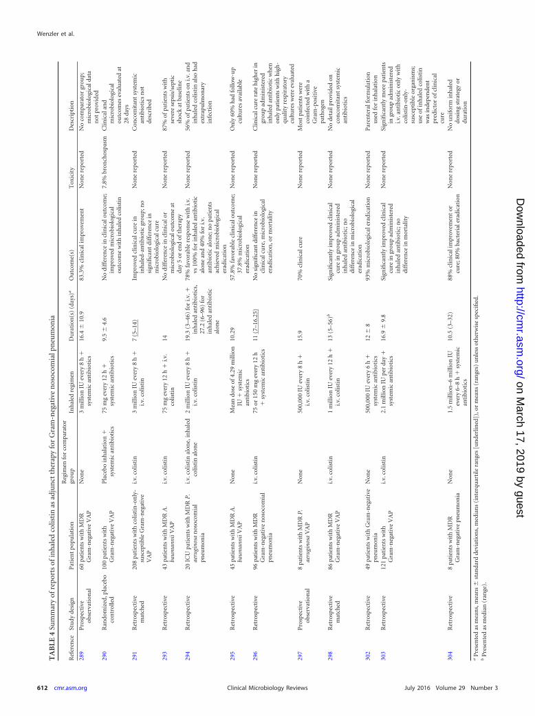
TA
BLE
4Su
mm
ary
ofre
port
sof
inh
aled
colis
tin
asad
jun
ctth
erap
yfo
rG
ram
-neg
ativ
en
osoc
omia
lpn
eum
onia
Ref
eren
ceSt
udy
desi
gnP
atie
nt
popu
lati
onR
egim
enfo
rco
mpa
rato
rgr
oup
Inh
aled
regi
men
Du
rati
on(s
)(d
ays)
aO
utc
ome(
s)T
oxic
ity
Des
crip
tion
289
Pro
spec
tive
obse
rvat
ion
al60
pati
ents
wit
hM
DR
Gra
m-n
egat
ive
VA
PN
one
3m
illio
nIU
ever
y8
h
syst
emic
anti
biot
ics
16.4
�10
.983
.3%
clin
ical
impr
ovem
ent
Non
ere
port
edN
oco
mpa
rato
rgr
oup;
mic
robi
olog
ical
data
not
prov
ided
290
Ran
dom
ized
,pla
cebo
con
trol
led
100
pati
ents
wit
hG
ram
-neg
ativ
eV
AP
Pla
cebo
inh
alat
ion
sy
stem
ican
tibi
otic
s75
mg
ever
y12
h
syst
emic
anti
biot
ics
9.5
�4.
6N
odi
ffer
ence
incl
inic
alou
tcom
e;im
prov
edm
icro
biol
ogic
alou
tcom
ew
ith
inh
aled
colis
tin
7.8%
bron
chos
pasm
Clin
ical
and
mic
robi
olog
ical
outc
omes
eval
uat
edat
28da
ys29
1R
etro
spec
tive
mat
ched
208
pati
ents
wit
hco
listi
n-o
nly
-su
scep
tibl
eG
ram
-neg
ativ
eV
AP
i.v.c
olis
tin
3m
illio
nIU
ever
y8
h
i.v.c
olis
tin
7(5
–14)
Impr
oved
clin
ical
cure
inin
hal
ed-a
nti
biot
icgr
oup;
no
sign
ifica
nt
diff
eren
cein
mic
robi
olog
ical
cure
Non
ere
port
edC
onco
mit
ant
syst
emic
anti
biot
ics
not
desc
ribe
d
293
Ret
rosp
ecti
ve43
pati
ents
wit
hM
DR
A.
baum
anni
iVA
Pi.v
.col
isti
n75
mg
ever
y12
h
i.v.
colis
tin
14N
odi
ffer
ence
incl
inic
alor
mic
robi
olog
ical
outc
ome
atda
y5
oren
dof
ther
apy
Non
ere
port
ed87
%of
pati
ents
wit
hse
vere
seps
is/s
epti
csh
ock
atba
selin
e29
4R
etro
spec
tive
20IC
Upa
tien
tsw
ith
MD
RP
.ae
rugi
nosa
nos
ocom
ial
pneu
mon
ia
i.v.c
olis
tin
alon
e,in
hal
edco
listi
nal
one
2m
illio
nIU
ever
y8
h
i.v.c
olis
tin
19.3
(3–4
6)fo
ri.v
.in
hal
edan
tibi
otic
s,27
.2(6
–96)
for
inh
aled
anti
biot
ical
one
78%
favo
rabl
ere
spon
sew
ith
i.v.
vs10
0%fo
rin
hal
edan
tibi
otic
alon
ean
d40
%fo
ri.v
.an
tibi
otic
alon
e;n
opa
tien
tsac
hie
ved
mic
robi
olog
ical
erad
icat
ion
Non
ere
port
ed56
%of
pati
ents
oni.v
.an
din
hal
edco
listi
nal
soh
adex
trap
ulm
onar
yin
fect
ion
295
Ret
rosp
ecti
ve45
pati
ents
wit
hM
DR
A.
baum
anni
iVA
PN
one
Mea
ndo
seof
4.29
mill
ion
IU
syst
emic
anti
biot
ics
10.2
957
.8%
favo
rabl
ecl
inic
alou
tcom
e;37
.8%
mic
robi
olog
ical
erad
icat
ion
Non
ere
port
edO
nly
60%
had
follo
w-u
pcu
ltu
res
avai
labl
e
296
Ret
rosp
ecti
ve96
pati
ents
wit
hM
DR
Gra
m-n
egat
ive
nos
ocom
ial
pneu
mon
ia
i.v.c
olis
tin
75or
150
mg
ever
y12
h
syst
emic
anti
biot
ics
11(7
–16.
25)
No
sign
ifica
nt
diff
eren
cein
clin
ical
cure
,mic
robi
olog
ical
erad
icat
ion
,or
mor
talit
y
Non
ere
port
edC
linic
alcu
rera
teh
igh
erin
grou
pad
min
iste
red
inh
aled
anti
biot
icw
hen
only
pati
ents
wit
hh
igh
-qu
alit
yre
spir
ator
ycu
ltu
res
wer
eev
alu
ated
297
Pro
spec
tive
obse
rvat
ion
al8
pati
ents
wit
hM
DR
P.
aeru
gino
saV
AP
Non
e50
0,00
0IU
ever
y8
h
i.v.c
olis
tin
15.9
70%
clin
ical
cure
Non
ere
port
edM
ost
pati
ents
wer
eco
infe
cted
wit
ha
Gra
m-p
osit
ive
path
ogen
298
Ret
rosp
ecti
vem
atch
ed86
pati
ents
wit
hM
DR
Gra
m-n
egat
ive
VA
Pi.v
.col
isti
n1
mill
ion
IUev
ery
12h
i.v
.col
isti
n13
(5–5
6)b
Sign
ifica
ntl
yim
prov
edcl
inic
alcu
rein
grou
pad
min
iste
red
inh
aled
anti
biot
ic;n
odi
ffer
ence
inm
icro
biol
ogic
aler
adic
atio
n
Non
ere
port
edN
ode
tail
prov
ided
onco
nco
mit
ant
syst
emic
anti
biot
ics
302
Ret
rosp
ecti
ve49
pati
ents
wit
hG
ram
-neg
ativ
epn
eum
onia
Non
e50
0,00
0IU
ever
y6
h
syst
emic
anti
biot
ics
12�
893
%m
icro
biol
ogic
aler
adic
atio
nN
one
repo
rted
Par
ente
ralf
orm
ula
tion
use
dfo
rin
hal
atio
n30
3R
etro
spec
tive
121
pati
ents
wit
hG
ram
-neg
ativ
eV
AP
i.v.c
olis
tin
2.1
mill
ion
IUpe
rda
y
syst
emic
anti
biot
ics
16.9
�9.
8Si
gnifi
can
tly
impr
oved
clin
ical
cure
ingr
oup
adm
inis
tere
din
hal
edan
tibi
otic
;no
diff
eren
cein
mor
talit
y
Non
ere
port
edSi
gnifi
can
tly
mor
epa
tien
tsin
grou
pad
min
iste
red
i.v.a
nti
biot
icon
lyw
ith
colis
tin
-on
ly-
susc
epti
ble
orga
nis
ms;
use
ofin
hal
edco
listi
nw
asin
depe
nde
nt
pred
icto
rof
clin
ical
cure
304
Ret
rosp
ecti
ve8
pati
ents
wit
hM
DR
Gra
m-n
egat
ive
pneu
mon
iaN
one
1.5
mill
ion
–6m
illio
nIU
ever
y6–
8h
sy
stem
ican
tibi
otic
s
10.5
(3–3
2)88
%cl
inic
alim
prov
emen
tor
cure
;80%
bact
eria
lera
dica
tion
Non
ere
port
edN
ou
nif
orm
inh
aled
dosi
ng
stra
tegy
ordu
rati
on
aP
rese
nte
das
mea
ns,
mea
ns
�st
anda
rdde
viat
ion
s,m
edia
ns
(in
terq
uar
tile
ran
ges
[un
derl
ined
]),o
rm
ean
s(r
ange
s)u
nle
ssot
her
wis
esp
ecifi
ed.
bP
rese
nte
das
med
ian
(ran
ge).
Wenzler et al.
612 cmr.asm.org July 2016 Volume 29 Number 3Clinical Microbiology Reviews
on March 17, 2019 by guest
http://cmr.asm
.org/D
ownloaded from

mannii and fewer were infected with K. pneumoniae than in thegroup that received i.v. colistin (69.2% versus 53.8% [P � 0.02]and 8% versus 20% [P � 0.01], respectively). More than 80% ofpatients in both groups received inadequate initial antibiotic ther-apy, with no difference between the groups. The median durationsof colistin treatment were 7 and 10 days in the group receivinginhaled plus i.v. colistin compared to the group receiving i.v. colis-tin, respectively, which did not differ statistically. The rate of clin-ical cure was statistically significantly better in the group receivinginhaled plus i.v. colistin than in the group receiving i.v. colistin(69.2% versus 54.8%; P � 0.03). The rate of microbiological cure(clearance of the infecting pathogen from posttreatment respira-tory cultures) in the group receiving inhaled plus i.v. colistingroup was 13.4% higher than that in the group receiving i.v. colis-tin group, although this did not reach significance (P � 0.08).There were no significant differences in ICU mortality or nephro-toxicity during colistin therapy between the groups. These authorsalso performed a logistic regression analysis, which indicated thattrauma-related admissions and inhaled and i.v. colistin therapywere independent predictors of clinical cure. Conversely, higherSAPS II and SOFA scores, septic shock at the onset of VAP, andacute kidney injury during colistin therapy were independentlyassociated with clinical failure. Although the isolates in this studywere determined to be susceptible to colistin only, these authorsdid not report whether the patients in either group received con-comitant systemic therapy with other agents. It is implied thatpatients in both groups received i.v. colistin alone with or withoutinhaled colistin, which can no longer be recommended given thedifficulty in achieving adequate PK/PD indices and the docu-mented regrowth and emergence of resistance with monotherapy(298). Emergence of colistin-resistant Gram-negative isolates wasnot explicitly reported in this study. The lack of difference in mor-tality rates with improved clinical cure rates in this study andothers argue that VAP has less of an impact on overall ICU survivalthan do age, comorbidities, severity of illness, and other factors.Even so, the improved clinical and microbiological outcomeswithout increased toxicity in this study warrant further explora-tion in larger, controlled trials.
In a study looking specifically at critically ill adult patients withVAP due to MDR A. baumannii, patients received either i.v. colis-tin only or i.v. and inhaled colistin and were evaluated retrospec-tively (299). The primary outcomes consisted of both clinical andmicrobiological responses to therapy on day 5 and at the end oftherapy. Clinical success was defined as resolution of signs andsymptoms of VAP with no need for additional antibiotic therapy,and microbiological clearance was defined as eradication of A.baumannii upon follow-up culture. Colistin was administered i.v.as either 2.5 mg/kg every 12 h (maximum of 300 mg) or 2.5 mg/kgevery 6 h (maximum of 600 mg), and inhaled colistin was given at75 mg twice daily via a nebulizer of an undefined type. Forty-threepatients were included in the study, 29 of whom received i.v. andinhaled colistin and 15 of whom received i.v. colistin alone. Therewere no significant differences in demographics between the twopopulations, and �30% of patients in each group received a con-comitant aminoglycoside. There were no differences in clinicaloutcomes at day 5 (44% for i.v. colistin only versus 35% for i.v.and inhaled colistin; P � 0.75) or at the end of colistin therapy(38% for i.v. colistin only versus 14% for i.v. and inhaled colistin;P � 0.13) between the two groups. There was also no difference inmicrobiological clearance between i.v. colistin only and i.v. plus
inhaled colistin (69% versus 76%; P � 0.73). Finally, nephrotox-icity and mortality rates also did not differ significantly betweenthe groups, although the proportion of patients with nephrotox-icity was 23% higher in the group that received i.v. and inhaledcolistin. These authors do not report the doses of i.v. colistin ad-ministered in patients receiving i.v. and inhaled colistin comparedto those receiving i.v. colistin alone. Overall, patients in this studywho received the higher dose of i.v. colistin had a lower rate ofclinical cure (7% versus 30%; P � 0.25) and a higher rate of mor-tality (67% versus 45%; P � 0.18). All patients were treated for 14days, and no patients were coinfected with any other pathogens.Importantly, no colistin-resistant A. baumannii organisms wereisolated during this study. Given the retrospective nature of thisstudy and the high percentage (87%) of patients with severe sepsisand/or septic shock on admission, the decreased cure rates in thegroups receiving high-dose i.v. colistin and i.v. plus inhaled colis-tin are likely a reflection of selection bias on the part of the treatingphysician.
In a 2-year retrospective case series of 20 ICU patients whodeveloped nosocomial pneumonia due to MDR P. aeruginosa, theoutcomes of colistin therapy were reported for i.v. colistin alone,inhaled colistin alone, or i.v. and inhaled colistin. Only 5 of the 20patients included had VAP, while the other 15 had undefined nos-ocomial pneumonia. In this series, colistin was given as 2 millionIU three times daily via nebulization. In total, 9 patients receivedboth i.v. and inhaled colistin, and 6 received inhaled colistin alone,while 5 received i.v. colistin only. All patients received colistin incombination with a �-lactam, most commonly piperacillin-tazo-bactam or meropenem. The groups were comparable with respectto baseline demographics and severity of illness at baseline, al-though 56% of the group receiving i.v. and inhaled colistin had anextrapulmonary source of infection. Treatment was administeredfor an average of 19 to 27 days between the groups. Follow-upcultures were available for 19 of 20 patients, and none of themachieved microbiological eradication. In contrast, the clinical re-sponse was deemed favorable in 100% and 78% of patients oninhaled colistin only and i.v. and inhaled colistin, respectively. Inthe group receiving i.v. colistin only, the response was favorable inonly 40% of patients (2/5 patients), and all 5 patients in this groupdied. Only 3 of the other 15 patients died during the study. Neph-rotoxicity was difficult to establish in this study, as many patientshad baseline renal dysfunction, although patients on only inhaledcolistin experienced less toxicity than did those in the other twogroups (300). Although this study included a very small number ofpatients, in general, the patients receiving inhaled colistin eitheralone or with i.v. colistin tended to have improved outcomes overthose of patients receiving i.v. colistin alone. The addition of an-tibiotics other than colistin to these patients’ therapy makes inter-pretation of these results difficult.
A retrospective analysis of the efficacy of inhaled colistin in-cluded 45 patients requiring mechanical ventilation in an ICUwho were diagnosed with MDR A. baumannii VAP (301). VAPwas diagnosed and microbiologically confirmed by positive cul-tures from either bronchial secretions or BAL fluid samples. Pa-tients received i.v. antimicrobial regimens in addition to inhaledcolistin, predominantly a carbapenem. The mean daily dose ofinhaled colistin was 4.29 million � 0.82 million IU, with a meanadministration duration of 10.29 days. A favorable microbiologi-cal outcome (defined as eradication) was noted for 17 patients(37.8%), and 26 patients (57.8%) had a favorable clinical outcome
Inhaled Antibiotics for Pulmonary Infections
July 2016 Volume 29 Number 3 cmr.asm.org 613Clinical Microbiology Reviews
on March 17, 2019 by guest
http://cmr.asm
.org/D
ownloaded from
(defined as clinical cure or improvement) without evidence ofsevere adverse effects. The all-cause mortality rate was 42.2%.
Another large retrospective study of critically ill patients withnosocomial pneumonia who received i.v. colistin alone or in com-bination with inhaled colistin evaluated the incidence of clinicalcure. This was a multicenter trial in which two sites used jet neb-ulizers and one used a vibrating-mesh nebulizer. The type ofpneumonia was not specifically reported, and inhaled colistin wasdosed at either 75 or 150 mg every 12 h depending on the studysite. Fifty-one patients received i.v. colistin only, and 44 receivedboth i.v. and inhaled colistin. The most commonly isolated patho-gens were A. baumannii and P. aeruginosa, and all isolates weresusceptible to colistin, although the majority of patients receivedadditional systemic agents, most commonly a carbapenem or tige-cycline. The mean durations of i.v. colistin were 11.2 and 12.2 daysin the groups receiving i.v. colistin only and i.v. and inhaled colis-tin, respectively. The median duration of inhaled colistin was 11days. Less than half (39.2%) of patients administered only i.v.therapy achieved a clinical cure, compared to 54.5% in the groupadministered i.v. and inhaled therapy, although this was not sig-nificantly different. There were also no differences in microbio-logical eradication in this study. The mortality rate was �16%lower in the group administered i.v. and inhaled colistin (P �0.106). The mortality rate due to pneumonia was �30% lower inthe group administered i.v. and inhaled colistin, although this wasnot significantly different (70.4% versus 40%; P � 0.055). Whenpatients with bronchoscopic BAL fluid cultures were evaluatedseparately from those with nonbronchoscopic BAL fluid or tra-cheal aspirate cultures, there was a statistically significant differ-ence in clinical cure, with better outcomes in the group adminis-tered i.v. and inhaled colistin (57.1% versus 31.3%; P � 0.033)(302).
A prospective observational case series included mechanicallyventilated patients treated with colistin for MDR P. aeruginosapneumonia (303). Ten patients were included, 8 of whom hadVAP. Tracheobronchial secretions or BAL fluid specimens wereutilized for microbiological diagnosis of VAP. All 10 patients re-ceived i.v. CMS at 3 million IU every 8 h, and patients diagnosedwith VAP received inhaled colistin at 500,000 IU every 8 h. Inhaledcolistin was continued even after the discontinuation of i.v. ther-apy until eradication of the pathogen was confirmed in two con-secutive cultures, for a median duration of 15.9 days. Clinical cure(defined as a resolution of clinical signs and symptoms, includingfever and leukocytosis, and improvement of chest radiographs) orimprovement was reported for 7 of the 10 patients. No adverseeffects attributable to colistin were observed. Most patients in thisanalysis were also coinfected with a Gram-positive pathogen.
In one of the more well-designed retrospective studies evalu-ating the efficacy and safety of inhaled antibiotics, Kofteridis et al.compared i.v. colistin alone to i.v. and inhaled colistin in patientswith VAP due to MDR Gram-negative pathogens (304). Thisstudy matched patients with monomicrobial VAP at a 1:1 ratio toreceive either combination therapy or monotherapy. Inhaledcolistin was delivered as 1 million IU twice daily; the type of neb-ulizer used was not mentioned. Forty-three patients in each groupwere evaluated, and the primary pathogen responsible for VAPwas A. baumannii (77%). All strains were colistin susceptible, andthe median durations of therapy were 10 days for the group ad-ministered i.v. colistin only and 13 days for the combinationgroup. There were no statistically significant differences in clinical
or microbiological cure, mortality, or adverse events between thegroups. The clinical cure rate in the combination group was al-most double that in the group that received i.v. colistin alone (54%versus 32.5%; P � 0.05). Interestingly, twice as many patients inthe combination group had a recurrence of their VAP, while themortality rate was almost half that of the group that received i.v.therapy alone (23% versus 42%; P � 0.06). In a logistic regressionmodel, combination therapy was not identified as an independentpredictor of clinical cure. No adverse events related to inhaledcolistin were reported, although 8 patients in each group devel-oped nephrotoxicity. This study demonstrates that although nostatistically significant differences were found, patients on combi-nation therapy tended to experience less morbidity and mortality.However, several limitations were noted in this study (305, 306),including a lack of information regarding the timing of initiationof colistin therapy, the absence of data on the concurrent use ofother antibiotics, and the omission of the total colistin dose ad-ministered in both study groups. Of note, the results of this studyshowed that inhaled colistin had no impact on bacterial growth ormicrobiological eradication, a finding in contrast to data fromprevious studies. This study differed from others, as the primarypathogen was A. baumannii and a lower dose of inhaled colistinwas employed. Repeat respiratory cultures were not dictated, asthis study was retrospective (304). Despite common hesitationsregarding the efficacy of the polymyxins, and colistin in particular,reported studies show a clinical efficacy of �50% despite a highbaseline severity of illness. This response rate is similar to thosereported in previous studies examining the use of piperacillin,imipenem-cilastatin, and ciprofloxacin for P. aeruginosa pneumo-nia (307). Given the good clinical efficacy in Gram-negative LRTIswhen given systemically, administration of colistin via inhalationshould maximize effectiveness by providing increased concentra-tions locally and minimize toxicity. This is especially true for dif-ficult-to-treat pathogens for which the polymyxins remain theonly viable treatment option.
Berlana et al. described 80 patients treated with colistin forinfections due to A. baumannii (86%) and P. aeruginosa (14%).Forty-nine of these patients suffered from pneumonia, and theyall received inhaled colistin for a mean duration of 12 days. Thetype of pneumonia was not categorized in this study. Twelve pa-tients infected with A. baumannii also received i.v. colistin, whileno patients infected with P. aeruginosa received parenteral colis-tin, although 85% of patients received some concomitant antibi-otic. The doses of inhaled colistin varied widely in this study, al-though the majority (79%) of patients received 0.5 million IUevery 6 h. For the 40 patients with pretreatment respiratory cul-tures, 37 of them were negative upon repeat culture at the end oftreatment. Clinical cure was not evaluated in this study, and theoverall mortality rate among all patients was 18%, although thisincluded patients with other foci of infection in addition to pneu-monia. Nephrotoxicity was specifically examined in this study andwas not observed in any patients with evaluable data. Of note, thisstudy used the parenteral form of CMS for inhalation therapy viaa nebulizer (308).
A similar retrospective cohort study compared i.v. colistin(n � 43) to inhaled colistin plus i.v. colistin (n � 78) in patientswith VAP (309). The most common pathogen responsible forVAP was A. baumannii, in �70% of patients in each group. Themean daily dose of inhaled colistin was 2.1 million IU, and therapywas started within 4 days of administration of i.v. colistin in �90%
Wenzler et al.
614 cmr.asm.org July 2016 Volume 29 Number 3Clinical Microbiology Reviews
on March 17, 2019 by guest
http://cmr.asm
.org/D
ownloaded from

of patients. Eighteen patients in the group administered i.v. andinhaled colistin also received another concomitant systemic anti-biotic, compared to 5 in the group administered i.v. colistin only.Of note, there were significantly more patients with colistin-only-susceptible pathogens in the group that received i.v. colistin only(72.1% versus 47.4%; P � 0.009). A greater incidence of clinicalcure among the group administered inhaled plus i.v. colistin thanin the group administered i.v. colistin only was observed (79.5%versus 60.5%; P � 0.025). This association remained true in thegroup of patients for which colistin was the only microbiologicallyactive antimicrobial (76.7% versus 57.9%; P � 0.049). Impor-tantly, upon multivariate analysis, administration of inhaled colis-tin was the only independent predictor for the cure of VAP (oddsratio [OR], 2.53; 95% confidence interval [CI], 1.11 to 5.76).There was no difference in all-cause or in-hospital mortality ratesbetween the two groups (39.7% for the combination group versus44.2% for the monotherapy group; P � 0.92). By multivariateanalysis, a higher APACHE II score (OR, 1.12; 95% CI, 1.04 to1.20), the presence of a malignancy (OR, 4.11; 95% CI, 1.18 to14.23), and a lower daily dosage of i.v. colistin (OR per millioninternational units, 0.81; 95% CI, 0.68 to 0.96) were all significantpredicators of mortality. Although adverse events were not specif-ically examined in this study, no events associated with inhaledcolistin were reported.
A small retrospective study examined the use of inhaled colis-tin for eight patients with MDR Gram-negative pneumonias, 88%of which were due to A. baumannii. Six of the eight patients in-cluded had VAP. The dose of inhaled colistin ranged from 1.5million to 6 million IU divided every 6 to 8 h for a mean durationof 10.5 days, and 7/8 patients received concomitant i.v. colistin oranother systemic agent. Seven of the eight patients achieved im-provement or cure in their pneumonia by the end of treatment,and 4/5 evaluable patients had bacterial eradication. No superin-fection with Gram-positive organisms or emergence of colistin-resistant Gram-negative organisms was detected. There were noreported adverse events related to inhaled colistin (310).
A small case series of two patients with HIV documented clin-ical success in the prevention of recurrence of P. aeruginosa pneu-monia using inhaled colistin for long durations, up to 17 months,without adverse effects (311).
Finally, a recent systematic review and meta-analysis examinedthe reported evidence regarding the efficacy and safety of adjunctinhaled colistin with i.v. colistin versus i.v. colistin alone in pa-tients with nosocomial pneumonia due to MDR Gram-negativepathogens. This review included some of the studies mentioned inthis section plus an additional two not discussed here (312, 313).In this analysis, clinical outcome was defined as the clinical re-sponse rate, classified as clinical cure (resolution of presentingsigns and symptoms of infection at the end of treatment) or im-provement (partial resolution of present signs and symptoms).Microbiological outcome was defined as eradication of the patho-gen in culture specimens at the end of hospitalization. The qualityof the included studies was evaluated on the Newcastle-Ottawascale, with point values of 7 to 8 and 5 to 6 indicating very goodand good studies, respectively. In total, 299 studies were identifiedin the initial search, although only 9 were included, comprising672 patients. All included studies were published from 2010 to2014, only one was prospective, all but two were performed inEurope, and five of the nine studies evaluated only VAP. In thegroups that received i.v. and inhaled colistin, i.v. colistin was given
for an average of 15.2 days at a mean dose of 6.3 million IU/day,compared to 12.5 days at a dose of 6.5 million IU/day in the groupsthat received i.v. colistin only. The daily dose of inhaled colistinwas 2 million to 4 million IU divided twice or three times. Fourstudies reported concomitant antibiotic use during administra-tion of colistin. The results from eight studies were available forevaluation of clinical response rates and showed significantly bet-ter clinical improvement and clinical cure in the groups that re-ceived i.v. plus inhaled colistin than in the groups that received i.v.colistin alone (odds ratio, 1.81; 95% CI, 1.30 to 2.53; P � 0.0005).Importantly, this outcome did not show evidence of statisticalheterogeneity based on the inconsistency index (I2 � 21%). In asubgroup analysis comparing i.v. and inhaled colistin to i.v. colis-tin alone, patients receiving i.v. and inhaled therapy tended tohave a higher clinical cure rate, although this was not statisticallysignificant (odds ratio, 1.69; 95% CI, 0.98 to 2.91; P � 0.06). Sixstudies commented on microbiological eradication and showedhigher rates of eradication in the groups that received i.v. andinhaled colistin (odds ratio, 1.66; 95% CI, 1.11 to 2.49; P � 0.01).All nine included studies were available to assess mortality, whichagain showed a significantly lower all-cause ICU or hospital mor-tality rate in groups that received i.v. and inhaled colistin (oddsratio, 0.69; 95% CI, 0.50 to 0.95; P � 0.02), without statisticalheterogeneity (I2 � 0%). Nephrotoxicity was the only safety out-come analyzed in this review, and no significant difference be-tween the groups was observed. All nine studies showed very-good-quality scores on the Newcastle-Ottawa scale, and statisticalevaluations indicated no evidence of publication bias. This sys-tematic review and meta-analysis demonstrates a favorable im-pact of i.v. and inhaled colistin on clinical improvement, clinicalcure, microbiological eradication, and mortality compared to i.v.colistin alone, without increased toxicity in the combinationgroup (314). It is important to remember that although statisticalheterogeneity was not observed when data from all nine studieswere combined, there are still contradictory conclusions betweenindividual studies owing to differences in sample size, selectionbias, pathogen distribution, and delivery device. The consistentdosing of inhaled colistin among all nine studies is in concordancewith existing literature and supports the anecdotal data discussedabove.
In the only study examining the use of inhaled polymyxin B forthe treatment of MDR Gram-negative LRTIs, 25 critically ill pa-tients who received i.v. and/or inhaled polymyxin B along withother systemic antimicrobials were evaluated retrospectively. Al-most all patients (92%) were admitted to the ICU, and 88% weremechanically ventilated. Inhaled polymyxin B was most fre-quently given as 2.5 mg/kg/day divided every 6 h and was used ineight patients, most often in combination with a carbapenem. A.baumannii was the most frequently isolated pathogen in 55% ofpatients, and all cultured Gram-negative isolates were susceptibleto polymyxin B. The overall end-of-treatment mortality rate was21%, while a favorable clinical response was reported for 76% ofpatients. No differences in outcomes between patients receivingi.v. and those receiving inhaled polymyxin B were observed (76%versus 67%; P � 0.63). Respiratory-related adverse events werenot reported in this study, although there were two instances ofneurotoxicity reported, but the route of polymyxin B administra-tion in these cases was unclear. The lack of standardization oftherapy and a control group, along with small numbers, limit theability to draw firm conclusions from this study. Although the lack
Inhaled Antibiotics for Pulmonary Infections
July 2016 Volume 29 Number 3 cmr.asm.org 615Clinical Microbiology Reviews
on March 17, 2019 by guest
http://cmr.asm
.org/D
ownloaded from

of reported pulmonary adverse events in this study is encouraging,further studies examining the use specifically of inhaled poly-myxin B as an adjunct therapy for Gram-negative LRTIs are ur-gently needed (315).
(iii) �-Lactams. In a 3-year prospective study of 25 mechani-cally ventilated patients with Enterobacteriaceae or P. aeruginosapneumonia, i.v. and inhaled cefotaxime, ceftazidime, or tobramy-cin were administered along with selective digestive decontamina-tion with polymyxin B, tobramycin, and amphotericin. The in-haled dose was given as half of the i.v. dose four times daily.Antibiotics were continued until tracheal aspirate cultures weresterile for two consecutive samples. Eradication was achieved in96% of patients within 9 days after the start of treatment, and onlytwo patients experienced a relapse. No emergence of resistant or-ganisms was observed (316).
A prospective, randomized, phase II trial was designed to assessthe efficacy and safety of inhaled ceftazidime and amikacin inpatients with VAP due to P. aeruginosa. Forty patients were in-cluded in this study, 20 of whom received inhaled ceftazidime andamikacin and 20 of whom received i.v. ceftazidime, amikacin, orciprofloxacin, depending on susceptibilities. Inhaled ceftazidimewas administered via a vibrating-plate nebulizer at 15 mg/kg 8times per day, and amikacin was administered at 25 mg/kg oncedaily. Notably, patients with extrapulmonary infections and thosewho received �24 h of antibiotics active against P. aeruginosa wereexcluded. In this study, the extrapulmonary depositions of cefta-zidime and amikacin were 37% each. Cure was assessed on day 9of therapy and was achieved in 70% and 55% of the patients in thegroups administered inhaled and i.v. therapy, respectively (P �0.33). There were also no differences in the recurrence of VAP,length of stay, or duration of ventilation between groups. Therewas no emergence of resistance in the group that received inhaledtherapy, and the 4 patients who initially had a P. aeruginosa strainintermediate to ceftazidime or amikacin had successful bacterialeradication at day 9. Patients in the group that received inhaledtherapy had more rapid microbiological clearance, with 94% ofpatients having negative BAL fluid cultures on day 3, compared to40% in the i.v. treatment group. No episodes of bronchospasmwere observed, although one patient was excluded from the studyafter nebulization-dependent alveolar derecruitment. Three ad-verse events related to obstruction of the expiratory filter, whichled to sudden cardiac arrest in one patient, were reported. Plasmaconcentrations were measured in both groups after 4 days of ther-apy, which showed significantly lower plasma concentrations ofboth drugs in the group that received inhaled therapy. The cefta-zidime trough concentration in plasma was �4-fold lower in thegroup that received inhaled therapy, and the amikacin peak con-centration in plasma was �5-fold lower, while trough concentra-tions were similar. Overall, the group administered inhaled ther-apy achieved more rapid bacterial eradication with similar clinicaloutcome results despite not receiving systemic therapy. Systemicconcentrations were significantly lower, although several adverseevents were reported in the group that received inhaled therapy.Notably, patients were treated for only 8 days in either groupduring this study (317).
In one case report of a patient with severe nosocomial pneu-monia due to P. aeruginosa, i.v. imipenem-cilastatin was given for10 days, without improvement in clinical signs and symptoms andgas exchange. After day 10, inhaled imipenem-cilastatin wasadded as 50 mg every 6 h over 30 min via a jet nebulizer connected
to the ventilator circuit. After 48 h of inhaled therapy, the patientshowed a marked decrease in tracheobronchial secretions andwhite blood cell counts. The patient’s fever resolved, and after 5days of inhaled therapy, he was able to be weaned off the ventilatorwithout any adverse events (318).
A short report detailed the use of inhaled ampicillin-sulbactamin 20 mechanically ventilated patients with A. baumannii pneu-monia. Ten patients received i.v. ampicillin-sulbactam alone,compared to 10 who received both i.v. and inhaled ampicillin-sulbactam dosed at 3 g every 8 h. After 2 to 3 days of therapy,patients who received both i.v. and inhaled ampicillin-sulbactamhad reductions in viable counts of bacteria to �102 CFU/ml, com-pared to no reduction in CFU per milliliter for those who receivedi.v. therapy alone (319).
(iv) Summary of inhaled antibiotics for treatment of VAP.Compared to the two disease states discussed above in this review,there is a wealth of reported data evaluating the use of inhaledantibiotics in the treatment of Gram-negative nosocomial pneu-monia. The bulk of these data is for patients with VAP; the largestcollection of studies utilized inhaled colistin and the majority ofreports are from institutions in Europe. The most commonly ad-ministered regimen for inhaled colistin is 3 million IU every 8 to12 h given via a jet nebulizer. There are also several studies usingaminoglycoside antibiotics via the inhaled route and only mini-mal data associated with �-lactam use. The vast majority of thestudies included in this section are retrospective and observationalin nature, and there are very few high-quality, randomized, place-bo-controlled studies in this field. Even so, the data taken togetherconsistently show positive clinical and microbiological outcomeswith inhaled therapy along with a lack of any serious pulmonarytoxicity. For those studies in which a comparator group was in-cluded, inhaled antibiotics as adjunct therapy often display im-proved clinical and microbiological outcomes over systemic ther-apy alone.
The PDDS used for inhalation of BAY41-6551 is an excitingimprovement over currently available delivery systems, especiallyfor patients undergoing mechanical ventilation. This drug-devicecombination is currently being evaluated as adjunct therapy intwo randomized, placebo-controlled, phase III clinical trials,INHALE 1 and INHALE 2 (ClinicalTrials registration numbersNCT01799993 and NCT00805168, respectively). These resultswill represent the first controlled trial utilizing a drug-devicecombination specifically formulated for inhaled administra-tion of an antimicrobial agent in mechanically ventilated pa-tients with Gram-negative nosocomial pneumonia and willhelp to definitively determine the place for this route of admin-istration in therapy.
Data from the currently available literature are insufficient tosupport the routine use of inhaled antibiotics as monotherapy inpatients with Gram-negative nosocomial pneumonia. For pa-tients who are not critically ill, have no extrapulmonary signs orsymptoms of infection, and have monomicrobial pneumonia dueto a Gram-negative pathogen, monotherapy with an inhaled an-tibiotic is a reasonable therapeutic selection. Inhaled antibioticsshould be utilized as adjunct therapy in patients with nosocomialpneumonia, particularly VAP, due to Gram-negative organisms,especially P. aeruginosa and other nonfermenters such as A. bau-mannii. Specifically, inhaled antibiotics should be started empiri-cally along with systemic therapy in patients with a history of orwho are at high risk for infections by MDR Gram-negative patho-
Wenzler et al.
616 cmr.asm.org July 2016 Volume 29 Number 3Clinical Microbiology Reviews
on March 17, 2019 by guest
http://cmr.asm
.org/D
ownloaded from
gens given the mortality associated with delayed appropriate an-tibiotic therapy in these patients. In this case, the inhaled agentmay represent the only microbiologically active agent despite invitro resistance, as discussed above. Inhaled antibiotics are essen-tial in order to penetrate the biofilm formed within the ET tubeduring mechanical ventilation and to reach the lung parenchymain sufficient concentrations in these patients suffering from anillness for which the morbidity and mortality rates are extremelyhigh.
Although a variety of agents have been studied for inhaled ad-ministration in cases of nosocomial pneumonia, the choice of theagent should depend on which appropriately formulated inhala-tion solution is available, along with the most effective deliverydevice, especially for those patients who are mechanically venti-lated. It is essential that inhaled therapy is administered with op-timized ventilator settings and given by a trained professional withexperience in delivering these agents, as discussed above (Table 3).Inhaled antibiotics should be given as long as the patient is intu-bated and for the same duration as that for systemic therapy. Ifpatients are extubated prior to the end of systemic therapy, con-tinuing to administer the inhaled agent via nebulization or a DPIis reasonable. If serious pulmonary adverse events occur, the in-haled agent should be discontinued.
Inhaled antibiotics for prevention Gram-negative LRTIs.Since the upper respiratory tract is rapidly colonized after hospi-talization for acute illness and given that nosocomial pneumoniasare often preceded by upper airway colonization, it is reasonableto assume that prevention of airway colonization via inhaled an-timicrobials may help prevent hospital-acquired Gram-negativeLRTIs.
(i) Polymyxins. In a population of critical-care patients whereMDR Gram-negative organisms are endemic, a single-center,open-label, randomized trial was conducted to evaluate the im-pact of prophylactic inhaled colistin on the incidence of VAP(320). Adult patients mechanically ventilated for �48 h withoutsigns and symptoms of active respiratory infection were included.Patients received either CMS or placebo three times daily for 10days of ventilation or until extubation, whichever was sooner. Ajet nebulizer was used, and CMS was delivered as 500,000 U. Theprimary outcome was the incidence of VAP at 30 days, with addi-tional secondary outcomes including the incidence of VAT, lengthof stay in the ICU, mortality, number of days without systemicantibiotic exposure, and emergence of colistin-resistant bacteria.A total of 84 patients received inhaled colistin for a median of 10days, compared to 84 who received placebo for a median of 9 days.Inhaled prophylaxis was initiated within a median of 8 h followingintubation in both groups, and the majority of patients (76.2% inthe colistin group versus 71.4% in the placebo group; P � 0.60)received systemic antibiotics during the 10-day prophylaxis pe-riod, with no differences in the numbers or types of antibioticsreceived. The overall incidence of VAP in the colistin group was16.7%, compared to 29.8% in the placebo group (P � 0.07), al-though the incidence of VAP due to Gram-negative organismswas lower in the colistin group (10.7% versus 17.9%; P � 0.03).Interestingly, the incidence of VAP in patients receiving systemicantibiotic therapy for treatment of Gram-negative organisms wasalso lower in the colistin group (14.1% versus 31.7%; P � 0.03).Among patients who developed VAP, the subsequent ICU mor-tality rate was lower for the patients receiving inhaled colistin(7.1% versus 44%; P � 0.03). There were no differences in the
incidence of VAT, hospital mortality, length of stay, or number ofdays without systemic antibiotics between the groups. No colistin-resistant isolates emerged during the study, and there were nodifferences in the incidences of adverse events, including bron-chospasm, between the groups. This study demonstrates a trendtoward improved outcomes for patients receiving VAP prophy-laxis with inhaled colistin. It is difficult to determine the true effectof prophylaxis, as patients continued the study even if they devel-oped VAP and were treated for an active infection. Of note, thedose of inhaled colistin in this study is roughly half of what isnormally used for the treatment of VAP. Although there was noobserved difference in the incidences of VAP between the groups,the patients receiving inhaled colistin as prophylaxis demon-strated a lower rate of VAP than what is normally expected, despitethe fact that the median duration of ventilation was �10 days inboth groups (12 versus 13.5 days; P � 0.26).
In a small case series of five patients colonized with P. aerugi-nosa, 1 million IU of colistin inhaled every 8 h reduced microbialgrowth and eventually sterilized cultures by day 6 (321). A meta-analysis from 2006 evaluating the effect of antibiotic administra-tion via the respiratory tract on the prevention of ICU-acquiredpneumonia revealed a decrease in the incidence of ICU-acquiredpneumonia in patients receiving prophylaxis. Although this anal-ysis combined both instillation and nebulization routes of admin-istration, these data support the potential for reducing theincidence of pneumonia in particularly vulnerable critically illpatients (276).
Inhaled polymyxin B was administered in a prospective studydesigned to evaluate its efficacy at preventing upper respiratorytract colonization with P. aeruginosa. Randomly selected patientswere given polymyxin B via a hand atomizer at a dose of 2.5 mg/kg/day divided every 4 h within 24 h of admission. The polymyxinB solution was atomized and sprayed into the posterior pharynx,tracheostomy tube, or endotracheal tube as applicable. Fifty-eightpatients were included in this study, 33 of whom received poly-myxin B and 25 of whom were controls. Only 7 of the 33 poly-myxin B-treated patients became colonized, compared to 17 of the25 controls. There were no significant demographic differencesbetween the groups. Administration of polymyxin B showed thegreatest reduction in colonization for patients admitted to theICU for 1 week or more. The vast majority of patients in bothgroups (88 and 76%) required systemic antibiotics during theirICU stay, although only 4 patients developed pneumonia duringthe study. Six patients developed renal failure, although detectableplasma concentrations of polymyxin B were found in only one ofthese patients. No other toxicities were observed (322). A fol-low-up study by the same group examined the effect of this ther-apy on the incidence of Gram-negative pneumonia. In this study,either saline or polymyxin B was given via inhalation in on/offcycles of 8 weeks. Approximately half (49% and 53%, respectively)of the patients in the saline and polymyxin B groups receivedsystemic antibiotics throughout the study. Only 1.6% of patientsreceiving polymyxin B had upper airway colonization by organ-isms susceptible to polymyxin B, compared to 9.7% of patients inthe placebo period. There was no difference in the numbers ofpatients acquiring pneumonia between the placebo and poly-myxin B cycles (17 versus 18 patients), although the majority ofthese cases were due to Gram-positive organisms or Gram-nega-tive organisms intrinsically resistant to polymyxin B (i.e., Proteusspp.). There was also no difference in mortality rates between
Inhaled Antibiotics for Pulmonary Infections
July 2016 Volume 29 Number 3 cmr.asm.org 617Clinical Microbiology Reviews
on March 17, 2019 by guest
http://cmr.asm
.org/D
ownloaded from

cycles (12.2 versus 12%) (40). Unfortunately, the results of thisstudy along with the results of the study by Feeley et al. in 1975 (7)significantly diminished the enthusiasm around inhaled antibiot-ics for years afterwards. Intratracheal colistin, as opposed to in-haled colistin, has also been shown to reduce the incidence ofnosocomial pneumonia by 10 to 25% in critically ill patients un-dergoing mechanical ventilation. Emergence of colistin-resistantorganisms was not observed in this study (323).
(ii) �-Lactams. A randomized, double-blind, placebo-con-trolled study evaluated the safety and efficacy of inhaled ceftazi-dime versus placebo for VAP prevention in 40 critically ill traumapatients (43). Adult patients admitted to the trauma ICU whowere expected to undergo mechanical ventilation for at least 7days were randomized to receive either ceftazidime at 250 mgevery 12 h or placebo for 7 days. Study treatment was initiatedwithin 48 h of hospital admission and continued until VAP devel-oped or until day 7. Inhaled ceftazidime was administered via a jetnebulizer with optimized ventilator settings by a respiratory ther-apist. In this study, BAL was performed pretreatment and onceposttreatment during days 4 to 7 for analysis of both inflammatorymarkers (TNF-, IL-1�, IL-6, and IL-8) and ceftazidime concen-trations. Blood samples were also collected during the time ofBAL. The primary outcomes of this study were the incidences ofVAP at days 7 and 14 and at the end of the ICU stay. There were nosignificant differences between the ceftazidime (n � 20) and pla-cebo (n � 20) groups at baseline. The mean numbers of doses inthe ceftazidime and placebo groups were 12.5 and 11.7, respec-tively. No significant difference in the number of patients whodeveloped VAP at day 7 was noted (P � 0.22). There was a statis-tically significant reduction in the incidence of VAP among pa-tients who received inhaled ceftazidime at day 14 (P � 0.021) andthroughout their ICU stay (P � 0.022). For the patients who de-veloped VAP, �85% of patients in each group received systemicantibiotics prior to developing VAP. This seems to imply thatthese patients had extrapulmonary foci of infection prior to devel-oping VAP, but the reason for this systemic usage is not describedby these authors. In the patients who developed VAP (n � 35), themost common (26%) infecting organism was P. aeruginosa. No-tably, 100% of these Pseudomonas isolates were susceptible to cef-tazidime. There was no difference in the duration of mechanicalventilation, ICU length of stay, total antibiotic therapy, and mor-tality between the two study groups. Ceftazidime concentrationswere detectable in the BAL fluid of 16 of 19 patients, with a medianconcentration of 56 mg/liter (range, 2.1 to 443.9 mg/liter). Inter-estingly, 3 of these 16 patients had BAL fluid concentrations thatwere lower than the 8-mg/liter breakpoint for ceftazidime, al-though none of these 3 patients went on to develop VAP. Serumconcentrations of ceftazidime were undetectable in 17 of 18 pa-tients. Additionally, BAL fluid concentrations of TNF-, IL-1�,and IL-8 were significantly decreased in the ceftazidime groupcompared to the placebo group, in which they were significantlyincreased from baseline concentrations. The 73% reduction in theincidence of VAP at day 14 in this study utilizing a �-lactam anti-biotic for VAP prevention in mechanically ventilated ICU patientsprovides key evidence for the role of inhaled agents in decreasingthe morbidity and mortality burdens associated with this seriousinfection. The lack of emergence of resistance and adverse eventssupports the need to validate these findings in a larger patientpopulation utilizing only inhaled therapy without systemic anti-microbials.
A similar prospective, double-blind study compared the inci-dence of VAP at 14 days and 30 days among 105 patients whoreceived inhaled ceftazidime to that among those who receivedplacebo (324). Patients were included if they were admitted to theICU and considered to be at high risk (�25%) for VAP. Ceftazi-dime was delivered as 250 mg every 12 h for 7 days or until thepatient was extubated. Again, respiratory therapists administeredinhaled ceftazidime via a jet nebulizer with optimized ventilatorsettings. Fifty-three patients received ceftazidime, and 52 receivedplacebo, and the overall rates of VAP at 14 and 30 days were 43%and 50%, respectively. There were no significant differences be-tween the groups at baseline, including the median number ofdoses of the study drug (14 in the ceftazidime group versus 13 inthe placebo group; P � 0.79). In this report, the difference in theincidences of VAP between the ceftazidime group and the placebogroup was not statistically different at 14 days (40% versus 46%;P � 0.50) or at 30 days (49% versus 50%; P � 0.90). Four of the sixmost common infecting pathogens in this study were Gram-pos-itive organisms and therefore not covered by ceftazidime, andthese authors failed to describe whether systemic antibiotic ther-apy was utilized. There were no significant differences in second-ary outcomes between the two groups, including mortality, dura-tion of mechanical ventilation, and length of stay. The reason forthe conflicting results between this study and the above-describedstudy are not clear, although the study described above includedfewer patients, and the incidence of VAP in the placebo group washigher than that in this study, possibly providing a larger marginfor effect. Also, 17% of the patients in this study received fewerthan six doses of the study drug. In this study, two patients devel-oped VAP due to ceftazidime-resistant P. aeruginosa. Adverse ef-fects were not described.
(iii) Summary of inhaled antibiotics for prevention of VAP.The efficacy data for the use of inhaled antibiotics for the preven-tion of VAP are encouraging but contradictory and remain to bedefinitively proven in future studies. Both inhaled colistin at500,000 IU three times daily and ceftazidime at 250 mg every 12 hhave demonstrated reduced incidences of Gram-negative VAPcompared to placebo in patients undergoing mechanical ventila-tion. Given the lack of an appropriately formulated compound ofinhaled ceftazidime, this agent cannot be recommended, althoughit is likely that other appropriately formulated agents would seesimilar success. There were no serious adverse events reported inthese studies, and the emergence of resistant Gram-negative or-ganisms was exceedingly rare. Therefore, patients undergoing me-chanical ventilation who are at high risk for VAP may benefit frominhaled antibiotic prophylaxis, especially considering the benefit-to-risk ratio of the morbidity and mortality associated with thedevelopment of VAP. The most appropriate patient for inhaledantibiotic prophylaxis needs to be carefully identified, and VAPrisk scoring systems such as those utilized in the above-describedstudies may be useful tools. Clinicians considering implementingthis strategy should pay very close attention to changes in theirinstitution’s microbial flora and be vigilant about monitoring forincreases in resistance among Gram-negative respiratory patho-gens, especially in the units where this therapy is being used.
SAFETY
In general, testing and proving the safety of inhaled antibiotics aremuch more difficult than for other routes of administration in invitro, preclinical, and clinical studies (325). In addition, current
Wenzler et al.
618 cmr.asm.org July 2016 Volume 29 Number 3Clinical Microbiology Reviews
on March 17, 2019 by guest
http://cmr.asm
.org/D
ownloaded from

pharmaceutical toxicology studies judge safety margins based ontarget organ toxicity measured as a function of the drug exposure(AUC) in the organ relative to plasma. As discussed above, an idealinhaled antibiotic should produce as little systemic exposure aspossible, therefore making only local pulmonary toxicity studiesrelevant, for which there is no standard method. In addition, an-imal studies may serve as a poor model, as rodent upper airwaysare extremely sensitive to insult from inhaled particles, andchanges are not usually predictive of irritancy in humans. Themost concerning adverse effects that one might expect with in-haled therapy, such as macrophage accumulation, cellular infiltra-tion, epithelial degeneration, necrosis, fibrosis, and hyperplasia,are not able to be routinely monitored in humans in a practicalmanner.
Two potential concerns with regard to the administration ofinhaled antibiotics are the development of antibiotic resistanceand the adverse effects sustained when administering the medica-tion. There have been two primary arguments supporting the con-cern of resistance development. First, there has been discussion ofan antibiotic gradient occurring due to the increased deposition ofinhaled antimicrobials at the proximal airway compared to thelower concentrations in the deeper airway (326). This conceptshould no longer be a concern for several reasons. For one, newer,more efficient delivery devices have been developed, and a betterunderstanding of the particle characteristics and inhalation tech-niques required to deliver adequate drug concentrations to thedistal airways has been attained. Additionally, a more thoroughcomprehension of PK/PD parameters and mathematical model-ing allowed the introduction of the concept of the mutant preven-tion concentration (MPC), that is, the capacity to severely restrictthe selection of resistant mutants during antibiotic treatment.This MPC is usually severalfold higher than the MIC and requireshigher exposures to be achieved. The gradient of drug concentra-tions in the lungs is not the cause of resistance development; it isactually the theoretical concern of subinhibitory concentrationsin the deep airways. In fact, administration of antimicrobials viainhalation deposits drug into the lungs at concentrations expo-nentially higher than would be possible with i.v. administration, ashas been demonstrated by both animal and human data presentedhere. Therefore, aerosolization of antibiotics is actually morelikely to achieve concentrations above both the MIC and MPC ofmost intrapulmonary organisms, while systemic administrationof the same agent would achieve high concentrations in theplasma but low concentrations in ELF, creating a gradient. Con-sequently, systemic administration theoretically increases thechances of subinhibitory concentrations in the lungs and failure toreach the MPC (327). Administering antimicrobials via inhalationmay actually help prevent resistance as opposed to inducing it.Finally, the data supporting increased resistance have come al-most solely from patients with CF who had been treated withinhaled antibiotics over extremely long periods of time (326).Even in the most commonly quoted study supporting an in-creased prevalence of resistance, pulmonary function improvedthroughout the study, even in patients with isolates for which theMIC of tobramycin exceeded 128 mg/liter, and at 6 months, therewas no increase in the rate of superinfection with tobramycin-resistant Gram-negative organisms.
Furthermore, in a recent randomized, placebo-controlled trialin critically ill patients by Palmer and Smaldone, inhaled antibi-otics eradicated 96% of organisms, compared to 9% with placebo,
and new drug resistance was not observed (328). On the contrary,the level of resistance to systemic antimicrobials was significantlyhigher in the placebo group. In fact, randomized clinical trialshave not demonstrated the emergence of drug-resistant organ-isms, even in CF patients (329, 330). A recent phase III trial inves-tigating long-term inhaled aztreonam for CF patients with P.aeruginosa infection found no changes in the MIC50 of aztreonamin any of the isolates. On the contrary, these authors found in-creases in susceptibility to tobramycin, suggesting that alternativetherapies may actually preserve susceptibilities to first-line thera-pies (331). Although the use of inhaled antimicrobials for the pre-vention of VAP fell out of favor due to the landmark study byFeeley et al. (7), Klick and colleagues performed a similar study,alternating polymyxin administration with placebo, and did notsee an increase in pseudomonal resistance (40), although in bothcases, the drugs were sprayed into the posterior pharynx and in-stilled into the ET tube. Of note, the study by Feeley et al. isolatedpolymyxin-resistant organisms, although these organisms wereFlavobacteria and Enterococcus isolates, which are not commonrespiratory pathogens. Similar results were observed by Brown etal. after implementing inhaled polymyxin B prophylaxis to con-trol an ICU outbreak of P. aeruginosa (332). More recent investi-gations have found that inhaled tobramycin, ceftazidime, andcolistin were effective at preventing VAP without the emergenceof resistance (276). It is reasonable to assume once again that highconcentrations administered over shorter, defined courses are ac-tually likely to prevent resistant colonies from forming when usedfor VAP, VAT, and non-CF bronchiectasis.
Another theoretical concern is the deposition of antibiotics inthe environment during inhalation. Nebulizers, in particular, canallow aerosolized particles to escape and be deposited in the pa-tient’s environment and may result in the inadvertent exposure ofothers to these particles. This may lead to bronchospasm or alter-ations in the microbial flora of healthy individuals, among othereffects. Education of patients and their caregivers regarding theproper technique for inhaled administration is paramount toachieving effective deposition and preventing a loss of medicationinto the environment. In addition, employees who administeraerosolized antibiotics in the inpatient setting need to be pro-tected and educated on precautionary practices for their use. Nei-ther the National Institute for Occupational Safety and Health(NIOSH) nor the Occupation Safety and Health Administration(OSHA) considers inhaled antibiotics hazardous drugs and hasnot developed guidelines for the safe handling of these medica-tions. Despite this, aerosolized antibiotics may pose an occupa-tional hazard to respiratory therapists and other health care work-ers who administer them, through unintentional inhalation andexposure to escaped aerosols. In a recent survey of health careworkers who administer aerosolized medications, only 52% ofrespondents reported receiving training on the safe handling ofinhaled antibiotics. In addition, �80% of respondents reportednot always using eye/face protection or a respirator when admin-istering these agents, citing that these measures were not in theinstitutional protocol (333). Appropriate controls, includingstandard procedures for personal protective equipment, should beput in place within institutions that administer inhaled antibioticsin order to minimize the likelihood of unnecessary exposure tohealth care workers who administer them.
Continuous in-line nebulizers have been shown to complicatemechanical ventilatory support, although these nebulizers are
Inhaled Antibiotics for Pulmonary Infections
July 2016 Volume 29 Number 3 cmr.asm.org 619Clinical Microbiology Reviews
on March 17, 2019 by guest
http://cmr.asm
.org/D
ownloaded from
now rarely used, and more is understood on how to optimallydeliver nebulizations during mechanical ventilation (334). Addi-tionally, nebulizer reservoirs can act as a cesspool for water-lovingGram-negative organisms, and therefore, contaminated nebulizerreservoirs can produce deleterious effects, especially in mechani-cally ventilated patients (335). Proper cleaning of nebulizers usedon multiple patients in the inpatient setting is essential, as is truefor any nondisposable hospital equipment.
As discussed above repeatedly, the appropriate formulation ofdrug must be used for inhalation. The use of parenteral formula-tions for inhaled therapy has been linked to fatal respiratory dis-tress syndrome and hypotension (336, 337). Inhalation of certainformulations of antimicrobial products can be associated withincreased rates of adverse events. Specifically, the presence of sul-fites in products has been shown to lead to bronchoconstriction(338). This is another reason why only products specifically for-mulated for inhalation should be used for inhalation antimicro-bial therapy whenever possible. For example, in a randomized,double-blind study of CF patients, measurements of lung functionwere significantly decreased after inhalation of colistin sulfatecompared to CMS (45). Colistin sulfate has also demonstrated ahigher rate of bronchoconstriction than CMS (339). Polymyxin Bhas led to bronchoconstriction when administered via inhalation,potentially due to histamine release upon inhalation (340, 341).Neurotoxicity was previously associated with polymyxins but isnow largely irrelevant with new formulations and modes ofadministration (342). The risk of nephrotoxicity is likely to beminimal given the limited systemic absorption after inhalation,as detailed in this review. In addition, newer, more advancedtechniques for both devices and particles for drug delivery, asdiscussed above, have decreased the side effects associated withinhaling these agents, provided that they are used correctly inthe appropriate setting.
LIMITATIONS OF INHALED ANTIMICROBIALS
The penetration of inhaled antibiotics into the lung parenchymaof patients with infected lungs is largely unknown, as the majorityof investigations reporting intrapulmonary concentrations viathis route were completed in phase I trials in healthy volunteers.Existing data for phase II studies of patients with VAP show wideinterpatient variability in pulmonary concentrations after inhaleddelivery.
In some studies, systemic absorption after inhalation reachedplasma concentrations high enough to cause toxicity, primarily inanimals and patients with diseased lungs. In contrast, the low sys-temic concentrations achieved after inhalation in most cases meanthat inhaled antibiotics should probably not be used alone in crit-ically ill patients with Gram-negative LRTIs or those with an ex-trapulmonary focus of infection.
Inhalation formulations are expensive and often require theassistance and time of knowledgeable respiratory therapists, par-ticularly when used on a patient undergoing mechanical ventila-tion. Cost issues related to commercially prepared inhalation so-lutions may lead to pressure to use extemporaneously preparedsolutions, often containing preservatives and with a lack of datasupporting pulmonary deposition, particle size, and efficacy. Inaddition, the cost associated with more modern, more efficientdelivery devices leads to the use of older nebulizers that are usedrepeatedly so that cost can be averaged over time, which may inturn negatively affect efficacy.
DISCUSSION
The prevalence and magnitude of bacterial resistance amongGram-negative pathogens will continue to increase, along with theyearly rates of morbidity and mortality related to antibiotic-resis-tant infections (29). This increase in bacterial resistance will con-tinue to affect the ability to achieve optimal clinical outcomes withthe current approaches in antimicrobial therapy. Despite ad-vances in our knowledge and understanding of human infectionsand the overwhelming wealth of data available on treatment ofpatients with respiratory infections, pneumonia continues to re-main the leading cause of death from infectious diseases world-wide. Compounding this issue is the frighteningly low number ofantibiotics in development for the treatment of Gram-negativerespiratory infections. To combat these issues, we must explorenovel ways to optimize our existing antimicrobial arsenal. Al-though the concept of delivering antibiotics directly to the lungsvia inhalation has been around for decades, recent advances inparticle engineering, delivery devices, administration techniques,and our understanding of PK and PD allow the improved use ofthis route of administration. Antibiotics delivered directly to thesite of infection are able to circumvent the layers of tissue andbody fluids that separate the drug in the bloodstream from thedrug in the lung and capitalize on maximizing efficacy while min-imizing systemic toxicity.
Appropriate formulations of antibiotics and delivery devicesmust be used when administering these agents via the inhalationroute to patients with Gram-negative LRTIs. Particle size and en-gineering characteristics are of the upmost importance for inhaledantibiotics in order to provide adequate pulmonary deposition tothe lower airway and lung parenchyma. The MMAD should bebetween 1 and 5 �m, and spray-drying is currently the most effec-tive manufacturing technique. Liposomal formulations can helpimprove residence time within the desired site of action and fur-ther decrease systemic distribution after inhalation. As clinicians,the choice of the appropriate delivery device is crucial to maximizethe delivered dose and improve patient compliance. Jet nebulizershave been the most widely used device but do not allow for con-sistency in regard to particle size. Vibrating-mesh or -plate nebu-lizers are the most reliable devices at present until newer technol-ogies such as the PDDS prove themselves definitively in phase IIItrials. Reliable in vitro PK and PD models are lacking in the field ofinhaled antibiotics but will need to be improved in order to con-tinue to advance this route of administration and support regula-tory drug approval in this area. Clinical microbiological break-points need to become site specific and take into consideration thehigh concentrations of drug achievable within the lungs after in-halation administration. The reported animal and human PKstudies evaluating inhaled antibiotics support the concept of beingable to achieve high pulmonary concentrations along with lowsystemic absorption after inhalation. Finally, the appropriate pro-cess of inhaled drug delivery in patients undergoing mechanicalventilation is vitally important, and certain steps must be followedin order to provide optimal drug delivery to these patients.
Antibiotics delivered via inhalation have consistently demon-strated intrapulmonary concentrations severalfold higher thanthose achieved after parenteral administration in both animalmodels and human patients. Importantly, in diseased animallungs with altered pathophysiology and ventilation-perfusionmismatch, they have demonstrated a significantly improved abil-
Wenzler et al.
620 cmr.asm.org July 2016 Volume 29 Number 3Clinical Microbiology Reviews
on March 17, 2019 by guest
http://cmr.asm
.org/D
ownloaded from

ity to penetrate areas of bronchopneumonia, resulting in bettermicrobiological eradication. In clinical studies, adverse events as-sociated with the use of inhaled antibacterials have been exceed-ingly rare and mild, even when these agents are given over thecourse of several years. Importantly, the theoretical concern of theemergence of resistant pathogens after administering inhaled an-tibiotics has not come to fruition in modern studies.
The majority of data regarding the treatment of Gram-negativeLRTIs with inhaled antibiotics as monotherapy or adjunctivetherapy demonstrate improved microbiological and clinical out-comes, although almost all of the literature consists of anecdotalreports and retrospective, uncontrolled case series. In the treat-ment of non-CF bronchiectasis, inhaled antibiotics alone or inaddition to systemic agents clearly increase the eradication of bac-teria from sputum. Additionally, inhaled antibiotics often alsoproduce better quality-of-life outcomes but rarely seem to influ-ence standard objective markers of lung function, including FEV1.The administration of systemic antibiotics to patients with VAThas been shown to improve the morbidity and mortality of thisdisease and decrease the progression to VAP. Data for the use ofinhaled antibiotics for VAT seem to mimic these results and maybe a viable option, even as monotherapy, to both treat this infec-tion and decrease the incidence and mortality associated withVAP. The use of inhaled antibiotics for the prevention of nosoco-mial pneumonia, especially in mechanically ventilated patients, iscontroversial but holds promise for future studies given the sig-nals of improved outcomes, overall lack of toxicity, and absence ofemergence of resistance seen throughout current studies. Simi-larly, in the treatment of Gram-negative nosocomial pneumonia,inhaled antibiotics as monotherapy or adjunct therapy have notbeen shown to lead to worse clinical or microbiological outcomesand have not produced significant toxicities or adverse events. Infact, many of the studies reviewed here reported improved micro-biological eradication and clinical cure albeit with often erraticdefinitions of primary and secondary outcomes. Given the mor-bidity and mortality associated with nosocomial pneumonia, par-ticularly VAP, the addition of inhaled antibiotics as adjunct ther-apy seems to be warranted in this scenario.
The majority of the studies included in this review did notcontain a comparator group and allowed heterogeneous admin-istration of concomitant systemic antimicrobials. Additionally,the dose, frequency, and duration of inhaled therapy were oftennot stated and varied widely among reports. Most of the studiesincluded in this review were observational in nature, and the de-cision to add inhaled antimicrobials often depended on the sus-ceptibility of the organism and the patient’s severity of illness.Unfortunately, many studies regularly do not report the specificdrug formulation used for inhalation or the exact type of deviceand specifications used. This information is vitally important andmust be included in future studies in order to improve the under-standing of the ideal clinical situations in which inhaled antibiot-ics can help maximize effects.
The many complex variables associated with the inhalationadministration of antibiotics to patients with Gram-negative re-spiratory LRTIs have been extensively reviewed here. Inhaled an-tibiotics can provide benefit to many patients with a variety ofpulmonary infectious complications; however, continued re-search in this area remains necessary. The current role for thistherapy is as monotherapy or adjunct therapy in specific patientswith non-CF bronchiectasis, VAT, or nosocomial pneumonia due
to Gram-negative pathogens, as outlined throughout this review.Advances in the treatment of respiratory infections will undoubt-edly include the more widespread use of inhaled antimicrobialsbased on the foundational principles laid out in this review.
FUTURE DIRECTIONS
The renewed interest in inhaled antibiotics is an exciting frontierbut also one fraught with challenges. In order to give clinicians theconfidence to use inhaled antimicrobials in patients with Gram-negative LRTIs, further studies assessing the efficacy and safety ofthese agents are needed. Guidelines regarding the appropriate andoptimal use of inhaled antibiotics in targeted patient populationsshould be introduced by societies such as the IDSA, the ATS, andthe Society of Critical Care Medicine in order to improve uponexisting practices and expand the knowledge base of cliniciansaround the world for this exciting field. We encourage pharma-ceutical companies that manufacture antibiotics to explore theinhalation route. Governmental initiatives such as the GeneratingAntibiotic Incentives Now (GAIN) Act should help fund the de-velopment of these novel administration routes given the limitednumber of new systemic agents being developed and brought tomarket. Public-private partnerships such as the Innovative Med-icines Initiative, including the New Drugs for Bad Bugs and Com-batting Bacterial Resistance in Europe programs, are also impor-tant models for moving drug development forward in areas ofunmet medical need (343, 344). This increased development mayallow us to preserve our antibiotic armamentarium by utilizingavailable agents in ways that maximize their efficacy and safety.Additionally, clinical trials evaluating systemic antibiotics in thetreatment of pneumonia should allow the utilization of inhaledantibiotics in order to establish their role in a controlled clinicaltrial environment. Finally, more of the currently available antibi-otics that are active against Gram-negative pathogens need to beexplored for their utility as inhaled agents. This has recently beenaccomplished with meropenem, cefepime, and piperacillin-tazo-bactam, showing MMADs within the ideal range, although paren-teral formulations were used in this evaluation (345).
One of the most challenging aspects of developing and usinginhaled antimicrobials is the drug delivery device. As discussedabove, there can be wide variability between devices and betweenmanufacturers of the same device. Also, the devices available areable to deposit only a small portion of the dose into the lungperiphery. Advanced devices such as the PDDS are needed to im-prove the delivery of these agents and in turn decrease the wasteassociated with inhalation, which should drive down costs andimprove engineering capabilities as bioavailability increases.
The recent advancements in our understanding of the PK/PDaspects of antimicrobials have revolutionized the way in which westudy and use antimicrobials in the clinical arena. From in vitropharmacodynamic models to Monte Carlo simulations, we arenow able to extrapolate results from small populations receivingan antibiotic to larger simulated populations. This gives us im-proved knowledge on PK/PD alterations that may occur and howbest to achieve the index of antibacterial efficacy in special patientpopulations. Unfortunately, these same PK/PD advancementshave not occurred in the realm of inhaled antibacterials. Futurestudies will need to focus on how the PK of inhaled agents changesin specific patient populations, with special consideration towardpatients with pneumonia and ventilation-perfusion mismatches.
In addition to the above-mentioned INHALE 1 and 2 trials,
Inhaled Antibiotics for Pulmonary Infections
July 2016 Volume 29 Number 3 cmr.asm.org 621Clinical Microbiology Reviews
on March 17, 2019 by guest
http://cmr.asm
.org/D
ownloaded from

there are currently six trials evaluating inhaled antibiotics listed onClinicalTrials.gov. Four of these six trials are evaluating Gram-negative antibacterials, one of which is a phase I PK study and theother three of which are for patients with pneumonia. The resultsof these trials will be instrumental in addressing some of the manyremaining unanswered questions regarding inhaled antibiotics:Which patients should receive inhaled therapy? Should it be addedto the standard of care or administered as monotherapy? Shouldinhaled antibiotics be used only for MDR pathogens or for allGram-negative LRTIs? What are the appropriate dose, frequency,and duration of inhaled antibiotics?
We hope that we have provided some insight into these linger-ing uncertainties based on the currently available data, and welook forward to future progress made within this field.
ACKNOWLEDGMENTS
There was no financial support for this work. We thank Melinda Sorianofor her contribution to this work.
REFERENCES1. Dessanges JF. 2001. A history of nebulization. J Aerosol Med 14:65–71.
http://dx.doi.org/10.1089/08942680152007918.2. Aiache J-M. 1990. Aerosol therapy in France. J Aerosol Med 3:85–120.
http://dx.doi.org/10.1089/jam.1990.3.85.3. Tiffeneau R, Brun E. 1947. Sur le diamètre des particules dans les
aérosols: titre inconnu. Sem Hop Paris 1947(27):5.4. Mearns MB, Hunt GH, Rushworth R. 1972. Bacterial flora of respira-
tory tract in patients with cystic fibrosis, 1950-71. Arch Dis Child 47:902–907. http://dx.doi.org/10.1136/adc.47.256.902.
5. Farber JE, Ross J. 1950. The use of aerosol penicillin and streptomycinin bronchopulmonary infections. Calif Med 73:214 –217.
6. Kuhn RJ. 2001. Formulation of aerosolized therapeutics. Chest 120:94S–98S. http://dx.doi.org/10.1378/chest.120.3_suppl.94S.
7. Feeley TW, Du Moulin GC, Hedley-Whyte J, Bushnell LS, Gilbert JP,Feingold DS. 1975. Aerosol polymyxin and pneumonia in seriouslyill patients. N Engl J Med 293:471– 475. http://dx.doi.org/10.1056/NEJM197509042931003.
8. Xu J, Murphy SL, Kochanek KD, Bastian BA. 2016. Deaths: final datafor 2013. Natl Vital Stat Rep 64:1–119. http://www.cdc.gov/nchs/data/nvsr/nvsr64/nvsr64_02.pdf. Accessed 1 July 2015.
9. Mandell LA, Wunderink RG. 2015. Pneumonia, chapter 153. In KasperD, Fauci A, Hauser S, Longo D, Jameson J, Loscalzo J (ed), Harrison’sprinciples of internal medicine, 19th ed. McGraw-Hill, New York, NY.
10. Rodvold KA, George JM, Yoo L. 2011. Penetration of anti-infectiveagents into pulmonary epithelial lining fluid: focus on antibacterialagents. Clin Pharmacokinet 50:637– 664. http://dx.doi.org/10.2165/11594090-000000000-00000.
11. Sylvester JT, Shimoda LA, Aaronson PI, Ward JP. 2012. Hypoxicpulmonary vasoconstriction. Physiol Rev 92:367–520. http://dx.doi.org/10.1152/physrev.00041.2010.
12. Densen P, Mandell GL. 1980. Phagocyte strategy vs. microbial tactics.Rev Infect Dis 2:817– 838. http://dx.doi.org/10.1093/clinids/2.5.817.
13. Krieg DP, Helmke RJ, German VF, Mangos JA. 1988. Resistance ofmucoid Pseudomonas aeruginosa to nonopsonic phagocytosis by alveo-lar macrophages in vitro. Infect Immun 56:3173–3179.
14. Stiver HG, Zachidniak K, Speert DP. 1988. Inhibition of polymorpho-nuclear leukocyte chemotaxis by the mucoid exopolysaccharide of Pseu-domonas aeruginosa. Clin Invest Med 11:247–252.
15. Baldwin DR, Honeybourne D, Wise R. 1992. Pulmonary disposition ofantimicrobial agents: methodological considerations. AntimicrobAgents Chemother 36:1171–1175. http://dx.doi.org/10.1128/AAC.36.6.1171.
16. Wang F, Daugherty B, Keise LL, Wei Z, Foley JP, Savani RC, Koval M.2003. Heterogeneity of claudin expression by alveolar epithelial cells. AmJ Respir Cell Mol Biol 29:62–70. http://dx.doi.org/10.1165/rcmb.2002-0180OC.
17. Campbell L, Abulrob AN, Kandalaft LE, Plummer S, Hollins AJ, GibbsA, Gumbleton M. 2003. Constitutive expression of p-glycoprotein innormal lung alveolar epithelium and functionality in primary alveolar
epithelial cultures. J Pharmacol Exp Ther 304:441– 452. http://dx.doi.org/10.1124/jpet.102.042994.
18. Michot JM, Seral C, Van Bambeke F, Mingeot-Leclercq MP, TulkensPM. 2005. Influence of efflux transporters on the accumulation andefflux of four quinolones (ciprofloxacin, levofloxacin, garenoxacin, andmoxifloxacin) in J774 macrophages. Antimicrob Agents Chemother 49:2429 –2437. http://dx.doi.org/10.1128/AAC.49.6.2429-2437.2005.
19. Hall MJ, DeFrances CJ, Williams SN, Golosinskiy A, Schwartzman A.2010. National Hospital Discharge Survey: 2007 summary. Natl HealthStat Rep 2010:1–20, 24.
20. Arancibia F, Bauer TT, Ewig S, Mensa J, Gonzalez J, Niederman MS,Torres A. 2002. Community-acquired pneumonia due to Gram-negative bacteria and Pseudomonas aeruginosa: incidence, risk, andprognosis. Arch Intern Med 162:1849 –1858. http://dx.doi.org/10.1001/archinte.162.16.1849.
21. Quartin AA, Scerpella EG, Puttagunta S, Kett DH. 2013. A comparisonof microbiology and demographics among patients with healthcare-associated, hospital-acquired, and ventilator-associated pneumonia: aretrospective analysis of 1184 patients from a large, international study.BMC Infect Dis 13:561. http://dx.doi.org/10.1186/1471-2334-13-561.
22. Jones RN. 2010. Microbial etiologies of hospital-acquired bacterialpneumonia and ventilator-associated bacterial pneumonia. Clin InfectDis 51(Suppl 1):S81–S87. http://dx.doi.org/10.1086/653053.
23. American Thoracic Society, Infectious Diseases Society of America.2005. Guidelines for the management of adults with hospital-acquired,ventilator-associated, and healthcare-associated pneumonia. Am JRespir Crit Care Med 171:388 – 416. http://dx.doi.org/10.1164/rccm.200405-644ST.
24. Chastre J, Fagon JY. 2002. Ventilator-associated pneumonia. Am JRespir Crit Care Med 165:867–903. http://dx.doi.org/10.1164/ajrccm.165.7.2105078.
25. Gaynes R, Edwards JR, National Nosocomial Infections SurveillanceSystem. 2005. Overview of nosocomial infections caused by gram-negative bacilli. Clin Infect Dis 41:848 – 854. http://dx.doi.org/10.1086/432803.
26. Mesaros N, Nordmann P, Plesiat P, Roussel-Delvallez M, Van EldereJ, Glupczynski Y, Van Laethem Y, Jacobs F, Lebecque P, Malfroot A,Tulkens PM, Van Bambeke F. 2007. Pseudomonas aeruginosa: resis-tance and therapeutic options at the turn of the new millennium. ClinMicrobiol Infect 13:560 –578. http://dx.doi.org/10.1111/j.1469-0691.2007.01681.x.
27. Chastre J, Wolff M, Fagon JY, Chevret S, Thomas F, Wermert D,Clementi E, Gonzalez J, Jusserand D, Asfar P, Perrin D, Fieux F,Aubas S, PneumA Trial Group. 2003. Comparison of 8 vs 15 days ofantibiotic therapy for ventilator-associated pneumonia in adults: a ran-domized trial. JAMA 290:2588 –2598. http://dx.doi.org/10.1001/jama.290.19.2588.
28. Infectious Diseases Society of America, Spellberg B, Blaser M, GuidosRJ, Boucher HW, Bradley JS, Eisenstein BI, Gerding D, Lynfield R,Reller LB, Rex J, Schwartz D, Septimus E, Tenover FC, Gilbert DN.2011. Combating antimicrobial resistance: policy recommendations tosave lives. Clin Infect Dis 52(Suppl 5):S397–S428. http://dx.doi.org/10.1093/cid/cir153.
29. Centers for Disease Control and Prevention. 2013. Antibiotic resistancethreats in the United States, 2013. Centers for Disease Control and Preven-tion, Atlanta, GA. http://www.cdc.gov/drugresistance/threat-report-2013/pdf/ar-threats-2013-508.pdf. Accessed 26 January 2015.
30. Hede K. 2014. Antibiotic resistance: an infectious arms race. Nature509:S2–S3. http://dx.doi.org/10.1038/509S2a.
31. Safdar N, Dezfulian C, Collard HR, Saint S. 2005. Clinical and eco-nomic consequences of ventilator-associated pneumonia: a systematicreview. Crit Care Med 33:2184 –2193. http://dx.doi.org/10.1097/01.CCM.0000181731.53912.D9.
32. Ambrose PG, Bhavnani SM, Ellis-Grosse EJ, Drusano GL. 2010. Phar-macokinetic-pharmacodynamic considerations in the design of hospi-tal-acquired or ventilator-associated bacterial pneumonia studies: lookbefore you leap! Clin Infect Dis 51(Suppl 1):S103–S110. http://dx.doi.org/10.1086/653057.
33. Hagerman JK, Hancock KE, Klepser ME. 2006. Aerosolised antibiotics:a critical appraisal of their use. Expert Opin Drug Deliv 3:71– 86. http://dx.doi.org/10.1517/17425247.3.1.71.
34. Le J, Ashley ED, Neuhauser MM, Brown J, Gentry C, Klepser ME,Marr AM, Schiller D, Schwiesow JN, Tice S, VandenBussche HL,
Wenzler et al.
622 cmr.asm.org July 2016 Volume 29 Number 3Clinical Microbiology Reviews
on March 17, 2019 by guest
http://cmr.asm
.org/D
ownloaded from

Wood GC, Society of Infectious Diseases Pharmacists AerosolizedAntimicrobials Task Force. 2010. Consensus summary of aerosolizedantimicrobial agents: application of guideline criteria. Insights from theSociety of Infectious Diseases Pharmacists. Pharmacotherapy 30:562–584. http://dx.doi.org/10.1592/phco.30.6.562.
35. Wood GC. 2011. Aerosolized antibiotics for treating hospital-acquiredand ventilator-associated pneumonia. Expert Rev Anti Infect Ther9:993–1000. http://dx.doi.org/10.1586/eri.11.126.
36. Patton JS. 1996. Mechanisms of macromolecule absorption by the lungs.Adv Drug Deliv Rev 19:3–36.
37. Patton JS, Fishburn CS, Weers JG. 2004. The lungs as a portal of entryfor systemic drug delivery. Proc Am Thorac Soc 1:338 –344. http://dx.doi.org/10.1513/pats.200409-049TA.
38. Ehrmann S, Roche-Campo F, Sferrazza Papa GF, Isabey D, BrochardL, Apiou-Sbirlea G. 2013. Aerosol therapy during mechanical ventila-tion: an international survey. Intensive Care Med 39:1048 –1056. http://dx.doi.org/10.1007/s00134-013-2872-5.
39. Klastersky J, Huysmans E, Weerts D, Hensgens C, Daneau D. 1974.Endotracheally administered gentamicin for the prevention of infectionsof the respiratory tract in patients with tracheostomy: a double-blindstudy. Chest 65:650 – 654. http://dx.doi.org/10.1378/chest.65.6.650.
40. Klick JM, du Moulin GC, Hedley-Whyte J, Teres D, Bushnell LS,Feingold DS. 1975. Prevention of gram-negative bacillary pneumoniausing polymyxin aerosol as prophylaxis. II. Effect on the incidence ofpneumonia in seriously ill patients. J Clin Invest 55:514 –519.
41. Drobnic ME, Sune P, Montoro JB, Ferrer A, Orriols R. 2005. Inhaledtobramycin in non-cystic fibrosis patients with bronchiectasis andchronic bronchial infection with Pseudomonas aeruginosa. Ann Phar-macother 39:39 – 44.
42. Palmer LB, Smaldone GC, Simon SR, O’Riordan TG, Cuccia A. 1998.Aerosolized antibiotics in mechanically ventilated patients: delivery andresponse. Crit Care Med 26:31–39. http://dx.doi.org/10.1097/00003246-199801000-00013.
43. Wood GC, Boucher BA, Croce MA, Hanes SD, Herring VL, FabianTC. 2002. Aerosolized ceftazidime for prevention of ventilator-associated pneumonia and drug effects on the proinflammatory responsein critically ill trauma patients. Pharmacotherapy 22:972–982. http://dx.doi.org/10.1592/phco.22.12.972.33596.
44. Dalhoff A. 2014. Pharmacokinetics and pharmacodynamics of aero-solized antibacterial agents in chronically infected cystic fibrosispatients. Clin Microbiol Rev 27:753–782. http://dx.doi.org/10.1128/CMR.00022-14.
45. Westerman EM, Le Brun PP, Touw DJ, Frijlink HW, Heijerman HG.2004. Effect of nebulized colistin sulphate and colistin sulphomethate onlung function in patients with cystic fibrosis: a pilot study. J Cyst Fibros3:23–28. http://dx.doi.org/10.1016/j.jcf.2003.12.005.
46. Patton JS, Byron PR. 2007. Inhaling medicines: delivering drugs to thebody through the lungs. Nat Rev Drug Discov 6:67–74. http://dx.doi.org/10.1038/nrd2153.
47. Stone KC, Mercer RR, Gehr P, Stockstill B, Crapo JD. 1992. Allometricrelationships of cell numbers and size in the mammalian lung. Am JRespir Cell Mol Biol 6:235–243. http://dx.doi.org/10.1165/ajrcmb/6.2.235.
48. Laube BL, Jashnani R, Dalby RN, Zeitlin PL. 2000. Targeting aerosoldeposition in patients with cystic fibrosis: effects of alterations in particlesize and inspiratory flow rate. Chest 118:1069 –1076. http://dx.doi.org/10.1378/chest.118.4.1069.
49. Kuhn RJ. 2002. Pharmaceutical considerations in aerosol drug delivery.Pharmacotherapy 22:80S– 85S. http://dx.doi.org/10.1592/phco.22.6.80S.33907.
50. Brain JD, Valberg PA. 1979. Deposition of aerosol in the respiratorytract. Am Rev Respir Dis 120:1325–1373.
51. Backman P, Adelmann H, Petersson G, Jones CB. 2014. Advances ininhaled technologies: understanding the therapeutic challenge, pre-dicting clinical performance, and designing the optimal inhaled prod-uct. Clin Pharmacol Ther 95:509 –520. http://dx.doi.org/10.1038/clpt.2014.27.
52. Byron PR. 1986. Prediction of drug residence times in regions of thehuman respiratory tract following aerosol inhalation. J Pharm Sci 75:433– 438. http://dx.doi.org/10.1002/jps.2600750502.
53. Li X, Vogt FG, Hayes D, Jr, Mansour HM. 2014. Physicochemicalcharacterization and aerosol dispersion performance of organic solutionadvanced spray-dried microparticulate/nanoparticulate antibiotic dry
powders of tobramycin and azithromycin for pulmonary inhalationaerosol delivery. Eur J Pharm Sci 52:191–205. http://dx.doi.org/10.1016/j.ejps.2013.10.016.
54. Haynes A, Nakamura J, Heng C, Heuerding S, Thompson G, Malcol-mson RJ. 2010. Aerosol performance of tobramycin inhalation powder.Respir Drug Deliv 3:701–706.
55. Geller DE, Weers J, Heuerding S. 2011. Development of an inhaleddry-powder formulation of tobramycin using PulmoSphere technology.J Aerosol Med Pulm Drug Deliv 24:175–182. http://dx.doi.org/10.1089/jamp.2010.0855.
56. Miller DP, Tan T, Nakamura J, Malcolmson R, Tarara T, Weers J. 30June 2015. Physical characterization of tobramycin inhalation powder. I.Rational design of a stable engineered-particle formulation for deliveryto the lungs. Mol Pharm http://dx.doi.org/10.1021/acs.molpharmaceut.5b00147.
57. d’Angelo I, Conte C, La Rotonda MI, Miro A, Quaglia F, Ungaro F.2014. Improving the efficacy of inhaled drugs in cystic fibrosis: challengesand emerging drug delivery strategies. Adv Drug Deliv Rev 75:92–111.http://dx.doi.org/10.1016/j.addr.2014.05.008.
58. Mansour HM, Rhee YS, Wu X. 2009. Nanomedicine in pulmonarydelivery. Int J Nanomedicine 4:299 –319.
59. Moreno-Sastre M, Pastor M, Salomon CJ, Esquisabel A, Pedraz JL. 22July 2015. Pulmonary drug delivery: a review on nanocarriers for anti-bacterial chemotherapy. J Antimicrob Chemother http://dx.doi.org/10.1093/jac/dkv192.
60. Zhou QT, Leung SS, Tang P, Parumasivam T, Loh ZH, Chan HK.2015. Inhaled formulations and pulmonary drug delivery systems forrespiratory infections. Adv Drug Deliv Rev 85:83–99. http://dx.doi.org/10.1016/j.addr.2014.10.022.
61. Stockmann C, Roberts JK, Yellepeddi VK, Sherwin CM. 2015. Clinicalpharmacokinetics of inhaled antimicrobials. Clin Pharmacokinet 54:473– 492. http://dx.doi.org/10.1007/s40262-015-0250-x.
62. Kelly C, Jefferies C, Cryan SA. 2011. Targeted liposomal drug deliveryto monocytes and macrophages. J Drug Deliv 2011:727241. http://dx.doi.org/10.1155/2011/727241.
63. Sackner MA, Hirsch J, Epstein S. 1975. Effect of cuffed endotrachealtubes on tracheal mucous velocity. Chest 68:774 –777. http://dx.doi.org/10.1378/chest.68.6.774.
64. Husain S, Capitano B, Corcoran T, Studer SM, Crespo M, Johnson B,Pilewski JM, Shutt K, Pakstis DL, Zhang S, Carey ME, Paterson DL,McCurry KR, Venkataramanan R. 2010. Intrapulmonary disposition ofamphotericin B after aerosolized delivery of amphotericin B lipid com-plex (Abelcet; ABLC) in lung transplant recipients. Transplantation 90:1215–1219. http://dx.doi.org/10.1097/TP.0b013e3181f995ea.
65. Montgomery AB, Pitlick WH, Nardella P, Tracewell WG, Ramsey BW.2000. Sputum concentrations and systemic pharmacokinetics of aerosol-ized tobramycin (Tobi) in diseased lungs. Respir Drug Deliv 1:19 –24.
66. Dolovich MB, Dhand R. 2011. Aerosol drug delivery: developments indevice design and clinical use. Lancet 377:1032–1045. http://dx.doi.org/10.1016/S0140-6736(10)60926-9.
67. Hickey AJ. 2013. Back to the future: inhaled drug products. J Pharm Sci102:1165–1172. http://dx.doi.org/10.1002/jps.23465.
68. Patton JS, Brain JD, Davies LA, Fiegel J, Gumbleton M, Kim KJ,Sakagami M, Vanbever R, Ehrhardt C. 2010. The particle has landed—characterizing the fate of inhaled pharmaceuticals. J Aerosol Med PulmDrug Deliv 23(Suppl 2):S71–S87. http://dx.doi.org/10.1089/jamp.2010.0836.
69. Hess D, Fisher D, Williams P, Pooler S, Kacmarek RM. 1996. Medi-cation nebulizer performance. Effects of diluent volume, nebulizer flow,and nebulizer brand. Chest 110:498 –505.
70. Coates AL, MacNeish CF, Meisner D, Kelemen S, Thibert R, Mac-Donald J, Vadas E. 1997. The choice of jet nebulizer, nebulizing flow,and addition of albuterol affects the output of tobramycin aerosols.Chest 111:1206 –1212. http://dx.doi.org/10.1378/chest.111.5.1206.
71. Vecellio L, Abdelrahim ME, Montharu J, Galle J, Diot P, Dubus JC.2011. Disposable versus reusable jet nebulizers for cystic fibrosis treat-ment with tobramycin. J Cyst Fibros 10:86 –92. http://dx.doi.org/10.1016/j.jcf.2010.10.004.
72. O’Riordan TG. 2002. Formulations and nebulizer performance. RespirCare 47:1305–1312; discussion 1312–1313.
73. Loffert DT, Ikle D, Nelson HS. 1994. A comparison of commercial jetnebulizers. Chest 106:1788 –1792. http://dx.doi.org/10.1378/chest.106.6.1788.
Inhaled Antibiotics for Pulmonary Infections
July 2016 Volume 29 Number 3 cmr.asm.org 623Clinical Microbiology Reviews
on March 17, 2019 by guest
http://cmr.asm
.org/D
ownloaded from

74. Waldrep JC, Keyhani K, Black M, Knight V. 1994. Operating charac-teristics of 18 different continuous-flow jet nebulizers with beclometha-sone dipropionate liposome aerosol. Chest 105:106 –110. http://dx.doi.org/10.1378/chest.105.1.106.
75. Daniels T, Mills N, Whitaker P. 2013. Nebuliser systems for drugdelivery in cystic fibrosis. Cochrane Database Syst Rev 4:CD007639. http://dx.doi.org/10.1002/14651858.CD007639.pub2.
76. Goode ML, Fink JB, Dhand R, Tobin MJ. 2001. Improvement inaerosol delivery with helium-oxygen mixtures during mechanical venti-lation. Am J Respir Crit Care Med 163:109 –114. http://dx.doi.org/10.1164/ajrccm.163.1.2003025.
77. Dhand R. 2002. Nebulizers that use a vibrating mesh or plate with mul-tiple apertures to generate aerosol. Respir Care 47:1406 –1416; discussion1416 –1418.
78. Geller DE, Rosenfeld M, Waltz DA, Wilmott RW, AeroDose TOBIStudy Group. 2003. Efficiency of pulmonary administration of to-bramycin solution for inhalation in cystic fibrosis using an improveddrug delivery system. Chest 123:28 –36. http://dx.doi.org/10.1378/chest.123.1.28.
79. Dhand R. 2008. Aerosol delivery during mechanical ventilation: frombasic techniques to new devices. J Aerosol Med Pulm Drug Deliv 21:45–60. http://dx.doi.org/10.1089/jamp.2007.0663.
80. Haddrell AE, Davies JF, Miles RE, Reid JP, Dailey LA, Murnane D.2014. Dynamics of aerosol size during inhalation: hygroscopic growth ofcommercial nebulizer formulations. Int J Pharm 463:50 – 61. http://dx.doi.org/10.1016/j.ijpharm.2013.12.048.
81. Heyder J. 2004. Deposition of inhaled particles in the human respiratorytract and consequences for regional targeting in respiratory drug deliv-ery. Proc Am Thorac Soc 1:315–320. http://dx.doi.org/10.1513/pats.200409-046TA.
82. Mainz JG, Schadlich K, Schien C, Michl R, Schelhorn-Neise P,Koitschev A, Koitschev C, Keller PM, Riethmuller J, Wiedemann B,Beck JF. 2014. Sinonasal inhalation of tobramycin vibrating aerosol incystic fibrosis patients with upper airway Pseudomonas aeruginosa col-onization: results of a randomized, double-blind, placebo-controlled pi-lot study. Drug Des Devel Ther 8:209 –217. http://dx.doi.org/10.2147/DDDT.S54064.
83. Kesser KC, Geller DE. 2009. New aerosol delivery devices for cysticfibrosis. Respir Care 54:754 –767; discussion 767–768. http://dx.doi.org/10.4187/002013209790983250.
84. Tiddens HA, Bos AC, Mouton JW, Devadason S, Janssens HM. 2014.Inhaled antibiotics: dry or wet? Eur Respir J 44:1308 –1318. http://dx.doi.org/10.1183/09031936.00090314.
85. Tiddens H. 2004. Inhaled antibiotics. Pediatr Pulmonol Suppl 26:92–94.86. Harrison MJ, McCarthy M, Fleming C, Hickey C, Shortt C, Eustace
JA, Murphy DM, Plant BJ. 2014. Inhaled versus nebulised tobramycin:a real world comparison in adult cystic fibrosis (CF). J Cyst Fibros 13:692– 698. http://dx.doi.org/10.1016/j.jcf.2014.04.004.
87. Konstan MW, Flume PA, Kappler M, Chiron R, Higgins M, Brock-haus F, Zhang J, Angyalosi G, He E, Geller DE. 2011. Safety, efficacyand convenience of tobramycin inhalation powder in cystic fibrosis pa-tients: the EAGER trial. J Cyst Fibros 10:54 – 61. http://dx.doi.org/10.1016/j.jcf.2010.10.003.
88. Gibson RL, Retsch-Bogart GZ, Oermann C, Milla C, Pilewski J, DainesC, Ahrens R, Leon K, Cohen M, McNamara S, Callahan TL, MarkusR, Burns JL. 2006. Microbiology, safety, and pharmacokinetics of az-treonam lysinate for inhalation in patients with cystic fibrosis. PediatrPulmonol 41:656 – 665. http://dx.doi.org/10.1002/ppul.20429.
89. Daddario MK, Hagerman JK, Klepser ME. 2010. Clinical perspectiveon aztreonam lysine for inhalation in patients with cystic fibrosis. InfectDrug Resist 3:123–132. http://dx.doi.org/10.2147/IDR.S7838.
90. Godden DJ, Borland C, Lowry R, Higenbottam TW. 1986. Chemicalspecificity of coughing in man. Clin Sci (Lond) 70:301–306. http://dx.doi.org/10.1042/cs0700301.
91. Lowry RH, Wood AM, Higenbottam TW. 1988. Effects of pH andosmolarity on aerosol-induced cough in normal volunteers. Clin Sci(Lond) 74:373–376. http://dx.doi.org/10.1042/cs0740373.
92. Eschenbacher WL, Boushey HA, Sheppard D. 1984. Alteration in os-molarity of inhaled aerosols cause bronchoconstriction and cough, butabsence of a permeant anion causes cough alone. Am Rev Respir Dis129:211–215.
93. Hess DR. 2000. Nebulizers: principles and performance. Respir Care45:609 – 622.
94. O’Doherty MJ, Thomas SH, Page CJ, Treacher DF, Nunan TO. 1992.Delivery of a nebulized aerosol to a lung model during mechanical ven-tilation. Effect of ventilator settings and nebulizer type, position, andvolume of fill. Am Rev Respir Dis 146:383–388.
95. O’Riordan TG, Greco MJ, Perry RJ, Smaldone GC. 1992. Nebulizerfunction during mechanical ventilation. Am Rev Respir Dis 145:1117–1122. http://dx.doi.org/10.1164/ajrccm/145.5.1117.
96. O’Doherty MJ, Thomas SH. 1997. Nebuliser therapy in the intensivecare unit. Thorax 52(Suppl 2):S56 –S59.
97. Chait R, Craney A, Kishony R. 2007. Antibiotic interactions that selectagainst resistance. Nature 446:668 – 671. http://dx.doi.org/10.1038/nature05685.
98. Rahal JJ. 2006. Novel antibiotic combinations against infections withalmost completely resistant Pseudomonas aeruginosa and Acinetobacterspecies. Clin Infect Dis 43:S95–S99. http://dx.doi.org/10.1086/504486.
99. Klemmer A, Kramer I, Kamin W. 2014. Physicochemical compatibilityand stability of nebulizable drug admixtures containing dornase alfa andtobramycin. Pulm Pharmacol Ther 28:53–59. http://dx.doi.org/10.1016/j.pupt.2013.08.003.
100. Kramer I, Schwabe A, Lichtinghagen R, Kamin W. 2009. Physicochem-ical compatibility of mixtures of dornase alfa and tobramycin containingnebulizer solutions. Pediatr Pulmonol 44:134 –141. http://dx.doi.org/10.1002/ppul.20955.
101. Rubin BK. 2015. Aerosol medications for treatment of mucus clearancedisorders. Respir Care 60:825– 832. http://dx.doi.org/10.4187/respcare.04087.
102. Adi H, Young PM, Chan HK, Stewart P, Agus H, Traini D. 2008.Cospray dried antibiotics for dry powder lung delivery. J Pharm Sci 97:3356 –3366. http://dx.doi.org/10.1002/jps.21239.
103. Tsifansky MD, Yeo Y, Evgenov OV, Bellas E, Benjamin J, KohaneDS. 2008. Microparticles for inhalational delivery of antipseudomo-nal antibiotics. AAPS J 10:254 –260. http://dx.doi.org/10.1208/s12248-008-9033-8.
104. Lee SH, Teo J, Heng D, Ng WK, Chan HK, Tan RB. 2013. Synergisticcombination dry powders for inhaled antimicrobial therapy: formula-tion, characterization and in vitro evaluation. Eur J Pharm Biopharm83:275–284. http://dx.doi.org/10.1016/j.ejpb.2012.09.002.
105. Durante-Mangoni E, Signoriello G, Andini R, Mattei A, De CristoforoM, Murino P, Bassetti M, Malacarne P, Petrosillo N, Galdieri N,Mocavero P, Corcione A, Viscoli C, Zarrilli R, Gallo C, Utili R. 2013.Colistin and rifampicin compared with colistin alone for the treatment ofserious infections due to extensively drug-resistant Acinetobacter bau-mannii: a multicenter, randomized clinical trial. Clin Infect Dis 57:349 –358. http://dx.doi.org/10.1093/cid/cit253.
106. Zhou QT, Sun SP, Chan JG, Wang P, Barraud N, Rice SA, Wang J, LiJ, Chan HK. 2015. Novel inhaled combination powder containing amor-phous colistin and crystalline rifapentine with enhanced antimicrobialactivities against planktonic cells and biofilm of Pseudomonas aerugi-nosa for respiratory infections. Mol Pharm 12:2594 –2603. http://dx.doi.org/10.1021/mp500586p.
107. Zhou QT, Gengenbach T, Denman JA, Yu HH, Li J, Chan HK. 2014.Synergistic antibiotic combination powders of colistin and rifampicinprovide high aerosolization efficiency and moisture protection. AAPS J16:37– 47. http://dx.doi.org/10.1208/s12248-013-9537-8.
108. Trapnell BC, McColley SA, Kissner DG, Rolfe MW, Rosen JM, McK-evitt M, Moorehead L, Montgomery AB, Geller DE, Phase 2 FTI StudyGroup. 2012. Fosfomycin/tobramycin for inhalation in patients withcystic fibrosis with Pseudomonas airway infection. Am J Respir Crit CareMed 185:171–178. http://dx.doi.org/10.1164/rccm.201105-0924OC.
109. Antoniu S. 2015. Novel inhaled combined antibiotic formulations in thetreatment of Pseudomonas aeruginosa airways infections in cystic fibro-sis. Expert Rev Anti Infect Ther 13:897–905. http://dx.doi.org/10.1586/14787210.2015.1041925.
110. Pilcer G, De Bueger V, Traina K, Traore H, Sebti T, Vanderbist F,Amighi K. 2013. Carrier-free combination for dry powder inhalation ofantibiotics in the treatment of lung infections in cystic fibrosis. Int JPharm 451:112–120. http://dx.doi.org/10.1016/j.ijpharm.2013.04.069.
111. Tamaoki J, Takeyama K, Tagaya E, Konno K. 1995. Effect of clarithro-mycin on sputum production and its rheological properties in chronicrespiratory tract infections. Antimicrob Agents Chemother 39:1688 –1690. http://dx.doi.org/10.1128/AAC.39.8.1688.
112. Kawamura-Sato K, Iinuma Y, Hasegawa T, Horii T, Yamashino T,Ohta M. 2000. Effect of subinhibitory concentrations of macrolides on
Wenzler et al.
624 cmr.asm.org July 2016 Volume 29 Number 3Clinical Microbiology Reviews
on March 17, 2019 by guest
http://cmr.asm
.org/D
ownloaded from

expression of flagellin in Pseudomonas aeruginosa and Proteus mirabilis.Antimicrob Agents Chemother 44:2869 –2872. http://dx.doi.org/10.1128/AAC.44.10.2869-2872.2000.
113. Nalca Y, Jansch L, Bredenbruch F, Geffers R, Buer J, Haussler S. 2006.Quorum-sensing antagonistic activities of azithromycin in Pseudomo-nas aeruginosa PAO1: a global approach. Antimicrob Agents Chemother50:1680 –1688. http://dx.doi.org/10.1128/AAC.50.5.1680-1688.2006.
114. Wallace SJ, Nation RL, Li J, Boyd BJ. 2013. Physicochemical aspects ofthe coformulation of colistin and azithromycin using liposomes for com-bination antibiotic therapies. J Pharm Sci 102:1578 –1587. http://dx.doi.org/10.1002/jps.23508.
115. Lee SH, Teo J, Heng D, Zhao Y, Ng WK, Chan HK, Tan RB. 2014.Steroid-decorated antibiotic microparticles for inhaled anti-infectivetherapy. J Pharm Sci 103:1115–1125. http://dx.doi.org/10.1002/jps.23874.
116. Blum CA, Nigro N, Briel M, Schuetz P, Ullmer E, Suter-Widmer I,Winzeler B, Bingisser R, Elsaesser H, Drozdov D, Arici B, Urwyler SA,Refardt J, Tarr P, Wirz S, Thomann R, Baumgartner C, Duplain H,Burki D, Zimmerli W, Rodondi N, Mueller B, Christ-Crain M. 2015.Adjunct prednisone therapy for patients with community-acquiredpneumonia: a multicentre, double-blind, randomised, placebo-controlled trial. Lancet 385:1511–1518. http://dx.doi.org/10.1016/S0140-6736(14)62447-8.
117. Anonymous. 2015. Global Initiative on Chronic Obstructive Lung Dis-ease diagnosis and management guidelines. http://www.goldcopd.org/uploads/users/files/GOLD_Report_2015.pdf. Accessed 18 January2016.
118. Berlutti F, Morea C, Battistoni A, Sarli S, Cipriani P, Superti F,Ammendolia MG, Valenti P. 2005. Iron availability influences aggrega-tion, biofilm, adhesion and invasion of Pseudomonas aeruginosa andBurkholderia cenocepacia. Int J Immunopathol Pharmacol 18:661– 670.
119. Moreau-Marquis S, Coutermarsh B, Stanton BA. 2015. Combinationof hypothiocyanite and lactoferrin (ALX-109) enhances the ability oftobramycin and aztreonam to eliminate Pseudomonas aeruginosa bio-films growing on cystic fibrosis airway epithelial cells. J Antimicrob Che-mother 70:160 –166. http://dx.doi.org/10.1093/jac/dku357.
120. Lee SH, Teo J, Heng D, Zhao Y, Ng WK, Chan HK, Tan LT, Tan RB.2015. A novel inhaled multi-pronged attack against respiratory bacteria.Eur J Pharm Sci 70:37– 44. http://dx.doi.org/10.1016/j.ejps.2015.01.005.
121. Elmore RL, Contois ME, Kelly J, Noe A, Poirier A. 1996. Stability andcompatibility of admixtures of intravenous ciprofloxacin and selecteddrugs. Clin Ther 18:246 –255. http://dx.doi.org/10.1016/S0149-2918(96)80005-1.
122. Weber B, Hochhaus G. 2013. A pharmacokinetic simulation tool forinhaled corticosteroids. AAPS J 15:159 –171. http://dx.doi.org/10.1208/s12248-012-9420-z.
123. De Backer JW, Vos WG, Vinchurkar SC, Claes R, Drollmann A,Wulfrank D, Parizel PM, Germonpre P, De Backer W. 2010. Valida-tion of computational fluid dynamics in CT-based airway models withSPECT/CT. Radiology 257:854 – 862. http://dx.doi.org/10.1148/radiol.10100322.
124. Delvadia RR, Longest PW, Byron PR. 2012. In vitro tests for aerosoldeposition. I. Scaling a physical model of the upper airways to predictdrug deposition variation in normal humans. J Aerosol Med Pulm DrugDeliv 25:32– 40. http://dx.doi.org/10.1089/jamp.2011.0905.
125. Miller DD, Amin MM, Palmer LB, Shah AR, Smaldone GC. 2003.Aerosol delivery and modern mechanical ventilation: in vitro/in vivoevaluation. Am J Respir Crit Care Med 168:1205–1209. http://dx.doi.org/10.1164/rccm.200210-1167OC.
126. Diot P, Morra L, Smaldone GC. 1995. Albuterol delivery in a model ofmechanical ventilation. Comparison of metered-dose inhaler and nebu-lizer efficiency. Am J Respir Crit Care Med 152:1391–1394.
127. Williams L, Fletcher GC, Daniel M, Kinsella J. 1999. A simple invitro method for the evaluation of an ultrasonic nebulizer for drugdelivery to intubated, ventilated patients and the effect of nebulizerand ventilator settings on the uptake of fluid from the nebulizerchamber. Eur J Anaesthesiol 16:479 – 484. http://dx.doi.org/10.1097/00003643-199907000-00008.
128. Thomas SH, O’Doherty MJ, Fidler HM, Page CJ, Treacher DF, NunanTO. 1993. Pulmonary deposition of a nebulised aerosol during mechan-ical ventilation. Thorax 48:154 –159. http://dx.doi.org/10.1136/thx.48.2.154.
129. Mendelman PM, Smith AL, Levy J, Weber A, Ramsey B, Davis RL.
1985. Aminoglycoside penetration, inactivation, and efficacy in cysticfibrosis sputum. Am Rev Respir Dis 132:761–765.
130. Moore JE, Maeda Y, Goldsmith CE, Rendall JC, Elborn JS. 2010. Directcomparison of in vitro susceptibility of wild-type clinical Pseudomonasaeruginosa isolated from adult patients with cystic fibrosis (CF) to TOBIand BRAMITOB (tobramycin inhalation solutions). J Cyst Fibros 9:237–238. http://dx.doi.org/10.1016/j.jcf.2010.03.005.
131. Montgomery AB, Rhomberg PR, Abuan T, Walters KA, Flamm RK.2014. Potentiation effects of amikacin and fosfomycin against selectedamikacin-nonsusceptible Gram-negative respiratory tract pathogens.Antimicrob Agents Chemother 58:3714 –3719. http://dx.doi.org/10.1128/AAC.02780-13.
132. Montgomery AB, Rhomberg PR, Abuan T, Walters KA, Flamm RK.2014. Amikacin-fosfomycin at a five-to-two ratio: characterization ofmutation rates in microbial strains causing ventilator-associatedpneumonia and interactions with commonly used antibiotics. Anti-microb Agents Chemother 58:3708 –3713. http://dx.doi.org/10.1128/AAC.02779-13.
133. MacLeod DL, Barker LM, Sutherland JL, Moss SC, Gurgel JL,Kenney TF, Burns JL, Baker WR. 2009. Antibacterial activities of afosfomycin/tobramycin combination: a novel inhaled antibiotic forbronchiectasis. J Antimicrob Chemother 64:829 – 836. http://dx.doi.org/10.1093/jac/dkp282.
134. Anonymous. 2005. Mesa Espanola de Normalizacion de la Sensibili-dad y Resistencia a los Antimicrobianos. Recomendaciones del grupoMENSURA para la seleccion de antimicrobianos en el estudio dela sensibilidad y criterios para la interpretacion del antibiograma.MENSURA, Madrid, Spain.
135. Morosini MI, Garcia-Castillo M, Loza E, Perez-Vazquez M, BaqueroF, Canton R. 2005. Breakpoints for predicting Pseudomonas aeruginosasusceptibility to inhaled tobramycin in cystic fibrosis patients: use ofhigh-range Etest strips. J Clin Microbiol 43:4480 – 4485. http://dx.doi.org/10.1128/JCM.43.9.4480-4485.2005.
136. So W, Crandon JL, Hamada Y, Nicolau DP. 10 November 2015.Antibacterial activity of achievable epithelial lining fluid exposures ofAmikacin Inhale with or without meropenem. J Antimicrob Chemotherhttp://dx.doi.org/10.1093/jac/dkv370.
137. Gatmaitan BG, Carruthers MM, Lerner AM. 1970. Gentamicin intreatment of primary Gram-negative pneumonias. Am J Med Sci 260:90 –94. http://dx.doi.org/10.1097/00000441-197008000-00003.
138. Pines A, Raafat H, Plucinski K. 1967. Gentamicin and colistin inchronic purulent bronchial infections. Br Med J ii:543–545. http://dx.doi.org/10.1136/bmj.2.5551.543.
139. Panidis D, Markantonis SL, Boutzouka E, Karatzas S, Baltopoulos G.2005. Penetration of gentamicin into the alveolar lining fluid of criticallyill patients with ventilator-associated pneumonia. Chest 128:545–552.http://dx.doi.org/10.1378/chest.128.2.545.
140. Carcas AJ, Garcia-Satue JL, Zapater P, Frias-Iniesta J. 1999. Tobramy-cin penetration into epithelial lining fluid of patients with pneumonia.Clin Pharmacol Ther 65:245–250. http://dx.doi.org/10.1016/S0009-9236(99)70103-7.
141. Li M, Byron PR. 2013. Tobramycin disposition in the rat lung followingairway administration. J Pharmacol Exp Ther 347:318 –324. http://dx.doi.org/10.1124/jpet.113.207415.
142. Marier JF, Brazier JL, Lavigne J, Ducharme MP. 2003. Liposomaltobramycin against pulmonary infections of Pseudomonas aeruginosa: apharmacokinetic and efficacy study following single and multiple intra-tracheal administrations in rats. J Antimicrob Chemother 52:247–252.http://dx.doi.org/10.1093/jac/dkg317.
143. Beaulac C, Sachetelli S, Lagace J. 1999. Aerosolization of low phasetransition temperature liposomal tobramycin as a dry powder in ananimal model of chronic pulmonary infection caused by Pseudomo-nas aeruginosa. J Drug Target 7:33– 41. http://dx.doi.org/10.3109/10611869909085490.
144. Beaulac C, Clement-Major S, Hawari J, Lagace J. 1996. Eradication ofmucoid Pseudomonas aeruginosa with fluid liposome-encapsulated to-bramycin in an animal model of chronic pulmonary infection. Antimi-crob Agents Chemother 40:665– 669.
145. Beaulac C, Sachetelli S, Lagace J. 1998. In-vitro bactericidal efficacy ofsub-MIC concentrations of liposome-encapsulated antibiotic againstgram-negative and gram-positive bacteria. J Antimicrob Chemother 41:35– 41. http://dx.doi.org/10.1093/jac/41.1.35.
146. Stratton CW, Warner RR, Coudron PE, Lilly NA. 1999. Bismuth-
Inhaled Antibiotics for Pulmonary Infections
July 2016 Volume 29 Number 3 cmr.asm.org 625Clinical Microbiology Reviews
on March 17, 2019 by guest
http://cmr.asm
.org/D
ownloaded from

mediated disruption of the glycocalyx-cell wall of Helicobacter pylori:ultrastructural evidence for a mechanism of action for bismuth salts. JAntimicrob Chemother 43:659 – 666. http://dx.doi.org/10.1093/jac/43.5.659.
147. Domenico P, Salo RJ, Novick SG, Schoch PE, Van Horn K, Cunha BA.1997. Enhancement of bismuth antibacterial activity with lipophilic thiolchelators. Antimicrob Agents Chemother 41:1697–1703.
148. Huang CT, Stewart PS. 1999. Reduction of polysaccharide productionin Pseudomonas aeruginosa biofilms by bismuth dimercaprol (BisBAL)treatment. J Antimicrob Chemother 44:601– 605. http://dx.doi.org/10.1093/jac/44.5.601.
149. Domenico P, O’Leary R, Cunha BA. 1992. Differential effects of bis-muth and salicylate salts on the antibiotic susceptibility of Pseudomonasaeruginosa. Eur J Clin Microbiol Infect Dis 11:170 –175. http://dx.doi.org/10.1007/BF01967072.
150. Veloira WG, Domenico P, LiPuma JJ, Davis JM, Gurzenda E, KazzazJA. 2003. In vitro activity and synergy of bismuth thiols and tobramycinagainst Burkholderia cepacia complex. J Antimicrob Chemother 52:915–919. http://dx.doi.org/10.1093/jac/dkg471.
151. Wu CL, Domenico P, Hassett DJ, Beveridge TJ, Hauser AR, Kazzaz JA.2002. Subinhibitory bismuth-thiols reduce virulence of Pseudomonasaeruginosa. Am J Respir Cell Mol Biol 26:731–738. http://dx.doi.org/10.1165/ajrcmb.26.6.2001-00020oc.
152. Alhariri M, Omri A. 2013. Efficacy of liposomal bismuth-ethanedithiol-loaded tobramycin after intratracheal administration in rats with pulmo-nary Pseudomonas aeruginosa infection. Antimicrob Agents Chemother57:569 –578. http://dx.doi.org/10.1128/AAC.01634-12.
153. Hirschl RB, Pranikoff T, Gauger P, Schreiner RJ, Dechert R, Bartlett RH.1995. Liquid ventilation in adults, children, and full-term neonates. Lancet346:1201–1202. http://dx.doi.org/10.1016/S0140-6736(95)92903-7.
154. Hirschl RB, Overbeck MC, Parent A, Hernandez R, Schwartz S,Dosanjh A, Johnson K, Bartlett RH. 1994. Liquid ventilation providesuniform distribution of perfluorocarbon in the setting of respiratoryfailure. Surgery 116:159 –167; discussion 167–168.
155. Smith DJ, Gambone LM, Tarara T, Meays DR, Dellamary LA,Woods CM, Weers J. 2001. Liquid dose pulmonary instillation ofgentamicin PulmoSpheres formulations: tissue distribution andpharmacokinetics in rabbits. Pharm Res 18:1556 –1561. http://dx.doi.org/10.1023/A:1013078330485.
156. Fox WW, Weis CM, Cox C, Farina C, Drott H, Wolfson MR, ShafferTH. 1997. Pulmonary administration of gentamicin during liquid ven-tilation in a newborn lamb lung injury model. Pediatrics 100:E5.
157. Zelinka MA, Wolfson MR, Calligaro I, Rubenstein SD, Greenspan JS,Shaffer TH. 1997. A comparison of intratracheal and intravenous ad-ministration of gentamicin during liquid ventilation. Eur J Pediatr 156:401– 404. http://dx.doi.org/10.1007/s004310050625.
158. Demaeyer P, Akodad EM, Gravet E, Schietecat P, Van Vooren JP,Drowart A, Yernault JC, Legros FJ. 1993. Disposition of liposomalgentamicin following intrabronchial administration in rabbits. J Micro-encapsul 10:77– 88. http://dx.doi.org/10.3109/02652049309015314.
159. Goldstein I, Wallet F, Nicolas-Robin A, Ferrari F, Marquette CH,Rouby JJ. 2002. Lung deposition and efficiency of nebulized amikacinduring Escherichia coli pneumonia in ventilated piglets. Am J RespirCrit Care Med 166:1375–1381. http://dx.doi.org/10.1164/rccm.200204-363OC.
160. Elman M, Goldstein I, Marquette CH, Wallet F, Lenaour G, Rouby JJ,Experimental ICU Study Group. 2002. Influence of lung aeration onpulmonary concentrations of nebulized and intravenous amikacin inventilated piglets with severe bronchopneumonia. Anesthesiology 97:199 –206. http://dx.doi.org/10.1097/00000542-200207000-00028.
161. Hashimoto S, Wolfe E, Guglielmo B, Shanks R, Sundelof J, Pittet JF,Thomas E, Wiener-Kronish J. 1996. Aerosolization of imipenem/cilastatin prevents Pseudomonas-induced acute lung injury. J Antimi-crob Chemother 38:809 – 818. http://dx.doi.org/10.1093/jac/38.5.809.
162. Ferrari F, Liu ZH, Lu Q, Becquemin MH, Louchahi K, Aymard G,Marquette CH, Rouby JJ. 2008. Comparison of lung tissue concentra-tions of nebulized ceftazidime in ventilated piglets: ultrasonic versus vi-brating plate nebulizers. Intensive Care Med 34:1718 –1723. http://dx.doi.org/10.1007/s00134-008-1126-4.
163. Tonnellier M, Ferrari F, Goldstein I, Sartorius A, Marquette CH,Rouby JJ. 2005. Intravenous versus nebulized ceftazidime in ventilatedpiglets with and without experimental bronchopneumonia: comparative
effects of helium and nitrogen. Anesthesiology 102:995–1000. http://dx.doi.org/10.1097/00000542-200505000-00019.
164. Marchand S, Lauda M, Le Moal G, Gobin P, Couet W, Roblot F. 2013.Pharmacokinetics of daptomycin in a patient with severe renal failure notreceiving dialysis. Antimicrob Agents Chemother 57:2898 –2899. http://dx.doi.org/10.1128/AAC.00230-13.
165. Sabet M, Miller CE, Nolan TG, Senekeo-Effenberger K, Dudley MN,Griffith DC. 2009. Efficacy of aerosol MP-376, a levofloxacin inhalationsolution, in models of mouse lung infection due to Pseudomonas aerugi-nosa. Antimicrob Agents Chemother 53:3923–3928. http://dx.doi.org/10.1128/AAC.00268-09.
166. Kassamali Z, Rotschafer JC, Jones RN, Prince RA, Danziger LH. 2013.Polymyxins: wisdom does not always come with age. Clin Infect Dis57:877– 883. http://dx.doi.org/10.1093/cid/cit367.
167. Nation RL, Li J, Turnidge JD. 2013. The urgent need for clear andaccurate information on the polymyxins. Clin Infect Dis 57:1656 –1657.http://dx.doi.org/10.1093/cid/cit522.
168. Kassamali Z, Rotschafer JC, Jones RN, Prince RA, Danziger LH. 2013.Reply to Nation et al. Clin Infect Dis 57:1657–1658. (Reply.) http://dx.doi.org/10.1093/cid/cit525.
169. Gales AC, Jones RN, Sader HS. 2011. Contemporary activity of colistinand polymyxin B against a worldwide collection of Gram-negativepathogens: results from the SENTRY Antimicrobial Surveillance Pro-gram (2006-09). J Antimicrob Chemother 66:2070 –2074. http://dx.doi.org/10.1093/jac/dkr239.
170. Yapa SWS, Li J, Porter CJH, Nation RL, Patel K, McIntosh MP. 2013.Population pharmacokinetics of colistin methanesulfonate in rats:achieving sustained lung concentrations of colistin for targeting respira-tory infections. Antimicrob Agents Chemother 57:5087–5095. http://dx.doi.org/10.1128/AAC.01127-13.
171. Brain JD, Knudson DE, Sorokin SP, Davis MA. 1976. Pulmonarydistribution of particles given by intratracheal instillation or by aero-sol inhalation. Environ Res 11:13–33. http://dx.doi.org/10.1016/0013-9351(76)90107-9.
172. Gontijo AV, Gregoire N, Lamarche I, Gobin P, Couet W, Marchand S.2014. Biopharmaceutical characterization of nebulized antimicrobialagents in rats: 2. Colistin. Antimicrob Agents Chemother 58:3950 –3956.http://dx.doi.org/10.1128/AAC.02819-14.
173. Lu Q, Girardi C, Zhang M, Bouhemad B, Louchahi K, Petitjean O,Wallet F, Becquemin MH, Le Naour G, Marquette CH, Rouby JJ.2010. Nebulized and intravenous colistin in experimental pneumoniacaused by Pseudomonas aeruginosa. Intensive Care Med 36:1147–1155.http://dx.doi.org/10.1007/s00134-010-1879-4.
174. Barrowcliffe MP, Jones JG. 1987. Solute permeability of the alveolarcapillary barrier. Thorax 42:1–10. http://dx.doi.org/10.1136/thx.42.1.1.
175. Marchand S, Bouchene S, de Monte M, Guilleminault L, Montharu J,Cabrera M, Gregoire N, Gobin P, Diot P, Couet W, Vecellio L. 4 June2015. Pharmacokinetics of colistin methansulphonate (CMS) and colis-tin after CMS nebulisation in baboon monkeys. Pharm Res http://dx.doi.org/10.1007/s11095-015-1716-0.
176. Newhouse MT, Hirst PH, Duddu SP, Walter YH, Tarara TE, ClarkAR, Weers JG. 2003. Inhalation of a dry powder tobramycin Pulmo-Sphere formulation in healthy volunteers. Chest 124:360 –366. http://dx.doi.org/10.1378/chest.124.1.360.
177. Le Conte P, Potel G, Peltier P, Horeau D, Caillon J, Juvin ME,Kergueris MF, Bugnon D, Baron D. 1993. Lung distribution and phar-macokinetics of aerosolized tobramycin. Am Rev Respir Dis 147:1279 –1282. http://dx.doi.org/10.1164/ajrccm/147.5.1279.
178. Baran D, de Vuyst P, Ooms HA. 1990. Concentration of tobramycingiven by aerosol in the fluid obtained by bronchoalveolar lavage. RespirMed 84:203–204. http://dx.doi.org/10.1016/S0954-6111(08)80035-2.
179. Burdette SD, Limkemann AJ, Slaughter JB, Beam WB, Markert RJ.2009. Serum concentrations of aerosolized tobramycin in medical, sur-gical, and trauma patients. Antimicrob Agents Chemother 53:4568. http://dx.doi.org/10.1128/AAC.00490-09.
180. Badia JR, Soy D, Adrover M, Ferrer M, Sarasa M, Alarcon A, CodinaC, Torres A. 2004. Disposition of instilled versus nebulized tobramycinand imipenem in ventilated intensive care unit (ICU) patients. J Antimi-crob Chemother 54:508 –514. http://dx.doi.org/10.1093/jac/dkh326.
181. Odio W, Van Laer E, Klastersky J. 1975. Concentrations of gentamicinin bronchial secretions after intramuscular and endotracheal administra-tion. J Clin Pharmacol 15:518 –524. http://dx.doi.org/10.1002/j.1552-4604.1975.tb01474.x.
Wenzler et al.
626 cmr.asm.org July 2016 Volume 29 Number 3Clinical Microbiology Reviews
on March 17, 2019 by guest
http://cmr.asm
.org/D
ownloaded from

182. Al-Amoud AI, Clark BJ, Assi KA, Chrystyn H. 2005. Determination ofthe bioavailability of gentamicin to the lungs following inhalation fromtwo jet nebulizers. Br J Clin Pharmacol 59:542–545. http://dx.doi.org/10.1111/j.1365-2125.2005.02360.x.
183. Omri A, Beaulac C, Bouhajib M, Montplaisir S, Sharkawi M, Lagace J.1994. Pulmonary retention of free and liposome-encapsulated tobramy-cin after intratracheal administration in uninfected rats and rats infectedwith Pseudomonas aeruginosa. Antimicrob Agents Chemother 38:1090 –1095. http://dx.doi.org/10.1128/AAC.38.5.1090.
184. Weers J, Metzheiser B, Taylor G, Warren S, Meers P, Perkins WR.2009. A gamma scintigraphy study to investigate lung deposition andclearance of inhaled amikacin-loaded liposomes in healthy male volun-teers. J Aerosol Med Pulm Drug Deliv 22:131–138. http://dx.doi.org/10.1089/jamp.2008.0693.
185. Ehrmann S, Mercier E, Vecellio L, Ternant D, Paintaud G, Dequin PF.2008. Pharmacokinetics of high-dose nebulized amikacin in mechani-cally ventilated healthy subjects. Intensive Care Med 34:755–762. http://dx.doi.org/10.1007/s00134-007-0935-1.
186. Luyt CE, Clavel M, Guntupalli K, Johannigman J, Kennedy JI, WoodC, Corkery K, Gribben D, Chastre J. 2009. Pharmacokinetics and lungdelivery of PDDS-aerosolized amikacin (NKTR-061) in intubated andmechanically ventilated patients with nosocomial pneumonia. Crit Care13:R200. http://dx.doi.org/10.1186/cc8206.
187. Montgomery AB, Vallance S, Abuan T, Tservistas M, Davies A. 2014.A randomized double-blind placebo-controlled dose-escalation phase 1study of aerosolized amikacin and fosfomycin delivered via the PARIinvestigational eFlow inline nebulizer system in mechanically ventilatedpatients. J Aerosol Med Pulm Drug Deliv 27:441– 448. http://dx.doi.org/10.1089/jamp.2013.1100.
188. Stass H, Nagelschmitz J, Willmann S, Delesen H, Gupta A, BaumannS. 2013. Inhalation of a dry powder ciprofloxacin formulation in healthysubjects: a phase I study. Clin Drug Invest 33:419 – 427. http://dx.doi.org/10.1007/s40261-013-0082-0.
189. Imberti R. 2010. Intravenous colistimethate administration and colistinlung tissue concentrations. Intensive Care Med 36:1795. http://dx.doi.org/10.1007/s00134-010-1960-z. (Reply, 36:1796-1797.)
190. Imberti R, Cusato M, Villani P, Carnevale L, Iotti GA, Langer M,Regazzi M. 2010. Steady-state pharmacokinetics and BAL concentrationof colistin in critically ill patients after IV colistin methanesulfonate ad-ministration. Chest 138:1333–1339. http://dx.doi.org/10.1378/chest.10-0463.
191. Markou N, Fousteri M, Markantonis SL, Boutzouka E, Tsigou E, Balto-poulo G. 2011. Colistin penetration in the alveolar lining fluid of critically illpatients treated with IV colistimethate sodium. Chest 139:232–233. http://dx.doi.org/10.1378/chest.10-1860. (Reply, 139:233-234.)
192. Athanassa ZE, Markantonis SL, Fousteri MZ, Myrianthefs PM, Bout-zouka EG, Tsakris A, Baltopoulos GJ. 2012. Pharmacokinetics of in-haled colistimethate sodium (CMS) in mechanically ventilated criticallyill patients. Intensive Care Med 38:1779 –1786. http://dx.doi.org/10.1007/s00134-012-2628-7.
193. Dudhani RV, Turnidge JD, Nation RL, Li J. 2010. fAUC/MIC is themost predictive pharmacokinetic/pharmacodynamic index of colistinagainst Acinetobacter baumannii in murine thigh and lung infectionmodels. J Antimicrob Chemother 65:1984 –1990. http://dx.doi.org/10.1093/jac/dkq226.
194. Westerman EM, de Boer AH, Le Brun PP, Touw DJ, Frijlink HW,Heijerman HG. 2007. Dry powder inhalation of colistin sulphomethatein healthy volunteers: a pilot study. Int J Pharm 335:41– 45. http://dx.doi.org/10.1016/j.ijpharm.2006.11.021.
195. Le Brun PP, de Boer AH, Mannes GP, de Fraiture DM, BrimicombeRW, Touw DJ, Vinks AA, Frijlink HW, Heijerman HG. 2002. Drypowder inhalation of antibiotics in cystic fibrosis therapy: part 2. Inha-lation of a novel colistin dry powder formulation: a feasibility study inhealthy volunteers and patients. Eur J Pharm Biopharm 54:25–32. http://dx.doi.org/10.1016/S0939-6411(02)00044-9.
196. de Boer AH, Le Brun PP, van der Woude HG, Hagedoorn P, Heijer-man HG, Frijlink HW. 2002. Dry powder inhalation of antibiotics incystic fibrosis therapy, part 1: development of a powder formulation withcolistin sulfate for a special test inhaler with an air classifier as de-agglomeration principle. Eur J Pharm Biopharm 54:17–24. http://dx.doi.org/10.1016/S0939-6411(02)00043-7.
197. Laennec RTH. 1819. On mediate auscultation, or a treatise on the diag-
nosis of diseases of the lungs and heart. J-A Brosson et J-S Chaudé, Paris,France.
198. McGuinness G, Naidich DP. 2002. CT of airways disease and bronchi-ectasis. Radiol Clin North Am 40:1–19. http://dx.doi.org/10.1016/S0033-8389(03)00105-2.
199. Seitz AE, Olivier KN, Adjemian J, Holland SM, Prevots R. 2012.Trends in bronchiectasis among Medicare beneficiaries in the UnitedStates, 2000 to 2007. Chest 142:432– 439. http://dx.doi.org/10.1378/chest.11-2209.
200. Cole PJ. 1986. Inflammation: a two-edged sword—the model of bron-chiectasis. Eur J Respir Dis Suppl 147:6 –15.
201. Pasteur MC, Helliwell SM, Houghton SJ, Webb SC, Foweraker JE,Coulden RA, Flower CD, Bilton D, Keogan MT. 2000. An investi-gation into causative factors in patients with bronchiectasis. Am JRespir Crit Care Med 162:1277–1284. http://dx.doi.org/10.1164/ajrccm.162.4.9906120.
202. Barker AF. 2002. Bronchiectasis. N Engl J Med 346:1383–1393. http://dx.doi.org/10.1056/NEJMra012519.
203. Rubin BK. 2003. Overview of cystic fibrosis and non-CF bronchiectasis.Semin Respir Crit Care Med 24:619 – 628. http://dx.doi.org/10.1055/s-2004-815658.
204. Tunney MM, Einarsson GG, Wei L, Drain M, Klem ER, Cardwell C,Ennis M, Boucher RC, Wolfgang MC, Elborn JS. 2013. Lung microbi-ota and bacterial abundance in patients with bronchiectasis when clini-cally stable and during exacerbation. Am J Respir Crit Care Med 187:1118 –1126. http://dx.doi.org/10.1164/rccm.201210-1937OC.
205. King PT, Holdsworth SR, Freezer NJ, Villanueva E, Holmes PW. 2007.Microbiologic follow-up study in adult bronchiectasis. Respir Med 101:1633–1638. http://dx.doi.org/10.1016/j.rmed.2007.03.009.
206. Chan ED, Iseman MD. 2013. Underlying host risk factors for nontuber-culous mycobacterial lung disease. Semin Respir Crit Care Med 34:110 –123. http://dx.doi.org/10.1055/s-0033-1333573.
207. Joish VN, Spilsbury-Cantalupo M, Operschall E, Luong B, BoklageS. 2013. Economic burden of non-cystic fibrosis bronchiectasis in thefirst year after diagnosis from a US health plan perspective. ApplHealth Econ Health Policy 11:299 –304. http://dx.doi.org/10.1007/s40258-013-0027-z.
208. Bilton D, Henig N, Morrissey B, Gotfried M. 2006. Addition of inhaledtobramycin to ciprofloxacin for acute exacerbations of Pseudomonasaeruginosa infection in adult bronchiectasis. Chest 130:1503–1510. http://dx.doi.org/10.1378/chest.130.5.1503.
209. Barker AF, Couch L, Fiel SB, Gotfried MH, Ilowite J, Meyer KC,O’Donnell A, Sahn SA, Smith LJ, Stewart JO, Abuan T, Tully H, VanDalfsen J, Wells CD, Quan J. 2000. Tobramycin solution for inhalationreduces sputum Pseudomonas aeruginosa density in bronchiectasis. AmJ Respir Crit Care Med 162:481– 485. http://dx.doi.org/10.1164/ajrccm.162.2.9910086.
210. Scheinberg P, Shore E. 2005. A pilot study of the safety and efficacy oftobramycin solution for inhalation in patients with severe bronchiecta-sis. Chest 127:1420 –1426. http://dx.doi.org/10.1378/chest.127.4.1420.
211. Ramsey BW, Pepe MS, Quan JM, Otto KL, Montgomery AB, Wil-liams-Warren J, Vasiljev KM, Borowitz D, Bowman CM, Marshall BC,Marshall S, Smith AL. 1999. Intermittent administration of inhaledtobramycin in patients with cystic fibrosis. Cystic Fibrosis Inhaled To-bramycin Study Group. N Engl J Med 340:23–30.
212. Chalmers JD, Smith MP, McHugh BJ, Doherty C, Govan JR, Hill AT.2012. Short- and long-term antibiotic treatment reduces airway and sys-temic inflammation in non-cystic fibrosis bronchiectasis. Am J RespirCrit Care Med 186:657– 665. http://dx.doi.org/10.1164/rccm.201203-0487OC.
213. Martinez-Garcia MA, Soler-Cataluna JJ, Perpina-Tordera M, Roman-Sanchez P, Soriano J. 2007. Factors associated with lung function de-cline in adult patients with stable non-cystic fibrosis bronchiectasis.Chest 132:1565–1572. http://dx.doi.org/10.1378/chest.07-0490.
214. Davies G, Wells AU, Doffman S, Watanabe S, Wilson R. 2006. Theeffect of Pseudomonas aeruginosa on pulmonary function in patientswith bronchiectasis. Eur Respir J 28:974 –979. http://dx.doi.org/10.1183/09031936.06.00074605.
215. Olsen AM. 1946. Streptomycin aerosol in the treatment of chronic bron-chiectasis; preliminary report. Proc Staff Meet Mayo Clin 21:53.
216. Pasteur MC, Bilton D, Hill AT, British Thoracic Society Bronchiec-tasis Non-CF Guideline Group. 2010. British Thoracic Society guideline
Inhaled Antibiotics for Pulmonary Infections
July 2016 Volume 29 Number 3 cmr.asm.org 627Clinical Microbiology Reviews
on March 17, 2019 by guest
http://cmr.asm
.org/D
ownloaded from

for non-CF bronchiectasis. Thorax 65(Suppl 1):i1–i58. http://dx.doi.org/10.1136/thx.2010.136119.
217. Vendrell M, de Gracia J, Olveira C, Martinez MA, Giron R, Maiz L,Canton R, Coll R, Escribano A, Sole A. 2008. Diagnosis and treatmentof bronchiectasis Spanish Society of Pneumology and Thoracic Surgery.Arch Bronconeumol 44:629 – 640. (In Spanish.) http://dx.doi.org/10.1016/S1579-2129(08)60117-2.
218. Vendrell M, Munoz G, de Gracia J. 2015. Evidence of inhaled tobra-mycin in non-cystic fibrosis bronchiectasis. Open Respir Med J 9:30 –36.http://dx.doi.org/10.2174/1874306401509010030.
219. Orriols R, Roig J, Ferrer J, Sampol G, Rosell A, Ferrer A, VallanoA. 1999. Inhaled antibiotic therapy in non-cystic fibrosis patientswith bronchiectasis and chronic bronchial infection by Pseudomonasaeruginosa. Respir Med 93:476 – 480. http://dx.doi.org/10.1016/S0954-6111(99)90090-2.
220. Murray MP, Govan JR, Doherty CJ, Simpson AJ, Wilkinson TS,Chalmers JD, Greening AP, Haslett C, Hill AT. 2011. A randomizedcontrolled trial of nebulized gentamicin in non-cystic fibrosis bronchi-ectasis. Am J Respir Crit Care Med 183:491– 499. http://dx.doi.org/10.1164/rccm.201005-0756OC.
221. Lin HC, Cheng HF, Wang CH, Liu CY, Yu CT, Kuo HP. 1997. Inhaledgentamicin reduces airway neutrophil activity and mucus secretion inbronchiectasis. Am J Respir Crit Care Med 155:2024 –2029. http://dx.doi.org/10.1164/ajrccm.155.6.9196111.
222. Wilson R, Welte T, Polverino E, De Soyza A, Greville H, O’Donnell A,Alder J, Reimnitz P, Hampel B. 2013. Ciprofloxacin dry powder forinhalation in non-cystic fibrosis bronchiectasis: a phase II randomisedstudy. Eur Respir J 41:1107–1115. http://dx.doi.org/10.1183/09031936.00071312.
223. Serisier DJ, Bilton D, De Soyza A, Thompson PJ, Kolbe J, GrevilleHW, Cipolla D, Bruinenberg P, Gonda I, ORBIT-2 Investigators.2013. Inhaled, dual release liposomal ciprofloxacin in non-cystic fi-brosis bronchiectasis (ORBIT-2): a randomised, double-blind, place-bo-controlled trial. Thorax 68:812– 817. http://dx.doi.org/10.1136/thoraxjnl-2013-203207.
224. Haworth CS, Foweraker JE, Wilkinson P, Kenyon RF, Bilton D. 2014.Inhaled colistin in patients with bronchiectasis and chronic Pseudomo-nas aeruginosa infection. Am J Respir Crit Care Med 189:975–982. http://dx.doi.org/10.1164/rccm.201312-2208OC.
225. Patel IS, Vlahos I, Wilkinson TM, Lloyd-Owen SJ, Donaldson GC,Wilks M, Reznek RH, Wedzicha JA. 2004. Bronchiectasis, exacerbationindices, and inflammation in chronic obstructive pulmonary disease. AmJ Respir Crit Care Med 170:400 – 407. http://dx.doi.org/10.1164/rccm.200305-648OC.
226. Steinfort DP, Steinfort C. 2007. Effect of long-term nebulized colistinon lung function and quality of life in patients with chronic bronchialsepsis. Intern Med J 37:495– 498. http://dx.doi.org/10.1111/j.1445-5994.2007.01404.x.
227. Kelly MG, Murphy S, Elborn JS. 2003. Bronchiectasis in secondary care:a comprehensive profile of a neglected disease. Eur J Intern Med 14:488 –492. http://dx.doi.org/10.1016/j.ejim.2003.10.002.
228. Berlana D, Llop JM, Manresa F, Jodar R. 2011. Outpatient treatment ofPseudomonas aeruginosa bronchial colonization with long-term inhaledcolistin, tobramycin, or both in adults without cystic fibrosis. Pharma-cotherapy 31:146 –157. http://dx.doi.org/10.1592/phco.31.2.146.
229. Barker AF, O’Donnell AE, Flume P, Thompson PJ, Ruzi JD, de GraciaJ, Boersma WG, De Soyza A, Shao L, Zhang J, Haas L, Lewis SA,Leitzinger S, Montgomery AB, McKevitt MT, Gossage D, Quittner AL,O’Riordan TG. 2014. Aztreonam for inhalation solution in patients withnon-cystic fibrosis bronchiectasis (AIR-BX1 and AIR-BX2): two ran-domised double-blind, placebo-controlled phase 3 trials. Lancet RespirMed 2:738 –749. http://dx.doi.org/10.1016/S2213-2600(14)70165-1.
230. Brodt AM, Stovold E, Zhang L. 2014. Inhaled antibiotics for stablenon-cystic fibrosis bronchiectasis: a systematic review. Eur Respir J 44:382–393. http://dx.doi.org/10.1183/09031936.00018414.
231. Yang JW, Fan LC, Lu HW, Miao XY, Mao B, Xu JF. 26 January 2015.Efficacy and safety of long-term inhaled antibiotic for patients with non-cystic fibrosis bronchiectasis: a meta-analysis. Clin Respir J http://dx.doi.org/10.1111/crj.12278.
232. Evans DJ, Bara AI, Greenstone M. 18 April 2007. Prolonged antibioticsfor purulent bronchiectasis in children and adults. Cochrane DatabaseSyst Rev http://dx.doi.org/10.1002/14651858.CD001392.pub2.
233. Hnin K, Nguyen C, Carson KV, Evans DJ, Greenstone M, Smith BJ.
2015. Prolonged antibiotics for non-cystic fibrosis bronchiectasis in chil-dren and adults. Cochrane Database Syst Rev 8:CD001392. http://dx.doi.org/10.1002/14651858.CD001392.pub3.
234. Chalmers JD, Goeminne P, Aliberti S, McDonnell MJ, Lonni S, Da-vidson J, Poppelwell L, Salih W, Pesci A, Dupont LJ, Fardon TC, DeSoyza A, Hill AT. 2014. The bronchiectasis severity index. An interna-tional derivation and validation study. Am J Respir Crit Care Med 189:576 –585. http://dx.doi.org/10.1164/rccm.201309-1575OC.
235. Craven DE, Chroneou A, Zias N, Hjalmarson KI. 2009. Ventilator-associated tracheobronchitis: the impact of targeted antibiotic therapyon patient outcomes. Chest 135:521–528. http://dx.doi.org/10.1378/chest.08-1617.
236. Craven DE. 2008. Ventilator-associated tracheobronchitis (VAT): ques-tions, answers, and a new paradigm? Crit Care 12:157. http://dx.doi.org/10.1186/cc6912.
237. Craven DE, Hjalmarson KI. 2010. Ventilator-associated tracheobron-chitis and pneumonia: thinking outside the box. Clin Infect Dis 51(Suppl1):S59 –S66. http://dx.doi.org/10.1086/653051.
238. Torres A, Valencia M. 2005. Does ventilator-associated tracheobron-chitis need antibiotic treatment? Crit Care 9:255–256. http://dx.doi.org/10.1186/cc3535.
239. Fabregas N, Torres A, El-Ebiary M, Ramirez J, Hernandez C,Gonzalez J, de la Bellacasa JP, de Anta J, Rodriguez-Roisin R. 1996.Histopathologic and microbiologic aspects of ventilator-associatedpneumonia. Anesthesiology 84:760 –771. http://dx.doi.org/10.1097/00000542-199604000-00002.
240. Rouby JJ, Martin De Lassale E, Poete P, Nicolas MH, Bodin L, JarlierV, Le Charpentier Y, Grosset J, Viars P. 1992. Nosocomial broncho-pneumonia in the critically ill. Histologic and bacteriologic aspects. AmRev Respir Dis 146:1059 –1066.
241. Gerber J, Smirnov A, Wellmer A, Ragheb J, Prange J, Schutz E,Wettich K, Kalich S, Nau R. 2001. Activity of LY333328 in experimentalmeningitis caused by a Streptococcus pneumoniae strain susceptible topenicillin. Antimicrob Agents Chemother 45:2169 –2172. http://dx.doi.org/10.1128/AAC.45.7.2169-2172.2001.
242. A’Court CH, Garrard CS, Crook D, Bowler I, Conlon C, Peto T,Anderson E. 1993. Microbiological lung surveillance in mechanicallyventilated patients, using non-directed bronchial lavage and quantitativeculture. Q J Med 86:635– 648. http://dx.doi.org/10.1093/qjmed/86.10.635.
243. Soler N, Ewig S, Torres A, Filella X, Gonzalez J, Zaubet A. 1999.Airway inflammation and bronchial microbial patterns in patients withstable chronic obstructive pulmonary disease. Eur Respir J 14:1015–1022. http://dx.doi.org/10.1183/09031936.99.14510159.
244. Nseir S, Di Pompeo C, Pronnier P, Beague S, Onimus T, Saulnier F,Grandbastien B, Mathieu D, Delvallez-Roussel M, Durocher A. 2002.Nosocomial tracheobronchitis in mechanically ventilated patients: inci-dence, aetiology and outcome. Eur Respir J 20:1483–1489. http://dx.doi.org/10.1183/09031936.02.00012902.
245. Palmer LB, Smaldone GC, Chen JJ, Baram D, Duan T, Monteforte M,Varela M, Tempone AK, O’Riordan T, Daroowalla F, Richman P.2008. Aerosolized antibiotics and ventilator-associated tracheobronchi-tis in the intensive care unit. Crit Care Med 36:2008 –2013. http://dx.doi.org/10.1097/CCM.0b013e31817c0f9e.
246. Athanassa ZE, Myrianthefs PM, Boutzouka EG, Tsakris A, Baltopou-los GJ. 2011. Monotherapy with inhaled colistin for the treatment ofpatients with ventilator-associated tracheobronchitis due to polymyxin-only-susceptible Gram-negative bacteria. J Hosp Infect 78:335–336. http://dx.doi.org/10.1016/j.jhin.2011.04.004.
247. Maskin LP, Setten M, Rodriguez PO, Bonelli I, Attie S, Stryjewski ME,Valentini R. 2015. Inhaled colistimethate sodium in ventilator-associated tracheobronchitis due to multidrug-resistant Gram-negativebacteria. Int J Antimicrob Agents 45:199 –200. http://dx.doi.org/10.1016/j.ijantimicag.2014.09.010.
248. Hamer DH. 2000. Treatment of nosocomial pneumonia and tracheo-bronchitis caused by multidrug-resistant Pseudomonas aeruginosa withaerosolized colistin. Am J Respir Crit Care Med 162:328 –330. http://dx.doi.org/10.1164/ajrccm.162.1.9910071.
249. Falagas ME, Sideri G, Korbila IP, Vouloumanou EK, Papadatos JH,Kafetzis DA. 2010. Inhaled colistin for the treatment of tracheobronchi-tis and pneumonia in critically ill children without cystic fibrosis. PediatrPulmonol 45:1135–1140. http://dx.doi.org/10.1002/ppul.21302.
250. Pereira GH, Muller PR, Levin AS. 2007. Salvage treatment of pneumo-
Wenzler et al.
628 cmr.asm.org July 2016 Volume 29 Number 3Clinical Microbiology Reviews
on March 17, 2019 by guest
http://cmr.asm
.org/D
ownloaded from

nia and initial treatment of tracheobronchitis caused by multidrug-resistant Gram-negative bacilli with inhaled polymyxin B. Diagn Micro-biol Infect Dis 58:235–240. http://dx.doi.org/10.1016/j.diagmicrobio.2007.01.008.
251. Agrafiotis M, Siempos II, Falagas ME. 2010. Frequency, prevention,outcome and treatment of ventilator-associated tracheobronchitis: sys-tematic review and meta-analysis. Respir Med 104:325–336. http://dx.doi.org/10.1016/j.rmed.2009.09.001.
252. Nseir S, Favory R, Jozefowicz E, Decamps F, Dewavrin F, Brunin G, DiPompeo C, Mathieu D, Durocher A, VAT Study Group. 2008. Anti-microbial treatment for ventilator-associated tracheobronchitis: a ran-domized, controlled, multicenter study. Crit Care 12:R62. http://dx.doi.org/10.1186/cc6890.
253. Bonten MJ, Bergmans DC, Ambergen AW, de Leeuw PW, van derGeest S, Stobberingh EE, Gaillard CA. 1996. Risk factors for pneumo-nia, and colonization of respiratory tract and stomach in mechanicallyventilated ICU patients. Am J Respir Crit Care Med 154:1339 –1346.http://dx.doi.org/10.1164/ajrccm.154.5.8912745.
254. Fagon JY, Chastre J, Domart Y, Trouillet JL, Pierre J, Darne C, GibertC. 1989. Nosocomial pneumonia in patients receiving continuous me-chanical ventilation. Prospective analysis of 52 episodes with use of aprotected specimen brush and quantitative culture techniques. Am RevRespir Dis 139:877– 884.
255. Kaye KS, Pogue JM. 24 October 2015. Infections caused by resistantGram-negative bacteria: epidemiology and management. Pharmaco-therapy http://dx.doi.org/10.1002/phar.1636.
256. Kumar A, Roberts D, Wood KE, Light B, Parrillo JE, Sharma S,Suppes R, Feinstein D, Zanotti S, Taiberg L, Gurka D, Kumar A,Cheang M. 2006. Duration of hypotension before initiation of effec-tive antimicrobial therapy is the critical determinant of survival inhuman septic shock. Crit Care Med 34:1589 –1596. http://dx.doi.org/10.1097/01.CCM.0000217961.75225.E9.
257. Tacconelli E, Cataldo MA, Dancer SJ, De Angelis G, Falcone M, FrankU, Kahlmeter G, Pan A, Petrosillo N, Rodriguez-Bano J, Singh N,Venditti M, Yokoe DS, Cookson B, European Society of ClinicalMicrobiology. 2014. ESCMID guidelines for the management of theinfection control measures to reduce transmission of multidrug-resistantGram-negative bacteria in hospitalized patients. Clin Microbiol Infect20(Suppl 1):1–55. http://dx.doi.org/10.1111/1469-0691.12427.
258. Ioannidou E, Siempos II, Falagas ME. 2007. Administration of antimi-crobials via the respiratory tract for the treatment of patients with noso-comial pneumonia: a meta-analysis. J Antimicrob Chemother 60:1216 –1226. http://dx.doi.org/10.1093/jac/dkm385.
259. Falagas ME, Kasiakou SK. 2007. Local administration of polymyxinsinto the respiratory tract for the prevention and treatment of pulmonaryinfections in patients without cystic fibrosis. Infection 35:3–10. http://dx.doi.org/10.1007/s15010-007-6104-1.
260. Melsen WG, Rovers MM, Groenwold RH, Bergmans DC, Camus C,Bauer TT, Hanisch EW, Klarin B, Koeman M, Krueger WA, LacheradeJC, Lorente L, Memish ZA, Morrow LE, Nardi G, van NieuwenhovenCA, O’Keefe GE, Nakos G, Scannapieco FA, Seguin P, Staudinger T,Topeli A, Ferrer M, Bonten MJ. 2013. Attributable mortality of venti-lator-associated pneumonia: a meta-analysis of individual patient datafrom randomised prevention studies. Lancet Infect Dis 13:665– 671.http://dx.doi.org/10.1016/S1473-3099(13)70081-1.
261. Bekaert M, Timsit JF, Vansteelandt S, Depuydt P, Vesin A, Garrouste-Orgeas M, Decruyenaere J, Clec’h C, Azoulay E, Benoit D, Outcom-erea Study Group. 2011. Attributable mortality of ventilator-associatedpneumonia: a reappraisal using causal analysis. Am J Respir Crit CareMed 184:1133–1139. http://dx.doi.org/10.1164/rccm.201105-0867OC.
262. Zampieri FG, Nassar AP, Jr, Gusmao-Flores D, Taniguchi LU, TorresA, Ranzani OT. 2015. Nebulized antibiotics for ventilator-associatedpneumonia: a systematic review and meta-analysis. Crit Care 19:150.http://dx.doi.org/10.1186/s13054-015-0868-y.
263. Fagon JY, Chastre J, Hance AJ, Montravers P, Novara A, Gibert C.1993. Nosocomial pneumonia in ventilated patients: a cohort study eval-uating attributable mortality and hospital stay. Am J Med 94:281–288.http://dx.doi.org/10.1016/0002-9343(93)90060-3.
264. Kollef MH. 1993. Ventilator-associated pneumonia. A multivariateanalysis. JAMA 270:1965–1970.
265. Rello J, Ollendorf DA, Oster G, Vera-Llonch M, Bellm L, Redman R,Kollef MH, VAP Outcomes Scientific Advisory Group. 2002. Epide-miology and outcomes of ventilator-associated pneumonia in a large US
database. Chest 122:2115–2121. http://dx.doi.org/10.1378/chest.122.6.2115.
266. Restrepo MI, Anzueto A, Arroliga AC, Afessa B, Atkinson MJ, Ho NJ,Schinner R, Bracken RL, Kollef MH. 2010. Economic burden of venti-lator-associated pneumonia based on total resource utilization. InfectControl Hosp Epidemiol 31:509–515. http://dx.doi.org/10.1086/651669.
267. Kollef MH, Hamilton CW, Ernst FR. 2012. Economic impact of venti-lator-associated pneumonia in a large matched cohort. Infect ControlHosp Epidemiol 33:250 –256. http://dx.doi.org/10.1086/664049.
268. Trouillet JL, Chastre J, Vuagnat A, Joly-Guillou ML, Combaux D,Dombret MC, Gibert C. 1998. Ventilator-associated pneumonia causedby potentially drug-resistant bacteria. Am J Respir Crit Care Med 157:531–539. http://dx.doi.org/10.1164/ajrccm.157.2.9705064.
269. Singh PK, Schaefer AL, Parsek MR, Moninger TO, Welsh MJ, Green-berg EP. 2000. Quorum-sensing signals indicate that cystic fibrosis lungsare infected with bacterial biofilms. Nature 407:762–764. http://dx.doi.org/10.1038/35037627.
270. Costerton JW, Stewart PS, Greenberg EP. 1999. Bacterial biofilms: acommon cause of persistent infections. Science 284:1318 –1322. http://dx.doi.org/10.1126/science.284.5418.1318.
271. Hoffman LR, D’Argenio DA, MacCoss MJ, Zhang Z, Jones RA, MillerSI. 2005. Aminoglycoside antibiotics induce bacterial biofilm formation.Nature 436:1171–1175. http://dx.doi.org/10.1038/nature03912.
272. Adair CG, Gorman SP, Byers LM, Jones DS, Feron B, Crowe M, WebbHC, McCarthy GJ, Milligan KR. 2002. Eradication of endotracheal tubebiofilm by nebulised gentamicin. Intensive Care Med 28:426 – 431. http://dx.doi.org/10.1007/s00134-002-1223-8.
273. Adair CG, Gorman SP, Feron BM, Byers LM, Jones DS, GoldsmithCE, Moore JE, Kerr JR, Curran MD, Hogg G, Webb CH, McCarthyGJ, Milligan KR. 1999. Implications of endotracheal tube biofilm forventilator-associated pneumonia. Intensive Care Med 25:1072–1076.http://dx.doi.org/10.1007/s001340051014.
274. Meers P, Neville M, Malinin V, Scotto AW, Sardaryan G, KurumundaR, Mackinson C, James G, Fisher S, Perkins WR. 2008. Biofilm pene-tration, triggered release and in vivo activity of inhaled liposomal amika-cin in chronic Pseudomonas aeruginosa lung infections. J AntimicrobChemother 61:859 – 868. http://dx.doi.org/10.1093/jac/dkn059.
275. Abazov VM, Abbott B, Abolins M, Acharya BS, Adams M, Adams T,Alexeev GD, Alkhazov G, Alton A, Alverson G, Alves GA, Ancu LS,Aoki M, Arnoud Y, Arov M, Askew A, Asman B, Atramentov O, AvilaC, BackusMayes J, Badaud F, Bagby L, Baldin B, Bandurin DV,Banerjee S, Barberis E, Baringer P, Barreto J, Bartlett JF, Bassler U,Beale S, Bean A, Begalli M, Begel M, Belanger-Champagne C, Bellan-toni L, Benitez JA, Beri SB, Bernardi G, Bernhard R, Bertram I,Besancon M, Beuselinck R, Bezzubov VA, Bhat PC, Bhatnagar V,Blazey G, Blessing S, Bloom K, Boehnlein A, et al. 2010. Search forsneutrino production in e� final states in 5.3 fb-1 of pp collisions atsquare root s �1.96 TeV. Phys Rev Lett 105:191802. http://dx.doi.org/10.1103/PhysRevLett.105.191802.
276. Falagas ME, Siempos II, Bliziotis IA, Michalopoulos A. 2006. Admin-istration of antibiotics via the respiratory tract for the prevention of ICU-acquired pneumonia: a meta-analysis of comparative trials. Crit Care10:R123. http://dx.doi.org/10.1186/cc5032.
277. Dhand R, Guntur VP. 2008. How best to deliver aerosol medications tomechanically ventilated patients. Clin Chest Med 29:277–296. http://dx.doi.org/10.1016/j.ccm.2008.02.003.
278. Luyt CE, Brechot N, Combes A, Trouillet JL, Chastre J. 2013. Deliv-ering antibiotics to the lungs of patients with ventilator-associated pneu-monia: an update. Expert Rev Anti Infect Ther 11:511–521. http://dx.doi.org/10.1586/eri.13.36.
279. Nseir S. 2008. Aerosolized antibiotics for ventilator-associated tracheo-bronchitis: let’s go with the flow! Crit Care Med 36:2191–2192. http://dx.doi.org/10.1097/CCM.0b013e31817c0a28.
280. Kemming GI, Kreyling W, Habler O, Merkel M, Kleen M, Welte M,Messmer K, Zwissler B. 1996. Aerosol production and aerosol dropletsize distribution during mechanical ventilation (IPPV) with a new ultra-sonic nebulizer. Eur J Med Res 1:321–327.
281. Harvey CJ, O’Doherty MJ, Page CJ, Thomas SH, Nunan TO, TreacherDF. 1995. Effect of a spacer on pulmonary aerosol deposition from a jetnebuliser during mechanical ventilation. Thorax 50:50 –53. http://dx.doi.org/10.1136/thx.50.1.50.
282. Davies JB, Bromilow J. 2011. Bacterial filter obstruction with the use of
Inhaled Antibiotics for Pulmonary Infections
July 2016 Volume 29 Number 3 cmr.asm.org 629Clinical Microbiology Reviews
on March 17, 2019 by guest
http://cmr.asm
.org/D
ownloaded from

ultrasonic nebulisation. Anaesthesia 66:394 –395. http://dx.doi.org/10.1111/j.1365-2044.2011.06683.x.
283. Ehrmann S, Roche-Campo F, Bodet-Contentin L, Razazi K, DugernierJ, Trenado-Alvarez J, Donzeau A, Vermeulen F, Thevoz D, Papaniko-laou M, Edelson A, Leon Yoshido H, Piquilloud L, Lakhal K, Lopes C,Vicent C, Desachy A, Apiou-Sbirlea G, Isabey D, Brochard L, RevaResearch Network, AT@ICU Study Group. 24 November 2015. Aerosoltherapy in intensive and intermediate care units: prospective observationof 2808 critically ill patients. Intensive Care Med http://dx.doi.org/10.1007/s00134-015-4114-5.
284. Lu Q, Luo R, Bodin L, Yang J, Zahr N, Aubry A, Golmard JL,Rouby JJ, Nebulized Antibiotics Study Group. 2012. Efficacy ofhigh-dose nebulized colistin in ventilator-associated pneumoniacaused by multidrug-resistant Pseudomonas aeruginosa and Acineto-bacter baumannii. Anesthesiology 117:1335–1347. http://dx.doi.org/10.1097/ALN.0b013e31827515de.
285. Kwa AL, Loh C, Low JG, Kurup A, Tam VH. 2005. Nebulized colistinin the treatment of pneumonia due to multidrug-resistant Acinetobacterbaumannii and Pseudomonas aeruginosa. Clin Infect Dis 41:754 –757.http://dx.doi.org/10.1086/432583.
286. Falagas ME, Siempos II, Rafailidis PI, Korbila IP, Ioannidou E, Mi-chalopoulos A. 2009. Inhaled colistin as monotherapy for multidrug-resistant gram (-) nosocomial pneumonia: a case series. Respir Med 103:707–713. http://dx.doi.org/10.1016/j.rmed.2008.11.018.
287. Falagas ME, Agrafiotis M, Athanassa Z, Siempos II. 2008. Administra-tion of antibiotics via the respiratory tract as monotherapy for pneumo-nia. Expert Rev Anti Infect Ther 6:447– 452. http://dx.doi.org/10.1586/14787210.6.4.447.
288. Nick JA, Moskowitz SM, Chmiel JF, Forssen AV, Kim SH, SaavedraMT, Saiman L, Taylor-Cousar JL, Nichols DP. 2014. Azithromycin mayantagonize inhaled tobramycin when targeting Pseudomonas aeruginosain cystic fibrosis. Ann Am Thorac Soc 11:342–350. http://dx.doi.org/10.1513/AnnalsATS.201310-352OC.
289. Mohr AM, Sifri ZC, Horng HS, Sadek R, Savetamal A, Hauser CJ,Livingston DH. 2007. Use of aerosolized aminoglycosides in the treat-ment of Gram-negative ventilator-associated pneumonia. Surg Infect(Larchmt) 8:349 –357. http://dx.doi.org/10.1089/sur.2006.041.
290. Arnold HM, Sawyer AM, Kollef MH. 2012. Use of adjunctive aerosol-ized antimicrobial therapy in the treatment of Pseudomonas aeruginosaand Acinetobacter baumannii ventilator-associated pneumonia. RespirCare 57:1226 –1233. http://dx.doi.org/10.4187/respcare.01556.
291. Hallal A, Cohn SM, Namias N, Habib F, Baracco G, Manning RJ,Crookes B, Schulman CI. 2007. Aerosolized tobramycin in the treat-ment of ventilator-associated pneumonia: a pilot study. Surg Infect(Larchmt) 8:73– 82. http://dx.doi.org/10.1089/sur.2006.051.
292. Niederman MS, Chastre J, Corkery K, Fink JB, Luyt CE, Garcia MS.2012. BAY41-6551 achieves bactericidal tracheal aspirate amikacin con-centrations in mechanically ventilated patients with Gram-negativepneumonia. Intensive Care Med 38:263–271. http://dx.doi.org/10.1007/s00134-011-2420-0.
293. Czosnowski QA, Wood GC, Magnotti LJ, Croce MA, Swanson JM,Boucher BA, Fabian TC. 2009. Adjunctive aerosolized antibiotics fortreatment of ventilator-associated pneumonia. Pharmacotherapy 29:1054 –1060. http://dx.doi.org/10.1592/phco.29.9.1054.
294. Ghannam DE, Rodriguez GH, Raad II, Safdar A. 2009. Inhaled amin-oglycosides in cancer patients with ventilator-associated Gram-negativebacterial pneumonia: safety and feasibility in the era of escalating drugresistance. Eur J Clin Microbiol Infect Dis 28:253–259. http://dx.doi.org/10.1007/s10096-008-0620-5.
295. Michalopoulos A, Fotakis D, Virtzili S, Vletsas C, Raftopoulou S,Mastora Z, Falagas ME. 2008. Aerosolized colistin as adjunctive treat-ment of ventilator-associated pneumonia due to multidrug-resistantGram-negative bacteria: a prospective study. Respir Med 102:407– 412.http://dx.doi.org/10.1016/j.rmed.2007.10.011.
296. Rattanaumpawan P, Lorsutthitham J, Ungprasert P, AngkasekwinaiN, Thamlikitkul V. 2010. Randomized controlled trial of nebulizedcolistimethate sodium as adjunctive therapy of ventilator-associatedpneumonia caused by Gram-negative bacteria. J Antimicrob Chemother65:2645–2649. http://dx.doi.org/10.1093/jac/dkq360.
297. Tumbarello M, De Pascale G, Trecarichi EM, De Martino S, Bello G,Maviglia R, Spanu T, Antonelli M. 2013. Effect of aerosolized colistin asadjunctive treatment on the outcomes of microbiologically documentedventilator-associated pneumonia caused by colistin-only susceptible
gram-negative bacteria. Chest 144:1768 –1775. http://dx.doi.org/10.1378/chest.13-1018.
298. Bergen PJ, Bulman ZP, Saju S, Bulitta JB, Landersdorfer C, Forrest A,Li J, Nation RL, Tsuji BT. 2015. Polymyxin combinations: pharmaco-kinetics and pharmacodynamics for rationale use. Pharmacotherapy 35:34 – 42. http://dx.doi.org/10.1002/phar.1537.
299. Kalin G, Alp E, Coskun R, Demiraslan H, Gundogan K, Doganay M.2012. Use of high-dose IV and aerosolized colistin for the treatment ofmultidrug-resistant Acinetobacter baumannii ventilator-associatedpneumonia: do we really need this treatment? J Infect Chemother 18:872– 877. http://dx.doi.org/10.1007/s10156-012-0430-7.
300. Naesens R, Vlieghe E, Verbrugghe W, Jorens P, Ieven M. 2011. Aretrospective observational study on the efficacy of colistin by inhalationas compared to parenteral administration for the treatment of nosoco-mial pneumonia associated with multidrug-resistant Pseudomonasaeruginosa. BMC Infect Dis 11:317. http://dx.doi.org/10.1186/1471-2334-11-317.
301. Lin CC, Liu TC, Kuo CF, Liu CP, Lee CM. 2010. Aerosolized colistinfor the treatment of multidrug-resistant Acinetobacter baumannii pneu-monia: experience in a tertiary care hospital in northern Taiwan. J Mi-crobiol Immunol Infect 43:323–331. http://dx.doi.org/10.1016/S1684-1182(10)60050-3.
302. Doshi NM, Cook CH, Mount KL, Stawicki SP, Frazee EN, PersonettHA, Schramm GE, Arnold HM, Murphy CV. 2013. Adjunctive aero-solized colistin for multi-drug resistant gram-negative pneumonia in thecritically ill: a retrospective study. BMC Anesthesiol 13:45. http://dx.doi.org/10.1186/1471-2253-13-45.
303. Mastoraki A, Douka E, Kriaras I, Stravopodis G, Manoli H, Geroula-nos S. 2008. Pseudomonas aeruginosa susceptible only to colistin inintensive care unit patients. Surg Infect (Larchmt) 9:153–160. http://dx.doi.org/10.1089/sur.2007.004.
304. Kofteridis DP, Alexopoulou C, Valachis A, Maraki S, Dimopoulou D,Georgopoulos D, Samonis G. 2010. Aerosolized plus intravenous colis-tin versus intravenous colistin alone for the treatment of ventilator-associated pneumonia: a matched case-control study. Clin Infect Dis51:1238 –1244. http://dx.doi.org/10.1086/657242.
305. Kwa AL, Falagas ME, Michalopoulos A, Tam VH. 2011. Benefits ofaerosolized colistin for ventilator-associated pneumonia: absence ofproof versus proof of absence? Clin Infect Dis 52:1278 –1279. http://dx.doi.org/10.1093/cid/cir134. (Reply, 52:1279-1280. http://dx.doi.org/10.1093/cid/cir135.)
306. van Leeuwen DHJ. 2011. A closer look at aerosolized colistin. Clin InfectDis 52:1472–1473. http://dx.doi.org/10.1093/cid/cir248. (Reply, 52:1473-1474.)
307. Linden PK, Paterson DL. 2006. Parenteral and inhaled colistin for treat-ment of ventilator-associated pneumonia. Clin Infect Dis 43(Suppl 2):S89 –S94. http://dx.doi.org/10.1086/504485.
308. Berlana D, Llop JM, Fort E, Badia MB, Jodar R. 2005. Use of colistin inthe treatment of multiple-drug-resistant gram-negative infections. Am JHealth Syst Pharm 62:39 – 47.
309. Korbila IP, Michalopoulos A, Rafailidis PI, Nikita D, Samonis G,Falagas ME. 2010. Inhaled colistin as adjunctive therapy to intravenouscolistin for the treatment of microbiologically documented ventilator-associated pneumonia: a comparative cohort study. Clin Microbiol In-fect 16:1230 –1236. http://dx.doi.org/10.1111/j.1469-0691.2009.03040.x.
310. Michalopoulos A, Kasiakou SK, Mastora Z, Rellos K, Kapaskelis AM,Falagas ME. 2005. Aerosolized colistin for the treatment of nosocomialpneumonia due to multidrug-resistant Gram-negative bacteria in pa-tients without cystic fibrosis. Crit Care 9:R53–R59. http://dx.doi.org/10.1186/cc3020.
311. Zylberberg H, Vargaftig J, Barbieux C, Pertuiset N, Rothschild C,Viard JP. 1996. Prolonged efficiency of secondary prophylaxis with colis-tin aerosols for respiratory infection due to Pseudomonas aeruginosa inpatients infected with human immunodeficiency virus. Clin Infect Dis23:641– 643. http://dx.doi.org/10.1093/clinids/23.3.641.
312. Amin M, Rashad A, Fouad A, Abdel Azeem A. 2013. Re-emerging ofcolistin for treatment of nosocomial pneumonia due to Gram negativemulti-drug resistant pathogens in critically ill patients. Egypt J Chest DisTuberc 62:447– 451. http://dx.doi.org/10.1016/j.ejcdt.2013.05.012.
313. Bogovic TZ, Baronica R, Tomasevic B, Miric M, Drvar Z, Pavlek M,Bratic V, Peric M, Budimir A, Bosnjak Z, Hrabac P. 2014. Inhalationplus intravenous colistin versus intravenous colistin alone for treatmentof ventilator associated pneumonia. Signa Vitae 9:29 –33.
Wenzler et al.
630 cmr.asm.org July 2016 Volume 29 Number 3Clinical Microbiology Reviews
on March 17, 2019 by guest
http://cmr.asm
.org/D
ownloaded from

314. Liu D, Zhang J, Liu HX, Zhu YG, Qu JM. 19 October 2015. Intravenouscombined with aerosolized polymyxin versus intravenous polymyxinalone in the treatment of pneumonia caused by multidrug-resistantpathogens: a systematic review and meta-analysis. Int J AntimicrobAgents http://dx.doi.org/10.1016/j.ijantimicag.2015.09.011.
315. Sobieszczyk ME, Furuya EY, Hay CM, Pancholi P, Della-Latta P,Hammer SM, Kubin CJ. 2004. Combination therapy with polymyxin Bfor the treatment of multidrug-resistant Gram-negative respiratory tractinfections. J Antimicrob Chemother 54:566 –569. http://dx.doi.org/10.1093/jac/dkh369.
316. Stoutenbeek CP, van Saene HK, Miranda DR, Zandstra DF, LangrehrD. 1986. Nosocomial gram-negative pneumonia in critically ill patients.A 3-year experience with a novel therapeutic regimen. Intensive CareMed 12:419 – 423.
317. Lu Q, Yang J, Liu Z, Gutierrez C, Aymard G, Rouby JJ. 2011. Nebu-lized ceftazidime and amikacin in ventilator-associated pneumoniacaused by Pseudomonas aeruginosa. Am J Respir Crit Care Med 184:106 –115. http://dx.doi.org/10.1164/rccm.201011-1894OC.
318. Radhakrishnan M, Jaganath A, Rao GS, Kumari HB. 2008. Nebulizedimipenem to control nosocomial pneumonia caused by Pseudomonasaeruginosa. J Crit Care 23:148 –150. http://dx.doi.org/10.1016/j.jcrc.2007.10.037.
319. Horianopoulou M, Kanellopoulou M, Paraskevopoulos I, KyriakidisA, Legakis NJ, Lambropoulos S. 2004. Use of inhaled ampicillin-sulbactam against multiresistant Acinetobacter baumannii in bronchialsecretions of intensive care unit patients. Clin Microbiol Infect 10:85– 86.http://dx.doi.org/10.1111/j.1469-0691.2004.00806.x.
320. Karvouniaris M, Makris D, Zygoulis P, Triantaris A, Xitsas S, Mant-zarlis K, Petinaki E, Zakynthinos E. 24 September 2015. Nebulisedcolistin for ventilator-associated pneumonia prevention. Eur Respir Jhttp://dx.doi.org/10.1183/13993003.02235-2014.
321. Horianopoulou M, Lambropoulos S, Papafragas E, Falagas ME. 2005.Effect of aerosolized colistin on multidrug-resistant Pseudomonasaeruginosa in bronchial secretions of patients without cystic fibrosis. JChemother 17:536 –538. http://dx.doi.org/10.1179/joc.2005.17.5.536.
322. Greenfield S, Teres D, Bushnell LS, Hedley-Whyte J, Feingold DS. 1973.Prevention of gram-negative bacillary pneumonia using aerosol polymyxinas prophylaxis. I. Effect on the colonization pattern of the upper respiratorytract of seriously ill patients. J Clin Invest 52:2935–2940.
323. Rouby JJ, Poete P, Martin de Lassale E, Nicolas MH, Bodin L, JarlierV, Korinek AM, Viars P. 1994. Prevention of gram negative nosocomialbronchopneumonia by intratracheal colistin in critically ill patients. His-tologic and bacteriologic study. Intensive Care Med 20:187–192. http://dx.doi.org/10.1007/BF01704698.
324. Claridge JA, Edwards NM, Swanson J, Fabian TC, Weinberg JA, WoodC, Croce MA. 2007. Aerosolized ceftazidime prophylaxis against venti-lator-associated pneumonia in high-risk trauma patients: results of adouble-blind randomized study. Surg Infect (Larchmt) 8:83–90. http://dx.doi.org/10.1089/sur.2006.042.
325. Wolff RK, Dorato MA. 1993. Toxicologic testing of inhaled pharmaceu-tical aerosols. Crit Rev Toxicol 23:343–369. http://dx.doi.org/10.3109/10408449309104076.
326. Rubin BK. 2008. Aerosolized antibiotics for non-cystic fibrosis bronchi-ectasis. J Aerosol Med Pulm Drug Deliv 21:71–76. http://dx.doi.org/10.1089/jamp.2007.0652.
327. Canton R, Morosini MI. 2011. Emergence and spread of antibioticresistance following exposure to antibiotics. FEMS Microbiol Rev 35:977–991. http://dx.doi.org/10.1111/j.1574-6976.2011.00295.x.
328. Palmer LB, Smaldone GC. 2014. Reduction of bacterial resistance withinhaled antibiotics in the intensive care unit. Am J Respir Crit Care Med189:1225–1233. http://dx.doi.org/10.1164/rccm.201312-2161OC.
329. Burns JL, Van Dalfsen JM, Shawar RM, Otto KL, Garber RL, QuanJM, Montgomery AB, Albers GM, Ramsey BW, Smith AL. 1999. Effectof chronic intermittent administration of inhaled tobramycin on respi-
ratory microbial flora in patients with cystic fibrosis. J Infect Dis 179:1190 –1196. http://dx.doi.org/10.1086/314727.
330. Sexauer WP, Fiel SB. 2003. Aerosolized antibiotics in cystic fibrosis.Semin Respir Crit Care Med 24:717–726. http://dx.doi.org/10.1055/s-2004-815667.
331. Oermann CM, McCoy KS, Retsch-Bogart GZ, Gibson RL, McKevittM, Montgomery AB. 2011. Pseudomonas aeruginosa antibiotic suscep-tibility during long-term use of aztreonam for inhalation solution(AZLI). J Antimicrob Chemother 66:2398 –2404. http://dx.doi.org/10.1093/jac/dkr303.
332. Brown RB, Phillips D, Barker MJ, Pieczarka R, Sands M, Teres D.1989. Outbreak of nosocomial Flavobacterium meningosepticum respi-ratory infections associated with use of aerosolized polymyxin B. Am JInfect Control 17:121–125.
333. Tsai RJ, Boiano JM, Steege AL, Sweeney MH. 2015. Precautionarypractices of respiratory therapists and other health-care practitionerswho administer aerosolized medications. Respir Care 60:1409 –1417.http://dx.doi.org/10.4187/respcare.03817.
334. Beaty CD, Ritz RH, Benson MS. 1989. Continuous in-line nebulizerscomplicate pressure support ventilation. Chest 96:1360 –1363. http://dx.doi.org/10.1378/chest.96.6.1360.
335. Craven DE, Lichtenberg DA, Goularte TA, Make BJ, McCabe WR.1984. Contaminated medication nebulizers in mechanical ventilator cir-cuits. Source of bacterial aerosols. Am J Med 77:834 – 838.
336. McCoy KS. 2007. Compounded colistimethate as possible cause of fatalacute respiratory distress syndrome. N Engl J Med 357:2310 –2311. http://dx.doi.org/10.1056/NEJMc071717.
337. Hakeam HA, Almohaizeie AM. 2006. Hypotension following treatmentwith aerosolized colistin in a patient with multidrug-resistant Pseu-domonas aeruginosa. Ann Pharmacother 40:1677–1680. http://dx.doi.org/10.1345/aph.1H019.
338. Alothman GA, Alsaadi MM, Ho BL, Ho SL, Dupuis A, Corey M,Coates AL. 2002. Evaluation of bronchial constriction in children withcystic fibrosis after inhaling two different preparations of tobramycin.Chest 122:930 –934. http://dx.doi.org/10.1378/chest.122.3.930.
339. Cunningham S, Prasad A, Collyer L, Carr S, Lynn IB, Wallis C. 2001.Bronchoconstriction following nebulised colistin in cystic fibrosis. ArchDis Child 84:432– 433. http://dx.doi.org/10.1136/adc.84.5.432.
340. Wilson FE. 1981. Acute respiratory failure secondary to polymyxin-Binhalation. Chest 79:237–239. http://dx.doi.org/10.1378/chest.79.2.237.
341. Marschke G, Sarauw A. 1971. Polymyxin inhalation therapeutic hazard.Ann Intern Med 74:144 –145. http://dx.doi.org/10.7326/0003-4819-74-1-144.
342. Falagas ME, Kasiakou SK. 2006. Toxicity of polymyxins: a systematicreview of the evidence from old and recent studies. Crit Care 10:R27.http://dx.doi.org/10.1186/cc3995.
343. Kostyanev T, Bonten MJ, O’Brien S, Steel H, Ross S, Francois B,Tacconelli E, Winterhalter M, Stavenger RA, Karlen A, Harbarth S,Hackett J, Jafri HS, Vuong C, MacGowan A, Witschi A, Angyalosi G,Elborn JS, deWinter R, Goossens H. 15 November 2015. The Innova-tive Medicines Initiative’s New Drugs for Bad Bugs programme: Euro-pean public-private partnerships for the development of new strategiesto tackle antibiotic resistance. J Antimicrob Chemother http://dx.doi.org/10.1093/jac/dkv339.
344. Kostyanev T, Bonten MJ, O’Brien S, Goossens H. 2015. InnovativeMedicines Initiative and antibiotic resistance. Lancet Infect Dis 15:1373–1375. http://dx.doi.org/10.1016/S1473-3099(15)00407-7.
345. Zarogoulidis P, Kioumis I, Ritzoulis C, Petridis D, Darwiche K,Porpodis K, Spyratos D, Parrish S, Browning R, Li Q, Turner JF,Freitag L, Zarogoulidis K. 2013. New insights in the production ofaerosol antibiotics. Evaluation of the optimal aerosol production systemfor ampicillin-sulbactam, meropenem, ceftazidime, cefepime and pip-eracillin-tazobactam. Int J Pharm 455:182–188. http://dx.doi.org/10.1016/j.ijpharm.2013.07.040.
Continued next page
Inhaled Antibiotics for Pulmonary Infections
July 2016 Volume 29 Number 3 cmr.asm.org 631Clinical Microbiology Reviews
on March 17, 2019 by guest
http://cmr.asm
.org/D
ownloaded from

Eric Wenzler, Pharm.D., is an Infectious Dis-eases Pharmacotherapy Fellow at the Universityof Illinois at Chicago in Chicago, IL. He gradu-ated from Ohio Northern University RaabeCollege of Pharmacy in 2012 and completed hisPostgraduate Year 1 training in 2013 and Post-graduate Year 2 in Infectious Diseases in 2014 atThe Ohio State University Wexner MedicalCenter in Columbus, OH. Dr. Wenzler’s re-search interests include pharmacokinetics,pharmacodynamics, antimicrobial steward-ship, and clinical microbiology.
Dustin R. Fraidenburg, M.D., is in his last yearof a Pulmonary Critical Care fellowship at theUniversity of Illinois at Chicago, where he pre-viously completed his Internal Medicine resi-dency training. He received his medical degreefrom Wayne State University in Detroit, MI.Dr. Fraidenburg’s main research interests arein pulmonary vascular disease, studying theimpact of lung inflammation on cellular andmolecular mechanisms involved in pulmonaryvasoreactivity.
Tonya Scardina, Pharm.D., is an InfectiousDiseases Clinical Pharmacist at Loyola Univer-sity Medical Center in Maywood, IL. She grad-uated from the University of Missouri—KansasCity School of Pharmacy in 2008. She com-pleted her Postgraduate Year 1 training in 2009and Infectious Diseases Pharmacotherapy Fel-lowship at the University of Illinois at Chicagoin 2011. Dr. Scardina’s main research interestsinclude Clostridium difficile infection, antibioticstewardship, and severe influenza.
Larry H. Danziger, Pharm.D., is a Professor ofPharmacy and Medicine at the University of Il-linois at Chicago (UIC) and Codirector of theSection of Infectious Diseases in the Depart-ment of Pharmacy Practice. He is also the Exec-utive Director of the Center for Advanced De-sign, Research and Exploration (CADRE), acenter which reports to the UIC Office of theVice Chancellor for Research. Dr. Danziger’sresearch interests include basic and clinical re-search regarding both new and old antimicro-bials. He played a critical role in the redevelopment of paromomycin, anantibiotic originally developed in the 1950s. In collaboration between theUniversity of Illinois at Chicago and the World Health Organization, paro-momycin was developed as a therapeutic agent for the treatment of visceralleishmaniasis.
Wenzler et al.
632 cmr.asm.org July 2016 Volume 29 Number 3Clinical Microbiology Reviews
on March 17, 2019 by guest
http://cmr.asm
.org/D
ownloaded from


