Influence of Luting Material Filler Content on Post Cementation
7
http://jdr.sagepub.com Journal of Dental Research DOI: 10.1177/0022034509342851 2009; 88; 951 J DENT RES M. Ferrari, C.A. Carvalho, C. Goracci, F. Antoniolli, A. Mazzoni, G. Mazzotti, M. Cadenaro and L. Breschi Influence of Luting Material Filler Content on Post Cementation http://jdr.sagepub.com/cgi/content/abstract/88/10/951 The online version of this article can be found at: Published by: http://www.sagepublications.com On behalf of: International and American Associations for Dental Research can be found at: Journal of Dental Research Additional services and information for http://jdr.sagepub.com/cgi/alerts Email Alerts: http://jdr.sagepub.com/subscriptions Subscriptions: http://www.sagepub.com/journalsReprints.nav Reprints: http://www.sagepub.com/journalsPermissions.nav Permissions: at Bond University Library on June 16, 2010 http://jdr.sagepub.com Downloaded from
-
Upload
nicoleta-marcu -
Category
Documents
-
view
3 -
download
0
Transcript of Influence of Luting Material Filler Content on Post Cementation
http://jdr.sagepub.com
Journal of Dental Research
DOI: 10.1177/0022034509342851 2009; 88; 951 J DENT RES
M. Ferrari, C.A. Carvalho, C. Goracci, F. Antoniolli, A. Mazzoni, G. Mazzotti, M. Cadenaro and L. Breschi Influence of Luting Material Filler Content on Post Cementation
http://jdr.sagepub.com/cgi/content/abstract/88/10/951 The online version of this article can be found at:
Published by:
http://www.sagepublications.com
On behalf of: International and American Associations for Dental Research
can be found at:Journal of Dental Research Additional services and information for
http://jdr.sagepub.com/cgi/alerts Email Alerts:
http://jdr.sagepub.com/subscriptions Subscriptions:
http://www.sagepub.com/journalsReprints.navReprints:
http://www.sagepub.com/journalsPermissions.navPermissions:
at Bond University Library on June 16, 2010 http://jdr.sagepub.comDownloaded from
951
RESEARCH REPORTSBiomaterials & Bioengineering
DOI: 10.1177/0022034509342851
Received July 13, 2008; Last revision April 21, 2009; Accepted April 24, 2009
A supplemental appendix to this article is published electroni-cally only at http://jdr.sagepub.com/supplemental.
M. Ferrari1, C.A. Carvalho1,2, C. Goracci1, F. Antoniolli3, A. Mazzoni4, G. Mazzotti4, M. Cadenaro3, and L. Breschi3*,5
1Department of Fixed Prosthodontics and Dental Materials, University of Siena, Italy; 2Department of Operative Dentistry, University of São Paulo, Bauru School of Dentistry, Bauru, Brazil; 3Department of Biomedicine, Unit of Dental Sciences and Biomaterials, University of Trieste, Via Stuparich, 1, I-34125 Trieste, Italy; 4Department of SAU&FAL, University of Bologna, Italy; and 5IGM-CNR, Unit of Bologna c/o IOR, Bologna, Italy; *corresponding author, [email protected]
J Dent Res 88(10):951-956, 2009
ABSTRACTLuting of fiber posts to intra-radicular dentin rep-resents the worst-case scenario in terms of control of polymerization shrinkage. This study tested the hypothesis that filler content of resin cements does not influence luting of fiber posts to intra-radicular dentin, by assaying polymerization stress, push-out bond strength, and nanoleakage expression. The polymerization stress of experimental cements containing 10%, 30%, 50%, or 70% in filler con-tent was investigated. Post spaces were prepared in endodontically treated teeth, and fiber posts were cemented with the experimental cements. A push-out test was performed, and interfacial nanoleak-age expression was analyzed. Results showed that luting cements with higher filler content were related to increased polymerization stress (p < 0.05), decreased push-out bond strength (p < 0.05), and increased interfacial nanoleakage expression (p < 0.05). Conversely, lower-stress luting materials increased bonding of fiber posts to intra-radicular dentin. Further in vivo studies are needed to inves-tigate the long-term clinical performance of these materials.
KEy wORdS: fiber post, resin-based cement, push-out bond strength, nanoleakage, polymeriza-tion stress.
Influence of Luting Material Filler Content on Post Cementation
INTROdUCTION
w hile bonding to coronal dentin is more reliable, several factors have been described to affect intra-radicular bonding of resin-based materials
(Schwartz and Robbins, 2004; Schwartz and Fransman, 2005; Schwartz, 2006; Breschi et al., 2008). The peculiar histological characteristics of root dentin (Ferrari et al., 2000), the presence of primary and secondary endodontic smear layers (i.e., created either by endodontic instruments and modified by irrigants or by post-space calibrated burs) (Schwartz and Fransman, 2005), negative clinical factors (i.e., minimal residual dentin structure), possible incompat-ibilities between simplified adhesives and dual-cure resin-based cements (Tay et al., 2003b), and adverse geometric factors (Tay et al., 2006) are consistent problems that affect bond strength within the endodontic space.
While the negative influence of some parameters may be reduced, the extremely high C-factor (defined as the ratio of bonded to unbonded surfaces of the restoration; Feilzer and Dauvillier, 2003) that characterizes the endodon-tic space cannot be solved. The endodontic C-factor has been estimated to be higher than 200, while coronal restoration values range between 1 and 5 (Bouillaguet et al., 2003). Since polymerization shrinkage is an unavoidable adverse effect occurring in resin-based materials, a high C-factor may generate sufficient stress to cause debonding of the luting material from the intra-radic-ular dentin, thereby decreasing retention and increasing microleakage (Tay et al., 2006). Additionally, since the unbonded surface area within the root canal is reduced, the direct consequence is insufficient stress relief (Tay et al., 2006). Previous reports revealed that cavity geometric factors influence contraction stress, and cavity depth has a stronger influence than diameter (Braga et al., 2006). Thus, considering the configuration of the endodontic cavity, the devel-opment of high contraction stress during polymerization cannot be avoided. Contraction stress and microleakage formation also depend on factors related to the rheological and viscoelastic properties of luting materials: Resin materi-als with high elastic modulus produce high shrinkage stress and consequently increase microleakage formation at the adhesive interface (Davidson and Feilzer, 1997; Moreira da Silva et al., 2007). Contraction stress and elastic modulus are directly proportional to filler content; however, the role played by filler content is controversial. While a significant correlation between filler content and contraction stress has been reported (Condon and Ferracane, 2000), hypothesizing a relevant influence of material stiffness on stress development, it has also been demonstrated that highly filled composites present lower volu-metric shrinkage and lower contraction stress (Baroudi et al., 2007).
A current trend in dental marketing is the use of dual-cure post luting cements that may also act as core build-up materials, due to their high filler content, which increases their physical properties. However, no studies have previously investigated the effect of filler content within resin-based cements
at Bond University Library on June 16, 2010 http://jdr.sagepub.comDownloaded from
952 Ferrari et al. J Dent Res 88(10) 2009
used to lute fiber posts to intra-radicular dentin in relation to polymerization stress development, bond strength, and quality of the adhesive interface. The hypothesis tested in this study was that stress occurring during polymerization, push-out bond strength of fiber posts, and interfacial nanoleakage expres-sion along the bonded interface to intra-radicular dentin are regardless of the filler content of dual-cure resin-based cements.
MATERIALS & METHOdS
Contraction Stress of dual-cure Cements
The polymerization stresses of 4 experimental dual-cure cements with different filler content (10%, 30%, 50%, and 70%; compo-sition reported in Table 1; Experimental Cement, GC, Tokyo, Japan) were analyzed by the use of stainless steel cylinders (6 mm in diameter and 40 mm in height) sandblasted with 150-200 μm alumina as bonding substrates. Two cylinders were fixed to the upper and lower clamps of a universal testing machine (Sun 500, Galdabini, Cardano al Campo, Italy). A silicon mold (6 mm in diameter) was adapted around the lower rod and filled with the mixed cement. After the insertion of the com-posite, the upper cylinder was lowered and inserted into the upper hole of the mold; the distance between the two cylinders was set to 1 mm. The C-factor of this study design was 3 (accord-ing to Feilzer et al., 1987). An extensometer (model 2630-101, Instron, Canton, MA, USA) attached to the rods kept specimen height constant during the test, with 0.1 μm accuracy. Five measurements were made for each cement immediately after materials were mixed. The force (N) recorded by the load cell necessary to keep specimen height constant was recorded for 2 hrs during self-polymerization; polymerization stress (MPa) and stress rate (expressed as MPa/sec) were calculated with data-analysis software (Logger Pro 3.5, Vernier Software & Technology, Beaverton, OR, USA).
Data were analyzed with a one-way analysis of variance (ANOVA) test, and differences between groups were calculated by Tukey’s post hoc test. Statistical significance was pre-set at p = 0.05.
Push-out Bond Strength
Twenty single-rooted premolars, extracted for orthodontic rea-sons, were selected for the study after informed consent was obtained from the donors under a protocol approved by the University of Siena. Treatment and filling of the endodontic space were done in accordance with previously described pro-cedures (Salameh et al., 2006). A standardized 7-mm post space was prepared by means of a low-speed calibrated bur (as recom-mended for RelyX Fiber Post size #2, 3M ESPE, St. Paul, MN, USA) in each root canal, with at least 4 mm of apical seal main-tained. Specimens were randomly and equally assigned to 4 groups (N = 5). The experimental cements were used in combi-nation with an etch-and-rinse bonding system (XP Bond, DeTrey-Dentsply, Kostanz, Germany) applied following the manufacturer’s instructions (Appendix). Light-curing was per-formed with the use of a conventional quartz–tungsten–halogen light (600 mW/cm2 output), with the light tip placed perpen-dicular to the post for 40 sec. The bonded roots were then placed in individually labeled containers in 100% humidity for 24 hrs at 37°C.
After 24 hrs, the specimens were transversely sectioned into 5 or 6 1-mm-thick serial slices by means of a low-speed saw (Isomet, Buehler, Lake Bluff, IL, USA) under water-cooling. The push-out test was performed on these slabs in a universal testing machine (Controls S.P.A., Milano, Italy) connected to a load cell operating at a crosshead speed of 0.5 mm/min in accordance with Radovic et al. (2008). To evaluate the area of the debonded interface and the mode of failure, we then care-fully investigated each slab by stereomicroscopy (Nikon type 102, Tokyo, Japan). Images were taken from both the upper and lower sides of each specimen, and by means of image analysis software (Image ProPlus 5.0, Media Cybernetics, Silver Spring, MD, USA), the failure limit was traced with a closed line, then measured after software calibration. Preliminary calculations revealed that the shape of the failed area (either elliptical or circular) did not influence the extent of the lateral surface and thus had no influence on push-out bond strength values (MPa calculation). Failure modes were classified as (A) adhesive between dentin and cementing agent, (M) mixed, (PA) adhesive
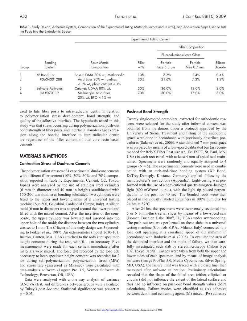
Table 1. Study Design, Adhesive System, Composition of the Experimental Luting Materials (expressed in wt%), and Application Steps Used to Lute the Posts into the Endodontic Space
Experimental Luting Cement
Filler Composition
Fluoroaluminosilicate Glass
Bonding Resin Matrix Filler Particle Particle Silicon Group System Composition wt% Size 5.3 μm Size 0.7 mm Dioxide
1 XP Bond: Lot Base: UDMA 80% wt, Methacrylic 10% 7.2% 2.4% 0.4% 2 #0604001288 Acid Ester 20% wt, amines 30% 21.6% 7.2% 1.2% < 1% wt, photo catalyst < 1% 3 Self-cure Activator: Catalyst: UDMA 80% wt, 50% 36.0% 12.0% 2.0% 4 Lot #070119 Methacrylic Acid Ester 70% 50.0% 17.0% 3.0% 20% wt, BPO < 1% wt
at Bond University Library on June 16, 2010 http://jdr.sagepub.comDownloaded from
J Dent Res 88(10) 2009 Effect of Filler on Fiber Post Luting 953
between post and cementing agent, or (C) cohesive in cementing agent failures.
Statistical analysis of the data was performed, with significance level set at p < 0.05. Since the tooth of origin was not a significant fac-tor for bond strength (regression analysis), push-out slices were con-sidered as statistical units. Since the data distribution was not nor-mal according to the Kolmogorov-Smirnov test, we applied the Kruskal-Wallis Non-parametric Analysis of Variance to assess the statistical significance of the differences among the groups, fol-lowed by Dunn’s Multiple-range test for post hoc comparisons.
Interfacial Nanoleakage Analysis
Additional post-luted roots (N = 4 per group) were selected for nanoleakage interfacial evaluation in accordance with previously described procedures (Saboia et al., 2008). Undemineralized and unem-bedded specimens were immersed in a 50 wt% ammoniacal AgNO3 solution, then in photo-developing solution (Tay et al., 2003a). Sec-tions were ground down to approx-imately 40 μm in thickness with wet carbide papers mounted on a specially designed grinding machine (Micromet, Remet, Bologna, Italy). Slides were stained with acid fuchsin and observed with a transmitted light micro-scope (Nikon Eclipse, Nikon). Images of all interfaces were obtained at a x100 magnification, and we evaluated the amount of tracer along the interface by scoring nanoleakage interfacial expression by two observers in accordance with previously described procedures (Saboia et al., 2008; Table 2).
We used the Kruskall-Wallis Non-parametric Analysis of Variance to assess the significance of the differences among the groups, followed by Dunn’s Multiple-range test for post hoc comparisons. The level of significance was set at p < 0.05.
RESULTS
Contraction Stress of dual-cure Cements
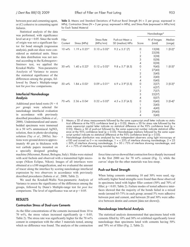
As the filler concentrations of the cements increased from 10 to 70 wt%, the stress values increased significantly (p < 0.05; Table 2). The stress rate was significantly higher for the 70 wt% cement in comparison with the other 3 materials tested, among which no difference was found. The analysis of the contraction
force/time curves showed that contraction force sharply increased in the first 2000 sec for 70 wt% cement (Fig. 1), while the curves’ slope for the other materials was less steep.
Push-out Bond Strength
When luting cements containing 10 and 30% were used, sig-nificantly higher bond strengths were found than those observed in specimens luted with higher filler content (50% and 70% of filler; p < 0.05; Table 2). Failure modes of tested adhesive inter-faces showed that the majority of the bonds failed in a mixed mode (around 65-75% in each group; around 5% were adhesive between post and cement, and between 20 and 30% were adhe-sive between dentin and cement [data not shown]).
Nanoleakage Interfacial Analysis
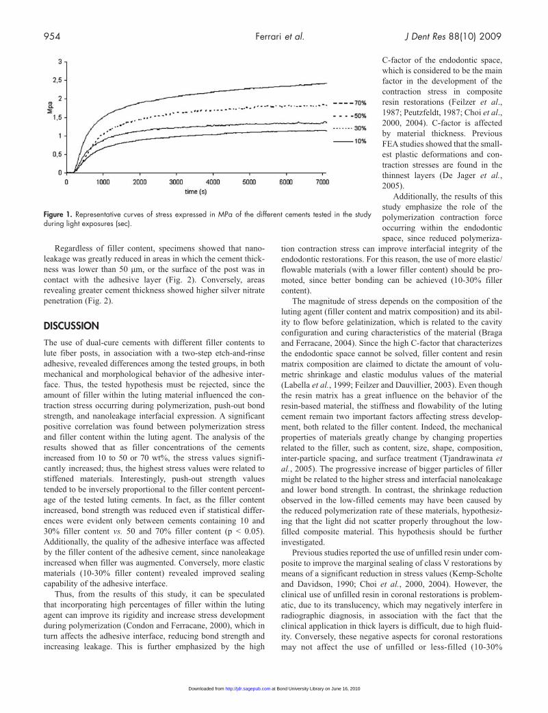
The statistical analysis demonstrated that specimens luted with cements filled by 10% and 30% wt exhibited significantly lower nanoleakage than specimens luted with cements having 50% and 70% wt of filler (Fig. 2, Table 2).
Table 2. Means and Standard Deviations of Push-out Bond Strength (N = 5 per group; expressed in MPa), Contraction Stress (N = 5 per group; expressed in MPa), and Stress Rate (expressed in MPa/min) for Each Tested Material
Nanoleakage*
Filler Stress Rate Push-out Mean ± N of Images Median Content Stress (MPa) (MPa/min) SD (median) MPa Score (total) (range)
10 wt% 1.19 ± 0.25a† 0.10 ± 0.02A 9.2 ± 5.51 (7) 0 13(38) 1 (0-2)# 1 22(38) 2 3(38) 3 0(38) 4 0(38)30 wt% 1.40 ± 0.22b 0.12 ± 0.02A 9.8 ± 5.71 (8.5) 0 9(40) 1 (0-3)# 1 27(40) 2 2(40) 3 2(40) 4 0(40)50 wt% 1.84 ± 0.02c 0.09 ± 0.01A 6.9 ± 3.92 (5.5) 0 2(37) 3 (0-3)$ 1 6(37) 2 7(37) 3 22(37) 4 0(37)70 wt% 2.36 ± 0.04c 0.22 ± 0.02B 4.5 ± 2.52 (4.5) 0 0(40) 3 (0-4)$ 1 2(40) 2 4(40) 3 29(40) 4 5(40)
† Means ± SD of stress measurements followed by the same superscript small letter indicate no statis-tical difference at the 95% confidence level (p > 0.05). Means ± SD for stress rate followed by the same superscript capital letter indicate no statistical difference at the 95% confidence level (p > 0.05). Means ± SD of push-out followed by the same superscript number indicate statistical differ-ence at the 95% confidence level (p > 0.05). Nanoleakage medians followed by the same super-script symbol indicate no statistical difference at the 95% confidence level (p > 0.05).
* Nanoleakage distribution was analyzed by two independent observers using light microscopy at x100 and scored as 0 = no nanoleakage, 1 = < 25% of interface showing nanoleakage, 2 = 25 ≤ 50% of interface showing nanoleakage, 3 = 50 ≤ 75% of interface showing nanoleakage, and 4 = > 75% of interface showing nanoleakage.
954 Ferrari et al. J Dent Res 88(10) 2009
Regardless of filler content, specimens showed that nano-leakage was greatly reduced in areas in which the cement thick-ness was lower than 50 μm, or the surface of the post was in contact with the adhesive layer (Fig. 2). Conversely, areas revealing greater cement thickness showed higher silver nitrate penetration (Fig. 2).
dISCUSSION
The use of dual-cure cements with different filler contents to lute fiber posts, in association with a two-step etch-and-rinse adhesive, revealed differences among the tested groups, in both mechanical and morphological behavior of the adhesive inter-face. Thus, the tested hypothesis must be rejected, since the amount of filler within the luting material influenced the con-traction stress occurring during polymerization, push-out bond strength, and nanoleakage interfacial expression. A significant positive correlation was found between polymerization stress and filler content within the luting agent. The analysis of the results showed that as filler concentrations of the cements increased from 10 to 50 or 70 wt%, the stress values signifi-cantly increased; thus, the highest stress values were related to stiffened materials. Interestingly, push-out strength values tended to be inversely proportional to the filler content percent-age of the tested luting cements. In fact, as the filler content increased, bond strength was reduced even if statistical differ-ences were evident only between cements containing 10 and 30% filler content vs. 50 and 70% filler content (p < 0.05). Additionally, the quality of the adhesive interface was affected by the filler content of the adhesive cement, since nanoleakage increased when filler was augmented. Conversely, more elastic materials (10-30% filler content) revealed improved sealing capability of the adhesive interface.
Thus, from the results of this study, it can be speculated that incorporating high percentages of filler within the luting agent can improve its rigidity and increase stress development during polymerization (Condon and Ferracane, 2000), which in turn affects the adhesive interface, reducing bond strength and increasing leakage. This is further emphasized by the high
C-factor of the endodontic space, which is considered to be the main factor in the development of the contraction stress in composite resin restorations (Feilzer et al., 1987; Peutzfeldt, 1987; Choi et al., 2000, 2004). C-factor is affected by material thickness. Previous FEA studies showed that the small-est plastic deformations and con-traction stresses are found in the thinnest layers (De Jager et al., 2005).
Additionally, the results of this study emphasize the role of the polymerization contraction force occurring within the endodontic space, since reduced polymeriza-
tion contraction stress can improve interfacial integrity of the endodontic restorations. For this reason, the use of more elastic/flowable materials (with a lower filler content) should be pro-moted, since better bonding can be achieved (10-30% filler content).
The magnitude of stress depends on the composition of the luting agent (filler content and matrix composition) and its abil-ity to flow before gelatinization, which is related to the cavity configuration and curing characteristics of the material (Braga and Ferracane, 2004). Since the high C-factor that characterizes the endodontic space cannot be solved, filler content and resin matrix composition are claimed to dictate the amount of volu-metric shrinkage and elastic modulus values of the material (Labella et al., 1999; Feilzer and Dauvillier, 2003). Even though the resin matrix has a great influence on the behavior of the resin-based material, the stiffness and flowability of the luting cement remain two important factors affecting stress develop-ment, both related to the filler content. Indeed, the mechanical properties of materials greatly change by changing properties related to the filler, such as content, size, shape, composition, inter-particle spacing, and surface treatment (Tjandrawinata et al., 2005). The progressive increase of bigger particles of filler might be related to the higher stress and interfacial nanoleakage and lower bond strength. In contrast, the shrinkage reduction observed in the low-filled cements may have been caused by the reduced poly merization rate of these materials, hypothesiz-ing that the light did not scatter properly throughout the low-filled composite material. This hypothesis should be further investigated.
Previous studies reported the use of unfilled resin under com-posite to improve the marginal sealing of class V restorations by means of a significant reduction in stress values (Kemp-Scholte and Davidson, 1990; Choi et al., 2000, 2004). However, the clinical use of unfilled resin in coronal restorations is problem-atic, due to its translucency, which may negatively interfere in radiographic diagnosis, in association with the fact that the clinical application in thick layers is difficult, due to high fluid-ity. Conversely, these negative aspects for coronal restorations may not affect the use of unfilled or less-filled (10-30%
Figure 1. Representative curves of stress expressed in MPa of the different cements tested in the study during light exposures (sec).
at Bond University Library on June 16, 2010 http://jdr.sagepub.comDownloaded from
J Dent Res 88(10) 2009 Effect of Filler on Fiber Post Luting 955
filler content) resin cements proposed in the present study to lute fiber posts in endodontically treated teeth.
Although the market has focused on the formulation of luting agents also suitable for core build-up, allowing for simultane-ous post cementation and core fabrication, from a research per-spective it is important to note that the increased percentage of filler in these materials needed for core build-up restoration and
retention leads to an irreversible higher stress during polymeri-zation, ultimately resulting in lower bond-strength values.
In conclusion, based on the satisfactory performance shown in the present in vitro study, a reduction in filler content for luting cements appears to offer a promising new approach to the luting of fiber posts in endodontically treated teeth. Further in vivo studies are currently ongoing to investigate the long-term clinical performance of these materials. Such studies are mandatory before a general recommendation can be made for their use.
ACKNOwLEdGMENTS
The authors thank GC (Tokyo, Japan) for generously donating the experimental cements, and Mr. Aurelio Valmori for photo-graphic assistance. The study was supported with MIUR (Italy) grants.
REFERENCESBaroudi K, Saleh AM, Silikas N, Watts DC (2007). Shrinkage behaviour of
flowable resin-composites related to conversion and filler-fraction. J Dent 35:651-655.
Bouillaguet S, Troesch S, Wataha JC, Krejci I, Meyer JM, Pashley DH (2003). Microtensile bond strength between adhesive cements and root canal den-tin. Dent Mater 19:199-205.
Braga RR, Ferracane JL (2004). Alter natives in polymerization contraction stress management. Crit Rev Oral Biol Med 15:176-184.
Braga RR, Boaro LC, Kuroe T, Azevedo CL, Singer JM (2006). Influence of cavity dimensions and their deri vatives (volume and ‘C’ factor) on shrinkage stress development and microleakage of composite restora-tions, Dent Mater 22:818-823.
Breschi L, Mazzoni A, Ferrari M (2008). Adhesion to Intraradicular dentin. In: Fiber posts and endodontically treated teeth: a compendium of scientific and clinical perspective. Ferrari M, Breschi L, Grandini S, editors. Johannesburg, South Africa: Modern Dentistry Media, pp. 21-35.
Choi KK, Condon JR, Ferracane JL (2000). The effect of adhesive thickness on polymerization contraction stress of composite. J Dent Res 79:812-817.
Choi KK, Ryu GJ, Choi SM, Lee MJ, Park SJ, Ferracane JL (2004). Effects of cavity configuration on composite restoration. Oper Dent 29:462-469.
Condon JR, Ferracane JL (2000). Assessing the effect of composite formula-tion on polymerization stress. J Am Dent Assoc 131:497-503.
Davidson CL, Feilzer AJ (1997). Polymerization shrinkage and polymeri-zation shrinkage stress in polymer-based restoratives. J Dent 25: 435-440.
De Jager N, Pallav P, Feilzer AJ (2005). Finite element analysis model to simulate the behavior of luting cements during setting. Dent Mater 21:1025-1032.
Feilzer AJ, Dauvillier BS (2003). Effect of TEGDMA/BisGMA ratio on stress development and viscolelastic properties of experimental two-paste composites. J Dent Res 82:824-828.
Feilzer AJ, De Gee AJ, Davidson CL (1987). Setting stress in composite resin in relation to configuration of the restoration. J Dent Res 66: 1636-1639.
Ferrari M, Mannocci F, Vichi A, Cagidiaco MC, Mjör IA (2000). Bonding to root canal: structural characteristic of the substrate. Am J Dent 13:255-260.
Kemp-Scholte CM, Davidson CL (1990). Complete marginal seal of Class V resin composite restorations effected by increased flexibility. J Dent Res 69:1240-1243.
Labella R, Lambrechts P, Van Meerbeek B, Vanherle G (1999). Polymerization shrinkage and elasticity of flowable composites and filled adhesives. Dent Mater 15:128-137.
Moreira da Silva E, dos Santos GO, Guimarães JG, Barcellos Ade A, Sampaio EM (2007). The influence of C-factor, flexural modulus and
Figure 2. Light-microscopy expression of interfacial nanoleakage analy-sis of the fiber posts luted within the endodontic space with 4 dual-cure cements differing in their filler content (N = 4 per group). All images were taken at the original magnification of x1000 (scale bar = 10 microns). (a) Image of a fiber post (P) luted with the resin-based cement (C) containing 10 wt% filler. Excellent adaptation was achieved, and almost no nanoleakage was evident along the adhesive interface (rep-resentative image of score 0); intra-radicular dentin (D). (b) Adhesive interface created by a fiber post (P) luted with the 30% filler-containing cement (C). Nanoleakage was scattered along the interface, and sil-ver aggregates were sometimes found (representative image of score 1); intra-radicular dentin (D). (c) Fiber post (P) cemented with 50 wt% filler-containing cement (C), revealing frequent silver deposits along the bonded interface, even in areas in which the cement layer was very thin (representative image of score 3). Silver deposits are clearly evi-dent both at the intra-radicular dentin (D)/adhesive/cement interface (pointing hand) and along the cement/fiber post interface. (d) Image revealing a fiber post (P) luted with the cement (C) with the highest tested filler concentration (i.e., 70 wt%). Nanoleakage was frequently visible along the adhesive interface, affecting both the intra-radicular dentin (D)/adhesive/cement interface (pointing hand) as well as the cement/fiber post interface, with extensive silver aggregates (representative image of score 4).
at Bond University Library on June 16, 2010 http://jdr.sagepub.comDownloaded from
956 Ferrari et al. J Dent Res 88(10) 2009
viscous flow on gap formation in resin composite restorations. Oper Dent 32:356-362.
Peutzfeldt A (1987). Resin composites in dentistry: the monomer systems. Eur J Oral Sci 105:97-116.
Radovic I, Monticelli F, Cury AH, Bertelli E, Vulicevic ZR, Ferrari M (2008). Coupling of composite cements to quartz fiber posts: a com-parison of industrial and “chairside” treatments of the post surface. J Adhes Dent 10:57-66.
Saboia VP, Nato F, Mazzoni A, Orsini G, Putignano A, Giannini M, et al. (2008). Adhesion of a two-step etch-and-rinse adhesive on collagen-depleted dentin. J Adhes Dent 10:419-422.
Salameh Z, Sorrentino R, Papacchini F, Ounsi H, Tashkandi E, Goracci C, et al. (2006). Fracture resistance and failure patterns of endodontically treated mandibular molars restored using resin composite with or with-out translucent (glass fiber) posts. J Endod 32:752-755.
Schwartz RS (2006). Adhesive dentistry and endodontics. Part 2: Bonding in the root canal system—the promise and the problems: a review. J Endod 32:1125-1134.
Schwartz RS, Fransman R (2005). Adhesive dentistry and endodontics: materials, clinical strategies and procedures for restoration of access cavities: a review. J Endod 31:151-165.
Schwartz RS, Robbins JW (2004). Post placement and restoration of endo-dontically treated teeth: a literature review. J Endod 30:289-301.
Tay FR, Hashimoto M, Pashley DH, Peters MC, Lai SC, Yiu CK, et al. (2003a). Aging affects two modes of nanoleakage expression in bonded dentin. J Dent Res 82:537-541.
Tay FR, Suh BI, Pashley DH, Prati C, Chuang SF, Li F (2003b). Factors contributing to the incompatibility between simplified-step adhesives and self-cured or dual-cured composites. Part II. Single-bottle, total-etch adhesive. J Adhes Dent 5:91-105.
Tay FR, Loushine RJ, Lambrechts P, Weller RN, Pashley DH (2006). Geometric factors affecting dentin bonding in root canals: a theoretical modelling approach. J Endod 31:584-588.
Tjandrawinata R, Irie M, Suzuki K (2005). Flexural properties of eight flow-able light-cured restorative materials, in immediate vs 24-hour water storage. Oper Dent 30:239-249.
at Bond University Library on June 16, 2010 http://jdr.sagepub.comDownloaded from