Influence of Human Milk on Very Preterms Gut Microbiota ...
13
nutrients Article Influence of Human Milk on Very Preterms’ Gut Microbiota and Alkaline Phosphatase Activity Juliana Morais 1,2 , Cláudia Marques 1,3 , Ana Faria 1,2 , Diana Teixeira 1,2,4 , Inês Barreiros-Mota 1,2 , Catarina Durão 1,5 , João Araújo 1,3 , Shámila Ismael 1,2 , Sara Brito 6 , Manuela Cardoso 7 , Israel Macedo 6 , Esmeralda Pereira 6 , Teresa Tomé 6 and Conceição Calhau 1,3,4, * Citation: Morais, J.; Marques, C.; Faria, A.; Teixeira, D.; Barreiros-Mota, I.; Durão, C.; Araújo, J.; Ismael, S.; Brito, S.; Cardoso, M.; et al. Influence of Human Milk on Very Preterms’ Gut Microbiota and Alkaline Phosphatase Activity. Nutrients 2021, 13, 1564. https://doi.org/10.3390/ nu13051564 Academic Editor: Bernhard Resch Received: 5 March 2021 Accepted: 27 April 2021 Published: 6 May 2021 Publisher’s Note: MDPI stays neutral with regard to jurisdictional claims in published maps and institutional affil- iations. Copyright: © 2021 by the authors. Licensee MDPI, Basel, Switzerland. This article is an open access article distributed under the terms and conditions of the Creative Commons Attribution (CC BY) license (https:// creativecommons.org/licenses/by/ 4.0/). 1 Faculdade de Ciências Médicas|NOVA Medical School, Universidade NOVA de Lisboa, 1169-056 Lisboa, Portugal; [email protected] (J.M.); claudia.sofi[email protected] (C.M.); [email protected] (A.F.); [email protected] (D.T.); [email protected] (I.B.-M.); [email protected] (C.D.); [email protected] (J.A.); [email protected] (S.I.) 2 CHRC-Comprehensive Health Research Centre, CEDOC-Chronic Diseases Research Center, Faculdade de Ciências Médicas|NOVA Medical School, Universidade NOVA de Lisboa, 1169-056 Lisboa, Portugal 3 CINTESIS-Center for Health Technology Services Research, Faculdade de Ciências Médicas|NOVA Medical School, Universidade NOVA de Lisboa, 1169-056 Lisboa, Portugal 4 NOVA Medical School, Unidade Universitária Lifestyle Medicine José de Mello Saúde, 1169-056 Lisboa, Portugal 5 EPIUnit-Institute of Public Health, Universidade do Porto, 4050-600 Porto, Portugal 6 Pediatrics Department, Maternidade Dr. Alfredo da Costa, Centro Hospitalar Universitário de Lisboa Central, 2890-495 Lisboa, Portugal; [email protected] (S.B.); [email protected] (I.M.); [email protected] (E.P.); [email protected] (T.T.) 7 Nutrition and Dietetics Unit, Maternidade Dr. Alfredo da Costa, Centro Hospitalar Universitário de Lisboa Central, 2890-495 Lisboa, Portugal; [email protected] * Correspondence: [email protected]; Tel.: +351-21-880-3000 Abstract: The FEEDMI Study (NCT03663556) evaluated the influence of infant feeding (mother’s own milk (MOM), donor human milk (DHM) and formula) on the fecal microbiota composition and alkaline phosphatase (ALP) activity in extremely and very preterm infants (≤32 gestational weeks). In this observational study, preterm infants were recruited within the first 24 h after birth. Meconium and fecal samples were collected at four time points (between the 2nd and the 26th postnatal days. Fecal microbiota was analyzed by RT-PCR and by 16S rRNA sequencing. Fecal ALP activity, a proposed specific biomarker of necrotizing enterocolitis (NEC), was evaluated by spectrophotometry at the 26th postnatal day. A total of 389 fecal samples were analyzed from 117 very preterm neonates. Human milk was positively associated with beneficial bacteria, such as Bifidobacterium, Bacteroides ovatus, and Akkermancia muciniphila, as well as bacterial richness. Neonates fed with human milk during the first week of life had increased Bifidobacterium content and fecal ALP activity on the 26th postnatal day. These findings point out the importance of MOM and DHM in the establishment of fecal microbiota on neonates prematurely delivered. Moreover, these results suggest an ALP pathway by which human milk may protect against NEC. Keywords: alkaline phosphatase; donor human milk; formula; gut microbiota; mother’s own milk; very preterm neonates 1. Introduction Very preterm infants (<32 weeks of gestational age) present an intestinal microbiota significantly different from that of full-term infants [1], being characterized by a highly permeable intestine surface and a marked vulnerability to dysbiosis [2]. Increasing data suggests that the type of infant feeding significantly influences the preterm infants’ devel- opment as well as their microbiota [3]. Mother’s own milk (MOM) is the gold standard for Nutrients 2021, 13, 1564. https://doi.org/10.3390/nu13051564 https://www.mdpi.com/journal/nutrients
Transcript of Influence of Human Milk on Very Preterms Gut Microbiota ...

nutrients
Article
Influence of Human Milk on Very Preterms’ Gut Microbiotaand Alkaline Phosphatase Activity
Juliana Morais 1,2, Cláudia Marques 1,3, Ana Faria 1,2 , Diana Teixeira 1,2,4 , Inês Barreiros-Mota 1,2,Catarina Durão 1,5, João Araújo 1,3, Shámila Ismael 1,2, Sara Brito 6, Manuela Cardoso 7, Israel Macedo 6 ,Esmeralda Pereira 6, Teresa Tomé 6 and Conceição Calhau 1,3,4,*
�����������������
Citation: Morais, J.; Marques, C.;
Faria, A.; Teixeira, D.; Barreiros-Mota,
I.; Durão, C.; Araújo, J.; Ismael, S.;
Brito, S.; Cardoso, M.; et al. Influence
of Human Milk on Very Preterms’
Gut Microbiota and Alkaline
Phosphatase Activity. Nutrients 2021,
13, 1564. https://doi.org/10.3390/
nu13051564
Academic Editor: Bernhard Resch
Received: 5 March 2021
Accepted: 27 April 2021
Published: 6 May 2021
Publisher’s Note: MDPI stays neutral
with regard to jurisdictional claims in
published maps and institutional affil-
iations.
Copyright: © 2021 by the authors.
Licensee MDPI, Basel, Switzerland.
This article is an open access article
distributed under the terms and
conditions of the Creative Commons
Attribution (CC BY) license (https://
creativecommons.org/licenses/by/
4.0/).
1 Faculdade de Ciências Médicas|NOVA Medical School, Universidade NOVA de Lisboa,1169-056 Lisboa, Portugal; [email protected] (J.M.); [email protected] (C.M.);[email protected] (A.F.); [email protected] (D.T.); [email protected] (I.B.-M.);[email protected] (C.D.); [email protected] (J.A.); [email protected] (S.I.)
2 CHRC-Comprehensive Health Research Centre, CEDOC-Chronic Diseases Research Center,Faculdade de Ciências Médicas|NOVA Medical School, Universidade NOVA de Lisboa,1169-056 Lisboa, Portugal
3 CINTESIS-Center for Health Technology Services Research, Faculdade de Ciências Médicas|NOVA MedicalSchool, Universidade NOVA de Lisboa, 1169-056 Lisboa, Portugal
4 NOVA Medical School, Unidade Universitária Lifestyle Medicine José de Mello Saúde,1169-056 Lisboa, Portugal
5 EPIUnit-Institute of Public Health, Universidade do Porto, 4050-600 Porto, Portugal6 Pediatrics Department, Maternidade Dr. Alfredo da Costa, Centro Hospitalar Universitário de Lisboa Central,
2890-495 Lisboa, Portugal; [email protected] (S.B.); [email protected] (I.M.);[email protected] (E.P.); [email protected] (T.T.)
7 Nutrition and Dietetics Unit, Maternidade Dr. Alfredo da Costa,Centro Hospitalar Universitário de Lisboa Central, 2890-495 Lisboa, Portugal;[email protected]
* Correspondence: [email protected]; Tel.: +351-21-880-3000
Abstract: The FEEDMI Study (NCT03663556) evaluated the influence of infant feeding (mother’sown milk (MOM), donor human milk (DHM) and formula) on the fecal microbiota composition andalkaline phosphatase (ALP) activity in extremely and very preterm infants (≤32 gestational weeks).In this observational study, preterm infants were recruited within the first 24 h after birth. Meconiumand fecal samples were collected at four time points (between the 2nd and the 26th postnatal days.Fecal microbiota was analyzed by RT-PCR and by 16S rRNA sequencing. Fecal ALP activity, aproposed specific biomarker of necrotizing enterocolitis (NEC), was evaluated by spectrophotometryat the 26th postnatal day. A total of 389 fecal samples were analyzed from 117 very preterm neonates.Human milk was positively associated with beneficial bacteria, such as Bifidobacterium, Bacteroidesovatus, and Akkermancia muciniphila, as well as bacterial richness. Neonates fed with human milkduring the first week of life had increased Bifidobacterium content and fecal ALP activity on the26th postnatal day. These findings point out the importance of MOM and DHM in the establishmentof fecal microbiota on neonates prematurely delivered. Moreover, these results suggest an ALPpathway by which human milk may protect against NEC.
Keywords: alkaline phosphatase; donor human milk; formula; gut microbiota; mother’s own milk;very preterm neonates
1. Introduction
Very preterm infants (<32 weeks of gestational age) present an intestinal microbiotasignificantly different from that of full-term infants [1], being characterized by a highlypermeable intestine surface and a marked vulnerability to dysbiosis [2]. Increasing datasuggests that the type of infant feeding significantly influences the preterm infants’ devel-opment as well as their microbiota [3]. Mother’s own milk (MOM) is the gold standard for
Nutrients 2021, 13, 1564. https://doi.org/10.3390/nu13051564 https://www.mdpi.com/journal/nutrients

Nutrients 2021, 13, 1564 2 of 13
preterm infants’ nutrition during hospitalization [4,5], leading them to develop a micro-biota resembling that of term infants [6]. When MOM is not available, or it is insufficient,pasteurized donor human milk (DHM) is recommended [7]. Bovine-based preterm for-mulas are the final alternative for preterm infants’ nutrition [7]. Even though formula isassociated with increased in-hospital weight gain and better growth, it is also linked with ahigher risk of necrotizing enterocolitis (NEC) [8,9], a very common cause of morbidity andmortality in preterm infants, closely associated with gut microbiota [10].
Recently, the intestinal isoform of alkaline phosphatase (ALP)—an enzyme that re-duces lipopolysaccharide-mediated inflammation [11]—was suggested to be a specificbiomarker for NEC [12]. Since preterm neonates have reduced ALP when compared toearly-term neonates, it has been suggested that the lack of ALP activity could increase therisk of inflammation and progression to NEC [13]. The modulation of ALP activity hasbeen poorly studied. In fact, the association between the triad of feeding type, ALP activity,and gut microbiota has never been evaluated in hospitalized preterm neonates.
Thus, the aim of this study was to evaluate the impact of different types of infantfeeding (MOM, DHM, and formula) during the first 4 weeks of life on the fecal microbiotaand on ALP activity of very and extremely preterm neonates.
2. Materials and Methods2.1. Study Design and Participants
The FEEDMI study is an observational longitudinal study conducted at the NeonatalIntensive Care Unit (NICU) of Maternidade Dr. Alfredo da Costa and Faculdade de CiênciasMédicas|NOVA Medical School, Universidade NOVA de Lisboa between May 2017 andApril 2019. This study, approved by both the hospital and faculty ethics committees,is registered on the ClinicalTrials.gov platform (NCT03663556) and was conducted inaccordance to the ethical principles expressed in the Declaration of Helsinki, the Portugueselaw, and Good Clinical Practice guidelines.
Extremely and very preterm neonates included in this study were recruited accordingto their admission in the NICU within the first 24 h post-birth. Eligible neonates wereborn at a gestational age of < 32 weeks and had no congenital malformations or metabolicdiseases. Signed written informed consent was obtained for each newborn after explainingthe study protocol to their legal representatives. The detailed study protocol was previouslypublished [14].
2.2. Enteral Nutrition Protocol
Exclusive human milk (MOM or DHM) was the recommended procedure. WhenMOM was insufficient or not available, DHM could be used for neonates until 35 weeks’postmenstrual age. Mothers were advised to collect milk with breast pumps every 3 heither in the hospital or at home. The MOM samples received were identified by dateand time of collection. MOM was frozen and stored, but never pasteurized. DHM wasfrozen and submitted to Holder pasteurization (62.5 ◦C for 30 min) in the maternity humanmilk bank. For each neonate, MOM samples were analyzed once a week and DHM wasanalyzed after the pasteurization using a mid-infrared human milk analyzer (Miris AB,Uppsala, Sweden). Formula was used if MOM was not sufficient and if the DHM stockwas limited. Nutritional daily plans were prescribed by physicians in collaboration witha nutritionist.
2.3. Clinical Data Collection
Detailed sociodemographic, perinatal, and postnatal clinical data were collectedthrough medical records. Clinical intrapartum and postpartum data included newborn’ssomatometric progression, antibiotic exposure and its duration, number of total days ofhospitalization, and other outcomes related to the preterm clinical evolution. Weight,length, and cephalic perimeter Z-scores at birth were determined using the growth curves

Nutrients 2021, 13, 1564 3 of 13
obtained by Fenton [15]. Additionally, type of infant feeding was recorded daily to selectthe most representative (>50%) type of feeding received during the study period.
2.4. Fecal Sample Collection and DNA Extraction
Meconium was collected within the first 72 h after birth and subsequent fecal sampleswere collected weekly over the first four weeks of life. All samples were collected bythe nursing team of the NICU. Bacterial DNA was extracted and purified from all fecalsamples using a NZY Tissue gDNA Isolation Kit (NZYtech, Lisbon, Portugal) as previouslydescribed [16].
2.5. Quantitative Analysis of Fecal Microbiota by RT-PCR
Specific bacterial populations were analyzed in duplicate by quantitative real-timePCR (RT-PCR) using LightCycler® (Roche Applied Science, Indianapolis, ID, USA). Specificmicroorganisms were assessed based on previous studies regarding preterm gut micro-biota composition and health [17–20]. Two phyla (Bacteroidetes and Firmicutes), one class(γ-Proteobacteria), four genera (Lactobacillus, Bifidobacterium, Bacteroides, and Enterococcus),and one species (Escherichia coli) were analyzed in preterm neonates’ samples. Primerssequences used to target bacterial 16S rRNA genes are described in Table 1. Microbiotaresults are expressed as log10 16S rRNA gene copies/10 ng of DNA. All of the analyseswere conducted using the appropriate negative controls, as previously described [21].
Table 1. Primer sequences used for gut microbiota analysis.
Target Group Primer Sequence (5′-3′) Genomic DNA Standard AT Ref.
Total bacteria AAA CTC AAA KGA ATT GAC GGCTC ACR RCA CGA GCT GAC
Bacteroides vulgatusATCC 8482 62 ◦C [22]
Bacteroidetes CAT GTG GTT TAA TTC GAT GATAGC TGA CGA CAA CCA TGC AG
Bacteroides vulgatusATCC 8482 60 ◦C [16]
Firmicutes ATG TGG TTT AAT TCG AAG CAAGC TGA CGA CAA CCA TGC AC
Lactobacillus gasseriATCC 33323 60 ◦C [16]
γ-Proteobacteria TCGTCAGCTCGTGTYGTGACGTAAGGGCCATGATG
E. coliATCC 25922 61 ◦C [22]
Lactobacillus GAG GCA GCA GTA GGG AAT CTT CGGC CAG TTA CTA CCT CTA TCC TTC TTC
Lactobacillus gasseriATCC 33323 60 ◦C [16]
Bifidobacterium CGC GTC YGG TGT GAA AGCCC CAC ATC CAG CAT CCA
Bifidobacterium longumATCC 15697 60 ◦C [16]
Bacteroides ATA GCC TTT CGA AAG RAA GATCCA GTA TCA ACT GCA ATT TTA
Bacteroides vulgatusATCC 33563 60 ◦C [16]
Enterococcus CCC TTA TTG TTA GTT GCC ATC ATTACT CGT TGT ACT TCC CT TGT
Enterococcus gilvusATCC BAA-350 61 ◦C [16]
Escherichia coli GTA AGT TAC ACT ATA AAA GCA CCG TCGTCT GTG TGG ATG GTA ATA AAT TTT TG
Escherichia coliATCC 25922 60 ◦C [23]
AT, annealing temperature.
2.6. Microbial 16S rRNA Sequence Analysis
Due to the limited volume of the fecal samples of preterm infants, a sub-set of fecalsamples collected in the fourth week of life was analyzed by 16S rRNA sequencing. Thefecal samples of preterm infants fed with MOM (n = 40), DHM (n = 14), and formula (n = 10)were investigated. Libraries were processed and sequenced following the 16S Metage-nomic Sequencing Library Preparation protocol from illumina (illumina; San Diego, CA,USA). Primers used to capture the region V3–V4 of the bacterial 16S region (primers 515F:GTGYCAGCMGCCGCGGTAA; 806R: GGACTACNVGGGTWTCTAAT) were previouslydescribed by Walters and colleges (2015) [24]. The samples were pooled and loaded into theillumina MiSeq System and sequenced using a 280-multiplex approach on a 2× 250 bp run,according to manufacturer’s procedures [24]. For all sequencing reads, QIIME 2.11 wasused with default parameters for demultiplexing, quality filtering, and clustering reads

Nutrients 2021, 13, 1564 4 of 13
into OTUs. The Greengenes database was used for taxonomy assignment. Gut microbialdiversity was evaluated by Shannon index, and gut microbial richness was measuredby Chao1.
2.7. Alkaline Phosphatase Activity Assay
ALP activity was assessed through dephosphorylation of p-nitrophenyl phosphate(pNPP) in p-nitrophenol, which results in a color change that was measured by absorbancespectrophotometry. The protocol was performed as previously described by Calhau(2000) [25] and Malo (2015) [26] with some modifications. Briefly, 50 µL of “extractionbuffer” (10 mM Tris-HCl and 1 mM MgCl2, pH 8.0) was added to 1 mg of fecal samplesfor homogenization and the sample was centrifuged at 10,000× g for 20 min. Using a1:50 ratio, the “dilution buffer” (200 mM Tris-base and 1 mM MgCl2, pH 10.4) was addedto the supernatant containing ALP, along with 10 µL of pNPP (5 mM), and the sample wasincubated for 2 min at 37 ◦C. The reaction was stopped with 5 mL of cold NaOH (0.02 M).Absorbance was read at 410 nm and results were expressed as U ALP/g stool.
2.8. Statistical Analysis
Statistical analysis was performed by SPSS® software, v25 (IBM SPSS Statistics corpo-ration, Chicago, IL, USA). The normality of the data was checked using the Kolmogorov–Smirnov test. Comparisons between groups were performed using the t-test, Mann–Whitney test, or Fisher’s exact test. Multivariate linear regression models adjusted for ges-tational age, mode of delivery, Z-scores’ growth parameters (weight, length, and cephalicperimeter at birth), and infant’s antibiotherapy were fitted to study the association be-tween infant feeding (independent variable) and microbiota composition of very pretermneonates at the 26th postnatal day (dependent variables). The sequencing data of fecal sam-ples was analyzed using MicrobiomeAnalyst [27]. Alpha-diversity of the fecal microbiotawas assessed through Chao1 and the Shannon index for microbial richness and diversityindex, respectively. Beta-diversity was performed using PERMANOVA. The differences inrelative abundance of fecal bacterial taxa between the three feeding types was assessed byKruskal–Wallis. Differences were considered statistically significant when p < 0.05. Dataare presented as mean ± SD.
3. Results3.1. Clinical Characterization of the Preterm Neonates
From the 159 preterm neonates recruited consecutively at the NICU, 117 were eligiblefor the study with forty-two were excluded: 14 for not meeting the inclusion criteria,13 due to early death, 7 for having nonconformities in their fecal samples, 4 for earlydischarge, 3 were transferred to another hospital, and 1 for unknown perinatal factors.The 117 eligible neonates were born with a gestational age between 25 and 31 weeks(28.6 ± 1.9) with birthweight ranging from 455 to 2020 g (1177 ± 419). Most of the neonateswere male (56%) and delivered by cesarean-section (62%). Enteral feeding was introducedbetween the 1st and 6th postnatal day and exclusive enteral feeding was achieved at15 ± 8 postnatal days. Preterm neonates were either fed with their MOM, DHM, and/orinfant formula. During the 26 days of the study, MOM was the main feeding type for mostneonates (n = 75, 64.1%), representing more than 50% of the total feedings. Twenty neonates(17.1%) were fed predominantly with DHM and 13 (11.1%) with infant formula. In somecases, neonates received mixed feeding of MOM + DHM (n = 7, 6.0%; 29.2 ± 1.2 weeks ofgestational age; 1389 ± 153 g birth weight) or MOM + formula (n = 1, 0.85%; 31 weeksof gestational age; 1800 g birth weight). Due to specific clinical conditions, one pretermneonate was fed mainly by parenteral feeding, reaching exclusive enteral feeding at the48th postnatal day and, therefore, was excluded from our analysis.
Extremely preterm neonates, those with lower birth weight, length, or cephalic perime-ter, were preferably fed with MOM and DHM rather than formula (Table 2). Neonates fed

Nutrients 2021, 13, 1564 5 of 13
predominantly with formula showed a greater weight gain, fewer days of antibiotherapy,and lower total days of hospitalization compared to MOM and DHM (Table 2).
Table 2. Clinical data of very preterm neonates receiving different types of infant feeding during study period.
MOM(n = 75)
DHM(n = 20)
Formula(n = 13)
p-Value(MOM vs.
DHM)
p-Value(MOM vs.Formula)
p-Value(DHM vs.Formula)
Extremely/very preterm n 28/47 5/15 0/13 0.205 0.004 0.065Gestational age (mean ± SD) 28.3 ± 2.1 28.6 ± 1.8 29.6 ±1.1 0.103 0.003 0.17Vaginal delivery/C-section, n 34/41 7/13 1/12 0.407 0.010 0.074
Somatometry at birth (mean ± SD)Weight, g 1123 ± 345 1173 ± 284 1416 ± 219 0.438 0.002 0.011
Length, cm 36.2 ± 3.3 36.8 ± 3.2 39.1 ± 2.0 0.428 0.003 0.027Cephalic perimeter, cm 25.4 ± 2.4 25.3 ± 1.8 27.2 ± 1.5 0.996 0.007 0.008
Z-scoreWeight at birth −0.27 ± 0.82 0.04 ± 1.45 0.15 ± 0.70 0.977 0.057 0.789Length at birth −0.44 ± 1.09 −0.25 ± 1.61 0.04 ±0.65 0.777 0.051 0.543
Cephalic perimeter at birth −0.46 ± 0.90 −0.68 ± 1.79 −0.77 ±1.28 0.731 0.147 0.331
Weight at 26th −0.08 ± 1.85 0.85 ± 1.62 1.65 ± 1.13 0.257 0.001 0.132∆ weight until 26th day, g 200 ± 190 200 ± 170 420 ± 160 0.671 <0.001 0.001
Postnatal day of full enteral nutrition(mean ± SD) 16.5 ± 8.5 16.4 ± 5.6 9.5 ± 4.2 0.401 0.002 <0.001
Days of antibiotherapy (mean ± SD) 12 ± 11 13 ± 8 5 ± 3 0.178 0.010 <0.001Days of hospitalization (mean ± SD) 58 ± 24 59 ± 21 42 ± 12 0.874 0.026 0.018
MOM, mother’s own milk. DHM, donor human milk.
3.2. Impact of Feeding on Intestinal Bacterial Establishment in Premature Neonates
A total of 389 fecal samples were collected weekly during the first four weeks of lifewith an average postnatal days of 2 ± 0.9 (corresponding to meconium samples), 10 ± 1.9,18 ± 2.1, and 26 ± 2.5 days. Feeding trends over time were assessed week-by-weekconsidering the predominant feeding type (Figure 1). In general, the level of total bacteriaincreased significantly during the first week of life. At day 26 of life, infants fed with MOMhad higher levels of total bacteria Bacteroidetes, Enterococcus, Bacteroides, Bifidobacterium, andLactobacillus when compared to that of formula fed infants (Figure 1).
Table 3 shows the cumulative effect of the predominant feeding type received duringthe first 26 days on preterm microbiota composition (total bacteria, Firmicutes, and Bifi-dobacterium; no other differences were observed). Compared to MOM, DHM and formulafeeding promoted lower levels of total bacteria in very preterm neonates at the 26th postna-tal day when adjusted for gestational age, mode of delivery, and for multivariable-adjustedmodel (Table 3). Generally, MOM promoted more Bifidobacterium content, and this differ-ence was significant after adjusting for gestational age when comparing to that of DMHand after adjusting for gestational age and for multivariable model when comparing tothat of formula (Table 3). After adjustment for multivariable model, Firmicutes were fewerin DHM and formula fed neonates (Table 3).
Table 3. Association between neonates feeding (independent variable) and neonates’ microbiota at the 26th postnatal day(dependent variables).
MOMDHM Formula
95% CI p-Value 95% CI p-Value
Total BacteriaGestational age-adjusted 1 (referent) −0.556 (−1.093 to −0.079) 0.023 −0.889 (−1.341 to −0.438) <0.001Mode of delivery-adjusted 1 (referent) −0.565 (−1.036 to −0.095) 0.018 −0.552 (−1.047 to −0.057) 0.029Multivariable-adjusted a 1 (referent) −0.659 (–1.082 to −0.236) 0.002 −0.721 (−1.158 to −0.284) 0.001
FirmicutesGestational age-adjusted 1 (referent) −0.585 (−1.183 to 0.013) 0.055 −0.789 (−1.322 to −0.256) 0.004

Nutrients 2021, 13, 1564 6 of 13
Table 3. Cont.
MOMDHM Formula
95% CI p-Value 95% CI p-Value
Mode of delivery-adjusted 1 (referent) −0.587 (−1.161 to −0.013) 0.045 −0.456 (−1.060 to 0.162) 0.139Multivariable-adjusted a 1 (referent) −0.610 (–1.114 to −0.107) 0.017 −0.759 (−1.279 to −0.238) 0.004
BifidobacteriumGestational age-adjusted 1 (referent) −2.002 (−3.315 to −0.689) 0.003 −2.092 (−3.262 to −0.922) <0.001Mode of delivery-adjusted 1 (referent) −0.291 (−1.016 to 0.434) 0.431 −0.684 (−1.447 to 0.079) 0.079Multivariable-adjusted a 1 (referent) −0.656 (−1.388 to 0.075) 0.079 −0.975 (−1.716 to −0.233) 0.010
a Multivariable-adjusted model included gestational age, mode of delivery, Z-scores’ growth parameters (weight, length, and cephalicperimeter at birth), and infant’s antibiotherapy received within 8 days prior to fecal collection. MOM, mother’s own milk. DHM, donorhuman milk.
Figure 1. Microbiota of neonates during the study period according to the predominant feeding received in the 8 dayspreceding each fecal collection. The means and standard mean errors of relative abundance are shown. * MOM vs. formula;** MOM vs. DHM; # DHM vs. formula. Comparisons between feeding groups in each time point were performed usingt-test or Mann–Whitney test, taking in account the distribution of the variables. Differences were statistically significantwhen p < 0.05. MOM, mother’s own milk. DHM, donor human milk.

Nutrients 2021, 13, 1564 7 of 13
3.3. Gut Microbiota Profile at the 26th Postnatal Day According to Feeding Types
Considering the results from 16S rRNA sequencing, three samples were excludedfrom analysis due their low sequencing reads, leaving 61 samples (MOM = 39, DHM = 14,Formula = 8). Preterm neonates fed with MOM had an increased microbial richness(Chao1 index) compared to that of DHM (p = 0.011) and formula (p < 0.001) (Figure 2). Nodifferences were found between groups regarding Shannon index
Figure 2. (A) Microbial richness (Chao1 index) and (B) diversity (Shannon index) of fecal microbiota in preterm infantsaccording to feeding types. * p < 0.05 and ** p < 0.01. MOM, mother’s own milk. DHM, donor human milk.
When assessing beta-diversity, no differences were found on the fecal microbiotaprofiles between infants fed with MOM, DHM, and formula (Figure 3A). Differences inthe relative abundance of phylum Proteobacteria, Bacteroidetes, Verrucomicrobia (p < 0.05and q < 0.01) among the three types of feeding (Figure 3B) were found. At the genuslevel, Bacteroides, Akkermansia, Octadecabacter, Parabacteroides, and Staphylococcus (p < 0.001and q < 0.05) were also changed with feeding type. However, the relative abundance ofBifidobacterium was not different between groups (p = 0.617) (Figure 3C) as observed in theRT-PCR analysis.
Figure 3. Microbiota composition in the fecal samples of preterm infants according to the predominant feeding type receivedduring the first 26 days of life (mother’s own milk, MOM; donor human milk, DHM; formula). Principal coordinatesanalyses (PCoA, Bray-Curtis index) (A), each point represents one individual, and each circle represents a microbialcommunity. Relative abundances of the dominant phylum (B) and genus (C).
A heatmap analysis was performed between the relative abundance of bacterial speciesin fecal samples and infant types of feeding. In Figure 4, it is possible to observe that,

Nutrients 2021, 13, 1564 8 of 13
contrary to formula, MOM and DHM were positively correlated with Streptococcus gordonii,Akkermansia muciniphila, and Bacteroides ovatus abundance (Figure 4).
Figure 4. Distribution of bacterial species by feeding type: heatmap analysis shows clustering ofbacterial species according to preterms’ feeding type (MOM, mother’s own milk; DHM, donor humanmilk; formula).
3.4. Influence of Infant Feeding on Fecal Alkaline Phosphatase Activity
To explore how the type of infant feeding modulates ALP activity, ALP activity wasmeasured in fecal samples collected from very preterm neonates at the 26th postnatalday. No differences were observed in the fecal ALP activity according to the predominantfeeding type that neonates received during the 26 days of the study. However, we noticedthat the type of infant feeding received in the sensitive window, that is the first week of life,impacted the activity of ALP in the fourth week of life (26th postnatal day). Very pretermneonates fed with MOM or DHM during the first week of life had significantly increasedALP activity at the 26th day of life, compared to that of formula fed neonates (p = 0.007and p = 0.002, respectively) (Figure 5A). Curiously, MOM and DHM administration in thefirst week of life also tended to increase Bifidobacterium content at the 26th postnatal day(Figure 5B).
Figure 5. Influence of infant feeding received in the first week of life (MOM, mother’s own milk; DHM, donor human milk;formula) on (A) alkaline phosphatase (ALP) activity and (B) Bifidobacterium in fecal samples collected at the 26th postnatalday from preterm neonates. * MOM vs. formula; # DHM vs. formula. Differences were considered statistically significantwhen p < 0.05.
Nutrients 2021, 13, 1564 9 of 13
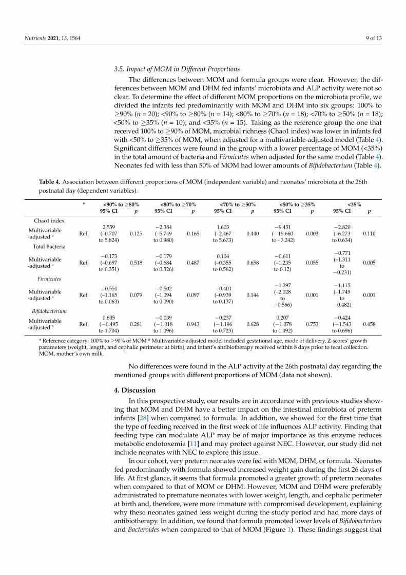
3.5. Impact of MOM in Different Proportions
The differences between MOM and formula groups were clear. However, the dif-ferences between MOM and DHM fed infants’ microbiota and ALP activity were not soclear. To determine the effect of different MOM proportions on the microbiota profile, wedivided the infants fed predominantly with MOM and DHM into six groups: 100% to≥90% (n = 20); <90% to ≥80% (n = 14); <80% to ≥70% (n = 18); <70% to ≥50% (n = 18);<50% to ≥35% (n = 10); and <35% (n = 15). Taking as the reference group the one thatreceived 100% to ≥90% of MOM, microbial richness (Chao1 index) was lower in infants fedwith <50% to ≥35% of MOM, when adjusted for a multivariable-adjusted model (Table 4).Significant differences were found in the group with a lower percentage of MOM (<35%)in the total amount of bacteria and Firmicutes when adjusted for the same model (Table 4).Neonates fed with less than 50% of MOM had lower amounts of Bifidobacterium (Table 4).
Table 4. Association between different proportions of MOM (independent variable) and neonates’ microbiota at the 26thpostnatal day (dependent variables).
* <90% to ≥80% <80% to ≥70% <70% to ≥50% <50% to ≥35% <35%95% CI p 95% CI p 95% CI p 95% CI p 95% CI p
Chao1 index
Multivariable-adjusted a Ref.
2.559(–0.707
to 5.824)0.125
−2.384(–5.749
to 0.980)0.165
1.603(–2.467
to 5.673)0.440
−9.451(−15.660to−3.242)
0.003−2.820(–6.273
to 0.634)0.110
Total Bacteria
Multivariable-adjusted a Ref.
−0.173(–0.697
to 0.351)0.518
−0.179(–0.684
to 0.326)0.487
0.104(–0.355
to 0.562)0.658
−0.611(–1.235to 0.12)
0.055
−0.771(–1.311
to−0.231)
0.005
Firmicutes
Multivariable-adjusted a Ref.
−0.551(–1.165
to 0.063)0.079
−0.502(–1.094
to 0.090)0.097
−0.401(–0.939
to 0.137)0.144
−1.297(–2.028
to−0.566)
0.001
−1.115(–1.749
to−0.482)
0.001
Bifidobacterium
Multivariable-adjusted a Ref.
0.605(−0.495to 1.704)
0.281−0.039(−1.018to 1.096)
0.943−0.237(−1.196to 0.723)
0.6280.207
(−1.078to 1.492)
0.753−0.424(−1.543to 0.696)
0.458
* Reference category: 100% to ≥90% of MOM a Multivariable-adjusted model included gestational age, mode of delivery, Z-scores’ growthparameters (weight, length, and cephalic perimeter at birth), and infant’s antibiotherapy received within 8 days prior to fecal collection.MOM, mother’s own milk.
No differences were found in the ALP activity at the 26th postnatal day regarding thementioned groups with different proportions of MOM (data not shown).
4. Discussion
In this prospective study, our results are in accordance with previous studies show-ing that MOM and DHM have a better impact on the intestinal microbiota of preterminfants [28] when compared to formula. In addition, we showed for the first time thatthe type of feeding received in the first week of life influences ALP activity. Finding thatfeeding type can modulate ALP may be of major importance as this enzyme reducesmetabolic endotoxemia [11] and may protect against NEC. However, our study did notinclude neonates with NEC to explore this issue.
In our cohort, very preterm neonates were fed with MOM, DHM, or formula. Neonatesfed predominantly with formula showed increased weight gain during the first 26 days oflife. At first glance, it seems that formula promoted a greater growth of preterm neonateswhen compared to that of MOM or DHM. However, MOM and DHM were preferablyadministrated to premature neonates with lower weight, length, and cephalic perimeterat birth and, therefore, were more immature with compromised development, explainingwhy these neonates gained less weight during the study period and had more days ofantibiotherapy. In addition, we found that formula promoted lower levels of Bifidobacteriumand Bacteroides when compared to that of MOM (Figure 1). These findings suggest that

Nutrients 2021, 13, 1564 10 of 13
formula delays colonization by anaerobic bacteria and, most likely, intestinal maturation.This is an expected result since Bifidobacterium, Bacteroides, and other anaerobes speciesuse human milk oligosaccharides (HMOs) as a substrate [29] and these were absent inthe formulas used at the time in our NICU. Even when adjusted for gestational age,mode of delivery, somatometry at birth, and neonatal antibiotherapy (factors known tostrongly affect the establishment of initial microbiota [30]), we found that neonates fedpredominantly with MOM had more absolute abundance of total bacteria, Firmicutes, andBifidobacterium when compared to those mainly fed with formula. Microbial richness andFirmicutes content were significantly lower when the feedings contained less than 50% ofMOM, when controlled for the same factors. However, no differences were observed inBifidobacterium in the groups that received different proportions of MOM. The effects ofDHM in very preterm neonates’ microbiota profile were less accentuated than those offormula, which could be explained by the similar composition in HMOs between MOMand DHM [31].
Regarding the sequencing data, we noticed that the results differed from those ob-tained by RT-PCR. Although this is a more complete approach, the analysis is based onrelative abundances, instead of absolute bacterial quantification, which may explain thediscrepancies found with respect to the species of Bifidobacterium. However, it allowed us tosee other finding: human milk (MOM and DHM) promoted higher amounts of Bacteroidesovatus and Akkermancia muciniphila than did formula. Both these bacteria are beneficial forthe intestines of preterm neonates. B. ovatus was described to be capable of inducing highmucosal IgA production when gnotobiotic mice were colonized with this strain comparedto that when they were colonized with others strains [32]. Reduced levels of A. muciniphilahave been associated with inflammatory bowel disease and metabolic disorders [33]. Inneonates, reduced levels of A. muciniphila was found in extensive intestinal ischemia [34].Actually, this mucin-degrading bacteria has been suggested to be a probiotic with promis-ing effects on inflammation prevention [35]. Due to structural similarities of mucin withHMOs, A. muciniphila is able to digest HMOs [36].
MOM promoted increased bacterial richness compared to that of DHM and formula.In fact, it is possible to see a tendency towards a ladder effect favorable to MOM, asif it were a dose-response regarding the preobiotic and probiotic content (Figure 2A).However, no statistically significant differences were found in microbiota profiles betweenthe groups (Figure 3A). This lack of difference can be explained by the discrepant numberof individuals in each group (MOM = 39, DHM = 14, Formula = 8).
It is important to note that the clinical characteristics of the neonates included in MOMvs formula groups were significant different (gestational age, mode of delivery and somatom-etry at birth). However, despite these limitations, our results highlighted the importance ofMOM administration in the first weeks of life on the intestinal microbiota development.
Besides having a beneficial microbial composition, MOM-fed neonates also showedincreased fecal ALP activity. To the best of our knowledge, the present study analyzedfor the first time the influence of neonate feeding in ALP activity in human very pretermneonates. Since the first week of life is considered to be a very important window tomodulate gut microbiota of neonates, we explored how the type of feeding during thistime-specific period influenced the ALP activity in fecal samples collected from verypreterm neonates. We found that neonates fed with MOM and DHM during the first weekof life had increased levels of ALP activity in fecal samples at postnatal day 26, comparedto that of formula-fed neonates in the same period. Although human milk is an exogenoussource of ALP [13], pasteurization destroys 99% of ALP present in the human milk [37].Thus, we propose that the effect of human milk on the neonatal activity of ALP is not onlydue an intake of exogenous ALP, but also to an induction of the enzyme by some agent inboth non-pasteurized and pasteurized human milk, and the agent is absent in neonatalformula. In fact, no differences were found in ALP activity between preterm rat pupsthat received formula with or without ALP supplementation [38–40]. Several moleculeshave been described to be involved in the induction of ALP activity such as omega-3,

Nutrients 2021, 13, 1564 11 of 13
whey protein, or the short chain fatty-acid butyrate [41–43]. As discussed earlier, thepresence of HMOs promote the growth of Bifidobacterium genus that have a role in neonatalintestinal maturation and immune tolerance [44] and are described as lipopolysaccharide(LPS)-suppressing bacteria [41]. During carbohydrate fermentation of HMOs present inhuman milk, Bifidobacterium enhances butyrate-producing bacteria by producing acetatethat is used as a co-substrate for butyrate synthesis [45]. In addition, butyrate has beenshown to increase ALP activity in an intestinal in vitro model [42]. Therefore, this could bea mechanism behind the Bifodobacterium correlation with ALP activity.
5. Conclusions
Overall, these findings point out the importance of MOM and DHM since both increasethe levels beneficial bacteria, such as Bifidobacterium, Bacteroides ovatus, and Akkermanciamuniciphila. Moreover, our results showed that human milk intake during the first week oflife increased the levels of Bifidobacterium and ALP activity measured at the fourth weekof life (26th postnatal day). Since ALP activity, by dephosphorylating LPS, may preventthe initiation of signaling cascades that trigger inflammation, avoiding LPS binding totoll-like receptor, we believe that the first week of life is a sensitive window of opportunityto prevent metabolic endotoxemia and to protect against NEC. Future studies are needed tobetter understand the potential mechanism raised in this study by which oligosaccharidespresent in MOM and DHM may protect against NEC through ALP activity.
Author Contributions: Conceptualization, J.M., C.M., D.T., S.B., M.C., I.M., T.T., and C.C.; Methodol-ogy, J.M., C.M., J.A., S.I., S.B., M.C., and E.P.; Formal Analysis, J.M., C.M., A.F., and C.D.; Writing–Original Draft Preparation J.M.; Writing–Review and Editing, C.M., A.F., D.T., I.B.-M., C.D., J.A., S.I.,S.B., M.C., I.M., T.T., and C.C.; Supervision, C.M., A.F., D.T., and C.C.; Funding Acquisition, J.M,C.M., A.F., D.T., S.B., M.C., I.M., T.T., and C.C. All authors have read and agreed to the publishedversion of the manuscript.
Funding: This study was supported by Milupa DN-ELN 2017 grant from the Portuguese NeonatalSociety, by ERDF through the operation POCI-01-0145-ERDF-007746 funded by the Programa Opera-cional Competitividade e Internacionalização–COMPETE2020, and by National Funds through FCT–Fundação para a Ciência e a Tecnologia within CINTESIS, R&D Unit (reference UID/IC/4255/2013).
Institutional Review Board Statement: The study was conducted in accordance with the ethicalprinciples expressed in the Declaration of Helsinki, the Portuguese law, and Good Clinical Practiceguidelines. The study was approved by both Ethical Committees of Centro Hospitalar Univer-sitário de Lisboa Central (Ref. 443/2017) and NOVA Medical School|Faculdade Ciências Médicas,Universidade Nova de Lisboa (56/2018/CEFCM).
Informed Consent Statement: Informed consent was obtained from legal representatives of allnewborns involved in the study.
Data Availability Statement: The data presented in this study are available on request from thecorresponding author.
Acknowledgments: We would like to thank the parents of the preterm neonates for participating inthis research and the nursing team of the NICU and the Human Milk Bank of Maternidade Alfredoda Costa for their assistance during sample collection. In addition, we would like to thank YourBiomefor the 16S rRNA sequencing data analysis.
Conflicts of Interest: The authors declare no conflict of interest.
References1. Hill, C.J.; Lynch, D.B.; Murphy, K.; Ulaszewska, M.; Jeffery, I.B.; O’Shea, C.A.; Watkins, C.; Dempsey, E.; Mattivi, F.; Tuohy, K.; et al.
Evolution of gut microbiota composition from birth to 24 weeks in the INFANTMET Cohort. Microbiome 2017, 5, 1–18. [CrossRef][PubMed]
2. Van Belkum, M.; Alvarez, L.M.; Neu, J. Preterm neonatal immunology at the intestinal interface. Cell. Mol. Life Sci. 2019, 77,1209–1227. [CrossRef] [PubMed]
3. Xu, W.; Judge, M.P.; Maas, K.; Hussain, N.; McGrath, J.M.; Henderson, W.A.; Cong, X. Systematic Review of the Effect of EnteralFeeding on Gut Microbiota in Preterm Infants. J. Obstet. Gynecol. Neonatal Nurs. 2018, 47, 451–463. [CrossRef] [PubMed]

Nutrients 2021, 13, 1564 12 of 13
4. Kumar, R.K.; Singhal, A.; Vaidya, U.; Banerjee, S.; Anwar, F.; Rao, S. Optimizing Nutrition in Preterm Low Birth WeightInfants—Consensus Summary. Front. Nutr. 2017, 4, 20. [CrossRef]
5. Agostoni, C.; Braegger, C.; Decsi, T.; Kolacek, S.; Koletzko, B.; Michaelsen, K.F.; Mihatsch, W.; A Moreno, L.; Puntis, J.;Shamir, R.; et al. Breast-feeding: A Commentary by the ESPGHAN Committee on Nutrition. J. Pediatr. Gastroenterol. Nutr. 2009,49, 112–125. [CrossRef]
6. Korpela, K.; Blakstad, E.W.; Moltu, S.J.; Strømmen, K.; Nakstad, B.; Rønnestad, A.E.; Brække, K.; Iversen, P.O.; Drevon, C.A.; DeVos, W. Intestinal microbiota development and gestational age in preterm neonates. Sci. Rep. 2018, 8, 1–9.
7. Arslanoglu, S.; Willemijn, C.; Guido, M.; Christian, B.; Cristina, C.; Virginie, C.; Tamas, D.; Magnus, D.; Mary, F.; Iva, H.; et al.Donor human milk for preterm infants: Current evidence and research directions. J. Pediatr. Gastroenterol. Nutr. 2013, 57, 535–542.[CrossRef]
8. Quigley, M.; Henderson, G.; My, A.; Mcguire, W. Formula milk versus donor breast milk for feeding preterm or low birth weightinfants (Review). Cochrane Database Syst. Rev. 2007, CD002971. [CrossRef]
9. Cheong, J.L.Y.; Burnett, A.C.; Treyvaud, K.; Spittle, A.J. Early environment and long-term outcomes of preterm infants. J. NeuralTransm. 2019, 127, 1–8. [CrossRef]
10. Neu, J. Necrotizing Enterocolitis: A Multi-omic Approach and the Role of the Microbiome. Dig. Dis. Sci. 2020, 65, 789–796.[CrossRef]
11. Rader, B.A. Alkaline Phosphatase, an Unconventional Immune Protein. Front. Immunol. 2017, 8, 897.12. Heath, M.; Buckley, R.; Gerber, Z.; Davis, P.; Linneman, L.; Gong, Q.; Barkemeyer, B.; Fang, Z.; Good, M.; Penn, D.; et al.
Association of Intestinal Alkaline Phosphatase With Necrotizing Enterocolitis Among Premature Infants. JAMA Netw. Open 2019,2, e1914996. [CrossRef]
13. Yang, Y.; Rader, E.; Peters-Carr, M.; Bent, R.C.; Smilowitz, J.T.; Guillemin, K.; Rader, B. Ontogeny of alkaline phosphatase activityin infant intestines and breast milk. BMC Pediatr. 2019, 19, 1–7. [CrossRef]
14. Morais, J.; Marques, C.; Teixeira, D.; Durão, C.; Faria, A.; Brito, S.; Cardoso, M.; Macedo, I.; Tomé, T.; Calhau, C. FEEDMI: A StudyProtocol to Determine the Influence of Infant-Feeding on Very-Preterm-Infant’s Gut Microbiota. Neonatology 2019, 116, 179–184.[CrossRef]
15. Fenton, T.R.; Kim, J.H. A systematic review and meta-analysis to revise the Fenton growth chart for preterm infants. BMC Pediatr.2013, 13, 59. [CrossRef]
16. Marques, C.; Meireles, M.; Norberto, S.; Leite, J.; Freitas, J.; Pestana, D.; Faria, A.; Calhau, C. High-fat diet-induced obesity Ratmodel: A comparison between Wistar and Sprague-Dawley Rat. Adipocyte 2015, 5, 11–21. [CrossRef]
17. Gale, C.; Logan, K.M.; Santhakumaran, S.; Parkinson, J.R.C.; Hyde, M.J.; Modi, N. Effect of Breastfeeding compared with FormulaFeeding on Infant Body Composition: A Systematic Review and Meta-Analysis. Am. J. Clin. Nutr. 2012, 95, 656–669. [CrossRef]
18. Guaraldi, F.; Salvatori, G. Effect of Breast and Formula Feeding on Gut Microbiota Shaping in Newborns. Front. Cell. Infect.Microbiol. 2012, 2, 94. [CrossRef]
19. Mshvildadze, M.; Neu, J.; Shuster, J.; Theriaque, D.; Li, N.; Mai, V. Intestinal Microbial Ecology in Premature Infants Assessedwith Non–Culture-Based Techniques. J. Pediatr. 2010, 156, 20–25. [CrossRef]
20. Gregory, K.E.; Samuel, B.S.; Houghteling, P.; Shan, G.; Ausubel, F.M.; Sadreyev, R.I.; Walker, W.A. Influence of maternal breastmilk ingestion on acquisition of the intestinal microbiome in preterm infants. Microbiome 2016, 4, 1–15. [CrossRef]
21. Morais, J.; Marques, C.; Teixeira, D.; Durão, C.; Faria, A.; Brito, S.; Cardoso, M.; Macedo, I.; Pereira, E.; Tomé, T.; et al. Extremelypreterm neonates have more Lactobacillus in meconium than very preterm neonates – the in utero microbial colonizationhypothesis. Gut Microbes 2020, 12, 1–9. [CrossRef]
22. De Gregoris, T.B.; Aldred, N.; Clare, A.S.; Burgess, J.G. Improvement of phylum- and class-specific primers for real-time PCRquantification of bacterial taxa. J. Microbiol. Methods 2011, 86, 351–356. [CrossRef]
23. Waitzberg, D.L.; Pereira, C.C.A.; Logullo, L.; Jacintho, T.M.; Almeida, D.; Da Silva, M.L.T.; Torrinhas, R.S.M.D.M. Microbiotabenefits after inulin and partially hydrolized guar gum supplementation: A randomized clinical trial in constipated women.Nutr. Hosp. 2012, 27, 123–129.
24. Walters, W.; Hyde, E.R.; Berg-Lyons, D.; Ackermann, G.; Humphrey, G.; Parada, A.; Gilbert, J.A.; Jansson, J.K.; Caporaso, J.G.;Fuhrman, J.A.; et al. Improved Bacterial 16S rRNA Gene (V4 and V4-5) and Fungal Internal Transcribed Spacer Marker GenePrimers for Microbial Community Surveys. mSystems 2015, 1, e00009-15. [CrossRef] [PubMed]
25. Calhau, C.; Martel, F.; Hipólito-Reis, C.; Azevedo, I. Differences between duodenal and jejunal rat alkaline phosphatase. Clin.Biochem. 2000, 33, 571–577. [CrossRef]
26. Malo, M.S. A High Level of Intestinal Alkaline Phosphatase Is Protective Against Type 2 Diabetes Mellitus Irrespective of Obesity.EBioMedicine 2015, 2, 2016–2023. [CrossRef] [PubMed]
27. Chong, J.; Liu, P.; Zhou, G.; Xia, J. Using MicrobiomeAnalyst for comprehensive statistical, functional, and meta-analysis ofmicrobiome data. Nat. Protoc. 2020, 15, 799–821. [CrossRef]
28. Parra-Llorca, A.; Gormaz, M.; Alcántara, C.; Cernada, M.; Nuñez-Ramiro, A.; Vento, M.; Collado, M.C. Preterm Gut MicrobiomeDepending on Feeding Type: Significance of Donor Human Milk. Front. Microbiol. 2018, 9, 1376. [CrossRef] [PubMed]
29. German, J.B.; Freeman, S.L.; Lebrilla, C.B.; Mills, D.A. Human milk oligosaccharides: Evolution, structures and bioselectiivty assubstrates for intestinal bacteria. Pers. Nutr. Divers. Needs Infants Child. 2008, 62, 205–222.

Nutrients 2021, 13, 1564 13 of 13
30. Arboleya, S.; Sánchez, B.; Milani, C.; Duranti, S.; Solís, G.; Fernández, N.; Reyes-Gavilán, C.G.D.L.; Ventura, M.; Margolles, A.;Gueimonde, M. Intestinal Microbiota Development in Preterm Neonates and Effect of Perinatal Antibiotics. J. Pediatr. 2015, 166,538–544. [CrossRef]
31. Marx, C.; Bridge, R.; Wolf, A.K.; Rich, W.; Kim, J.H.; Bode, L. Human Milk Oligosaccharide Composition Differs between DonorMilk and Mother’s Own Milk in the NICU. J. Hum. Lact. 2013, 30, 54–61. [CrossRef] [PubMed]
32. Yang, C.; Mogno, I.; Contijoch, E.J.; Borgerding, J.N.; Aggarwala, V.; Li, Z.; Siu, S.; Grasset, E.K.; Helmus, D.S.;Dubinsky, M.C.; et al. Fecal IgA Levels Are Determined by Strain-Level Differences in Bacteroides ovatus and Are Mod-ifiable by Gut Microbiota Manipulation. Cell Host Microbe 2020, 27, 467–475.e6. [CrossRef] [PubMed]
33. Derrien, M.; Belzer, C.; de Vos, W.M. Akkermansia muciniphila and its role in regulating host functions. Microb. Pathog. 2017, 106,171–181. [CrossRef] [PubMed]
34. Romani, L.; Del Chierico, F.; Chiriaco, M.; Foligno, S.; Reddel, S.; Salvatori, G.; Cifaldi, C.; Faraci, S.; Finocchi, A.; Rossi, P.; et al.Gut Mucosal and Fecal Microbiota Profiling Combined to Intestinal Immune System in Neonates Affected by Intestinal IschemicInjuries. Front. Cell. Infect. Microbiol. 2020, 10, 59. [CrossRef]
35. Bavineni, M.; Wassenaar, T.M.; Agnihotri, K.; Ussery, D.W.; Lüscher, T.F.; Mehta, J.L. Mechanisms linking preterm birth to onset ofcardiovascular disease later in adulthood. Eur. Heart J. 2019, 40, 1107–1112. [CrossRef]
36. Ottman, N. Host Immunostimulation and Substrate Utilization of the Gut Symbiont Akkermansia Muciniphila. Ph.D. Thesis,Wageningen University, Wageningen, The Netherlands, 2015.
37. Neu, J. Mother’s Own Milk: How Does It Differ from Donor Milk for the Baby. Breastfeed. Med. 2019, 14, S3–S4. [CrossRef]38. Heinzerling, N.P.; Liedel, J.L.; Welak, S.R.; Fredrich, K.; Biesterveld, B.E.; Pritchard, K.A.; Gourlay, D.M. Intestinal alkaline
phosphatase is protective to the preterm rat pup intestine. J. Pediatr. Surg. 2014, 49, 954–960. [CrossRef]39. Biesterveld, B.E.; Koehler, S.M.; Heinzerling, N.P.; Rentea, R.M.; Fredrich, K.; Welak, S.R.; Gourlay, D.M. Intestinal alkaline
phosphatase to treat necrotizing enterocolitis. J. Surg. Res. 2015, 196, 235–240. [CrossRef]40. Rentea, R.; Rentea, M.; Biesterveld, B.; Liedel, J.; Gourlay, D. Factors Known to Influence the Development of Necrotizing
Enterocolitis to Modify Expression and Activity of Intestinal Alkaline Phosphatase in a Newborn Neonatal Rat Model. Eur. J.Pediatr. Surg. 2018, 29, 290–297. [CrossRef]
41. Kaliannan, K.; Wang, B.; Li, X.-Y.; Kim, K.-J.; Kang, J.X. A host-microbiome interaction mediates the opposing effects of omega-6and omega-3 fatty acids on metabolic endotoxemia. Sci. Rep. 2015, 5, 11276. [CrossRef]
42. Gonçalves, P.; Catarino, T.A.; Gregório, I.; Martel, F. Inhibition of butyrate uptake by the primary bile salt chenodeoxycholic acidin intestinal epithelial cells. J. Cell. Biochem. 2012, 113, 2937–2947. [CrossRef]
43. Navis, M.; Muncan, V.; Sangild, P.T.; Willumsen, L.M.; Koelink, P.J.; Wildenberg, M.E.; Abrahamse, E.; Thymann, T.; Van Elburg,R.M.; Renes, I.B. Beneficial Effect of Mildly Pasteurized Whey Protein on Intestinal Integrity and Innate Defense in Preterm andNear-Term Piglets. Nutrients 2020, 12, 1125. [CrossRef]
44. Underwood, M.A.; Gaerlan, S.C.; De Leoz, M.L.A.; Dimapasoc, L.M.; Kalanetra, K.M.; Lemay, D.G.; German, J.B.; Mills, D.A.;Lebrilla, C.B. Human milk oligosaccharides in premature infants: Absorption, excretion, and influence on the intestinal microbiota.Pediatr. Res. 2015, 78, 670–677. [CrossRef]
45. Rivière, A.; Selak, M.; Lantin, D.; Leroy, F.; De Vuyst, L. Bifidobacteria and Butyrate-Producing Colon Bacteria: Importance andStrategies for Their Stimulation in the Human Gut. Front. Microbiol. 2016, 7, 979. [CrossRef]


![Gut microbiota and metabolite alterations …...the existence of a gut microbiota-bone axis [14–18], and the gut microbiota is a major regulator of bone mineral density (BMD) via](https://static.fdocuments.us/doc/165x107/5f0ecd4a7e708231d441023f/gut-microbiota-and-metabolite-alterations-the-existence-of-a-gut-microbiota-bone.jpg)